schwann cells - form myelin sheaths in pns...
TRANSCRIPT

1
Oligodendrocytes
Form myelin sheaths in CNS
http://cti.itc.virginia.edu/~psyc220/astro2.gif
http://www.emc.maricopa.edu/faculty/farabee/BIOBK/BioBookNERV.html
This image is copyright Dennis Kunkel at www.DennisKunkel.com
Cross section
of myelin
sheaths that
surround
axons
Schwann Cells - form myelin sheaths in PNS
Oligodendrocytes - envelop an average of 15 axonal
internodes each
Schwann cells - envelop only one internode
http://www.zoobotanica.plus.com/portfolio%20med
icine%20pages/neuronst.htmhttp://cti.itc.virginia.edu/~psyc220/oligo.gif
Microglia
-phagocytes
- mobilized after
injury, infection or
disease
- arise from
macrophages outside
of NS
2
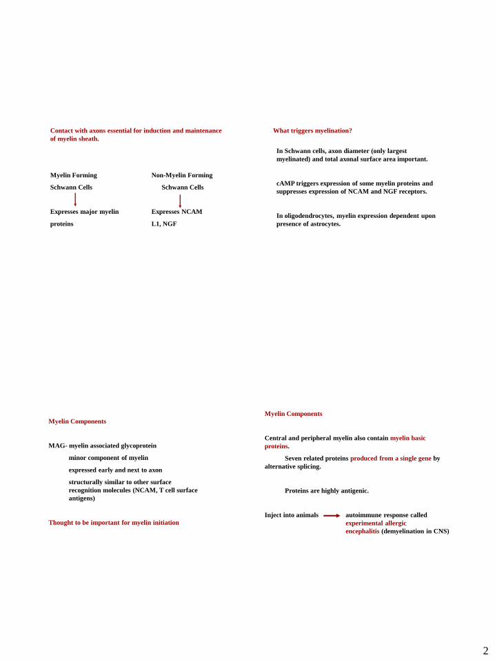
Contact with axons essential for induction and maintenance
of myelin sheath.
Myelin Forming Non-Myelin Forming
Schwann Cells Schwann Cells
Expresses major myelin Expresses NCAM
proteins L1, NGF
In Schwann cells, axon diameter (only largest
myelinated) and total axonal surface area important.
cAMP triggers expression of some myelin proteins and
suppresses expression of NCAM and NGF receptors.
In oligodendrocytes, myelin expression dependent upon
presence of astrocytes.
What triggers myelination?
Myelin Components
MAG- myelin associated glycoprotein
minor component of myelin
expressed early and next to axon
structurally similar to other surface
recognition molecules (NCAM, T cell surface
antigens)
Thought to be important for myelin initiation
Myelin Components
Central and peripheral myelin also contain myelin basic
proteins.
Seven related proteins produced from a single gene by
alternative splicing.
Proteins are highly antigenic.
Inject into animals autoimmune response called
experimental allergic
encephalitis (demyelination in CNS)
3
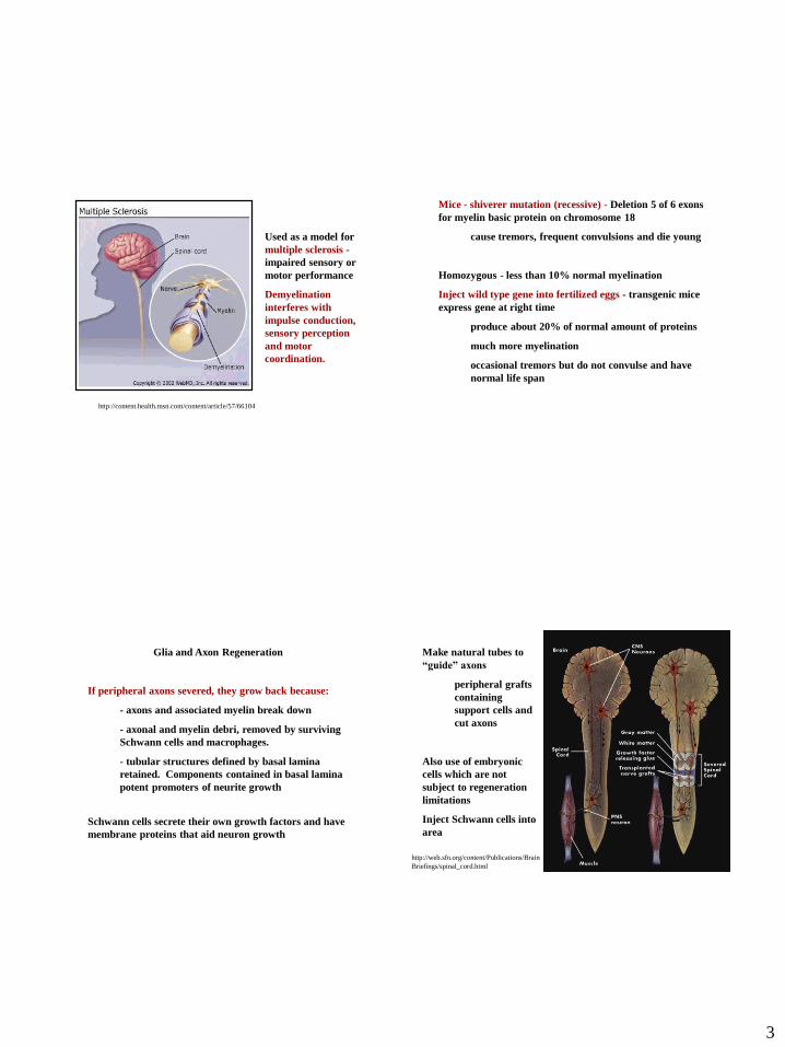
Used as a model for
multiple sclerosis -
impaired sensory or
motor performance
Demyelination
interferes with
impulse conduction,
sensory perception
and motor
coordination.
http://content.health.msn.com/content/article/57/66104
Mice - shiverer mutation (recessive) - Deletion 5 of 6 exons
for myelin basic protein on chromosome 18
cause tremors, frequent convulsions and die young
Homozygous - less than 10% normal myelination
Inject wild type gene into fertilized eggs - transgenic mice
express gene at right time
produce about 20% of normal amount of proteins
much more myelination
occasional tremors but do not convulse and have
normal life span
Glia and Axon Regeneration
If peripheral axons severed, they grow back because:
- axons and associated myelin break down
- axonal and myelin debri, removed by surviving
Schwann cells and macrophages.
- tubular structures defined by basal lamina
retained. Components contained in basal lamina
potent promoters of neurite growth
Schwann cells secrete their own growth factors and have
membrane proteins that aid neuron growth
Make natural tubes to
“guide” axons
peripheral grafts
containing
support cells and
cut axons
Also use of embryonic
cells which are not
subject to regeneration
limitations
Inject Schwann cells into
area
http://web.sfn.org/content/Publications/Brain
Briefings/spinal_cord.html

4
Myelin in the brain and spinal cord gets in the way of axon
regeneration
Interfering with myelin can aid axon repair and restore some
function in rodents with spinal cord injuries.
- a vaccine against myelin prompted axons regrowth and
treated animals regained some movement in their hind legs
Other possible approaches?
Identify specific molecules signaling macrophages to
ingest and remove myelin from the damaged spinal
cord.
Target specific components of myelin, instead
of whole sheath
Some proteins present in CNS myelin:
At least MAG and Nogo are capable of causing growth
cone collapse and inhibiting neurite outgrowth in vitro.
Have a common receptor (NgR).
Nogo, may be partly responsible for the inability of damaged
axon fibers to repair.
Normal neuron Neuron treated with
synthesized Nogo
http://web.sfn.org/content/Publications/BrainBriefings/brain_spinalcord.html
The Nervous System
1) Central Nervous System
Brain, spinal cord, retina
2) Peripheral Nervous System
Everything (except the retina) outside of
the brain and spinal cord

5
Peripheral Nervous System
1) Somatic - carries voluntary motor and sensory information
both to and from the CNS.
2) Autonomic
3) Enteric - meshwork of nerve fibers that innervate the viscera
(gastrointestinal tract, pancreas, gall bladder).
a. sympathetic
b. parasympathetic
Peripheral Nervous System
1) Somatic - peripheral nerve fibers that send sensory information to the
central nervous system AND motor nerve fibers that project to skeletal
muscle.
Somatic Nervous System
The cell body is located in either the brain or spinal cord
and projects directly to a skeletal muscle.
http://faculty.washington.edu/chudler/nsdivide.html
Peripheral Nervous System
1) Somatic
2) Autonomic - controls smooth muscle of the viscera (internal organs) and
glands.
3) Enteric
a. sympathetic - "fight" or take "flight"
(run away)
b. parasympathetic - "rest" and "digest"
6
http://faculty.washington.edu/chudler/nsdivide.html
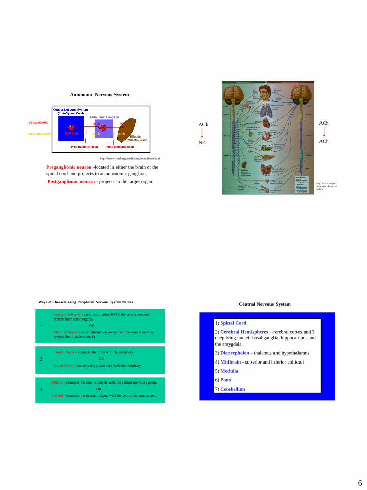
Autonomic Nervous System
Preganglionic neuron -located in either the brain or the
spinal cord and projects to an autonomic ganglion.
Postganglionic neuron - projects to the target organ.
AChSympathetic NE
Parasympathetic ACh ACh
http://home.swipnet.
se/sympatiska/nervo
us.htm
ACh
NE
ACh
ACh
Sensory (afferent) - carry information INTO the central nervous
system from sense organs.
OR
Motor (efferent) - carry information away from the central nervous
system (for muscle control)
1
Cranial Nerve - connects the brain with the periphery.
OR
Spinal Nerve - connects the spinal cord with the periphery.
Somatic - connects the skin or muscle with the central nervous system.
OR
Visceral - connects the internal organs with the central nervous system.
2
3
Ways of Characterizing Peripheral Nervous System NervesCentral Nervous System
1) Spinal Cord
2) Cerebral Hemispheres - cerebral cortex and 3
deep lying nuclei: basal ganglia, hippocampus and
the amygdala.
3) Diencephalon - thalamus and hypothalamus
4) Midbrain - superior and inferior colliculi
5) Medulla
6) Pons
7) Cerebellum
7
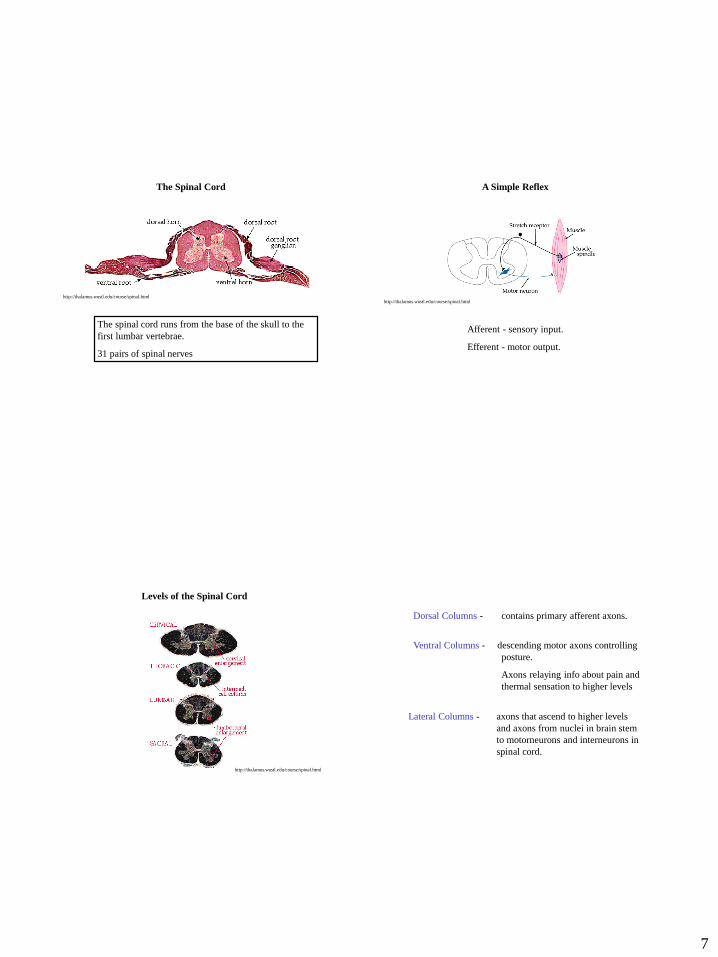
The Spinal Cord
The spinal cord runs from the base of the skull to the
first lumbar vertebrae.
31 pairs of spinal nerves
http://thalamus.wustl.edu/course/spinal.html
Afferent - sensory input.
Efferent - motor output.
A Simple Reflex
http://thalamus.wustl.edu/course/spinal.html
Levels of the Spinal Cord
http://thalamus.wustl.edu/course/spinal.html
Dorsal Columns - contains primary afferent axons.
Lateral Columns - axons that ascend to higher levels
and axons from nuclei in brain stem
to motorneurons and interneurons in
spinal cord.
Ventral Columns - descending motor axons controlling
posture.
Axons relaying info about pain and
thermal sensation to higher levels
8
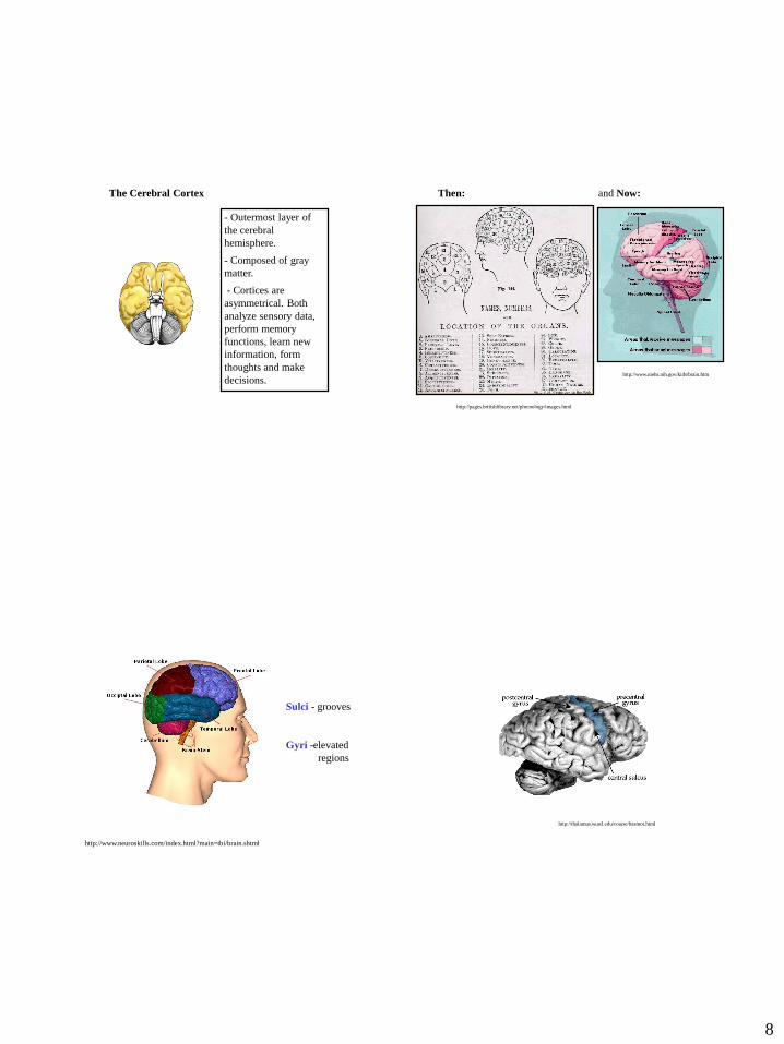
- Outermost layer of
the cerebral
hemisphere.
- Composed of gray
matter.
- Cortices are
asymmetrical. Both
analyze sensory data,
perform memory
functions, learn new
information, form
thoughts and make
decisions.
The Cerebral Cortex
http://www.niehs.nih.gov/kids/brain.htm
Then: and Now:
http://pages.britishlibrary.net/phrenology/images.html
http://www.neuroskills.com/index.html?main=tbi/brain.shtml
Sulci - grooves
Gyri -elevated
regions
http://thalamus.wustl.edu/course/basmot.html

9
The Frontal Lobes
Divided into:
a) prefrontal area-
emotional control center and
home to our personality.
Involved in motor function,
problem solving, spontaneity,
memory, language, initiation,
judgement, impulse control,
and social and sexual
behavior.
b) premotor area -contains
neurons that produce
movements.
Two functional regions:
1) Involves sensation
and perception. Integrates
sensory information to form a
single perception (cognition).
2) Integrates sensory
input, primarily with the visual
system to construct a spatial
coordinate system to represent
the world around us.
The Parietal Lobes
Center of our visual
perception system.
Disorders of this lobe
can cause visual
hallucinations (visual
images with no external
stimuli) and illusions. http://www.neuroskills.com/index.html?main=tbi/brain.shtml
The Occipital Lobes
Involved in the primary
organization of sensory input and
also highly associated with
memory skills. Left temporal
lesions result in impaired memory
for verbal material. Right side
lesions result in impaired recall of
non-verbal material, such as music
and drawings.
Language can also be affected by
temporal lobe damage. Left lesions
disturb recognition of words. Right
damage can cause a loss of
inhibition of talking.
http://www.neuroskills.com/index.html?
main=tbi/brain.shtml
The Temporal Lobes
10
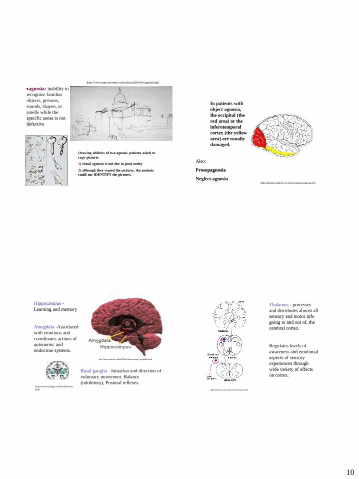
Drawing abilities of two agnosic patients asked to
copy pictures
1) visual agnosia is not due to poor acuity
2) although they copied the pictures, the patients
could not IDENTIFY the pictures.
http://www.cquest.utoronto.ca/psych/psy280f/ch4/agnosia.html
agnosia: inability to
recognize familiar
objects, persons,
sounds, shapes, or
smells while the
specific sense is not
defective
In patients with
object agnosia,
the occipital (the
red area) or the
inferotemporal
cortex (the yellow
area) are usually
damaged.
http://ahsmail.uwaterloo.ca/kin356/agnosia/agnosia.htm
Also:
Prosopagnosia
Neglect agnosia
Basal ganglia - Initiation and direction of
voluntary movement. Balance
(inhibitory), Postural reflexes.http://www.waiting.com/brainfunction.
html
http://ahs.uwaterloo.ca/kin356/ltm/hippocampus_amygdala.html
Hippocampus -
Learning and memory
Amygdala -Associated
with emotions and
coordinates actions of
autonomic and
endocrine systems.
Thalamus - processes
and distributes almost all
sensory and motor info
going to and out of, the
cerebral cortex.
Regulates levels of
awareness and emotional
aspects of sensory
experiences through
wide variety of effects
on cortex.
http://thalamus.wustl.edu/course/bassens.html

11
Hypothalamus - Main function is homeostasis. Factors such as
blood pressure, body temperature, fluid and electrolyte balance, and
body weight are held to set-points.
- Receives inputs about the state of the body, and initiates
compensatory changes.
- Extensive afferent and efferent connections with thalamus, midbrain
and some cortical areas.
http://thalamus.wustl.e
du/course/hypoANS.ht
ml
The Brainstem
- Lower extension of the brain
where it connects to the spinal
cord.
- Functions include those
necessary for survival (breathing,
digestion, heart rate, blood
pressure) and for arousal (being
awake and alert).
- Consists of:
1) medulla oblongata
2) pons
3) cerebellum
4) midbrain
medulla oblongata - primarily a relay station for the crossing of motor
tracts between the spinal cord and the brain. It also contains the
respiratory, vasomotor and cardiac centers, as well as many mechanisms
for controlling reflex activities such as coughing, gagging, swallowing and
vomiting.
pons - links different parts of the brain and serves as a relay station
from the medulla to the higher cortical structures of the brain. It
contains the respiratory center.
Midbrain - serves as the nerve pathway of the cerebral hemispheres
and contains auditory and visual reflex centers.
- Involved in the
coordination of voluntary
motor movement, balance
and equilibrium and muscle
tone.
- Located just above the
brain stem and toward the
back of the brain.
- Cerebellar injury results in
movements that are slow
and uncoordinated.
The Cerebellum
http://thalamus.wustl.edu/course/cerebell.html

12
http://www.hark.com/clips/sfpbhcvhzh-
jeopardy-theme
INSIDE THE BRAIN OF MEGAN FOX
Megan's corpus callosum connects the right & left halves of her
brain, also called these, like halves of the earth
Jeopardy Show #6297 - Tuesday, January 24, 2012
A 2010 paper shows Megan's amygdala maintains her "loss aversion"
when faced with decisions about risking this
When Megan enjoys a fine meal, she's employing the parietal
these, right behind the frontal ones
The thalamus, part of this botanical-sounding part of Megan's brain,
receives all sensory input except smell
Megan's higher functions use her cerebral this; when she "hears" a song in
her head, she's using her brain's auditory this
400
800
1200
1600
2000
hemispheres
money
lobes
brainstem ???
cortex
The cerebellum
("little brain")
convolutions similar
to those of cerebral
cortex
Has an outer cortex,
an inner white
matter, and deep
nuclei below the
white matter.
http://thalamus.wustl.edu/course/cerebell.html
http://thalamus.wustl.edu/course/cerebell.html
molecular layer-
outermost layer and is
nearly cell-free.
Purkinje cell- monolayer
of large cells
granule cells- dense
layer of tiny neurons.
In the center of each
folium is the white matter.

13
Purkinje Cells
Sole output from cerebellum
Receive input from granule cells
http://thalamus.wustl.edu/course/cerebell.html
Purkinje cells arise
from ventricular zone
Granule cells born in
external germinal
zone
Migration of granule cells arises late in
development.
Must migrate along paths apparently blocked by
obstacles.
Follow radial path because of Bergmann glia.
14
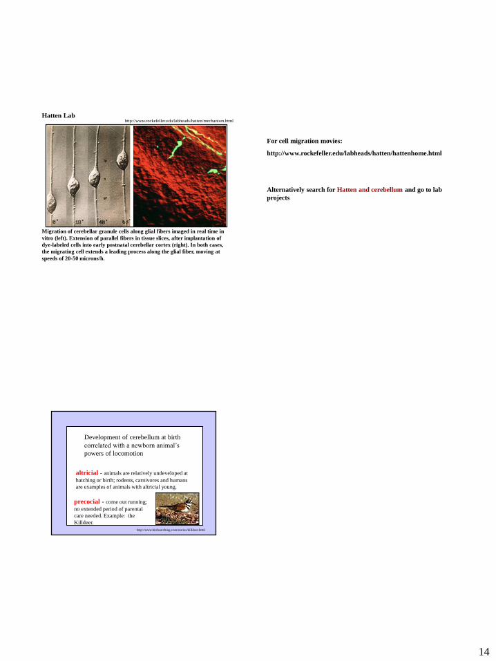
Migration of cerebellar granule cells along glial fibers imaged in real time in
vitro (left). Extension of parallel fibers in tissue slices, after implantation of
dye-labeled cells into early postnatal cerebellar cortex (right). In both cases,
the migrating cell extends a leading process along the glial fiber, moving at
speeds of 20-50 microns/h.
http://www.rockefeller.edu/labheads/hatten/mechanism.htmlHatten Lab
For cell migration movies:
http://www.rockefeller.edu/labheads/hatten/hattenhome.html
Alternatively search for Hatten and cerebellum and go to lab
projects
Development of cerebellum at birth
correlated with a newborn animal’s
powers of locomotion
altricial - animals are relatively undeveloped at
hatching or birth; rodents, carnivores and humans
are examples of animals with altricial young.
precocial - come out running;
no extended period of parental
care needed. Example: the
Killdeer.
http://www.birdwatching.com/stories/killdeer.html