sau xignon .. , science - nzimls
TRANSCRIPT

ISSN 1171-0195
Sau_xignon ..__, science
Volume 63 Number 1 April 2009

THE GLOBAL HAEMOSTASIS
SOLUTIO
A wide range of reagents A complete line of analysers
A f u II series of services
~ Stago
Diagnostica Stago Ply. Ltd. 651 Doncaster Road P.O. Box 106 • Doncaster, 3108 Australia Phone: 1800 4 STAGO (Aus) 0508 4 STAGO (NZ)
Fax: +61 3 9855 8999 AUSTRALIA - NEW ZEALAND [email protected]
At the Heart of Haemostasis www.stago.com

Editor
Rob Siebers, PGCertPH, FIBio l, FNZIC, FNZIMLS; School of Med icine & Hea lth Sciences, Otago University, Wellington.
Deputy Editor
Ann Thornton, FNZIMLS; Schoo l of Med icine & Hea lt h Sciences, Otago University, Wellington
Editorial Board
Gloria Evans, MM LSc, FNZIMLS; Otago University, Christchurch Chris Kendrick, MSc, MNZIMLS; Massey Univers ity Mike Legge, PhD, FNZIMLS; Otago University, Duned in Kevin Tay lor, BMLSc, PGDipMLSc; Canterbury Hea lth Laboratories John Stirli ng, BSc (Hons), MLett, FRMS, MAIMS; Co-Editor Austr J Med Sci Tony Woods, PhD, MAIMS, Co-Editor Austr J Med Sci
Statistical Advisers
Gordon Purdie, BSc and Nevil Pierse, MSc; School of Medicine & Health Sciences, Otago University, We llington.
About the Journal
The New Zea land Journal of Med ical Laboratory Science (the Journal) is the official publication of the New Zea land Institute of Medica l Laboratory Science (NZIMLS) who owns the copyright. No parts of th is pub lication may be reproduced in any form w ithout the written perm ission of The NZIM LS. The journa l is a peerreviewed biomed ica l publicat ion since 1946 and is published t hree t imes per year in April, August and November. It is circulat ed to NZIM LS members and universiti es and research institutes in New Zea land and overseas. Current circu lation is about 2,000 cop ies per issue. Print ing by Red_i, Auck land.
Brief instructions t o authors
Submit all material electron ica lly to the Editor (rob.siebers@otago. ac.nz or [email protected]) . Comprehensive instruction on layout, etc can be found in t he New Zealand Journal of Medica l Laboratory Science, vol. 54, issue 3, pages 108-110 or on t he NZIMLS web site (www.nzimls.org.nz) . With your submission provide a covering letter stating t hat t he work is original, has not previously been published (except as an abstract at a scientifi c meeting), is not under considerat ion by another journal, and that all named authors justify authorship by either contributing to the planning, execution, ana lysis, or crit ical writing of the study and that all authors approve submission of the final version. Additionally, one author (not necessarily the 1st author) must take responsibility for the integrity of the work as a whole. Please state who this author is. Also, specifically state what contributions each author has made. Th is information w il l be published with the accepted paper. Authors are responsible for scientif ic content and views. Opinions expressed in the Journal are not necessari ly those of the Editors or Counci l of t he NZIMLS.
Indexing
The Journal is abstracted by the Cu mulative Index to Nursing and Alli ed Hea lt h Literature, Index Copernicus, Excerpta Med ica/ EMBASE, Australian Medica l Index, Scopus, and the Thomson Gale Group. The Editor and Deputy Editor are members of the World Association of Med ical Editors (www.wame.org) and the Editor is current ly a Board Director of WAME.
Subscription
Enquiri es regarding subscriptions and address changes should be addressed to the Executive Offi cer of the NZIMLS, Fran van Ti l at PO Box 505, Rang iora. Phone: (03) 313 4761. Ema il : [email protected] .nz
Advertising
Advertisment book ings and rates enqu iries shou ld be addressed to t he Advertis ing Manager, Trish Re illy, 48 Towa i Street, St Heliers, Auck land 5. Phone: (09) 575 5057. Fax: (09) 575 0698. Emai l: journa ladvertising@nzim ls.org.nz.
New Zealand Jour n a l of
Medical Laboratory Science
L
Editorial
Volume 63 April ISSN 1171 -0195
Number 1 2009
The Journal: electronic or print, or both? Rob Siebers .................................................................................. 2
Original articles
Estimated plate let and different ial leucocyte counts by microscopy, Sysmex XE-21 00 and Ce llaVision™ DM96 Anthea Po vall, Christopher John Kendrick ................... .. .. ... 3-1 0
Attenuation of serum lam in in concentrat ions upon t reatment of chron ic hepatit is Hadi Parsian, Mohammad Nouri, Mohammad Hossein Soumi, A li Rahimipour, Durdi Qujeq, Rasoul Estakhri, Mehrdad Kashi Fard, Karim Agcheli, Golamininar Majidi ... ... .... .... ..... ....... 12-17
Letter to the Editor
Ta lk about f ly ing blind!! ! A young lab rat from Dunedin (Erolia Rooney) ...... .... .. ... .. .. .. . 18
Obit uary - Ba rry Edwards By Sue Carnoutsos ................................ ..................................... 18
After match function for Barry Edwards Mike Southern, Christine Hickton ....... ..................................... 19
Obituary- Robert (Bob) Allen By Jan Parker .............................................. ............................... 20
Regular features Abstracts from the British Journal of Biomedical Science 28-32 Advertisers in this issue ............................................................. 33 Index to Volume 62 ................................................................... 34 In this issue ................... ............................................................... 2 Instructions to authors ................................................................ 1 Journa l-based questionna ire ..................................... ... ............ 22 Just t he Job- medica l laboratory science ............. ........ .... 24-25 Med-Bio journa l prize .. ....... ........................ ....... .......... ............. 23 News from t he universit ies ...... .. ............................................. .. 32 NZIMLS ASM 2009, Blenheim ............................. ....... Inside back NZIMLS journal pr ize ....... .... ............ .. ...... .. ... .... ... ...................... 23 Pacifi c Way ............... ........ .... ...... ...... ... ......... .............. .. .. ... .. . 26-27 Specia l Interest Groups .... ..... .... ....... .. .... ....... .. .. ... .. ............. 21, 23
NZ J Med Lab Sdence 2009

Editorial: The Journal: electronic or print, or both? Rob Siebers, FNZIMLS, Editor School of Medicine and Health Sciences, University of Otago, Wellington
There has been concern by some members of the NZIMLS regarding the print version of the Journal. They believe that since Journal articles are freely accessible on the Institute's web site the printed version is no longer requ ired and that el imination of the printed version would help the sustainability of forestry worldwide as 40% of the world's forests are now used for paper.
The NZIMLS Journal is printed by Red-i, Auckland. They became the first print company in New Zealand in 2007 to obtain certification to two premier international standards from the Forest Stewardship Council and the Programme for the Endorsement of Forest Certification Schemes Chain of Custody for sustainable renewable forestry. This ensures that paper used for printing the Journal is sourced from forests that are sustainable managed with a chain of custody up to the printed sheet. Paper sourced by Red-i also meets the New Zealand Government Sustainability Procurement Policy. Additionally, Red-i uses vegetable-based inks for printing that are based on renewable raw materials which are non-mineral oil based.
In this issue Anthea Povall compared the leukocyte differentia l of the Ce llaVision™ DM96 imaging device to the manua l differential and differential values ofthe Sysmex XE-2100 as part of the requirement for the Massey University BMLSc degree. In their artic le, Anthea and Chris Kendrick report that the CellaVision™ DM96 correctly pre-classified 69% of all samples, with the reclassified data showing strong corre lations with the neutrophil, lymphocyte and eosinoph il populations using the Sysmex analyser. The authors conclude that the CellaVision™ DM96 is particu larly suited to laboratories processing large numbers of normal samples, but that microscopy and the experienced morphologist continues to occupy an important place in the haematology laboratory.
In their article Parsian and colleagues determined the serum laminin level cutoff point for predicting liver fibrosis. They determined laminin concentrations in chronic hepatitis patients by ELISA and compared them to healthy controls. Serum laminin concentrations were significantly higher in the patient group and determined a cutoff for the various stages of liver fibrosis that showed a good sensitivity and specificity. After treatment laminin levels decreased but were still higher than the control group. The authors conclude that serum lamin in is a useful marker of liver fibrosis.
In th is issue are two obituaries of prominent members of our profession. Barry Edwards served for 18 years on the NZIM LS Council, 15 years as Secretary. On New Year's Day 2009 an "after match function" was held in memory of Barry, a report of which is elsewhere in the Journal. Robert (Bob) Allan was for many years charge technologist in the biochemistry department of Dunedin Hospital. He previously was Ed itor of the Institute's Journal for ten years. Both Barry and Bob were pass ionate about the profession and devoted a great dea l of their time to the profession and its members.
Another Journa l questionnaire appears in th is issue giving members the opportunity to earn 5 CPD po ints. Since its inception, the
Certa inly, if the Journal was on ly ava ilab le electron ically, the reader can use the computer to read articles of interest. However, read ing the print version of the Journal is better on the eye than staring into the light source of a cathode ray tube. Additionally a journal is like a book, always on hand to go back for interesting bits and can be taken to a quit corner for perusal. You can even scribble your own notes in the print version of the Journal.
So, when you sit down in your comfortable chair with the print version of the Journal in one hand and a glass of chardonnay in the other, you can be assured that in the Institute's choice of Red-ito print the Journal we are ensuring the sustainability of renewable forests worldwide. Sustainability as defined by the United Nations is: "The capacity to meet the needs of the present without compromising the ability of future generations to meet their needs" .
Journal questionnaire has proven a popu lar avenue to accrue CPD po ints with over 500 members subm itting their answers each issue. A lthough the majority experience no problems subm itting through the NZIMLS web site, a few do. One such prob lem that has been identified is that some emp loyers 'time out' from the web site after a specified time. We suggest that members f irst write their answers in a word doc and then cut & paste their answers on the NZIMLS web site. If your submission was successful then you will receive your submitted answers in a confirmatory email to the address you have supplied. Because of the shear volume of responses the Editor tries to mark and respond within one to two days of submission. It must be remembered that the Editor is not a paid employee of the NZIMLS and does this work voluntarily. He is allowed to go on annual leave and thus was disappointed to receive a fair number of emails upon his return in February querying why the member had not received an immediate reply to his/her submission.
Annually the NZIMLS awards a prize for the best case study published in the Journal during the calendar year. Joint winners for 2008 were Sarla Naran and Sharda Lallu from Anatomic Patho logy at Wellington Hospital for their case study on pulmonary mucormycosis. Congratulations to both . As Editor I look forward to rece iving subm issions of case stud ies for 2009. Many excellent case studies are presented at SIG meetings and the North and South Island Sem inars. Expect the Ed itor to attend some of those meetings and tap you on the shou lder to subm it to the Journal. Your case study presentation deserves a wider presentation that at the meeting . You will also obta in CPD points and be eligible (if you are a financial member of the Institute) for either (or both) the MedB io Prize for the best paper in each issue of the Journal and the NZIMLS Journa l Prize. Indeed, the w inners of the NZIMLS Journa l Prize for 2008 were also the winners of the MedBio prize for the August 2008 issue.
Rob Siebers Editor
NZ J Med Lab Science 2009
2

Estimated platelet and counts by microscopy, CellaVision™ DM96
differential leucocyte Sysmex XE-2100 and
Anthea PovaiJ1, BMLSc, Medical Laboratory Scientist Christopher John Kendrick2, Grad Dip Sci, MSc, MNZIMLS, Senior Lecturer
1 Haematology Department, Canterbury Health Laboratories, Christchurch 2Massey University, Palmerston North
Abstract Modern imaging techniques and specialised computer software has enabled the differential counting of leucocytes to become automated. This study compared the leucocyte differential of the CellaVision™ DM96 to the manual differential and the differential values from the Sysmex XE-2100. The evaluation was performed on 50 samples submitted for routine CBC testing at Canterbury Health Laboratories in Christchurch. The DM96 correctly preclass ified 69% of all samples, with the reclassif ied data showing strong corre lation with the neutrophil, lymphocyte and eos inophil popu lations us ing the Sysmex ana lyser. The reclassified DM96 resu lts showed a strong correlation with the resu lts obta ined by manual different ial counting. This study also evaluated the platelet estimation funct ion of t he DM96 software and compared the results to manual estimations and the platelet count from the Sysmex ana lyser. Th is DM96 method produced a consistent over estimate of plate let numbers and was more time consum ing than microscopy. Using a correct ion to t he DM96 mu ltip licat ion factor, resu lts closer to the actual platelet were obtained.
Key words: leucocyte differential; platelet count; CellaVision DM96; Sysmex XE-2100
N Z J Med Lab Sci 2009; 63 (1): 3-10
Introduction The increased workload experienced by many haematology laboratories coupled with reductions in staff numbers has lead to the introduction of automated morphological analysis in laboratories. The improved processing capacity of computers combined with high resolution digital imaging technology has allowed for the development of systems which can pre-classify haematological cells, mostly leucocytes (1). The CellaVision™ DM96 is an imaging device intoduced into Canterbury Health Laboratories (CHL) in 2007. This paper reports on the performance of the DM96 to determine the leucocyte differential and compared the resu lts w ith both manual differentia ls and the differentia l result from the Sysmex XE-2100 automated cell counter. In addition, the DM96 derived p late let estimate was performed and compared to the standard microscopic platelet estimate and the platelet count from the Sysmex XE-21 00.
Materials and methods Equipment The study ut ilised the Sysmex XE-2 100 haematology analyser and the Sysmex SP1000i slidemaker/autostainer. The Ce llaVision DM96 system was used for the automated leucocyte differential counting and p late let count est imates. These ana lysers are used in the core laboratory at CHL and provide CBC test ing of sa mples submitted from inpatients and outpatients of the pub lic hospita l in Christchurch. Standard light microscopes were used for microscopy.
Sample selection Samples were selected from the routine work which arrived in the haematology laboratory during a sing le 24-hour period. Flagging functions on the Sysmex XE-21 00 indicated which samples requ ired blood film examination and fi lms were prepared and sta ined by the Sysmex SP1 OOOi slidemaker/autostainer. Fifty slides were selected for the project by the author's (AP) supervisors from the resu lts of the CBC values. Samples were selected to include both norma ls and abnormals containing qualitative and/or quantitative abnormalities in the main cell classes. Slides from patients with known acute leukaem ia were excluded. The slides were anonym ised to show only the laboratory identi fication numbers. Of the fifty slides four samples w ere excl uded from plate let analysis due to the presence of platelet clumps observed during microscopy. Three samples were also excluded from the differential count ana lysis as the Sysmex w as not ab le to count suffficient ce lls to produce a valid differential.
Data collection Each slide was analysed by the DM96 with 100 cells counted. Ce lls were pre-classified into: band neutroph ils, segmented neutrophi ls, lymphocytes, monocytes, eosinoph ils, basoph il s, metamyelocytes, myelocytes, promyelocytes, blast ce ll s, lymphocyte variant forms and plasma ce lls. For data collection purposes, band and segmented neutroph ils were counted together. Lymphocytes, lymphocyte variants and plasma cells were counted as one group. Precursor cells from myeloid blast cells through to metamyelocytes were counted together as immature granulocytes. Unidentified cells were categorised as 'other'. Results were stored as the preclassification DM96 data for each slide and were then viewed. Incorrectly classified cells were moved to their correct classification based on their morphological appearance. Cells classified as 'other' were sim ilarly reclassified where possible and moved into one ofthe leucocyte classification groups. Non-leucocyte classifications were also examined and the ce lls incorrectly identified as nucleated red cells, giant plate lets, plate let aggregates, smudge ce lls or artefacts were reclassified. Results were recorded as the reclassified DM96 data.
For each slide a manual 1 DO-cell differential was performed using a light microscope at 500x magnification. Cells were recorded in the same categories as for the reclassified DM96 data.
The p late let count estimation facility on the DM96 was used to derive an estimate of p latelet numbers on each of the stained blood films. Plat elet numbers present in 16 sub-images were counted, and the average value for those f ields multiplied by the plate let estimation factor (pre-set at 1 0) provided the est imated plate let resu lt. For the DM96, the plate let estimation corresponded to counting the equiva lent of eight high power microscopy f ields as per the Ce llaVision Users Manua l (4). Resu lts were recorded as t he DM96 p late let estimation .
Manua l plate let estimates were performed by counting the
NZ J Med Lab Science 2009
3

Table 1. Platelet count raw data (x 10 9/L) for slides 1-50.
Slide No Sysmex Platelets Manual Slide No Sysmex Platelets Manual DM96 DM96
1 358 450 354 24 362 539 413
2 173 264 246 25 338 420 419
3 171 151 147 26 255 303 260
4 254 194 233 27 889 1105 974
5 59 66 64 28 190 264 200
6 38 51 30 29 236 280 211
7 165 171 180 30 165 203 221
8 197 259 207 31 232 295 247
9 594 663 545 32 495 483 504
10 142 150 152 33 159 158 158
11 291 345 319 34 514 624 594
12 241 229 201 35 341 510 423
13 128 186 174 36 32 43 40
14 225 283 295 37 175 234 173
15 222 355 244 38 449 556 602
16 222 288 250 39 333 388 355
17 357 496 424 40 445 484 437
18 229 324 275 41 251 225 262
19 480 493 441 42 345 305 285
20 197 270 222 43 354 504 481
21 158 208 23 1 44 350 326 346
22 371 543 490 45 105 79 96
23 367 449 382 46 360 391 424
* Four samples were clotted and were excluded from all platelet analyses.
NZJMedlabScience2009
4

number of plate lets in 10 successive high power f ields at 1 OOOx magnification and mu lt iplying t he count by 109 to give the plate let estimation as n/1 09/L. These results were recorded as the manua l platelet estimation.
Fo llowing co llation of manual and DM96 resu lts for both the leucocyte differentials and the plate let estimates, patient data from the laboratory information system was accessed and the verified data for the Sysmex d ifferential and plate let count were obtained. Correlation data was derived using Bland-Aitman plots and regression analysis.
Results Platelets The platelet estimates from the DM96 and the visual method fol lowed closely the platelet counts generated by the Sysmex XE-2100 (Table 1 and Figure 1). Of interest was the tendency for the DM96 to over estimate the platelet count as compared with the manual method . In comparison with the Sysmex results, the R2 values were 0.91 and 0.93 for the DM96 and visual p latelet estimation methods, respectively (Figure 2) .
The average ratio of Sysmex to DM96 platelet counts was 0.86 with the DM96 counts an overestimate in 80% of samples. In absolute values the average platelet overestimate was about 66 platelets. The average ratio of Sysmex to visual plate let counts was 0.93 with the visual result either over- or underestimating the Sysmex result by about 40 platelets.
Leucocyte Differential The corre lation between the Sysmex data and either reclassified DM96 or visual results for cel l lines are summarised by the R2 values in Table 2. The correlation data presented for the leucocyte counts in Tab le 2 was derived from the data presented in figures 3-14.
Both DM96 and manual methods showed a strong correlat ion with the Sysmex data for the neutrophil counts, and an acceptable corre lation was observed for lymphocyte and eosinophil counts. The corre lat ions for monocytes, basophils and immature granulocytes were weak. Tab le 2 also shows the R2 values for the pre-classification data. The pre-classification data was correct in 69% of cases. In most cases the results paralle led the reclassified results, except for the monocyte population, where the pre-classification count correlated strongly with the Sysmex data.
The percentage agreement with Sysmex data for each cell class according to test method is presented in Figure 15. The results from each method were divided into the proportion of test results which were in agreement with the Sysmex data, and those results which were not. Results which were not in agreement were further divided according to whether the result was an over- or underestimate of the Sysmex value.
A ll test methods identified neutrophils with the highest percentage agreement, followed by lymphocytes. Cell classes with sma ller populations represented in the blood general ly showed a lower percentage agreement.
Figure 16 shows the proportion of each cell class in an average differential for each test method. On average, all test methods had higher neutrophil, and lower lymphocyte and monocyte counts compared to the Sysmex. All methods were approximately equal in their estimates of smal ler cell classes. Overall, values obtained by the DM96 and manual methods were almost identical.
Discussion Both DM96 and visual platelet estimates corre lated well with the Sysmex XE-21 00 platelet counts, however, there was a consistent overestimation of the DM96 platelet count. The overestimation could be accounted for by the platelet estimation factor used on the machine, which set at 10 appeared to be too high. A change
of the estimation factor to 0.86 wou ld yield a plate let estimation very close to t he Sysmex plate let count making th is method more accurate t han the manual method. Forty-seven samples were ana lysed in this study which was more than the 30 sample minimum recommended by t he manufacturer in t he setup of the plate let estimate function (3). The slides used in the study included 3 sa mples with plate let counts be low 100 x 109/L and 5 above 450 x 109/L. Given these sma ll numbers it may be worthwhile repeating the estimated platelet count on larger numbers of samples containing platelet numbers above and below the normal range.
While both the platelet estimate methods were time consuming, the DM96 method took longer to scan slides to estimate the platelet numbers in the film . An advantage of the DM96 method was the overlay grid which aided counting, but the major disadvantage was poor image qua lity. In our hands the imaging was sometimes out of focus. With traditional microscopy a scratched section could be bypassed, but the DM96 has no 'reserve' images for such a situation.
While the platelet estimate function of the DM96 could at times be useful in the laboratory, platelet estimates are rarely required. The DM96 is capable of producing accurate platelet estimates once properly setup, but its use for screening al l slides probably cannot be justified given the extra time taken.
In this study the identification and counting of neutrophils and lymphocytes by manual and DM96 methods showed good correlation with the Sysmex data although the results as mentioned showed both over- and under-estimation by both techniques. The smaller ce ll populations (eosinophils, basophi ls and blast ce lls) showed less variation in the results between the the manua l and DM96 methods, however, these did not correlate well with the Sysmex data, the eosinophil population being the exception. The DM96 to Sysmex correlation was particularly poor for the monocyte population, and immature granulocytes were not well identified by the manual method when compared to the Sysmex values.
The pre-classification data correlated well with the reclassification values in most cases except for the monocyte population, which had a high pre-classification R2 va lue. This resulted from other cells (often smudge ce lls) being incorrectly classified as monocytes.
In a previous study by Briggs et al the pre-classification monocyte count as compared to the manual method resulted in a low correlation (2). Monocytes are generally poorly classified by both automated and manual differential systems, mainly due to smear method and the area of the film examined (4). The relatively low number of monocytes in most samples is also likely to contribute to this error.
Sample size is an important factor to consider when assessing the relevance of results. In this study 50 samples were initially selected, of which 46 were used for the platelet estimates and 47 for leucocyte ana lyses. Three other stud ies compared the DM96 and manual differential, ana lysing 136, 322 and 400 samples respective ly, and performed either 200 or 400-cell differentials (2,4,5). The manual 100 cell differential is the standard method used in laboratories today. This method lacks both accuracy and precision which can be improved on by counting 200-400 cells. This was not performed in this study. Additionally, the comparison of the manual differential results to the Sysmex XE-2100 differential, which counts many thousands of ce lls, highlighted the manual different ial inaccuracies. The DM96 and manual methods counted the same number of ce lls and neither had any statistical advantage over t he other.
One recurrent source of error was the total number of cells present in the reclassification differential. As only 100 cells were counted and classified by the DM96 in our study, when reclassification took place the differentia l was more or less than 100 ce lls in
NZ J Med Lab Science 2009
5
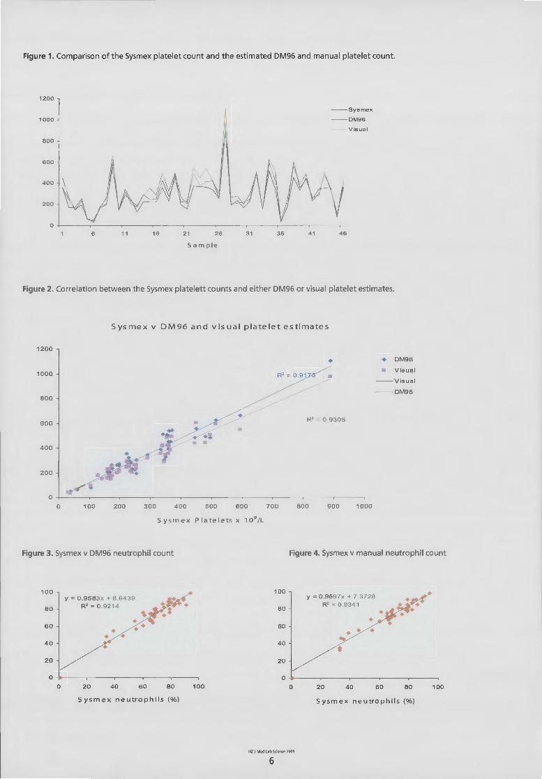
Figure 1. Comparison of the Sysmex plate let count and the estimated DM96 and manual plate let count.
1200
~ -- Sysmex
1000 DM96
Visua l
BOO
600
400
2 00 v 0
6 11 16 21 26 31 36 41 46
Sam pie
Figure 2. Correlation between the Sysmex platelett counts and either DM96 or visual platelet estimates.
1200
1000
800
600
0 100
Sysmex v DM96 and visual p latelet estimates
• R2 = 0.91
• R C 93DE
•
200 300 400 500 600 700 800 900
S y s m e x P I a te I e ts x 1 0 9 /L
1000
+ DM96
Visua l
Visual
--DM96
Figure 3. Sysmex v DM96 neutrophil count Figure 4. Sysmex v manual neutrophil count
100
80
60
40
2 0
y = 0.9583x + 8 .6439 R2 = 0.9214
• • • •
0 ~----~----~----~----~----.
0 2 0 40 60 80 100
Sysmex n e utrophil s (% )
100
80
60
40
20
y = 0.9597x + 7.3728 R2 = 0.9341
••• ~
• 0 ~---.---.----.---.----,
0 20 40 6 0 8 0 100
Sysmex n e utrophil s (%)
NZ J Med lab Smmce 2009
6

Figure 5. Sysmex v DM96 eosinophi l count Figure 6. Sysmex vs manual eosinoph il count
14 1 2 y = 1 .1486x- 0 .5598 • y = 0 .974 1 x- 0 .00 7 1 2 R 2 = 0.8 0 75 10 R 2 = 0.7 1 62
• 1 0
• 8 8
• 6 6 • • • 4 • • 4
• • • 2 • • •
• • • • 2
• • 0
0 2 4 6 8 10 12 14 2 4 6 10 12 14 8 0
S ysm ex eosinophils (o/o) S ysm ex eosinophils (o/o)
Figure 7. Sysmex v DM96 lymphocyte count Figure 8. Sysmex v manual lymphocyte count
60 80 y = 0.8423x - 0.6175 • y = 0.8264x + 0.3746
R 2 = 0.8449 R 2 = 0.7746
• ~ 60 • 40 • • • •
40 • •• • •
20 ~ • • • 20 •
0 0
0 20 40 60 80 0 20 40 6 0 80
Sysmex lymphocytes (o/o) Sysmex lymphocytes (o/o)
Figure 9. Sysmex v DM96 basophil count Figure 10. Sysmex v manual basophil count
10 10 y = 0.3098x + 0.4097 y = 0.5222x + 0 .3605
R 2 = 0.1242 R2 = 0.2406 8 8
6 6 • 4 • 4 •
• • 2 2
0 0 0 2 4 6 0 2 4 6
Sysmex ba sop hi l s (o/o) S ysmex basoph il s (o/o )
NZ J Med lab SCience 2009
7

Figure 11. Sysmex v DM96 monocyte count
14 y = 0 .3971 x + 2.2888
12 R2 = 0.1691 • • • 1 0 • • •
8
6
4
2 • • • 0
0 2 4 6 8 10 12 14
Sysmex monocytes (o/o)
Figure 13. Sysmex v DM96 immature granulocyte count
10 y = 0.5584x + 0.1 0 94
R 2 = 0 .47 4 8 8
6 • • 4 • •
• 2 •
• • 0
0 2 4 6 8 10
Sysmex imm gran s (o/o)
14
12
10
8
6
4
2
0
10
8
6
4
2
0
Figure 12. Sysmex v manual monocyte count
y = 0.6469x + 1 .2204 R 2 = 0 .4533 • • • • • • • • • • • • • • • • • • • • • • • • • • • • • • •
• • 0 2 4 6 8 10 12 14
Sysmex m onocytes (o/o)
Figure 14. Sysmex v manual imm granulocyte count
•
0
y = 0.228 5x + 1 .126 R 2 = 0.0464
• •
2 4 6
Sysmex imm grans (o/o)
8 10
NZ J Med Lab Science 2009
8

Figure 15. The percentage agreement with Sysmex data for each cel l class according to test method .
r:::J Agreeme nt • Overestimate 0 Underestimate
Neutroph ils Lymphocytes Monocytes Eosinophils Basophils
Test method for ana lysis of ce II I ine s
* Pre-classif ication data.
Immature
Granulocytes
Figure 16. The proportion of each ce ll type in an averaged differential for each test method.
100
95
90
85
80
75
70
65
60 Sysmex
* Pre-classif ication data.
Av era g e diffe r e nti a l by m e thod of a n a ly s i s
DM96• DM96 Manual
Method of analysIs
NZ J Med Lab Science 2009
9
•ott1er
• Immature Granulocytes
• Basophils
D Eosinophlls
0 Monocytes
• Lymphocytes
• Neutrophils

half of the samples. Where a 'reciprocal rec lassification ' took p lace (eg . a neutrophil classified as an eosinophi l as well as an eosinophi l classified as a neutrophil), the differential remained 100. However, when ce lls were classified as non-leucocytes or vice versa, the differentia l was higher or lower than 100 ce lls after the reclassification. Other studies have set the automated microscope to count 105, 110 or 200 ce lls to take this problem into account (2,4,5) . In our experience setting the count value to 110 should be adequat e for most samples.
It is also worth remembering t hat t he DM96 on ly exam ines t he area of the sl ide where it can detect an erythrocyte monolayer, thus if a sample does not conta in many leucocytes in that area, the d ifferential value may be provided on less than 100 WBCs. In this respect tradit iona l microscopy has the advantage in that an area with overlapp ing erythrocytes can sti ll be examined and leucocytes accurately ident if ied.
Another important factor to take into account when examining correlation data, is the experience of the morphologists exam ining the blood films. Logica lly it wou ld be expected that more experience wou ld corre late w it h greater accuracy of identificat ion, and this has been shown to be the case in other stud ies (2) .
In conclusion, th is study has show n that the resu lts from a 6-part differential (including immature granulocytes) performed by t he DM96 is similar to the results obtained by a manual differenti al. Neutrophil, lymphocyte and eos inophil values can be expected to correlate well with resu lts obtained from the ana lyser differential using the Sysmex XE-2100, however monocyte, basophil and immature granulocyte numbers can be expected to corre late less well. This work supports the findings of others that the DM96 is particu larly suited to laboratories (eg . community laboratories) processing large numbers of normal samp les (2). More complex samples from patients with haematological ma lignancies, recent bone marrow transplantation and morphological changes associated with some infections, require the attention of a morpholog ist to accurat ely ident ify and class if y immature cells. In
the sett ing of a larger laboratory, such as that at Canterbury Health Laboratories, the DM96 can reduce the number of 'normal' films examined by morphologists, and can be used in conjunction with microscopy to investigate abnormal cases. The results of this study have demonstrated that microscopy and the trained morphologist still occupy an important place in the haemato logy laboratory. For the moment at least the human eye continues to occupy the high ground ahead of the modern mechanical morphologist.
Acknowledgements This w ork w as part of t he requirement for t he Massey Univers ity BMLSc degree and was conducted during the 4th year cl inical laboratory placement in Haematology at Canterbury Hea lth Laboratories in 2008. Many thanks to the haematology staff at Canterbury Hea lth Laboratories, particularly Mr. Ken Beechey, Mrs. Linda Henshaw, and Mrs. Christine Harper for the ir assistance with th is project.
References 1. Hutchinson CV, Brereton ML, Burthem J. Digital imaging of
haematological morphology. Clin Lab Haematol 2005; 27: 357-62.
2. Briggs C, Longair I, Slavik M, Thwaite K, M ill s R, Thavaraja V, et al. Can automated b lood film ana lysis rep lace the manual differential? An evaluation of t he CellaVision DM96 automated image analysis system. tn t J Lab Hemato/ 2009; 31: 48-60.
3. CellaVisionTM DM96 User's Manual. CellaVision A.B. 2006. 4. Cornet E, Perol JP, Troussard X. Performance evaluation and
relevance of the CellaVision TM DM96 system in routine analysis and in patients with malignant hematological disease. International Journal of Hematology. tnt J Lab Hemato/2008; 30: 536-42.
5. Swolin B, Simonsson P, Backman S, Lofqvist I, Bred in I, Johnsson M. Differential counting of blood leukocytes using automated microscopy and a decision support system based on artificial neural networks - evaluation of DiffMaster Ocavia. lnt J lab Hematol 2003; 25: 139-47.
Address for correspondence: Anthea Povell, Haematology, Canterbury Hea lth Laboratories, PO Box 151, Christchurch.
NZ J Med Lab Science 2009
10

Introducing UniCel® DxH. A new day dawns in cellular analysis
0 Transform your laboratory, with unparalleled quality of results,
innovative efficiency solutions and revolutionary scalability.
0 Flow Cytometric Digital Morphology and Data Fusion
deliver the ultimate in high-definition cellu lar analysis.
0 Lean Efficiency - eliminate unnecessary work while improving patient care .
To learn more, contact your Beckman Coulter NZ representative on 0800 442 346
or visit us at beckmancoulter.com/DxH. • BECKMAN COULTER
We're better together

Attenuation of serum laminin concentrations upon treatment of chronic hepatitis Hadi Parsian 1
, PhD Student Mohammad Nouri1•2, Assistant Professor Mohammad Hossein SoumP, MD, Assistant Professor Ali Rahimipour, Professor Durdi Qujeq3, Assistant Professor Rasoul Estakhri4, MD, Pathologist and Associate Professor Mehrdad Kashi fard5,MD, Associate Professor
Karim Agcheli6, MD, Associate Professor Golamininar Majidj2, MD
1Gastroenterology Research Centre, 2Department of Biochemistry, 41mam Reza Hospital, Tabriz, University of Medical Sciences, Tabriz, Iran; 3Department of Biochemistry and Biophysics,5Yahya Nez had Hospital, Sabol University of Medical Sciences, Sabol, Iran; and 6Gastroenterology Research Centre, Golestan University of Medical Sciences, Gargan, Iran.
Abstract Objectives: The aim of this work was to determ ine the serum lam inin level cutoff po int for pred icting liver fibrosis high lighting its diagnostic value and determining the effect of treatment on serum laminin concentrations. Methods: Serum laminin concentrations in chronic hepatitis patients (n=62) and contro ls (n=20) were compared by ELISA and stages of fibrosis were assessed according to the modified Knodel I score system. Results: Mean serum lam inin concentration in patients (91.9 ± 20.9 ng/ml) was greater than controls (46.2 ± 10.2 ng/ml; p <0.001 ). Serum concentrat ions of lam inin in al l stages of hepatic fibrosis were significantly higher than those of healthy controls (p <0.05). A cutoff point of 52ng lamin in/ml of serum was obta ined for the discrimination of various stages of liver fibrosis showing a good sensitivity (96.8%) and specificity (80%). After 6 months of treatment, a gradual decrease in serum lam in in concentrations were observed, however the level was still higher than that of the healthy group (p<0.05). Conclusions: Our findings suggest that the serum laminin concentration is a useful noninvasive marker of liver fibrosis and shows a strong positive correlat ion with different stages of the disease.
Key words: chronic hepatitis; hepatic fibrosis; lam in in; treatment
N Z J Med Lab Sci 2009; 63 (1): 12-17
Introduction Laminin was initially identified by Timpl and Martin in 1979, from a murine fibrosarcoma (1). Laminin is one of the main glycoproteins of the basement membrane and participates in a series of such biologica l phenomena such as adhesion, migration, cel lular differentiation and the maintenance of the cytoskeleton upon its b inding to severa l components of the matrix, such as co llagen type IV, heparin sulphate and entac in (2-7).
In the liver, lam inin is norma lly found around the vesse ls and bil iary ducts, where basement membranes are identified . Litt le or on ly a slight reaction for antibod ies against laminin can be observed in the hepatic sinusoids (8, 9) . In this organ, glycoproteins are also involved in intracellu lar activities, such as the normal differentiation of the
bi liary ducts, expression of albumin messenger RNA in hepatocytes, and regenerat ion with normal lobular organ ization following partia l hepatectomy (1 0-12). Laminin is thought to be synthesized by hepatocytes and sinusoidal ce lls (13). Among all ce ll ular types in the sinusoids, special attention should be given to stellate cells or lipocytes, which produce the largest amount of serum laminin . With the development of hepatic cirrhosis, laminin and collagen deposition occurs both along the fibers of septal fibrosis and subendothelial sinusoids or Disse's space. At the latter site, laminin deposition, together with collagen deposition, determine the formation of a true basement membrane along sinusoids. Th is phenomenon is cal led capi llarization of Disse's space (14).
Increased concentrations of laminin were observed in the more advanced stages of fibrosis in patients with hepatic disease (15-19). Kropf et al have proposed laminin serum concentrations as a sensitive screening test for hepatic fibrotic disease and portal hypertension (18, 19).
An important component of the management of hepatic fibrosis is the clinical assessment of disease severity. Liver histo logy is frequent ly considered the gold standard for establishing the severity of hepatic necro inflammation and fibrosis. However, liver biopsy is an invasive procedure that may cause undesirable events, such as pain in 20% to 30% of cases, major complications in 0.5%, and even death. In add ition, because of the complications derived from the procedure and frequent poor patient acceptance, the direct costs of such procedures are high (20). Thus, the finding of surrogate markers of liver fibrosis could be relevant to reduce the number of liver biopsies in patients with hepatitis.
The first aim of th is study was to determine the serum laminin level cut off point to predict both presence and absence of fibrosis. The second aim was to obtain a relationship between the diagnostic values of serum lam inin concentrations for differentiation of various stages of hepati c fibrosis in patients with chronic hepatitis. The final aim was to determ ine serum lamin in changes during treatment of these patients.
NZJMedlabSc.ence2009
12

Materials and methods Study population 62 patients (35 men and 27 women, mean age ± SD: 35.4 ± 11.3; range: 15-65 years) were enro lled in the study. Among these, 35 patients had hepatit is B virus (HBV), 14 had hepatitis C virus (HCV), and 13 were autoimmune hepatitis (AI H). The subjects were selected from persons who were referred to the Gastroenterology Research Centres in Tabriz and Gonbad, Iran. Patients were included in the study if they were positive for serum hepatitis B surface antigen or C antibodies and had pers istently elevated serum am inotransferase concentrations greater than 1.5 t imes the upper limit of the reference range for at least six months. All patients were diagnosed according to the International Autoimmune Hepatitis Group Report protocol (21).
For assessment of liver fibrosis scores all patients underwent liver biopsy as part of the normal diagnostic procedure and were subclassified according to the score for the histological activity index (HAl). Patients with a history of gastrointestinal bleeding and chronic liver disease (Wilson's disease, hemochromatosis, a1 ph a 1-antitrypsin deficiency, bi liary disease, hepatocellu lar carcinoma), active intravenous drug abuse, and liver transplantation were excluded.
Control sera for the determination of laminin were obtained from 20 healthy individuals; 10 women and 10 men, 20-69 years old (mean ± SD: 42 ± 14.7 years).These healthy persons had normal serum concentrat ions of aminotransferases and alkaline phosphatase (ALP) and had no history of gastrointesti nal bleeding or chronic liver disease, smoking, alcohol intake, no fam ily history of hepatitis or liver disease, and no act ive intravenous drug abuse, or liver transplantation . A ll patients gave written informed consent to use these data for scient ific purposes and the study was approved by Tabriz Un ivers ity of Medical Sciences Ethical Committee.
Blood sample collection and analysis Fasting venous blood (5ml) was co llected on the day before t he beginning of the treatment and three times at two monthly intervals, i.e. two, four and six months after the beginn ing of treatment. Serum was separated (2500 g for 5 minutes) within one hour of blood co llection . Standard liver function tests (LFT), includ ing aspartate aminotransferase (AST), alan ine aminotransferase (ALT), total and direct b ili rubin, albumin (A lb), and hepatitis sero logy were performed on aliquots of each sample at entry and recorded. The rest of the serum samples were stored at - 20oc. Serum Jaminin concentrations were determined in one analytical batch . The controls were dealt with in the same manner, except that the control group provided blood only once at entry. Routine LFT were performed using commercially available kits (Ziestchem, Iran).
Patients treatments were begun if they met the inclusion criteria and they were fol lowed up for at least six months. Treatment of each patient was according to a standard protocol as follows. Hepatitis C patients were treated with Pegylated Interferon + Ribaverin or Interferon + Ribaverin. Hepatitis B patients were treated with Interferon or Adefovir and the AIH patients with Predniso lone and lmuran (22).
Serum laminin concentrat ions were assayed using a laminin EIA Kit (Takara Bio, code number: MK1 07) on an ELISA reader (BDSL, lmmunoscan, Switzerland, Lab System). The laminin EIA kit is a so lid phase EIA based on a sandwich method t hat utili zes two mouse monoclonal anti-lam inin ant ibodies to detect lam inin by a two-st ep procedure. One of the monoclonal antibodies is bound to the microtitre plate to create the solid phase. Non-specific binding is blocked using a block ing buffer. Samples and standards are then incubated in the microtitre plate wel ls. After wash ing the plate, the second anti- lam in in monoclona l antibody that is labeled w ith peroxidase (POD) is added to t he we lls and incubated . Duri ng these steps, lam inin is captured onto t he so lid support on one side and tagged on t he other by POD-anti-lam inin. The react ion between
POD and substrate (H 20 2 and tetremethybenzidine) results in co lor development with intensities proportional to t he amount of lam inin present in the samp les and standards. The amount of lamin in was determ ined by measuring the absorbances using an EIA plate reader. A standard curve of 5, 10, 20, 40, 80, 160 and 320 ng/ml laminin was used to convert sample absorbances into ng lamin in/ml serum.
Histological assessment of liver damage All patients underwent a liver biopsy for assessing the presence and severity of liver disease. The biopsy fragments were fixed in a 10% formalin solution for 12 hours and embedded in paraffin. Sections were stained with hematoxylin-eosin, Masson's trichrome and reticulin stain to establish the histological diagnosis and the extent of the liver lesions.
Specimens were graded and staged according to the modified Knodel! scoring system (23, 24). The grading system scores 0-18 and was based on sum of four indices: 1. Periportal or periseptal interface hepatitis (piecemeal necrosis,
score 0-4) 2. Confluent necrosis (score 0-6) 3. Focal (spotty) lytic necrosis, apoptosis, and focal inflammation
(score 0-4) 3. Portal inflammation (score 0-4)
The f ibrosis scores were determined as Stage 0 if there was no fibrosis, Stage 1 if there was f ibrous expansion of some portal areas, w ith or without short fibrous septa, Stage 2 if there was fibrous expansion of most porta l areas, w ith or w ithout short fibrous septa, Stage 3 if there was fibrous expansion of most portal areas with occasional porta l to portal (P-P) bridging, Stage 4 if there was fibrous expansion of porta l areas with marked bridging [porta l to porta l (P-P) as well as porta l to central (P-C) ], Stage 5 if there was marked bridging (P-P and/or P-C) with occasional nodu les (incomplete cirrhosis), and Stage 6 if there was probable or defin ite cirrhosis (23,24).
Statistical analysis All statistical ana lyses were done by SPSS version 12.0 for Microsoft Windows (SPSS Inc.) and the data were considered statistica lly signif icant at a two-s ided p < 0.05. Numerical data were expressed as mean ± SD. According to the Gaussian distribution (1 sample Kolmogorov-Smirnov test), mean of serum laminin concentrations of patients and various chronic hepatitis stages, as well as the control group, were compared using the Mann-Whitney U-test or Student's t test. Spearman's correlation coefficients were calculated to assess the relationship between the histological degree of severe liver fibrosis and the concentrations of serum laminin.
To assess and compare the diagnostic accuracy of laminin for differentiating chronic hepatitis patients with severe liver fibrosis from those without fibrosis, we plotted ROC curves (25) and ca lculated the areas under the curves (AUC) for comparison. Rece iver operating characterist ic (ROC) curves were generated by plotting the relationship of the true positivity (sensitivity) and the false positivity (1 - specificity) at various cutoff po ints of the test. An AUC of 1.0 is characteristic of an ideal test, whereas 0.5 indicates a test of no diagnostic value (26). The diagnostic sensit ivity, specificity, positive predictive values (PPV) and negative predict ive values (NPV) values were also calculated.
Results Serological and biochemical profiles of t he patients are summarized in Table 1. Hepatitis serology revealed that 56.4% of the patients were suffer ing from chronic hepatitis B, 22.5% from chronic hepatitis C and 20.9% from autoimmune hepatitis . Histo log ica l examination of liver for f ibros is scoring revea led 35% of patients to be suffering from sign if icant f ibrosis (stage ~3) .
The mean serum lamin in concentrat ions (ng/ml ± SD) in patients
NZ J Med Lab Science 2009
13

Table 1. Serological and biochemical profile of patients and control group.
Variables Patients (n=62) Control group (n=20)
M ale/Female (No) 35/27 10/10
Age in years (mean ± SD) 35.4 ± 11.3 42 ± 14.7
ALT (mean ± SD) 132.7 ± 141 .7 27.3 ± 6.4
Reference range: < 38 IU/L
AST (mean ± SD) 97.0 ± 138.3 28.3 ± 6.5
Reference range: < 42 IU/L
ALP (mean ± SD) 376.4 ± 413.7 130.6 ± 38
Reference range: women 64-305 lUlL; men 80-306 lUlL
Total bilirubin (mean± SD) 32.5 ± 58.1 -
Reference range: <20.5 IJmoi!L
Direct bilirubin Direct (mean ± SD) 11.5±23.1 -
Reference range: < 6.8 IJmoi/L - --
Albumin (mean± SD) 41.0 ± 6.0 -
Reference range: 35.0-S2.0 g/L
Hemoglobin (mean ± SD) 13.1 ± 2.6 -Reference range: women 12.0-16.0 g/L; men 14.0-18.0 g/L
Hematocrit (mean ± SD) 41 ± 9.1 -
Reference range: women 37.0-47.0%; men 40.0-54.0%
Platelets (mean ± SD) 220,511 ± 132,732 -
Reference range: 160,000-450,000
Smoking (No, %) 7 (11 %) 0 (0%)
Alcohol intake (No, %) 1 (1 .6%) 0 (0%)
Family history of hepatitis (No, %) 5 (8%) 0 (0%)
Family history of chronic liver disease (No, %) 1 (1.6%) 0 (0%)
History of drug abuse (No, %) 2 (3.2%) 0 (0%)
Chronic hepatitis B (No, %) 35 (56.4%) 0 (0%)
Chronic hepatitis C (No, %) 14 (22.5%) 0 (0%)
Autoimmune hepatitis (No, %) 13 (20.9%) 0 (0%) -
Fibrosis Stage 0 (No, %) 10 (16.1%) 0 (0%) - ·-
Fibrosis Stage 1 (No, %) 19 (30.6%) 0 (0%) --
Fibrosis Stage 2 (No, %) 11, 17.7% 0, 0% --
Fibrosis Stage 3 (No, %) 9, 14.5% 0, 0% --
Fibrosis Stage 4 (No, %) 8, 12.9% 0, 0%
Fibrosis Stage 5 (No, %) 4, 6.4% 0, 0%
Fibrosis Stage 6 (No, %) 1, 1.6% 0, 0%
~ ~
Table 2. Comparison of serum laminin concentrations (ng/ml, mean ± SD) of patients in various stages of liver fibrosis and various stages of sampling vs. healthy controls.
Laminin Fibrosis Stage 0 Fibrosis Stage 1 Fibrosis Stage 2 Fibrosis Stage 3 Fibrosis Stage 4 Fibrosis Stage 5
concentrations
At entry 63.0 ± 12.1t 85.7 ± 6.3:1: 94.0 ± 10.2:1: 100.6 ± 8.6:1: 104.2 ± 20.2:1: 130.2 ± 13.7:1:
2nd month 55.4 ± 8.5t 79.5 ± 6.6:1: 90.9 ± 12.0:1: 96.0 ± 7.0 :1: 100.4 ± 18.4:1: 118.2 ± 19.0:1:
4th month 49.2 ± 6.0* 75.3 ± 7.0:1: 85.7 ± 12.4:1: 90.0 ± 5.0:1: 93.0 ± 15.2:1: 111.7 ± 12.6:1:
6th month 46.2 ± 4.5* 75.2 ± 6.0:1: 83.8 ± 11.1:1: 87.6 ± 5.9:1: 88.9 ± 13.0:1: 110.2 ± 9.5:1:
Resu lts are mean ± SD. Differences: *statistica lly not sign ificant; tp <0.05; :t: p <0.001
NZJ Med labScience2009
14

with HBV, HCV and AIH were 92.0 ± 20.9 (range: 63-149), 92.8 ±
24.2 (range: 43-128), 90.6 ± 18.4 (range: 69-128), respectively. However, the mean serum lam inin concentrat ions (ng/ml ± SD) in hea lthy control subjects were statistically significantly lower than the patients serum laminin concentrat ions (46.2 ± 10.2; p < 0.001, Figure 1 ).
In Tab le 2, mean ± SD of serum lam inin concentrations in various chronic hepatitis stages and variousstagesofsampling are presented. As shown in this table, differences in serum concentrat ions of laminin, almost in all stages of hepatic fibrosis as compared with the healthy controls, were not statistica lly signif icant (p <0.05). An exception was in 3rd and 4th samples of patients in Stage 0 (p=0.313 and 0.985, respectively). Also in fibrosis Stage 6, which was represented by only one patient, we could not compare this single patient with the control group (laminin serum concentration in this stage: 143 ng/ml). As the degree of liver fibrosis stage increased, there was a gradual rise in basal serum laminin concentrations at entry (rS=0.788, p-value<0.001, Figure 2).
After the beginning of treatment, a decrease in serum laminin concentrations was observed. We compared serum laminin concentrations of patients after six months of treatment with the basal laminin concentrations (at entry) of each fibrosis stage. Although there was a gradual decrease in serum laminin concentrations of progressive fibrosis stages (i.e. Stages 4 and 5), differences were not statistically sign ificant (p <0.093 and <0.054, respective ly). Conversely in early stages of fibrosis (i.e. Stages 0-3) differences between the serum laminin concentrations at entry compared with the lam inin concentrations after the beg inning of treatment were statistica lly sign ificant (for Stages 0 and 1, p <0.001 ; and for Stages 2 and 3 , p <0.05).
Tab le 3 shows the cutoff point, sens itivity, and specificity of serum laminin concentrations. In this tab le the ROC curve data, for patients before and after the treatment, are presented. Figure 4 illustrates the ROC curve of serum laminin concentrations in patients (at entry) for differentiation of patients with liver fibrosis vs. control group.
Discussion Several serum markers have been developed to assess fibrogenesis and investigations have been made to replace liver biopsy with non-invasive markers of liver fibrosis, which have been assessed in many studies. However, questions remain regarding their sensitivity and significance and whether changes in concentrations of these markers during the treatment protocol can be detected (27). In our study we have assessed serum laminin concentrations in patients with chronic hepatitis in an attempt to evaluate its predictive value for the risk of fibrosis progression. In addition, we have investigated changes in serum lam in in concentrations during the treatment protocol.
As shown in Table 2, the mean serum laminin concentrations in patients with chron ic liver disease were significantly higher than those of healthy controls. These resu lts are cons istent with other reported studies (16, 28). As the stage of liver fibrosis increases, there is a rise in serum laminin concentrations (Table 2) and patients in a higher fibrosis stage show higher serum laminin concentrations. In addition, as is clear from Figure 2, the correlation of serum laminin concentrat ions with various histological stages of liver fibrosis revealed a strong pos itive corre lation (rS= 0.788). This indicates a positive relat ionship between serum laminin concentrat ions and the degree of liver fibrosis, and the two variabl es are linearly related (p < 0.001). As shown in Table 2, differences in serum concentrations of lam inin at various stages during the treatment protocol, compared to healthy controls, were statistical ly sign ificant (p <0.05) . It seems that during the treatment protocol there was a decrease in serum lam inin concentrations. After six months of treatment, gradua l decreases in serum lam inin concentrations were observed. When we compared basal serum laminin
concentrat ions in patients (at entry) w ith the concentrations of lam inin after 6 months of treatment, we observed that the serum lam inin concentrations did not differ statist ica lly in stages 4 and 5 of liver fibrosis. Converse ly, in the ea rly stages of liver fibrosis (0-3) the differences were statistically signif icant (p <0.05). Therefore, it appears that treatment is more effect ive in the ear ly stages of liver fibrosis, because only in patients with a liver fibrosis score of less than 4 a decrease in serum lam inin concentrat ions occurred after treatment. An increase in the inflammation grade of liver damage led to a rise in serum laminin concentrations, and the correlat ion is stat istica lly significant (data not shown).
As shown in Table 3, a cutoff point of 52 ng laminin/ml serum was obtained for discrimination of various stages of liver fibrosis with a reasonably good sensitivity, specificity, PPV and NPV. The AUC of serum laminin ROC curve was found to be 0.974 indicating that serum laminin is a useful diagnostic index of liver fibrosis. We also used this cutoff point (52 ng/ml) for creating the ROC curve after treatment. Again, the results showed a reasonably good AUC, sensitivity, specificity, PPV and NPV. We conclude that this cutoff point is also suitable for discrimination of our patients with fibrosis during treatment.
Our data are consistent with the work of other groups who reported that serum lam inin concentrations increase in chronic liver disease (28-36). Schneider et al (16) and Castera et al (37) reported that laminin concentrations increase in early stages of chron ic liver disease and the highest concentrations were in active cirrhosis and chronic active hepatitis.
Severa l mechanisms are proposed for t he elevation of serum lam inin concentrations in chronic hepatitis patients. Besides the increased production of laminin in the liver, an additional effect due to a lack of degradation of t his protein by liver endothelial ce lls shou ld also be considered. As demonstrated by Smedsrod et al (38), apart from an increase in t issue deposition or turnover, there wou ld be a decrease in the liver's abi lity to degrade this protein. In a study of alcoholic liver disease patients, serum laminin P1 -peptide concentrat ions were higher on admission (alcohol intake period) and rapidly returned toward the normal range through alcohol abstinence (39).
In our study we observed that after the beginning of treatment a gradual decrease occurred in serum laminin concentrations. Possibly treatment of liver fibrosis causes the liver endothelial cells to regenerate and new endothelial cells degrade this glycoprotein. Further studies are needed to gain a complete understanding of laminin metabolism.
In conclusion, the findings of our study suggest that serum laminin is a useful non-invasive marker of liver fibrosis. There was a strong positive correlation between serum laminin concentrations and the degree of liver fibrosis and inflammation (all stages and grades). Serum laminin concentrations may also be used for the follow-up of liver fibrosis in patients with chronic liver disease as well as for the assessment of liver fibrosis where liver biopsy is contraindicated.
Acknowledgment This work was supported by a grant from the Tabriz Gastroenterology Research Centre and Tabriz Faculty of Medicine, Tabriz University of Medical Sciences, Iran. We thank Professor Mahmood Vessal for his gratefu l revision of the manuscript.
NZ J Med lab Science 2009
15

Table 3. ROC curve of laminin serum level for discrimination of patients with liver fibrosis vs. control group (laminin cutoff point =52 ng/ml) before and after treatment.
Stages of AUC p Sensitivity Specificity PPV sampling (95 % Cl)
Before treatment 0.974
<0.001 96.8% 80% 93 .7% (0.945 -1.004)
After treatment 0.926
<0.00 1 83.9% 80% 92.8% (0.837-0.980)
AUC: area under the curve. 95% Cl: 95% confidence interval. PPV: positive predictive va lue. NPV: negative predictive value.
150.00
125.00 E --Ol c
Qi >
J!! 100.00 E :::1 .... Q) VI
c c 75.00 .E ~
50.00
25.00
160.00
140.00
E --Ol 120.00 c Qi >
J!! E :::1
100.00 .... Q) VI
z _J
60.00
40.00
0
T
HBV
---r HCV
2 3
Fibrosis stage
--
AIH
4
Healthy control
5 6
NZ J Mcd Lab Science 2009
16
Figure 1. Box plot for serum laminin concentrat ions (ng/m l) in patients w ith hepat it is B virus (HBV), hepatitis C vi rus (HCV), autoimmune hepatitis (AI H) and control groups, at entry.
Figure 2. Correlation between serum laminin concentrat ions (at entry) and stages of fibrosis in liver b iopsies (r5=0.788, p < 0.001).
NPV
88.8%
61.5%

References
1. Timpl R, Rohde H, Robey PG, Rennard 51, Foidart JM, Martin GR. Laminin--a glycoprotein from basement membranes. J Bioi Chem 1979; 254: 9933-7.
2. Burgeson RE, Chiquet M, Deutzmann R, Ekblom P, Engel J, Kleinman H, et al. A new nomenclature f or t he laminins. Matrix Bio/ 1994; 14: 209-11 .
3. Aumail ley M, Smyth N. The rol e of laminins in basement membrane f unction . J Anat 1998; 193 (Pt 1): 1-21 .
4. Kleinman HK, Cannon FB, Laurie GW, Hassell JR, Auma iiley M, Terranova VP, et al. Biolog ica l activit ies of lam inin. J Cell Biochem 1985; 27: 317-25.
5. Kershenobich Sta lamininikowitz D, Weissbrod AB. Liver f ibrosis and inflammation. A review. Ann Hepato/2003; 2: 159-63.
6. Mecham RP. Receptors for lam inin on mammalian cel ls. FASEB J 1991; 5: 2538-46.
7. Haas TA, Plow EF. lntegrin-ligand interactions: a year in review. Curr Opin Cell Bio/ 1994; 6: 656-62.
8. Martinez-Hernandez A. The· hepatic extracell ular matrix. I. Electron immunohistochemical stud ies in norma l rat liver. Lab Invest 1984; 51: 57-74.
9. Parise ER, Summerfield JA. Hahn E, Wiedmann KH, Doenhoff MJ. Basement membrane prote ins and t ype Ill proco llagen in murine schistosomiasis. Tra ns R Soc Trop Med Hyg 1985; 79: 663-70.
10. Shah KD, Gerber MA. Development of intrahepat ic bile duct s in humans. Possib le ro le of lam inin . Arch Patho/ Lab Med 1990; 114: 597-600.
11. Caron JM. Induction of albumin gene transcription in hepat ocytes by extrace llular matrix proteins. Mol Cell Bioi 1990; 10: 1239-43.
12. Martinez-Hernandez A, Delgado FM, Amenta PS. The extracellular matrix in hepatic regeneration. Loca lizat ion of co llagen types I, Ill, IV, laminin, and f ibronectin. Lab Invest 1991; 64: 157-66. Erratum in: Lab Invest 1991; 65: 257.
13. Gressner AM, Bachem MG. Ce llu lar sources of noncollagenous matrix proteins: rol e of f at-storing ce lls in fibrogenesis. Semin Liver Dis 1990; 10: 30-46.
14. Schaffner F, Poper H. Capillarization of hepatic sinuso ids in man. Gastroenterology 1963; 44: 239-42.
15. Hahn E, W ick G, Pencev D, Timpl R. Distribution of basement membrane prote ins in normal and f ibrotic human liver: collagen type IV, laminin, and fibronectin . Gut 1980; 21: 63 71.
16. Schneider M, Voss B, Hogemann B, Eberhardt G, Gerlach U. Evaluation of serum laminin P1, procollagen Ill peptides, and N-acetyl-beta-glucosaminidase for monitoring the activity of liver fibrosis. Hepatogastroenterology 1989; 36: 506-10.
17. Niemela 0, Riste li J, Blake JE, Riste li L, Compton KV, Orrego H. Markers of fibrogenesis and basement membrane format ion in alcoholic liver disease. Re lation to severity, presence of hepatit is, and alcoho l intake. Gastroenterology 1990; 98: 1612-9.
18. Korner T, Kropf J, Gressner AM . Serum laminin and hyaluronan in liver cirrhosis: markers of progression w it h high prognostic value. J Hepato/ 1996; 25: 684-8.
19. Kropf J, Gressner AM, Tittor W. Logist ic-regression model for assess ing portal hypertension by measuring hyaluron ic acid (hya lu ronan) and lam inin in serum. Clin Chem 1991; 37: 30-5.
20. Forns X, Ampurdanes 5, Llovet M, Aponte J, Qui nto L, Matinez-Bauer E, et al. Ident if icati on of chronic hepatit is C patients without hepatic fi brosis by a simple predictive model. Hepatology 2002; 36 (4 Pt 1) :986-92.
21. Alvarez F, Berg PA, Bianchi FB, et al .. International Autoimmune Hepat it is Group Report: review of criteria for diagnosis of auto immune hepatitis. J Hepatol 1999; 31: 929-38.
22. HC Thomas, 5 Lemon, AJ Zuckerman (Ed itors). Viral Hepatitis, 3'd Edition. Wiley-Blackwell, USA 2005: 149-553 .
23. Knodell RG, Ishak KG, Black WC, Chen TS, Craig R, Kaplowitz N, et al. Formulation and application of a numerical scoring
24.
25.
26.
27.
28.
29.
30.
31.
system for assessing histological activity in asymptomatic chronic active hepatitis. Hepatology 1981; 1:431 - 5. Ishak K, Baptista A, Bianchi L, Callea F, De Groote J, Gudat F, et al. Histological grading and staging of chronic hepat itis. J Hepato/1 995; 22 : 696-9. Zweig M H, Ca mpbell G. Receiver-operating characteristic (ROC) plots: a fundamental eva luation tool in cl inical med icine. Clin Chem 1993; 39 ; 561 - 77. Erratum in: Clin Chem 1993; 39: 1589. Huguet J, Castineiras MJ, Fuentes-Arderiu X. Diagnostic accuracy evaluation using ROC curve analysis. Scand J Clin Lab Invest 1993; 53 : 693- 9. Ponomarenko Y, LeoMA, Krol l W, Lieber CS. Effects of alcohol consumption on eight circu lating markers of liver fibrosis. Alcohol A lcohol 2002; 37: 252-5. Annoni G, Colombo M, Cantaluppi MC, Khlat B, Lampertico P, Rojkind M. Serum type Ill proco llagen peptide and lamin in (Lam-P1) detect alcoholic hepatit is in chron ic alcoho l abusers. Hepatology 1989; 9: 693-7. Gressner AM, Tittor W. Serum laminin--its concentration increases w ith portal hypertension in cirrhot ic liver disease. Klin Wochenschr 1986; 64: 1240-8. Mal F, Hartmann DJ, Trinchet JC, Lacombe F, Vill e G, Beaugra nd M. [Serum lam inin and porta l pressure in alcoho lic cirrhosis. A study of 39 patients] in French . Gastroenterol Clin Bioi 1988; 12:841-4. Kondo M, M iszputen SJ, Le ite-mor MM, Parise ER. The predict ive va lue of serum lamin in for the risk of varicea l bleed ing re lated to porta l pressure concent rations. Hepatogastroenterology 1995; 42: 542-5.
32. Gressner AM, Tittor W, Negw er A. Serum concent rations of N-terminal propepti de of t ype Ill proco ll agen and laminin in t he outflow of f ibrotic livers compared w ith liver-dista l reg ions. Hepatogastroenterology 1986; 33: 191 -5.
33. Parise ER, Leite-Mor MM, Rosa H. Serum laminin in hepatosplenic human schistosomiasis. Mem lnst Oswaldo Cruz 1992; 87 Suppl 4: 127-8.
34. Lebensztejn DM, Skiba E, Sobaniec-Lotowska ME, Kaczmarski M. Serum hyaluronan and lamin in level in chi ldren w ith chronic hepatitis B during long-term lam ivudine treatment. Hepatogastroenterology 2007; 54: 834-8.
35. Atta llah AM, Toson EA, Shiha GE, Omran MM, Abde l-Aziz MM, EI-Dosoky I. Evaluation of serum procollagen aminoterminal propeptide Ill, laminin, and hydroxyproline as predictors of severe fibrosis in patients with chronic hepatitis C. J Immunoassay lmmunochem 2007; 28: 199-211.
36. Santos VN, Leite-Mor MM, Kondo M, Martins JR, Nader H, Lanzoni VP, et al. Serum laminin, type IV collagen and hyaluronan as f ibrosis markers in non-alcoholic fatty liver disease. Braz J Med Bioi Res 2005; 38: 747-53.
37. Castera L, Hartmann DJ, Cha pel F, Guettier C, Mall F, Lons T, et al. Serum laminin and type IV col lagen are accurate markers of histo logica lly severe alcoholic hepatit is in patients with cirrhosis. J Hepato/ 2000; 32: 412-8.
38. Smedsrod B, Paulsson M, Johansson 5. Uptake and degradation in vivo and in vi tro of laminin and nidogen by rat liver cell s. Biochem J 1989; 261 : 37-42.
39. Nouch i N, Worner TM, Sato 5, Liebers CS. Serum procollagen Type Ill N-term inal peptides and lam inin P1 peptide in alcoholic liver disease. Alcohol Clin Exp Res 1987; 11: 287-91.
Address for correspondence: Dr. Mohammad Nouri, Gastroentero logy Research Centre, Tabri z University of Med ica l Sciences, Daneshgah Avenue, Tabriz, Iran. Ema il : nourimd2@yahoo. com
NZ J Med Lab Science 2009
17

Letter to the Editor Talk about flying blind!!!
In early November 2007 a notice came around cal ling for volunteers. 1 love the word 'vo lunteer' so be ing the do gooder I am (aka sucker), up my hand went .. . for what I did not know but hey, I f elt good about myse lf !! And here t he journ ey starts ....
The Organising Committee started out w ith 12 members, all ready and rearing to go. Trevor was the man at the helm (or that's what us females let him think), a few old hands and a couple of fresh, youthful, wonderful faces (if you hadn't guessed this is the group I like to include myself in). Meetings were set to occur monthly and so it began with Fran always at the ready with her laptop.
First order of business was a flyer and catch phrase. Next, a scientific Committee for the Educational sessions and workshops.
Again 1 answered the call for volunteers (by this stage I was feeling absolutely great about myself ... no need to sponsor a child in Africa this year!!) but things were looking up, another fresh face in the form of Cat stepped in to bolster us. Again meetings for the scientific committee were set down to be a monthly occurrence . .. 1 am sure the free lunches had nothing to do with the 100% attendance (the student in us is hard to hide when it comes to seek ing out free food) . We were set to go.
Workshop: my history in t his w as next to nil having being to on ly one other (there was free f ood at that too). Tit le? Speakers? Full day or half day? Costs? Duration of ta lks? At this point my brain was about to exp lode and on top of this was the weight of planning the .... Food, I knew most attendees would measure the success or f ailure of my workshop on how w ell they at e afterwards (see I do know you all!!). The poor long suffering team at the Dunedin
Barrie Edwards
Blood Bank became my sounding post and discussions about the conference were the norm. The Blood Ba nk teams support and contr ibution was immense. Hence, my education of be ing in an Organ ising committee ... .... .
While it was a busy f ew months of organizing, stress ing and hair discoloring etc etc it was all w orth wh ile. Fran kept us all in line. It was f un, reward ing and best of all (no not t he free lunches) I got to work with some great people! ! ! These people were on t he committee, in our broader lab community and also included the diverse and wonderful people outside of the lab. The Green Man Breweries, Pasta D'oro, The University Book Shop, Velvet Burger and Custom print were some of the local businesses that answered our asking.
These business people and local identities stepped in without hesitation and helped us out. They gave prizes for our competitions during the conference ..... Many of whom had absolutely no idea that when you spoke of labs techs you weren't referring to rodents in running wheels with spare ears growing on their backs. Being able to let people in our community know what we do and how important we are was probably the best part of the entire process, 1 made many fr iends and would strongly recommend getting involved if you have the chance. Show cas ing what we do is important, so is meeting and socializing w ith peop le doing the same job. It was an honor to be involved ... YAY for vo lunteering, 1 knew I did it for a reason !! !
So don't become an albino rat, get outs ide and spread t he word, have some fun and open you mind ... you may also get some free food on the way.
A young lab rat f rom Dunedin
21 August 1941 - 19 September 2008 The medical laboratory science community lost a fine scientist and manager, as well as a strong advocate for the profession, when we learnt of the untimely death of Barrie Edwards on the 19th September, 2008. Barrie worked at Canterbury Health Laboratories for 46 years starting as a tra inee in 1961 before progressing to Charge Technolog ist of Haemato logy in 1969 and f ina lly as the ir Service/ Business Development Manager from 1993 unti l his sudden death in September.
His profess ional life saw him elect ed to the NZIMLS Council (1 972 - 1990) where for 15 years he served as secretary (1975- 1990) and the Medical Laboratory Technologists Board (1979 - 1991) where he held the office of Deputy Cha ir (1986 - 1991). His contributions to the profession were many and included advocacy for a regu lar
------
scientific forum with our Australian counterparts which resulted in the South Pacific Congress every four years.
Barrie has been variously described as a mentor, a great networker, and a person that was universally liked by those he came in contact with. He was someone on whom you could rely for a sound opinion as well as a positive and pragmatic response. His entrepreneurial flair was always evident and it was this talent that he utilised well over his years w ith Canterbury Health Laboratories. Be it in the way he promoted Canterbury Hea lth Laboratories to the profess ion or the idea to se ll handbags to the laboratory staff! He had a great love of fam il y, fr iends and yachting- often combining all three in a social occas ion- and was, in recent years, ab le to successfu lly divide his work ing life between Canterbury Hea lth Laboratories and his home in the Keneperu Sound.
He is greatly missed by all his coll eagues from within Canterbury Hea lth Laboratories and across the w ider New Zealand laboratory sector as well as by many who worked and socia lised with Barrie over the years.
NZ J Med Lab Scienc:e 2009
18

After Match Function for Barrie Edwards Karamarina Bay, New Years Day 2009.
Picture this: a beautifu l sundrenched afternoon in Karamarina Bay Keneperu Sound, a gentle northerly breeze causing the anchored yachts and runabouts at Karamarina to bounce and tug at the ir moorings. It was a perfect sailing day and almost two hundred guests were gathered on the lawn of the Edward's home to hold the after-match function that was always Barrie's wish and was clearly signalled at his recent funeral.
The formalities were few. At 4pm there was a compulsory toast of rum, which was followed through-out the rest of the afternoon and evening by many voluntary ones, and Jill's son Tony greeted the assembled guests. Tony mentioned that earlier in the day the family had marked the occasion by planting three commemorative Waratah trees, one at the top of the drive to greet visitors as they arrived, with another two being planted closer to the house. The brilliant red flowers of these trees will appear every year to mark the time that Barrie died.
The guests were then invited to one of Barrie's favourite spots on the beach, known by the family as "Drink Rock" where Barrie's ashes were scattered. A beautifu l and spontaneous moment then just happened as each of the Grandchildren scattered a handful of Barrie's ashes at this spot. Sand was then mingled with the ashes ensuring Barrie will always be a part of his be loved beach. Many a tear was w iped from many an eye at this stage.
This sombre mood was soon overtaken with laughter as stories of Barrie's exp loits, sa iling, skiing and laboratory were recalled . Much was made of "spud gun" batt les that took place between the Edwards and their surround ing neighbours, and Barrie's sons gave a pract ica l demonstrat ion of these awesome devices much to the del ight of the guests, whi le any neighbours not present at the gathering took cover, or fired back! It was a Barrie sort of 21 gun sa lute!
Many people spoke of Barrie's willingness to assist others, and that nothing was ever too much troub le when friends or colleagues needed a hand.
When darkness overtook the proceedings, the remaining guests migrated toward the house, sitting or dancing on the deck while they enjoyed some of Barrie's favourite music. At the end of the evening a firework display added to the celebrations.
The poem written by the family and read at Barrie's funeral was repeated at this gathering and these verses summed up that beautiful day.
At sunset, at drinks rock or for that evening cruise, when having a party and pouring the booze, I'll be there.
In the sun on your face, the breeze in your hair, for a gentle walk in the bush. I'll be there.
There can be little doubt that on that sunny afternoon at Karamarina Bay on New Years day 2009, Barrie was there.
Mike Southern Christ ine Hickton
NZ J Med Lab Science 2009
19

Robert (Bob) Allan. 24th March 1921- 4th December 2008
Robert Allan, known as 'Bob' to his work colleagues, started his work ing life as a lab assistant in a veterinary clinic in Ed inburgh and transferred from there to the Edinburgh Royal Infirmary as a techn ician. He was always an achiever and in the days when d iplomas in two subjects were needed to qua lify as a Technologist, he completed all four. He married his life- long sou l mate, Ena, at Christchurch in Mornington in 1959 and their three ch ildren (Robert, Barbara and Janet) were all born in the UK. He brought his family to New Zealand for what he saw as better opportunities, but Scotland was always very close to his heart and there were many trips 'home' over the years. Each trip, like everything he did, was meticulously planned and documented. The following is an excerpt from one such trip: "It was Plockton that we had in our sights today, for we hoped to see Hamish McBeth standing in the middle of the road, dumb and inarticulate as the Highland Beesties gathered round him. We followed the fine road by the gaping maw of Loch Linnhe, dipping round the wee inlet of Loch (reagan under the auld auld brig and over the grand new brig then up that ploutering wee twisty road that takes a body across the heid o' Loch Leven and need a body cry? Only in relief that a body is still travelling north. Our silver car skinkled in the sun, the sea glittered like diamonds and the air was so intoxicating that we had to get out and dance a wee jig!" Bob had a great love of music and literature, and in fact completed a music degree at Otago University after his retirement from the laboratory. He had a good singing voice and was a long time, and
very active member of the local Opera Company. He won his gold bar in ballroom dancing and was a del ight to watch partnering a grand-daughter in a joyous reel at his sixt ieth wedd ing anniversary. A consummat e sportsman, Bob t r ied his hand at most t hings -yachti ng, mounta ineering, skiing and in later life running. He was in his mid-fifties w hen he took up running and in typ ica l Bob fash ion it was not just a hobby, he was a very competit ive marathon runner and completed the London Marathon with his daughter Barbara the year he turned 65. As he got older- much older- his joints got a touch of the arthritis and he switched to walking. Not short gentle strolls of course, he was often to be seen striding out many miles from home. At home, as at work, Bob was very organised and multiskilled. Tuesday was his cooking night. Ballet tutus for the girls, no problem, out with the trusty old sewing machine. A large vege garden, home made compost, and every ten years the house and roof got painted. It was with very real chagrin that he was obliged to get someone else in to paint the roof the last time around, although he still climbed up to inspect the quality of the workmanship. He once said he could not shift into a smaller house because there was 30 years worth of compost invested in the garden! Bob was one of a group of technologists imported from the UK by Professor D'arth to establish a fu ll Pathology Service at the Medical School in Dunedin. The group included Henry Shott, John Case, Derek Tingle, Brian Glynn-Jones and Derek Ford. He was involved in moving the tra ining and education of Technologists from learning at the bench to the first formal Polytechnic course, and saw Biochemistry move from manual techniques to full automation and computerisation. Along the way he sat on the Grad ing Committee for many years, served as an examiner, and indulged his literary bent as Ed itor of the professiona l Journal. He was number 6 on the register with the Med ical Laboratory Technolog ists Board when they began in 1974 and was appointed an Ora l Examiner in 1979. He was an excellent lecturer with an in-depth knowledge of his subject and a prodigious memory for complexities like the structure of the porphyrin ring. Within the Laboratory as Charge Technologist in Biochemistry he was a firm but fair boss who was both loved and respected by the staff. When the laboratory was re located from the Medical School to the new Clinical Services building in the late 60's he was largely responsible for the planning of the new facility which has only recently been rebuilt. As with everything he did, it was carefully planned and nothing was forgotten or left to chance. In retirement, while he did not often visit the laboratories, he maintained a keen and close interest in the profession and kept ongoing contact with many of his erstwhile co I leagues. This included attending both Dunedin Hospital reunions and participating as part of a very talented quartet. Bob not only achieved so much himself, he also encouraged us lesser mortals to reach for the stars. It was a long life, well lived, and the world is a richer place for his having passed this way.
NZ J Med Lab Science 2009
20

Haematology Special Interest Group Journal Article Questionnaire
Artic le: "The classification and diagnosis of erythrocytosis" M.F.McMullin International Journal of Laboratory Hematology, December 2008, Vol 30, Issue 6. p 447 - 459.
Questions 1. What FBC parameter most accurately reflects blood viscosity?
2. In what situation, and how can this accuracy be affected?
3. In what patient type may measured and predicted red cell mass measurement (RCM) be inaccurate?
4. Using RCM predicted values define an "absolute" and "apparent" erythrocytosis.
5. An absolute erythrocytosis can be assumed from what Hct values?
6. Erythrocytosis is classified into three main groups, what are they?
7. What genetic mutation has been demonstrated in the majority of patients with polycythaemia vera (PV)?
8. Erythropoietin (EPO ) levels in PV patients is expected to be low/high?
9. Secondary eryth rocytosis is classified into two main groups, what are they?
10. Name the six causes of congenita l secondary erythrocytosis.
11. How many EPO receptor mutations have been described?
12. How are high oxygen affinity Hb's best detected, and what is the expected resu lt?
13. What other characteristic may high oxygen affinity Hb's have?
14. Can Hb electrophoresis detect these variants?
15. What does VHL stand for?
16. What is the Chuvash mutation?
17. Acquired erythrocytosis is classified into 4 groups, what are they?
18. High altitude and carbon monoxide poisoning are examples of which group of acquired erythrocytosis.
19. What percentage of renal transplant recipients can develop erythrocytosis?
20. Idiopathic erythrocytosis is divided into two groups, what are they?
21. What has allowed revision and simplification of diagnostic criteria for PV?
22. What two findings are sufficient to make a diagnosis of PV?
23. When investigating an erythrocytosis, what test should be done fo llowing a negative JAK2 and why?
Questions compiled by Jacquie Case, Special Haematology section, Middlemore Hospital, Auckland. Contact details for a copy of the journal; Ph (09) 276 0044, ext: 8515. e-mail: [email protected]
Answers on page 33
NZ J Med l ab Science 2009
21
Therrno Fisher SC ENT FIC
Anatomical Pathology • Tissue Processors
• Embedding Centre • Microtomes • Cryotomes • Automated Stainers
• Automated Coverslippers • Cassette MicroWr1ters
• Slide MicroWriters • Cytospin Cytocentrifuge • Flotation Baths
• Dissecting Instruments • Grossing Workstations
• Autopsy Tables/Sinks
• Embalming Station • Cadaver Carriers
••
Shandon Excelsior ES
Shandon Finesse Microtomes
• Mortuary Racks and Refrigeration Units
Shandon Cryotome
For catalogues, specification sheets and pricing contact
MEDICA ~PACIFICA LTD
Freephone: 0800 106 100 Website: www.medica.co.nz Freefax: 0800 688 883 Email [email protected] Business Manager: Bhoo Gautam Email: [email protected] Territory Manager: Dennis Yep Email: [email protected] Territory Manager: Aaron Watson Email: [email protected]

Journal questionnaire Below are 10 questions based on articles in the Apri l 2009 issue of the Journal. Read the articl es fu lly and carefully, most quest ions req uire more than one answer.
Answers are to be subm itted t hroug h the NZIMLS web site. Make sure you supply your correct ema il address and membership number. Due to a number of members experiencing problems in subm itting it is recommend that you write your answers in a word doc and then cut & paste your answ ers on the web site.
The site will remain open until Friday 12th June 2009. You must get a minimum of 8 questions right to obtain 5 CPO points.
Journal questions 1. What biological phenomena does laminin participate in.
2. At which anatomical site does laminin deposition determine the formation of a true basement membrane along sinusoids and what is this phenomenon ca lled.
3. What were the sensitivit ies, specificit ies, pos it ive predictive values and negative pred ict ive values for serum laminin before treatment and after treatment.
4. What mechanisms are proposed for the elevation of serum laminin concentrations in chronic hepatiti s patients.
5. By what mechanism do the authors think that the gradual decrease in serum laminin after the beginning of treatment occurs.
6. What was the average rat io of Sysmex to DM96 plate let counts and by what percentage and in absolute number does the DM96 overest imate the plate let count.
7. To what do the authors attribute the consistent overestimation of the DM96 platelet count to and what do they propose to make the DM96 platelet estimation very close to the Sysmex platelet count.
8. What did the authors consider t he advantage and major advantage of the DM96 to be.
9. What d id the authors state what the recurrent source of error was.
10. What did the authors conclude that the results of their study have demonstrated.
Questions and answers for the November 2008 Journal questionnaire 1. What is the typical blood film appearance in Homozygous
Hb E patients and what other condit ion is it similar to. Hypochromia, microcytosis & presence of target cells. Similar to beta thalassaemia trait.
2. What are the usual leve ls of Hb F in Hb E/B0 tha lassaemia and what range of levels have been documented in the literature. 30-60%. 5-87%.
3. What may prove helpful in detecting a beta tha lassaemia mutation. Sequencing of the globin gene.
4. Why is a correct diagnosis of Hb EB0 thalassaemia important. Disease monitoring and treatment of the patient, and in regard to reproductive choices.
5. At what percentage are reticulocytes normally present in the peripheral circu lation and what can enhance their re lease. About 1% of the red cells. Intense erythropoetic stimulation.
6. In the quoted study by the Span ish Multicentric Study Group what was haematopoietic recovery indicated by. ANC of >0.5 x 10⁹/ L or an INF >5 % or HFR >3%.
7. What was the d iag nosis and treatment of the paediatric patients in the study by Yeu-Sheuan Khor. Malignant lymphoma, primary thrombocytopenia, neuroblastoma. Autologous PSC infusion, allogenic CB infusion, autologous HPC & marrow infusion.
8. How many and what percentage of patients had an immature reticulocyte fraction of ~5% and what parameter at which level was this fraction preceded by. 6 out of 7, 86%. ANC of+- 0.5 x 10⁹/ L.
9. In Ross Hewett's viewpoint art icle what does he perceive to be the biggest cha llenge for anyone work ing in a laboratory. Managing fellow scientists.
10. Where and when is the next NZIMLS Annual Scientific Meeting and what is its theme. Blenheim, 24-28 August 2009 (or 17-20 August 2009), Sauvignon and Science.
Editor's note: when the Journal was printed the dates of 24-28 August were correct. Subsequently, due to a clash with another scientific meeting in Christchurch the ASM was rescheduled to a week earlier. Either answers were deemed correct.
NZ J Med Lab Science 2009
22
Med-Bio Journal Award ~~ med·bio
Med-Bio offers an award for the best article in each issue of the New Zealand Journal of Medical Laboratory Science. All financial members of the NZIMLS are eligible. The article can be an Original, Review or Technical Article, a Case Study or a Scientific Letter. Excluded are Editorials, Reports, or Fellowship Treatises. No application is necessary. The Editor and Deputy Editor will decide which article in each issue is deemed worthy of the award. If, in their opinion no article is worthy, then no award will be made. Their decision is final and no correspondence will be entered into.
Winners of the Med-Bio Journal Award from the November 2008 issue was Jaine Duncan, Haematology, Canterbury Health Laboratories for her article "A retrospective review of homozygous Haemoglobin E patients". N Z J Med Lab Science 2008; 62(3): 61-62
NZIMLS Journal Prize Council of the NZIMLS has approved an annual Journal prize for the best case study accepted and published in the Journal during the calendar year. The prize is worth $200.
Case studies bring together laboratory results with the patient's medical condition and are very educational. Many such studies are presented at the Annual Scientific Meeting, SIG meetings, and the North and South Island Seminars, yet are rarely submitted to the Journal for wider dissemination to the profession. Consider submitting your case study presentation to the Journal. If accepted, you are in consideration for the NZIMLS Journal Prize and will also earn you CPD points. Please contact the Editor or any Editorial Board Member for advice and help. Contact details are on the NZIMLS web site (www.nzimls.org.nz) as are instructions to authors.
No formal application is necessary but you must be a financial member of the NZIMLS during the calendar year to be eligible. All case stud ies accepted and published during the calendar year (April, August and November issues) will be considered. The Editor, Deputy Ed itor and the President of the NZIMLS w ill judge all eligib le articles in December each ca lendar year. Their decision will be final and no correspondence will be entered into.
Joint winners of the NZ/MLS Journal Prize for 2008 were Sarla Naran and Shards Lallu, Department of Cyto logy, Anatomic Patho logy, Wellington Hospital for their article "Pulmonary mucormycosis diagnosed by brushing cytology. A case report". N Z J Med Lab Sci 2008; 62 (2): 32-34.
The judges commented that this was a well written and interesting case study, had good use of figures and the discussion was structured and informative.
NZ J Med Lab Science 2009
23

Just the Job - Medical Laboratory Science
The Council of the New Zealand Institute of Medical Laboratory Science is pleased to announce, as part of its commitment to promote the profession, the screening of a segment on Medical Laboratory Science on the "Just the Job" TV series. This will be aired on TV2 Saturday 9'h May at 9.30am with two other hea lthcare professions in this episode.
The NZIMLS has been working with Dave Mason, the producer of the series in the making of this segment, and committed time and funds to its final production.
Every Laboratory in New Zealand will be gifted a copy of the DVD by council via the regional representatives. More copies are ava ilab le and wil l help each Lab with any promot iona l activities with in the ir area.
The NZIM LS w ill use the DVD w it h its activit ies at Ca reer Expo's in 2009 and w ill also give cop ies to AUT, Massey and Otago Medica l Laboratory Science Schools.
From the website www.davemason.co.nz
"Just the Job is a TV series giving high school students a unique upclose and personal insight into the myriad of career paths on their doorstep and elsewhere in New Zea land. We all know just how d ifficult it can be decid ing exactly what career might suit us best. That's where Just the Job steps in ...
Each series of 10 half hour episodes features three careers each programme, the series has been designed as an informative and entertaining tool to help secondary school students gain a first hand understanding of what happens "on the job" .
Each week, students from the 14 to 18 year old age range try out their possible future careers. By adding a mentor and some hands-
on experience int o t he mix they have every chance to f ind out t he pro 's and con's of t he job bef ore deciding whether it 's for t hem or not.
The series not only creates awareness of a variety of careers but shows what is ava ilab le in one part ofthe country to young career seekers in another part of the country. Just the Job helps students realise they have a raft of opportunities available to them and that they can have exciting, challenging careers if they just know where to look and get started.
Clinton Randel l, a NZ Idol top 10 finisher, presents the lively introductions to each segment. The great aspect of this programme is it demonstrates there is a lot more to particular careers than people may have ever thought. Often preconceived ideas are just blown out of the water.
Career Services Advisors, Sarah Mcindoe and Selwyn Insley wrap up each episode with some expert advice designed to help high school scho lars kick start their own career investigations."
NZ J Med lab Science 2009
24

Synopsis Just the Job - Series 3 - Episode 10 - To be Aired TV2 - Saturday 9th May 2009 at 09.30am Just the Job wraps up another successfu l series this week with a special show featuring a number of exceptiona l career opportunities in the medical profession. You don't need to become a doctor to enjoy a medical careerthere are many other career paths offering challenge, variety and a rewarding way to help others, as you'll see in this medical spec ial.
Hospital and Operating Theatre
Community Healthcare
Medical Laboratory Scientist It's one of t he unseen sciences but in this story, join Wynand as he gets to find out some of the fascinating work that goes on at Labplus. If you love sciences and solving puzz les, then th is could be just the job for you. Wynand meets Kristen who gives him an introductory look at what a medical laboratory scientist does. He f inds out there are many different aspects to the ro le (which requ ires a degree) and samples that need testing and ana lysing come in many d ifferent forms.
Kristen shows Wynand ce lls under the microscope and exp lains what is unusua l and what the unusual element means. The first sample they investigate shows malaria and Kristen deduces the person has recently returned from overseas where they would have contracted the illness.
Demand for people in this career is high so if Wynand decides to pursue this occupation, he has exce llent chances of securing a great job that ultimately helps others. We hope NZIMLS members apprec iate this initiative the NZIMLS counci l is making on your beha lf in rais ing the profile of Medical Laboratory Scientists and encouraging high school leavers to pursue a career in our profession. Ross Hewett.
NZ J Med Lab Scien<e 1009
25

News from the PPTC New Staff Member
Phil Wakem has been appointed as the PPTC's Programme Coordinator. Prior to his appointment to the PPTC, Phil was already known to many of the laboratory staff from the Pacific through his involvement in the Haematology courses run here at the PPTC and also as the co-ordinator of the Haematology EQA surveys. He is very enthusiastic and passionate when it comes to the teaching of haematology and he has already been in contact with all the laboratories in the Pacific regarding enhancements to
Training Courses for 2009
We have rece ived funding from WHO to repeat the very successful distance learning programme that was run through WHO's POLHN website over the last two years leading to the PPTC's Diploma in Medical Laboratory Technology. We are very pleased to report that 20 students from Pacific Island Countries have successfully graduated with this DipMLT and they will be receiving their Diplomas very shortly. A number of lab staff have on ly one or two
The students who graduated with the Diploma in 2008 are:
Tonga
Timote
Mele Vea
Fele'unga
Sokopeti
Vie Ia
Falekakala
Aiona
Palau
Catherine
March
April Lynn
Romuel
Evelyn
Fakasi'eiki
Fonua
Vaka'uta
lketau
Tapa'atoutai
Tomu
Ha'unga
Ksano
Kloulubak
So lang
Loquinario
Tomokane
Kiribati
Tiaon
ltaaken
Boutu
Yap
Paula
Maria
Lucy
Elizabeth
Cook Islands
Victoria
Romuel
Evelyn
Tongaibeia
Barieti
Berenateta
Matyed
Marfel
Dibay
Namgey
Wuatai
Loquinario
Tomokane
Courses to be run at the PPTC during 2009 are; Blood Cell Morphology in April/May, Blood Bank Technology in September and Microbiology in November.
the Haematology teaching programme. With Phil'on-board' it will give us the opportunity to expand our training programme and we are looking at running courses in-country. We have discussed this with NZAID, our principle funders, and they also are enthusiastic about us conducting training courses in-country as well as here at the PPTC. It is great to have Phil as part of the PPTC Team and so keep a look-out for these new initiatives.
of the modules left to complete and so it is hoped that they will complete these during this cycle that commences with Biochemistry at the beginning of March. It is anticipated, that commencing in 2010 this Diploma course will be upgraded and will include a sign ificant amount of practical work that will have to be completed by the student in their own laboratory. This work will be supervised by their Charge Technologist and signed off in a logbook.
NZ J Med Lab S<ience 2009
26

WHO Strategy for Strengthening Health Laboratory Services
A meeting of sign ifi ca nt importance to laboratories in t he Pacif ic Reg ion was held at WH O Man ila in early December. This meeting reviewed a document ca lled "Asia-Pac if ic Strategy for Strengthen ing Hea lth Laboratory Services [201 0 - 2015] " . In its introduction t he document states:
Laboratories are an essential and fundamental part of all health systems and their goals to improve health. Reliable and timely results from laboratory investigations are crucial elements in decision making in almost all aspects of health services. This ranges from critical decisions concerning health security, national
Visit to Fiji
Early in December, John and Phil visited Fiji for discussions with WHO and Fiji School of Medicine on training programmes. A distance learning course on Laboratory Diagnosis of STis including HIV is being planned for teaching through POLHN commencing early 2010. This course will also include laboratory workshops conducted in the student's laboratories.
Retirement
We have just recently heard that Catherine Betiei Ksano from the laboratory in Pa lau has ret ired . Catheri ne has worked in the lab in Palau for many years and attended courses here at the PPTC. We wish her all the very in her retirement.
Catherine is second from left in the front row of this PPTC class photo of 1994
economies and meeting obligations such as IHR to equa lly vita l decisions about the hea lth and well being of individuals.
When fina lised, this strategy document wi ll be put to the WHO Regional Meeting later this year. If approved, it will mean that every country in the region is committed to introducing a national laboratory po licy to cover all aspects of their laboratory services including adequate resourcing and the setting up of national standards. We will be hearing lot more about this in the next few months.
During this time in Fiji we also visited laboratories in Suva, Nadi and Lautoka, and Phil conducted a blood film training session in Lautoka. It was a good chance to introduce Phil and for him to meet lab staff.
NZ J Med Lab S<ienc;e 2009
27

Abstracts of articles in the British Journal of Biomedical Science Liu XS, Zhang XQ, Tian T, Liu l, Ming J. Influence of homeobox B2 antisense oligodeoxynucleotides on the biological characteristics of in vitro cultured primary human umbilical vein endothelial cells. Br J Biomed Sci 2008; 65(1): 22-7.
This study aims to explore the influence of homeobox B2 (HOXB2) antisense oligodeoxynucleotides (asodn) on the biological characteristics of in vitro cultured primary human umbilical vein endothelial cells (HUVECs). The distribution of HOXB2 asodn in the HUVECs was observed by fluorescent labelling, and the influence of different concentrations of HOXB2 asodn on the DNA synthesis of HUVECs was assessed. Flow cytometry and a reverse transcriptase-polymerase chain reaction (RT- PCR) method were employed to observe the influence of HOXB2 asodn on HOXB2 expression and the HUVEC cell cycle. After the induction of liposome, the nuclear fluorescent staining of HOXB2 asodn was weaker 15 min after transfection and the staining reached the strongest level at 4-8 h but then weakened and disappeared by 16 h after transfection. This indicated that endothe lial DNA synthesis cou ld be inhibited by HOXB2 asodn in a dose-dependent manner. Furthermore, the HUVECs could be delayed in their passage from G1 to S. Simultaneously, expression of HOXB2 mRNA had decreased significantly by 24-48 h after transfection. Clearly, HOXB2 plays important roles in the proliferation of endothelia l ce lls and also affects the ce ll cycl e.
Clarke l, Moore JE, Millar BC, Crowe M, Xu J, Goldsmith CE, et al. Molecular epidemiology of Pseudomonas aeruginosa in adult patients with cystic fibrosis in Northern Ireland. Br J Biomed Sci 2008; 65(1 ): 18-21.
Isolates (n = 51) of Pseudomonas aeruginosa obtained from the sputa of 29 adult patients attending the Regional Cystic Fibrosis Centre in Northern Ireland were compared using an enterobacterial repetitive intergenic consensus sequence (ERIC2) primer in a random amplification of polymorphic DNA (RAPD) polymerase chain reaction (PCR) method. Resulting banding patterns showed a high degree of genetic heterogeneity among all isolates from the patients examined, suggesting a non-clonal relationship between isolates from these patients, when employing this genotyping technique.
Davies S, Mcintyre S, Fowler J, Tovey M. Methicillinresistant Staphylococcus aureus detection using chromogenic media: the Sheffield experience. Br J Biomed Sci 2008; 65(1): 13-7.
Methicillin-resistant Staphylococcus aureus (MRSA) continues to cause major problems, both in hospitals and the community. Microbiology departments need to review their methodology regularly to ensure that they are contributing in the most appropriate manner to the battle against MRSA. Media employing chromogenic enzymes to aid the isolation and identification of MRSA is a re latively new approach. In this study, 192 swabs from 112 different patients were inoculated on two chromogencontaining media and four other commonly used so lid MRSA media to determ ine which gave the appropriate combination of sensititivity, spec ificity and speed of result. Methicillin-resistantS. au reus was iso lated on at least one of the six media from 102 of the 192 swabs. Both chromogenic media proved to be statistica lly sign if icant ly more sensitive than the other media after overnight incubation and had a sens it ivity of 96% after 48 hours' incubation . The recent introduction of chromogen-containing MRSA media offers microbio logy laboratories the opportunity to iso late and
confirm the majority of MRSA infections/colonisations in 24 hours, which should resu lt in better patient care. The poss ible slight increase in costs should not provide a valid excuse for using infer ior methodologies.
Barber R. Evaluation of the BD ProbeTec ET system for the direct detection of Mycobacterium tuberculosis from clinical samples. Br J Biomed Sci 2008; 65(1): 7-12.
Early detection of disease caused by members of the Mycobacterium tuberculosis complex (MTBC) is essential in controlling transmission of tuberculosis. Clinical evaluation of the BD ProbeTec ET system for the direct detection of MTBC organisms in clinical specimens is carried out by comparing results obtained with conventional liquid culture and reference laboratory identification . In 186 specimens (159 respiratory, 27 non-respiratory), the BD ProbeTec ET assay detected eight out of 11 MTBC culture-positive specimens, with overall sensitivity, specificity, positive predictive value and negative predictive value of 85%, 100%, 100% and 99%, respectively. The data show that the BD ProbeTec ET assay is a highly sensitive and specific technique, and gives an accurate result from a clinical specimen with in three to four hours.
Hannon-Fietcher MP, Barnett YA. lymphocyte cytochrome P450 expression: inducibility studies in male Wistar rats. Br J Biomed Sci 2008; 65(1): 1-6. The cytochrome P450 system plays a key ro le in the metabolism of endogenous and exogenous compounds. The system is distributed widely in body tissues, with t he highest concentration of the enzymes found in liver hepatocytes. Extrahepatic expression of the P450 system has been documented in the lung, pancreas and kidney, and the enzymes are induced by many disease states, including diabetes mellitus and cancer. Little attention has been paid to the expression and inducibility of the system in peripheral blood lymphocytes. In this study, specific P450 inducers are administered in vivo to male Wistar rats. The expression and in vivo induction of the P450 isoforms CYP2B, CYP2E, CYP3A and CYP4A in liver and lymphocyte samples is determined using Western blot analysis. Following in vivo induction, the lymphocyte P450 proteins showed an average three-fold increase in expression (0.003-0.005 microg P450/microg microsomal protein), compared to the control lymphocyte samples. Expression in the induced lymphocyte samples was up to 11-fold lower than that in the induced liver samples, as expected. These results indicate that lymphocytes may provide a relatively simple method by which to monitor the P450 profile in human subjects.
Hardy SR, Watson FR. Early municipal bacteriology in Brighton, Aberdeen and Bristol: blessing or burden? Br J Biomed Sci 2008; 65(2): 109-18. In contrast to the idea that bacteriology was introduced as a tool for the diagnosis and management of the individual patient, this review highlights the work of the municipal bacteriological laboratory in the United Kingdom to illustrate how bacteriological laboratories were introduced as means to control community epidemic disease. Using t he examples of municipal laboratori es in Brighton, Bristo l and Aberdeen, it shows how public hea lth considerations of community infectious diseases such as diphtheria and typhoid dominated the early development and workload of the mun icipal laboratory, rather than exam ination of patients with pathologica l stat es of uncerta in aetiology. It argues that this public health focus of the Medica l Officer of Hea lth limited the range of diagnostic tests carr ied out in such laboratories for over two decades. The growing number of pathogen ic microbes being discovered in the late 19th century appears to have had little impact on the tests being carried out in the municipa l
NZJ Mcd LabScience2009
28

laboratory. Municipal bacteriological facilities in three towns, a centra l municipa l laboratory (in Brighton), a central university pathologica l department (Aberdeen) or a combination of both (Bristo l) all provided the same limited set of tests. Th is restr icted set of bacterio logica l examinations is likely to have dim inished the value and status of bacterio logy in what shou ld have been a period of increasing scope.
Shitara M, Tsuboi Y, Sekizuka T, Tazumi A, Moorei JE, Millar BC, et al. Genetic heterogeneity of the dnaK gene locus including transcription terminator region (TTR) in Campylobacter lari. Br J Biomed Sci 2008; 65(2): 95-101.
Nucleotide sequences of approx imately 3.1 kbp consist ing of the fu ll-length open reading frame (ORF) for grpE, a non-coding (NC) region and a putative ORF for the full -length dnaK gene (1860 bp) were identified from a urease-positive thermophilic Campylobacter (UPTC) CF89-12 isolate. Then, following the construction of a new degenerate po lymerase chain reaction (PCR) primer pair for ampli f ication of the dnaK structura l gene, includ ing the transcription term inator region of C. lari iso lates, the dnaK region was amplified successfu lly, TA-cloned and sequenced in nine C. /ari isolates. The dnaK gene sequences commenced with an ATG and term inated with a TAA in all 10 iso lates, includ ing CF89-12. In addit ion, t he putative ORFs f or t he dnaK gene locus from seven UPTC isolates consisted of 1860 bases, and the fou r urease-negative (UN) C. lari isolates included C. lari RM2100 reference strain 1866. Interestingly, different probable ribosome binding sites and hypothetically intrinsic p-independent terminator structures were identified between the seven UPTC and four UN C. lari isolates, respective ly. Moreover, it is interesting to note that 20 out of a total of 28 polymorphic sites occurred among am ino acid sequences of the dnaK ORF from 11 C. lari iso lates, identified to be alternatively UPTC-specific or UN C. /ari-specific. In the neighbour-joining tree based on the nucleotide sequence information of the dnaK gene, C. lari forms two major distinct clusters consisting of UPTC and UN C. lari isolates, respectively, with UN C. lari being more closely related to other t hermophilic campylobacters t han to UPTC.
EI-Abd E, EI-Tahan R, Fahmy l, Zaki S, Faid W, Sobhi A, et al. Serum metastasin mRNA is an important survival predictor in breast cancer. Br J Biomed Sci 2008; 65(2): 90-4. This study investigates the possib le prognostic ro le of serum metastasin messenger RNA (mRNA) in breast carcinoma as a noninvasive screen ing tool, and determines metastasin mRNA in the serum of breast cancer patients with high sensitivity (85%) and specificity (100%). A significant difference (P = 0.05) was observed between serum metastasin mRNA and the number of involved lymph nodes. Patients with higher expression of serum metastasin showed poor surviva l (six times worse) than those with lower levels. Patients negative for serum metastasin mRNA suffered recurrences, w hile t hose pos itive for serum metastasin mRNA suffered distant metastases. The resu lts of th is study suggest that serum metastasin mRNA represents an important survival marker in breast carcinoma.
Crowley 0, Cryan B, Lucey B. First detection of a class 2 integron among cl inical isolates of Serratia marcescens. Br J Biomed Sci 2008; 65(2): 86-9. Serratia marcescens is a frequent nosocomial isolate and is associated with a variety of cli nical sources, including b lood, urine and sputum, and can cause significant infection. Infections can be d ifficu lt to treat due to its resista nce to a variety of antimicrob ial agents. An investigation of a population of 30 clinica l stra ins revea led the presence of a class 2 integron among nine of the iso lates, wh ich represents the first iso lation of th is integron in Serratia species. Th is integron conta ined the gene cassettes dfrA 1, sat1 and a adA 1, conferring res ista nce to tr imethoprim, streptothricin and streptomycin/ spectinomycin, respective ly. One of these isolates also carried a class 1 integron identified by sequence ana lysis as
containing the open reading frames aacC1 (encod ing gentam icin res istance), ORFX, ORFY and aadA 1. Po lymerase chain reaction analysis confirmed the presence of the qac eps ilon delta1 and su l1 markers, which are common among class 1 integrons.
George S, Chaturvedi P. Protective role of Ocimum canum plant extract in alcohol-induced oxidative stress in albino rats. Br J Biomed Sci 2008; 65(2): 80-5. Ethanol is the most frequently abused drug and causes a variety of pathologi ca l d isturbances. It causes t oxicity t o t issues by generating free rad ica ls during the course of its metabolism that can damage ce ll ular structure and function, especia lly in hepatocytes. Th is study investigates the preventive and protective effects of Ocimum canum on alcoho l-induced oxidative stress. Ma le Wistar rats were used in three separate experiments. First, two groups of six rats each (normal control and alcohol-treated) were used to estab lish hepatotoxicity. The alcohol-treated group showed a significant increase in TBARS and decreased activities of SOD, catalase, GSH, alpha-tocopherol and ascorbic acid. Second, the preventive effect of the 0. canum extract was assessed. Four groups of rats (s ix in each group) were used and the experimenta l groups were treated with ethanol and graded doses of the extract for four weeks. Normal control and alcohol-treated groups were also assessed. Lipid peroxidation and antioxidant potential were quantified in plasma samples, which showed t hat t he extract had a preventive effect. Third, the curative effect of t he extract w as assessed. The rats were divided into f our groups comprising a normal contro l group on a normal diet and three other groups given alcohol for four weeks to establish alcohol toxicity. One of the alcohol groups was used as a control and the other two alcoho l groups were given graded doses of the extract. After four weeks the rats were sacrificed in order to assess the li pid peroxidation and ant ioxidant potentials. The results indicated t hat the 0. canum extract had hepatoprotective abilities against alcohol-induced oxidative stress.
EI-Nabi Kamel MA, Shehata M. Effect of toluene exposure on the antioxidant status and apoptotic pathw ay in organs of the rat. Br J Biomed Sci 2008; 65(2): 75-9. The chron ic abuse of t he solvent t oluene results in structura l and fu nctional impa irment of various organs. However, the pathophysiologica l mechanisms that cause these impairments of funct ion are not clearly understood. This study aims to assess the effect of chronic toluene exposure {15, 30 and 45 days) on the oxidative stress and antioxidant status of different organs in the rat. Also, cyclooxygenase-2 and caspase-3 activities (a marker of apoptosis) are stud ied. Forty male albino rats were used and divided into four groups: controls (group I) and three other groups receiving a single daily dose of toluene (650 mg/kg) for 15 days (group II), 30 days (group Ill) and 45 days (group IV). The animals were then sacrificed and the bra in cortex, cerebe ll um, liver, kidney and testis were separated for the determination of thiobarb ituric acid reactive substance (TBARS), GSH, glutathione disulphide (GSSG) and glutathione reductase (GR), g lutath ione peroxidase (GPx), g lutath ione-S-transferase (GST), superoxide dismutase (SOD), cycl ooxygenase-2 (COX-2) and caspase-3 activity. Resu lts showed a signifi ca nt and t ime-dependent increase in t he levels of TBARS, GSSG and in GST. SOD, COX-2 and caspase-3 activity, whil e GSH, GR and GPx showed a marked decline in most tissues. The brain (cortex and cerebellum) was the most affected organ, showing the greatest increase in one apoptotic marker (caspase-3), while the testis and kidneys were least affected. In conclusion, oxidative stress and derangement of the GSH:GSSG rat io, induced chronic inflammatory change and apoptosis may play an essentia l role in to luene toxicity.
Whiting PH, Kalansooriya A, Holbrook I, Haddad F, Jennings PE. The relationship between chronic glycaemic control and oxidative stress in type 2 diabetes mellitus. Br J Biomed Sci 2008; 65(2): 71-4.
NZ J Med lab Scien<e 2009
29

Th is study compares the lipid peroxidation marker urinary t hiobarbituric acid reactive substances (TBARS) and antioxidants includ ing plasma alpha-t ocophero l (vit amin E), plasma (P-GSH-Px) and erythrocyte g lutathione peroxidase (E-GSH-Px) activit ies, and p lasma se lenium levels in two groups of type 2 diabetic subjects (both n=20) w ith a d isease durat ion of < or =2 (GP1) and 4-6 years (GP2), and non-diabeti c age and gender-matched cont ro l subjects (CG, n=20). The mean (standard deviation [SD]) age of t he groups w as sim ilar at 41(10) years . Fasting blood and midstream urine samples w ere obtained f rom diabetic and non-diabet ic subjects attend ing t he diabetic clinic and HbAic, fructosamine, ur ine TBARS, total antiox idant (TAS) levels, P-GSH-Px, E-GSHPx and p lasma se lenium and vitamin E concentrat ions were measured. HbA 1 c (%) and fructosamine levels in the GP1 and GP2 d iabetic subjects and the controls were 5.75 (0 .67), 11.43 (2.01) and 4.33 (0.47), and 3.09 (0.57), 6.09 (1.15) and 1.67 (0.31 ), respectively (GP1 vs. GP2, GP1 vs. GC and GP2 vs. CG, all P < 0.001). Elevated urinaryTBARS (micromol/ mmol urinary creatinine) in the GP1, GP2 and GC groups were 2.47 (0.37), 3. 73 (0.93) and 1.18 (0.24), respectively (GP1 vs. GP2, GP1 vs. GC and GP2 vs. CG, all P < 0.001). A significant correlation between HbA 1 c and TBARS was also noted (r2 = 0.894, P < 0.001) but only in the GP2 subjects. TAS levels were on ly decreased in the GP2 group compared to control values (0.59 [0.18) vs. 1.74 [0.21). P < 0.001). Plasma vitamin E concentrat ions (micromoi/L) of 34.11 (3.31 ), 9.57 (2.20) and 39.01 (2.91) were observed in t he GP1, GP2 and GC groups, respect ively (GP1 vs. CG, P < 0.05 and GP1 vs. GP2 and GP vs. CG, both P < 0.001). E-GSH-Px (U/g Hb) and P-GSH-Px (U/L) act ivities in GP1, GP2 and CG groups were also decreased at 57.04 (4.31), 24.0 (8.94) and 67.6 (4.29), and 6.16 (1.56), 2.67 (0.47) and 8.72 (0.31 ), respectively (E-GSH-Px: CG vs. GP1, P < 0.01 , CG vs. GP2 and GP1 vs. GP2, both P < 0.001 ; P-GSH-Px: CG vs. GP1, CG vs. GP2 and GP1 vs. GP2, all P < 0.001). Pl asma se lenium levels (mi romo i!L) w ere only signifi cantly decreased in GP2 compa red to both GP1 and CG values (0.49 [0.29) vs. 1.67 [0.80) vs. 1.79 [0 .26). both P < 0.001 ). These observations support t he suggestion t hat chroni c hyperg lycaem ia can influence t he generation of free radica ls, w hich may lead ult imate ly to increased li pid peroxidation and depletion of antioxidants, and thereby enhanced oxidative stress in subjects with t ype 2 d iabetes mellitus.
Orchard GE, Torres J, Sounthararajah R. Use of softening agents to improve the production of formalin-fixed, paraffin-embedded sections of nail tissue: an assessment. Br J Biomed Sci 2008; 65(2): 68-70. The use of tissue softeners to enhance the quality of tissue sections of heavily keratotic tissue is not widely published. There are very few indicators in the scientific literature that attempt to compare and contrast the benefits and disadvantages of such techniques, as most are passed down through word of mouth rather than through published data. This study attempts to present a preliminary evaluation of several methods employing tissue softeners to facilitate the preparation of reproducible, good-quality formalin-fixed, paraffin-embedded sections of nail tissue. A standard 1 0-minute surface application of each softener is employed for all paraffin-embedded t issue in order to ensure consistency. The results show t hat t he use of Veet (hair remover), Fa iry Liq uid or fa bric conditioner provides t he most benef icial results. Thus, widely available products can be used in preference to speci f ic commercially produced reagents that have no clear benefits and can cost considerab ly more t o purchase. This study will form t he basis of a more in-depth evaluation of t he most beneficial softeners, in an att empt to determine opti mal para met ers for their use in routine histopatho logy laboratories.
Fujimori F, Shimizu T, Takada T, Narita J, Suzuki E. Gejyo F. Differences in lymphocyte profi le between BAL fluid and human lung tissue from patients with interstitial lung disease. Br J Biomed Sci 2008; 65(2): 63-7.
Bronchoa lveo lar lavage (BAL) is a technique t hat samp les t he
inflammatory cel ls from dist al airways and alveoli ; however, it is unclear whether or not cel lular prof iles in the BAL f luid refl ect the ce llu lar components of t he lung parenchyma in interstitia l lung d isease (ILD). The aim of t his study is to compa re immunophenotypes of lymphocytes between BAL f luid and human lung tissue from patients with ILD . Fourteen consecutive patients with ILD who underwent BAL and surgica l lung biopsy w ere enro lled. The d iag nos is of ILD was confirmed by t he presence of clinica l symptoms and impa ired resp iratory funct ion and on highreso lution computed tomography (CT) of the chest. Mononuclear ce lls in BAL w ere immunophenotyped f or t he express ion of CD3, CD4, CD8, CD19, CD45, and CD 103 by f low cytometry. Lung t issue obta ined by surg ica l biopsy w as d igested with co llagenase and then centr ifuged to extract parenchyma l ce lls. Iso lated cells were also immunophenotyped for the same CD expression. Frequencies of positive ce lls were compared statistica lly between BAL and different lobes. Seven out of 14 patients were diagnosed clinically as suffering idiopathic interstitial pneumonia. Frequency of CD19+ ce lls from BAL was significantly lower than that from the upper/ middle lobes (P < 0.05). Frequency of CD103+ ce lls from BAL was significantly higher than that from the upper/midd le lobes and the lower lobe (P = 0.01 and P < 0.05, respectively). Comparison between different lobes demonstrated that the frequency of CD4+ ce lls from the upper/middle lobes was sign if icant ly low er than that from t he low er lobe (P < 0.05). The resu lts suggest t hat lymphocyte immunophenotype profil es f rom BAL may not refl ect t hose in t he inflammatory t issue of ILD. Kakinuma Y, lida H, Sekizuka T, Taneike I, Takamiya S, Moore JE, et al. Molecular characterisation of urease genes from urease-positive thermophilic campylobacters (UPTC). Br J Biomed Sci 2008; 65(3): 148-52.
This study aims to clarify t he molecular characteristics of t he urease gene operon from urease-positive t hermophilic campylobact ers (UPTC) obta ined from different sources and in various countries. Sequence heterogeneity was observed for the promoter structures at the -35-like region among the 12 iso lates exam ined. The most probable TTG start codon w as suggest ed for the ureB and ureH genes, and for t he ureA, E, F and G genes, ATG w as suggested among all the iso lates examined. Overlap was detected between ureA and ureB and between ureB and ureE among all the iso lates examined. UPTC is the first example of an overlap between the two structural genes ureA and ureB. When the completely sequenced open reading frames (ORFs) for ureE, ureF, ureG and ureH were identified, non-coding regions between ureE and ureF, ureF and ureG, and ureG and ureH were also demonstrated. All six start codons of the six urease genes were demonstrated to be preceded by Shine-Dalgarno sequences among all the isolates examined. The Cys-His sequence corresponding to urease active sites were aligned perfectly and fu lly conserved among the three UPTC iso lates exam ined. A putative and intrinsic p- independent transcript ional terminator was identified to be identical among all the iso lates exam ined. A partial and putative ORF of about 200 bp in length showing high sequence simi larity to GTP cyclohydro lase I was observed dow nstream of ureH. Denny BJ, West PW, Mathew TC. Antagonistic interactions between the flavonoids hesperetin and naringenin and beta-lactam antibiotics against Staphylococcus aureus. Br J Biomed Sci 2008; 65(3): 145-7. Flavonoids are a group of po lypheno lic plant compounds with a range of bio log ica l activiti es. This study shows t hat t he f lavonoids hesperetin and naringenin have antibact erial act ivity aga inst methi cil li n-sens it ive and methicilli n-res istant Staphylococcus aureus iso lates. Min imum inh ibitory concentrations for hesperet in were 250 and 500 microg/mL, respect ively, and for naringenin were 125 and 250 microg/m L, respectively. This effect w as reversed by the bet a-lact am antibiotics methicill in, penicill in and oxacill in, but not by cefoxitin . For bacteri a growin g in t he presence of these antibioti cs, t he flavon oids had no effect on the levels of bet alact amase enzymes and PBP-2 ' compared t o controls. Elect ron
NZ J Med Lab Science 2009
30

microscopy showed abnorma l morphology in bacteria treated with sub inhibitory concentrations of flavono ids. These results are interesting beca use previous studies have reported synergisti c interactions between fl avonoids and beta- lactam ant ibioti cs. It is suggested t hat an inte raction removes both inhibito rs f rom t he bacterial growth milieu.
Davies S, Mcintyre SM, Mckinven A. Evaluation of a new Staphylococcus aureus latex agglutination kit, Prolex Staph Xtra, against other third-generation kits. Br J Biomed Sci 2008; 65(3): 142-4.
Staphylococcus aureus, including methicillin-resistant strains, continues to be a common cause of infection/colonisation, which necessitates accurate and prompt diagnosis in the laboratory. Several rapid agglutination tests that aid this function are available, and some have been modified to improve their performance. One such kit, Prolex Staph Xtra, has been released recently. This study aims to compare this kit with other improved kits (i.e., Pastorex Staph-Plus, Staphaurex Plus and Staphytect Plus) and investigate their ability to confirm the identity of 100 strains of 5. au reus. Results showed that 50 were resistant to methicillin. Specificity was checked against 30 strains of coagulase-negative staphylococci and 20 Enterococcus species isolates. Of the four kits tested, Pro lex Staph Xtra and Pastorex Staph-Plus proved superior in terms of sensitivity and speed.
Nwose EU, Richards RS, Kerr RG, Tinley R, Jelinek H. Oxidative damage indices for the assessment of subclinical diabetic macrovascular complications. Br J Biomed Sci 2008; 65(3): 136-4 1.
Subcli nica l cardiovascu lar disease (SCVD), inc lud ing complications in diabetes, is associated with oxidative damage and precedes future cardiovascular disease (CVD) . Hence, assessment and management of oxidative damage is imperative. This study investigates biomarkers associated with CVD, diabetes and oxidative stress in order to determine a set of indices that could be useful to assess oxidative damage in diabetic macrovascular pathogenesis. A total of 266 participants were selected and divided into seven groups (control, family history of diabetes, prediabetes, prediabetes with CVD, diabetes mell itus [DM]. DM+CVD and CVD) based on clinical history/status. Blood glucose (BG) level, erythrocyte glutathione (GSH), malondialdehyde, methaemoglobin, D-dimer, homocysteine, blood viscosity and cholesterol profile were determined. Factorial MANOVA and independent univariate analyses were performed. Prevalence of significant biomarkers was assessed following a 3.5-year retrospective study. Multivariate analysis showed statistically significant differences between groups (P < 0.0001) with post hoc tests identifying a statistically significant association for BG level (P < 0.0001 ), GSH (P < 0.0001), D-dimer (P < 0.02) and total cholesterol (P < 0.0001). Of the subjects who showed hyperg lycaemiaassociated progression in clinical and biochemistry status, 89% had low-level GSH and 44% had high-level D-dimer. Four individuals exhib ited prediabetic status at some stage and would qua lify for macrovascular disease intervention. The resu lts of this study suggest that BG level , D-dimer, GSH and total cho lestero l contribute sign ificantly to a diabetic oxidative damage panel of markers that cou ld assist in evidence-based pharmacological intervention with anti-aggregation and/or antioxidant agents against future CVD in diabetes.
Lippi G, Montagnana M, Salvagno Gl, Guidi GC. Influence of stable, long-term treatment with phenobarbital on the activity of serum alanine aminotransferase and gammaglutamyltransferase. Br J Biomed Sci 2008; 65(3): 132-5.
Phenobarbital, a long-acting barbiturate, is general ly considered t o be a fa ir ly saf e and effective drug; however, hepatotoxicity is an infrequent but potential ly fatal adverse effect and there is
little information on the serum activity of liver enzymes in patients on stab le, long-term monotherapy. The serum activity of alanine aminotransferase (ALT) and gamma-glutamyltransferase (GGT) are measured along wit h phenobarbital as part of the routine biochemica l measurement in 128 consecut ive adult out-patient s on stab le, long-term phenobarb ita l treatment. The contro l popu lat ion consists of 2468 consecutive outpat ients matched for age and gender. The patients on long-term phenobarb ital therapy had significantly higher serum activities of ALT (27 IU/L versus 23 IU/L, P < 0.001) and GGT (79 IU/L versus 24 IU /L, P < 0.001). The preva lence of subjects with abnorma l GGT values, but not ALT. was significantly higher than that in the control population. No significant differences were observed in either the mean activity or the prevalence of abnormal values of ALT or GGT between patients with suboptimal and therapeutic concentrations of the drug. These results suggest that chronic phenobarbital therapy may be associated with a clinically significant elevation of serum GGT activity. If confirmed, a specific GGT reference range should be adopted. Moreover, in those patients presenting with high serum GGT activity, it would not be necessary to reduce the dosage, discontinue the drug or change to a different anti-epileptic medication. Chow E, Fox N, Gama R. Effect of low serum total protein on sodium and potassium measurement by ion-selective electrodes in critically ill patients. Br J Biomed Sci 2008; 65(3): 128-31.
Hypoproteinaemia may lead to spuriously high electrolyte values us ing ind irect ion-se lective electrodes (ISE) compared to direct ISEs. Th is study eva luates the impact on electrolyte status assessment of direct compared to ind irect ISEsod ium and potass ium measurements in samples from critica lly ill patients who have a high preva lence of hypoproteinaemia . Serum sodium and potassium measurements were compared using ind irect and direct ISE in 190 samples rece ived from critical care units over a three-week period. Serum sod ium and potassium measurements were higher (P < 0.0001) using indirect ISE (140.0 +1- 5.0 and 4.5 +1- 0.6, respectively) compared to direct ISE (136.5 +/- 5.2 and 4.5 +1- 0.6, respect ively). The calcu lated difference between indirect and direct ISE values for sodium increased as total protein concentration decreased (Y = 7.2-0.07X, 95% Cl slope -0.1 to -0.05, P<0.0001, r2 = 0.14). Hypoproteinaemia was present in 85% of samples. Indirect ISE, compared to direct ISE, misclassified 28% of samples as pseudonormonatraemia (19%), pseudohypernatraemia (8%), pseudonormokalaemia (0.8%) and pseudohyperkalaemia (0.4%). Hypoproteinaemia is common in critically ill patients and this may lead to spuriously high indirect ISE electrolyte measurements, resulting in significant misclassification of electrolyte (particularly sodium) status. In such patients, direct ISE (as employed in point-of-care testing) offers more accurate and consistent electrolyte results than does indirect ISE (commonly used in major laboratory analysers).
Ogunwobi 00, Beales ll. leptin stimulates the proliferation of human oesophageal adenocarcinoma cells via HB-EGF and Tgfalpha mediated transactivation of the epidermal growth factor receptor. Br J Biomed Sci 2008; 65(3): 121-7.
Obesity increases the risk of developing oesophageal adenocarcinoma (OAC) as w ell as severa l other ca ncers. Leptin is secreted by adipocytes and serum leptin levels rise with body mass index. Leptin st imu lates pro liferation and inh ibits apoptosis in OAC ce lls but the mechanisms are not fu lly elucidated, Transact ivation of t he ep iderma l growth factor receptor (EGFR) is an important signalling mechanism for G-prote in-coupled recept ors, but t he relationship with leptin-type receptors has not been exa mined and the authors hypothesise that lept in-induced pro li ferat ion involves EGFR signa lli ng . This study exam ines the effect of leptin on EGFR signalling in cultured cell li nes. Leptin stimulated pro liferation in four OAC lines expressing leptin receptors (OE33, OE19, BIC-1 and FLO) and this was abolished by specific EGFR inhibitors (PD153035
NZ J Med lab Sden(e 2009
31

and AG1478) . Lept in- induced pro li feration was inh ibited by neutra lising antibod ies to transforming growth factor-a lpha (TGFalpha and HB-EGF) but not by anti-amphiregulin. Leptin significantly increased gene express ion of HB-EGF and TGFalpha as measured by a quantitative real-t ime polymerase cha in react ion (PCR) method but d id not alter amphiregu lin and EGFR gene express ion . Leptin increased extrace llular release of HB-EGF and TGFa lpha and this was blocked by matrix metalloproteinase (MMP) inhibitors. The MMP inhibitors also abo lished leptininduced proliferation as well as leptin-induced EGFR tyrosine phosphorylation, but did not affect proliferation or EG FR activation induced by TGFa lpha. The aut hors conclude t hat leptin st imulates OAC proliferat ion via increased gene expression of HB-EGF and TGFalpha, MMP-med iated extrace ll ular re lease of HB-EGF and TGFa lpha and subsequent activation of EGFR.
Brazier JS. Clostridium difficile: from obscurity to superbug. Br J Biomed Sci 2008; 65(3): 39-44. According to the UK media and popular press, Clostridium difficile is now a fu lly fledged member of that notorious but ill -defined group of microorganisms portrayed to the general pub lic as superbugs. Following t he trail blazed by methicillin-resistant Staphylococcus aureus (MRSA), C. difficile has made the transit ion from being an obscure anaerobic bacterium, mainly of interest to specialist anaerobic microbiologists, to that of an infamous superbug responsible fo r outbreaks of hospital-acquired infection t hat commonly result in ser ious disease and death. This review tracks t he ri se in scientif ic knowledge and public aw areness of t his organ ism.
News from the Universities science degree programs
laboratory
AUT University has three new post graduate programmes in medical laboratory sc ience avai lab le from semester 1, 2009.
Master of Medical Laboratory Science Postgraduate Dip loma in Medical Laboratory Science Postgraduate Certificate in Medical Laboratory Science
The Masters programme is a full -time 2 year Masters by Research. Two pathways are available; specialist scientist, for those wishing to advance thei r knowledge in technica l areas, and management, for t hose wishing to add management papers to t heir quali f icat ions.
The first year of the Masters consists of papers selected from specialist read ings in medical laboratory science, selected topics in medical laboratory science, ethics, research methods, quality assurance, management. health law and leadership papers. The second year of the Masters is the thesis.
The post graduate certificate and diploma consist of papers only, and can either be completed as distinct qua lificat ions, or taken on for cred it in the Masters.
All three qua li fications can be studied part-time. Papers are offered by a mixture of weekly lectures, block courses and self directed study.
For more information please go t o www.aut .ac.nz or contact Kay Vopel, Post Graduate Programme Leader, Facult y of Hea lth and Environmental Sciences e-mail : [email protected]
t~Z J Med Lab Science 2009
32

Attention all Blood Bankers
20th NICE weekend 1st to 3rd May 2009
Bayview Wairakei Resort
Come and stretch your scientific mind, and ce lebrate our 20th anniversary!
Applications are limited. Apply early on-line at www.nzimls.org.nz or contact [email protected] o r Raewyn [email protected]. nz
REED HEALTHCARE
•••
Thinking of working in the UK?
Whether you're planning a working holiday or seeking to advance your career in the UK, the smartest move you can make is to register with Reed Health Care.
Why not take advantage of the new two year "Youth Mobility'' visa available for Australian and New Zealand passport holders aged 18 - 30?
We have on going contracts in Scotland, England and Wales and offer great pay rates and benefits including part reimbursement of your professional registration (HPC) fees a dedicated International Centre based in London to help with tax, accommodation and health advice as well as setting up a UK bank account for you free of charge.
Our friendly, experienced consultants will guide you through the necessary paperwork and give you all the help you need to become a front runner for the best jobs available in your field.
As a Reed HealthCare candidate, you'll be way ahead right from the start. Prospective employers know that your credentials have been verified, you comply with all the current regulations and when you're available to start work on arrival in the UK.
For more information on our services or to recommend a friend call us now.
Freecall: 0800 803 854 Email: [email protected] Web: www.reedhealthcare.com.au
Answers to the HSIG journal questionnaire 1. Haematocrit (Hct) or PCV.
2. Vi scosit y can be underestimated by some ana lysers in the presence of iron deficiency.
3. In obese pat ients, low measured value and high pred ict ed normal value.
4. Abso lute erythrocytos is = RCM > 125%. Apparent erythrocytosis =< 125%
5. Above 0.60 in males and 0.56 in a f emale.
6. Pri mary, secondary and idiopathic.
7. JAK2 V617F mutation .
8. Low.
9. Congen ital and acquired.
10. EPO receptor mutations, high oxygen affinity haemoglobins, biphosphoglycerate mutase deficiency, oxygen sensing pathway gene mutations, PHD2 mutations and HIF-2a lpha mutations.
11. Eleven . 12. By measuring the p50 in the oxygen d issociation curve. High
oxygen affinity variants shift the curve to the left.
13. They may be al pha or beta chain variants, also may be stab le or unstable .
14. Yes, but 20-25% do not exhibit an abnormal band .
15. von Hippel Lindau 16. A sing le VH L mutation, C598T, am ino acid 2000 arginine
changed to t rypt ophan. Affected ind ividuals are homozygous and come from the Chuvash reg ion in Russ ia. Mutation has since been identif ied in famil ies from outside the Chuvash area (8 f ami lies from Pakistan or Bang ladesh, and another clust er f rom Ischia Is. Italy).
17. Centra l hypoxia, loca l rena l hypoxia, excess EPO production due to a pathologica l process(tumour), and exogenous administration .
18. Central hypoxia.
19. 10-15% 20. Those w it h be low norma l EPO levels (one t hird) and t hose
with inappropr iate ly norma l or raised EPO leve ls (two th irds).
21. Discovery of the JAK2 mutations.
22. A raised Hb level (>0.52 ma le, and >0.48 fema le) and a JAK2 mutation.
23. A red cell mass (RCM) to determine a "true" increase in red cells.
Advertisers in this issue
Beckman Cou lter ............................................................................ 11
Med ica Pacif ica ................... ........................... ... .. ............................. 21
Reed Specialists Recru it ments ........................................................ 33
Roche Diagnostics .. .. ....... .. ...... ....... .. .. .. ................ outside back cover
Stago .. ........ .. .. ...... ....... .. ... .. .. ..... .... .... .. ............ .... .. .. ins ide front cover
NZ J Med Lab S<.ience 2009
33

Index to volume 62, 2008 wws Authorship (and non-a uthorship) criter ia and new author requirements Rob Siebers ... .... .. ... ... ... .... ..... ....... .. .............. ....... ......... .......... ..... .. .... . 3
Are we the problem and is "Y" the answer? Sue Carnoutsos .. ...... ...... ... ...... .................. .. .... ... .. ...... ....... .... .... .. 27-28
Original Articles Comparison of methicillin-res istant Staphylococcus aureus (MRSA) carr iage rate in the general popu lation with the health-worker popu lation Zena H Fadheel, Holly E Perry, Ross A Henderson ........................ 4-6
Observed changes in the prevalence of uropathogens in Benin City, Nigeria Richard Omoregie, Joyce E Erebor, ljeoma Ahonkhai, Joe 0 lsibor, Helen 0 Ogefere ................................................... 29-31
A retrospective review of homozygous haemoglobin E pat ients Jaine M Duncan .......................................................................... 61-62
Evaluation of the immature reticulocyte fraction as an ear ly indicator for bone marrow engraftment in patients treated with bone marrow/stem cell transplantation Yeu-Sheuan Khor ....................................................................... 63-69
Case Studies Accurate diagnosis of high-affinity vWF platelet disorders: a case study of pseudo von Willebrand disease Anna Denholm, Mark P Smith, David M Patterson ........ ..... .. .. ..... 7-9
Pulmonary mucormycosis diagnosed by brushing cyto logy. A case report Sari a Naran, Shard a Lallu, Diane Kenwright.. ....... ................... 32-34
An unusual fluid. The advantages of cross discipline training G Gibbs, KM Reid ....................................................................... 40-42
Viewpoint The future of medical laboratory science- a personal perspective of Dr Who Ross Hewett ................................................................................ 56-60
TH Pullar Memorial Address Finding Rumplestiltskin by crossing the Southern Alps Kevin Taylor ........ .................. ...... .... .................. .. .......... .... .......... 54-55
Education A new graduate's experience: the challenges and advantages Caroline Knight .......................................................................... 10-11
Technical Communication Comparison of intact parathyroid hormone (iPTH) assays in the diagnostic laboratory: Roche Modular E170 and Advia Centaur Christian Christian .................. .. ......................................... .. ....... 35-36
Reports Pres ident's report Robin A llen .......... ............. ............. .......... ................... .......... ...... 72-73
Minutes of the 64th AGM of the NZIMLS ................................. 74-75
Abstracts of the NZIMLS ASM, Dunedin, August 2008 ............ 83-91
Book Review Pulmonary Patho logy by Zander & Farver Diane Kenwright ... ............................................. .. ......................... .. 71
Obituary- Rod Kennedy Stuart Duncan, Bruce White ...................................................... 70-71
Author index Ahonkhai I ................................. .. ......... ..... ................... ................... 29
Allen R ... ............ ........................ ..... .................... .... .......................... 72
Carnoutsos S .............................. ...................................................... 27
Christian C. ...... .. ........... .................. .. ........................... ..................... 35
Denholm A ......................................................................................... 7
Duncan JM ...................... ............. ....... ... .......... .... ... ... ..... ..... ............ 61
DuncanS .......................................................................................... 70
Erebor JO ......................................................................................... 29
Fad heel ZH ........................................................................................ .4
Gibbs G ............................................................................................. 40
Henderson RA ................................................. .. ... ........ ......... ......... .... 4
Hewett R ...... ........... ... ......... .. ........... ............................... .......... ....... 56
lsibor JO ........................................................................................... 29
Kenwright D .............................................................................. 32, 71
Khor YS ..... .................. ................... .... ..................... .. .... ....... ........... . 63
Knight C ............................................ .. ............................................. 10
La llu S ...... .. ............................................................................ .. ... .. .... 32
Naran S ................. .. ................. .... ....... ...... ....... ...... ... .. ................. .... . 32
Ogefere HO ...... ..... ...... ............... ..................... .... ..... .... ................... 29
Omoregie R .. .. ..... ..... ......................... ... .... ...... .. .. .... ........... ............... 29
Patterson DM ................................................................................. .... 7
Perry HE .. .. ............... ... ..................... .................... ..... ......................... 4
Read KM ..... .................................. ... .. .......... .............. ... .... ........ .. ... .. 40
Siebers R ..................................................................................... ........ 3
Smith MP ............................................................................................ 7
Taylor K ............................................................................................ 54
White B ............................................................................................ 70
NZ J Med Lab Sden<e 2009
34

'~J()~JJOYJ l -.\.\· ,~~_.~c NZIMLS

Engineered for Peace of Mind. The co bas® 4800 System.*
The state-of-the-art co bas • 4800 System is designed
to provide maximum lab efficiency. Improve your peace
of mind with fully automated sample preparation and
real-time PCR technology for amplification and detection,
plus easy-to-use software that seamlessly integrates both
processes. The cobas 4800 System allows you to run the
cobas41 4800 HPVTest and the cobaslt 4800 CT/NG Test
and offers these important advance technology features:
Extend your workday but not your workload- the cobasl8> 4800 System can run 376 samples per day and
continue to run after personnel have left for the day.
Streamlined workflow-reduce hands-on time with
fewer manual steps, ready to use bar-coded reagents,
and primary sample tube loading.
Superior results with refined componentsoptimal, unambiguous, and uniform results with a
custom ized 96-well heater/shaker platform.
Simply reliable cobas"' 4800 software-intuitive and easy test ordering with bi-directional
communication for your laboratory information system.
ROCHE, COBAS and LIFE NEEDS ANSWERS are trademarks of Roche.
© 2009 Roche
A world leader in molecular diagnostic technology, Roche
is dedicated to providing you with fast, dependable, and
convenient service. One call gives you direct access to a
world class support organization.
For more information on how the cobas~ 4800 System
can bring you peace of mind, contact your local Roche
Molecular Diagnostics representative.
c<o)bas® Life needs answers
Roche Diagnostics NZ Ltd. PO Box 62 089, 15 Rakino Way, Mt Wellington, Auckland 1 Phone 0800 652 6341 Fax 09 276 89171 www.roche-diagnostics.co.nz