s023%hair and scalp dermoscopy s023...keratotic plugs thick arborizing vessels ......
TRANSCRIPT

S023 Hair and Scalp Dermoscopy
Discoid Lupus Erythematosus
Bruna Duque Estrada, M.D.
Instituto de Dermatologia Prof. Rubem David Azulay
Rio de Janeiro, Brazil.

Disclosure of Relationship with Industry
Bruna Duque-‐Estrada, MD
S023 Hair and Scalp Dermoscopy
Disclosures
I have no conflicts to disclosure

Discoid Lupus Erythematosus (DLE)
Primary Cicatricial AlopeciaAlthough the hair follicle is notthe primary target of inflammatory process
Onset 20-‐40s
Scalp involvement is often the presenting symptom
50-‐80% of patients with Chronic Cutaneous Lupus
Discoid Lupus Erythematosus
Well-‐demarcated alopecic patch
Follicular plugging, adherent scale and erythema
Hypo-‐ or hyperpigmentation
Symptoms: itching, pain, burning and ternderness
Early diagnosis may avoid scarring and atrophy

Trichoscopy of DLE -‐ Objectives
• Diagnosis: present trichoscopic features of DLE
• Avoid scarring: call attention for early diagnosis
• Indicate biopsy sites
• Follow-‐up: differentiate active from non-‐active lesions
• Make differential diagnosis with scarring and non-‐scarring alopecias

Discoid Lupus Erythematosus
Diagnostic features
Keratotic plugs
Thick arborizing vessels
Blue-‐grey dots
Red dots
Scattered brown discoloration
White patches
Ref Trabalho Ross, nosso, Lidia e Tosti
Discoid Lupus Erythematosus
Keratotic plugs
”Large yellow dots”
Use dry dermoscopy
Dark yellow to yellow brown

Hyperkeratosis and plugging of the
follicular ostia with keratotic material
Acute and periphery of chronic lesions
Discoid Lupus Erythematosus
Thick arborizing vessels
Deep plexus + Epidermal atrophy
+ 90% of DLE lesions
May last in late-‐stage disease
A. Rakowska et al. J Drugs Dermatol 2012
Discoid Lupus Erythematosus
”Red spider in a yellow dot”
Yellow dots + thin arborizing vessels
Epidermal atrophy
Late stage DLE
A. Rakowska et al. J Drugs Dermatol 2012

Discoid Lupus Erythematosus
Blue-‐grey dots
Speckled pattern
Melanin andmelanophages in the dermis
Interfollicular interface dermatitis
African descendents
Duque-‐Estrada B et al. An Bras Dermatol. 2010

Discoid Lupus Erythematosus
Scattered browndiscoloration
Pigment incontinence
African descendents and Caucasians
A. Rakowska et al. J Drugs Dermatol 2012

Discoid Lupus Erythematosus
Blue-‐white veil
Pigment incontinence + hyperkeratosis
Use immersion fluid
African descendents

Discoid Lupus Erythematosus
Blue-‐white veil
Pigment incontinence + hyperkeratosis
Use immersion fluid
African descendents

Discoid Lupus Erythematosus
Red dots
Early Diagnosis, active disease
Follicular opening surrounded by dilated vessels
Good prognostic factor for hair regrowth
Tosti A. et al. Arch Dermatol 2009

RosettesShiny whiteFour oval-‐shaped points
Optical effect between of crossed polarizationin the follicular and perifollicular structures
Narrowing of infundibulaor blockage by keratin
Early disease – follicle preserved
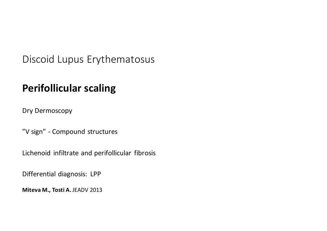
Discoid Lupus Erythematosus
Perifollicular scaling
Dry Dermoscopy
”V sign” -‐ Compound structures
Lichenoid infiltrate and perifollicular fibrosis
Differential diagnosis: LPP
MitevaM., Tosti A. JEADV 2013

Discoid Lupus Erythematosus
Interfollicular scaling
Hyperkeratosis
Differential: psoriasis, seb dermatitis

Discoid Lupus Erythematosus
Milky red/Pink areas White patches

Trichoscopy of DLEActive featuresKeratotic plugsRed dotsThick arborizing vesselsBlue-‐gray dots
Scattered brown discolorationBlue-‐white veilPerifollicular scaleFine interfollicular scalingRosettes
Inactive (end-‐stage) features
Loss of follicular openingsPink areas
White patchesThin Arborizing vessels
Yellow dots with spider vessels

DLE & Frontal Fibrosing Alopecia
DLE may coexist with FFA
1: Trüeb RM, El Shabrawi-‐Caelen L, Kempf W. Cutaneous Lupus ErythematosusPresenting as Frontal Fibrosing Alopecia: Report of 2 Patients. Skin
Appendage Disord. 2017 Oct;3(4):205-‐210.
2: Contin LA, Martins da Costa Marques ER, Noriega L. Frontal Fibrosing Alopecia Coexisting with Lupus Erythematosus: Poor Response to
Hydroxychloroquine. SkinAppendage Disord. 2017 Jan;2(3-‐4):162-‐165
3: del Rei M, Pirmez R, Sodré CT, Tosti A. Coexistence of frontal fibrosingalopecia and discoid lupus erythematosus of the scalp in 7 patients: just a
coincidence? J Eur Acad Dermatol Venereol. 2016 Jan;30(1):151-‐3.
4: Khan S, Fenton DA, Stefanato CM. Frontal fibrosing alopecia and lupus overlap in a man: guilt by association? Int J Trichology. 2013 Oct;5(4):217-‐9.
5: Gaffney DC, Sinclair RD, Yong-‐Gee S. Discoid lupus alopecia complicated byf rontal fibrosing alopecia on a background of androgenetic alopecia. Br J
Dermatol. 2013 Jul;169(1):217-‐8
6: Duque-‐Estrada B, Tamler C, Sodré CT, Barcaui CB, Pereira FB. Dermoscopy patterns of cicatricial alopecia resulting from discoid lupus erythematosus
and lichen planopilaris. An Bras Dermatol. 2010 Mar-‐Apr;85(2):179-‐83.
Take home message
1. Early LesionsTrichoscopy helps you to be suspicious of DLE Keratotic plugs , Red dots, Rosettes – prefibrotic stage

Take home message
2. Clinically evident DLETrichoscopy confirms diagnosis, helps recognizing active/late stage areasand choosing biopsy sites
Take home message

3. Diffuse scaling may be a sign of DLERemove the scales before perfoming trichoscopy
Take home messageTake home message

Take home message
4. Try not miss late stage DLECicatricial alopeciaTrichoscopy will suggest DLE, and helps choosing biopsy sites

Thank [email protected]