glenn d. goldman, md - university of vermont d. goldman, md . ... at least 30% of white men in...
TRANSCRIPT
Glenn D. Goldman, MD
Fletcher Allen Health Care
University of Vermont College of Medicine
Recognize and identify the main types of skin cancer
Understand how and why Mohs surgery is utilized for the treatment of skin cancer
Increasing incidence w/ more than 1Mil cases per/yr in US
Basal Cell Carcinoma 85%-90% Squamous Cell Carcinoma 10%-15% At least 30% of white men in southern USA get
skin cancer at some point in their lives Melanoma 1/45 lifetime, Rapid increase
Photoaging: Wrinkling, mottled pigment and onset of actinic keratoses and skin cancer
UVB (290-320 nm) › Acute sunburn, mutations
UVA (320-400 nm)
› Photoaging, promotion
Premalignant sun induced gritty erythematous macules and keratotic papules - No invasion of dermis
May progress to squamous carcinoma Often require destruction with liquid nitrogen or
5-fluorouracil A marker for the potential development of
cutaneous malignancy Commonly harbor mutations in p53
Majority of Skin Cancer › Arises from follicular (basal) germ › Well over one million cases annually US
Indolent but Destructive › Doubling time 1 year › Rarely metastasizes › Locally highly destructive
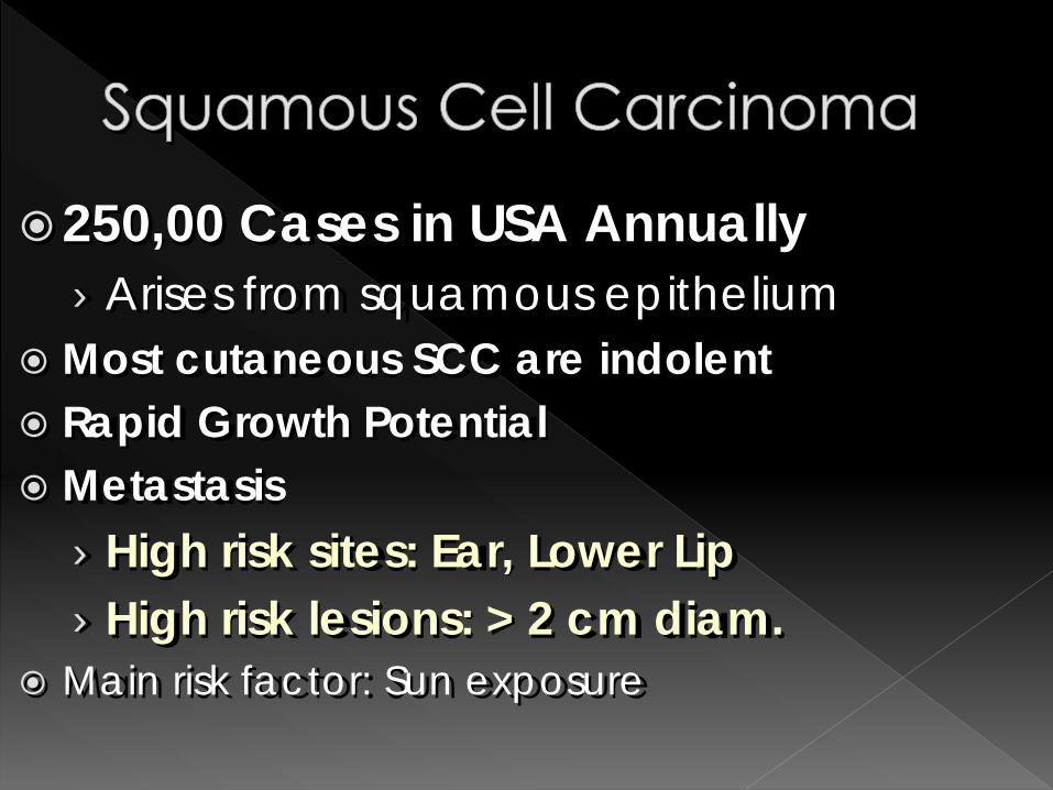
250,00 Cases in USA Annually › Arises from squamous epithelium
Most cutaneous SCC are indolent Rapid Growth Potential Metastasis
› High risk sites: Ear, Lower Lip › High risk lesions: > 2 cm diam.
Main risk factor: Sun exposure
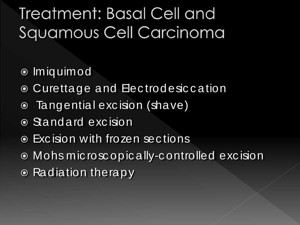
Imiquimod Curettage and Electrodesiccation Tangential excision (shave) Standard excision Excision with frozen sections Mohs microscopically-controlled excision Radiation therapy
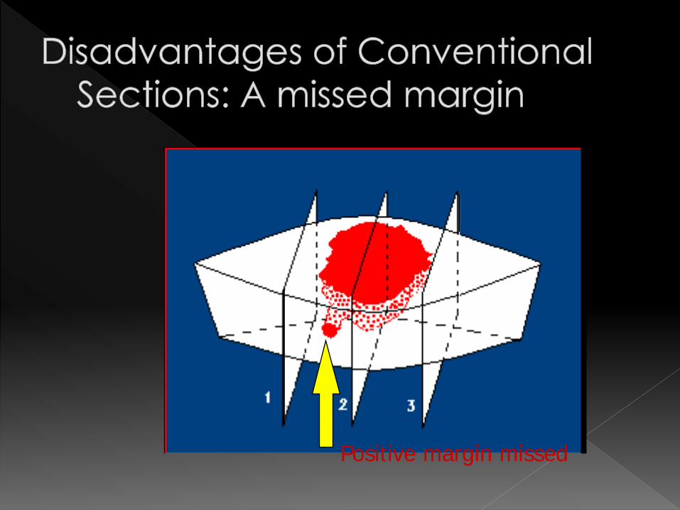
Positive margin missed
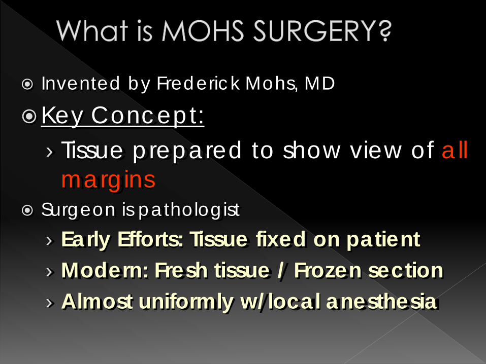
Invented by Frederick Mohs, MD
Key Concept: › Tissue prepared to show view of all
margins Surgeon is pathologist
› Early Efforts: Tissue fixed on patient › Modern: Fresh tissue / Frozen section › Almost uniformly w/local anesthesia
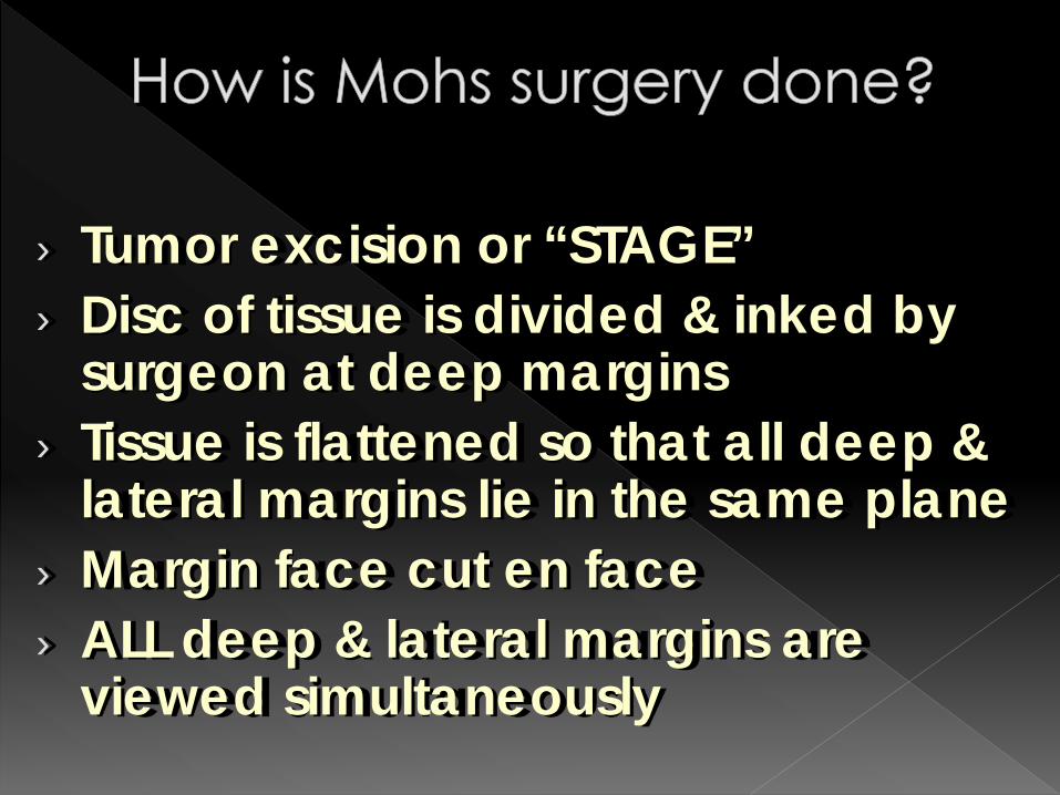
› Tumor excision or “STAGE” › Disc of tissue is divided & inked by
surgeon at deep margins › Tissue is flattened so that all deep &
lateral margins lie in the same plane › Margin face cut en face › ALL deep & lateral margins are
viewed simultaneously
All margins evaluated Highest cure rate Tissue Sparing Surgeon as pathologist
› Surgeon has precise knowledge of tumor location
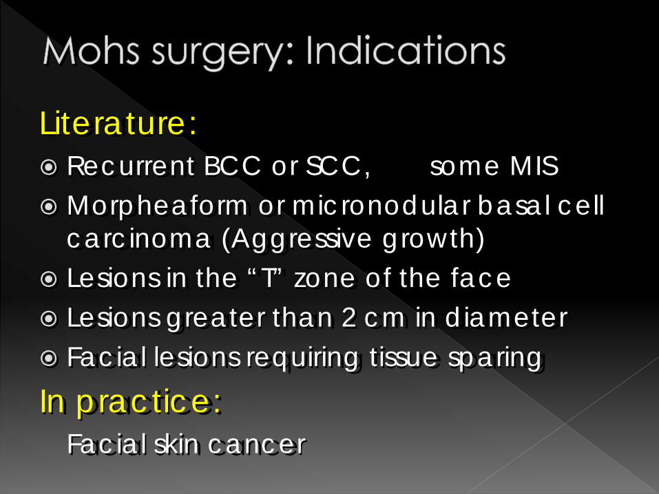
Literature: Recurrent BCC or SCC, some MIS Morpheaform or micronodular basal cell
carcinoma (Aggressive growth) Lesions in the “T” zone of the face Lesions greater than 2 cm in diameter Facial lesions requiring tissue sparing In practice: Facial skin cancer
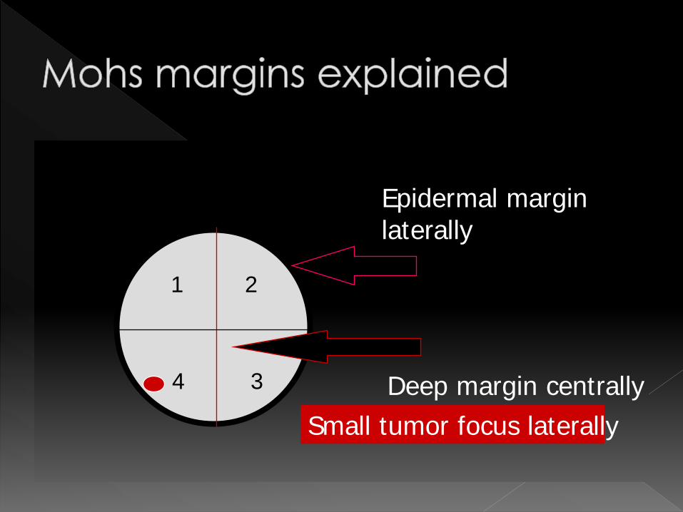
Deep margin centrally
Epidermal margin laterally
Small tumor focus laterally
1 2
3 4
Mainly dermatologists Must be board certified to read
dermatopathology 1 to 2 year surgery fellowship required for
certification by Mohs College Fellowship includes extensive instruction
in reconstructive surgery
Usually local anesthesia No hospital stay Extensive surgery without hospitalization Moderately expensive, but no hospital
fees, no anesthesia fees Very low complication rate
Basal cell and squamous cell carcinoma should be respected as malignancies
Early recognition & definitive treatment provide high cure rate & prevent serious deformity and/or death
Malignant tumor of melanocytes Incidence increasing faster than any other
maliglancy Current US lifetime incidence approximately 1 in
90 Approximately 7500 deaths in US per year Curable if treated definitively at an early stage
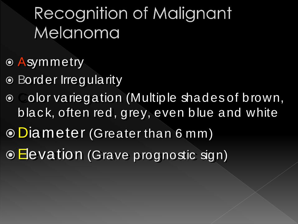
Asymmetry Border Irregularity Color variegation (Multiple shades of brown,
black, often red, grey, even blue and white Diameter (Greater than 6 mm)
Elevation (Grave prognostic sign)
Overtaking superficial spreading as most commonly diagnosed form of melanoma
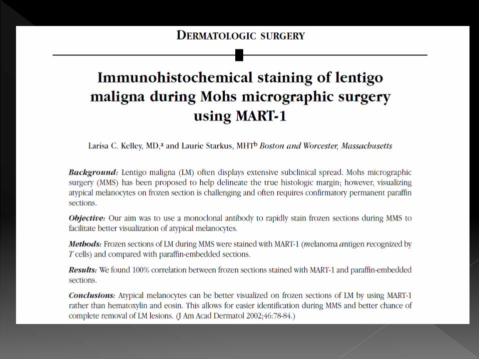
Long (many years) preinvasive phase as lentigo maligna (melanoma in situ)
Peak incidence is in 6th to 7th decade By far most common on face
Excision with at least 5 mm peripheral margin Frequently surgical margins are found to be
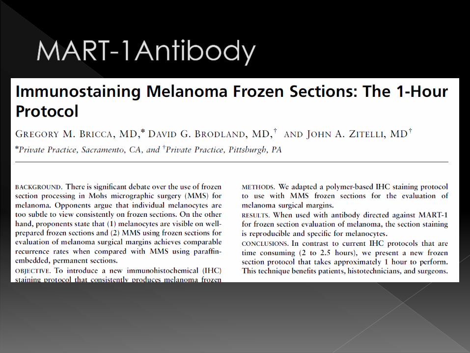
positive, necessitating further surgery Mohs surgery with specialized immunostaining
can assist in definitive tumor removal.