rvc open access repository copyright … · detection of porcine cysticercosis in the absence of a...
TRANSCRIPT
RVC OPEN ACCESS REPOSITORY – COPYRIGHT NOTICE
This is the peer-reviewed, manuscript version of the following article:
Porphyre, V., Betson, M., Rabezanahary, H., Mboussou, Y., Zafindraibe, N. J., Rasamoelina-
Andriamanivo, H., Costard, S., Pfeiffer, D. U. and Michault, A. (2016) 'Taenia solium porcine
cysticercosis in Madagascar: Comparison of immuno-diagnostic techniques and estimation of
the prevalence in pork carcasses traded in Antananarivo city', Veterinary Parasitology, 219, 77-
83.
The final version is available online: http://dx.doi.org/10.1016/j.vetpar.2015.08.027. © 2016. This manuscript version is made available under the CC-BY-NC-ND 4.0 license
http://creativecommons.org/licenses/by-nc-nd/4.0/.
The full details of the published version of the article are as follows:
TITLE: Taenia solium porcine cysticercosis in Madagascar: Comparison of immuno-
diagnostic techniques and estimation of the prevalence in pork carcasses traded in
Antananarivo city
AUTHORS: Porphyre, V; Betson, M; Rabezanahary, H; Mboussou, Y; Zafindraibe, N J;
Rasamoelina-Andriamanivo, H; Costard, S; Pfeiffer, D U; Michault, A
JOURNAL: VETERINARY PARASITOLOGY
PUBLISHER: Elsevier
PUBLICATION DATE: 30 March 2016
DOI: 10.1016/j.vetpar.2015.08.027

1
Taenia solium porcine cysticercosis in Madagascar: comparison of immuno-diagnostic
techniques and estimation of the prevalence in pork carcasses traded in Antananarivo
city
V. Porphyrea,*, M. Betsonb, H. Rabezanaharyc, Y. Mboussouc, N.J. Zafindraibec, H.
Andriamanivod, S. Costardb,e, D. U. Pfeifferb, A. Michaultc
aCIRAD, UMR112 SELMET, F-97410, Saint Pierre, La Réunion, France
bRoyal Veterinary College, Hatfield, Herts AL9 7TA , UK
cCHU de La Réunion - Groupe Hospitalier Sud Réunion, F-97448, Saint Pierre, La Réunion
dFOFIFA-DRZV, Antananarivo, Madagascar
eEpiX Analytics, 1643 Spruce Street, Boulder, Colorado 80302, USA
(V. Porphyre and M. Betson are co-first authors)
* Corresponding author: [email protected]
ABSTRACT
Taenia solium cysticercosis was reported in official veterinary and medical statistics to be
highly prevalent in pigs and humans in Madagascar, but few estimates are available for pigs.
This study aimed to estimate the seroprevalence of porcine cysticercosis among pigs
slaughtered in Antananarivo abattoirs. Firstly, the diagnostic performance of two antigen-
ELISA techniques (B158B60 Ag-ELISA and HP10 Ag-ELISA) and an immunoblotting
method were compared with meat inspection procedures on a sample of pigs suspected to be
infected with (group 1; n=250) or free of (group 2; n=250) T. solium based on direct
veterinary inspection in Madagascar. Sensitivity and specificity of the different tests were
then estimated using a Bayesian approach for detection of porcine cysticercosis in the
absence of a gold standard. Then, a third set of pig sera (group 3, n=250) was randomly
2
collected in Antananarivo slaughterhouses and tested to estimate the overall prevalence of T.
solium contamination in pork meat traded in Antananarivo.
The antigen ELISAs showed a high sensitivity (>84%), but the B158B60 Ag-ELISA
appeared to be more specific than the HP10 Ag-ELISA (model 1: 95% vs 74%; model 2:
87% vs 71%). The overall prevalence of porcine cysticercosis in Antananarivo
slaughterhouses was estimated at 2.3% (95% credibility interval [95%CrI]: 0.09–9.1%) to
2.6% (95%CrI: 0.1–10.3%) depending on the model and priors used. Since the sample used
in this study is not representative of the national pig population, village-based surveys and
longitudinal monitoring at slaughter are needed to better estimate the overall prevalence,
geographical patterns and main risk factors for T. solium contamination, in order to improve
control policies.
Keywords: Taenia solium; cysticercosis; immunodiagnostic; Enzyme-linked
immunoelectrotransfer blot; ELISA; pigs; Madagascar

3
1. Introduction
Taenia solium cysticercosis is a neglected parasitic disease involving humans and pigs and is
endemic in developing countries where pigs roam freely and scavenge human feces around
villages (Torgerson, 2013). T. solium cysticercosis was reported to be highly prevalent in
humans and pigs in Madagascar, with seroprevalences of cysticercosis in humans ranging
from 7% to 21% in the 1990s and 7% to 48% in pigs (Andriantsimahavandy et al., 1997;
Andriantsimahavandy et al., 2003; Michelet et al., 2010; Rasamoelina-Andriamanivo et al.,
2013; Ribot and Coulanges, 1988). Cysticercosis has been described in other islands in the
Indian Ocean, in particular in La Réunion during the 1990s (Michault et al., 1990; Michault
et al., 1989).
Treatment of cysticercosis in humans is problematic, as the subsequent inflammatory
response can be harmful for the patient. To reduce the need for treatment, prophylaxis should
be improved through mass screening, treatment of adult-worm carriers and control of
cysticercosis in pigs (Boussard et al., 2012). For this reason, continuous efforts are being
made to develop rapid and efficient diagnostic tests, and evaluations of the performance of
laboratory techniques for the detection of T. solium in humans are regularly reported (Carod
et al., 2012; Deckers and Dorny, 2010; Hernandez et al., 2000; Hubert et al., 1999; Prasad et
al., 2008; Simac et al., 1995; Villota et al., 2003). Several methods have been previously
described to detect antibodies to T. solium infections in humans and in pigs, such as
radioimmunoassay, hemagglutination, the complement fixation test, dipstick assay, latex
agglutination, enzyme-linked immunosorbent assay (ELISA) and immunoblot techniques
(Deckers and Dorny, 2010). These assays measure exposure to the parasite. In contrast, the
ELISAs which have been developed to detect parasite antigens (Ag) circulating in the host
demonstrate the presence of the living parasite. Such Ag-ELISAs have also been trialed in

4
both humans and pigs (Deckers and Dorny, 2010; Rodriguez et al., 2012; Sciutto et al.,
1998).
In developing countries, the routine diagnosis of porcine cysticercosis in pigs is based (i) for
live animals on lingual palpation that is efficient only when moderate to heavy infection
occurs in individual animals (da Silva et al., 2012; Phiri et al., 2006), and (ii) for carcasses on
visual postmortem and incisional examination during veterinary inspection at abattoirs.
Although several of the laboratory diagnostic techniques described above have been used to
estimate the prevalence of the zoonotic T. solium cysts in pigs, the interpretation of test
results can be difficult, especially in detecting cysticercosis in pigs with low levels of cysts
(Dorny et al., 2004, Krecek et al., 2008, Krecek et al., 2012, Ramahefarisoa et al., 2010, and
Sciutto et al., 1998b).
In the present study, we aimed to determine the diagnostic performance of different tests for
detection of porcine cysticercosis in the absence of a gold standard and to estimate the
prevalence of cysticercosis in pigs slaughtered in Antananarivo, Madagascar.
1. Materials and methods
1.1. Serum sample collection
From April to December 2010, blood was collected from pigs in the four main
slaughterhouses in Antananarivo city, the capital of Madagascar, namely Ampasika,
Ankadindratombo, Anosipatrana, and Anosizato. Information regarding sampling date,
slaughterhouse, region of origin, breed, sex and age was recorded for each animal. Blood was
sampled from the jugular vein directly into plain BD Vacutainer® tubes and allowed to clot
at 4°C. Serum was obtained by centrifugation, dispensed into 2 ml aliquots, stored in labeled
vials and kept at -80°C until shipped on dry ice for testing.
A total of 750 blood samples were collected from pigs raised in 11 different regions (out of a
5
total 22) in Madagascar. Samples were split into three groups: group 1 samples (n=250) came
from animals considered to be infected based on visual inspection, group 2 (n=250) consisted
of samples from animals considered free from infection based on absence of lesions on visual
inspection, and group 3 consisted of blood samples (n=250) randomly collected from
slaughtered pigs in November and December 2010.
1.2. Examination of pigs
The T. solium cysticercosis status of carcasses was determined by an extensive visual
postmortem and incisional examination according to the local meat inspection regulations
(Phiri et al., 2006; Phiri et al., 2002). Heart, masseters, diaphragm, and tongue were visually
examined. Long and parallel incisions were made in external and internal masseter muscles.
The tongue was palpated and a longitudinal incision was made at the base of the tongue to
check for cysts. The heart was cut open to detect cysts in the septum (Boa et al., 2002). No
information was recorded about the number of larvae in muscles making the investigation of
the infection intensity impossible. The cysticerci stages, i.e. viable or degenerated, were not
registered. Only the location of cysticerci lesions for animals considered in group 1 were
registered. In group 1, cysticerci lesions were observed in limbs (100%), pork shoulder
(49.6%), masseter (12.4%) , tongue (39.6%), heart and pericardium (5.2 %), as predilection
sites. Cysticerci were reported in only one location (limbs) in 30.4% (n=76) of pigs in group
1.
1.3. Serological tests
Sera in groups 1 and 2 were analysed using three serological tests. Enzyme-linked
immunoelectrotransfer blot (EITB) analysis was carried out using the Cysticercosis Western
Blot Kit (LDBio Diagnostics, Lyon, France) according to the manufacturer’s instructions.
This test was considered positive if the pig serum detected at least two specific bands. Two
different Ag-ELISAs were also used. ELISAs were performed in a single test and positive
6
samples were confirmed by duplicate test. The first was the Cysticercosis Ag-ELISA (ApDia
Ltd., Turnhout, Belgium), which makes use of the B158C11A10 and B60H8A4 monoclonal
antibodies to detect circulating antigens released by viable cysticerci (Brandt et al., 1992;
Draelants et al., 1995). The assay was carried out according to the manufacturer’s
instructions, the optical density (OD) was read at 450 nm and the Ag index was calculated as
described. The cut-offs recommended by the manufacturer were used, where an Ag index less
than 0.8 was considered a negative result, an Ag index greater than 1.3 was classified as a
positive result and values in between were considered “doubtful”. The manufacturer reports
that upon testing 99 animals infected with viable cysticerci of Taenia species using the
B158B60 Ag-ELISA, all gave positive results. Based on repeated testing of 300 negative
porcine samples, the manufacturer claims a specificity of 99.6% in diagnosis of porcine
cysticercosis (Dorny et al., 2004; Nguekam et al., 2003). The second Ag-ELISA detects a
metacestode antigen using the HP10 monoclonal antibody (Harrison et al., 1989), and was
carried out according to the method described by Sciutto et al. (Sciutto et al., 1998).
In this case an OD greater than 0.177 was considered a positive result, an OD less than 0.129
was classified as a negative result and ODs in between were considered “doubtful results”.
This latter Ag-ELISA was also used to screen the group 3 sera. All ELISAs were performed
once and all positive samples were retested to confirm results.
1.4. Statistical methods
As a first step the diagnostic performance of the three immunodiagnostic tests was
determined using carcass visual and incisional examination as the “gold standard”. In
addition, receiver operator characteristic (ROC) curve analysis was performed. “Doubtful
results” were removed from the dataset (8 for HP10 Ag-ELISA and 1 for B158B60 Ag-
ELISA). The statistical analysis was carried out in R v3.0.3 (R development core team, 2008)

7
using the caret and pROC packages.
However, carcass inspection is not a true gold standard for validation of diagnostic tests for
porcine cysticercosis unless complete carcass dissection and enumeration of cysts is carried
out, which is rarely logistically and economically feasible. Thus, a Bayesian approach
(Markov chain Monte Carlo [MCMC] simulation with Gibbs sampling) was adopted to
estimate test sensitivity and specificity in the absence of a gold standard (Berkvens et al.,
2006; Branscum et al., 2005). To maximise the number of samples with complete test results,
EITB results were excluded from the analysis. Data from groups 1 and 2 on carcass
inspection and the two Ag-ELISAs were included in the analysis (n=117). As both ELISAs
detect circulating parasite antigens, an assumption of conditional dependence between these
two tests was made and two co-variance parameters were included in the model (Branscum,
2005). In contrast, carcass inspection was assumed to be conditionally independent of both
ELISAs due to a biologically different outcome being measured (i.e. visible pathology rather
than antigen). An initial model was constructed which included prior information about
sensitivities and specificities of the three tests. The BetaBuster software
(http://www.epi.ucdavis.edu/diagnostictests/betabuster.html) was employed to calculate beta
(α, β) distributions based on published estimates (see Table 2). Two models (Models 1 and 2)
were run using two sets of priors for the sensitivity and specificity of B158B60 Ag-ELISA,
based on two different studies conducted in Africa (Dorny et al., 2004; Krecek et al., 2011).
The priors for the other diagnostic tests were the same in the two models.
The Bayesian models were run using the WinBUGS software (v14) (Lunn et al., 2000). An
initial burn-in of 5,000 iterations was discarded, and followed by 50,000 further iterations.
The median and 95% credibility intervals of the posterior distributions of the parameters of
interest were obtained using MCMC with Gibbs sampling. Model convergence was assessed

8
by running five chains simultaneously and visually inspecting time-series plots for each
parameter. Models were validated by comparing the number of parameters estimated by the
model (pD) and the Deviance Information Criterion (DIC) values calculated in the posterior
mean of the multinomial probabilities and in the posterior mean of the parameters of the
model (Berkvens et al., 2006). After running the initial models, a sensitivity analysis was
carried out by replacing the informative priors with non-informative priors or partially
informative priors. In the latter case, prior beta distributions were substituted with uniform
(a,b) distributions. The two parameters, a and b, which are the minimum and maximum
values of the random variable, were defined according tests and models: a=0.5, b=1 for HP10
Ag-ELISA sensitivity, B158B60 Ag-ELISA sensitivity (Model 1) and specificity and carcass
inspection specificity; a=0.4, b=0.9 for HP10 Ag-ELISA specificity and B158B60 Ag-ELISA
sensitivity (Model 2); a=0, b=0.5 for sensitivity of carcass inspection.
The true prevalence of porcine cysticercosis in pork carcasses slaughtered and retailed in
Antananarivo was then estimated using a Bayesian approach based on the apparent
prevalence determined through testing of sera from the group 3 pigs with the HP10 Ag-
ELISA (http://www.epi.ucdavis.edu/diagnostictests/aptoprev.html). The sensitivity and
specificity estimates for the HP10 Ag-ELISA from Models 1 and 2 were used to generate
informative Beta priors. Models were run in WinBUGS as described above and median
values and 95% credibility intervals for the true prevalence were estimated.
3. Results
Diagnostic test results for pigs which were deemed positive and negative for cysticercosis
based on carcass visual and incisional examination are summarized in Table 2. In group 1,
carcasses, cysticerci lesions were observed in limbs (100%), pork shoulder (49.6%), masseter
(12.4%), tongue (39.6%), heart and pericardium (5.2%), as predilection sites. Cysticerci were

9
reported in only one location (limbs) in 30.4% (n = 76) of pigs in group 1.
EITB results were obtained for 108 pigs (64 in group 1 and 44 in group 2), B158B60 Ag-
ELISA results for 145 pigs (128 in group 1 and 17 in group 2) and HP10 Ag-ELISA results
for 288 pigs (159 in group 1, and 129 in group 2). HP10 Ag-ELISA results were also
obtained for 175 pigs in group 3 (Table 2).
When diagnostic performance was assessed using visual and incisional examination of
carcass as the “gold standard”, all three immunodiagnostic tests showed a high sensitivity
(>90%). However, the HP10 Ag-ELISA was less specific than EITB and B158B60 Ag-
ELISA (see Table 3). When ROC curve analysis was carried out, the area under the curve
(AUC) was 0.916 for the EITB, 0.971 for the B158B60 Ag-ELISA, but 0.802 for the HP10.
Of the 117 pigs for which complete results were available for three diagnostic tests (carcass
visual and incisional examination, B158B60Ag-ELISA and HP10 Ag-ELISA), there was full
agreement between all three tests in 111 cases (94.8%). The cross-classified results of the
three tests are presented in Table 4. This dataset was used to estimate the sensitivity and
specificity of the three diagnostic tests using Bayesian analysis. The results are summarized
in Table 5. Similar estimates of diagnostic test performance were generated by the two
models based on different prior distributions for the sensitivity and specificity of B158B60
Ag-ELISA. The median estimates of sensitivity and specificity for the B158B60 Ag-ELISA
were somewhat higher for model 1 than for model 2. The B158B60 Ag-ELISA was the most
sensitive test overall and also showed a high specificity. Visual inspection was very highly
specific but showed a lower sensitivity, whereas the HP10 Ag-ELISA was the least specific
of the three tests.
When sensitivity analysis was carried out using non-informative or partially informative
priors, all median estimates of test sensitivity and specificity fell within 8% of the original
values, except for the specificity of the HP10 Ag-ELISA in Model 1, which increased by

10
12% with a non-informative prior, and the sensitivity of the B158B60 Ag-ELISA in Model 2,
which increased by 12% with a non-informative prior. The sensitivity of the visual and
incisional inspection of carcasses increased by 46% with a non-informative prior and
decreased by 22–26% with a partially informative prior.
Of the 175 pigs in group 3 tested with the HP10 Ag-ELISA, 19 were positive. Based on this
result and the estimates of the diagnostic performance of this ELISA reported above, the
prevalence of porcine cysticercosis was estimated as 2.3% (95% credibility interval [CrI]:
0.09–9.1%) if results from Model 1 were used to generate priors, and 2.6% (CrI: 0.1–10.3%)
if results from Model 2 were used to generate priors.
4. Discussion
The aim of this study was to evaluate the performance of different diagnostic tests for porcine
cysticercosis in Madagascar using samples collected from pigs upon slaughter and to estimate
the prevalence of porcine cysticercosis among pigs slaughtered in Antananarivo, Madagascar.
Since there can be variation in performance of diagnostic tests in different locations and
populations, it is important to validate tests in the area in which they will be used (Deckers
and Dorny, 2010).
The diagnosis of porcine cysticercosis remains challenging. The gold standard of detailed
carcass dissection and cyst enumeration is time-consuming, expensive and requires skilled
personnel, and so was not logistically feasible for this study. A Bayesian approach was thus
adopted to estimate the sensitivity and specificity of diagnostic tests in the absence of a gold
standard, as has been carried out for porcine cysticercosis in Zambia and South Africa (Dorny
et al., 2004; Krecek et al., 2008).
Both Ag-ELISAs compared in this study were highly sensitive in diagnosis of porcine
cysticercosis but the B158B60 Ag-ELISA was more specific than the HP10 Ag-ELISA,

11
probably due to the fact that the tests use different monoclonal antibodies, which likely target
different circulating antigens or epitopes.
Serological test results should be interpreted carefully considering possible cross-reactions
with other parasites. Recently concerns have been raised about the specificity of Ag-ELISA
and EITB for diagnosis of porcine cysticercosis. Gavidia et al. (2013) and Jayashi et al.
(2014) found that pigs from endemic areas that were EITB positive had no cysts upon
necropsy. Similar results were reported by Devleesschauwer et al. (2013) using the B158B60
Ag-ELISA: in sentinel pigs that tested Ag-ELISA positive, no T. solium cysts could be found
in the carcass. It is well-documented that infection with T. hydatigena causes false positives
in B158B60 and HP10 Ag-ELISAs ( Rodriguez et al., 2012), however other potential sources
of false positive reactions in Ag-ELISAs in pigs have not been investigated (for example
exposure to T. saginata or to the eggs of other taeniid cestodes). There is very little
information on how much T. hydatigena exists in Madagascar. As some areas of the country
have a serious problem with both household and feral dogs ( Ratsitorahina et al., 2009), there
is a possibility that T. hydatigena or Echinococcus spp. are circulating between dogs and
pigs. However, the meat inspection noted the presence of no other parasites apart from T.
solium cysts. In addition, when carcass inspection was used as a reference standard, the EITB
assay and the B158B60 Ag-ELISA were found to highly specific for detection of porcine
cysticercosis (specificities of 90.9% and 94.1%, respectively), suggesting that for these
assays cross-reactivity with other parasites is not a major concern in this setting.
In the current Bayesian analysis, carcass inspection was found to be highly specific in
diagnosis of porcine cysticercosis, consistent with previous reports (Dorny et al., 2004; Phiri
et al., 2006). However, the sensitivity of this method was also surprisingly high in
comparison with earlier estimates (Boa et al., 2002; Dorny et al., 2004). This suggests that

12
either the inspection was carried out more thoroughly in this study than in previous surveys,
thus increasing the likelihood of detecting cysts. However, no information was recorded on
cyst numbers or whether the pigs were considered heavily, medium or light infections, which
is known to be related to the sensitivity of meat inspection and serological tests (Sciutto et al.,
1998b).
There are limitations to the estimates of diagnostic performance obtained during this study.
Firstly, since our 3 tests required significant amounts of sera, our samples were not
systematically analyzed with the three laboratory-based diagnostic tests; this technical
difficulty may have introduced bias if certain types of samples were more likely to have
failed testing than others. Secondly, the sample size for the Bayesian analysis was small
(n=117) and only 17 “negative” (by carcass inspection) samples were included, providing a
potential further source of bias. The results of the sensitivity analysis fell within 8% of the
original model results when partially informative priors were used and within 12% of the
model results when non-informative priors were used. This suggests that the prior
distributions employed were appropriate for the analysis. The one exception to this was the
sensitivity of carcass inspection which showed a dramatic change when partially informative
or non-informative priors were employed, indicating that the prior distribution very strongly
influenced the posterior estimate of this parameter. The models each contained seven degrees
of freedom (seven independent data cells) and were used to estimate nine parameters
(sensitivity and specificity for each test, “prevalence” and two co-variance parameters). Thus,
they were not “identifiable”, meaning that there were insufficient data to estimate the
parameters of interest, unless prior information was included (Branscum et al., 2005). A
possible explanation for the results of the sensitivity analysis is that the prior estimate of
carcass inspection sensitivity was too low, perhaps because the inspection was conducted
more carefully in this study than in previous surveys or due to a difference in infection

13
intensity (as discussed previously). Thus the actual estimate of the sensitivity of carcass
inspection in this setting may actually be higher than reported here.
The prevalence of porcine cysticercosis in Madagascar estimated here is slightly higher than
the official prevalence of 0.5-1% reported for the 2008-2012 period, which was based on
visual inspection of carcasses in urban abattoirs (Direction des Services Veterinaires de
Madagascar, 2012). Our estimate is surprisingly low given that Madagascar is considered to
be a hotspot for human taeniasis and pig cysticercosis is often reported in local abattoirs and
markets (Rasamoelina-Andriamanivo et al., 2013). Since the majority of pigs in Madagascar
are not slaughtered at abattoirs, but rather in villages or at home (Rasamoelina-Andriamanivo
et al., 2013), we could not estimate the overall prevalence of T.solium in pig population at
country level; indeed, the samples in this study were not representative of the national pig
population in Madagascar, but representative of the commercial pigs slaughtered during a
short period of time in Antananarivo city for urban consumers only. Moreover, traders use
lingual palpation to detect heavily infected animals at rural live pig markets. Although the
efficacy of such control, and, in consequence, the prevalence of infected pigs at urban market,
may be influenced by fluctuations in demand for pork through the year, it is likely that the
most “healthy” pigs are sent for slaughter at abattoirs (Praet et al., 2010). Thus, abattoir-based
surveys may underestimate the prevalence of porcine cysticercosis.
Recent village-based surveys in other African countries revealed porcine cysticercosis
prevalence as high as 41% (Assana et al., 2010; Eshitera et al., 2012; Ganaba et al., 2011;
Komba et al., 2013; Ngowi et al., 2010; Pondja et al., 2010; Praet et al., 2010). Thus, village-
based studies may be necessary to gain a better understanding of the overall burden of
porcine cysticercosis in Madagascar.
Results from the assessment of diagnostic tests performance reported here suggest that the
B158B60 Ag-ELISA would be the most appropriate laboratory-based diagnostic for such

14
surveys. However, Ag-ELISA tests techniques remain challenging for farm-based testing as
they require laboratories and trained staff. Detection of cysts through lingual palpation, the
method which is used most widely in developing countries due to its simplicity and low cost,
is notoriously insensitive for detecting low-intensity infections in individual animals (Phiri et
al., 2006). Thus there is an urgent need for the development of simple, sensitive and
inexpensive point-of-care tests which do not require additional equipment and can be
deployed on-farm by farmers and animal health workers to inform treatment and control
decisions on the ground.
In conclusion, our results provide a first laboratory-based description of the burden of
cysticercosis in pigs slaughtered in Antananarivo city in Madagascar; they noted an apparent
low percentage of pork carcasses contaminated with T. solium cysts at urban market level. To
better define appropriate surveillance and control measures for cysticercosis in Madagascar
several questions need to be investigated in further studies, including: (i) what is the
prevalence of porcine cysticercosis in different Malagasy regions? (ii) What are the main risk
factors for infection in farms? (iii) What is the seasonal variation in disease burden? (iv) How
much understanding of the disease exists in rural communities? (v) How acceptable would
potential new control measures be in rural communities?
Acknowledgements
We are extremely grateful to the technical staff of the FOFIFA-DRZV, and especially Mr
Samuel Rakotonindrina for his invaluable work in the pig slaughterhouses. We thank Denis
Limonne, LDBio Diagnostic, for his involvement in the EITB analysis. We are grateful to
Raf Berghmans, Advanced Practical Diagnostics NV (ApDia ltd.) for providing the Ag-
ELISA kit material. Equally, we warmly thank Dr Michael Parkhouse for his technical
assistance regarding the HP10 Ag-ELISA method.

15
Conflict of interest
Authors declare that they have no conflicts of interest relating to this paper.
Role of the funding source
The main financial supports were provided by the Wellcome Trust Fund, the Regional
Council of La Réunion, the European Regional development Funds (ERDF) and French
Government through the QualiREG research network in the Indian Ocean
(www.qualireg.org).
Authors' addresses:
Vincent Porphyre, International Center for Agronomical Research for Development
(CIRAD), UMR112 SELMET, Saint Pierre, La Réunion, France; E-mail :
Martha Betson, Dept. of Production and Population Health, Royal Veterinary College, North
Mymms, Hatfield, Herts, UK; E-mail: [email protected]
H. Rasamoelina-Andriamanivo, FOFIFA, Department of Veterinary and Husbandry
Research, Ministry of Agriculture, Antananarivo, Madagascar; E-mail: [email protected]
Norosoa Julie Zafindraibe, CHU de La Réunion, F-97448, Saint Pierre, La Réunion ; E-mail :
Solenne Costard, EpiX Analytics, 1643 Spruce Street, Boulder, CO, 80302, USA; E-mail:
Alain Michault, CHU, F-97448, Saint Pierre, La Réunion; E-mail: alain.michault@chu-
reunion.fr
Yoan Mboussou, CHU, F-97448, Saint Pierre, La Réunion; E-mail: [email protected]

16
Henintsoa Rabezanahary, CHU, F-97448, Saint Pierre, La Réunion; E-mail:
Dirk Pfeiffer, Dept. of Production and Population Health, Royal Veterinary College, North
Mymms, Hatfield, Herts, UK; E-mail: [email protected]
Corresponding author:
V. Porphyre, International Center for Agronomical Research for Development (CIRAD),
UMR112 SELMET, Saint Pierre, La Réunion, France; Tel.: +262 262 49 92 55; fax: +262
262 49 92 95; E-mail: [email protected]
References
Andriantsimahavandy, A., Lesbordes, J.L., Rasoaharimalala, B., Peghini, M., Rabarijaona,
L., Roux, J., Boisier, P., 1997. Neurocysticercosis: a major aetiological factor of late-
onset epilepsy in Madagascar. Trop Med Int Health 2, 741-746.
Andriantsimahavandy, A., Ravaoalimalala, V.E., Rajaonarison, P., Ravoniarimbinina, P.,
Rakotondrazaka, M., Raharilaza, N., Rakotoarivelo, D., Ratsitorahina, M.,
Rabarijaona, L.P., Ramarokoto, C.E., Leutscher, P., Migliani, R., 2003. The current
epidemiological situation of cysticercosis in Madagascar. Arch Inst Pasteur
Madagascar 69, 46-51.
Assana, E., Amadou, F., Thys, E., Lightowlers, M.W., Zoli, A.P., Dorny, P., Geerts, S., 2010.
Pig-farming systems and porcine cysticercosis in the north of Cameroon. J Helminthol
84, 441-446.
Berkvens, D., Speybroeck, N., Praet, N., Adel, A., Lesaffre, E., 2006. Estimating disease
prevalence in a Bayesian framework using probabilistic constraints. Epidemiology 17,
17
145-153.
Boa, M.E., Kassuku, A.A., Willingham, A.L., 3rd, Keyyu, J.D., Phiri, I.K., Nansen, P., 2002.
Distribution and density of cysticerci of Taenia solium by muscle groups and organs
in naturally infected local finished pigs in Tanzania. Vet Parasitol 106, 155-164.
Boussard, M., Millon, L., Grenouillet, F., Jambou, R., 2012. Prevention and treatment of
cysticercosis. J Anti-Infect 14, 143-150.
Brandt, J.R.A., Geerts, S., Dedeken, R., Kumar, V., Ceulemans, F., Brijs, L., Falla, N., 1992.
A monoclonal antibody-based ELISA for the detection of circulating excretory
secretory antigens in Taenia saginata cysticercosis. Int J Parasitol 22, 471-477.
Branscum, A.J., Gardner, I.A., Johnson, W.O., 2005. Estimation of diagnostic-test sensitivity
and specificity through Bayesian modeling. Prev Vet Med 68, 145-163.
Carod, J.F., Randrianarison, M., Razafimahefa, J., Ramahefarisoa, R.M., Rakotondrazaka,
M., Debruyne, M., Dautigny, M., Cazal, P., Andriantseheno, M.L., Charles, E.R.,
2012. Evaluation of the performance of 5 commercialized enzyme immunoassays for
the detection of Taenia solium antibodies and for the diagnosis of neurocysticercosis.
Diagn Microbiol Infect Dis 72, 85-89.
da Silva, M.R.M., Uyhara, C.N.S., Silva, F.H., Espindola, N.M., Poleti, M.D., Vaz, A.J.,
Meirelles, F.V., Maia, A.A.M., 2012. Cysticercosis in experimentally and naturally
infected pigs: Parasitological and immunological diagnosis. Pesq Vet Bras 32, 297-
302.
Deckers, N., Dorny, P., 2010. Immunodiagnosis of Taenia solium taeniosis/cysticercosis.
Trends Parasitol 26, 137-144.
Direction des Services Veterinaires de Madagascar 2012. Situations zoosanitaires de
Madagascar de 2001 à 2011 (Ministere de l’Elevage).

18
Dorny, P., Phiri, I.K., Vercruysse, J., Gabriel, S., Willingham, A.L., 3rd, Brandt, J., Victor,
B., Speybroeck, N., Berkvens, D., 2004. A Bayesian approach for estimating values
for prevalence and diagnostic test characteristics of porcine cysticercosis. Int J
Parasitol 34, 569-576.
Draelants, E., Brandt, J.R.A., Kumar, V., Geerts, S., 1995. Characterization of epitopes on
excretory-secretory antigens of Taenia saginata metacestodes recognized by
monoclonal-antibodies with immunodiagnostic potential. Parasite Immunol 17, 119-
126.
Eshitera, E.E., Githigia, S.M., Kitala, P., Thomas, L.F., Fevre, E.M., Harrison, L.J., Mwihia,
E.W., Otieno, R.O., Ojiambo, F., Maingi, N., 2012. Prevalence of porcine
cysticercosis and associated risk factors in Homa Bay District, Kenya. BMC Vet Res
8, 234.
Ganaba, R., Praet, N., Carabin, H., Millogo, A., Tarnagda, Z., Dorny, P., Hounton, S., Sow,
A., Nitiema, P., Cowan, L.D., 2011. Factors associated with the prevalence of
circulating antigens to porcine cysticercosis in three villages of Burkina Faso. PLoS
Negl Trop Dis 5, e927.
Harrison, L.J.S., Joshua, G.W.P., Wright, S.H., Parkhouse, R.M.E., 1989. Specific detection
of circulating surface secreted glycoproteins of viable cysticerci in Taenia saginata
cysticercosis. Parasite Immunol 11, 351-370.
Hernandez, M., Beltran, C., Garcia, E., Fragoso, G., Gevorkian, G., Fleury, A., Parkhouse,
M., Harrison, L., Sotelo, J., Sciutto, E., 2000. Cysticercosis: towards the design of a
diagnostic kit based on synthetic peptides. Immunol Lett 71, 13-17.
Hubert, K., Andriantsimahavandy, A., Michault, A., Frosch, M., Muhlschlegel, F.A., 1999.
Serological diagnosis of human cysticercosis by use of recombinant antigens from
19
Taenia solium cysticerci. Clin Diagn Lab Immunol 6, 479-482.
Komba, E.V., Kimbi, E.C., Ngowi, H.A., Kimera, S.I., Mlangwa, J.E., Lekule, F.P.,
Sikasunge, C.S., Willingham, A.L., 3rd, Johansen, M.V., Thamsborg, S.M., 2013.
Prevalence of porcine cysticercosis and associated risk factors in smallholder pig
production systems in Mbeya region, southern highlands of Tanzania. Vet Parasitol
198, 284-291.
Krecek, R.C., Michael, L.M., Schantz, P.M., Ntanjana, L., Smith, M.F., Dorny, P., Harrison,
L.J.S., Grimm, F., Praet, N., Willingham, A.L., 2008. Prevalence of Taenia solium
cysticercosis in swine from a community-based study in 21 villages of the Eastern
Cape Province, South Africa. Vet Parasitol 154, 38-47.
Krecek, R.C., Michael, L.M., Schantz, P.M., Ntanjana, L., Smith, M.F., Dorny, P., Harrison,
L.J.S., Grimm, F., Praet, N., Willingham, A.L., 2011. Corrigendum to “Prevalence of
Taenia solium cysticercosis in swine from a community-based study in 21 villages of
the Eastern Cape Province, South Africa”. Vet Parasitol 183, 198-200.
Lunn, D.J., Thomas, A., Best, N., Spiegelhalter, D., 2000. WinBUGS - A Bayesian modelling
framework: Concepts, structure, and extensibility. Stat Comput 10, 325-337.
Michault, A., Duval, G., Bertil, G., Folio, G., 1990. [Sero-epidemiological study of
cysticercosis in Reunion Island]. Bull Soc Pathol Exot 83, 82-92.
Michault, A., Leroy, D., Coubes, P., Laporte, J.P., Bertil, G., Mignard, C., 1989.
Immunological diagnosis in the cerebrospinal fluid and serum in patients with
evolutive cerebral cysticercosis by ELISA. Trans Zool Soc London 37, 249-253.
Michelet, L., Carod, J.F., Rakontondrazaka, M., Ma, L., Gay, F., Dauga, C., 2010. The pig
tapeworm Taenia solium, the cause of cysticercosis: Biogeographic (temporal and
spacial) origins in Madagascar. Mol Phylogenet Evol 55, 744-750.

20
Ngowi, H.A., Kassuku, A.A., Carabin, H., Mlangwa, J.E., Mlozi, M.R., Mbilinyi, B.P.,
Willingham, A.L., 3rd, 2010. Spatial clustering of porcine cysticercosis in Mbulu
district, northern Tanzania. PLoS Negl Trop Dis 4, e652.
Nguekam, A., Zoli, A.P., Vondou, L., Pouedet, S.M., Assana, E., Dorny, P., Brandt, J.,
Losson, B., Geerts, S., 2003. Kinetics of circulating antigens in pigs experimentally
infected with Taenia solium eggs. Vet Parasitol 111, 323-332.
Phiri, I.K., Dorny, P., Gabriel, S., Willingham, A.L., 3rd, Sikasunge, C., Siziya, S.,
Vercruysse, J., 2006. Assessment of routine inspection methods for porcine
cysticercosis in Zambian village pigs. J Helminthol 80, 69-72.
Phiri, I.K., Dorny, P., Gabriel, S., Willingham, A.L., Speybroeck, N., Vercruysse, J., 2002.
The prevalence of porcine cysticercosis in Eastern and Southern provinces of Zambia.
Vet Parasitol 108, 31-39.
Pondja, A., Neves, L., Mlangwa, J., Afonso, S., Fafetine, J., Willingham, A.L., 3rd,
Thamsborg, S.M., Johansen, M.V., 2010. Prevalence and risk factors of porcine
cysticercosis in Angonia District, Mozambique. PLoS Negl Trop Dis 4, e594.
Praet, N., Kanobana, K., Kabwe, C., Maketa, V., Lukanu, P., Lutumba, P., Polman, K.,
Matondo, P., Speybroeck, N., Dorny, P., Sumbu, J., 2010. Taenia solium cysticercosis
in the Democratic Republic of Congo: how does pork trade affect the transmission of
the parasite? PLoS Negl Trop Dis 4.
Prasad, A., Gupta, R.K., Pradhan, S., Tripathi, M., Pandey, C.M., Prasad, K.N., 2008. What
triggers seizures in neurocysticercosis? A MRI-based study in pig farming community
from a district of North India. Parasitol Int 57, 166-171.
R development core team, 2008. R: A language and environment for statistical computing, In:
http://www.r-project.org. Vienna.

21
Rasamoelina-Andriamanivo, H., Porphyre, V., Jambou, R., 2013. Control of cysticercosis in
Madagascar: beware of the pitfalls. Trends Parasitol 29, 538-547.
Ribot, J.J., Coulanges, P., 1988. Malagasy zoonoses. Rev Elev Med Vet Pays Trop 41, 9-22.
Rodriguez, S., Wilkins, P., Dorny, P., 2012. Immunological and molecular diagnosis of
cysticercosis. Pathog Glob Health 106, 286-298.
Sciutto, E., Martinez, J.J., Villalobos, N.M., Hernandez, M., Jose, M.V., Beltran, C., Rodarte,
F., Flores, I., Bobadilla, J.R., Fragoso, G., Parkhouse, M.E., Harrison, L.J., de Aluja,
A.S., 1998. Limitations of current diagnostic procedures for the diagnosis of Taenia
solium cysticercosis in rural pigs. Vet Parasitol 79, 299-313.
Simac, C., Michel, P., Andriantsimahavandy, A., Esterre, P., Michault, A., 1995. Use of
enzyme-linked-immunosorbent-assay and enzyme-linked immunoelectrotransfer blot
for the diagnosis and monitoring of neurocysticercosis. Parasitol Res 81, 132-136.
Torgerson, P.R., 2013. One world health: Socioeconomic burden and parasitic disease control
priorities. Vet Parasitol 195, 223-232.
Villota, G.E., Gomez, D.I., Volcy, M., Franco, A.F., Cardona, E.A., Isaza, R., Sanzon, F.,
Teale, J.M., Restrepo, B.I., 2003. Similar diagnostic performance for
neurocysticercosis of three glycoprotein preparations from Taenia solium
metacestodes. Am J Trop Med Hyg 68, 276-280.
22
Figure Legends
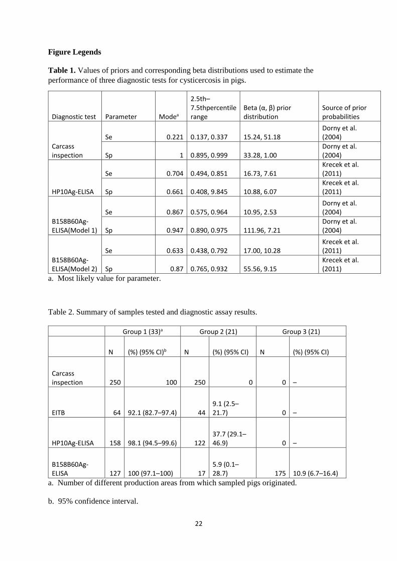
Table 1. Values of priors and corresponding beta distributions used to estimate the
performance of three diagnostic tests for cysticercosis in pigs.
Diagnostic test Parameter Modea
2.5th–7.5thpercentile range
Beta (α, β) prior distribution
Source of prior probabilities
Carcass inspection
Se 0.221 0.137, 0.337 15.24, 51.18 Dorny et al. (2004)
Sp 1 0.895, 0.999 33.28, 1.00 Dorny et al. (2004)
HP10Ag-ELISA
Se 0.704 0.494, 0.851 16.73, 7.61 Krecek et al. (2011)
Sp 0.661 0.408, 9.845 10.88, 6.07 Krecek et al. (2011)
B158B60Ag-ELISA(Model 1)
Se 0.867 0.575, 0.964 10.95, 2.53 Dorny et al. (2004)
Sp 0.947 0.890, 0.975 111.96, 7.21 Dorny et al. (2004)
B158B60Ag-ELISA(Model 2)
Se 0.633 0.438, 0.792 17.00, 10.28 Krecek et al. (2011)
Sp 0.87 0.765, 0.932 55.56, 9.15 Krecek et al. (2011)
a. Most likely value for parameter.
Table 2. Summary of samples tested and diagnostic assay results.
Group 1 (33)a Group 2 (21) Group 3 (21)
N (%) (95% CI)b N (%) (95% CI) N (%) (95% CI)
Carcass inspection 250 100 250 0 0 –
EITB 64 92.1 (82.7–97.4) 44 9.1 (2.5–21.7) 0 –
HP10Ag-ELISA 158 98.1 (94.5–99.6) 122 37.7 (29.1–46.9) 0 –
B158B60Ag-ELISA 127 100 (97.1–100) 17
5.9 (0.1–28.7) 175 10.9 (6.7–16.4)
a. Number of different production areas from which sampled pigs originated.
b. 95% confidence interval.

23
Table 3. Summary of performance of three diagnostic tests for cysticercosis in pigs using
visual and incisional examination of carcasses as the “gold standard”.
Sensitivity(%) (95% CI)a
Specificity(%) (95% CI)
PPVb(%) (95% CI)
NPVc(%) (95% CI) AUCd(95% CI)
EITB 92.2 90.9 93.7 88.9 0.916
(n = 108) (82.7–97.4) (78.3–97.5) (84.5–98.2) (75.9–96.3) (0.861–0.970)
HP10 Ag-ELISA 98.1 62.3 77.1 96.2 0.802
(n = 180) (94.6−99.6) (53.1−70.9) (70.7−82.7) (89.3−99.2) (0.758−0.847)
B158B60 Ag-ELISA 100 94.1 99.2 100 0.971
(n = 144) (97.1−100) (71.3−99.9) (95.7−100.0) (79.4−100) (0.912−1)
a. 95% Confidence interval.
b. Positive predictive value.
c. Negative predictive value.
d. Area under the curve.

24
Figure 1. ROC plot for EITB (green line), HP10 Ag-ELISA (red line) and B158B60 Ag-
ELISA (blue line) for detection of porcine cysticercosis using carcass inspection as the
reference standard.