rule 17, exhibit 2a - colorado · objective functional gains include, but are not limited to:...
TRANSCRIPT

Mild Traumatic Brain Injury Exhibit Page Number 1
RULE 17, EXHIBIT 2A
Mild Traumatic Brain Injury
Medical Treatment Guideline
Revised: 12/7/2018
Effective: 1/30/2019
Revised: January 8, 1998 Effective: March 15, 1998
Revised: March 1, 2005 Effective: May 1, 2005
Revised: September 29, 2005 Effective: January 1, 2006
Revised: November 26, 2012 Effective: January 14, 2013
Presented by:
DIVISION OF WORKERS' COMPENSATION

Mild Traumatic Brain Injury Exhibit Page Number 2
Table of contents A. Guideline introduction ............................................................................................................. 5
B. General guideline principles .................................................................................................... 6
B.1 Application of the guidelines .......................................................................................................... 6
B.2 Education ................................................................................................................................................ 6
B.3 Informed decision making ............................................................................................................... 6
B.4 Treatment parameter duration ...................................................................................................... 6
B.5 Active interventions ........................................................................................................................... 7
B.6 Active therapeutic exercise program ........................................................................................... 7
B.7 Positive patient response ................................................................................................................. 7
B.8 Re-evaluation of treatment no less than every 3 to 4 weeks.............................................. 7
B.9 Surgical interventions ........................................................................................................................ 7
B.10 Six-month time frame ........................................................................................................................ 7
B.11 Return to work ..................................................................................................................................... 8
B.12 Delayed recovery ................................................................................................................................. 8
B.13 Guideline recommendations and inclusion of medical evidence ...................................... 8
B.14 Treatment of pre-existing conditions .......................................................................................... 9
B.15 Post maximum medical improvement (MMI) care................................................................. 9
C. Introduction to traumatic brain injury (TBI) ......................................................................... 10
C.1 Definitions of TBI .............................................................................................................................. 10
C.1.a Mild TBI (mTBI) .................................................................................................... 10
C.1.b Complicated mTBI.................................................................................................. 11
C.1.c Moderate/severe TBI (M/S TBI) ............................................................................ 11
C.1.d Other terminology ................................................................................................... 11
C.2 Prevention ........................................................................................................................................... 12
C.3 Interdisciplinary rehabilitation professionals ...................................................................... 14
C.4 Disability .............................................................................................................................................. 17
D. Overview ................................................................................................................................ 19
D.1 Prognosis and risk factors ............................................................................................................. 19
D.2 First 2 weeks of post-injury care ................................................................................................ 19
D.3 Weeks 2-8 post-injury symptom management..................................................................... 21
D.4 Assessment of current function .................................................................................................. 23
D.5 Resumption of normal activities ................................................................................................. 23
D.6 Return to work or study................................................................................................................. 24
D.7 Further testing ................................................................................................................................... 24
D.8 8-12 weeks post-injury care ......................................................................................................... 25
D.9 Multiple mTBIs versus chronic exposure to concussive forces ...................................... 25
E. Diagnosis ............................................................................................................................... 31
E.1 Initial diagnostic procedures ....................................................................................................... 31

Mild Traumatic Brain Injury Exhibit Page Number 3
E.1.a History of injury ...................................................................................................... 31
E.1.b Physical examination .............................................................................................. 34
E.1.c Neurological examination ....................................................................................... 34
E.1.d Neuropsychological evaluation ............................................................................... 34
E.1.e Neurodiagnostic tests .............................................................................................. 36
E.2 Further diagnostic procedures .................................................................................................... 39
E.2.a Electrodiagnostic studies ........................................................................................ 39
E.2.b Electroencephalography .......................................................................................... 40
E.2.c Neuroimaging ......................................................................................................... 41
E.2.d Laboratory testing ................................................................................................... 44
E.2.e Lumbar puncture ..................................................................................................... 44
E.2.f Nerve blocks – diagnostic ....................................................................................... 45
E.2.g Further neuropsychological assessment and testing ............................................... 45
E.2.h Psychological, psychiatric, or psychosocial evaluations ........................................ 46
E.2.i Neuro-otology: vestibular and audiological evaluations ........................................ 50
E.2.j Swallowing evaluation ............................................................................................ 53
E.2.k Vision evaluation .................................................................................................... 53
E.2.l Return-to-work assessment and special tests .......................................................... 56
F. Treatment ............................................................................................................................... 60
F.1 Post-traumatic headache treatments ....................................................................................... 60
F.2 Visual treatment................................................................................................................................ 69
F.3 Neuro-otologic treatment: vestibular and audiologic ........................................................ 72
F.4 Sleep disturbances ........................................................................................................................... 78
F.5 Cognitive treatment ......................................................................................................................... 82
F.6 Psychological interventions ......................................................................................................... 84
F.7 Medications ......................................................................................................................................... 88
F.8 Communication and swallowing ................................................................................................ 91
F.9 Therapeutic exercise ....................................................................................................................... 92
F.10 Education ............................................................................................................................................. 93
F.11 Hyperbaric oxygen (HBO2) .......................................................................................................... 94
F.12 Interdisciplinary rehabilitation programs .............................................................................. 95
G. Return to work & vocational rehabilitation ........................................................................... 97
G.1 Return to work .................................................................................................................................. 97
G.1.a Recommended vocational rehabilitation assessment .............................................. 99
G.2 Driving ................................................................................................................................................ 100
G.3 Vocational rehabilitation ............................................................................................................. 100
G.4 Work conditioning ......................................................................................................................... 101
G.5 Work simulation ............................................................................................................................. 102
H. Maintenance management ................................................................................................... 103
Mild Traumatic Brain Injury Exhibit Page Number 4
H.1 Exercise programs requiring gym memberships or special facilities ........................ 103
H.2 Home exercise programs and exercise equipment ........................................................... 104
H.3 Medication management ............................................................................................................. 104
H.4 Patient education management ................................................................................................ 106
H.5 Cognitive/behavioral/psychological management ........................................................... 106
H.6 Neuromedical management ....................................................................................................... 107
H.7 Physical, occupational, and speech-language therapy ..................................................... 107
H.8 Durable medical equipment: purchase, rental, and maintenance ............................... 108

Mild Traumatic Brain Injury Exhibit Page Number 5
DEPARTMENT OF LABOR AND EMPLOYMENT
Division of Workers’ Compensation
CCR 1101-3
RULE 17 EXHIBIT 2A
Mild Traumatic Brain Injury Medical Treatment Guideline
A. Guideline introduction
This document has been prepared by the Colorado Department of Labor and Employment,
Division of Workers’ Compensation (Division) and should be interpreted within the context of
guidelines for physicians/providers treating individuals who qualify as injured workers with
traumatic brain injury (TBI) under the Colorado Workers’ Compensation Act.
Although the primary purposes of this document for practitioners are advisory and educational,
this guideline is enforceable under the Workers’ Compensation Rules of Procedure, 7 CCR
1101-3. The Division recognizes that acceptable medical practice may include deviations from
this guideline, as individual cases dictate. Therefore, this guideline is not relevant as evidence of
a provider’s legal standard of professional care.
To properly utilize this document, the reader should not skip or overlook any sections.

Mild Traumatic Brain Injury Exhibit Page Number 6
B. General guideline principles
The principles summarized in this section are key to the intended implementation of all Division
of Workers’ Compensation medical treatment guidelines and critical to the reader’s application
of the guidelines in this document.
B.1 Application of the guidelines
The Division provides procedures to implement medical treatment guidelines and to foster
communication to resolve disputes among the provider, payer, and patient through the Workers’
Compensation Rules of Procedure. In lieu of more costly litigation, parties may wish to seek
administrative dispute resolution services through the Division or the office of administrative
courts.
B.2 Education
Education of the individual and family and/or support system, as well as the employer, insurer,
policy makers, and the community, should be the primary emphasis in the treatment of TBI.
Currently, practitioners often think of education last, after medications, manual therapy, and
surgery. Practitioners must implement strategies to educate individuals with TBI, employers,
insurance systems, policy makers, and the community as a whole. An education-based paradigm
should always start with inexpensive communication that provides recovery, function-focused,
patient-centered, and evidence-based information to the individual with TBI. More in-depth
education is currently a component of treatment regimens that employ functional, restorative,
preventive, and rehabilitative programs. No treatment plan is complete without addressing issues
of individual and/or group patient education as a means of facilitating self-management of
symptoms and prevention. Facilitation through language interpretation, when necessary, is a
priority and part of the medical care treatment protocol.
B.3 Informed decision making
Providers should implement informed decision making as a crucial element of a successful
treatment plan. Patients, with the assistance of their health care practitioner and support system,
should identify their personal and professional functional goals of treatment at the first visit.
Progress towards the individual’s identified functional goals should be addressed by all members
of the health care team at subsequent visits and throughout the established treatment plan. Nurse
case managers, psychologists, physical therapists, and other members of the health care team
play an integral role in informed decision making and achievement of functional goals. Patient
education and informed decision making should facilitate self-management of symptoms and
prevention of further injury.
B.4 Treatment parameter duration
Time frames for specific interventions commence once treatments have been initiated, not on the
date of injury. Obviously, duration will be impacted by the individual’s adherence, as well as
availability of services. Clinical judgment may substantiate the need to accelerate or decelerate
the time frames discussed in this document.
Mild Traumatic Brain Injury Exhibit Page Number 7
B.5 Active interventions
Active interventions emphasizing patient responsibility, such as therapeutic exercise and/or
functional treatment, are generally utilized over passive modalities, especially as treatment
progresses. Generally, passive interventions are viewed as a means to facilitate progress in an
active rehabilitation program with concomitant attainment of objective functional gains.
B.6 Active therapeutic exercise program
Exercise program goals should incorporate patient strength, endurance, flexibility, coordination,
and education. This includes functional application in vocational or community settings.
B.7 Positive patient response
Positive results are defined primarily as functional gains that can be objectively measured.
Objective functional gains include, but are not limited to: positional tolerances, range of motion
(ROM), strength, endurance, activities of daily living, ability to function at work, cognition and
communication, psychological behavior, and efficiency/velocity measures that can be quantified.
Subjective reports of pain and function should be considered and given relative weight when the
pain has anatomic and physiologic correlation. Anatomic correlation must be based on objective
findings. Patient completed functional questionnaires such as those recommended by the
Division as part of Quality Performance and Outcomes Payments (QPOP, see Rule 18-8), the
Patient Specific Functional Scale, or other validated function scales can provide useful additional
confirmation.
B.8 Re-evaluation of treatment no less than every 3 to 4 weeks
If a given treatment or modality is not producing positive results within 3 to 4 weeks or within
the time to produce effect in the guidelines, the treatment should be either modified or
discontinued. Before discontinuing the treatment, the provider should have a detailed discussion
with the patient to determine the reason for failure to produce positive results. Reconsideration of
diagnosis should also occur in the event of a poor response to a seemingly rational intervention.
B.9 Surgical interventions
Surgery should be contemplated within the context of expected functional outcome and not
purely for the purpose of pain relief. The concept of “cure” with respect to surgical treatment by
itself is generally a misnomer. All operative interventions must be based upon positive
correlation of clinical findings, clinical course, and diagnostic tests. A comprehensive
assimilation of these factors must lead to a specific diagnosis with positive identification of
pathologic conditions.
B.10 Six-month time frame
The prognosis drops precipitously for returning an injured worker to work once he/she has been
temporarily totally disabled for more than six months. The emphasis within these guidelines is to
move patients along a continuum of care and return to work within a six-month time frame,

Mild Traumatic Brain Injury Exhibit Page Number 8
whenever possible. It is important to note that time frames may be less pertinent for injuries that
do not involve work-time loss or are not occupationally related.
B.11 Return to work
When considering return-to-work options following TBI, the practitioner must skillfully match
the individual's abilities (physical, cognitive, communicative, psychological, and behavioral) and
the work requirements.
The practitioner must write detailed restrictions when returning an individual with TBI to limited
duty. An individual with TBI should never be released to "sedentary or light duty" without
specific physical, psychological, and cognitive limitations. The practitioner must understand
essential job functions and job requirements/duties, as well as all of the physical, visual,
cognitive, psychological, and behavioral demands of the individual's job position before
returning him/her to full duty. Job duty clarification should be obtained from the employer or
others if necessary, including but not limited to: employer supervisor or co-worker, an
occupational health nurse, occupational therapist, physical therapist, speech-language
pathologist, vocational rehabilitation specialist, case manager, industrial hygienist, or other
appropriately trained professional.
B.12 Delayed recovery
For individuals with mild TBI (mTBI), strongly consider requesting a neuropsychological
evaluation, if not previously provided. Interdisciplinary rehabilitation treatment and vocational
goal setting may need to be initiated for those who are failing to make expected progress 6 to 12
weeks after an injury. In individuals with mTBI, neurological recovery is generally achieved
within a range of weeks/months up to one year post-injury, but functional improvements may be
made beyond one year. The Division recognizes that 3–10% of all industrially injured
individuals will not recover within the timelines outlined in this document despite optimal care.
Such individuals should have completed a full neuropsychological evaluation. These individuals
may require treatment beyond the limits discussed within this document, but such treatment will
require clear documentation by the authorized treating provider focusing on objective functional
gains afforded by further treatment.
B.13 Guideline recommendations and inclusion of medical evidence
All recommendations are based on available evidence and/or consensus judgment. A Division
staff methodologist (MD, MSPH) researched and adopted literature critique criteria. Literature
critiques were performed in a manner congruent with national standards and were completed
independent of the multidisciplinary task force group which drafted initial recommendations.
The methodology is described in detail on the Division’s website. Please also refer to the
Division’s website for evidence tables and study critiques which provide details on the studies
used to develop the evidence statements.
When possible, guideline recommendations note the level of evidence supporting the treatment
recommendation. It is generally recognized that early reports of a positive treatment effect are

Mild Traumatic Brain Injury Exhibit Page Number 9
frequently weakened or overturned by subsequent research. When interpreting medical evidence
statements in the guideline, the following apply:
● Consensus means the judgment of experienced professionals based on general medical
principles. Consensus recommendations are designated in the guidelines as “generally
well-accepted,” “generally accepted,” “acceptable/accepted,” or “well-established.”
● “Some evidence” means the recommendation considered at least one adequate scientific
study, which reported that a treatment was effective. The Division recognizes that further
research is likely to have an impact on the intervention’s effect.
● “Good evidence” means the recommendation considered the availability of multiple
adequate scientific studies or at least one relevant high-quality scientific study, which
reported that a treatment was effective. The Division recognizes that further research may
have an impact on the intervention’s effect.
● “Strong evidence” means the recommendation considered the availability of multiple
relevant and high-quality scientific studies, which arrived at similar conclusions about the
effectiveness of a treatment. The Division recognizes that further research is unlikely to
have an important impact on the intervention’s effect.
There is limited and varied literature on TBI. Therefore, many of the studies cited focus on
athletes, military personnel, or stroke survivors.
All recommendations in the guideline are considered to represent reasonable care in
appropriately selected cases, irrespective of the level of evidence or consensus statement
attached to them. Those procedures considered inappropriate, unreasonable, or unnecessary are
designated in the guideline as “not recommended.”
B.14 Treatment of pre-existing conditions
The conditions that preexisted the work injury/disease will need to be managed under two
circumstances: (a) A pre-existing condition exacerbated by a work injury/disease should be
treated until the patient has returned to their objectively verified prior level of functioning or
Maximum Medical Improvement (MMI); and (b) A pre-existing condition not directly caused by
a work injury/disease but which may prevent recovery from that injury should be treated until its
objectively verified negative impact has been controlled. The focus of treatment should remain
on the work injury/disease.
The remainder of this document should be interpreted within the parameters of these guideline
principles that may lead to more optimal medical and functional outcomes for injured workers.
B.15 Post maximum medical improvement (MMI) care
This document includes recommendations for post-MMI care in appropriate cases. (Refer to
Section H, Maintenance management.)
Mild Traumatic Brain Injury Exhibit Page Number 10
C. Introduction to traumatic brain injury (TBI)
C.1 Definitions of TBI
Before a diagnosis of TBI is made, the physician should assess the level of trauma exposure to
the individual using available objective evidence. According to the Institute of Medicine of the
National Academies, TBI is an injury to the head or brain caused by externally inflicted trauma.
The Department of Defense defines TBI as a “traumatically induced structural injury and/or
physiological disruption of brain functions as a result of an external force.” TBI may be caused
by a blow to the head from an object or by striking an object, by acceleration or deceleration
forces without impact, or by blast injury or penetration to the head that disrupts the normal
function of the brain.
A diagnosis of TBI is based on acute injury parameters and should be determined by the criteria
listed below. Severity of initial impairment following TBI is subdivided into two major
categories, mild TBI (mTBI) and moderate/severe TBI (M/S TBI). These definitions apply to the
initial severity of impairment and do not necessarily define or describe the degree of subsequent
impairment or disability.
After sustaining a TBI, whether initially diagnosed as mTBI (including complicated mTBI) or
M/S TBI, assessment, evaluation, and testing under the Division’s Moderate/Severe TBI Medical
Treatment Guideline is appropriate when there are complex questions related to differential
diagnosis (brain injury versus other diagnosis) or when the patient is not progressing in cognitive
function and/or activities of daily living (ADLs). There should be a clear rationale for
undertaking testing and/or treatment under the M/S TBI Guideline.
C.1.a Mild TBI (mTBI)
mTBI is a traumatically induced physiological disruption of brain function, as manifested by at
least one of the following, documented within 24 to 72 hours of an injury:
● any loss of consciousness
● any loss of memory for events immediately before or after the injury
● any alteration of mental status at the time of the injury (e.g., feeling dazed, disoriented, or
confused)
● focal neurological deficit(s) that may or may not be transient but where the severity of the
injury does not exceed the following:
○ loss of consciousness for approximately 30 minutes or less,
○ at 30 minutes, a Glasgow Coma Scale (GCS) of 13–15, and
○ post-traumatic amnesia (PTA) not greater than 24 hours.
If the GCS is not available, the closest approximation to the patient’s state at 30 minutes
post-injury should be used.

Mild Traumatic Brain Injury Exhibit Page Number 11
If the patient presents with any of the above after 72 hours, the clinician will need to use
available information to construct a diagnosis.
C.1.b Complicated mTBI
Complicated mTBI is an mTBI accompanied by structural brain damage visualized on initial
structural neuroimaging. More patients in this group have slow or incomplete recovery as
compared to patients without this finding. However, the imaging finding alone may not fully
predict the clinical course of an individual with mTBI. Based on a reanalysis of data from the
Dikmen study, there is some evidence that mTBI and complicated mTBI – whether the GCS is
15 or 13-14 – are similar with respect to the frequency of persistent concussion symptoms at one
month and one year. The term is not separately addressed in this guideline to determine care, but
it should be understood that complicated mTBI cases will frequently require more extensive
treatment than that described under mTBI and may be given access to care listed under
M/S TBI as appropriate for the individual. For those cases, refer to the Division’s
Moderate/Severe TBI Medical Treatment Guideline.
C.1.c Moderate/severe TBI (M/S TBI)
M/S TBI is a traumatically induced physiological and/or anatomic disruption of brain function as
manifested by at least one of the following:
● altered state of consciousness or loss of consciousness for greater than 30 minutes,
● an initial GCS of 12 or less, and/or standardized structural neuro-imaging evidence of
trauma, and/or
● post-traumatic amnesia (PTA) greater than 24 hours.
If the GCS is not available, the closest approximation to the patient’s state at 30 minutes
post-injury should be used.
C.1.d Other terminology
Once a patient has met the criteria defined above in C.1.a for mTBI or C.1.b for complicated
mTBI, the treatment patterns and diagnostic tools of this guideline apply. The following terms
are noted for information only.
● Acquired brain injury (ABI): ABI refers to any type of brain injury that occurs after birth
and that is not related to a congenital disorder or degenerative disease. In addition to TBI,
ABI also includes damage to the brain from internal factors such as lack of oxygen, brain
bleed, exposure to toxins, infection, or pressure from a tumor. Although non-traumatic
ABI may have symptoms and treatments in common with TBI, this guideline was
developed specifically for TBI. It is possible that some of these treatments may be useful
for other types of ABI.
● Concussion: There is some disagreement in the literature regarding definitions and
terminology. “Concussion” is used synonymously with mTBI in many papers. The term
is only referenced in this guideline when describing studies using the terminology.

Mild Traumatic Brain Injury Exhibit Page Number 12
● Post-Concussive Syndrome (PCS): PCS is an accepted diagnosis that is generally
determined by the number of symptoms present after an mTBI and how long they persist.
However, the symptoms used to determine the presence of PCS are frequently present in
those without mTBI. In this guideline, once a person has been diagnosed with mTBI, any
of the treatments for continuing symptoms may be used. Thus, the diagnostic category of
PCS is not necessary and should not be used in isolation to access the treatments in this
guideline.
C.2 Prevention
Prevention of injuries such as TBI is an essential component of any medical treatment guideline
or injury management program. TBI is a dynamic condition, and patients may deteriorate over
time in the areas of physical and mental health, cognition, employment, and activities of daily
living (ADLs). The following guideline-specific definitions of the various types and levels of
prevention are necessary to prevent the deterioration from a healthy state to pathology and to
successfully intervene at the levels of disablement described in section C.4, Disability.
● Primary prevention
The goal is the prevention of disease in a susceptible, or potentially susceptible,
population through specific measures, including general health promotion efforts. All
health providers should remind individuals, supervisors, and employers of the primary
measures for preventing recurring TBIs.
Always use appropriate protective equipment on jobs that require protection, including
following all of the employment policy and procedures related to the safety of the
individual, co-workers, or external customers. Examples of primary prevention include:
○ Provide safety guidelines for employer premises;
○ Wear protective helmets, complying with the American National Standards Institute
(ANSI), on jobs requiring protection from falling objects or electrical hazards;
○ Wear protective helmets and headwear when involved in contact, collision, and other
sports such as biking, horseback riding, skating, skiing, and snowboarding;
○ Wear safety goggles or glasses on jobs that require protection from flying objects or
debris;
○ Avoid walking on wet, slippery floors on the worksite, or wear the appropriate
footwear for the conditions;
○ Ensure that scaffolding has appropriate railings and/or harnesses, and that they are in
good working order;
○ Use ladders in accordance with Occupational Safety and Health Administration
(OSHA) recommendations (e.g., make sure that ladders over 20 feet tall have cages);

Mild Traumatic Brain Injury Exhibit Page Number 13
○ Provide and use airbags, safety belts, etc., in motor vehicles;
○ Avoid alcohol and other drug use, including marijuana, during recreational activities
such as boating, hunting, skiing, snowboarding, etc., while driving or operating
equipment, when working from elevated surfaces, and at work;
○ Avoid distracted driving (e.g., driving while texting, using cell phone, etc);
○ Practice fatigue management techniques, such as limiting duty hours and night shifts,
to maintain optimal energy levels for the required work tasks;
○ Weight management and regular exercise may decrease the likelihood of an injury as
well as length of recovery when an injury occurs.
● Secondary prevention
Secondary prevention includes efforts to decrease duration of illness, severity of disease,
and sequelae through early diagnosis and prompt intervention.
mTBI is one of the most common neurologic disorders. Health care providers may play a
key role in improving outcomes following mTBI. Early diagnosis of individuals with
mild and moderate/severe TBI is critical in helping to avoid secondary symptoms and
problems in living. Individuals with a previous history of TBI, comorbid conditions,
psychiatric disorders, cognitive disorders, and substance abuse are also at greater risk for
poor outcome and represent an opportunity to reduce the effects of TBI. Such individuals
should receive appropriate referrals for the comorbid conditions, and treatment of these
comorbid conditions should be integrated into the individual’s rehabilitation program.
For mTBI, providing education about symptoms, their management, and their probable
positive outcome is an essential component of treatment. Using the available diagnostic
information as the basis for providing education and providing written instructions on the
discharge sheet regarding high-risk activities and timing for return to regular activities
may help to improve outcomes and prevent further injury. Written materials and internet
references that provide appropriate education for individuals with TBI and family and/or
support system about TBI care and prevention are available in English and Spanish from
the Centers for Disease Control and Prevention.
Workers who have sustained a recent TBI should be especially cautious about returning
to work activities that may lead to a second TBI since second injuries occurring prior to a
full recovery from the initial mTBI may have more serious consequences. Providers
should practice secondary prevention by setting appropriate restrictions for these workers
and workers who are suffering from impairment, such as dizziness, that could lead to falls
in some work environments. (Refer to Section G, Return to work.)
● Tertiary prevention
Tertiary prevention encompasses the effort to decrease the degree of disability and
promote rehabilitation and restoration of function in individuals with chronic and
irreversible diseases and to prevent disease and disability. Life-long management and

Mild Traumatic Brain Injury Exhibit Page Number 14
follow-up services may be required for select individuals with TBI with persistent
medical, cognitive, psychological, and/or functional skill deficits.
The majority of this guideline addresses secondary and tertiary prevention of disability for
workers with TBI.
C.3 Interdisciplinary rehabilitation professionals
An interdisciplinary treatment team is an alliance of professionals from different medical or
therapeutic disciplines (as described below) that provides a coordinated treatment program. The
particular treatment needs of the individual with TBI will determine the disciplines that make up
the team. Those with mTBI will generally require fewer disciplines involved in their care. (Refer
to Section D, Overview.) The team establishes treatment priorities and goals and provides
treatment. Team members contribute their respective skills, competencies, insight, and
perspectives to the rehabilitation process. This includes education, communication, and
alignment of expectations to optimize treatment outcomes. It is highly recommended that the
individual with TBI participate in team planning, along with his or her family and/or support
system, insurance carrier, case manager, and sometimes the employer or return-to-work
specialist when addressing return-to-work planning. (Refer to Section G, Return to work.)
The most common disciplines, in alphabetical order, involved in the medical and rehabilitation
treatment of TBI include but are not limited to:
● Behavioral psychologist: a psychologist with special training, credentials, and licensing
who specializes in the area of behavior analysis and treatment.
● Behavioral analyst: a master’s level, certified behavioral analyst who designs and
supervises behavioral interventions. Behavioral assessments by an analyst do not
substitute for neuropsychological assessments.
● Case manager: Case managers are initially trained under a variety of disciplines such as
nursing, social work, and other health and human services fields and should be certified
through the Commission for Case Manager Certification (CCMC). In order to achieve the
best possible outcome for everyone involved, it is best to provide case management
services in an environment in which the case manager, the client, the client’s family
and/or support system, and the appropriate service personnel are able to communicate
directly. It is crucial that the case manager be thoroughly educated in the complexities of
treating individuals with TBI.
Case managers may perform Utilization Review (UR) as a part of case management
duties, but UR alone is not case management.
The primary functions of TBI case management are:
o to obtain information through a comprehensive assessment of the injured individual
and his/her family and/or support system;
o to work with the health care team, the injured worker, and family and/or support
system in development, monitoring, and implementation of a comprehensive case
management plan. Plan reassessment should be completed on a regular basis;

Mild Traumatic Brain Injury Exhibit Page Number 15
o to optimize access to appropriate health care services and maintain cost effectiveness;
o to integrate and coordinate service delivery among all providers and to prevent
fragmentation of services by facilitating communication and by involving the injured
worker and family and/or support system in the decision-making process;
o to educate and collaborate with the injured worker, family and/or support system, and
the health care team when necessary about treatment options, compliance issues, and
community resources;
o to predict and avoid potential complications.
● Chiropractor: a credentialed and licensed doctor of chiropractic who assesses and treats
human illness and injury, including, but not limited to: musculoskeletal injuries;
movement dysfunction; impairments in strength, muscle tone, motor control, posture
coordination, endurance, and functional mobility; neurological injuries; and loss of
function. Chiropractic utilizes joint manipulation and spinal and joint rehabilitation, along
with various therapies and modalities.
● Clinical pharmacist: a pharmacist with expertise in medication management. He/she
might be useful for patients with multiple medication regimens.
● Clinical psychologist: a psychologist with special training, credentials, and licensing who
specializes in the assessment and treatment of personality and psychological disorders,
education and adjustment counseling, psychotherapy, and management of behavior.
● Driver rehabilitation specialist: an individual who is trained in the health care field and
certified by the Association for Driver Rehabilitation and the American Occupational
Therapy Association.
● Independent life skills trainer: an individual with documented training to develop and
maintain an individual’s ability to independently sustain him or herself physically,
emotionally, and economically. Services may include: assessment, training, and
supervision or assistance to an individual with self-care; medication supervision; task
completion; communication skill building; interpersonal skill development; socialization;
therapeutic recreation; sensory motor skills; mobility or community transportation
training; reduction or elimination of maladaptive behaviors; problem solving skill
development; benefits coordination; resource coordination; financial management; and
household management.
● Music therapist: an individual who is board certified and trained to use music within a
therapeutic relationship to improve cognitive, sensory, motor, communication, and
behavioral functions that have been affected by neurologic disease.
● Neurologist: a physician with special training and credentials in the area of the nervous
system who has successfully completed an approved residency in neurology.
● Neuro-ophthalmologist: an ophthalmologist or neurologist who has completed an
approved residency in ophthalmology or neurology, who has completed a fellowship in

Mild Traumatic Brain Injury Exhibit Page Number 16
neuro-ophthalmology, and who specializes in the treatment of visual disorders related to
the nervous system.
● Neuro-otologist: a physician who has completed a fellowship in neurotology or oto-
neurology.
● Neuropsychologist: a licensed psychologist with knowledge of and special training in
brain-behavior relationships, including neuropsychological assessment, causality of
neurobehavioral changes, and treatment and management of neurobehavioral disorders.
● Neuroscience nurse: a registered nurse (RN) who has certification in the treatment of
individual and family and/or support system responses to nervous system function and
dysfunction across the healthcare continuum.
● Neurosurgeon (neurological surgeon): a physician who has special training and
credentials in the surgery of nervous system disorders and who has successfully
completed an approved residency in neurological surgery.
● Nurse: an RN with specialty training, credentials, and licensing who specializes in the
collection and assessment of health data, health teaching, and the provision of treatment
that is supportive and restorative to life and well-being.
● Occupational therapist: a registered and licensed therapist who specializes in participation
in activities of daily living (ADLs). He/she assesses and treats the physical, perceptual,
behavioral, and cognitive skills needed to perform self-care, home maintenance, and
community skills. He/she also provides patient and family and/or support system
education.
● Occupational medicine physician: a physician who has education and training in
occupational medicine and preferably qualifies for board certification.
● Optometrist: a specialist with training, credentials, and licensing who examines, assesses,
diagnoses, and treats select abnormal conditions of the eye and adnexa. Optometric scope
of practice varies from state to state. It is defined by statute and may include topical or
systemic medical therapy. Neuro-optometrists are preferred.
● Ophthalmologist: a physician with training and credentials in the diagnosis and treatment
of visual disorders, including related systemic conditions, who has successfully
completed an internship and an approved residency in ophthalmology. Ophthalmologists
are able to perform medical and surgical procedures on the eye, orbit, and adnexa. Neuro-
ophthalmologists are preferred.
● Otolaryngologist: a physician who specializes in ear, nose, and throat medical treatment.
He/she has completed a residency in otolaryngology.
● Physical therapist: a licensed therapist with expertise in managing movement dysfunction
who specializes in the assessment and treatment of individuals with impairments, deficits
and functional limitations in the areas of strength, muscle tone, motor control, posture,
coordination, balance, endurance, and general functional mobility. He/she works to

Mild Traumatic Brain Injury Exhibit Page Number 17
improve functional independence, as well as provide family and/or support system and
patient education.
● Physiatrist / physical medicine and rehabilitation physician: a physician with special
training, credentials, and licensing in the field of physical medicine and rehabilitation.
He/she has successfully completed an approved residency.
● Psychiatrist/neuropsychiatrist: a physician with special training, credentials, and licensing
who specializes in the field of mental health and psychological disorders. He/she has
successfully completed an approved residency in psychiatry. A neuropsychiatrist is a
psychiatrist who has specialized training, credentials, and licensing in neurologically
based behavioral, cognitive, and emotional disturbances, including specialized training in
TBI.
● Rehabilitation counselor: a bachelor’s or master’s level counselor who specializes in
assisting individuals in the process of independent living, productive activity, and
vocational pursuits. This includes assistance with financial resources, housing,
community resources, social skills, vocational evaluation and treatment, integration back
into the workforce, and patient and family and/or support system counseling.
● Rehabilitation nurse: an RN who has certification in rehabilitation nursing. Rehabilitation
nursing is a specialty practice area within the field of nursing. It involves recognizing,
reporting, and treating human responses of individuals and groups to present or future
health problems resulting from changes in functional ability and lifestyle.
● Rehabilitation psychologist: a specialty within psychology requiring additional training
that focuses on interdisciplinary teamwork to achieve optimal physical, psychological,
and interpersonal functioning for those with chronic or traumatic injuries.
● Social worker: a master’s level, licensed social worker who specializes in patient and
family relationships, as well as housing, financial resources, and society reintegration.
● Speech-language pathologist: a certified, licensed, and master’s or doctoral level therapist
who specializes in the assessment and treatment of individuals in the areas of
communication (speech, language, social skills, voice), cognition, swallowing, and
family and/or support system patient education.
● Therapeutic recreation specialist: a bachelor’s or master’s level therapist who specializes
in the assessment and treatment of individuals in the areas of planning and management
of leisure activities, time management, mental health through recreation, and community
access.
C.4 Disability
The World Health Organization (WHO) conceptualizes disability as the interaction of health
conditions with environmental factors (such as social and legal structures) and personal factors
(including age, education, and coping styles).
For the purposes of this guideline, we are adopting the International Classification of
Functioning, Disability, and Health (ICF).

Mild Traumatic Brain Injury Exhibit Page Number 18
This model recognizes the interaction between the health condition and three major components:
body functions and structures, activity, and participation. These in turn are influenced by
environmental and personal issues. The following definitions are used:
● Body functions: physiological functions of body systems, including psychological
functions.
● Activity limitations: difficulties an individual may have in executing activities.
● Participation restrictions: problems an individual may experience in involvement in life
situations.
● Disability: activity limitations and/or participation restrictions in an individual with a
health condition, disorder, or disease.
Because of the nature of TBI and the nature of learning and memory, functional skills often
cannot be generalized across work environments. Therefore, the assessment of function,
evaluation, and treatment should not only consider the injured worker but also include
evaluations of the individual’s “real world” environment, conducted by qualified practitioners.

Mild Traumatic Brain Injury Exhibit Page Number 19
D. Overview
The overview is intended to assist providers in caring for patients post mTBI. Related evidence
statements and supporting literature can be found at the end of this section. Recommended
considerations should be accomplished in a timely manner.
Payers and providers should refer to specific treatment and diagnostic sections to determine
coverage for payment.
D.1 Prognosis and risk factors
In general, 75–90% of people with mTBI fully recover in less than 90 days. Those who suffer an
mTBI may continue to report symptoms for several months or years.
A number of factors appear to increase the risk for symptom prolongation:
● Glasgow Coma Scale score of less than 15 at 2 hours post-injury;
● work risk factors, such as very demanding or stressful vocations or being employed in the
current job for a short period of time;
● age above 40 years;
● injury complicated by the presence of intracranial lesions, current or previous;
● history of prior brain injury, cognitive impairment, learning disabilities, or developmental
delay;
● associated orthopedic, soft tissue, or organ injuries;
● pre-injury issues with general health or psychosocial well-being;
● psychological factors such as depression, post-traumatic stress disorder, or anxiety (see
evidence statement below);
● pre-injury history of migraines or other recurrent headaches.
CT or MRI findings that do not necessitate surgery nor result in significant initial neurologic
findings on physical exam may still result in a complex recovery. All patients with any CT or
MRI findings should be evaluated by providers specializing in brain injury care. Upon initial
presentation, in-hospital observation may be required. These patients are usually labeled
complicated mTBI and will often need care under the Division’s Moderate/Severe Traumatic
Brain Injury Medical Treatment Guideline.
D.2 First 2 weeks of post-injury care
● Patient education regarding the expectation for recovery is an important component of
initial care. Although initially symptoms are common, these can usually be managed with
conservative measures and avoidance of aggravating factors.
Because recovery is expected in the majority of cases, it is important to initially focus the

Mild Traumatic Brain Injury Exhibit Page Number 20
patient on the likelihood of full recovery over a relatively short period of time. Common
post-injury complaints are listed below.
● There should be a detailed, focused neurological exam by a physician experienced in
mTBI within the first week with documentation of symptoms and risk factors. By seven
days post-injury, a complete history and neurologic exam must be performed by a
physician knowledgeable in mTBI protocols. The exam should define any symptoms that
are continuing and also identify risks for persistence of mTBI symptoms.
Common mTBI symptoms include the following:
● headaches;
● sleep disturbances;
● dizziness;
● nausea;
● visual disturbances;
● photophobia;
● phonophobia/hyperacusis;
● tinnitus;
● attention and memory problems;
● slow processing of information;
● difficulty multi-tasking;
● increased distractibility;
● losing one’s train of thought;
● feeling foggy;
● fatigue, likely multi-factorial.
In order to decrease symptoms of distress and concern over normal reactions to mTBI injury, the
second visit should allow for expanded time if there are any remaining symptoms. Patients often
express cognitive problems (e.g., difficulty with attention, memory, or solving problems) or
emotional issues (e.g., increased intolerance or irritability) secondary to the mTBI. The provider
must realize that these symptoms may arise from other secondary problems related to the injury.
For example, if there is significant cervical spasm or myofascial pain in the neck, this may cause
or contribute to the severity of headaches. Also, it is not uncommon for patients to have mild
vestibular problems and/or issues with visual tracking. Patients frequently report the symptoms
associated with these abnormalities as dizziness or difficulty with reading and comprehension.
Also, non-brain related injuries may contribute to difficulty with physical activities or to
sensation of pain, which interferes with reading, attention, and sleep.

Mild Traumatic Brain Injury Exhibit Page Number 21
Small studies of mTBI have noted that PTSD can increase the risk for long-term symptoms.
Several studies evaluating models for mTBI predictors have noted that years of education, pre-
injury psychiatric disorders, and prior TBI were strong predictors of 6-month post-concussive
symptoms, and these should be considered as risk factors for patients with continuing symptoms.
In the same manner, patients should be screened for psychological issues in addition to PTSD.
Detailed questioning in all of these areas is important. It is equally important to report when no
symptoms are present in these common areas of mTBI complaints.
The essential areas to address in the initial two weeks post-injury are:
● obtaining sufficient sleep;
● avoiding drinking alcohol or use of medication not prescribed by the provider;
● identifying and treating stress including anxiety, depression, and other psychological
symptoms;
● conservative treatment of headaches and other minor pain.
D.3 Weeks 2-8 post-injury symptom management
● All patients with concerning clinical findings on exam should be referred expeditiously to
a physician specialist. However, a referral to a physician specialist is usually not required
for most patients with symptoms not accompanied by neurological findings on exam due
to the high-expected recovery rate for uncomplicated mTBI.
● It is recommended that the first set of early treatment includes sleep hygiene
recommendations; limited use of caffeine, tobacco, and alcohol; progressive return to
normal work activity and exercise; prevention of subsequent head injuries; and
reassurance and self-management.
● When symptoms persist beyond seven days, a number of conservative measures aimed at
the causes identified in the thorough physical exam can be effective.
● Early conservative treatments for the following conditions include:
○ Pain: Over the counter medication for headaches and neck pain should be
encouraged. Ergonomic positioning of the head/neck at work or home may also be
important. For headaches persisting beyond four weeks or causing incapacitating
symptoms, refer to the headache portion of this guideline for a number of available
treatments.
○ Photophobia: Lights may disturb patients with mTBI. FL-41 tinted lenses, neutral
tinted lenses, and/or other ambient lighting changes may be beneficial in managing
this symptom.
○ Hyperacusis: Some patients may suffer from acute sensitivity to sounds encountered
in everyday life. In these situations, the use of ear filters that do not compromise
safety are likely to be beneficial.

Mild Traumatic Brain Injury Exhibit Page Number 22
○ Sleep disturbances: Problems with sleep should be addressed early and followed
throughout the course for mTBI. Lack of sleep may be a primary instigator of
cognitive complaints and is common in mTBI. Patients should be advised regarding
normal sleep hygiene measures. These include: avoiding use of computers or mobile
phones for several hours before bedtime, limiting use of caffeine or alcohol, and
following a regular schedule for going to bed and arising. Use of a sleep diary may
assist in adjusting sleep times. Persistent sleep disturbance may also be addressed
with cognitive behavioral therapy. Refer to Section F.4, Sleep disturbances.
○ Cognitive complaints: In most cases, persistent cognitive complaints are associated
with other risk factors such as additional medical problems, negative self-beliefs or
expectations, or low coping skills. When symptoms persist beyond 2 weeks and are
exerting any influence on daily functioning, they must be addressed by the physician.
In mTBI, it is rarely appropriate to supplement treatment with medication for
cognitive issues. If medication is prescribed, the provider should follow the patient
closely, especially when prescribing medication with a risk for long-term addiction.
Use of calendars or reminders on mobile phones or computers are likely to be useful
during the initial stage of recovery. The patient’s support system may also provide
assistance.
○ Balance and visual problems: Balance and vestibular complaints, such as dizziness
and disequilibrium, and visual changes may be subtle and not detected using common
neurological testing.
The provider should consider limited treatment by qualified personnel if the
vestibular or visual symptoms continue beyond the first 2 weeks. Limited treatment
can usually be provided by a therapist with a specialty in these areas (e.g., vestibular
therapist).
Visual symptoms may be addressed with visual tracking rehabilitation in a limited
number of visits. The efficacy of visual training / vision therapy without the guidance
of a specialist is unproven. Therefore, it should be provided by a trained therapist
such as a vestibular therapist.
o Hearing loss or tinnitus: Any reported hearing loss requires evaluation. If tinnitus
persists, no specific treatment during the acute recovery period is usually
recommended; however, evaluation by a specialist is recommended for persistent
symptoms.

Mild Traumatic Brain Injury Exhibit Page Number 23
o Psychological concerns: It is appropriate to screen patients for anxiety, depression,
stress, and post-traumatic stress disorder and to begin treatment if one of these is
identified.
o Fatigue: Fatigue associated with TBI is distinct from tiredness or sleepiness.
Treatment for fatigue should include exploration of other accompanying issues.
Patient-reported fatigue can also have a psychological basis, so it is important to
account for the impact of stress and/or psychiatric diagnoses on the patient’s fatigue.
Treatment may require a combination of rest, sleep, and change of task. Blue light
therapy has been shown to be useful to relieve complaints of fatigue and could be
suggested for morning and afternoon use in those with fatigue.
o Persistent symptoms without likely identifiable pathologic findings: These should be
acknowledged and providers may initially treat them with the simple conservative
measures described. Not all reported symptoms in mTBI require prolonged or
complex treatment.
D.4 Assessment of current function
This may be done by open-ended questioning supplemented with a reliable, generally accepted
functional self-report assessment tool, such as the Rivermead Post Concussion Symptoms and
Neurobehavioral Symptom Inventory. The Rivermead Post Concussion Symptoms and
Neurobehavioral Symptom Inventory is not to be used as a diagnostic tool. Functional status
evaluations not only assist the provider in choosing appropriate return-to-work modifications,
they also establish the efficacy, or lack thereof, for current treatment. See Positive Patient
Response under General Guideline Principles above. Payers may temporarily discontinue
coverage of treatment if providers are not documenting patient functional improvement or
progress. Payers are required to request that providers document improvement or lack thereof
before discontinuing coverage.
D.5 Resumption of normal activities
The provider should individualize recommendations for rest and timelines for resumption of
modified and normal activities based on the individual circumstances of the patient and his/her
work. It is suggested that the patient receive recommendations for decreased cognitive and
physical activities during the first 24 hours, particularly if those activities increase any symptoms
for the patient such as headache and/or photophobia. In many cases, the patient should return to
limited work activities after the first 24 hours with limitations on intensive physical or cognitive
activities. General guidelines have suggested that the progress of activities be based on the
intensity or recurrence of physical symptoms by the patient. However, the provider will need to
weigh all this information in conjunction with the specific work load for the patient.
Literature supports an individualized approach. One study looked at young patients (11-18 years
old) with mTBI and compared the treatment of rest or usual care for those patients with signs of
injury based on neurocognitive and balance testing and those only reporting symptoms. The
patients with signs of injury on testing benefited from the rest. In contrast, those with only
symptoms were less likely to benefit from rest, and they were more likely to remain symptomatic
three days after injury when prescribed rest. A recent review of the literature found no quality

Mild Traumatic Brain Injury Exhibit Page Number 24
studies but suggested that low level of exercise could benefit athletes post mTBI. Of note, in a
randomized trial of adults with mTBI who received emergency room discharge instructions of
two kinds of rest (cognitive rest or gradual return to duty), there was no difference between the
groups regarding time off work or school, change in the post-concussion symptom score, or
follow up physician visits. 30% had some symptoms at 4 weeks. Other reviews and consensus
statements confirm the lack of evidence for long-term rest or specific non-individualized return
to activity. Literature suggests that people who have had multiple TBIs may recover more slowly
and thus may take more time to return to normal function.
D.6 Return to work or study
Most patients can return to work within the first week when modified duty is available. The
provider should carefully consider the patient’s reported symptoms and order modifications to
address these. Psychological factors also need to be considered when evaluating the
appropriateness of returning the patient back to work. Restricting work during the initial stages
of recovery may be indicated to promote recovery. Initially, many patients may need reduced
hours at work, rest breaks, and reduced task assignments. Both physical and cognitive duties
should be non-stressful initially, with a gradual increase in activity based on improvement and/or
resolution of symptoms. The individual should be competent in most basic ADLs before return
to work is considered. The provider may need to engage a return-to-work specialist or employer
contact to obtain a job description and assess the patient’s ability to return to the workforce.
Safety sensitive positions such as responsibilities that require driving or work on ladders, at
heights, or across scaffolding may require reduced duties for some time until adverse symptoms
which might affect job duties have abated. Repeated evaluation of both symptoms and cognitive
status is recommended to help guide management considerations. Physiologic changes on a
variety of diagnostic tests after sports concussion appear to persist longer than clinical changes.
Clinical assessment should be used to determine relevant activity and work.
D.7 Further testing
Studies support the concept that for most patients, neuropsychological testing is not advocated
and is not required for mTBI during the first 30 days and that full neuropsychological battery can
be delayed. However, if the provider has clearly delineated psychological complaints such as
post traumatic symptoms, anxiety, or other areas that may require testing, neuropsychological or
psychological testing may be recommended earlier. If earlier testing is thought to be necessary to
verify or establish specific information in a case, it should assess specific issues such as patient’s
concerns, behavioral deficits, and/or cognitive deficits. It should also consider if secondary
psychological issues may be interfering with cognitive functions. Serial testing may be helpful in
tracking the patient’s status over time.
One study documented productivity loss in patients with mTBI and persistent post-concussion
symptoms and comorbid psychiatric condition. Thus, treatment of psychological conditions is
important for full functional recovery.
Screening psychological tests focused on anxiety, stress, depression, and/or post traumatic
symptoms, such as those adopted by the Division of Workers’ Compensation Quality
Performance and Outcome Payments (QPOP, see Rule 18-8) program may be performed by

Mild Traumatic Brain Injury Exhibit Page Number 25
primary care providers at any time.
D.8 8-12 weeks post-injury care
If a patient has persistent symptoms or complaints at 60 days and the initial portion of this
guideline has been completed, it is suggested that a referral be made to a neurologist or
physiatrist with extensive experience in mTBI treatment. It is important for the primary provider
to remember that many frequently reported symptoms may be related to other physiologically
based diagnoses, psychological issues, or pain issues. The considerations that exist for chronic
pain patients also may be present for these patients. The Division’s Chronic Pain Disorder
Medical Treatment Guideline may be more appropriate for patients with multiple injuries.
When continuing symptoms persist:
● Repeat detailed neurological examination by 90 days post-injury.
● Complete neuropsychological testing, if not already performed.
● Refer to other qualified specialists, such as neuropsychologists, ophthalmologists, or
neuro-otologists when continuing symptom complaints suggest additional pathology.
● Reassure patients that for most people with mTBI, persistent symptoms are unlikely to
significantly interfere with most daily living activities and can be ameliorated with
rehabilitation techniques.
● Reinforce the positive prognosis with the injured worker. Overall, the need for long-term
treatment of a patient with mTBI is uncommon.
In approximately 10-25% of patients, chronic symptoms requiring treatment are associated with
mTBI. In individuals with mTBI, neurological recovery is generally achieved at one year post-
injury or sooner, but functional changes occur beyond one year. In the absence of secondary or
tertiary complications like hydrocephalus, seizures, initial abnormal MRI findings, or extra-axial
fluid collections (e.g., subdural or epidural fluid collections), ongoing improvement with
eventual stability of symptoms is the general expectation after mTBI. Deterioration over time
after mTBI is uncommon. In situations where patients have worsening complains or continuing
symptoms affecting function after mTBI, other issues such as psychosocial issues, sleep
disturbance, pain, or other medical issues should be considered in the differential diagnosis.
D.9 Multiple mTBIs versus chronic exposure to concussive forces
Studies on chronic traumatic encephalopathy (CTE) are based on the respective number of
recalled traumatic brain events and autopsy findings. Currently, a diagnosis of CTE can only be
made post mortem. The applicability of these data outside of sports-related injury, in which a
stereotypic application of force may have occurred multiple times and without opportunity for
recovery, is not known. These limitations do not allow for a reasonable determination of the type
or number of previous events that might correlate to permanent damage.
Thus, any cases alleging multiple events as causative for permanent neurological impairment
will need to be determined on an individual basis. A comprehensive diagnostic evaluation should
be completed to rule out dementias or other neurological factors.

Mild Traumatic Brain Injury Exhibit Page Number 26
Tables of evidence statements and supporting literature for the Overview
Evidence statements regarding prognosis and risk factors
Good evidence Evidence statement Design
Psychosocial factors such as pre-injury general
health are important determinants of recovery from
acute mild head injury and may be as predictive or
more predictive of recovery than such phenomena
as abnormal CT findings.
Systematic
review of
observational
studies
TBI is associated with an important increase in
risk of all-cause mortality six months and more
after injury. This includes death from suicide,
assault, and unintentional injuries. The increase in
risk is approximately threefold, and it appears to
be independent of sociodemographic factors such
as income and marital status.
Longitudinal
cohort study
from a
population
registry
database
Some evidence Evidence statement Design
While neuropsychological testing scores resolve in
complicated and uncomplicated mTBI patients
during the year after injury, a significant level of
physical, cognitive, and emotional symptoms
persist for some patients 1 year after injury when
compared to symptoms reported by patients who
had been admitted to hospital emergency
departments with non-head injuries.
Prospective
cohort study
Patients who have been seen in an emergency
department for an uncomplicated mTBI do as well
on a battery of standard neuropsychological tests
as patients who have been treated in an emergency
department for non-head injuries when these tests
are administered one month after the date of injury.

Mild Traumatic Brain Injury Exhibit Page Number 27
Studies regarding prognosis and risk factors not resulting in evidence statements
In one study, 16% of patients with mTBI had lower executive function at 1 year. This
was related to post-concussion symptoms, mood, and self-report. Another study noted
that those with symptoms at 1 year had significant psychological risk factors at 1 month.
Perceived injustice, a belief that one has been treated unfairly and disrespectfully, is also
associated with persistent post-concussive symptoms.
An additional study identified worse performance on complex attention, cognitive
flexibility, processing speed, and executive function for those with PTSD and mTBI
compared to PTSD alone or controls.
A study of young military members experiencing reported post-concussion symptoms in
a questionnaire found that depression, traumatic stress, loss of consciousness greater
than 15 minutes, post traumatic amnesia 24 hours or more, initial normal CT scans, and
poor effort or symptom magnification were all associated with positive post-concussion
symptoms. The study emphasized for the need for clinical evaluations rather than sole
reliance on checklists.
Other studies confirm the association of increased self-reported symptoms with injury-
related stress and premorbid mental and physical health issues.
Evidence statements regarding vestibular symptoms and treatment
Some evidence Evidence statement Design
In patients with a sport-related concussion who
have persistent dizziness, neck pain, and/or
headache 10 days after injury and who are
suspected by a physician of having vestibular
involvement or cervical spine involvement, an 8
week program of combined cervical physiotherapy
and vestibular rehabilitation is likely to improve the
rate of medical clearance for return to sport.
Randomized
clinical trial

Mild Traumatic Brain Injury Exhibit Page Number 28
Evidence statements regarding psychological treatment
Some evidence Evidence statement Design
From a small study: 5 individual sessions, 1.5
hours long, of Cognitive Behavioral Therapy
(CBT) initiated for patients diagnosed with acute
stress disorder early after TBI are significantly
more effective than supportive counseling in
preventing chronic PTSD in patients who develop
acute stress disorder following mTBI.
Single-blind
randomized
clinical trial
Evidence statements regarding fatigue
Strong evidence Evidence statement Design
Subjective fatigue is more prevalent following
mTBI than in healthy controls.
It is important to note that studies differ in how
fatigue is defined, how it is tested for, and how
results are interpreted. This leads to uncertainty in
estimates of the frequency of fatigue.
Systematic
review of
prognostic
studies of TBI
Good evidence Evidence statement Design
Baseline fatigue, medical comorbidity, and
litigation are likely to be risk factors for fatigue in
patients recovering from mTBI.
Systematic
review of
prognostic
studies of TBI
Some post-traumatic symptoms such as fatigue
are not specific to head injury but also occur with
non-head injuries such as fractures, sprains, and
other injuries which are not associated with TBI.
Systematic
review of
observational
studies
Some evidence Evidence statement Design
A blue light therapy device with a wavelength of
465 nm, used in the morning upon awakening,
can alleviate the severity of fatigue associated
with TBI, but the benefits do not persist after the
use of the light has been discontinued.
Randomized
clinical trial

Mild Traumatic Brain Injury Exhibit Page Number 29
Evidence statements regarding early symptoms
Some evidence Evidence statement Design
There is little symptomatic or functional gain for
patients who have persisting symptoms, such as
headaches, fatigue, blurred vision, sleep
disturbance, and the like 10 days after an mTBI, and
are referred for an early single follow-up office visit
with a specialist.
Randomized
clinical trial
Early and active individual rehabilitation treatment
initiated within 2 to 8 weeks after an mTBI injury
for patients with post-concussion symptoms does
not significantly reduce post-concussion symptoms
or improve life satisfaction one year after injury,
compared with a non-intervention control group.
Single-blind
randomized
clinical trial
Evidence statements regarding return to work
Some evidence Evidence statement Design
Predictors of delayed return to work include a
lower level of education, nausea or vomiting on
admission to an emergency room, extracranial
injuries in addition to mTBI, and severe pain early
after injury. Most workers with mTBI return to
work within 3 to 6 months after injury, but there is
a small percentage (5% to 20%) who face
persisting problems 1 to 2 years after injury.
Systematic
review of
observational
studies

Mild Traumatic Brain Injury Exhibit Page Number 30
Evidence statements regarding persistent mTBI symptoms
Good evidence Evidence statement Design
In the setting of mTBI, patients are likely to report
their pre-injury status as more favorable than it
was likely to have been since they tend to report
fewer pre-injury problems with common
phenomena such as misplacing car keys and
forgetting where they parked than are reported by
healthy uninjured volunteers.
Systematic
review of
observational
studies
Studies not resulting in evidence statements
Many symptoms reported after mTBI are also frequently reported after other traumatic
injuries and are not pathognomonic for mTBI. Pure self-evaluation questionnaires
regarding function may not accurately reflect long-term changes and can be affected by
IQ. In general, return to normal function can be expected.
Evidence statements regarding chronic traumatic encephalopathy (CTE)
Some evidence Evidence statement Design
A history of repeated mTBI is a risk factor for the
development of chronic traumatic encephalopathy,
and among football players, the number of seasons
of play may be correlated with the severity of
disease.
Descriptive
study of autopsy
findings
Mild Traumatic Brain Injury Exhibit Page Number 31
E. Diagnosis
E.1 Initial diagnostic procedures
E.1.a History of injury
In order to establish the TBI diagnosis and treatment plans/goals, it is a generally accepted and
widely used practice for a qualified practitioner to obtain a thorough history of the injury.
Recommended data obtained in the history-taking generally should include:
● Identification data: Identification data should include name, address, age, gender, and
marital/relationship status.
● Precipitating event: Information regarding the detailed circumstances of the TBI should
include where and when the injury occurred, how the injury occurred, what the individual
was doing at the time of the injury, and what happened. This may necessitate acquiring
information from other sources if the patient does not have full recall. Reports from first
responders should be obtained. If possible, collaborative information (e.g., witnesses,
paramedic report, etc.) should be obtained to seek details of the event and the injured
person’s behavioral and cognitive responses immediately following the injury. The
presence of alcohol and/or drug use at or prior to the time of the injury should be noted.
All of this history should be used when establishing the presence of a TBI caused by a
work-related event.
If the injury occurred as a result of a motor vehicle crash, information should be obtained
as to: the speed of the vehicle; position or location of the injured worker; use of restraints
or helmet; degree of damage to the vehicle; all other involved vehicles, if known;
involvement of EMS system, if any; and acute or sub-acute accident-related physical
complaints or injuries, including other people involved, if known. The crash outcome
regarding non-TBI complaints/injuries may enhance an understanding of the forces
involved in the accident and will minimize the possibility of unrecognized physical
injury. The accident report and any police records should be obtained and reviewed if
available.
If the injury occurred as a result of a fall, information should be obtained regarding the
type of fall, distance of the fall, type of surface, etc. The goal is to provide a review of the
biomechanical forces involved in the event.
● Alteration in consciousness: History should include a review of chief complaints,
presenting problems, and symptoms, with the goal of obtaining additional information
regarding the alteration of consciousness associated with the index injury. Generally
accepted data should include information about duration of alteration of mental status,
including consciousness, degree and length of retrograde and PTA, as well as cognitive,
behavioral, and physical impairments, with collateral sources of information when
possible. Information should be collected regarding various time intervals for the
following:
Mild Traumatic Brain Injury Exhibit Page Number 32
o Current status: This is a report of the individual’s current condition, symptoms,
complaints, functional problems, etc.
o Initial status: This is a report of the individual’s neurological condition at the time of
the injury, symptoms, complaints, functional problems, etc. The GCS, when
performed in the field and the emergency department, may aid in grading the severity
of TBI. Individuals with mTBIs may have a normal score on the GCS. Serial GCS
scores may be helpful when intoxication may be a factor. It may be helpful to ask the
patient to describe in detail the first event they remember after the injury in order to
assess PTA or loss of time sequence and what events they remember immediately
prior to the injury. When evaluating alteration in mental state at the time of the injury,
it is also important to consider the individual’s emotional reaction to the distressing
event. As a practical example, the provider should contemplate whether the feeling of
“being dazed” could be a manifestation of emotional numbing. It is possible to have
dazing due to TBI including emotional reactions (e.g., numbing and/or detachment)
or even acute pain in relation to the event. The diagnosis of acute stress disorder
should be considered in evaluating individuals with possible mTBI.
o Evolution of neurological status: This is a report of change in the individual’s
recovery between the time of the injury and the present, including symptoms,
complaints, and functional problems. The individual’s report of when he/she was able
to return to independent activity is relevant to understanding the course of the injury.
A family and/or support system member’s history of the patient’s ability to perform
his/her usual duties is often helpful. Other measures of functional activity that are
standardized and can be repeated during treatment may also be useful.
● Review of medical records: In addition to the individual’s self-report, practitioners should
attempt to obtain and review any external sources of data, including police reports,
ambulance reports, emergency department records, eyewitness reports, pre-injury
medical records, etc. The practitioner should utilize this information to establish or verify
the probable degree of trauma involved in the incident and the consistency between these
reports and current symptoms.
● Medical/health history: Taking a history is a generally accepted practice and should
include a history of past and current illnesses, injuries, previous TBIs or other disabilities,
seizures/epilepsy, stroke, cerebrovascular disease, developmental/intellectual disabilities,
neurodegenerative disorders, any previous intracranial pathology (such as infections,
tumors, congenital malformations), pain, previous surgeries of any kind, mental health
and medication history, sleep disorders, educational history, and other medical/health
data. A report from family and/or support system members or other persons
knowledgeable about the individual with TBI relevant to pre-injury as compared to post-
injury function should be obtained.
● Activities of daily living (ADLs): A thorough history should be taken of daily activities.
Basic ADLs include: self-care and personal hygiene, communication, ambulation,
attaining all normal living postures, travel, non-specialized hand activities, sexual
function, sleep, and social and recreational activities. Instrumental activities of daily
living (IADLs) are complex self-care activities that may be delegated to others (e.g.,

Mild Traumatic Brain Injury Exhibit Page Number 33
financial management, medications, meal preparation). This assessment should delineate
the changes in the individual’s ability to perform ADLs prior to and after the injury and
any assistance needed from family members or others.
● Family history: Family history should include psychiatric, including substance use and
abuse, medical-legal involvement, and medical history of illness or disability within the
family that is relevant to the individual’s condition.
● Social history
o Living situation: This should include marital history, family and/or support system
members, household makeup, significant others, etc.
o Occupational history: This should include the name of the individual’s current
employer, job title, primary job duties, special licenses or certifications, length of
employment, prior places and dates of employment, and previous work-related
injuries and their outcomes.
o Developmental history: This should include educational history, highest level of
education obtained, learning disabilities or disorders, any developmental delay, abuse,
or neglect, etc.
o Avocations: This should include common non-occupational activities, including
leisure activities such as sports, hobbies, and personal interests.
o Substance use history: This should be obtained (particularly if there is data to suggest
substance abuse was involved in the injury) along with information related to the
amount and duration of alcohol, drug, and marijuana use, licit and illicit, including
prescription drug use and/or abuse.
o Legal history: DUIs, violence, speeding/reckless driving violations, and other
medical/legal issues.
● Review of systems: This is a generally accepted practice and should include a complete
review of body systems and functions.
● Pain diagnosis: This is recommended, especially during the first visit to document all
body parts involved. This should include a pain diagram completed by the patient, if able.
● Psychiatric history: Psychiatric history should be assessed at the initial visit and at
follow-up visits. Depression and anxiety are common conditions pre-injury and following
TBI, and symptoms may be subtle or unapparent unless directly assessed. Individuals
may not always present with complaints of sadness or anxiety, but instead they may
express feeling other symptoms that are commonly seen in clinical depression or anxiety,
particularly disturbances of sleep and energy. Many individuals also tend to focus on
somatic complaints that do not always correlate with objective findings. Therefore, it is
crucial to question the individual, family and/or support system, and pre-injury medical
records about significant changes in appetite, sleep disturbances (including nightmares),
decreased interest in pleasurable activities, loss of energy, diminished ability to think or
concentrate, irritability, suicidal ideation, history of suicide attempts, psychiatric

Mild Traumatic Brain Injury Exhibit Page Number 34
hospitalizations, mental health treatment, and feelings of emptiness, worthlessness, and
excessive guilt.
E.1.b Physical examination
This is a well-accepted practice and should be performed by a qualified practitioner. A thorough
trauma exam should be done during the initial exam and the first follow-up visit to ensure all
complaints are addressed. The exam should include a complete cervical spine exam. It is
hypothesized that dysfunction of the brain stem secondary to TBI may significantly contribute to
the usual balance and sleep complaints accompanying mTBI.
E.1.c Neurological examination
A neurological examination should be performed by a qualified practitioner and should include a
mental status examination. A comprehensive neurological examination includes, but is not
limited to, mental status, cranial nerves, motor status, sensory status, balance and coordination,
and gait and station. The mental status examination involves both formal and informal
observations. It includes observations about the individual’s presentation, social/behavioral
decorum, personal hygiene, ability to provide a history, and ability to follow directions. A formal
(structured) cognitive examination should be performed to the extent indicated by the situation. It
includes an assessment of the individual’s alertness, orientation, attention, concentration,
memory, affect, mood, thought process and content, language, ability to perform simple
calculations, and higher order assessments of reasoning, judgment, and insight. Using a standard
approach for all visits assists serial functional assessment.
E.1.d Neuropsychological evaluation
This is the evaluation of cognitive processes and behavior using psychological and
neuropsychological testing to assess central nervous system function and to diagnose specific
behavioral or cognitive deficits or disorders. Neuropsychological assessments are generally
accepted and widely used as a valuable component of the diagnosis and management of
individuals with TBI. They include sensitive tests that are used to detect cognitive deficits,
severity of impairment, and improvement over time. Neuropsychological assessment assists in
the differential diagnosis of neurobehavioral disorders and the cumulative effect of multiple
TBIs.
Neuropsychological assessments may be utilized to formulate how the individual's underlying
TBI impacts behavior and the ability to function effectively in daily life. These assessments are
also used as a basis for formulating rehabilitation strategies and may provide information related
to prognosis and outcome.
Neuropsychological assessments utilize standardized testing procedures. Test reliability and
validity are important considerations. Examiners should be aware that abnormal cognitive
function may occur in the setting of chronic pain, psychological disorders, sleep deprivation,
medication use, malingering, developmental/intellectual disabilities, acute or chronic substance
abuse, and comorbid or pre-existing cognitive or neurologic disorders. In cases where comorbid
diagnoses are suspected, formal psychological evaluation should accompany the
neuropsychological battery to assist in characterization and differentiation of diagnoses. Multiple
sources of data (self-report information, medical history, Emergency Medical Services [EMS]

Mild Traumatic Brain Injury Exhibit Page Number 35
records, psychosocial history, family report, etc.) are integrated with test performance factors to
draw inferences about brain-behavior relationships. The individual’s cultural background, race,
age, primary language, and developmental and educational history should be considered.
Neuropsychological testing may not be valid when English is not the patient’s primary language.
When practical, educational records including history of learning disability should be obtained
and reviewed.
The specific neuropsychological tests used may vary according to the symptom presentation of
the individual and the purpose of the evaluation. Tests usually assess the following cognitive
domains: level of orientation, attention, language, memory, praxis, executive function, speed of
processing, visual-spatial ability, recognition, personality, and function. All reports should
include a clinical interview that notes the patient and family medical / psychiatric / substance
abuse history, developmental milestones, educational history, psychosocial issues, and current
medical conditions and treatment. Interpretation of these tests should always discuss the impact
of information from the clinical interview that might affect test results, such as medications
causing confusion or drowsiness, lack of sleep, anxiety, depression, and similar issues.
Based on the evidence listed in the table at the bottom of this subsection, early
neuropsychological testing may not validly differentiate changes due directly to mTBI from
changes due to experiencing a physically traumatic event. Therefore, neuropsychological testing
is not typically recommended prior to three months post-injury.
However, there are permitted exceptions for earlier evaluation. Individuals with mTBI should be
considered for testing, prior to three months when:
● the patient is not recovering well early in the course of treatment, or
● the patient has a cognitively demanding job or works in a safety sensitive position.
The referral for neuropsychological assessment prior to three months post mTBI may be
advantageous for those patients meeting the indications above because it documents the
attentional, memory, emotional status, other cognitive deficits and strengths, and preserved
cognitive capabilities.
Neuropsychological consultation and/or assessment may be useful prior to three months for:
● documenting a post-injury baseline and the time course of improvements in attentional
functioning, memory, and executive functions in order to contribute to treatment
planning;
● providing relevant information regarding the individual’s current functioning in domains
such as speed of information processing, memory, and executive functions. A test battery
that permits serial testing focused on attention/concentration skills, memory, speed of
processing, executive functions, and emotional/personality status may be indicated.
In some cases, brief neuropsychological testing may be beneficial in increasing patient
awareness of the presence of impaired functioning.
Validity testing is required for all neuropsychological testing to assess performance and
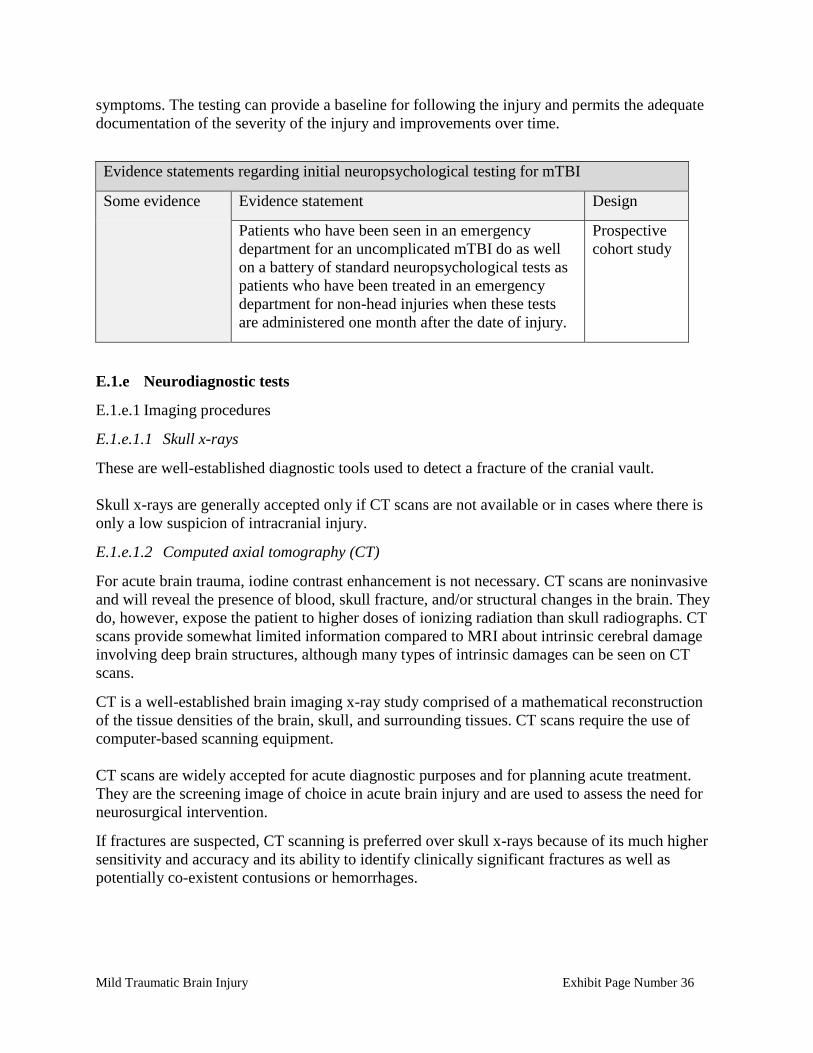
Mild Traumatic Brain Injury Exhibit Page Number 36
symptoms. The testing can provide a baseline for following the injury and permits the adequate
documentation of the severity of the injury and improvements over time.
Evidence statements regarding initial neuropsychological testing for mTBI
Some evidence Evidence statement Design
Patients who have been seen in an emergency
department for an uncomplicated mTBI do as well
on a battery of standard neuropsychological tests as
patients who have been treated in an emergency
department for non-head injuries when these tests
are administered one month after the date of injury.
Prospective
cohort study
E.1.e Neurodiagnostic tests
E.1.e.1 Imaging procedures
E.1.e.1.1 Skull x-rays
These are well-established diagnostic tools used to detect a fracture of the cranial vault.
Skull x-rays are generally accepted only if CT scans are not available or in cases where there is
only a low suspicion of intracranial injury.
E.1.e.1.2 Computed axial tomography (CT)
For acute brain trauma, iodine contrast enhancement is not necessary. CT scans are noninvasive
and will reveal the presence of blood, skull fracture, and/or structural changes in the brain. They
do, however, expose the patient to higher doses of ionizing radiation than skull radiographs. CT
scans provide somewhat limited information compared to MRI about intrinsic cerebral damage
involving deep brain structures, although many types of intrinsic damages can be seen on CT
scans.
CT is a well-established brain imaging x-ray study comprised of a mathematical reconstruction
of the tissue densities of the brain, skull, and surrounding tissues. CT scans require the use of
computer-based scanning equipment.
CT scans are widely accepted for acute diagnostic purposes and for planning acute treatment.
They are the screening image of choice in acute brain injury and are used to assess the need for
neurosurgical intervention.
If fractures are suspected, CT scanning is preferred over skull x-rays because of its much higher
sensitivity and accuracy and its ability to identify clinically significant fractures as well as
potentially co-existent contusions or hemorrhages.

Mild Traumatic Brain Injury Exhibit Page Number 37
CT scans are recommended for abnormal mental status (GCS less than 13 on admission), focal
neurologic deficits, or acute seizure. CT scans are recommended for the following patients:
● High risk
○ GCS less than 15 at two hours post-injury;
○ suspected open or depressed skull fracture;
○ any sign of basilar skull fracture (e.g., hemotympanum, raccoon eyes, Battle’s Sign,
CSF oto-/rhinorrhea);
○ greater than or equal to two episodes of vomiting;
○ age equal to or greater than 60.
● Other risk
○ retrograde amnesia to the event greater than or equal to 30 minutes;
○ “dangerous” mechanism (e.g., pedestrian struck by motor vehicle, occupant ejected
from motor vehicle, fall from greater than three feet or more than five stairs);
○ coagulopathy, including use of use of anticoagulant medication;
○ focal neurologic deficits;
○ acute seizure;
○ severe and persistent headache;
○ physical evidence of trauma above the clavicles and/or multiple trauma and/or basilar
skull fracture;
○ drug or alcohol intoxication;
○ any recent history of TBI, including mTBI.
E.1.e.1.3 Magnetic resonance imaging (MRI)
MRI is a well-established brain imaging study for patients with TBI in which the individual is
positioned in a magnetic field and a radio-frequency pulse is applied. Hydrogen proton energy
emission is translated into visualized structures. Altered signal intensity compared to normals
may indicate trauma or other disease.
CT is superior to MRI in detecting acute intracranial bleeds and remains the preferred initial
imaging study in the first 24 hours following TBI.
MRI should not be used to diagnose mTBI. Initially, MRI scans are clinically useful in the
following situations to:
● determine neurological deficits in TBI not explained by CT;

Mild Traumatic Brain Injury Exhibit Page Number 38
● evaluate prolonged intervals of disturbed consciousness or other prolonged alteration in
mental status;
● define evidence of acute changes super-imposed on previous trauma or disease.
MRI scans are also useful to assess transient or permanent changes, to determine the etiology of
subsequent clinical problems, and to plan treatment.
MRI may reveal an increased amount of pathology compared to CT. Due to their high contrast
resolution, MRI scans are superior to CT scans for the detection of some intracranial pathology
(e.g., axonal injury, subtle cortical contusions, small extra-axial fluid collections, etc.) but not
bone injuries such as fractures. MRI is more sensitive than CT for detecting traumatic cerebral
injury.
Specific MRI sequences and techniques are very sensitive for detecting acute traumatic cerebral
injury. They may include, but are not limited to: diffusion weighted imaging (DWI),
susceptibility weighted imaging, gradient echo weighted imaging, and fluid attenuated inversion
recovery (FLAIR). Some of these techniques are not available on an emergency basis.
E.1.e.2 Vascular imaging tests
Vascular imaging tests reveal arterial or venous abnormalities in the chest, neck, head, or
extremities (e.g., thrombosis, dissection, spasm, emboli, or tearing).
These tests are generally used if standard CT/MRI scans fail to demonstrate suspected vascular
abnormalities. However, only rarely are they useful in mTBI. If needed, refer to the Division’s
Moderate/Severe TBI Medical Treatment Guideline.
Brain Acoustic Monitor: This device identifies turbulent blood flow in the brain. It is considered
investigational for the purpose of detecting deficits requiring CT scanning in the emergency
room. Based on the evidence listed in the table below, it is not recommended at the time of this
guideline.
Evidence against use of a brain acoustic monitor as an initial diagnostic procedure for TBI
Some evidence Evidence statement Design
A Brain Acoustic Monitor cannot reliably predict
the development of post-concussive symptoms.
Diagnostic
cohort study
Mild Traumatic Brain Injury Exhibit Page Number 39
E.2 Further diagnostic procedures
E.2.a Electrodiagnostic studies
These are limited to electromyogram (EMG), nerve conduction studies, and multisensory evoked
potentials including visual evoked potentials (VEP), somatosensory evoked potentials (SSEP),
and brain stem auditory evoked responses (BSAER).
E.2.a.1 EMG and nerve conduction studies
These are generally accepted, well-established diagnostic procedures. These studies may be
useful for individuals with brain injury and EMG associated suspected peripheral nervous system
involvement. They are often used to differentiate peripheral versus central spinal cord or brain
deficits. These electrodiagnostic studies are possibly complementary to other imaging procedures
such as CT, MRI, and/or myelography. These studies provide useful correlative
neuropathophysiologic information that is unattainable from standard radiologic studies.
E.2.a.2 Electroneuronography (EnoG)
EnoG is a well-established and generally accepted test that measures facial nerve function. This
test measures the action potential of different branches of a facial nerve. It is used in individuals
with TBI resulting in a facial paralysis and is key in determining the need for surgical
intervention. This test is most useful within the first three weeks of facial nerve dysfunction. If
the action potentials on the affected side are 90–100% less than those on the normal side, it
suggests significant injury to the nerve and calls for surgical exploration. Individuals with TBI
whose nerve is less than 90% decreased in function have a reasonably good outcome with
observation alone.
E.2.a.3 Dynamic electromyographies
These are electrodiagnostic studies utilized to distinguish the voluntary capacity of a muscle
from a spastic reaction. This aids the clinician in better planning specific rehabilitative treatment.
This study is helpful in the differential diagnosis and diagnostic work-up of disordered muscle
tone. This is a generally accepted procedure.
E.2.a.4 Evoked potential responses (EP)
EPs are generally accepted, well-established diagnostic procedures. EPs are central nervous
system electrophysiologic responses to a stimulus, either externally generated via one or more
sensory modalities or internally generated via the processing of information. Multisensory EP
studies are limited to visually evoked potentials, brain stem auditory evoked potentials,
somatosensory evoked potentials, and cognitive evoked potentials.
E.2.a.4.1 Auditory brainstem response (ABR)
ABR is a generally accepted diagnostic procedure useful in assessing damage to the brain stem,
midbrain, and other neural structures that govern hearing and/or balance. A normal test does not
rule out structural damage, and the test may be abnormal in middle ear and non-traumatic disease
affecting the auditory pathway. Waves one, three, and particularly five are most useful in
Mild Traumatic Brain Injury Exhibit Page Number 40
assessing injury. While amplitude and the presence of wave are important, the latency and
interwave latency are equally important. This test is often sensitive but non-specific. It may be
useful in some cases and requires prior authorization.
E.2.a.4.2 Electroretinogram (ERG)
ERG is a generally accepted diagnostic procedure for occult retinal trauma accompanying TBI.
Most traumatic retinal pathology presents as a field deficit detected by direct examination. ERG
cannot detect mild changes in retinal function, and normal results should not be taken as
evidence against ocular dysfunction. ERG requires prior authorization.
E.2.a.4.3 Cognitive event-related potential
Event-related potential provides no diagnostic information in mTBI that cannot be obtained
through other diagnostic procedures and is not recommended in mTBI. It may be justified if
other neurological diagnoses are suspected. It requires prior authorization.
E.2.a.4.4 Somatosensory evoked potential (SSEP)
SSEP provides no information in mTBI that cannot be obtained through other diagnostic means.
SSEP is not recommended in mTBI. It may be used when other diagnoses are suspected. It
requires prior authorization.
E.2.a.4.5 Visual evoked potential (VEP)
VEP is a generally accepted diagnostic procedure. Pattern reversal monocular VEP recording
may detect pathology in the anterior-posterior visual pathway from the retina to the occipital
cortex. It may be indicated in the event of compromised acuity or visual field defect. The VEP
may occasionally be normal in cases of severe structural damage if there is enough preserved
central visual field. Unfortunately, VEP is highly susceptible to artifact and has a low specificity
for structural injury to the visual pathways. Therefore, prior authorization is required.
E.2.a.4.6 Vestibular evoked myogenic potentials (VEMP)
Refer to Section E.2.i, Neuro-otology.
E.2.b Electroencephalography
E.2.b.1 Electroencephalography (EEG)
EEG is a well-established diagnostic procedure that monitors brain wave activity using scalp
electrodes and provocative maneuvers such as hyperventilation and photic strobe for the purpose
of seizure diagnosis. Information generated includes alterations in brain wave activity such as
frequency changes (non-specific) or morphologic (seizures). EEG is not generally indicated in
the immediate period of emergency response or during acute evaluation and treatment.
Following initial assessment and stabilization, the individual’s course should be monitored. If
during this period there is failure to improve or the medical condition deteriorates, an EEG may
be indicated to assess seizures, focal encephalopathy due to persistent effects of hemorrhage,
diffuse encephalopathy due to the injury, or other complicating factors such as hydrocephalus or
medications. A normal EEG does not definitively rule out a seizure disorder. If there is sufficient

Mild Traumatic Brain Injury Exhibit Page Number 41
clinical concern that a seizure disorder may exist despite a normal EEG, then a 72-hour
ambulatory EEG or inpatient video-EEG monitoring may be appropriate.
E.2.b.2 Quantified electroencephalography (QEEG) (Computerized EEG)
QEEG is a modification of standard EEG using computerized analysis of statistical relationships
between power, frequency, timing, and distribution of scalp recorded brain electrical activity.
These statistically generated values are then compared to those recorded from selected control
and specific populations, generally using multiple regression analysis of multiple measurements
and calculated parameters.
Recent studies suggest that in the future, QEEG may become a useful tool in the retrospective
diagnosis of TBI and its severity, but this application remains investigational. QEEG is not
recommended for diagnosing mTBI or M/S TBI.
E.2.c Neuroimaging
Practitioners should be aware of the radiation doses associated with various procedures.
Coloradans have a background exposure to radiation, and unnecessary CT scans or x-rays
increase the lifetime risk of cancer death.
E.2.c.1 Structural imaging
E.2.c.1.1 Computed axial tomography (CT)
CT may be used to follow identified pathology or to screen for late pathology. Subsequently, CT
scans are generally accepted when there is suspected intracranial blood, extra-axial blood,
hydrocephalus, altered mental state, or a change in clinical condition, including development of
new neurological symptoms or post-traumatic seizure (within the first days following trauma).
Once the initial acute stage has passed, MRI scans are frequently ordered as opposed to CT.
Repeat CT scans are usually not necessary for most patients with mTBI (loss of consciousness
and/or retrograde amnesia and Glasgow Coma Scale greater than 12). In one study, 95 mTBI
adult patients were admitted to the hospital for observation and did not have a repeat CT if their
Glasgow Coma Scale was 15 at 24 hours. Patients with repeat head trauma, coagulopathy, or
other medical problems were excluded. No patients needed a neurosurgical intervention, and
only 8% had repeat CTs.
Another systematic review concluded that routine CT repetition rarely identified conditions
needing surgical intervention. Nevertheless, risk factors for a further bleed or neurological
deterioration from the following conditions would warrant repeat CTs:
● skull fracture,
● coagulopathy or anticoagulation,
● age over 60,
● epidural hematoma,

Mild Traumatic Brain Injury Exhibit Page Number 42
● suspected open or depressed skull fracture,
● continuing severe headache,
● moderate to severe TBI, and
● continuing TBI symptoms.
E.2.c.1.2 Magnetic resonance imaging (MRI)
MRI is the image of choice to detect the late, sub-acute, and chronic structural changes in the
brain which underlie abnormal functioning. It is a well-accepted technique for follow-up
imaging. Complications of TBI that may be explained by MRI include, but are not limited to:
post-traumatic epilepsy, post-traumatic movement disorder, post-traumatic cranial neuropathy,
post-traumatic infection, or failure to recover within the expected time frame. (Refer to Section
E.2.c.3, Advanced MRI techniques, for more advanced imaging.)
Diffusion tensor imaging (DTI), susceptibility-weighted imaging, and magnetic transfer imaging:
These have been used to explore the effects of mTBI. They remain research tools because, as of
the time of this guideline, there are no studies validating their clinical use to differentiate patients
with cognitive deficits from those without. They are not recommended to diagnose mTBI. DTI
may be useful for identifying pathology and guiding treatment in patients with documented
physiological deficits, such as hemianopsia, but interpretation of results is very dependent upon
the experience and skill of the neuroradiologist. DTI may be used when an accompanying MRI is
ordered for purposes other than diagnosing mTBI. DTI may not be used in isolation to diagnose
mTBI.
E.2.c.2 Dynamic imaging
In contrast to anatomical imaging procedures, the following procedures are designated to detect
physiologic activity of the brain, including cerebral blood flow and cerebral metabolism. Both
PET and SPECT scans can subject the patient to significant radiation levels. Prior authorization
is required for these procedures.
E.2.c.2.1 Single photon emission computed tomography (SPECT)
SPECT is not generally accepted as a diagnostic test for TBI of any severity and is considered
investigational for diagnostic purposes. SPECT may not be used in isolation to diagnose mTBI.
It is a functional image of the brain created by a flow tracer or a receptor-binding substance
tagged with a radionuclide and injected intravenously into the individual. The radiotracer is
assumed to accumulate in different areas of the brain proportionately to the rate of delivery of
nutrients to that volume of brain tissue. Using a gamma camera and the techniques of CT, a 3-D
image of the distribution of a radionuclide in the brain is obtained. SPECT may identify areas of
decreased perfusion and provide a qualitative estimate of regional cerebral blood flow (CBF),
which correlates with metabolism in many neurologic disorders. There is a variable correlation
of SPECT with other measures, such as neuropsychological test findings. Its interpretation
should take into account its low specificity, making the predictive value of SPECT no better than
CT.
Although it should not be used to diagnose mTBI, there is some evidence that SPECT may
provide useful information in some cases in which the prognosis is in question, particularly if

Mild Traumatic Brain Injury Exhibit Page Number 43
structural neuroimaging is normal. Given its high sensitivity, SPECT may be useful when
expected recovery from mTBI is not occurring within several months from the time of injury.
Thus, the primary use of SPECT is as a tool for prognosis in patients with mTBI who are not
progressing and have continued symptoms. A normal SPECT scan in this setting indicates a
likelihood of resolution of symptoms within twelve months. However, due to its lack of
specificity, an abnormal SPECT scan does not mean that symptoms will persist. Symptoms may
resolve even when areas of abnormal perfusion continue to be seen on the SPECT scan.
In all severities of TBI, prior authorization is required and it is recommended that medical
necessity and clinical usefulness for this study be justified.
E.2.c.2.2 Positron emission testing (PET)
PET is a functional brain imaging procedure. A tracer molecule tagged with a positron-emitting
radioisotope is injected into the body. Biodistribution of the tracer is imaged, producing
information about local cerebral glucose utilization and cerebral perfusion. This procedure
requires on-site access to a cyclotron.
PET can reveal areas of decreased metabolism in the brain. Little information is available about
its use and results in mTBI. In all severities of TBI, it is recommended that medical necessity and
clinical usefulness for this diagnostic study be justified. It is not generally accepted as a
diagnostic study and should not be used solely to diagnose the presence of TBI. Any requested
use requires prior authorization.
Evidence statements regarding follow-up diagnostic procedures
Some evidence Evidence statement Design
Although it should not be used to diagnose mTBI,
SPECT may provide useful information in some
cases in which the prognosis is in question,
particularly if structural neuroimaging is normal.
Consecutive case
series
E.2.c.3 Advanced MRI techniques
At the time of writing this guideline, all advanced MRI techniques are not recommended for
diagnostic purposes. While they can identify anatomic physiologic variation, the changes cannot
clearly be related to the need for specific treatment. They require prior authorization and
justification of the medical necessity and clinical usefulness of the study.
E.2.c.3.1 Magnetic resonance (MR) spectroscopy
This is a noninvasive test that applies a burst of radio frequency energy to tissue inside an
applied magnetic field. The resulting excitation and relaxation of nuclei generates a signal that
carries information about the chemical environment of those nuclei. MR spectroscopy may
detect changes in levels of n-acetyl-aspartate, an intermediate in neurotransmitter synthesis that

Mild Traumatic Brain Injury Exhibit Page Number 44
is present in large amounts in normal functioning neurons but is decreased in damaged brain
tissue. Its spectral signal may correlate with neuronal integrity and function and may show loss
of function in tissue, which appears normal on conventional CT or MRI studies. MR
spectroscopy may increase the sensitivity of MR imaging for traumatic lesions. This sensitivity
may allow for increased correlation to more specific neuro-cognitive deficits and guide treatment
planning. It may be useful information in determining long-term outcome. MR spectroscopy
remains predominantly a research tool at this time and should not be used solely to diagnose the
presence of TBI. MR spectroscopy requires prior authorization for patients with mTBI. It may
be considered with adequate documentation of its medical necessity in unusual cases, such as in
patients with a minimally conscious state, when the information will assist in clarifying the
pathology to direct a therapeutic approach to the individual with TBI.
E.2.c.3.2 Functional MRI (fMRI)
This uses MRI to detect physiologic responses of brain tissue to various tasks. Blood
oxygenation level dependent (BOLD) contrast is the most popular fMRI technique. It derives an
image from differences in the magnetic properties, and therefore differences in MR decay
parameters, of oxygenated and deoxygenated hemoglobin. A typical fMRI study compares
images under two or more behavioral conditions, which may involve motor, cognitive, or visual
tasks. Functional MRI studies have shown functional reorganization as a general response to
TBI. Alterations in patterns of cerebral activity seen on fMRIs may correlate with cognitive
deficits in individuals with TBI, but the specificity of the test is not sufficient to make fMRI a
diagnostic tool. At the time of this guideline, it is a research tool and not recommended for
clinical use. Recent publications report problems with the mathematical formulas used, relating
false positives and false negatives.
E.2.d Laboratory testing
Laboratory testing is a generally accepted, well-established procedure. In mTBI, laboratory tests
are rarely indicated at the time of initial evaluation unless there is suspicion of systemic illness,
infection, neoplasm, drug or alcohol intoxication, endocrine dysfunction, or underlying disease.
A number of blood tests have been proposed to identify brain damage in mTBI. It is currently not
clear that results from these tests can more accurately diagnosis mTBI or identify mTBI which
requires treatment or follow up. Thus, they are not recommended at this time. Any individual
with TBI on medication will require laboratory testing to monitor therapeutic drug levels and the
effects on organ function.
Endocrine testing is frequently appropriate because hypopituitarism occurs in approximately
17% of mTBI cases.
E.2.e Lumbar puncture
Lumbar puncture is a well-established diagnostic procedure for examining cerebrospinal fluid
(CSF) in neurological disease and injury. The procedure should be performed by qualified and
trained physicians under sterile conditions.
Lumbar puncture is contraindicated in acute trauma to the spinal column, certain infections,
increased intracranial pressure due to space occupying lesions, and in some coagulation disorders
or defects. Additionally, it should be avoided if there are cutaneous infections in the region of the

Mild Traumatic Brain Injury Exhibit Page Number 45
puncture site. In individuals with suspected or known increased intracranial pressure, lumbar
puncture should be preceded by fundoscopic examination and a CT scan or MRI.
E.2.f Nerve blocks – diagnostic
These are generally accepted procedures involving percutaneous needle injection techniques to a
specific nerve. These diagnostic blocks are typically performed with quick-acting, short duration
local anesthetics such as lidocaine or bupivacaine. Temporary diagnostic nerve blocks evaluate
limb ROM, dystonia, or spasticity and assist in planning subsequent, specific therapy.
E.2.g Further neuropsychological assessment and testing
Neuropsychological assessment after three months is appropriate in the following situations
when:
● input is needed to plan treatment to maximize long-term cognitive and overall functional
outcomes;
● documentation of accommodations is needed to establish adjustments to the
neurocognitive challenges;
● assessment will assist in increasing insight and be used to assist with supportive
psychotherapy;
● there is a question of the individual’s ability to perform work-related duties and/or there
are safety issues (i.e., possible harm to self or others) or when the person’s vocation
necessitates more extensive testing prior to vocational re-entry or return to
school/training;
● assistance is needed with differential diagnosis including the diagnosis of TBI;
● it is deemed necessary to evaluate and/or monitor effectiveness of treatment approaches
(i.e., cognitive rehabilitation therapy, somatic therapies, or medication trials) in specific
individuals;
● the patient’s presentation is such that symptom validity testing and performance validity
testing may be helpful in treatment planning;
● subjective complaints are disproportionate to the clinical history or objective findings as
observed by provider(s);
● the degree of disability is disproportionate to the clinical history and objective findings as
observed by provider(s);
● there are questions of competency, guardianship, or conservatorship.
Neuropsychological testing should be used to document the patient’s level of effort and to
provide data regarding symptom validity. Testing should not be used to diagnose malingering.
Neuropsychological testing may take into account validated testing in other areas without a
direct relationship to psychological issues.

Mild Traumatic Brain Injury Exhibit Page Number 46
Evidence statements regarding neuropsychological assessment
Some evidence Evidence statement Design
There is an association between poor effort on verbal
memory tests and poor effort on computerized tests
of postural stability in patients with TBI who are
being evaluated for disability ratings.
Retrospective
review of
consecutive charts
The following information may aid in delineating when a full neuropsychological battery is
necessary versus more limited testing:
● mTBI three months post-injury: Serial testing with specialized tests that are sensitive to
effort, speed of processing, memory, and executive functions will usually be appropriate
for treatment planning and monitoring progress. The administration of a full
neuropsychological test battery (typically including assessment of effort) may become
necessary in this time period when:
○ there is no witnessed history of TBI, or there is other uncertainty regarding the
diagnosis of mTBI;
○ the patient is not progressing and/or symptoms indicate mTBI may be more severe;
○ it is necessary to address issues on the initial indicators list.
E.2.h Psychological, psychiatric, or psychosocial evaluations
These are generally accepted and well-established diagnostic procedures with selective use in the
TBI population. They have more widespread use after three months. Diagnostic testing may be
indicated for individuals with symptoms of post-traumatic disturbances of sleep, mood, anxiety,
psychosis, substance use, aggression/agitation, and pain, as well as depression, delayed recovery,
chronic pain, recurrent painful conditions, and disability problems. An individual with a PhD,
PsyD, or psychiatric MD/DO credentials may perform these evaluations if listed as an authorized
user by the test publisher. Practitioners’ familiarity with patients with TBI is preferred.
Psychosocial evaluations can help to determine if further psychosocial or behavioral
interventions are indicated for patients diagnosed with TBI. The interpretations of the evaluation
can provide clinicians with a better understanding of the patient in his or her social environment,
thus allowing for more effective rehabilitation. Psychosocial assessment requires consideration
of variations in experience and expression resulting from affective, cognitive, motivational, and
coping processes, as well as other influences such as gender, age, race, ethnicity, national origin,
religion, learning disability, language, or socioeconomic status.
A comprehensive psychological evaluation should attempt to identify both primary psychiatric
risk factors (e.g., psychosis, active suicidality, lack of awareness) as well as secondary risk
factors (e.g., moderate depression, job dissatisfaction). Significant personality disorders should
also be taken into account in treatment planning.

Mild Traumatic Brain Injury Exhibit Page Number 47
Psychometric testing is a valuable component of a consultation to assist the physician and other
members of the treatment team in making a more effective treatment plan. Psychometric testing
can assist in enhancing general medical outcomes and in predicting a patient’s likely adherence
to and cooperation with medical treatment plans.
Several meta-analyses have evaluated the occurrence of depression and anxiety with non-
penetrating TBI. Both anxiety and depression appear to occur at a rate about one-third higher
than the general population. Both conditions are likely to increase during the initial two to five
years post-injury, although anxiety may decrease after five years. Patients with mild and
moderate/severe TBI are both likely to suffer from psychological stress. Increased physical
activity and decreased alcohol consumption may be useful to decrease symptoms. One study
found that pre-injury alcohol abuse and longer PTA predicted symptoms lasting longer than six
months.
Even in cases where no diagnosable psychological condition is present, these evaluations can
identify social, cultural, coping, and other variables that may be influencing the patient’s
recovery process and may be amenable to various treatments, including behavioral therapy.
● Qualifications
○ A psychologist with a PhD, PsyD, or EdD credentials or a physician with psychiatric
MD/DO credentials may perform the initial comprehensive evaluations. It is
preferable that these professionals have experience in diagnosing and treating mTBI
in injured workers.
○ Psychometric tests should be administered by psychologists with a PhD, PsyD, or
EdD credentials or health professionals working under the supervision of a doctorate
level psychologist. Administration and interpretation of
psychological/neuropsychological measures must adhere to standards set forth by test
publishers.
● Indications
A psychological assessment may be necessary if symptoms do not correlate with a
diagnosis of TBI. Complaints of cognitive dysfunction may also be associated with a
variety of conditions that do not involve neurological disease, TBI, or mTBI. This
includes conditions that may have been pre-existing or are concurrent, such as
depression, anxiety, chronic pain, somatoform disorders, and factitious disorders. At
times, a set of symptoms may not coincide with expected objective findings for those
with a diagnosis of TBI. To identify non-neurological contributions to cognitive or other
functional complaints, a psychological evaluation focusing on mental disorder diagnoses
is appropriate when:
○ delayed recovery is present,
○ there is delayed access to appropriate care,
○ there is a question of whether a brain injury has occurred,

Mild Traumatic Brain Injury Exhibit Page Number 48
○ neuropsychological testing yields a pattern of test results that is not consistent with
the clinical history,
○ neurologically improbable symptoms are present, or
○ it is necessary to assess for accompanying psychological components.
● Clinical evaluation
Special note to health care providers: Most providers are required to adhere to the federal
regulations under the Health Insurance Portability and Accountability Act (HIPAA).
Unlike general health insurers, workers’ compensation insurers are not required to adhere
to HIPAA standards. Thus, providers should assume that sensitive information included
in a report sent to the insurer could be forwarded to the employer. The Colorado statute
provides a limited waiver of medical information regarding the work-related injury or
disease to the extent necessary to resolve the claim. It is recommended that the health
care provider either (1) obtain a full release from the patient regarding information that
may go to the employer or (2) not include sensitive health information that is not directly
related to the work-related conditions in reports sent to the insurer.
The clinical evaluation should address the following areas:
○ History of injury
The history of the injury should be reported in the patient’s words or using similar
terminology. Certified medical interpreters are preferred. Collateral information
should be obtained as appropriate. This may include family, support systems,
witnesses, and EMS records.
■ nature of injury
■ psychosocial circumstances of the injury
■ current symptomatic complaints
■ extent of medical corroboration
■ treatment received and results
■ compliance with treatment
■ coping strategies used, including perceived locus of control, catastrophizing, and
avoidance behaviors
■ perception of medical system and employer
■ history of response to prescription medications
■ medication history related to this injury
○ Health history
■ nature of injury

Mild Traumatic Brain Injury Exhibit Page Number 49
■ medical history
■ psychiatric history
■ history of alcohol or substance abuse, including abuse of prescription medication
■ ADLs
■ previous injuries, including disability, impairment, and compensation
■ complete medication history, including prescription and over-the-counter
medications
○ Psychosocial history
■ childhood history, including abuse/neglect and developmental/intellectual
disability or delay
■ educational history
■ family history, including disability
■ relationship/marital history and other significant adulthood activities and events
■ legal history, including criminal and civil litigation
■ employment history
■ military duty: Because post-traumatic stress disorder (PTSD) might be an
unacceptable condition for many military personnel to acknowledge, it may be
prudent to screen initially for signs of depression or anxiety – both of which may
be present in PTSD.
■ symptoms of pre-injury psychological dysfunction
■ current and past interpersonal relations, support, and living situation
■ financial history
○ Mental status exam including cognition, affect, mood, orientation, thinking, and
perception. May include the Mini-Mental Status Examination or the Frontal
Assessment Battery, if appropriate, and detailed neuropsychological testing.
○ Assessment of any danger posed to self or others.
○ Barriers to care should be considered as the patient may experience problems with
transportation and access to appropriate care.
○ Psychological test results, if performed.
○ Current psychiatric/psychological diagnosis consistent with the standards of the
American Psychiatric Association’s most recent Diagnostic and Statistical Manual of
Mental Disorders.

Mild Traumatic Brain Injury Exhibit Page Number 50
○ Pre-existing psychiatric conditions. Treatment of these conditions is appropriate when
the pre-existing condition affects recovery.
○ Causality (to address medically probable cause and effect, distinguishing pre-existing
psychological symptoms, traits, and vulnerabilities from current symptoms or
aggravation of prior symptoms).
○ Treatment recommendations with respect to specific goals, frequency, timeframes,
and expected outcomes.
Evidence statements regarding psychometric testing
Good evidence Evidence statement Design
Psychometric testing can predict medical treatment
outcome.
Prospective cohort
study and
observational
cohort studies
E.2.i Neuro-otology: vestibular and audiological evaluations
Neurotologic evaluation is a widely used and generally accepted practice in cases of hearing loss,
dizziness, balance problems, facial nerve injury, and cerebrospinal fluid leak. An individual with
TBI may experience these symptoms. Any patient with complaints of vertigo or significant
neurological findings on clinical exam, such as ataxia, should be referred to appropriate
specialists expeditiously. Diagnostic testing for significant pathology usually requires the listed
evaluations (audiometry, tympanometry, and vestibular function tests [E.2.i.3.1 – E.2.i.3.5]). It is
recommended that all patients with mTBI and continual balance complaints and no clinical
findings after 6 weeks be referred to clinicians with experience in balance assessment and
training (see subsection E.2.i.3.6, Acquired visual dysfunction, below). Some tests may need to
be repeated to clarify diagnosis.
E.2.i.1 Audiometry
Audiometry is a generally accepted and well-established procedure that measures hearing. An
audiologist or skilled trained technician administers the test using an audiometer. The machine
presents individual frequencies to the person with TBI (typically ranging from 125–8000 Hz) at
different levels of loudness (in dB HL). The individual is asked to respond to the sound at its
lowest detectable intensity (threshold). Normal thresholds are from 0-25 dB HL and are depicted
on an audiogram. The audiologist or physician should determine the presence and type (non-
organic, conductive, sensorineural, presbycusis, or mixed) of hearing loss based on the
audiogram and other tests reasonably deemed necessary.
If available, obtain pre-injury baseline audiograms / audiometry studies to include a summary of
past audiometric history, if known (e.g., prior hearing loss, prior tinnitus, prior vestibular
problems, prior injury, etc.).
Baseline audiometry following TBI is indicated when the individual with TBI presents with

Mild Traumatic Brain Injury Exhibit Page Number 51
hearing loss, dizziness, tinnitus, or facial nerve dysfunction.
Audiograms may be obtained in serial fashion to monitor inner ear function in response to time
and treatment.
Specific audiometric testing can be done to assess the presence of tinnitus and complaints of
unilateral hearing loss (Stinger test).
E.2.i.2 Tympanometry
Tympanometry is a generally accepted and well-established procedure that measures middle ear
air pressures. It is used to help identify the presence of tympanic membrane perforations,
ossicular abnormalities, and the presence of fluid in the middle ear.
E.2.i.3 Vestibular function tests
The most common type of vertigo is benign paroxysmal positional vertigo (BPPV), which
usually does not require additional testing because it is diagnosed with the clinical Dix-Hallpike
maneuver and treated with a variety of canalith repositioning maneuvers (CRM), such as Epley
and Semont maneuvers. (Refer to Section F.3, Neuro-otology Treatment, Benign Paroxysmal
Positional Vertigo [BPPV].) The following tests are used to verify the presence of vestibular
dysfunction and specify the origin when possible.
E.2.i.3.1 Electro- or video-nystagmography (ENG/VNG)
This is a generally accepted and well-established procedure that measures inner ear/central
balance function. The test measures eye movement responses to inner ear balance stimulation
making use of the vestibulo-ocular reflex. There are several components to the ENG/VNG. They
include oculomotor testing, positional and positioning nystagmus testing, and caloric testing.
This series of tests may identify peripheral and central abnormalities, abnormalities in
oculomotor function, positional nystagmus, and unilateral and bilateral vestibular dysfunction.
The ENG/VNG can be helpful in identifying the affected ear. This test is often used in
individuals with TBI complaining of dizziness or dysequilibrium and may help diagnose
conditions such as labyrinthine concussion, vestibular hypofunction, and central vertigo. It is
often used in conjunction with other tests such as the audiogram and clinical history to help
arrive at a diagnosis.
E.2.i.3.2 Rotary chair testing
This is a generally accepted, although not commonly used, test that evaluates the ocular
responses of the inner ear to rotation. It is used to identify the extent of bilateral vestibular loss
and is more accurate than VNG caloric tests for this purpose. It is also useful in assessing the
ability of vision to compensate for vestibular impairments and so provides prognostic
information regarding recovery.
E.2.i.3.3 Video head impulse testing (vHIT)
This is a generally accepted, although not commonly used, test that evaluates the ocular
responses of the inner ear to high acceleration head rotation. vHIT is used to evaluate the ability
to maintain visual focus during head movement that can be impaired with moderate to high grade

Mild Traumatic Brain Injury Exhibit Page Number 52
unilateral or bilateral vestibular injuries. It detects dysfunction of individual vertical semicircular
canals in vestibular patients as accurately as scleral search coils but is non-invasive and easy to
use. It is used to identify the extent of bilateral vestibular loss and is more accurate than VNG
caloric tests for this purpose.
E.2.i.3.4 Computerized dynamic platform posturography
This is a generally accepted, although not commonly used, test that assesses the contributions of
vision, somatosensation, and the inner ear to balance control. It separately evaluates the role of
lower extremity motor control to balance. It can be used to determine whether a vestibular lesion
is present, but it does not localize the lesion. The purpose of this procedure is to identify the
integral components of a functional balance deficit that may help in treatment planning. This
technique also may be useful in monitoring neurologic recovery in individuals with TBI and
balance deficits. These functional methods of evaluation are considered generally accepted
practices in the evaluation of persistent vestibular and balance deficits that may require specific
treatment and remediation strategies. Non-physiologic findings on this exam can result from
either symptom exaggeration, anxiety, psychiatric disorders, atypical results, or malingering and
should not be interpreted as malingering without other evidence. One study demonstrated
positive VNG testing in a number of cases where dynamic posturography was non-physiologic.
E.2.i.3.5 Electrocochleography (ECoG)
This is a well-established and generally accepted procedure that indriectly tests endolymphatic
fluid pressures. It identifies the affected ear in cases of post-traumatic endolymphatic hydrops
and post-traumatic perilymphatic fistula.
The inner ear has two fluid chambers – the perilymphatic and the endolymphatic. After TBI, it is
not uncommon for patients to develop an increase in the endolymphatic fluid pressure; this
condition is called hydrops. When the endolymphatic pressures are abnormally high, the inner
ear membranes distend, and the ear malfunctions. Symptoms include hearing loss, sporatic
dizziness, tinnitus, aural fullness, and sensitivity to sound.
The ECoG is a test that uses evoked potentials. The patient listens to a series of clicks. Monitors,
including one sitting on the tympanic membrane, measure three potentials: the cochlear
microphonic, the summating potential (SP), and the action potential (AP). An increase in the
ratio of the summating potential of the action potential (SP/AP) suggests the presence of hydrops
or perilymphatic fistula. The test varies in sensitivity and specificity. Diagnosis of endolymphatic
hydrops requires a characteristic clinical picture with progressive hearing loss, fluctuating
hearing, and recurrent vertigo episodes lasting for hours. (Refer to the Division’s
Moderate/Severe TBI Medical Treatment Guideline, section F.4.l, Neuro-otology: vestibular and
audiology, under treatment of progressive otologic disorders.) In the absence of these clinical
features, diagnosis should not be based solely on an abnormal ECoG test result.
E.2.i.3.6 Vestibular evoked myogenic potentials (VEMP)
This is a generally accepted test that evaluates the function of the saccule, one of the gravity-
sensing organs of the inner ear. It is the only objective test of these organs. It is a form of
auditory evoked response and is measured using the ABR and EMG equipment. A loud sound

Mild Traumatic Brain Injury Exhibit Page Number 53
stimulus is introduced into the ear, and a vestibulo-colic reflex response from the saccule is
recorded as a brief relaxation of the ipsilateral sternocleidomastoid muscle by EMG. A
characteristic wave form is recorded for each ear that is analyzed for presence or absence,
threshold, amplitude, and latency. Absence of a response in persons under age 60 suggests
saccular damage. Reduced thresholds are indicative of semicircular canal dehiscence. It is not
useful in diagnosing benign paroxysmal positional vertigo or vestibular migraine.
E.2.i.3.7 Acquired visual dysfunction
This is usually not the primary cause of imbalance. However, if ocular and/or visual
abnormalities are found on testing, the patient may be further evaluated by a qualified
optometrist or ophthalmologist with training and experience in TBI, balance disorders, and
underlying neurology, preferably a neuro-ophthalmologist or neuro-optometrist. The goal is to 1)
determine if there is a visual component or not, 2) determine if it is central or peripheral, 3)
determine if the problem is a manifestation of an underlying vestibular disorder or a true vision
problem, and 4) coordinate with the treating providers regarding most appropriate treatment.
E.2.i.3.8 Other clinical referrals
The treating physician may refer individuals with TBI who have balance problems to other
clinicians with appropriate training in balance dysfunction, such as neuro-otologists,
neurologists, and otolaryngologists, to assist in their assessment. The referrals may include, but
are not restricted to: neuro-ophthamology, optometry, physical therapy, vestibular therapists,
occupational therapy, and chiropractic therapy. There should be a coordinated approach between
these disciplines and the physician specialist in the individual’s treatment.
E.2.j Swallowing evaluation
Swallowing impairment or dysphagia may be due to neurologic, structural, or cognitive deficits
and may result from head trauma. Dysphagia may result in aspiration, airway obstruction,
pneumonia, inadequate nutrition, dehydration, weight loss, failure to thrive, and death.
Dysphagia is uncommon in mTBI. If treatment is needed, refer to the Division’s
Moderate/Severe TBI Medical Treatment Guideline.
E.2.k Vision evaluation
There are standard examination techniques and ancillary tests to establish the diagnosis of visual
disorders. It is a generally accepted practice for a qualified practitioner to provide a
comprehensive vision evaluation. The qualified practitioner (optometrist, neuro-optometrist,
ophthalmologist, neuro-ophthalmologist) should have training and experience in the neurology
of TBI.
The comprehensive visual evaluation should assess afferent visual function (visual acuity, visual
fields / peripheral vision), efferent function (ocular movement, fixational stability/instability,
binocular alignment), and anatomic integrity of the eye and its adnexal structures. In doing so,
the practitioner will obtain information about the functional status of the eyes and visual system
including the presence or absence of refractive error; loss of visual acuity and/or visual field;
oculomotor dysfunction with or without diplopia; ocular, orbital, and adnexal injuries; and other
pathology involving intraocular structures. Vision evaluation may be necessary to evaluate
Mild Traumatic Brain Injury Exhibit Page Number 54
acquired/traumatic central and peripheral nervous system disorders, visual acuity loss, visual
field loss, nystagmus, ocular motility impairment, cranial nerve palsy, diplopia, suspected or
noted ocular and visual pathology, pupillary disorders, and visual perceptual disorders.
The diagnosis/diagnoses determined following a comprehensive neuro-ophthalmic examination
should be based upon objective findings that correlate with the known or suspected underlying
neuro-pathology and symptoms. A simple description of symptoms may be used but should not
be understood as explanatory or diagnostic, as the practitioner should seek to identify the
underlying pathology.
Signs and symptoms of visual dysfunction commonly include, but are not limited to:
● Signs
○ ocular mis-alignment (strabismus),
○ nystagmus or other instability of fixation,
○ ocular discharge,
○ red or swollen eyes,
○ ptosis,
○ lagophthalmos (inability to fully close the eyelid),
○ globe dystopia (lack of orbit alignment).
● Symptoms, including complaints of
○ blurred vision or loss of vision,
○ difficulty with visual tracking or scrolling,
○ focusing problems,
○ double vision,
○ having to close or cover an eye to improve vision,
○ problems with depth perception,
○ impaired peripheral vision,
○ headache or eye strain with use of eyes,
○ head tilt to improve vision,
○ dizziness or balance problems with use of eyes,
○ photophobia,
○ reduced attention or concentration for visual tasks.

Mild Traumatic Brain Injury Exhibit Page Number 55
Visual evaluation is indicated when signs or symptoms consistent with a visual problem are
reported by the individual or observed by others. Significant signs and symptoms not directly or
solely attributable to other causes (e.g., cognitive, vestibular, medication, psychological) indicate
the need for vision evaluation as soon as reasonably possible post-injury. Mild signs and
symptoms do not require referral but may be monitored for several weeks to allow for resolution
or improvement.
In mTBI, self-reported photosensitivity and blurred vision are relatively common, but there is
usually resolution by three months post-injury. Persistent functional related visual symptoms
may require specialty evaluation.
A formal vision examination may be intermediate, extended, or comprehensive, depending on
the nature of the deficits. The vision examination may include, but is not limited to: case history;
visual acuity at a distance and near; refraction; color vision testing; pupillary examination; visual
field by confrontation; Amsler grid testing; ocular motility examination; binocularity
examination; accommodation testing; external/adnexal examination; intraocular pressure testing;
and anterior and posterior segment examinations.
Ancillary diagnostic tests may include, but are not limited to: visual field testing (tangent screen
campimetry, manual or automated perimetry), ultrasonography, fluorescein angiography, anterior
segment and fundus photography, optical coherence tomography, electrodiagnostic studies, low
vision assessment, and visual perceptual testing.
E.2.k.1 Visual field testing
This is a well-established technique to evaluate central and peripheral vision. It is indicated when
a deficit is suspected by the practitioner or noted by the patient and should be considered in any
patient with TBI and subjective visual field loss. Visual field testing beyond the basic
examination should be performed using a procedure and tool that is well-established and
standardized. Examples include computerized perimetry and Goldmann perimetry.
E.2.k.2 Ultrasonography
Ultrasonography is a well-established diagnostic test that is indicated for evaluation of ocular or
orbital pathology. It is indicated for ocular lesions that are suspected but poorly visualized due to
opaque ocular media or for further evaluation of ocular or orbital pathology.
E.2.k.3 Fluorescein angiography
This is a well-established diagnostic test to evaluate the retinal and choroidal circulation. It is
indicated when lesions of one or both of these circulations are suspected.
E.2.k.4 Visual perceptual testing
This testing may be conducted informally by an ophthalmologist or optometrist or with a
standardized battery of tests employed by a neuropsychologist. Testing consists of functional
assessments to evaluate an individual’s recognition and interpretation of visual sensory
information. Visual perceptual testing is indicated for determination of the level of visual
perceptual impairment and/or confirmation of suspected impairment. Perceptual areas assessed
Mild Traumatic Brain Injury Exhibit Page Number 56
include visual memory, judgment of visual spatial relationships, visual discrimination, visual
motor integration, visual figure-ground discrimination, and visual attention. Numerous tests are
used for the evaluation of visual perception. Some of these tests are well-established. It is
suggested that only tests with established norms be used in a standardized battery, and caution
should be exercised in using other instruments. Visual perception testing should not be used in
isolation to diagnose mTBI.
E.2.k.5 Low vision evaluation
Low vision evaluation is well-established and indicated in the presence of subnormal bilateral
visual acuity or visual field. The goal is to provide vision aids for distance or near vision that
improve visual functioning.
E.2.k.6 Electrodiagnostic studies
These are well-established and possibly indicated in the presence of reduced visual acuity or
visual fields, ocular pathology, or suspected optic nerve or visual pathway deficit. (Refer to
Section E.2.a, Electrodiagnostic studies, for further description.)
E.2.k.7 Optical coherence tomography
This is an interferometric technique, usually with near-infrared wavelengths, used to evaluate
optic nerve and retinal structural integrity. This study should be used in conjunction with tests of
visual function to establish the possible causes of visual deficits. Serial assessments can provide
objective longitudinal data about retinal and optic nerve structure.
E.2.l Return-to-work assessment and special tests
A return-to-work procedure should be part of a company’s policies and procedures, knowing that
return to work can decrease anxiety, reduce the possibility of depression, and reconnect the
worker with society. Evaluations used to define these abilities, such as the functional capacity
evaluation (FCE) and the worksite analysis, should be objective. The professional performing the
FCE and worksite analysis should be specifically trained and familiar with the unique
presentation of the individual who has sustained a TBI. The ability to tolerate these evaluations
and follow commands may be limited due to TBI and should not be construed as non-
cooperative or suggestive of malingering.
Caution should be used in returning an individual to work and other activities too early. Both
physical and cognitive duties should generally be non-stressful initially, with a gradual increase
in activity based on improvement and/or resolution of symptoms. The individual should be
competent in most basic ADLs before return to work is considered. Return to full duty depends
on the rate of decrease of symptoms. Generally, if symptoms recur during increasing job duties
or exertion, duties should be decreased accordingly. Because a prolonged period of time off work
will decrease the likelihood of return to work, the first weeks of treatment are crucial in
preventing and/or reversing chronicity and disability mindset. In complex cases, experienced
nurse case managers or occupational therapists may be required to assist in return to work. Other
services, including psychological evaluation and/or treatment and vocational assistance should
be employed. Two evaluations that may be used are job site evaluations and alterations and
FCEs.

Mild Traumatic Brain Injury Exhibit Page Number 57
E.2.l.1 Job site evaluations and alterations
For many patients with TBI, job alterations may be needed. These may be in the form of: (1)
instructing the worker how specific duties might be performed to avoid excessive mental stress;
(2) actual job worksite or duty changes; and/or (3) a formal job site evaluation and alterations at
the worksite.
Job site evaluation and alteration should include input from the employee, the employer, and a
health care professional with experience with TBI cases. The employee should be observed
performing all job functions in order for the job site evaluation to be a valid representation of a
typical workday.
A formal job site evaluation is a comprehensive analysis of the physical, mental, and sensory
components of a specific job and may be important initially to determine causation. These
components may include, but are not limited to: (a) postural tolerance (static and dynamic), (b)
aerobic requirements, (c) ROM, (d) torque/force, (e) lifting/carrying, (f) cognitive demands, (g)
social interactions, (h) interpersonal skills management, (i) visual perceptual challenges, (j)
environmental requirements of a job, (k) repetitiveness, and (l) essential functions of a job.
Changes that provide a therapeutic benefit or relieve the patient’s ongoing symptoms are part of
the required medical treatment for TBI, and therefore, it is assumed that the insurer will be
responsible for paying for reasonably necessary job site alterations.
Job descriptions provided by the employer are helpful but should not be used as a substitute for
direct observation.
A job site evaluation may include observation and instruction of how work is done, what
material changes should be made, and determination of readiness to return to work. Refer to the
Division’s Chronic Pain Disorder Medical Treatment Guideline.
Requests for a job site evaluation should describe the expected goals for the evaluation. Goals
may include, but are not limited to:
● Provide a detailed description of the physical and cognitive job requirements;
● Make recommendations for and assess the potential for job site changes;
● Assist the patient in his/her return to work by educating on how he/she may be able to do
his/her job more safely; and/or
● Give detailed work/activity restrictions.

Mild Traumatic Brain Injury Exhibit Page Number 58
Time frames
Time frames regarding job site evaluations and alterations
Frequency One time with additional visits as needed for follow-up.
E.2.l.2 Functional capacity evaluation (FCE)
FCE may be indicated to identify residual physical limitations. FCE is a comprehensive
assessment of the various aspects of physical and cognitive function as they relate to the
individual’s ability to perform functional activities necessary for return to work. When cognitive,
emotional, and/or behavioral sequelae are also present, a comprehensive FCE may provide
indications of return-to-work readiness.
Components of the physical portion of the FCE may include, but are not limited to:
musculoskeletal screen, cardiovascular profile/aerobic capacity, coordination, lift/carrying
analysis, job specific activity tolerance, maximum voluntary effort, pain assessment, non-
material and material handling activities, balance/dizziness, climbing, physical fatigue,
endurance, and visual skills. The physical portion of any FCE should include all of the physical
skills required for specific job placement.
Components of the cognitive portion of the FCE may include, but are not limited to: memory,
executive skill function, attention and concentration, communication, speed of information
processing, multi-tasking, new learning, and cognitive fatigue and endurance.
Components of the emotional portion of the FCE may include, but are not limited to:
temperament, ability to manage stress, adaptation to change, mood changes, toleration of
feedback, and anger control.
Components of the behavioral portion of the FCE may include, but are not limited to: appropriate
social and behavioral interactions. This may present as inability to complete or cooperate with
the tests, inconsistent or erratic behavior, or the inability to get along with coworkers and
supervisors.
FCEs include tools that are an extension of the basic medical examination and may be useful for
the determination of impairments, functional/cognitive restrictions, determination of progress,
and planning and monitoring of the rehabilitation program. Whenever possible, FCEs should be
supplemented with information from neuropsychology, speech therapy, occupational therapy,
and physical therapy to determine physical, cognitive, and psychological abilities in order for the
patient to function safely and productively in a work setting. FCEs are typically conducted in
four to six hours, but for individuals who have sustained a TBI, additional time may be required
or it may be necessary to conduct the evaluation in two or three separate sessions to allow for the
potential variability of cognitive and physical fatigue. Total time for an FCE would rarely exceed
eight to ten hours.
When an FCE is being used to determine return to a specific job site, the provider is responsible
for fully understanding the job duties. A job site evaluation is frequently necessary. FCEs cannot
be used in isolation to determine work restrictions. The authorized treating provider must
interpret the FCE in light of the individual patient’s presentation and medical and personal

Mild Traumatic Brain Injury Exhibit Page Number 59
perceptions. FCEs should not be used as the sole criteria to diagnose malingering.
FCEs may be beneficial in the TBI population to assist in return to work.
Time frames
Time frames regarding functional capacity evaluation
Frequency 2 times. When the patient is unable to return to the pre-injury position and
further information is desired to determine permanent work restrictions. A
repeat FCE may be needed if additional treatment is deemed necessary after the
first FCE. Prior authorization is required for more than 3 FCEs.
Mild Traumatic Brain Injury Exhibit Page Number 60
F. Treatment
Due to the complex nature of the brain, individuals with mTBI may require coordinated
interdisciplinary treatment. Usually, the impairment(s) and functional limitations are
appropriately treated by more than one therapeutic discipline. Treatment should include
functional, outcome-oriented, and community reintegration goals. Treatment session duration
and frequency will vary depending on the individual’s tolerance and may evolve over time.
All treatment plans begin with shared decision making with the patient. Before initiation of any
therapeutic procedure, an authorized treating provider, employer, and insurer must consider these
important issues in the care of the injured worker:
● Patients undergoing therapeutic procedure(s) should be released or returned to modified
or restricted duty during their rehabilitation at the earliest appropriate time. Refer to
Section G, Return to work, for detailed information.
● Reassessment of the patient’s functional improvement status should be documented after
each treatment. If patients are not responding within the recommended time periods,
alternative treatment interventions, further diagnostic studies, or consultations should be
pursued. Continued response to treatment should be monitored using objective measures
such as:
○ Return to work or maintaining work status;
○ Fewer restrictions at work or performing activities of daily living (ADLs);
○ Decrease in usage of medications related to the work injury; and
○ Measurable functional gains, such as: increased range of motion; documented
increase in strength; increased ability to stand, sit, or lift; or patient completed
functional evaluations.
● Clinicians should provide and document education to the patient. No treatment plan is
complete without addressing issues of individual and/or group patient education as a
means of facilitating self-management of symptoms.
● Neuropsychological testing should be performed on all patients with mTBI requiring
treatment beyond 3 months.
Interdisciplinary care among all of the disciplines providing care to the patient is required for
treatment of TBI.
F.1 Post-traumatic headache treatments
Definition and background
Headaches are one of the most common symptoms seen in general medical practices. Following
TBI, 50% or more of injured individuals experience headache throughout the first year post-
injury. The majority of these are self-limited, but headache persisting for more than three months
may occur. Evolving brain pathology is unlikely to be responsible for post-traumatic headache,

Mild Traumatic Brain Injury Exhibit Page Number 61
which is seen more commonly after mTBI than after moderate/severe TBI. Involvement of
extracranial structures, including cervical dysfunction, may be the mechanism of headache
persistence following TBI. (Refer to the Division’s Cervical Spine Injury Medical Treatment
Guideline when appropriate.)
Headaches are more common in those with a prior history of headaches and may be associated
with other symptoms such as dizziness, memory problems, or weakness. Migraines and
cervicogenic headaches are the most common type of post-injury headache.
Supporting literature and evidence tables
Evidence statements regarding acupuncture for headache
Good evidence Evidence statement Design
True acupuncture has small positive effects in
reducing headache frequency in adults with episodic
or chronic tension-type headache over 6 months
when compared to no treatment / routine care or
“sham” (placebo) acupuncture.
Systematic
review and meta-
analyses of
randomized
clinical trials
True acupuncture has small positive effects in
reducing migraine frequency over 6 months when
compared to “sham” (placebo) acupuncture, small
positive effects after treatment compared to
prophylactic drug treatment, and moderate positive
effects in reducing migraine frequency after
treatment compared to no treatment / routine care in
adults with episodic migraines.
Systematic
review and meta-
analyses of
randomized
clinical trials
Evidence statements regarding exercise, manipulation, and patient education for headache
Good evidence Evidence statement Design
Therapeutic patient education has small to moderate
positive effects in improving quality of life and in
reducing headache disability and the frequency of
migraines in patients with migraines when
compared to controls or usual care.
Systematic review
and meta-analyses
of randomized
clinical trials
Some evidence Evidence statement Design
6 to 8 sessions of upper cervical and upper thoracic
manipulation over 4 weeks are significantly more
effective in reducing headache intensity, disability,
headache frequency and duration, and medication
intake than mobilization combined with exercises in
Single-blind
randomized
clinical trial

Mild Traumatic Brain Injury Exhibit Page Number 62
Evidence statements regarding exercise, manipulation, and patient education for headache
patients with cervicogenic headache, and the effects
are maintained at 3 months.
Some evidence,
continued
Spinal manipulation is effective for treatment of
cervicogenic headaches.
Exercise is equally efficacious as manipulation and
can be used in combination with manipulation.
The usual course of treatment was 3–6 weeks and
effects were still found at 1 year.
Systematic review
of clinical trials
Summary of evidence regarding manipulation for cervicogenic headaches
There is good evidence that cervicogenic headaches may benefit from manipulation based
on the combined studies above with some level of evidence.
Evidence statements regarding pharmaceutical treatment for headache
Strong evidence Evidence statement Design
Aspirin is better than placebo for acute migraine
headaches.
Meta-analysis of
randomized
clinical trials
Topiramate at a dose of 100 mg/day is more
effective than placebo in reducing the frequency of
migraine headache.
Meta-analysis of
clinical trials
Sumatriptan is more effective than placebo for
rapid relief of acute migraine headache in adults.
The subcutaneous route of administration at a dose
of 4 mg or 6 mg is likely to be more effective than
the oral route of either 50 or 100 mg. There is
insufficient evidence to support the oral dose of 25
mg, although it may be effective as well. The
intranasal route of 20 mg is supported by the
evidence, but there is insufficient support for the
10 mg route. There is insufficient evidence
regarding the rectal route of 25 mg due to limited
data, but it is also a reasonable option under
appropriate circumstances.
Meta-analyses of
randomized
clinical trials
Propranolol is superior to placebo for migraine Meta-analysis of

Mild Traumatic Brain Injury Exhibit Page Number 63
Evidence statements regarding pharmaceutical treatment for headache
prophylaxis. clinical trials
Good evidence Evidence statement Design
Amitriptyline is beneficial for chronic tension
headaches.
Randomized
crossover trial
Acetaminophen at a dose of 1000 mg/day is
effective for acute migraines.
Meta-analysis of
randomized
clinical trials
A single dose of 200-400 mg of ibuprofen is
effective for acute migraines.
Meta-analysis of
randomized
clinical trials
Valproate is more effective than placebo in
reducing the frequency of migraine headache.
Meta-analysis of
clinical trials
Adding an antiemetic to aspirin makes it more
effective for headache and associated symptoms.
Meta-analysis of
randomized
clinical trials
Indications/recommendations for post-traumatic headache treatment
Every effort should be made to eliminate the “cause” and treat headaches and other symptoms as
early as possible. A headache diary should be completed by the patient. This will help identify
modifying factors. Migraines not present prior to injury should be termed migraine-like
headaches and may not respond to traditional migraine treatment.
● Initial management
Management of post-traumatic headache should be tailored to the class of headache into
which it fits. Less common headache diagnosis may be present and require unique
clinical options. Migraine patients should be provided with the migraine’s diet advisories
and restrictions as appropriate. Relaxation techniques, stress management, biofeedback,
and good sleep hygiene can all assist in decreasing headaches. Both traumatic and non-
traumatic headaches may be made worse by overuse of analgesics and cause chronic
daily headache, otherwise known as medication overuse headaches. Treatment should be
directed toward modifying causative factors with re-establishment of activities and away
from rumination on the injury.
Education, medication adjustment, and interdisciplinary team approaches may be
necessary. Chronic daily headache should be considered as a diagnosis in patients whose
daily headache may be in response to iatrogenic complications of other medications or

Mild Traumatic Brain Injury Exhibit Page Number 64
substances. Patients who are prescribed analgesics or who use caffeine, alcohol, or
nicotine chronically may experience chronic daily headaches as serum levels of these
substances fluctuate. These patients may require treatment to carefully titrate these
substances. Patients with a blow to the side of the head, with chronic neck/shoulder pain,
or with bruxism may develop temporal mandibular joint pain that will result in headache.
Patients with sinus involvement, sometimes evidenced on early CT imaging, may
develop chronic headache pain that requires treatment of the underlying sinus pathology.
A sedating antidepressant such as amitriptyline may alleviate the insomnia that often
complicates headache. See the Headache Treatment Algorithm below. Referral to a
specialist may be necessary if initial treatment is not effective.
● Non-pharmacologic treatments
Widely accepted treatments for post-traumatic headache may include, but are not limited
to: interdisciplinary treatment, pharmacology, joint manipulation, physical therapy,
massage, acupuncture, biofeedback, psychotherapy (i.e., cognitive behavioral therapy),
and diet.
Manipulation is appropriate for cervicogenic headaches. There is good evidence that
cervicogenic headaches may benefit from manipulation based on the studies listed in the
evidence table above. This may be appropriate when cervical trauma has not caused
instability. However, if headache follows trauma, imaging should be done before
manipulation. Refer to the Division’s Cervical Spine Injury Medical Treatment Guideline
for parameters.
Acupuncture may be useful as prophylaxis for migraines. See related evidence statement
in the table above. These procedures should only be continued if functional gains are
documented.
Psychological evaluation is a generally accepted intervention to identify factors for
delayed recovery associated with pain and the potential need for cognitive assessment.
Refer to the Division’s Chronic Pain Disorder Medical Treatment Guideline for specific
time frames.
Special procedures may be useful for specific or intractable head pain syndromes
including nerve blocks for neuralgia, trigger point injections for myofascial pain
syndromes (refer to Section F.4.j, Muscle tone and joint restriction management
including spasticity, in the Division’s Moderate/Severe TBI Medical Treatment
Guideline), and the use of dental splinting for temporomandibular joint syndrome.
Repeat imaging is generally not required unless there has been a change in symptoms.

Mild Traumatic Brain Injury Exhibit Page Number 65
● Pharmaceutical treatment
The etiology of the headache should be carefully determined prior to initiation of any
drug regimens. See the evidence table above for evidence statements regarding specific
medications. Prescribers should take into account the significant side effects and
contraindications associated with these medications.
Tension type headaches should generally be treated with limited analgesics (NSAIDS or
acetaminophen) initially and/or accompanied by physical therapy modalities for neck and
shoulder treatments.
The following drugs are not recommended. Petasites hybridus root (butterbur) is not
recommended due to the difficulty assuring the quality. Opioid treatment should be
avoided. Opioids are not recommended for headache treatment or control. Combination
therapies using caffeine and barbiturates should not be used as a first line of therapy or on
a chronic basis due to a likely rebound and the drug combination.
There also is inadequate evidence to support the use of selective serotonin reuptake
inhibitors (SSRIs) or serotonin-norepinephrine reuptake inhibitors (SNRIs) to prevent
tension headaches.
The recommendations below in the Headache Treatment Algorithm take into account the
American Academy of Neurology, the American Headache Society’s latest guideline
recommendations, and the Institute of Heath Economics.
● Frequent re-occurrence and maintenance
Emergency treatment or inpatient admission is sometimes required when intravenous
medications (e.g., dihydroergotamine or other IV medications) and close monitoring are
necessary to control migraine or analgesia rebound, especially in individuals with severe
depression, suicidal ideation, or complicated medical problems. An individualized
interdisciplinary outpatient treatment program may be appropriate when: greater than two
disciplines are necessary; there is significant dysfunction secondary to headache; the
individual has not returned to work for greater than 3 months; or treatment is
geographically inaccessible.
Long-term maintenance plans are necessary in chronic headache management.
Medications may be necessary for an indefinite period; however, a distinction should be
made between headache conditions that were pre-existing and those caused by the TBI.
In mTBI, most cases will not result in debilitating frequent headaches. If the patient is
suffering from debilitating headaches, a full review of the diagnosis, triggering events,
and psychosocial issues should take place. All headache treatment modalities should be

Mild Traumatic Brain Injury Exhibit Page Number 66
focused on independence and return to function. Even if headaches are permanent, it is
expected that the individual will be functional and able to return to work.

Mild Traumatic Brain Injury Exhibit Page Number 67
Headache Treatment Algorithm
Initial Evaluation
HEADACHES INTERFERING WITH FUNCTION INCLUDING MIGRAINES
↓
History and Physical Evaluation
Establish Diagnosis
Lab Studies
Possible Brain and Cervical Imaging
↓
Initiate Treatment
Pharmacological - Preventives Pharmacological - Abortives Non-Pharmacological
To be used if 2+ headaches/week or
increased headache severity or duration
Limit use to prevent medication
overuse headache
Education and identification of triggers
Medication if terminated rebound
present
Tricyclic antidepressants: amitriptyline
and/or nortriptyline
NSAIDs or acetaminophen Physical therapy
Beta blockers
Calcium channel blockers
Sumatriptan and other “triptans” for
migraine type headaches
Biofeedback
SNRIs and SSRIs may also be useful for
some patients (such as venlafaxine)
An anti-emetic may accompany the
medications in either of the above
cells (examples include:
metoclopramide, promethazine, and
prochlorperazine)
Acupuncture (refer to the Division’s
Chronic Pain Disorder Medical
Treatment Guideline)
Anti-epileptic drugs
Topiramate
Divalproex/Valproate
Gabapentin
Psychology including cognitive
behavioral therapy
Others with possible effect
ACE inhibitors, angiotensin receptor
blockers, a-agonists, and memantine
Biofeedback relaxation training
Joint manipulation therapy (refer to the
Division’s Cervical Spine Injury Medical
Treatment Guideline for time
parameters)
↓
Headache control
Good Sub-optimal
Continue treatment 3–6 months, then
taper.
Preventive medications as appropriate.
Reassess diagnosis, optimize treatment,
and identify other contributors.
Referral to additional specialists as
needed.
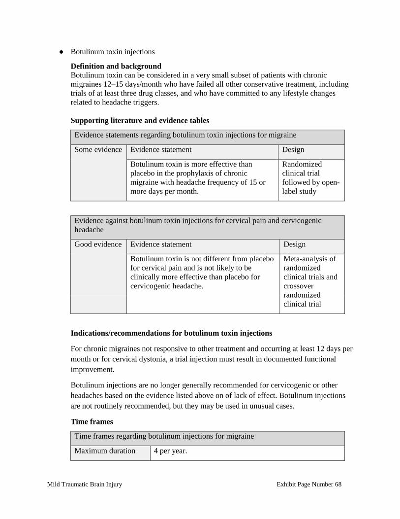
Mild Traumatic Brain Injury Exhibit Page Number 68
● Botulinum toxin injections
Definition and background Botulinum toxin can be considered in a very small subset of patients with chronic
migraines 12–15 days/month who have failed all other conservative treatment, including
trials of at least three drug classes, and who have committed to any lifestyle changes
related to headache triggers.
Supporting literature and evidence tables
Evidence statements regarding botulinum toxin injections for migraine
Some evidence Evidence statement Design
Botulinum toxin is more effective than
placebo in the prophylaxis of chronic
migraine with headache frequency of 15 or
more days per month.
Randomized
clinical trial
followed by open-
label study
Evidence against botulinum toxin injections for cervical pain and cervicogenic
headache
Good evidence Evidence statement Design
Botulinum toxin is not different from placebo
for cervical pain and is not likely to be
clinically more effective than placebo for
cervicogenic headache.
Meta-analysis of
randomized
clinical trials and
crossover
randomized
clinical trial
Indications/recommendations for botulinum toxin injections
For chronic migraines not responsive to other treatment and occurring at least 12 days per
month or for cervical dystonia, a trial injection must result in documented functional
improvement.
Botulinum injections are no longer generally recommended for cervicogenic or other
headaches based on the evidence listed above on of lack of effect. Botulinum injections
are not routinely recommended, but they may be used in unusual cases.
Time frames
Time frames regarding botulinum injections for migraine
Maximum duration 4 per year.

Mild Traumatic Brain Injury Exhibit Page Number 69
F.2 Visual treatment
Definition and background
Visual treatment is appropriate to consider for mTBI. Visual impairments may occur secondary
to mTBI and in one or more of the following categories:
● visual acuity and visual field function;
● ocular motor control and ocular alignment;
● visual perception.
Note: Visual rehabilitation is also performed for dizziness. (Refer to Section F.3, Neuro-otologic
treatment, under Vestibular rehabilitation for details.)
Supporting literature and evidence tables
Studies not resulting in evidence statements
Prevalence of mTBI visual findings, including eye movement disorders and
accommodation, appears to be approximately 30 – 60% in blast induced mTBI. Other
studies of populations such as athletes confirm presence of visual issues in mTBI.
Currently, there are no easy to perform clinical measures to clearly identify these conditions
in the primary care settings. These findings generally decrease as cognition improves.
Indications/recommendations for visual treatment
An ophthalmologist, neuro-ophthalmologist, neurologist, occupational therapist, certified vision
therapist, or optometrist may treat visual impairment resulting from TBI. Treatment of visual
impairments should be based on a comprehensive evaluation and diagnosis. When possible,
therapy should be provided in an interdisciplinary integrated approach for the purposes of best
outcomes and greatest convenience for the patient.
Treatment should be functionally-based and goal-directed. Individuals should be evaluated at
intervals depending on his/her impairment, and progress should be clearly documented. Reliance
on unvalidated outcome measures, such as performance on tests of “eye teaming” or “eye
tracking,” should be avoided.
● Visual acuity and visual field function
These are determined by the eye, optic nerve, optic chiasm, optic tracts, optic radiations,
and visual cortex. If visual acuity deficits are caused by optic nerve trauma, the best data
argues against the use of corticosteroid in almost all cases, in part because of the risk of
increased morbidity and mortality from a concomitant head injury. Surgery may be
indicated if the trauma results in progressive visual acuity loss in the setting of
demonstrable compression of the nerve or if a hematoma is present within the optic nerve
sheath. These cases are extremely rare. If visual acuity or visual field deficits are caused
by intracranial visual pathway damage, acute treatment should be directed toward the
specific injury.

Mild Traumatic Brain Injury Exhibit Page Number 70
Vision aids may be prescribed for those individuals with documented visual acuity or
visual field loss after the acute injury. Lenses may be used to improve visual acuity.
Tinted FL41 lenses may be useful to treat photophobia and glare sensitivity.
The use of optical and digital compensatory devices may benefit some individuals with
documented visual field loss from visual pathway disorders that affect the visual fields in
both eyes.
Depending on the level of adaptation to the visual field loss, some individuals may need
training and education in strategies to improve compensation. Efforts to use visuospatial
interventions to improve visual field loss directly without developing compensatory
visual scanning are not recommended. The use of computers as a primary and
independent form of visual treatment has limited application due to: (1) limitations in the
rationale and specific application of software programs to address the needs of the
individual with TBI; and (2) difficulty with generalization of learned computer skills into
functional environments. Integrated computer-based treatment (i.e., both individualized
cognitive and interpersonal therapies) may improve functioning within the context of an
interdisciplinary, neuropsychological rehabilitation program. Sole reliance on repeated
exposure and practice on computer-based tasks without extensive involvement and
intervention by a therapist is not recommended. Virtual reality tools may prove useful for
ADL assessment and training; however, they are experimental at the time of this
guideline, as there are no strong studies supporting its success. Computerized visual
restoration therapy programs or other computerized visual treatment programs, such as
virtual reality, are not recommended due to lack of proven clinically meaningful efficacy
and cost.
● Disorders involving ocular motility and binocular vision
These should be treated according to the underlying diagnosis. Ocular motility includes:
ductions, versions, smooth pursuit, saccade, vergence, and vestibular eye movements.
Disorders of binocular vision include strabismus with double vision and disturbances of
accommodation and vergence.
Treatment may include the use of lenses, prisms, vision rehabilitation, and/or surgery. For
individuals with disorders of ocular motor and ocular alignment that result in diplopia,
the following should be considered based on severity and duration of impairments:
monocular eye patching, occlusion of central or peripheral vision, prisms lenses, or
strabismus surgery.
Lenses may be used to help accommodation. Because of the interaction between
accommodation and vergence, lenses may also at times be used to assist in the treatment
of a vergence disorder.
Prisms may be prescribed to provide an immediate improvement in diplopia and other
disorders with symptoms. If diplopia is not stable, then appropriate patching (partial
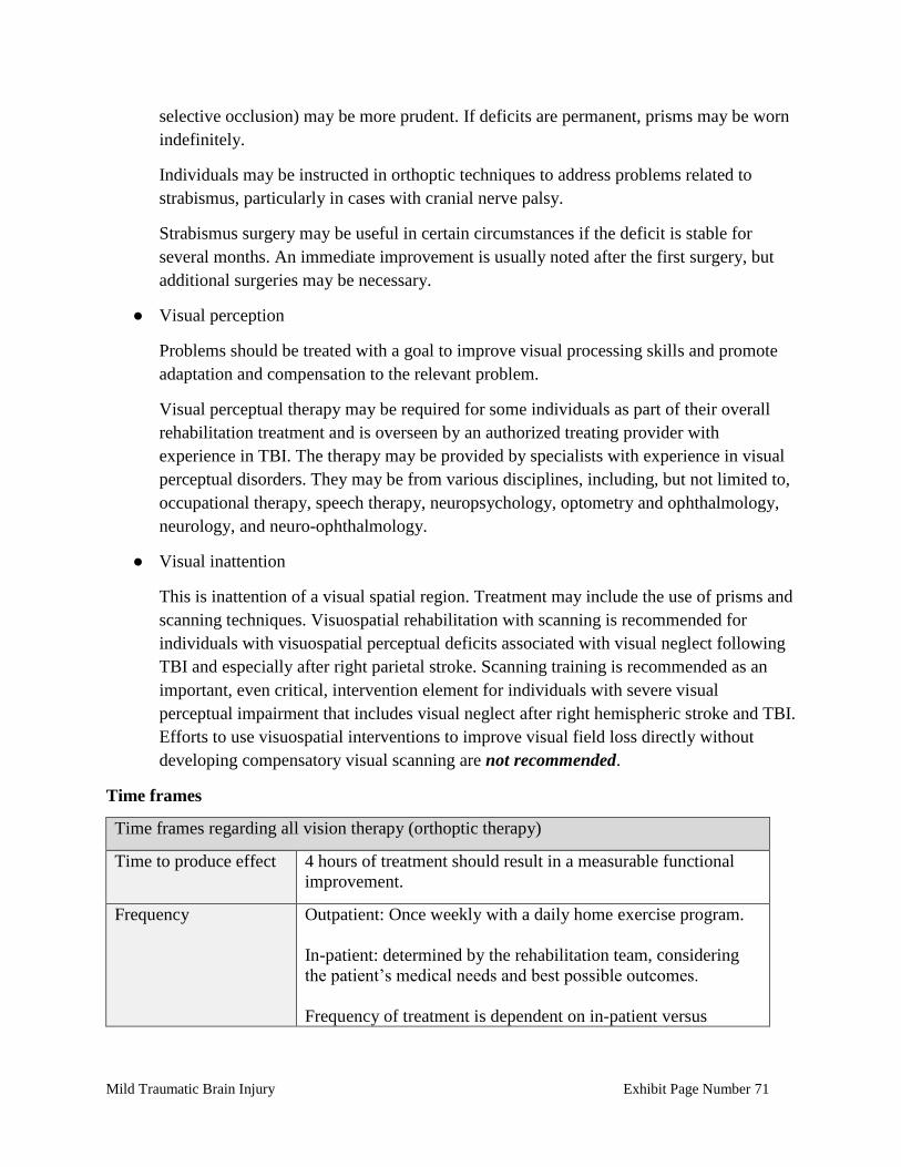
Mild Traumatic Brain Injury Exhibit Page Number 71
selective occlusion) may be more prudent. If deficits are permanent, prisms may be worn
indefinitely.
Individuals may be instructed in orthoptic techniques to address problems related to
strabismus, particularly in cases with cranial nerve palsy.
Strabismus surgery may be useful in certain circumstances if the deficit is stable for
several months. An immediate improvement is usually noted after the first surgery, but
additional surgeries may be necessary.
● Visual perception
Problems should be treated with a goal to improve visual processing skills and promote
adaptation and compensation to the relevant problem.
Visual perceptual therapy may be required for some individuals as part of their overall
rehabilitation treatment and is overseen by an authorized treating provider with
experience in TBI. The therapy may be provided by specialists with experience in visual
perceptual disorders. They may be from various disciplines, including, but not limited to,
occupational therapy, speech therapy, neuropsychology, optometry and ophthalmology,
neurology, and neuro-ophthalmology.
● Visual inattention
This is inattention of a visual spatial region. Treatment may include the use of prisms and
scanning techniques. Visuospatial rehabilitation with scanning is recommended for
individuals with visuospatial perceptual deficits associated with visual neglect following
TBI and especially after right parietal stroke. Scanning training is recommended as an
important, even critical, intervention element for individuals with severe visual
perceptual impairment that includes visual neglect after right hemispheric stroke and TBI.
Efforts to use visuospatial interventions to improve visual field loss directly without
developing compensatory visual scanning are not recommended.
Time frames
Time frames regarding all vision therapy (orthoptic therapy)
Time to produce effect 4 hours of treatment should result in a measurable functional
improvement.
Frequency Outpatient: Once weekly with a daily home exercise program.
In-patient: determined by the rehabilitation team, considering
the patient’s medical needs and best possible outcomes.
Frequency of treatment is dependent on in-patient versus

Mild Traumatic Brain Injury Exhibit Page Number 72
Time frames regarding all vision therapy (orthoptic therapy)
outpatient and the medical condition of the individual.
Optimum duration 12 hours.
Maximum duration 20 hours. Throughout the treatment process, exams are
performed to evaluate status. When progress is no longer
occurring, then therapy should be stopped unless there are
mitigating circumstances. If after 20 hours of treatment there is
documented progress but the individual is not at maximum
therapeutic gain, then additional therapy may be indicated.
Additional therapy should take into consideration the overall
rehabilitation plan for the individual.
These time frames are not meant to be applied to each section separately. The time frames are to
be applied to the totality of all vision rehabilitation regardless of the type or combination of
therapies being provided.
F.3 Neuro-otologic treatment: vestibular and audiologic
Definition and background
For patients with dizziness causing nausea or affecting balance, treatment of these conditions
may be necessary before other rehabilitative therapy can be accomplished.
Supporting literature and evidence statements
The following recommendations were based on consensus. Supporting literature and evidence
statements are also included in the subsections below when available.
Indications/treatment for neuro-otology: vestibular and audiology
● Post-traumatic tinnitus
Individuals with TBI may experience debilitating tinnitus (ringing in the ears). Tinnitus
may be associated with pressure or noise related trauma and ossicular or perilymphatic
fistula disorder. Tinnitus can be evaluated with specific audio-metric testing techniques.
Patients may benefit from anti-depressants, anti-seizure medicines, and anxiolytics. In
many situations, devices are recommended. These may include hearing aids, maskers,
and tinnitus trainers. Tinnitus trainers require a 30-day trial to determine the effectiveness
of masking. More sophisticated devices that use music as opposed to masking are not
recommended due to no proof of their superiority.
● Hyperacusis
Individuals with TBI may experience significant sensitivity to sound. This is more
common in association with tinnitus. These individuals may benefit from devices such as

Mild Traumatic Brain Injury Exhibit Page Number 73
tinnitus trainers, musician’s plugs, and simple noise plugs. Continuous exposure to
broadband sound may also be used.
● Benign paroxysmal positional vertigo (BPPV)
BPPV is the most common cause of post-traumatic vertigo. It is an otologic disorder in
which particles normally adherent to the gravity sensors of the ear become displaced into
the semicircular canals, which cause the sensation of spinning. It is characterized by
recurrent, brief spells of vertigo triggered by head movements such as getting in and out
of bed, rolling over in bed, tipping the head upward, or bending over. It is diagnosed by
the Dix Hallpike maneuver and treated with canalith repositioning maneuvers (CRM)
specific to the affected semicircular canals. Patients treated by CRM should be re-
evaluated within the first month to ensure resolution of symptoms. Recurrences are
common after trauma. These may be treated by repeating the CRM, home exercises, or
referral to physical therapy.
Some individuals may require an exercise-based approach following, or instead of, the
CRM. Home exercises are safe and effective in this disorder.
Time frames
Time frames regarding vestibular rehabilitation for benign paroxysmal positional
vertigo
Frequency 1 to 3 sessions with repeated CRM at each session and
follow-up at 1 month.
Optimum duration 1 month with re-evaluation.
Maximum
duration
Reoccurrence can occur randomly for many years following
trauma. Home exercises are necessary for those with frequent
recurrences. Some patients are unable to perform home
exercises, so repeated visits for CRM may be required.
● Semicircular canal dehiscence
This is an abnormal communication between the CSF space in the skull and perilymph
surrounding the inner ear. It can result from blunt head trauma with fracture of the bone
separating these spaces. Symptoms include vertigo brought on by loud sounds or
straining and autophony, which is the magnification of internal bodily sounds (chewing,
eye movement, joint movement, heartbeat) in the affected ear. Vestibular suppressants
and avoidance of provoking sounds can be used; surgery is required in severe cases.
● Vestibular migraine
Individuals experiencing an exacerbation of migraine after TBI frequently have an
associated dizziness. Treatment includes trigger avoidance, vestibular suppressants, and
migraine prophylactic medications such as calcium channel blockers, anti-seizure

Mild Traumatic Brain Injury Exhibit Page Number 74
medication, beta blockers, and SSRIs. (Refer to Section F.1, Post-traumatic headache
treatment.)
● Vestibular rehabilitation
Individuals with TBI may experience loss of inner ear balance function resulting in
dizziness and imbalance. This can result from labyrinthine concussion, penetrating
injuries, injury to the eighth cranial nerve, and explosive pressure changes. Patients may
compensate for vestibular deficits more quickly with vestibular rehabilitation than
without it.
Persistent postural perceptual dizziness: This is a condition in which patients continue to
experience persistent dizziness due to mis-calibration of the sensorimotor, cerebellar, and
vestibular inputs. This condition may be comorbid with anxiety. Treatment with
aggressive vestibular rehabilitation and either SSRI or SNRI may be beneficial.
Symptoms of vestibular system dysfunction following TBI may be due to damage of
central or peripheral structures. These symptoms may include vertigo, eye-head
dyscoordination affecting the ability to stabilize gaze during head movements, and
imbalance affecting stability in standing or walking. Dizziness is commonly associated
with TBI. Dizziness and balance disorders may or may not co-exist in the same
individual with TBI.
Vestibular rehabilitation is performed by qualified practitioners, e.g., audiologists,
otologists, trained nurses, vestibular therapists, physical therapists (preferably neurology
certified) or occupational therapists.
Evidence statements regarding vestibular rehabilitation
Good evidence Evidence statement Design
Vestibular rehabilitation incorporating visual
motion performed by the patient alone with
brief instruction from a health care provider
reduces dizziness and improves function.
Meta-analysis of
randomized
clinical trials
Some evidence Evidence statement Design
2 to 6 sessions of Mulligan sustained natural
apophyseal glides (SNAGs) or Maitland
mobilizations over 6 weeks are significantly
more effective in reducing the intensity and
frequency of cervicogenic dizziness than a
placebo intervention in patients with chronic
cervicogenic dizziness. The effects are
maintained at 12 weeks post treatment.
Double-blind
randomized
clinical trial

Mild Traumatic Brain Injury Exhibit Page Number 75
Evidence statements regarding vestibular rehabilitation
Some
evidence,
continued
An 8-week program of combined cervical
physiotherapy and vestibular rehabilitation is
likely to improve the rate of medical clearance
for return to sport for patients with a sport-
related concussion who have persistent
dizziness, neck pain, and/or headache 10 days
after injury and who are suspected by a
physician of having vestibular involvement or
cervical spine involvement.
Randomized
clinical trial
Studies not resulting in evidence statements
One study of patients with mTBI 4 years after the incident found 30% continuing
complaints of balance problems. A patient with mTBI may complain of continuing
dizziness with minor clinical findings. It is suggested that these patients be referred
to a vestibular therapist or physical or occupational therapists with vestibular
training and experience.

Mild Traumatic Brain Injury Exhibit Page Number 76
○ Balance disorders
Balance disorders occur frequently following TBI. This may be due to a peripheral
vestibular lesion or central vestibular lesion secondary to trauma, fracture,
hemorrhage, or intracranial pressure changes.
Balance is a complex motor control task, requiring integration of sensory information,
neural processing, and biomechanical factors. It is the ability to control the center of
gravity (COG) over the base of support in a given sensory environment.
Assessment includes evaluation of the motor system, ROM, and sensory systems that
affect the person’s ability to maintain equilibrium. Movement strategies to maintain
balance require functional ROM and adequate strength. Sensory information from the
vestibular, visual, and somatosensory systems are integrated at the central level
between the two sides of the body and three sensory systems. These key areas are
associated with maintenance of balance or posture. Central motor planning is
essential for proper strategies that are then transmitted to the peripheral motor system
for execution. Deficits at the central level, peripheral motor level, or peripheral
sensory level will affect balance and equilibrium.
Common clinical tests used to evaluate balance include the Balance Error Scoring
System (BESS), Berg Balance Scale (BBS), Community Balance and Mobility Scale
(CB&M), Clinical Test of Sensory Interaction on Balance (CTSIB), Motion
Sensitivity Quotient, Sensory Organization Test (SOT), tandem gait task from the
Sport Concussion Assessment Tool-3, Functional Gait Assessment (FGA), and an
instrumented gait speed assessment. The disadvantage to most of these tests is that
they were developed for specific age groups and may not have been studied for TBI.
However, the CB&M has been studied for TBI. Self-report instruments are also
available to evaluate balance: ABC scale for dizziness, Dizziness Handicap Inventory
(DHI), Vertigo Handicap Questionnaire (VHQ), and the Vestibular Disorder
Activities of Daily Living Scale (VADL).
Sources of imbalance symptoms include vestibular, ocular, and cervicogenic.
Vestibular-ocular type may have visual abnormalities on testing and impaired clinical
balance. It is usually treated with a vestibular rehabilitation program. Visual or ocular
motor training alone has no demonstrated benefit, and treatment should be done using
an interdisciplinary approach. Cervicogenic imbalance is associated with neck pain
and headaches associated with head movement. It is usually treated with head and
neck proprioceptive re-training and cervical manual therapy.

Mild Traumatic Brain Injury Exhibit Page Number 77
The dynamic systems model recognizes that balance and dynamic equilibrium are the
result of the interaction between the individual, the functional task, and the
environment. Emphasis of treatments performed by a qualified physical or
occupational therapist in vestibular and balance dysfunction are head exercises for
habituation of vertigo, eye-head coordination exercises for improvement of gaze
stabilization, and sensorimotor retraining to remediate postural dyscontrol in all
functional movement positions. Exercises may be directed at habituation,
desensitization by repeat exposure; substitution, alternative strategies; or adaption,
improved use of remaining functioning vestibular system.
Special equipment for vestibular treatment in the clinic may include dynamic
platform posturography or a foam/dome apparatus for sensory integration and balance
as well as tilt or rocker boards. Other virtual reality devices used in isolation are not
suggested for use with this treatment because therapist intervention and supervision
are important for success. No special equipment is needed at home unless identified
by the treating professional and documented as medically necessary.
Individuals with central traumatic vestibular lesions take longer to improve than those
with dizziness from other causes. Studies indicate that at six months, only one-third
of individuals with unilateral loss from trauma were symptom-free as compared with
other causes. At 18 months, many individuals continued to show symptoms. Of those
with central vestibular loss, 60-70% had persisting symptoms at five years and half
were unable to return to work.
For additional related complaints, refer to the Division’s Moderate/Severe TBI
Medical Treatment Guideline treatment section for neuro-otology.
Time frames
Time frames for vestibular rehabilitation for balance disorders
Time to produce
effect
6 to 12 weeks depending on severity.
Frequency 1-2 times per week initially for training, with 2-4 follow-
up visits to reinforce treatment. Individuals are expected
to perform self-directed exercises twice daily at home,
but they may require supervision for guidance and safety.
Optimum duration For mTBI, 12 visits.
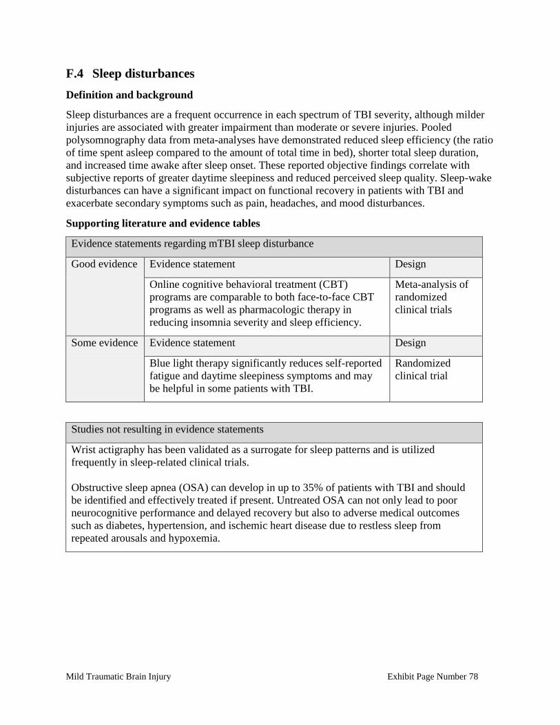
Mild Traumatic Brain Injury Exhibit Page Number 78
F.4 Sleep disturbances
Definition and background
Sleep disturbances are a frequent occurrence in each spectrum of TBI severity, although milder
injuries are associated with greater impairment than moderate or severe injuries. Pooled
polysomnography data from meta-analyses have demonstrated reduced sleep efficiency (the ratio
of time spent asleep compared to the amount of total time in bed), shorter total sleep duration,
and increased time awake after sleep onset. These reported objective findings correlate with
subjective reports of greater daytime sleepiness and reduced perceived sleep quality. Sleep-wake
disturbances can have a significant impact on functional recovery in patients with TBI and
exacerbate secondary symptoms such as pain, headaches, and mood disturbances.
Supporting literature and evidence tables
Evidence statements regarding mTBI sleep disturbance
Good evidence Evidence statement Design
Online cognitive behavioral treatment (CBT)
programs are comparable to both face-to-face CBT
programs as well as pharmacologic therapy in
reducing insomnia severity and sleep efficiency.
Meta-analysis of
randomized
clinical trials
Some evidence Evidence statement Design
Blue light therapy significantly reduces self-reported
fatigue and daytime sleepiness symptoms and may
be helpful in some patients with TBI.
Randomized
clinical trial
Studies not resulting in evidence statements
Wrist actigraphy has been validated as a surrogate for sleep patterns and is utilized
frequently in sleep-related clinical trials.
Obstructive sleep apnea (OSA) can develop in up to 35% of patients with TBI and should
be identified and effectively treated if present. Untreated OSA can not only lead to poor
neurocognitive performance and delayed recovery but also to adverse medical outcomes
such as diabetes, hypertension, and ischemic heart disease due to restless sleep from
repeated arousals and hypoxemia.

Mild Traumatic Brain Injury Exhibit Page Number 79
Indications/recommendations for sleep disturbances
Although physiologic sleep is most effectively measured by polysomnography (PSG), it is often
not feasible or necessary to undertake such overnight testing. Subjective self-reporting
questionnaires that have been validated against objective measures are the Pittsburgh Sleep
Quality Index and the Epworth Sleepiness Scale. These tests are useful tools that can evaluate
sleep patterns. Sleep diaries are another subjective tool that may be used to assess self-reported
sleep patterns, although there is a paucity of research validating sleep diaries with objective
testing. Objective testing measures to assess sleep-related behaviors include actigraphy and PSG.
However, the treating clinician must consider the feasibility of such testing in this population.
Obstructive sleep apnea (OSA) should be identified and effectively treated if present (see studies
listed in table above). Patients with OSA will complain of excessive daytime sleepiness, fatigue,
and morning headaches. Bed partners may note that patients snore or appear to choke/gasp for air
during the night. OSA should be suspected in patients with symptoms and/or with elevated risk
factors such as obesity, a crowded oropharyngeal airway, increased neck circumference, and
hypertension. As mentioned above, PSG can be used to diagnose sleep apnea and other sleep
related disorders.
Patients identified as having OSA should be treated with a positive airway pressure device such
as continuous positive airway pressure (CPAP), bilevel positive airway pressure (BiPAP), or
autotitrating positive airway pressure (APAP). Patients should also be counseled regarding
weight loss strategies if indicated. Frequent evaluations are important to evaluate for adherence
to therapy. This may be accomplished through patient interview and review of downloaded data
from their device. Objective testing is usually not indicated unless symptoms do not resolve after
several weeks of therapy.
In addition to OSA, clinicians should have a low threshold to obtain PSG in patients with TBI
and hypersomnia (to rule out narcolepsy) or complaints suspicious for periodic limb movement
disorder (uncomfortable urge to move legs and arms during inactivity). Several small studies
have suggested that sleep-related movement disorders and parasomnias occur at an increased rate
in patients with TBI. If present, mild symptoms of periodic limb movement disorder may be
treated with sleep hygiene measures and behavioral therapies previously discussed.
Other sleep disorders should be ruled out when appropriate symptoms are present before
proceeding with the following treatments.
● Counseling on sleep hygiene
Non-pharmacologic treatments for sleep-wake disturbances following TBI, including
modification of sleep hygiene, should be undertaken prior to instituting pharmacologic
therapy. Clinicians should be sure to take a comprehensive medical history to include
sleep patterns (such as taking naps during the day) and an occupational history to identify
any high-risk job duties such as commercial driving or piloting. Patients suffering from a
sleep-wake disorder should first receive basic behavioral sleep hygiene and stimulus
control counseling.

Mild Traumatic Brain Injury Exhibit Page Number 80
Counseling should include the following:
o Set an alarm clock to wake up at a fixed time each morning, including weekends.
o Try not to force sleep.
o Avoid caffeinated beverages after lunch and maintain consistent eating schedules,
particularly dinner.
o Avoid alcohol.
o Avoid smoking or other nicotine intake, particularly during the evening.
o Adjust the bedroom environment as needed to decrease stimuli (e.g., reduce ambient
light, turn off the television or radio).
o Avoid prolonged use of light-emitting screens (laptops, tablets, smartphones, ebooks)
before bedtime.
o Resolve concerns or worries before bedtime.
o Exercise regularly for at least 20 minutes, preferably more than 4 to 5 hours prior to
bedtime.
o Avoid daytime naps, especially if they are longer than 20 to 30 minutes or occur late
in the day.
o Go to bed only when sleepy.
o Do not watch television, read, eat, or worry while in bed. Use bed only for sleep and
sex.
o Get out of bed if unable to fall asleep within 20 minutes and go to another room.
Return to bed only when sleepy. Repeat this step as many times as necessary
throughout the night.
Additional conservative measures include the following:
o Mental alerting activities, such as working on a computer or doing crossword puzzles,
at times of rest or boredom.
o Avoidance of aggravating factors, including consideration of withdrawal of possibly
predisposing medications (antidepressants, antihistamines, dopamine-blocking
antiemetics, and neuroleptic agents).
o Moderate regular exercise.
o Reduced caffeine intake.
o For symptomatic relief: walking, bicycling, soaking the affected limbs, and leg
massage, including pneumatic compression.

Mild Traumatic Brain Injury Exhibit Page Number 81
● Cognitive behavioral therapy
Patients with sleep disturbances refractory to these basic counseling measures may
benefit from a cognitive behavioral treatment (CBT) program for insomnia. When
utilizing internet delivered CBT, the clinician should institute a program of a minimum of
six weeks duration and consider including some measure of personal support as opposed
to fully automated programs. Additionally, these programs require technical resources
which may not be feasible in all patients and a formal in-person CBT program may be
more appropriate.
The general components of an effective CBT program may include the following:
o Basic education regarding sleep and the nature and causes of chronic insomnia should
be reviewed.
o Behavioral therapy targeted problems with sleep maintenance and sleep onset using
sleep restriction and stimulus control (see sleep hygiene and stimulus control
recommendations). These interventions are designed to reestablish the bed as a
dominant cue for sleep onset, regulate sleep-wake schedules, and consolidate sleep
over a short period of time. Specifically, sleep restriction induces a state of mild sleep
deprivation, which accelerates sleep onset and improves quality of sleep by
establishing a “sleep window” for which patients stay in bed. To start, patients should
use a sleep diary to determine the total amount of hours spent in bed as well as the
amount of hours spent asleep. The amount of time the patient actually spends asleep
will determine their initial window and the patient will only spend this specified
amount of time in bed. For example, if a patient spends 8 hours in bed on average
every night but only 6 hours is spent sleeping, then the patient’s initial window would
be 6 hours. Patients can then increase or decrease the time spent in bed by 20 to 30
minutes weekly depending on their sleep efficiency (time spent asleep/total time in
bed). If sleep efficiency is >90%, then the patient can increase their time in bed. If
sleep efficiency is <80% the patient should decrease time spent in bed. Sleep
restriction allows the patient to learn to associate the bed with sleep and sex only.
o Relaxation training, such as the imagery method, provided as an additional coping
skill for patients. Guided imagery helps to inhibit cognitive stimulation by distracting
patients and helps to refocus their attention on pleasant scenes that evoke positive
emotions. Patients should practice these exercises every night to help them fall asleep.
o Cognitive components designed to assist patients in exploring how their thoughts
affect their sleep behaviors and emotions. Patients should be guided through cognitive
restructuring procedures focused on reducing negative self-talk and negative attitudes
that can be maladaptive.
o Sleep hygiene measures as previously mentioned should be continuously reinforced.
● Pharmacologic therapy for insomnia
The use of sleep-inducing agents such as benzodiazepines and nonbenzodiazepine
hypnotics should generally be avoided as randomized clinical trials directly assessing

Mild Traumatic Brain Injury Exhibit Page Number 82
their effects on insomnia and sleep-wake disorders are scarce. However, they may be
useful for the first few weeks for some patients with mTBI and for anxiety.
● Fatigue and day time sleepiness
o Blue light therapy: Blue light therapy should be administered for 30 to 45 minutes in
the morning, within two hours of awakening, with a device containing 66 light-
emitting diodes (LED). Patients can self-administer light therapy at home. They
should be instructed to sit in front of the light panel with the center approximately
50cm (1.5 feet) in front the eyes and look into the light source for a few seconds at
least every few minutes.
o Pharmacologic agents for fatigue: Although there is a paucity of evidence
demonstrating the effectiveness of Modafinil for treatment of fatigue, it may be
beneficial for the short-term treatment of excessive daytime sleepiness. Armodafinil,
the enantiomer of Modafinil with a slightly longer time to peak serum concentration,
may be effective for the short-term treatment of patients with excessive daytime
sleepiness despite good sleep efficiency on PSG. The use of these wakefulness-
promoting agents should be restricted to short term use (4 to 6 weeks) in conjunction
with nonpharmacologic therapy measures.
F.5 Cognitive treatment
Definition and background
In mTBI, acute cognitive deficits are common. In the majority of injured individuals,
spontaneous cognitive improvement is expected within the first three months, frequently within
days or weeks.
For patients needing treatment, therapists or speech-language pathologists may work with the
individual with mTBI in order to teach individuals adaptive skills, compensatory techniques, or
new ways of solving problems that assist them in coping more effectively during recovery. The
interdisciplinary treatment team approach is particularly beneficial in these cases, and it is
strongly encouraged, especially during the first 12 months post-injury. The team may include
specialists in neurology, rehabilitative medicine, neuropsychology, and ocular-motor, vestibular,
and balance functioning.
“Cognitive rehabilitation is a systematic, functionally oriented service of therapeutic cognitive
activities, based on an assessment and understanding of the person’s brain-behavior deficits.
Services are directed to achieve functional changes by (1) reinforcing, strengthening, or
reestablishing previously learned patterns of behavior, or (2) establishing new patterns of
cognitive activity or compensatory mechanisms for impaired neurological systems.”
All cognitive rehabilitation should be focused on functional goals relative to the individual
patient’s needs in performing activities of daily living and work. Progress toward achievement of
specific goals should continually be monitored during treatment. Strategies should always be
task or goal specific or demonstrate generalization of the learned concepts to the daily living
activities of the specific patient with external cueing as necessary. Compensatory or cognitive
support strategies are useful in this population.

Mild Traumatic Brain Injury Exhibit Page Number 83
For most patients with mTBI, cognitive therapy will focus on memory, executive function, and
in some cases attention. External memory compensation tools, such as memory notebooks, smart
phones, and computers can be very helpful. Self-talk strategies may be useful as patients with
mTBI return to more complex executive function. When patients with mTBI are required to
perform skills requiring alternating or selective attention, specific strategies can be applied to
improve the success rate.
Supporting literature and evidence tables
Evidence statements regarding mTBI and cognitive treatment
Good evidence Evidence statement Design
Cognitive training has small to moderate
positive effects in improving cognitive and
functional outcomes in patients with mild to
severe TBI who are at least 1-year post-acute
TBI when compared to waiting list controls or
standard rehabilitation.
Systematic review
and meta-analyses of
randomized clinical
trials
mTBI without post-traumatic amnesia does not
require routine rehabilitation.
Systematic review of
randomized trials,
quasi-randomized
trials, and quasi-
experimental studies
Some evidence Evidence statement Design
Routine scheduling for cognitive rehabilitation
for uncomplicated mTBI is not likely to improve
outcomes, and mTBI cases with a psychiatric
history are more likely to benefit from routine
assessment for cognitive rehabilitation
treatment.
Randomized clinical
trial
Studies not resulting in evidence statements
There is inadequate evidence to support that computerize cognitive rehabilitation adds any
benefits over conventional rehabilitation for patients with mild to severe TBI or stroke.

Mild Traumatic Brain Injury Exhibit Page Number 84
Indications for mTBI cognitive treatment
Not all patients with mTBI require cognitive treatment. Rehabilitation of cognitive impairments
should be initiated if:
● the individual is not demonstrating the expected cognitive improvement;
● the individual exhibits more severe cognitive impairments on formal evaluation;
● the individual’s vocation or other life circumstances necessitate the learning of
compensatory strategies;
● there are safety issues in question (e.g., possible harm to self or others).
Recommendations for mTBI cognitive treatment
If therapy is required, use the time frames listed below. In individual cases, the physician may
determine the need for further evaluation and treatment.
Computer-based treatment with active therapist involvement: Computer-based treatment must be
accompanied by oversight from the treating provider either in person or by telehealth. Computer-
based treatment is not recommended in isolation.
Assistive technology: These devices must be used in consultation with specialists and within a
rehabilitation therapy program by physical therapists, occupational therapists, and speech-
language pathologists to determine which tools are most suitable for individual cases.
Time frames
Time frames regarding mTBI cognitive treatment
Frequency Weekly 1-hour sessions initially during the first month to determine
specific goals and objective outcomes as part of the primary
treatment and return-to-work evaluations. Once the patient has
returned to normal function without impairing symptoms, visit
frequency should decrease or treatment should be terminated.
Optimum duration 1 to 3 months. Additional sessions may be required as justified. For
example, in cases with complicated mTBI or a number of comorbid
conditions, treatment patterns may resemble that for
moderate/severe injuries.
F.6 Psychological interventions
Definition and background
Early interventions that educate individuals, their family and/or support system, or the employer
about the symptoms, natural history, prognosis, and management of mTBI symptoms are very
important.

Mild Traumatic Brain Injury Exhibit Page Number 85
Psychological and educational interventions may include, or be performed in conjunction with,
cognitive and behavioral treatment. Cognitive behavioral therapy (CBT) is a specialized goal-
oriented systematic process used to problem solve that focuses on changing thought processes. It
is usually provided by a trained therapist or psychologist.
The acute symptoms of mTBI (e.g., feeling dazed, disoriented, or confused) overlap with those
of emotional trauma and acute stress disorder. Over the course of recovery, the symptoms of
mTBI also overlap with a variety of psychological conditions, such as depression, anxiety,
insomnia, and PTSD. Consequently, the possibility that the symptoms are neuropsychiatric rather
than neuropsychological in origin should be evaluated when the degree of cognitive symptoms
exceeds what would be expected given objective findings, the mechanism of injury, or acute
signs of mTBI or if there is an unexplained, marked worsening of cognitive symptoms over time.
A psychological evaluation is especially important if the injury occurred in an emotionally
traumatic context or if there are clinical indications of another mental health disorder.
Supporting literature and evidence tables
Evidence statements regarding mTBI psychological/educational interventions
Some evidence Evidence statement Design
5 individual sessions, 1.5 hours long, of Cognitive
Behavioral Therapy (CBT) initiated for patients
diagnosed with acute stress disorder early after TBI
are significantly more effective than supportive
counseling in preventing chronic PTSD in patients
who develop acute stress disorder following mTBI.
Single-blind
randomized
clinical trial

Mild Traumatic Brain Injury Exhibit Page Number 86
Evidence statements regarding mTBI psychological/educational interventions
Some evidence,
continued
For patients with complicated mTBI and moderate
TBI who have completed initial therapy, 12 weeks
of telephone-based and in-person Cognitive
Behavioral Therapy (CBT) interventions are no
more effective than usual care for treating Major
Depressive Disorder (MDD).
Due to the differences noted between groups in this
study, it is not possible to determine if telephone
CBT is preferable to in-person CBT after initial
treatment has been completed. However, telephone
CBT allowed more participation by support persons,
and this may be important to patients with mTBI.
It is interesting that secondary data showed high
satisfaction with CBT, 84%, and only 26% with
usual care.
Single-blind
randomized
clinical trial
Studies not resulting in evidence statements
One study noted sustained improvement after 6 months of CBT with either face-to-face or
telephone contact. This should always be performed within the construct of a more
complete therapy program.
Indications for mTBI psychological/educational interventions
Early intervention is appropriate in the acute stage of mTBI for patients at increased risk of
prolonged symptoms to promote positive coping skills and to manage symptoms. Risk factors for
prolonged symptoms include, but are not limited to:
● Glasgow Coma Scale less than 15 at 2 hours post-injury;
● work risk factors, such as very demanding or stressful vocations or being employed in the
current job for a short period of time;
● age above 40 years;
● injury complicated by the presence of intracranial lesions, current or previous;
● history of prior brain injury, cognitive impairment, learning disabilities, or developmental
delay;

Mild Traumatic Brain Injury Exhibit Page Number 87
● associated orthopedic, soft tissue, or organ injuries;
● pre-injury issues with general health or psychosocial well-being;
● psychological factors such as depression, post-traumatic stress disorder, or anxiety – see
evidence statement above.
The presence of other injuries requiring medical attention should not exclude anyone from
appropriate psychological treatment.
During the first 12 weeks following mTBI, the following should always be treated:
● problems that are secondary to the injury (e.g., anxiety, depression, adjustment disorder,
difficulties with self-acceptance, and difficulties in adapting to one’s work demands due
to diminished cognitive capacity), and
● post-traumatic stress disorder (PTSD). PTSD may be present in a minority of patients
with mTBI and should be assessed early on and treated.
● It is also appropriate to consider intervention to address persistent specific problems that
are directly caused by the injury (e.g., memory deficits).
Recommendations for mTBI psychological/educational interventions
Early intervention in the acute stage: Psychological treatment after the first 12 weeks, by
physicians with experience in brain injury care, is recommended when recovery is hindered by
symptoms caused by the injury or by symptoms secondary to it as indicated above. For
psychological symptoms lasting longer than six months, refer to the Division’s Moderate/Severe
TBI Medical Treatment Guideline. Primary care providers should be made aware of these
recommendations as often there is delay in treatment.
Treatment may include individual psychotherapy, marital/family therapy, group therapy,
instruction in relaxation and related techniques, cognitive/behavioral therapy, medication
management, social skills training, repetitive transcranial magnetic stimulation (rTMS) for
treatment-resistant depression, and interventions/consultation in the community. When cognitive
interventions are needed, refer to Section F.5, Cognitive treatment.
Functional gains are to be documented and achieved with therapy. They may include, but are not
limited to improved mood, irritability, frustration tolerance, concentration, memory, sleep
quality, and interpersonal skills (such as empathy and capacity to effectively interact with family
and/or support system members and co-workers).
● During the acute stage: Psychological interventions to educate regarding coping
mechanisms for common symptom amelioration may include the individual and family
and/or support system or alternatively close friends and co-workers.
● During the first 12 weeks: Mental health interventions to address PTSD should be
evidence based and may include individual psychotherapy, cognitive/behavioral therapy,
instructions in specific techniques such as relaxation training or biofeedback, instruction
in symptom management, trauma resolution techniques (e.g., EMDR), group therapy,
medications, and interventions in the community.

Mild Traumatic Brain Injury Exhibit Page Number 88
Time frames
Time frames regarding mTBI psychological/educational interventions
Frequency Weekly 1-hour sessions initially during the first month as part of
the primary treatment and return-to-work evaluations. Once a
patient has returned to normal function without impairing
symptoms, visit frequency should decrease or treatment should be
terminated.
Optimum duration Optimum Duration: 1 to 3 months.
Additional sessions may be required as justified.
F.7 Medications
Definition and background
Most mTBI cases do not require prescription medication as the majority of cases resolve without
them. The most common needs for medication in mTBI are anxiety, depression, nausea, and
headaches.
Indications/recommendations for medications
A number of patients with mTBI have a constellation of complaints including insomnia,
headache, and depressive symptoms. When conservative measures are not effective and
symptoms are still interfering with activity after seven to ten days, amitriptyline or an SSRI and
low dose amitriptyline may be reasonable considerations.
If other medication use can be justified based on specific patient needs, refer to the Division’s
Moderate/Severe TBI Medical Treatment Guideline, Section F.4.h, Medications. Specialists
should generally be involved for TBI cases requiring multiple medications.
When medication is used for mTBI, a trial of tapering by 6-12 months post-injury is
recommended.
● Acetaminophen
Acetaminophen is an effective analgesic with anti-pyretic but not anti-inflammatory
activity.
Acetaminophen is generally well-tolerated. It causes little or no gastrointestinal (GI)
irritation and is not associated with ulcer formation.
However, acetaminophen can exacerbate headache when used chronically.
Acetaminophen also has been associated with liver toxicity in overdose situations or in

Mild Traumatic Brain Injury Exhibit Page Number 89
chronic alcohol use. Patients may not realize that many over-the-counter preparations
contain acetaminophen. The total daily dose of acetaminophen is recommended not to
exceed three grams per 24-hour period, from all sources, including narcotic-
acetaminophen combination preparations.
Time frames
Time frames regarding acetaminophen
Optimum
duration
7 to 10 days.
Maximum
duration
Long-term use as indicated on a case-by-case basis. Use of this
substance long-term (for 3 days per week or greater) may be
associated with rebound pain upon cessation.
● Non-steroidal anti-inflammatory drugs (NSAIDs)
NSAIDs are useful for pain and inflammation. In mild cases, they may be the only drugs
required for analgesia. Chronic use of NSAIDs is generally not recommended due to
increased risk of cardiovascular events and GI bleeding.
There are several classes of NSAIDs. The response of the injured worker to a specific
medication is unpredictable. For this reason, a range of NSAIDs may be tried in each
case, with the most effective preparation being continued.
Patients should be closely monitored for adverse reactions. The FDA advises that many
NSAIDs may cause an increased risk of serious cardiovascular thrombotic events,
myocardial infarction, and stroke, which can be fatal. Administration of proton pump
inhibitors, histamine 2 blockers, or prostaglandin analog misoprostol along with these
NSAIDs may reduce the risk of duodenal and gastric ulceration in patients at higher risk
for this adverse event (e.g., age > 60, concurrent antiplatelet or corticosteroid therapy).
They do not impact possible cardiovascular complications.
NSAIDs are associated with abnormal liver function and renal function, including renal
failure. Patients with hepatic or renal disease may need increased dosing intervals with
chronic use.
Due to the cross-reactivity between aspirin and NSAIDs, NSAIDs should not be used in
aspirin-sensitive patients, and they should be used with caution in all patients with
asthma.
Topical NSAIDs may be more appropriate for some patients; see evidence statements in
table below.
NSAIDs may be associated with non-unions. Thus, their use with fractures is
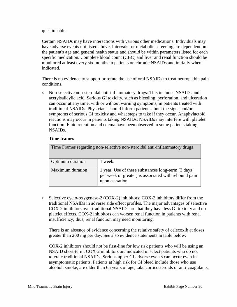
Mild Traumatic Brain Injury Exhibit Page Number 90
questionable.
Certain NSAIDs may have interactions with various other medications. Individuals may
have adverse events not listed above. Intervals for metabolic screening are dependent on
the patient's age and general health status and should be within parameters listed for each
specific medication. Complete blood count (CBC) and liver and renal function should be
monitored at least every six months in patients on chronic NSAIDs and initially when
indicated.
There is no evidence to support or refute the use of oral NSAIDs to treat neuropathic pain
conditions.
○ Non-selective non-steroidal anti-inflammatory drugs: This includes NSAIDs and
acetylsalicylic acid. Serious GI toxicity, such as bleeding, perforation, and ulceration
can occur at any time, with or without warning symptoms, in patients treated with
traditional NSAIDs. Physicians should inform patients about the signs and/or
symptoms of serious GI toxicity and what steps to take if they occur. Anaphylactoid
reactions may occur in patients taking NSAIDs. NSAIDs may interfere with platelet
function. Fluid retention and edema have been observed in some patients taking
NSAIDs.
Time frames
Time Frames regarding non-selective non-steroidal anti-inflammatory drugs
Optimum duration 1 week.
Maximum duration 1 year. Use of these substances long-term (3 days
per week or greater) is associated with rebound pain
upon cessation.
○ Selective cyclo-oxygenase-2 (COX-2) inhibitors: COX-2 inhibitors differ from the
traditional NSAIDs in adverse side effect profiles. The major advantages of selective
COX-2 inhibitors over traditional NSAIDs are that they have less GI toxicity and no
platelet effects. COX-2 inhibitors can worsen renal function in patients with renal
insufficiency; thus, renal function may need monitoring.
There is an absence of evidence concerning the relative safety of celecoxib at doses
greater than 200 mg per day. See also evidence statements in table below.
COX-2 inhibitors should not be first-line for low risk patients who will be using an
NSAID short-term. COX-2 inhibitors are indicated in select patients who do not
tolerate traditional NSAIDs. Serious upper GI adverse events can occur even in
asymptomatic patients. Patients at high risk for GI bleed include those who use
alcohol, smoke, are older than 65 years of age, take corticosteroids or anti-coagulants,

Mild Traumatic Brain Injury Exhibit Page Number 91
or have a longer duration of therapy. Celecoxib is contraindicated in sulfonamide
allergic patients.
Time frames
Time frames regarding selective cyclo-oxygenase-2 (COX-2) inhibitors
Optimum duration 7 to 10 days.
Maximum duration Chronic use is appropriate in individual cases. Use
of these substances long-term (3 days per week or
greater) is associated with rebound pain upon
cessation.
Supporting literature and evidence tables
Evidence statements regarding nonsteroidal anti-inflammatory drugs (NSAIDs)
Good
evidence
Evidence statement Design
Celecoxib in a dose of 200 mg per day,
administered over a long period, does not have
a worse cardiovascular risk profile than
naproxen at a dose of up to 1000 mg per day or
ibuprofen at a dose of up to 2400 mg per day.
Randomized
noninferiority
trial
Celecoxib has a more favorable safety profile
than ibuprofen or naproxen with respect to
serious GI adverse events, and it has a more
favorable safety profile than ibuprofen with
respect to renal adverse events.
Topical NSAIDs are associated with fewer
systemic adverse events than oral NSAIDs,
e.g., reduced risk of gastrointestinal adverse
effects by approximately one third.
Meta-analysis
of randomized
clinical trials
F.8 Communication and swallowing
Communication and swallowing issues are uncommon in mTBI. However, refer to the Division’s
Moderate/Severe TBI Medical Treatment Guideline if treatment is needed.

Mild Traumatic Brain Injury Exhibit Page Number 92
F.9 Therapeutic exercise
Definition and background
Therapeutic exercises, with or without mechanical assistance or resistance, may include a
number of modalities as part of the integrated occupational therapy or physical therapy program.
A therapeutic exercise program should be initiated at the start of any treatment program and
coordinated in an interdisciplinary approach. Such programs should emphasize education,
independence, and the importance of an on-going exercise regime.
Supporting literature and evidence tables
Studies not resulting in evidence statements
Both animal and human studies suggest therapeutic exercise has a direct relationship on
recovery from a TBI.
Indications for therapeutic exercise
Indications include the need for:
● cardiovascular fitness,
● improved muscle strength,
● improved connective tissue strength and integrity,
● increased bone density,
● promotion of circulation to enhance soft tissue healing,
● improvement of muscle recruitment,
● improved proprioception and coordination, and
● increased ROM.
Recommendations for therapeutic exercise
Therapeutic exercise may be done as part of the overall occupational therapy or physical therapy
program. It is not to be used in isolation. It should occur in an interdisciplinary treatment setting.
As with all treatments, specific objective goals should be set initially and monitored during
treatment.
The patient and/or caregiver should be instructed in and receive a home or community exercise
program that is progressed as functional status improves.
Upon discharge from inpatient or residential rehabilitation, the patient and/or caregiver would be
independent in the performance of the home exercise program and would have been educated in
the importance of continuing such a program. Educational goals would be to maintain or further
improve function and to minimize the risk for aggravation of symptoms in the future.
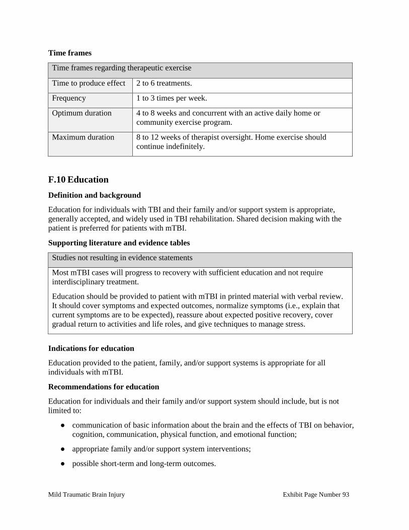
Mild Traumatic Brain Injury Exhibit Page Number 93
Time frames
Time frames regarding therapeutic exercise
Time to produce effect 2 to 6 treatments.
Frequency 1 to 3 times per week.
Optimum duration 4 to 8 weeks and concurrent with an active daily home or
community exercise program.
Maximum duration 8 to 12 weeks of therapist oversight. Home exercise should
continue indefinitely.
F.10 Education
Definition and background
Education for individuals with TBI and their family and/or support system is appropriate,
generally accepted, and widely used in TBI rehabilitation. Shared decision making with the
patient is preferred for patients with mTBI.
Supporting literature and evidence tables
Studies not resulting in evidence statements
Most mTBI cases will progress to recovery with sufficient education and not require
interdisciplinary treatment.
Education should be provided to patient with mTBI in printed material with verbal review.
It should cover symptoms and expected outcomes, normalize symptoms (i.e., explain that
current symptoms are to be expected), reassure about expected positive recovery, cover
gradual return to activities and life roles, and give techniques to manage stress.
Indications for education
Education provided to the patient, family, and/or support systems is appropriate for all
individuals with mTBI.
Recommendations for education
Education for individuals and their family and/or support system should include, but is not
limited to:
● communication of basic information about the brain and the effects of TBI on behavior,
cognition, communication, physical function, and emotional function;
● appropriate family and/or support system interventions;
● possible short-term and long-term outcomes.

Mild Traumatic Brain Injury Exhibit Page Number 94
Written information and material and referral to credible internet resources may be helpful as the
individual and family and/or support system may not be able to remember the vast amount of
information provided to them.
For similar reasons, they may need to be provided repeated or ongoing information. Further in-
depth education may be required to maximize the individual’s potential for functional living.
Treatment plans should include individual and group education as a means of facilitating self-
awareness, self-management, and prevention of secondary disability.
In order to maximize treatment outcomes and the durability of those outcomes, treatment
providers are highly encouraged to provide: hands-on, personal consultations; education (written,
verbal, internet); and support services to families.
Time frames
Time frames regarding mTBI education
Frequency Weekly 1-hour sessions initially during the first month as part of the
primary treatment and return-to-work evaluations. Once the patient
has returned to normal function without impairing symptoms, visit
frequency should decrease or treatment should be terminated.
Optimum duration 1 to 3 months.
Additional sessions may be required as justified.
F.11 Hyperbaric oxygen (HBO2)
Definition and background
HBO2 was initially thought to improve outcomes.
Supporting literature and evidence tables
Evidence statements regarding hyperbaric oxygen
Good evidence Evidence statement Design
HBO2 is unlikely to be beneficial in the setting of
mTBI.
Systematic review
of randomized and
nonrandomized
studies of HBO2
Complications
Complications can occur, including tension pneumothrorax.

Mild Traumatic Brain Injury Exhibit Page Number 95
Recommendations for HBO2
Hyperbaric oxygen is not recommended acutely or chronically. Ongoing studies could affect this
recommendation.
F.12 Interdisciplinary rehabilitation programs
Definition and background
Consider referral to an interdisciplinary program based on the results of a comprehensive
neuropsychological and/or psychiatric assessment, which should be conducted post-injury in
individuals with mTBI and delayed recovery and as soon as appropriate for more severe cases.
Informal programs with community-based services are usually most appropriate for patients with
mTBI. However, formal inpatient programs may be necessary as described below. The
sequencing of treatment is based on the individual’s ability to tolerate and benefit from the
specific therapies.
Supporting literature and evidence tables
Evidence statements regarding mTBI interdisciplinary rehabilitation programs
Good evidence Evidence statement Design
mTBI without post-traumatic amnesia does not
require routine rehabilitation.
Systematic review
of randomized
trials, quasi-
randomized trials,
and quasi-
experimental
studies
Indications/recommendations for mTBI interdisciplinary rehabilitation programs
Informal interdisciplinary programs in the workers’ compensation system should be considered
for patients who are currently employed, those who cannot attend all day programs, those with
language barriers, or those living in areas not offering formal programs. Before treatment has
been initiated, the patient, patient’s family and/or support system, physician, and insurer should
agree on the treatment approach, methods, and goals. Generally, the type of outpatient program
needed will depend on the degree of impact the injury has had on the patient’s medical, physical,
psychological, social, and/or vocational functioning.
However, formal inpatient rehabilitation programs may be necessary for patients with mTBI with
any of the following conditions: (a) high risk for medical instability; (b) moderate-to-severe
impairment of functional status; (c) moderate impairment of cognitive and/or emotional status;
(d) dependence on medications from which he or she needs to be withdrawn; and (e) the need for
24-hour supervision. For formal programs, refer to the Division’s Moderate/Severe TBI Medical
Treatment Guideline.

Mild Traumatic Brain Injury Exhibit Page Number 96
Patients with addiction and/or substance abuse problems or high dose opioid or other drugs of
potential abuse may require inpatient and/or outpatient chemical dependency treatment programs
before or in conjunction with other interdisciplinary rehabilitation. Guidelines from the
American Society of Addiction Medicine are available and may be consulted relating to the
intensity of services required for different classes of patients in order to achieve successful
treatment.

Mild Traumatic Brain Injury Exhibit Page Number 97
G. Return to work & vocational rehabilitation
G.1 Return to work
Definition and background
In addition to the treatment strategies described below, practitioners should be familiar with how
various state and federal statutes and regulations may impact return-to-work planning. These
may include, but are not limited to, Family and Medical Leave Act (FMLA), Americans with
Disabilities Act (ADA), Occupational Safety and Health Administration (OSHA), Federal Motor
Carrier Safety Administration (FMCSA), and the Department of Transportation (DOT).
Supporting literature and evidence tables
Evidence statements regarding return to work
Strong evidence Evidence statement Design
In the setting of TBI, there is a negative association
between psychiatric comorbidity (anxiety,
depression, PTSD) and return to work; however, the
magnitude of this effect has not been clearly
established.
Systematic review
of observational
studies
Studies not resulting in evidence statements
One study found a relationship between perceived self-efficacy in cognitive areas and life
satisfaction. The same study found a relationship with employed or volunteer work and
satisfaction.
Indications/recommendations for return to work
During the first five days post-injury, symptoms can be severe and significantly disrupt normal
daily function. Initial considerations should include lightening task load and allowing extra time
to complete normal tasks. Thus, shortening the work day or adding breaks, along with decreasing
responsibility for the first several weeks, are generally suggested. Driving, heavy lifting, working
with dangerous machinery, using ladders, and heights may be restricted because of possible
safety risk.
Following mTBI, many individuals are able to resume normal work duties with secondary
prevention precautions and education. They typically require little or no additional therapeutic

Mild Traumatic Brain Injury Exhibit Page Number 98
intervention. A small percentage of individuals with mTBI - such as those with age greater than
40, prior TBI, loss of consciousness close to 30 minutes, or mental status changes lasting up to
24 hours - may require more assistance in return to work and accommodations. For individuals
with mTBI, it may be necessary to temporarily reduce the time, amount, type, and/or intensity of
their work duties or temporarily remain out of work entirely for a period of time, at the discretion
of the clinician, and gradually increase hours and complex cognitive and physical duties based
on symptomology.
For example, if workers with mTBI have any loss of consciousness or prolonged disorientation,
providers should consider restricting higher risk job duties (such as working at heights, working
with power tools, and operating heavy machinery) until they have been free from the symptoms,
including dizziness and imbalance, for two consecutive weeks. Second impact syndrome (refer to
Section C.2, Prevention) has been seen in younger age groups who suffer severe life threatening
effects after a second TBI within a short time after the first TBI. Physicians should take this into
account when writing work restrictions.
Return to full duty depends on the rate of decrease of symptoms. The individual should be
competent in most basic ADLs before return to work is considered. Generally, if symptoms recur
during increasing job duties or exertion, duties should be decreased slightly. Very gradual return
to activity over weeks may be necessary for cases with symptoms lasting longer than 15 minutes
at the time of the injury, unconsciousness lasting minutes, or prolonged amnesia.
Physicians should be aware that symptoms in workers with mTBI may include cognitive deficits
in memory, attention, and executive function, even if the worker has no complaints/symptoms.
Memory, attention, and executive function should be tested by asking specific questions
regarding recent events and having the individual perform specified tasks. Physicians should
educate the individual with TBI and his/her supervisor to be aware of possible memory and
attention deficits and to accommodate accordingly. Time to return to baseline function will differ
according to the individual’s pre-accident condition, age, and medication, as well as other pre-
injury, injury, and post-injury factors.
Physicians should attempt to clearly and specifically document vocational restrictions and have a
graded or gradual plan for re-entry to work and to communicate with the employer (e.g.,
supervisor, safety officer, employee health nurse). Having a significant physical disability,
psychosocial impairment, cognitive impairment, or a history of alcohol and other substance
abuse are factors that impede return to work. Other factors impeding return to work include
difficulties regarding transportation, coordination, and vision. An interdisciplinary team
approach may be recommended, which may include a neuropsychological assessment, vocational
evaluation, job site analysis, early contact with employer, assessment of vocational feasibility,
supervisor education, transferable skills analysis, skillful increased titration of job duties and

Mild Traumatic Brain Injury Exhibit Page Number 99
demands, job coaching, physical therapy, occupational therapy, speech-language therapy, and
psychological services.
In work situations where the employer is unable to accommodate and a return to the previous
occupation is not suitable or appropriate, other options include volunteer placements or
supported employment opportunities. See the above return-to-work literature table for a study on
volunteer work and life satisfaction. Consider vocational re-training, individualized/specialized
job placement services, and job coaching as needed.
Based on the evidence listed in the evidence table above, all patients with TBI should have
psychological/neuropsychological evaluations and treatment as needed.
For individuals with mTBI who have persistent deficits or who have difficulty once back at
work, a return-to-work program should occur, which requires a carefully designed and managed
plan involving the person with TBI, his/her employer, and the treatment team. Physicians should
consider evaluation and treatment for comorbid conditions such as chronic pain, stress level, pre-
existing personality disorders, depression, anxiety, and/or substance abuse. Communication
among all involved parties and the avoidance of fragmentation among treatment professionals is
critical to successful outcome. Case management may be indicated to facilitate communication.
Following return to work, maintenance support services are appropriate to best ensure the
durability of the outcome.
G.1.a Recommended vocational rehabilitation assessment
According to Inter-Professional Clinical Practice Guidelines, a vocational evaluation should
include some of the following:
● initial intake process, including:
○ pre-injury history;
○ educational and work histories;
○ current social status;
○ pre-injury job performance and performance evaluations; and
○ successes and failures in post-injury work trials;
● assessment of the person (individual’s perspective), including:
○ work goals, values, and meaning he/she attached to work pre- and post-injury;
○ work performance, strengths, weaknesses, current work competency;
○ compensatory strategies and support needs;
Mild Traumatic Brain Injury Exhibit Page Number 100
○ readiness to work and anticipated challenges/barriers to work or return to work;
○ individual’s own assessment of the costs and benefits of working;
○ individual’s view of the implications of a decision not to work (e.g., insurance and
benefits);
● assessment of an individual’s functional status and level of independence;
● observations of an individual’s work-related skills and behavior during performance in
real work setting, or if unavailable, simulated work tasks;
● assessment of the physical workplace environment;
● assessment of supports (i.e., formal and informal) and opportunities within the workplace
and the individual’s support network including availability of accommodations and/or job
modifications;
● assessment of the occupational/job requirements.
G.2 Driving
If evaluation and treatment for driving is needed, refer to the Division’s Moderate/Severe TBI
Medical Treatment Guideline.
G.3 Vocational rehabilitation
Definition and background
Vocational rehabilitation is a generally accepted intervention, but the Colorado Workers’
Compensation statute limits its use.
Supporting literature and evidence tables
Studies not resulting in evidence statements
In one study, a brain injury vocational rehabilitation program was successful at returning
41% of clients to competitive employment. The majority of the cases were 2 years or more
from date of injury and had injuries classified as severe (post-traumatic amnesia duration of
1 or more days). These cases were also without significant behavioral problems and able to
function independently for ADLs. The program included cognitive training for those who
had not previously received it and job trials with job coach support.

Mild Traumatic Brain Injury Exhibit Page Number 101
Indications/recommendations for vocational rehabilitation
Initiation of vocational rehabilitation requires adequate evaluation of individuals with TBI for
quantification of highest functional level, motivation, and achievement of MMI. Vocational
rehabilitation should involve a comprehensive job analysis and a carefully planned return-to-
work strategy with input from the treating physician and interdisciplinary team. In some
instances, retraining may need to occur to access new job markets. (Refer to Section G, Return to
work.)
G.4 Work conditioning
Definition and background
These well-accepted programs are work-related, outcome-focused, and individualized treatment
programs. Objectives of the program include, but are not limited to, improvement of
cardiopulmonary and neuromusculoskeletal functions (strength, endurance, movement,
flexibility, stability, and motor control functions), patient education, and symptom relief. The
goal is for patients to gain full or optimal function and return to work.
Supporting literature and evidence tables
The following recommendations were based on consensus.
Indications for work conditioning
These programs are usually initiated once re-conditioning has been completed, but they may be
offered at any time throughout the recovery phase. It should be initiated when imminent return of
a patient to modified or full duty is not an option but the prognosis for returning the patient to
work at completion of the program is at least fair to good.
Recommendations for work conditioning
The service may include the time-limited use of modalities, both active and passive, in
conjunction with therapeutic exercise, functional activities, general conditioning body
mechanics, and re-training of lifting techniques. The patient should be assisted in learning to
pace activities to avoid exacerbations.
Time frames
Time frames regarding work conditioning
Length of visit 1 to 4 hours per day.
Frequency 2 to 5 visits per week.

Mild Traumatic Brain Injury Exhibit Page Number 102
Time frames regarding work conditioning
Optimum duration 2 to 4 weeks.
Maximum duration 6 weeks. Participation in a program beyond 6 weeks should be
documented with respect to need and the ability to facilitate positive
symptomatic and functional gains.
G.5 Work simulation
Definition and background
Work simulation is a generally accepted program where an individual completes specific work-
related tasks for a particular job and return to work.
Supporting literature and evidence tables
The following recommendations were based on consensus.
Indications/recommendations for work simulation
Use of this program is appropriate when modified duty can only be partially accommodated in
the work place, when modified duty in the work place is unavailable, or when the patient
requires more structured supervision. The need for work-place simulation should be based on the
results of a functional capacity evaluation and/or job site analysis.
Time frames
Time frames regarding work simulation
Length of visit 2 to 6 hours per day.
Frequency 2 to 5 visits per week.
Optimum duration 2 to 4 weeks.
Maximum duration 6 weeks. Participation in a program beyond 6 weeks should be
documented with respect to need and the ability to facilitate positive
symptomatic and functional gains.

Mild Traumatic Brain Injury Exhibit Page Number 103
H. Maintenance management
Most individuals following mTBI make a good neurological and functional recovery with
minimal or no intervention, although the possibility of subtle residual impairments or functional
limitations exists. Some individuals with mTBI experience impairments, functional limitations,
and disabilities. Individuals with mTBI who have comorbid conditions and/or have experienced
a longer period of confusion or loss of consciousness are more likely to have a poorer outcome
and require longer treatment or maintenance care. For patients with mTBI with specific
additional issues, further care may require treatment or follow-up with specialty services such as
optometry, neuro-ophthalmology, neuro-otology, rehabilitation psychology, neuropsychology,
and physical medicine and rehabilitation including brain injury medicine. Medical and
rehabilitation providers are encouraged to educate individuals and their family and/or support
systems regarding anticipated ongoing medical and rehabilitation needs.
Patients and families and/or support systems should understand that failure to comply with the
elements of the self-management program or therapeutic plan of care may affect consideration of
other interventions.
Periodic reassessment of the individual’s condition will occur as appropriate. The overall
maintenance plan should be reassessed at least annually by the authorized treating provider.
Programs should be individualized to specific needs and may include the following.
H.1 Exercise programs requiring gym memberships or special facilities
Definition and background
Some individuals with mTBI may have higher compliance with an independent exercise program
at a health club or a community activity-based wellness program versus participation in a home
program, although individuals with mTBI may require supervision or guidance.
Supporting literature and evidence tables
The following recommendations were based on consensus.
Indications/recommendations for exercise programs requiring gym membership or special
facilities
All exercise programs completed through a health club facility should be approved by the
treating therapist and/or physician and focus on the same parameters of an age-adjusted and
diagnosis-specific program for aerobic conditioning, flexibility, balance, stabilization, and
strength. Prior to purchasing a membership, a therapist and/or exercise specialist who has treated
the individual should visit the facility with the individual to ensure proper use of the equipment.
Periodic program evaluation and upgrading may be necessary by the therapist. The use of a
personal trainer may be necessary.
Mild Traumatic Brain Injury Exhibit Page Number 104
Time frames
Time frames regarding exercise programs requiring special facilities: maintenance
Frequency Approximately 2 times per week. Regular attendance is necessary
for continuation, with an exception for a medical or sufficient
intervening cause.
Maximum maintenance
duration
Continuation beyond 3 months after MMI should be based on
functional benefit and compliance. At MMI, health club
membership should not extend beyond 3 months if attendance
drops below 2 times per week on a regular basis without a medical
cause.
H.2 Home exercise programs and exercise equipment
Definition and background
Most patients have the ability to participate in a home exercise program after completion of a
supervised exercise rehabilitation program.
Supporting literature and evidence tables
The following recommendations were based on consensus.
Indications/recommendations for home exercise maintenance programs
Programs should incorporate an exercise prescription including the continuation of an age-
adjusted and diagnosis-specific program for aerobic conditioning, flexibility, stabilization,
balance, and strength. Home exercise programs are most effective when done three to five times
a week. For chronic pain, refer to the Division’s Chronic Pain Disorder Medical Treatment
Guideline.
Time frames
Follow-up evaluations in the home should occur to ensure compliance and to upgrade the home
program.
H.3 Medication management
Definition and background
Medications may be necessary for long-term management of some individuals with mTBI.
Supporting literature and evidence tables
The following recommendations were based on consensus.
Mild Traumatic Brain Injury Exhibit Page Number 105
Indications/recommendations for medication maintenance management
Medications may be used for medical, physical, perceptual, cognitive, neuroendocrine, and
psychological reasons, and they should be prescribed by physicians experienced in mTBI
medication management. Reasons for possible medications and the types and names of
medication are numerous, are individualized for each person, and are beyond the scope of these
guidelines.
In situations where there are multiple providers for multiple clinical issues, coordination of the
total medication regimen is essential. It is strongly recommended that changes in medication be
discussed with the physician who is primarily managing the case.
As with all prescriptive regimens, physicians periodically reassess the efficacy and side effects of
each medication. This is particularly true for individuals who are on long-term medication use.
Physicians must follow patients who are on any chronic medication or prescription regimen for
compliance, efficacy, and side effects. Individuals with mTBI are particularly susceptible to
certain medication side effects, including compromised cognitive function, decreased seizure
threshold, and other neurological effects. Follow-up visits should document the individual’s
ability to perform routine functions. Laboratory or other testing is usually required on a regular
basis to monitor medication effects on organ function. For some, medications and drug levels
should be closely monitored.
Individuals with mTBI may forget to take medications and/or have difficulty with complicated
medication regimens. They may need assistance with medication management, such as
reminders, medication boxes, assistance with filling medication boxes, or medication
administration supervision. Some medications may need to be prescribed in small amounts or
locked due to safety in patients who are impulsive, forgetful, inconsistent, or otherwise unsafe in
independent medication management.
Time frames
Time frames regarding medication management: maintenance
Frequency Medication and medical management reviews may need to be
monthly or more frequently if necessary for changes in
medication. Frequency depends on the medications prescribed,
with laboratory and other monitoring performed as appropriate.
Maintenance duration As new medications become available and side effects of other
medications are established, there may need to be changes in
medical management.

Mild Traumatic Brain Injury Exhibit Page Number 106
H.4 Patient education management
Definition and background
Functional abilities and support systems for individuals with mTBI may change over time and
frequently require additional support.
Supporting literature and evidence tables
The following recommendations were based on consensus.
Indications/recommendations for patient education maintenance management
Educational classes or sessions may be necessary to reinforce self-management techniques to
help the individual adjust to life changes. This may be performed as formal or informal
programs, either group or individual.
Time frames
Time frames regarding patient education management: maintenance
Frequency 2 to 6 educational sessions during one 12-month period. Changes
in life circumstances or the individual’s condition may require
greater frequency of educational sessions.
H.5 Cognitive/behavioral/psychological management
Definition and background
In some cases, additional treatment may be necessary, especially when support systems or
employment has changed.
Supporting literature and evidence tables
The following recommendations were based on consensus.
Indications/recommendations for cognitive/behavioral/psychological maintenance
management
For select patients with mTBI, longer supervised psychological/psychiatric treatment may be
required, especially if there are ongoing medical procedures or complications. The treatment
plan/progress must show severity.
Time frames
If counseling beyond six months is indicated, the management of psychosocial risks, functional
progress, or functional stability to prevent deterioration must be documented. Treatment may be
sporadic and may vary depending on the life change requirements of the patients with mTBI.

Mild Traumatic Brain Injury Exhibit Page Number 107
H.6 Neuromedical management
Definition and background
Some patients with mTBI will have ongoing medical issues requiring treatment on a regular
basis.
Supporting literature and evidence tables
The following recommendations were based on consensus.
Indications/recommendations for neuromedical maintenance management
Examples of related medical diagnoses include, but are not limited to: neuro-endocrine
dysfunction, urinary incontinence, heterotrophic ossification, seizures, and other conditions
described in the treatment sections of this guideline.
Time frames
Time frames regarding neuromedical management: maintenance
Frequency Medical management visit frequency will depend on the severity
of the medical condition but may occur monthly or more
frequently.
Maximum maintenance
duration
Visits should occur at least at 6-month intervals for extremely
stable conditions.
H.7 Physical, occupational, and speech-language therapy
Definition and background
Exacerbation of symptoms or decline in functional status may require short-term intensive
treatment to return the individual to maximized function.
Supporting literature and evidence tables
The following recommendations were based on consensus.
Indications/recommendations for PT/OT/ST
Therapy may be indicated on a continued basis if the therapy maintains objective physical
function, decreases pain, or decreases medication use. In those situations, frequency and duration
parameters as defined in this guideline apply. Changes in the individual’s living situation, role,
and responsibilities at home or work, support systems, and/or life’s stressors may require
monitoring to determine the impact on daily function. Short-term speech-language therapy
emphasizing patient education, compensatory strategies, and functional goals measured
objectively may be indicated. Participation in a PT, OT, or ST maintenance program must be

Mild Traumatic Brain Injury Exhibit Page Number 108
documented with respect to need and the ability of the program to facilitate positive symptomatic
gains or functional gains.
Time frames
For time frames to address changes, refer to Section F, Treatment.
H.8 Durable medical equipment: purchase, rental, and maintenance
Definition and background
It is recognized that some patients may require ongoing use of equipment for the purpose of
maintaining MMI in the areas of strength, ROM, balance, tone control, functional mobility,
ADLs, and/or analgesic effect.
Supporting literature and evidence tables
The following recommendations were based on consensus.
Indications for purchase or rental of durable medical equipment
Purchase or rental of this equipment should be done only if the assessment by the physician
and/or therapist has determined the safety, effectiveness, compliance, and improved or
maintained function by its application.
Recommendations for purchase, rental, and maintenance of durable medical equipment
It is generally felt that large expense purchases (such as spas, whirlpools, and special mattresses)
are not necessary to maintain function for patients with mTBI, but gym memberships is
encouraged to keep the patient as active as possible to prevent deterioration.
Periodic maintenance and replacement of the equipment may be indicated and should be
considered in the maintenance plan.
Time frames
Time frames regarding durable medical equipment: maintenance
Maximum maintenance
duration
Not to exceed 6 months for rental of large equipment. Purchase
and maintenance should occur if effective.