roto evo gb.qxd:strato2it evo brochure en.pdf · the dental studio software package (option) brings...
TRANSCRIPT

Dental Panoramic - Digital & Film
DENTALLINE

A history of Evolution
Since the very first model introducedin 1974, the Rotograph name hasalways been synonymous withpanoramic radiography.
Rotograph Evo takes the experience of five decades of dedication to X-ray diagnostic imaging to the next level. You can rely on over 25.000 manufactured panoramic units that have been suc-cessfully operating worldwide for years, from the smallest dental practice to the largest universityhospital.
And the Villa experience extends far beyond dental imaging. From general purpose radiologicalrooms to direct digital real time angiographic X-ray systems, Villa products help saving patientlives in thousands of hospitals worldwide.
Evolution comes from experience
1974 Rotograph I 1983 Rotograph 230 1997 Rotograph Plus
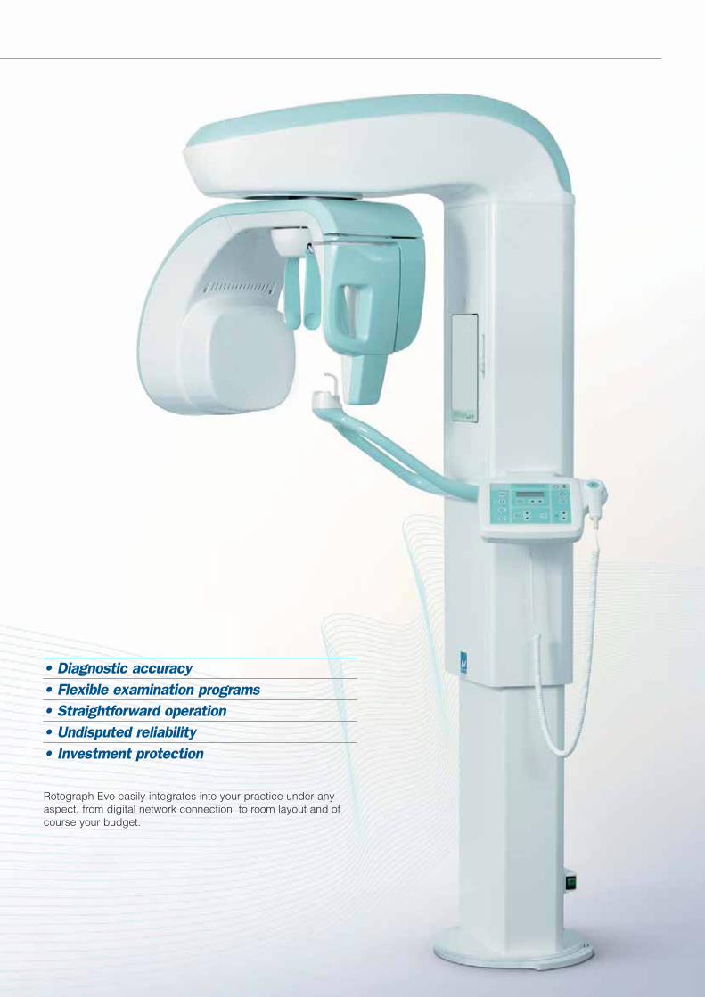
• Diagnostic accuracy• Flexible examination programs• Straightforward operation• Undisputed reliability• Investment protection
Rotograph Evo easily integrates into your practice under anyaspect, from digital network connection, to room layout and ofcourse your budget.

Diagnostic Accuracy
Your patient relies on you for dental treatment and you can rely on Rotograph Evo for an accurate diagnosis.
Not only high frequency: we give you high efficiency, too
The 200kHz High Frequency generator provides accurateand efficient X-ray emission and produces excellentimages with lower tube current than previous generationproducts. Detail-rich images can be obtained with minimum patient dose and reduced energy consumption.
Spine compensation
Shadows produced by the spinal column are reduced byan effective modulation of the kV value during the rotationof the overhead assembly. The result is a more uniformimage in the incisors area.
Constant Magnification
Panoramic images are acquired with constant magnifica-tion, translating into an accurate geometrical representa-tion of anatomic structures.
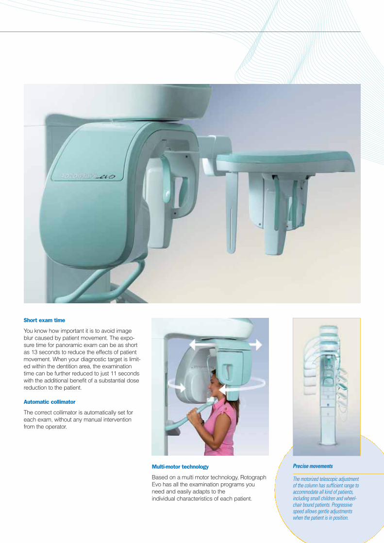
Short exam time
You know how important it is to avoid imageblur caused by patient movement. The expo-sure time for panoramic exam can be as shortas 13 seconds to reduce the effects of patientmovement. When your diagnostic target is limit-ed within the dentition area, the examinationtime can be further reduced to just 11 secondswith the additional benefit of a substantial dosereduction to the patient.
Automatic collimator
The correct collimator is automatically set foreach exam, without any manual interventionfrom the operator.
Multi-motor technology
Based on a multi motor technology, RotographEvo has all the examination programs youneed and easily adapts to the individual characteristics of each patient.
Precise movements
The motorized telescopic adjustmentof the column has sufficient range toaccommodate all kind of patients,including small children and wheel-chair bound patients. Progressivespeed allows gentle adjustmentswhen the patient is in position.

The key to a good panoramic image
Anatomic programs
Exposure factors and movement geometry can easily be adapted to the individual charac-teristics of each patient by selecting betweenadult and child programs and three patientsizes. The result is an optimal compromisebetween X-ray dose and image quality.
Self-centering
As the patient can see him/herself reflected in the mirror, he/she automatically tends to self-centre the mid-sagittal plane, resulting in fasterpositioning.
Accurate alignment
A good patient alignment to the reference axis isthe most important factor for a good panoramicimage. Two laser beams are used for propercentering of mid-sagittal and Frankfurt planes,without the need for a third positioning light.
Focal layer adjustment
Patients with different jawstructure are not a problem.The focal layer adjustmentallows to compensate dif-ferent arc shapes in theanterior region. The focallayer is electronicallyadjusted, without reposi-tioning the patient.

Chin rests
Three different chin rests are sup-plied with the unit to accommodateall patients and applications.• Standard chin rest with bite:
provides stable chin support and accurate location of the incisors in the focal layer
• Edentulous chin rest: provides a reference position for the patientchin when use of bite is not possible.
• Reduced chin rest: for Sinus and TMJ exams
Stable positioning
The patient support structure makes use ofmultiple contact points to ensure correct patientalignment and stability during the exam.• Three-point headrest provides centering of
mid-sagittal and Frankfurt planes• Chin rest and bite stick provide stability
and proper localization of the focal layer• Angulated hand grips provide for a natural
extension of the cervical vertebræ to reduce image shadows in the incisor area.
Child friendly
Dedicated paediatric exams ensure consistentdose reduction and reduced exam time. The analog version is also equipped with a spe-cific collimator, limiting the X-ray beam to furtherreduce dose to patient. Color combination androunded shapes of the unit help in creating acomfortable and stress free environment for thepatient, thereby contributing to successfulexams.

The Power of Digital
Environment and budget friendly
Say goodbye to films and polluting chemicalsand contribute to the preservation of both theenvironment and your budget.Images can be displayed and shown to thepatient in seconds and become an importanttool to show treatment planning and progress. If needed, printouts can easily be producedusing inexpensive inkjet printers.
Less dose to patient
The high sensitivity of the Cs-I digital detectormakes for a reduced X-ray dose and in case of wrong exposure settings, the digital imageallows to extract useful information withoutretakes. Rotograph Evo D is already compliantto existing regulations on patient dose monitor-ing. The dose readout is calculated for eachexposure and stored with the image without the need for add-on DAP measuring devices.
Even more tools
The Dental Studio software package (option)brings additional possibilities to image treat-ment. The 16-bit image processing engineallows a more precise control of image con-trast. Patient archive and image database areextremely powerful and can be integrated withpractice management software.
DICOM functionalities (option) can also beadded for integration into hospital networks.
All the Tools you need
Rotograph Evo D comes bundled with the QuickVision software package that offers all the image processing tools required for yourprofessional activity.

Simple is beautiful
The integrated keyboard features large, easy to find pushbuttons and a clear display.Controls are grouped in logical areas consis-tent with the typical operating workflow:patient selection, exam protocol, exposureadjustments.A virtual version of the keyboard can also bedisplayed on the PC screen and allows theoperator to preset exposure parameters fromwithin the image acquisition program.
Instant network integration
Rotograph Evo is easily deployed into your network environment. The integrated Ethernetconnection is compatible with existing com-puter networks and requires no dedicatedboards to be installed in the computer, making it possible to acquire images from any PC, including notebooks.With integrated networking capability, imagescan be acquired and transferred from anyworkstation in your practice.
Dual storage
Rotograph Evo offers the uniquecapability of saving images directly to a USB memory stick, without inter-rupting your workflow. Images canthen be transferred to the PC at anytime, even if the PC or network wasmomentarily unavailable at the time of the exam.

Cephalometric Imaging
Precise geometry
The digital ceph imaging principle ofRotograph Evo combines the scanning move-ment of the detector with the stationary positionof the X-ray source. This method provides thesame projection geometry as with a regularfilm, allowing precise orthodontic analysis.
Single or Dual Detector: it's your choice
What are your requirement for ceph exams?If you’re doing just a few studies per week, or are bound to a limited budget, the singledetector unit is your choice. The same digitaldetector can be moved from the Pan to theCeph position with a quick and simple opera-tion. If orthodontics is your field of expertise, or you just want to avoid handling the detector,than a dual detector unit is what you need.
Versatile upgrade paths
Predisposed digital pan units can be upgraded to ceph with severalupgrade options to help you plan your budget according to your needs, protecting your investment over time.

Automatic soft tissue filter
For lateral projections a copper filter is automatically inserted intothe x-ray beam to enhance the visibility of the patient's profile and a calibrated ruler is superimposed on the image for proper geometriccalibration.
Optimized protocols
A total of eight digital cephalometric programsgives you the flexibility you need for all patients,with the best combination of image area,acquisition speed and resolution.The cephalostat provides simple and gentlepatient alignment for all projections, includinglateral, frontal and postero-anterior.
Carpus Exam
Bone age can be assessed with the dedicated Carpus Exam. A dedicated hand support plate makes positioning fast and easy.
Fast and detailed
Depending on the exam type and patient size,several image areas can be chosen, from18x22 to 22x30cm.Two scanning modes can be selected:• high resolution mode delivers highly detailed
images• high speed mode acquires a standard lateral
ceph in just 4.5 seconds, and is especially suitable for children
No unnecessary exposure
During the digital ceph scan, each portion of the skull is exposed for just fractions of asecond by a perfectly collimated x-ray beam,limiting the overall patient exposure to a mini-mum. This is particularly beneficial for childrenand teenagers, who are often subject to cephexams.

Examination Programs
Standard Exams
Standard Panoramic with Constant Magnification
Sinus. Extended visibility of the paranasal sinuses
Child Panoramic. Reduced examination time
TMJ - Temporo-Mandibular Joint with open and closed mouth. The true Lateral view shows the exact location of the condyle

Exams with Ceph Arm Additional exams with Evo XP eXtended Package
Lateral Ceph with Soft Tissue Filter
Postero-Anterior Ceph
Carpus Exam.A removable hand support is supplied with the ceph arm
The optional Evo XP exams packageincreases the scope of application of your unit with the addition of severalexams to target specific diagnosticneeds.
Half Panoramic, left and right. Provides reduced exposure when the diagnostic target is in one orthe other half of the dentition
Reduced dose. A panoramic limited to the dentition only.Used when the diagnostic target does not require the visibility of the Condyles
Orthogonal projection. Reduced overlapping of adjacent teeth for improved detection of interproximal caries
Frontal dentition. A further segmentationof the panoramic, limitedto the frontal dentitionarea including incisorsand canines only
Analog version
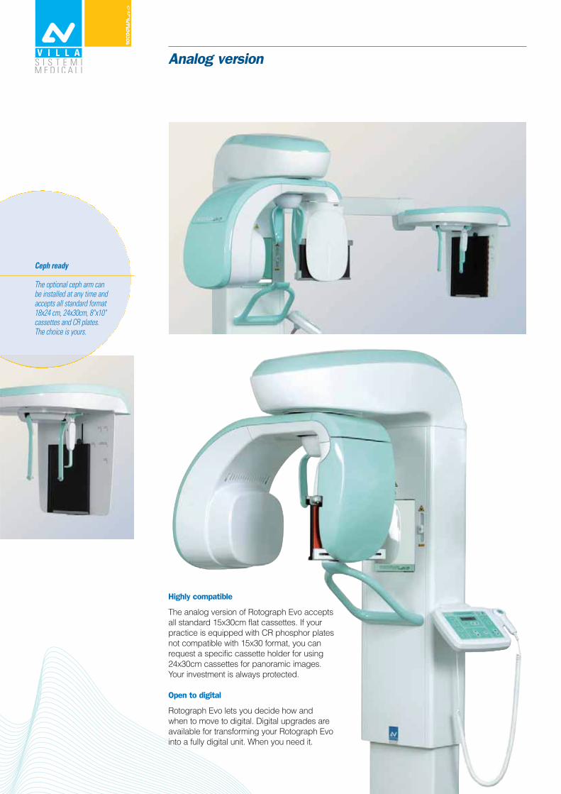
Highly compatible
The analog version of Rotograph Evo acceptsall standard 15x30cm flat cassettes. If yourpractice is equipped with CR phosphor platesnot compatible with 15x30 format, you canrequest a specific cassette holder for using24x30cm cassettes for panoramic images.Your investment is always protected.
Open to digital
Rotograph Evo lets you decide how and when to move to digital. Digital upgrades areavailable for transforming your Rotograph Evointo a fully digital unit. When you need it.
Ceph ready
The optional ceph arm can be installed at any time and accepts all standard format 18x24 cm, 24x30cm, 8"x10" cassettes and CR plates. The choice is yours.

Examination Programs
Basic unit Adult PanoramicChild PanoramicOpen-Closed mouth lateral TMJP-A Sinus (rotational)
Evo XP package Half Panoramic adultHalf Panoramic childOrthogonal ProjectionLow Dose PanoramicFrontal Dentition
Ceph version Lateral Ceph A-P and P-A CephCarpus (hand)
Technical data
Villa Sistemi Medicali reserves the right to change the product specification without notice. X-ray images in this booklet are shown only as a representation of possible applica-tions are and may have been taken with other devices than those indicated in this brochure. Some of the features and accessories described in this brochure may be optional.
Floor stand
The optional self-supporting floorstand can be used when wall mountis not possible and allows to performexams on wheelchairs.
Rotograph Evo D - Digital Rotograph Evo - AnalogExposure time PAN 7.3 – 13.8 sec PAN 7.3 – 13.8 sec
CEPH 4.5 – 15 sec CEPH 0.7 – 2 secDose x area evaluation (DAP) Standard OptionImage area PAN 14.6 x 30cm PAN 15x30 or 24x30cm
CEPH 18x22, 24x22, 29x22 cm CEPH 18x24 cm, 24x30cm, 8"x10"Preferred cassette size to be specified at order
Image transfer Ethernet – USB memory stick N/ASensor technology CCD with high resolution Cs-I N/A
(Cesium Iodide) scintillatorCCD Pixel size 48 μm N/AEffective image resolution 5.2 lp/mm N/AImage size 1536x2800 (standard PAN) - 3000 x 2305 (CEPH, max) N/AAcquisition depth 12 bit (4096 levels) N/ADICOM connectivity (option) Print, Store, Worklist N/AGenerator High frequency, 200kHz constant potentialHigh Voltage 60 – 86kVCurrent 6 – 12 mAFocal spot 0.5 IEC 336Spine compensation Automatic kV modulationWeight PAN 157kg (346 lb.)
CEPH 177kg (390 lb.)Power supply voltage 110-120 / 220-240 Vac, 50/60HzAbsorbed current 6.6 A @ 220-240 V – 15A @ 110-120 V
Prod
ucts
are
con
tinuo
usly
unde
r rev
iew
in th
e lig
ht o
f tec
hnica
l adv
ance
men
t. Th
e ac
tual
spe
cifica
tions
may
ther
efor
e be
sub
ject
to im
prov
emen
t or m
odifi
catio
n w
ithou
t not
ice. A
ll rig
hts
rese
rved
- Pr
inte
d in
Ital
y - 1
0/08
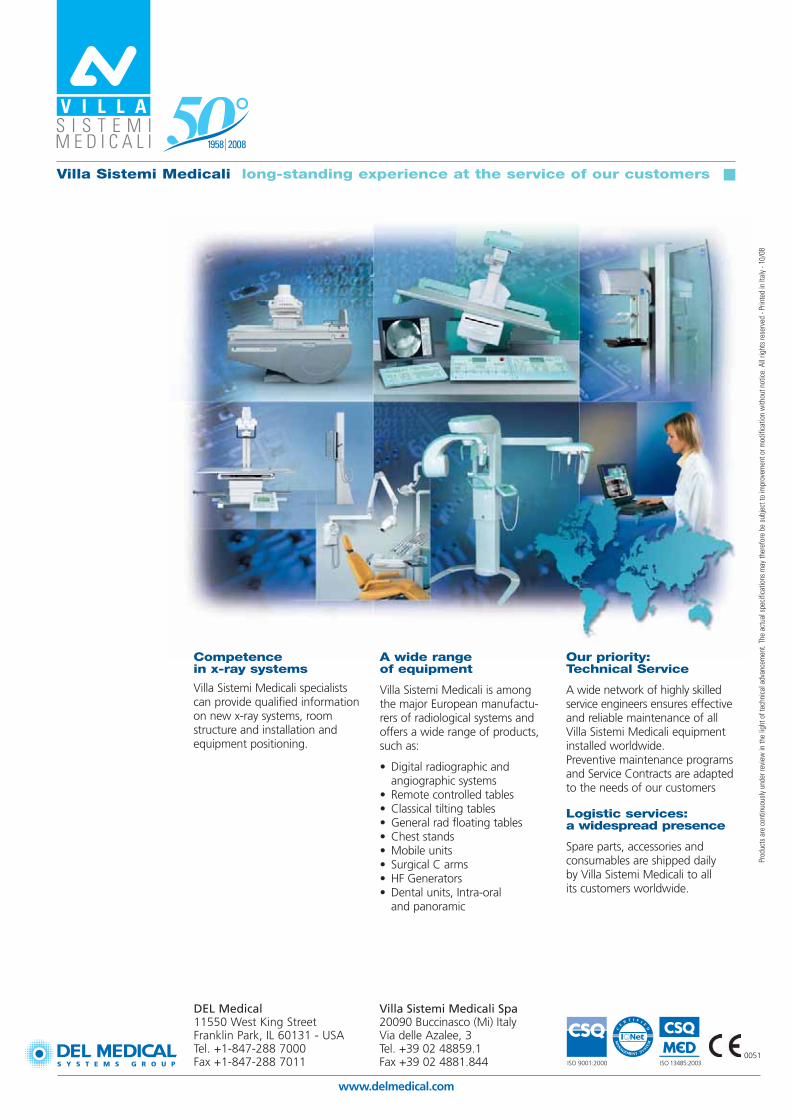
Competence in x-ray systems
Villa Sistemi Medicali specialistscan provide qualified informationon new x-ray systems, roomstructure and installation andequipment positioning.
A wide range of equipment
Villa Sistemi Medicali is amongthe major European manufactu-rers of radiological systems andoffers a wide range of products,such as:
• Digital radiographic and angiographic systems
• Remote controlled tables• Classical tilting tables• General rad floating tables• Chest stands• Mobile units• Surgical C arms• HF Generators• Dental units, Intra-oral
and panoramic
Our priority: Technical Service
A wide network of highly skilledservice engineers ensures effectiveand reliable maintenance of allVilla Sistemi Medicali equipmentinstalled worldwide. Preventive maintenance programsand Service Contracts are adaptedto the needs of our customers
Logistic services: a widespread presence
Spare parts, accessories and consumables are shipped daily by Villa Sistemi Medicali to all its customers worldwide.
Villa Sistemi Medicali long-standing experience at the service of our customers
Villa Sistemi Medicali Spa20090 Buccinasco (Mi) Italy Via delle Azalee, 3 Tel. +39 02 48859.1Fax +39 02 4881.844
www.delmedical.com
DEL Medical11550 West King Street Franklin Park, IL 60131 - USATel. +1-847-288 7000Fax +1-847-288 7011 ISO 9001:2000 ISO 13485:2003
0051