role of imprint/exfoliative cytology in ulcerated skin neoplasms
TRANSCRIPT
ORIGINAL ARTICLE
Role of Imprint/Exfoliative Cytology in UlceratedSkin Neoplasms
Vishnu Prasad Nelamangala Ramakrishnaiah &
Ravindra Babu & Dinker Pai & Surendra Kumar Verma
Received: 30 March 2013 /Accepted: 2 September 2013 /Published online: 22 September 2013# Indian Association of Surgical Oncology 2013
Abstract Imprint cytology is a method of studying cells bytaking an imprint from the cut surface of a wedge biopsyspecimen or from the resected margins of a surgical specimen.It is rapid, simple and fairly accurate. Exfoliative cytology isan offshoot from the imprint cytology where in cells obtainedfrom the surface of ulcers, either by scrape or brush, areanalyzed for the presence of malignant cells. We undertookthis study to see the role of imprint/exfoliative cytology in thediagnosis of ulcerated skin neoplasm and to check the ade-quacy of resected margins intra-operatively. This was a pro-spective investigative study conducted from September 2003to July 2005. All patients presenting to surgical clinic withulcerated skin and soft tissue tumours were included in thestudy. A wedge biopsy obtained from the ulcer and imprintsmears were taken from the cut surface. Exfoliative cytologywas analyzed from the surface smears. Wedge biopsy speci-men was sent for histopathological (HPE) examination. Thecytology and HPE were analyzed by a separate pathologist.Imprint cytology was also used to check the adequacy ofresected margins in case of wide excision. This was comparedwith final HPE. Total of 107 patients was included in thepresent study and 474 imprint smears were done, with anaverage of 4.43 slides per lesion. Out of 59 wide excisionsamples, 132 imprint smears were prepared for assessingresected margins accounting for an average of 2.24 slidesper each excised lesion. On combining imprint cytology withexfoliative cytology the overall sensitivity, specificity andpositive predictive value were 90.38 %, 100 % and 90.38 %
respectively. Only one out of 59 cases had a positive resectedmargin which was not picked by imprint cytology. Imprintcytology can be used for rapid and accurate diagnosis ofvarious skin malignancies. It can also be used to check theadequacy of the resected margin intraoperatively.
Keywords Imprint cytology . Squamous cell carcinoma .
Basal cell carcinoma
Introduction
Imprint cytology is a method of studying cells by takingimprint from the cut surface of a wedge biopsy specimen orfrom the resected margins of a surgical specimen. It is a rapid,simple and fairly accurate method of analyzing the margin of aresected specimen for the presence of malignant cells [1].Exfoliative cytology is an offshoot from the imprint cytologywhere in, the cells obtained from the surface of ulcers either byscrape or brush is analyzed for the presence of malignant cells.Some use swab stick to obtain the exfoliated epidermal cells,which are then smeared onto glass slides, fixed and stained.Both the methods of cytology have been used successfully fordiagnosing and classifying various tumours, by various au-thors [1–3].
Various authors have successfully used imprint cytology todiagnose skin malignancies [2, 3], upper gastrointestinal ma-lignancies [4]. Imprint cytology of sentinel lymph node, incarcinoma breast [5] and malignant melanoma [6], is useful inon-table decision making. Imprint cytology has reduced therole of frozen-section in the intraoperative assessment ofadequacy of resected margins [7]. One of the prerequisitesfor successful impression smears is that the cells of the lesionshould exfoliate easily. This is especially true for the epithelialtumours. Cells from mesenchymal tissues typically do notexfoliate well, and impression smears will usually be cell-
V. P. N. Ramakrishnaiah (*) :R. Babu :D. PaiDepartment of Surgery, Jawaharlal Institute of Postgraduate MedicalEducation and Research (JIPMER), Puducherry 605006, Indiae-mail: [email protected]
S. K. VermaDepartment of Pathology, Jawaharlal Institute of PostgraduateMedical Education and Research (JIPMER), Puducherry, India
Indian J Surg Oncol (December 2013) 4(4):385–389DOI 10.1007/s13193-013-0270-7

poor. As a rule, if a diagnostic slide cannot be produced byaspiration from a tissue, it is unlikely that an impression smearwill be of good quality. Ulcerated skin malignancies exfoliateeasily as they are epithelial tumours. Hence they can be easilyimprinted for a quick cytological examination. They requirerapid diagnosis and early treatment. There have been fewstudies that have evaluated the role of imprint cytology inthe management of these lesions. Hence this study was doneto see the role of imprint cytology in the rapid diagnosis ofulcerated skin neoplasms and to check the adequacy ofresected margins intra-operatively.
Methods
This is a prospective, hospital based study, conductedover a period of 22 months, from September 2003 toJuly 2005. All patients with ulcerated neoplasm of theskin and ulcerated soft tissue tumours, attending theout-patient clinic of department of surgery, were includ-ed in the study. Patients with ulcerated neoplasms of themucosal surfaces like carcinoma tongue and cheek wereexcluded from the study. A detailed history was takenand clinical examination was done. Imprint smears weretaken from the surface of the ulcer. A wedge biopsywas obtained from the ulcer edge. Imprint smears weremade from the cut surface of the wedge biopsy speci-men. The specimen was kept in 10 % formalin and sentfor histopathology. All the imprints were made using thevertical squash technique [8]. A total of 4–6 slides weretaken from each patient depending on the surface areathat need to be covered. Exfoliative cytology was ana-lyzed from the surface smears.
Imprint smears were stained with May-Grunwald Giemsastain for the cytoplasmic features and Papanicolaou stain forstudying the nuclear features, as per the standard procedure.The wedge biopsy specimen was sent for histopathologicalexamination to confirm the diagnosis. The cytology smearsand histopathology slides were analyzed by separate patholo-gists. The cytologist was blinded to the clinical as well as thehistopathological diagnosis. In all the cases the definitivemanagement was excisional surgery, either wide local exci-sion or amputation. Those cases that underwent wide localexcision were included for checking the adequacy of resectedmargins by imprint cytology.
The resected margins of the surgical specimen wereimprinted to obtain cytology smears. A total of 2–4 slideswere prepared from each case depending on the surface area tobe covered. The results of the imprint smear cytology werecompared with those of histopathology. The sensitivity, spec-ificity and the predictive values of imprint cytology and exfo-liative cytology have been calculated using the final histopa-thology as the gold standard.
Results
The study included a total of 107 patients with ulcerated skinor soft tissue tumours, who attended the out-patient clinic ofdepartment of surgery, between September 2003 and July2005. There were 86 males and 21 females with age rangingfrom 24 to 78 years, with a mean age of 51.1±12.1 years.Most of the patients were in 50–59 years age group (Fig. 1). Atotal of 474 imprint smears were made from 107 patients. Anaverage of 4.43 slides for each ulcerated lesion. For the 59cases of resected margin assessment group, 132 imprintsmears were prepared. An average of 2.24 slides was madefor each excised lesion.
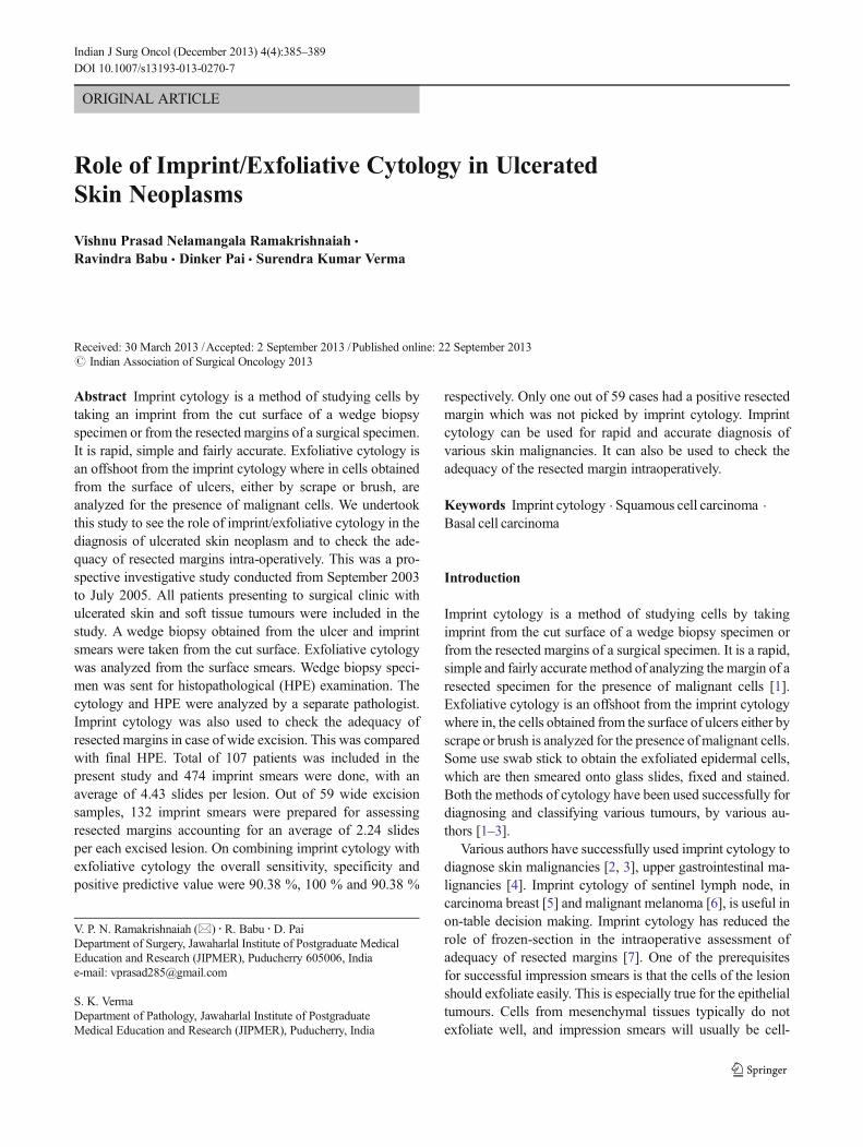
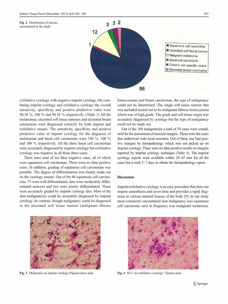
The distribution of various lesions is shown in Fig. 2.Squamous cell carcinoma (Fig. 3) was the most commonlyencountered malignant lesion, comprising 86 cases. Othersincluded 12 malignant melanomas (Fig. 4), three basal cellcarcinomas, two ulcerated breast carcinomas and one ulcerat-ed soft tissue tumour. There were three cases of chronic non-healing ulcers, which clinically looked malignant but bothimprint and histopathology diagnosed otherwise. These havebeen included in the results but excluded in the final analysis.Site wise distribution of squamous cell carcinomas include, 34penile carcinomas, 36 in the lower limbs, seven in upperlimbs, two in scalp, two in the anterior neck, five over thesacrum, gluteal and chest wall regions.
There were 104 malignancies and three chronic non-healing ulcers diagnosed by histopathology. Among the 104malignancies, imprint cytology was positive in 93 cases andexfoliative cytology in 76 cases. Both were positive in 94cases. There was only one patient in whom imprint cytologywas negative with a positive exfoliative cytology. On com-paring exfoliative cytology with histopathology we found thatexfoliative cytology had sensitivity of 70.93 %, specificity of100 % and a positive predictive value of 70.93 % (Table 1).On comparing imprint cytology with histopathology, wefound that imprint cytology had sensitivity of 87.21 %, spec-ificity of 100 % and a positive predictive value of 87.21 %(Table 2). Among the 86 squamous cell carcinomas 75 caseswere diagnosed by imprint smears and 61 by exfoliativecytology. Only one squamous cell carcinoma had a positive
Fig. 1 Age distribution of patients
386 Indian J Surg Oncol (December 2013) 4(4):385–389
exfoliative cytology with negative imprint cytology. On com-bining imprint cytology and exfoliative cytology the overallsensitivity, specificity and positive predictive value were90.38 %, 100 % and 90.38 % respectively. (Table 3) All themelanomas, ulcerated soft tissue tumours and ulcerated breastcarcinomas were diagnosed correctly by both imprint andexfoliative smears. The sensitivity, specificity and positivepredictive value of imprint cytology for the diagnosis ofmelanomas and basal cell carcinomas were 100 %, 100 %and 100 % respectively. All the three basal cell carcinomaswere accurately diagnosed by imprint cytology but exfoliativecytology was negative in all these three cases.
There were total of ten false negative cases, all of whichwere squamous cell carcinomas. There were no false positivecases. In addition, grading of squamous cell carcinomas waspossible. The degree of differentiation was clearly made outon the cytology smears. Out of the 86 squamous cell carcino-mas, 75 were well differentiated, nine were moderately differ-entiated tumours and two were poorly differentiated. Thesewere accurately graded by imprint cytology also. Most of theskin malignancies could be accurately diagnosed by imprintcytology. In contrast, though malignancy could be diagnosedin the ulcerated soft tissue tumour (malignant fibrous
histiocytoma) and breast carcinomas, the type of malignancycould not be determined. The single soft tissue tumour thatwas included turned out to be malignant fibrous histiocytomawhich was of high grade. The grade and soft tissue origin wasaccurately diagnosed by cytology but the type of malignancycould not be made out.
Out of the 104 malignancies a total of 59 cases were consid-ered for the assessment of resectedmargins. Thesewere the casesthat underwent wide local resection. Out of these one had posi-tive margins by histopathology which was not picked up onimprint cytology. There were no false positive results on marginsreported by imprint cytology technique (Table 4). The imprintcytology reports were available within 30–45 min for all thecases but it took 5–7 days to obtain the histopathology report.
Discussion
Imprint/exfoliative cytology is an easy procedure that does notrequire anaesthesia and saves time and provides a rapid diag-nosis in various tumoral lesions of the body [9]. In our studymost commonly encountered skin malignancy was squamouscell carcinoma; next in frequency was malignant melanoma.
Fig. 2 Distribution of lesionsencountered in the study
Fig. 3 Melanoma on imprint cytology-Papanicolaou stain Fig. 4 SCC on exfoliative cytology: Giemsa stain
Indian J Surg Oncol (December 2013) 4(4):385–389 387

Similar studies done by Aryya et al [3], Vega-Memije et al [2]had squamous cell carcinoma as the commonest skin malig-nancy encountered. Unlike our study, Brown et al [10], Bartonet al [11] studied basal cell carcinomas exclusively. Amongsquamous cell carcinomas majority were located in lowerextremity and penis. Aryya et al [3] and Vega-Memije et al[2] predominantly encountered extremity lesions and fewpenile carcinomas.
We found that imprint cytology was very useful in rapidaccurate diagnosis of skin malignancies. The sensitivity andspecificity of imprint cytology were 89.42 % and 100 %respectively. All the malignant melanomas and basal cellcarcinomas and 87.21 % of squamous cell carcinomas weresuccessfully identified. The combined (imprint cytology andexfoliative cytology) sensitivity, specificity and positive pre-dictive values were 90.38 %, 100 % and 90.38 % respectively.Similar results have been reported by several authors. Brownet al [10] successfully identified all the squamous cell carci-nomas and malignant melanomas and 95 % of basal cellcarcinomas using imprint cytology when compared with his-topathology. Aryya et al [3] found 100 % correlation between
imprint cytology and histopathology in the diagnosis of skinneoplasms. Vega-Memije et al [2] found that imprint cytologyhad a 100 % sensitivity and specificity for the diagnosis ofsquamous cell and basal cell carcinomas. Barton et al [11]found the sensitivity and predictive accuracy of imprint cytol-ogy in the diagnosis of basal cell carcinomas to be 92 % and75 % respectively. They included benign lesions along withbasal cell carcinomas in their study. In this study malignantlesions alone have been included in the statistical analysis.
When we compared the results of imprint cytology with theexfoliative cytology, Imprint smears were more useful and hada higher sensitivity and accuracy. There was a single squamouscell carcinoma that had a positive exfoliative cytology and anegative imprint smear report. Exfoliative cytology was notuseful in the diagnosis of basal cell carcinomas. None of thethree basal cell carcinomas could be identified with exfoliativecytology. Imprint cytology was more useful in that situation.This is probably because of the dry nature of all the three basalcell carcinomas that were encountered in this study.
There was nearly 100 % correlation between imprint cy-tology and histopathology in the assessment of adequacy ofresected margins. Imprint smears failed to identify the singlecase that had a positive margin on histopathology. There wereno false positive reports on imprint smears. In contrast, Florellet al [12] found that the sensitivity of imprint smear foridentifying a positive margin was 50 % compared to 70 %with histopathology with respect toMohs surgical margins forbasal cell carcinomas. We found that imprint smear report wasavailable within 45 min in all the cases compared to histopa-thology report that took 5 to 7 days. This enabled us to have anearly diagnosis and avoid the undue delay in starting treatmentfor the skin malignancies. The imprint smear reports on theadequacy of resected margins were available to the operating
Table 1 Comparison of exfoliative cytology with histology
Type ofmalignancy
Histopathologypositive cases
Positiveexfoliativecytology
Sensitivity(%)
Specificity(%)
Squamous cellcarcinoma
86 61 70.93 100
Basal cellcarcinoma
3 0 0 0
Melanoma 12 12 100 100
Ulcerated breastcarcinoma
2 2 100 100
Ulcerated softtissue tumour
1 1 100 100
Grand total 104 76 72.11 100
Table 2 Comparison of imprint cytology with histology
Type ofmalignancy
Histopathologypositive cases
Positiveimprintcytology
Sensitivity(%)
Specificity(%)
Squamous cellcarcinoma
86 75 87.21 100
Basal cellcarcinoma
3 3 100 100
Melanoma 12 12 100 100
Ulcerated breastcarcinoma
2 2 100 100
Ulcerated softtissue tumour
1 1 100 100
Grand total 104 93 89.42 100
Table 3 Comparison of imprint cytology with exfoliative cytology in thediagnosis of squamous cell carcinoma
Sensitivity(%)
Specificity(%)
Positive predictivevalue (%)
Imprint cytology 87.21 100 87.21
Exfoliative cytology 70.93 100 70.93
Combined 90.38 100 90.38
Table 4 Results of resected margin assessment
Types of malignancy(number of cases)
Positive marginHistopathology
Positive marginImprint cytology
Squamous cell carcinoma (46) 1 Nil
Basal cell carcinoma (3) Nil Nil
Melanoma (10) Nil Nil
Total (59) 1 Nil
388 Indian J Surg Oncol (December 2013) 4(4):385–389

surgeon during the same time, enabling him to provideoncologically sound excisional surgery. Similar findings werereported by Aryya et al [3], Cubuku et al [4] and Creager et al[13] who found that imprint cytology was able to diagnosemalignant lesions rapidly and accurately.
Thus Imprint cytology was technically simple and gaverapid, accurate results. It was very sensitive and 100 % spe-cific for diagnosing skin malignancies. The type of skin ma-lignancy was also diagnosed accurately. The grading of squa-mous cell carcinomas was possible with imprint smear tech-nique and it correlated well with that of histopathology. Ma-lignancies that caused skin ulceration but not arising from itcould also be diagnosed. However the type of malignancycould not be diagnosed with imprint cytology. Exfoliativecytology was less sensitive but 100 % specific for diagnosingskin malignancies. It was not useful for the diagnosis of basalcell carcinomas probably because these tumours were drylesions and did not exfoliate well. Our results were compara-ble to those reported by most of the authors mentioned in thereview of literature.
Imprint smear technique was very useful in intra-operativeassessment of resected margins. Though there was one falsenegative margin there were no false positive margins. More-over the reports were available in a short time and operatingsurgeon could make on-table decisions regarding the adequa-cy of resected margins. He could proceed with further resec-tion, if positive margins were reported. It can be effectivelyused to cover large surface areas, which is otherwise not easilydone with frozen-section technique.
Conclusions
Considering the simplicity of the procedure, minimal discom-fort to the patient and rapidity of results, imprint/exfoliativecytology can be used for accurate diagnosis of various skinmalignancies. It can also be used intra-operatively, to checkthe adequacy of resected margins of these lesions. It helps toinitiate early treatment by saving the time of waiting for thedefinitive histopathology report.
Acknowledgments Dr. Mangala G, Assistant Professor of Pathology,Department of Pathology, MGMC&RI, Puducherry, for helping in pre-paring this manuscript.
Sources of Support and Financial Disclosure Nil
References
1. George LW, Marluce B, Catherine MK (1991) Diagnostic qualityassurance in cytopathology. In: Marluce B (ed) Comprehensive cy-topathology, 2nd edn. W B Saunders, Philadelphia
2. Vega-Memije E, De Larios NM, Waxtein LM, Dominguez-Soto L(2000) Cytodiagnosis of cutaneous basal and squamous cell carcino-ma. Int J Dermatol 39(2):116–120
3. Aryya NC, Khanna S, Shukla HS, Tripathi FM, Shukla VK (1992)Role of rapid imprint cytology in the diagnosis of skin cancer andassessment of adequacy of excision. Indian J Pathol Microbiol 35(2):108–112
4. Cubuku A, Gonullu NN, Lacar SO, Alponat A, Pakosy N (2002)Imprint cytology in the endoscopic diagnosis of gastrointestinalmalignancies. Hepatogastroenterology 49(43):198–200
5. Llatjós M, Castellà E, Fraile M, Rull M, Julián FJ, Fusté F, Rovira Cet al (2002) Intraoperative assessment of sentinel lymph nodes inpatients with breast carcinoma: accuracy of rapid imprint cytologycompared with definitive histologic workup. Cancer 96(3):150–156
6. Creager AJ, Shiver SA, Shen P, Geisinger KR, Levine EA (2002)Intraoperative evaluation of sentinel lymph nodes for metastaticmelanoma by imprint cytology. Cancer 94(11):3016–3022
7. Suen KC, Wood WS, Syed AA, Quenville NF, Clement PB (1978)Role of imprint cytology in intraoperative diagnosis: value andlimitations. J Clin Pathol 31(4):328–337
8. Powers CN (1998) Diagnosis of infectious diseases: acytopathologist’s perspective. Clin Microbiol Rev 11(2):341–365
9. Sharifi N, Yazdanpanah MJ (2007) Cytodiagnosis of cutaneous basaland squamous cell carcinoma. Iran J Pathol 2(3):97–99
10. Brown CL, Klaber MR, Robertson MG (1979) Rapid cytologicaldiagnosis of basal cell carcinoma of skin. J Clin Pathol 32(4):361–367
11. Barton K, Curling OM, Parideans AD, Hungerford JL (1996) Therole of cytology in the diagnosis of periocular basal cell carcinomas.Ophthal Plast Reconstr Surg 12(3):190–194
12. Frorell SR, Layfield LJ, Gerwels JW (2001) A comparison of touchimprint cytology and Mohs frozen-ection histology in the evaluationof Mohs micrographic surgical margins. J Am Acad Dermatol 44(4):660–664
13. Creager AJ, Shaw JA, Young PR, Geisinger KR (2002)Intraoperative evaluation of lumpectomy margins by imprint cytol-ogy with histologic correlation: a community hospital experience.Arch Pathol Lab Med 126(7):846–848
Indian J Surg Oncol (December 2013) 4(4):385–389 389