rf coils used in mri. rf coils the simplest rf probe or coil is a set of wire wrapped around a...
TRANSCRIPT

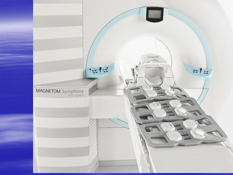
RF Coils Used In MRI
RF Coils
The simplest RF probe or coil is a set of wire wrapped around a patient or placed on the body part being under examination but separated by a covering

Why are coils used?
To serve as an antenna for
radio frequency
To create as uniform a
magnetic field as possible
To get the best images possible

Intensity of the signal
The intensity of the emitted RF signal and the sensitivity to the signal received from the patient are maximum in a volume approximately equal to the diameter of the coil.
Thant means the coil must cover the area you want to scan

Three General Types Of RF Coils
Transmit Receiver
Coils (T/R)
Receiver Only Coils
Transmit Only Coils

Transmit Receiver Coils
Act as a transmitter and receiver for signal; “transceivers”
Examples:
Volume Coils-T/R coils that engulf the entire anatomy of interest
Body Coil-largest T/R volume coil used
Bird Cage Coil-volume coil used mostly for heads/brain and joints;
has a high homogeneity factor

Transmit Receiver Coils

Receiver Only Coils
Connected directly to the receiver circuitry to receive RF signal
Examples:
Surface Coils-placed over part of anatomy of interest
Phased Array Coils-multiple coils used simultaneously with multiple separate receivers

Receiver Only Coils

Transmit Only Coils
Coil is located inside
the bore of magnet
Usually used with receiver only coils to
detect/receive signal

Coils types according to their Homogeniety
1) Homogeneous volume coils are typically used both to transmit RF and to receive the MR signal. These include the head and body coils and other special application coils, such as an upper extremity/shoulder coil

Coils types according to their Homogeniety
2) Inhomogeneous coils are surface coils, and the word inhomogeneous is used because these coils do not transmit RF in a homogeneous fashion. For this reason, they usually used as receive coils only. When these coils are used, the RF is transmitted by the head or body coil
Coils types according to their design and body part
1- Saddle coil Body Coils was the most widely used design for the RF coils
Old and not used anymore

2-Quadrature Coils
Quadrature coil design
improves SNR by around 40%
by detecting the MR signal from multiple directions,
Quadrature coils are also constructed to be more homogeneous for RF transmission and reception.
The “bird cage” coil is popular version of Quadrature coils
Example: Head, body, extremity coils

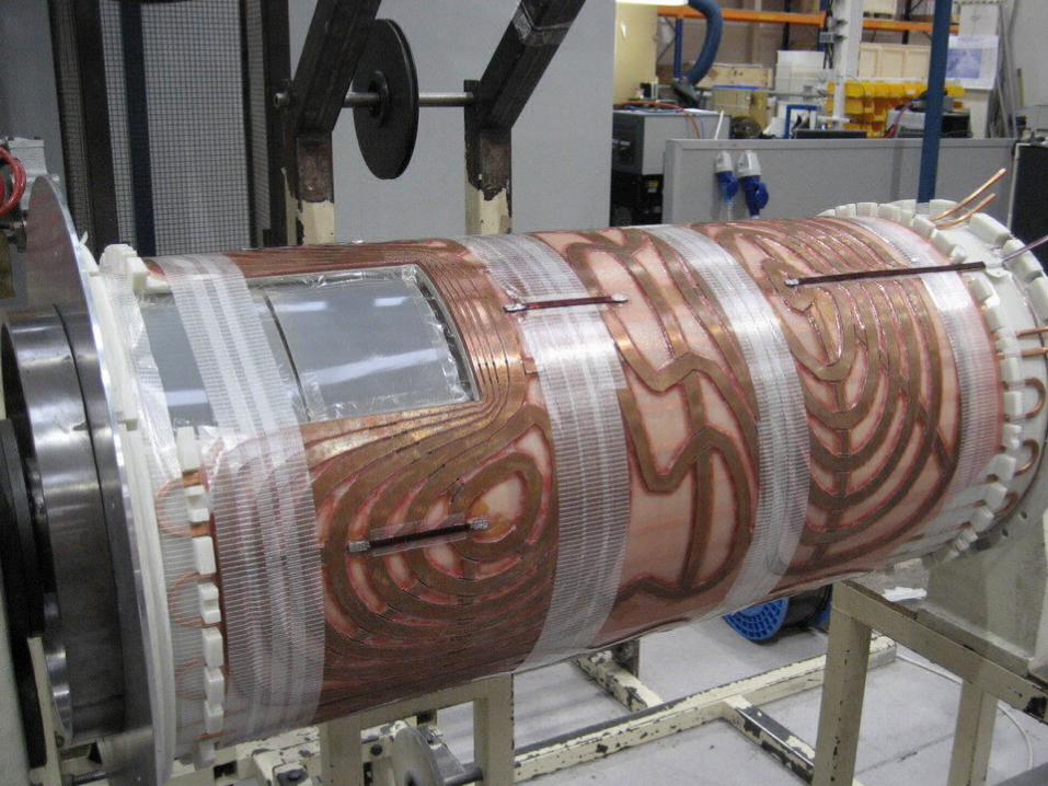
3- Body Coils
The body coil is wound inside the gradient coils.
It is surrounding the Patient, for this the body coil can image any part of the human anatomy and it is always a transmit/receive device

Body coil

4-Special-purpose coils
are designed to optimize the SNR from a given region of the body. For example Hemholtz Pair is a coil of two parts with one receiver channel. It is used for neck or cervical spine to improve S/N
Examples
breast, prostate (endo-rectal), extremities, joints, or orbit coils.

Special-purpose coils

5- A surface coil is a specially designed coil that is usually flat or any other shapes and is used to obtain high SNR images of anatomy close to the surface.
Examples of surface coils are breast, prostate (endo-rectal), extremities, joints, or orbit coils.
Surface coils provide better contrast resolution because of its higher SNR and better spatial resolution because of smaller FOV.

Examples of surface coils

Endo-rectal coil


Coil Safety
Coils are conductive..they produce heat and can burn the patient
Make sure coils are connected properly/not tangled
Use proper positioning when using coils