review open access role of carotenoid b-cryptoxanthin in ... · review open access role of...
TRANSCRIPT
REVIEW Open Access
Role of carotenoid b-cryptoxanthin in bonehomeostasisMasayoshi Yamaguchi
Abstract
Bone homeostasis is maintained through a balance between osteoblastic bone formation and osteoclastic boneresorption. Aging induces bone loss due to decreased osteoblastic bone formation and increased osteoclastic boneresorption. Osteoporosis with its accompanying decrease in bone mass is widely recognized as a major publichealth problem. Nutritional factors may play a role in the prevention of bone loss with aging. Among variouscarotenoids (carotene and xanthophylls including beta (b)-cryptoxanthin, lutein, lycopene, b-carotene, astaxanthin,and rutin), b-cryptoxanthin, which is abundant in Satsuma mandarin orange (Citrus unshiu MARC.), has been foundto have a stimulatory effect on bone calcification in vitro. b-cryptoxanthin has stimulatory effects on osteoblasticbone formation and inhibitory effects on osteoclastic bone resorption in vitro, thereby increasing bone mass. b-cryptoxanthin has an effect on the gene expression of various proteins that are related osteoblastic bone formationand osteoclastic bone resororption in vitro. The intake of b-cryptoxanthin may have a preventive effect on boneloss in animal models for osteoporosis and in healthy human or postmenopausal women. Epidemiological studiessuggest a potential role of b-cryptoxanthin as a sustainable nutritional approach to improving bone health ofhuman subjects. b-Cryptoxanthin may be an osteogenic factor in preventing osteoporosis in human subjects.
Keywords: Carotenoid, β-cryptoxanthin, Osteoblastic bone formation, Osteoclastic bone resorption, Osteoporosis
IntroductionBone is a dynamic tissue that preserves skeletal size,shape, and structural integrity and to regulate mineralhomeostasis. Bone homeostasis is maintained through abalance between osteoblastic bone formation and osteo-clastic bone resorption. Aging and numerous pathologi-cal processes induce decrease in bone formation andincrease in bone resorption, leading to osteoporosis, adevastating bone disease [1]. Osteoporosis, which isinduced with decrease in bone mass, is widely recog-nized as a major public health problem [1]. The mostdramatic expression of the disease is represented byfractures of the proximal femur for which the numberincreases as the population ages [2].Nutritional factors may have the potential effect to
prevent bone loss with increasing age. There is growingevidence that the supplementation of nutritional andfood factors may have the preventive effect on bone lossthat is induced in animal model of osteoporosis and in
human subjects [3-6]. Chemical compounds in food andplants, which regulate on bone homeostasis, have beento be worthy of notice in maintaining of bone healthand prevention of bone loss with increasing age [7-13].Carotenoids (carotene and xanthophyll) are present in
fruit and vegetables. Carotenoids, which are a provita-min A, may have an anabolic effect on bone metabo-lism. Vitamin A (retinol, retinal, and retinoic acid),which is formed from carotenoids in animal and human,has been shown to have a role in the regulation of bonecells and it may have an anabolic effect on bone [14-16].However, vitamin A is also known to have a detrimentaleffect on bone at high doses [17-20]. In laboratory ani-mals, high levels of vitamin A lead to accelerated boneresorption, bone fractures, and osteoporotic bone lesions[17].Beta (b)-cryptoxanthin, a kind of xanthophyll, is abun-
dant in Satsuma mandarin orange (Citrus unshiuMARC.). Of various carotenoids, b-cryptoxanthin hasbeen found to have a potential-anabolic effect on bonedue to stimulating osteoblastic bone formation and inhi-biting osteoclastic bone resorption [21,22]. This review
Correspondence: [email protected] of Foods and Nutrition, The University of Georgia, 425 RiverRoad, Rhodes Center, Room 448, Athens, GA 30602-2771, USA
Yamaguchi Journal of Biomedical Science 2012, 19:36http://www.jbiomedsci.com/content/19/1/36
© 2012 Yamaguchi; licensee BioMed Central Ltd. This is an Open Access article distributed under the terms of the Creative CommonsAttribution License (http://creativecommons.org/licenses/by/2.0), which permits unrestricted use, distribution, and reproduction inany medium, provided the original work is properly cited.

has been written to outline the recent advances con-cerning the role of b-cryptoxanthin in the regulation ofbone homeostasis and in the prevention of osteoporosis,especially the cellular and molecular mechanisms bywhich b-cryptoxanthin stimulates osteoblastic bone for-mation and inhibits osteoclastic bone resorption.
Effect of carotenoids in bone homeostasisBone is a dynamic tissue that undergoes continual adap-tation during vertebrate life to attain and preserve skele-tal size, shape, and structural integrity and to regulatemineral homeostasis. Bone homeostasis is regulated bythe functions of osteoblasts, osteoclasts, and osteocyteswhich are major cells in bone tissues [23,24]. Osteo-clasts, which develop from hematopoietic progenitors,are recruited to the site and excavate the calcifiedmatrix. Osteoblasts arising from local mesenchymalstem cells assemble at the bottom of the cavity andbone formation begins. Bone acts as major storage sitefor growth factors [25]. Growth factors, which are pro-duced by osteoblasts, diffuse into newly depositedosteoid and are stored in the bone matrix including isu-lin-like growth factors (IGF- I and II), transforminggrowth factor-b1 (TGF-b1), platelet-derived growth fac-tor (PDGF), and bone morphologic protein (BMP).These bone-derived factors, which can be liberated dur-ing subsequent periods of bone resorption, act in anautocrine, paracrine, or delayed paracrine fashion in thelocal microenvironment of the bone surface. Thus, bonehomeostasis is regulated through complex mechanism.Whether functional food factors can regulate bonehomeostasis has been poorly understood.Carotene, which is contained in plant and fruits, is
generically named for carotene (a, b, g, δ, ε) and lyco-pene, which is a natural compound with the derivativeof fundamental form of C40H56 in chemical structure.Other derivatives with hydroxyl group for C40H56 arenamed as xanthophyll, which are lutein, zeaxanthin,canthaxanthin, fucoxanthin, antheraxanthin, violax-anthin, and b-cryptoxanthin. Carotenoids are namedgenerically for carotene and xanthophyll.The effects of carotenes, canthaxanthin, fucoxanthin,
antheraxanthin, and violaxanthin on bone homeostasishave not been shown clearly. Among various carotenesand xanthophylls (including b-carotene, lycopene, lutein,astaxanthin, and b-cryptoxanthin), b-cryptoxanthin hasbeen found to have a potential-anabolic effect on bonecalcification in vitro [21,22]. Myricetin, kaempferol, iso-rhamnetin, curcumin, hesperidin, and rutin (quercetin-3-rutinoside) of various flavonoids do not have an effecton bone formation and calcification in vitro [22].Vitamin A (retinol, retinal, and retinoic acid) is
formed from carotenoids, which are provitamin A, inthe animals and humans. The retinoic acid receptors
(RAR) a, b and g (RARa, RAR b and RARg) are nuclearhormone receptors that regulate fundamental processesduring embryogenesis, but their roles in skeletal devel-opment and growth are investigated.Mice deficient in RARa and RARg (or RAR b and
RARg) have been shown to exhibit severe growth retar-dation obvious by about 3 weeks postnatally [14]. Reti-noic acid receptors may be required for skeletal growth,matrix homeostasis and growth plate function in postna-tal mouse, suggesting a role of retinoic acid in bonegrowth [14].Retinol and b-carotene have been shown to inhibit the
proliferation of MC3T3-E1 cells as well as DNA synth-esis of the cells in a dose-dependent manner [15] andstimulate differentiation of MC3T3-E1 cells by increas-ing alkaline phosphatase activity dose dependently [15].a-Carotene, canthaxanthin, and lycopene also have beenshown to inhibit MC3T3-E1 cell proliferation and toincrease alkaline phosphatase activity and osteopontinmRNA expression [15].The effects of retinoids on osteoclast differentiation in
cultured mouse bone marrow cells (BMCs), bone mar-row macrophages (BMMs), spleen cells, and RAW264.7cells are shown by analyzing osteoclast formation andexpression of important genes in signal transductionand osteoclast function [16]. All-trans-retinoic acid(ATRA) did not stimulate osteoclastogenesis in BMCs,but inhibited hormone and RANK (receptor activator ofnuclear factor kappa B; NF-�B) ligand (RANKL)-induced gene expression and formation of osteoclasts[16]. ATRA abolished an increase in the transcriptionfactors c-Fos and the nuclear factor of activated T cells(NFAT) NFAT2 stimulated by RANKL and suppresseddown-regulation of the anti-osteoclastogenic transcrip-tion factor MafB [16].Excessive intakes of vitamin A have been shown to
have adverse skeletal effects in animals [17]. High vita-min A intake may lead to an increased risk of fracturein humans. Association between vitamin D deficiencyand excess of vitamin A as a potential risk factor ofosteoporosis and fracture is shown [18]. Whereas inwomen with vitamin D deficiency the risk of osteoporo-sis increased was up 5 times higher than women in thelowest quintile of retinol [18]. High retinol levelstogether with vitamin D deficiency may be hitherto anoverlooked risk factor for osteoporosis.Retinol is derived from both retinoids, which are con-
tained in animal food, and carotenoids, which are con-tained in vegetables and fruits. A possible role ofcarotenoids in involutional osteoporosis is evaluated.When plasma levels of b-carotene and other carote-noids, in addition to those of retinol, were measured infree-living, non-supplemented, elderly women with orwithout severe osteoporosis, plasma levels of retinol and
Yamaguchi Journal of Biomedical Science 2012, 19:36http://www.jbiomedsci.com/content/19/1/36
Page 2 of 13
of all carotenoids tested, with the exception of lutein,were consistently lower in osteoporotic than in controlwomen [26]. A weak association was found onlybetween retinol and femoral neck bone mineral densityin osteoporotic women [26]. A bone sparing effect ofretinol, to which the provitamin A activity of some caro-tenoids, may be contributed [26].Carotenoid lycopene has been shown to have an inhi-
bitory effect on bone resorption in vitro [27], althoughlycopene is not shown to have a stimulatory effect onmineralization using rat femoral tissues in vitro [21].Lycopene (10-5 M) has been found to inhibit basal andparathyroid hormone (PTH)-stimulated osteoclasticmineral resorption and formation of tartrate-resistantacid phosphatase (TRAP) activity-possitive multinu-cleated osteoclasts [27]. Lycopene may have an inhibi-tory effect on osteoclastic bone resorption, suggestingthat lycopene has a role in the prevention ofosteoporosis.In a cross-sectional study by using 33 postmenopausal
women aged 50-60 years, groups with higher lycopeneintake, as determined from the dietary records, showedhigher serum lycopene [28]. A higher serum lycopenehas been found to be associated with a low cross-linkedN-telopeptides of type I collagen [28]. Similarly, groupswith higher serum lycopene have lower protein oxida-tion [28]. These observations suggest that the dietaryantioxidant lycopene reduces oxidative stress and thelevels of bone turnover markers in postmenopausalwomen, and may be beneficial in reducing the risk ofosteoporosis [28].Postmenopausal women supplemented with lycopene
are shown to increase antioxidant capacity and decreaseoxidative stress and the bone resorption marker N-telo-peptide [29]. Lycopene decreases bone resorption mar-kers and may reduce the risk of osteoporosis [29]. Sixtypostmenopausal women, 50-60 years old, were recruited[29]. Following a 1-month washout without lycopeneconsumption, participants consumed either regulartomato juice, lycopene-rich tomato juice, tomato lyco-pene capsules, or placebo capsules, twice daily for totallycopene intakes of 30 and 70 mg/day respectively for 4months [29]. Lycopene supplementation for 4 monthssignificantly increased serum lycopene and decreasedlipid peroxidation, and cross-linked aminoterminal N-telopeptide as compared to placebo [29]. The antioxi-dant lycopene may be beneficial in reducing oxidativestress parameters and the bone resorption marker [29].
b-Cryptoxanthine and bone homeostasisXanthophyll b-cryptoxanthin, which is a kind of carote-noid, is abundant in Satsuma mandarin orange (Citrusunshiu MARC). The molecular weight of b-cryptox-anthin is 552. The biological function of b-
cryptoxanthin in animal and human, however, has beennot determined fully. Yamaguchi et al. found that b-cryptoxanthin has been shown to have a unique ana-bolic effect on bone calcification; such an effect is notseen in lutein, lycopene, or astaxanthin, which is othercarotenoids and flavonoid rutin (quercetin-3-rutinoside)[21,22].b-Cryptoxanthin stimulates bone formation and inhibitsbone resorption in bone tissue cultureb-Cryptoxanthin has been shown to have a unique ana-bolic effect on bone calcification. Culture with b-cryp-toxanthin (10-7 or 10-6 M) has been found to cause anincrease in calcium content and alkaline phosphataseactivity in the femoral-diaphyseal (cortical bone) and-metaphyseal (trabecular bone) tissues in vitro. Lutein,lycopene, and rutin (10-8 to 10-6 M) did not have ana-bolic effects on alkaline phosphatase activity and cal-cium contents in rat femoral tissues [21]. Astaxanthinand b-carotene did not have an effect on the femoralcalcium contents [22]. Myricetin, kaempferol, isorham-netin, curcumin, or hesperidin (10-7 to 10-5 M) had noeffect on bone calcium content in tissue cultures invitro [22]. Quercetin significantly increased calcium con-tent in femoral diaphyseal tissues but not metaphysealtissues. b-Cryptoxanthin has a unique anabolic effect onbone calcification in vitro. The effect of b-cryptoxanthinincreasing bone components was completely preventedin the presence of cycloheximide, an inhibitor of proteinsynthesis, suggesting that the effect is needed newly pro-tein synthesis [22].b-Cryptoxanthin has also been shown to inhibit bone
resorption in bone tissue cultures in vitro [22]. Parathyr-oid hormone (PTH) or prostaglandin E2 (PGE2), whichis a bone-resorbing factor, can stimulate osteoclasticbone resorption in vitro [30-32]. Culture with PTH orPGE2 caused a decrease in calcium content in the dia-physeal and metaphyseal tissues [22]. This decrease wascompletely inhibited in the presence of b-cryptoxanthin(10-8 to 10-6 M). Likewise, culture with b-cryptoxanthincompletely inhibited the PTH- or PGE2-inducedincrease in medium glucose consumption and lactic acidproduction by bone tissues [22]. b-Cryptoxanthin has aninhibitory effect on bone resorption in tissue culture invitro.Thus, b-cryptoxanthin has been shown to have stimu-
latory effect on osteoblastic bone formation and inhibi-tory effects on osteoclastic bone resorption in bonetissue culture in vitro. Serum concentration of b-cryp-toxanthin due to consumption of vegetable juice inwomen is shown to be in the range of 1.3 × 10⁻⁷ to 5.3× 10⁻⁷ M [33]. b-Cryptoxanthin in the range of 10-8 to10-6 M has an anabolic effect on biochemical compo-nents in rat femoral tissues in vitro, suggesting a physio-logic role in the regulation of bone metabolism.
Yamaguchi Journal of Biomedical Science 2012, 19:36http://www.jbiomedsci.com/content/19/1/36
Page 3 of 13

b-Cryptoxanthin stimulates osteoblastogenesisThe cellular and molecular mechanisms by which b-cryptoxanthin stimulates bone formation in bone tissueshas been examined by using osteoblastic MC3T3-E1cells in vitro. b-Cryptoxanthin has been found to stimu-late the proliferation of osteoblastic cells in subconfluentmonolayers in a medium containing 10% fetal bovineserum [34]. Culture with b-cryptoxanthin also caused anincrease in biochemical components (alkaline phospha-tase activity, protein, and DNA contents) of osteoblasticcells [34]. This effect was abolished in the presence ofstaurosporine, an inhibitor of protein kinase C, orPD98059, an inhibitor of mitogen- activated proteinkinase (MAPK), although the effect of b-cryptoxanthinin increasing cellular biochemical components was notprevented in culture with dibucain, an inhibitor of Ca2+/calmodulin-dependent protein kinase [34]. The stimu-latory effect of b-cryptoxanthin on osteoblastic cell com-ponents seems to be partly mediated through signalingfactors of protein kinase C or MAPK in the cells.The effects of b-cryptoxanthin in increasing the bio-
chemical components in osteoblastic cells were comple-tely inhibited in the presence of 5,6-dichloro-1- b-D-ribofuranosylbenzimidazole (DRB), an inhibitor of RNApolymerase II, suggesting that the effect of carotenoidresults from a stimulatory effect on transcriptional activ-ity in osteoblastic cells.The mineralization in osteoblastic cells is shown to
stimulate in the prolonged culture with b-cryptoxanthin[35]. The stimulatory effect of b-cryptoxanthin onmineralization may result from the carotenoid-inducedproliferation and differentiation of osteoblastic cells.b-Cryptoxanthin may stimulate gene expression for
proteins that are involved in bone formation and miner-alization in osteoblastic cells. The effect of b-cryptox-anthin on gene expression in osteoblastic cells usingreverse transcription-polymerase chain reaction (RT-PCR) is examined. Culture with b-cryptoxanthin wasfound to stimulate the mRNA expression of IGF-I orTGF- b1 in osteoblastic cells [34]. This finding may sup-port the view that b-cryptoxanthin has a stimulatoryeffect on transcriptional activity in osteoblastic cells.IGF-I or TGF-b1 is a bone growth factor produced fromosteoblasts [36-38]. The stimulatory effect of b-cryptox-anthin on the proliferation of osteoblastic cells may bepartly mediated through the action of IGF-I or TGF-b1produced from the cells.TGF-b1 has a potent activity on osteoblast-lineage
commitment an event that is partly mediated throughSmad transcription factors [38]. It has been reportedthat NF-�B signaling represses basal osteoblast differen-tiation and mineralization in MC3T3 cells and antago-nizes TGF-b1 and BMP-2 mediated MC3T3mineralization by downregulating Smad activation [39].
Interestingly, b-cryptoxanthin has been shown to havethe capacity to suppress the receptor activator of nuclearfactor-kappa B (NF-�B) activity in MC3T3 preosteoblas-tic cells [40], suggesting that one mechanism by whichb-cryptoxanthin may stimulate osteoblast differentiationmay be by promoting Smad activation. This finding alsosuggests that b-cryptoxanthin may lead to enhancedSmad signaling. While, b-cryptoxanthin failed to directlystimulate Smad activity, it did however amplify TGF-b1-induced Smad signaling [41].Furthermore, whether b-cryptoxanthin directly stimu-
lates Smad activation or regulates TGF-b1-inducedSmad activation in osteoblastic cells in vitro is examined[41]. b-Cryptoxanthin was found to potentiate TGF-b1-induced, but not BMP-2-induced, Smad activation inMC3T3 preosteoblastic cells, suggesting that onemechanism by which b-cryptoxanthin stimulates boneformation is by potentiating the TGF-b1-mediated com-mitment of preosteoblasts to differentiate along theosteoblastic pathway [41].Runx2 (Cbfa1) is a member of the runt domain family
of transcription factors and a master regulator of osteo-blast differentiation [42]. a1 (I) Collagen is a matrixprotein that is related to bone formation and mineraliza-tion in osteoblast lineage cells [43]. Alkaline phospha-tase participates in the mineralization process inosteoblastic cells [44]. b-Cryptoxanthin (10-7 or 10-6 M)was found to increase the mRNA expression of Runx2,a1 (I) collagen, and alkaline phosphatase in osteoblasticMC3T3-E1 cells [35].b-Cryptoxanthin had a stimulatoryeffect on the gene expression of various proteinsinvolved in osteoblasic bone formation [35]. Such effectsof b-cryptoxanthin were blocked in the presence of DRB[34], supporting the view that the carotenoid stimulatestranscriptional activity in osteoblastic MC3T3-E1 cells.Vitamin A (retinol) may be able to bind to nuclear
receptors in cells. Retinol and b-carotene has beenshown to inhibit the proliferation of osteoblasticMC3T3-E1 cells as well as DNA synthesis of the cells,due to increasing alkaline phosphatase activity dosedependently (10-9 to 10-7 M) [15]. Vitamin A (10-7 or10-6 M) increases alkaline phosphatase activity inosteoblastic cells [34]. b-Cryptoxanthin (10-7 or 10-6
M) caused an increase in alkaline phosphatase activityand protein content in osteoblastic cells. This effectwas also seen in the presence of vitamin A (10-6 M)[34]. Moreover, the stimulatory effect of b-cryptox-anthin on the expression of Runx2 type 1 and a1 (I)collagen mRNA was observed in the presence of vita-min A [34]. Vitamin A did not have a significant effecton Runx2 type 1 mRNA expression in osteoblasticMC3T3-E1 cells [34]. Thus, the mode of action of b-cryptoxanthin on gene expression in osteoblastic cellsmay differ from that of vitamin A, which is mediated
Yamaguchi Journal of Biomedical Science 2012, 19:36http://www.jbiomedsci.com/content/19/1/36
Page 4 of 13

through the retinoid X receptor (RXR) in the nucleusof the cells [34].It is speculated that b-cryptoxanthin can bind to other
receptors (including orphan receptors that ligands arenot unknown), and that the carotenoid may stimulatetranscriptional activity in osteoblastic cells. The mechan-ism of b-cryptoxanthin action in stimulating prolifera-tion, differentiation, and mineralization in osteoblasticcells is summarized in Figure 1.b-Cryptoxanthin suppresses osteoclastogenesisNF-�B ligand (RANKL) plays a pivotal role in osteoclas-togenesis from bone marrow cells. RANKL expression isinduced in osteoblastic cells and bone marrow stromalcells in response to osteoporotic factors, such as PTH,PGE2, and 1,25-dihydroxyvitamin D3 (VD3), and com-bined treatment of hematopoietic cells with macrophagecolony-stimulating factor (M-CSF) and the soluble formof RANKL (sRANKL) induce osteoclast differentiationin vitro [45,46]. The receptor protein RANK (receptoractivator of NF-�B) is expressed on the surface of osteo-clast progenitors.b-Cryptoxanthin (10-8 to 10-6 M) is shown to have a
potent inhibitory effect on osteoclast-like cell forma-tion in mouse marrow culture in vitro [47]. The inhibi-tory effect of b-cryptoxanthin on osteoclast-like cellformation was seen at the later stage of osteoclast dif-ferentiation in bone marrow cultures [47]. Culturewith b-cryptoxanthin caused a marked inhibition ofosteoblast-like cell formation induced in the presenceof PTH, PGE2, VD3, lipopolysaccharide, or tumor
necrosis factor-a (TNF-a). b-Cryptoxanthin also hadan inhibitory effect on osteoclast-like cell formationinduced by RANKL [47]. The inhibitory effect of b-cryptoxanthin was equal to that of 17 b-estradiol (E2),calcitonin, genistein, and zinc sulfate, which can inhibitosteoclast-like cell formation induced by bone-resorb-ing factors [47].The interaction of RANKL with its receptor RANK
leads to the recruitment of the signaling adaptor mole-cules TRAFs (TNF receptor-associated factors) to thereceptor complex and the activation of NF-�B and c-JunN-terminal kinase (JNK) [48,49]. Protein kinase C familyenzyme has a role in regulation of osteoclast formationand function potentially by participating in the extracel-lular signaling-regulated kinase (ERK) signaling pathwayof M-CSF and RANKL [49].Phorbol 12-myristate 13-acetate (PMA), an activator
of protein kinase C, stimulated osteoclast-like cell for-mation in mouse marrow cultures, and the PMA-induced osteoclastogenesis was found to inhibit in thepresence of b-cryptoxanthin [47]. Moreover, b-cryptox-anthin had an inhibitory effect on dibutyryl cyclic ade-nosine monophosphate (DcAMP)-induced osteoclast-like cell formation in mouse marrow cultures [47]. It isassumed that activation of protein kinase C and proteinkinase A pathways leads to increased RANKL expres-sion, and that b-cryptoxanthin can inhibit protein kinaseC- or protein kinase A-related RANKL expression inosteoclastogenesis.The effect of b-cryptoxanthin on mature osteoclasts
is demonstrated [50]. M-CSF-dependent bone marrowmacrophages were cultured in the presence of M-CSFand RANKL for 4 days [50]. The osteoclastic cellsformed were further cultured in medium containingb-cryptoxanthin with or without M-CSF and RANKLfor 24-72 hours. The number of osteoclastic cells hasbeen found to decrease in culture with b-cryptox-anthin (10-7 or 10-6 M) in the presence or absence ofM-CSF and RANKL for 72 hours. The b-cryptox-anthin-induced decrease in osteoclastic cells wasinhibited in the presence of caspase-3 inhibitor. Theresults of agarose gel electrophoresis showed the pre-sence of low-molecular-weight DNA fragments ofadherent cells cultured with b-cryptoxanthin. Thesefindings indicate that the carotenoid induces apopto-tic cell death.Apoptosis-related gene expression was determined by
using RT-PCR [50]. The expression of caspase-3 mRNAor Apaf-2, which involves apoptosis, in osteoclastic cellswas found to stimulate when cultured with b-cryptox-anthin in the presence or absence of M-CSF andRANKL [50]. b-Cryptoxanthin-induced apoptotic celldeath may be partly mediated through caspase-3 expres-sion in osteoclastic cells.
β
Target genes
IGF-ITGF- 1Runx2Alkaline phosphatase
mRNA
Figure 1 The cellular and molecular mechanism by which b-cryptoxanthin (BCX) stimulates bone formation andmineralization in osteoblastic cells. BCX may bind to orphanreceptors in the nucleus of osteoblastic cells, and it stimulates geneexpression of bone formation-related proteins. BCX also stimulatesnuclear transcriptional activity mediated through activation ofprotein kinase C (PKC) or mitogen-activated protein kinase (MAPK)in osteoblastic cells.
Yamaguchi Journal of Biomedical Science 2012, 19:36http://www.jbiomedsci.com/content/19/1/36
Page 5 of 13
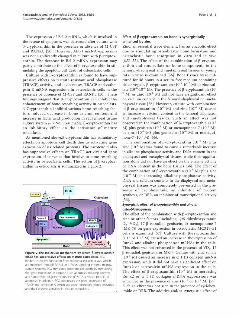
The expression of Bcl-2 mRNA, which is involved inthe rescue of apoptosis, was decreased after culture withb-cryptoxanthin in the presence or absence of M-CSFand RANKL [50]. However, Akt-1 mRNA expressionwas not significantly changed in culture with b-cryptox-anthin. The decrease in Bcl-2 mRNA expression maypartly contribute to the effect of b-cryptoxanthin in sti-mulating the apoptotic cell death of osteoclastic cells.Culture with b-cryptoxanthin is found to have sup-
pressive effects on tartrate-resistant acid phosphatase(TRACP) activity, and it decreases TRACP and cathe-psin K mRNA expressions in osteoclastic cells in thepresence or absence of M-CSF and RANKL [50]. Thesefindings suggest that b-cryptoxanthin can inhibit theenhancement of bone-resorbing activity in osteoclasts.b-Cryptoxanthin inhibitd various bone-resorbing fac-tors-induced decrease in bone calcium content andincrease in lactic acid production in rat femoral tisuueculture system in vitro. Presumably, b-cryptoxanthin hasan inhibitory effect on the activation of matureosteoclasts.As mentioned above,b-cryptoxanthin has stimulatory
effects on apoptotic cell death due to activating geneexpression of its related proteins. The carotenoid alsohas suppressive effects on TRACP activity and geneexpression of enzymes that involve in bone-resorbingactivity in osteoclastic cells. The action of b-cryptox-anthin in osteoclasts is summarized in Figure 2.
Effect of b-cryptoxanthin on bone is synergisticallyenhanced by zincZinc, an essential trace element, has an anabolic effectdue to stimulating osteoblastic bone formation andosteoclastic bone resorption in vitro and in vivo[6,51-55]. The effect of the combination of b-cryptox-anthin and zinc sulfate on bone components in thefemoral-diaphyseal and -metaphyseal tissues of youngrats in vitro is examined [56]. Bone tissues were cul-tured for 48 hours in a serum-free medium containingeither vegicle, b-cryptoxanthin (10-9-10-7 M) or zinc sul-fate (10-6-10-4 M). The presence of b-cryptoxanthin (10-9 M) or zinc (10-6 M) did not have a significant effecton calcium content in the femoral-diaphyseal or -meta-physeal tissue [56]. However, culture with combinationof b-cryptoxanthin (10-9 M) and zinc (10-6 M) causedan increase in calcium content in the femoral-diaphysealand -metaphyseal tissues. Such an effect was notobserved in the combination of b-cryptoxanthin (10-9
M) plus genistein (10-6 M) or menaquinone-7 (10-6 M),or zinc (10-6 M) plus genistein (10-6 M) or menaqui-none-7 (10-6 M) [56].The combination of b-cryptoxanthin (10-9 M) plus
zinc (10-6 M) was found to cause a remarkable increasein alkaline phosphatase activity and DNA content in thediaphyseal and metaphyseal tissues, while their applica-tion alone did not have an effect on the enzyme activityor DNA content in the bone tissues [56]. The effect ofthe combination of b-cryptoxanthin (10-9 M) plus zinc(10-6 M) in increasing alkaline phosphatase activity,DNA and calcium contents in the diaphyseal and meta-physeal tissues was completely prevented in the pre-sence of cycloheximide, an inhibitor of proteinsynthesis, or DBR, an inhibitor of transcriptional activity[56].Synergistic effect of b-cryptoxanthin and zinc inosteoblastogenesisThe effect of the combination with b-cryptoxanthin andzinc or other factors [including 1,25-dihydroxyvitaminD3 (VD3), 17 b-estradiol, genistein, or menaquinone-7(MK-7)] on gene expression in osteoblastic MC3T3-E1cells is examined [57]. Culture with b-cryptoxanthin(10-7 or 10-6 M) caused an increase in the expression ofRunx2 and alkaline phosphatase mRNAs in the cells.This effect was not enhanced in the presence of VD3, 17b-estradiol, genistein, or MK-7. Culture with zinc sulfate(10-5 M) caused an increase in a 1 (I) collagen mRNAexpression, while it did not have a significant effect onRunx2 or osteocalcin mRNA expression in the cells.The effect of b-cryptoxanthin (10-7 M) in increasingRunx2 or a 1 (I) collagen mRNA expressions wasenhanced in the presence of zinc (10-6 or 10-5 M) [57].Such an effect was not seen in the presence of cyclohex-imide or DRB. The additive and/or synergistic effect of
H+ Lactic acidTRACP Cathepsin K
Bone
TRACP
Cathepsin K
Gene expressionCaspase 3
Bcl-2
Apoptosis
RANKLRANK
TRAF6
Osteoclast
BCX
Nucleus
Figure 2 The molecular mechanism by which b-cryptoxanthin(BCX) has suppressive effects on mature osteoclasts. BCXinhibits osteoclast formation from mononuclear osteoclasts whichare mediated through RANKL and RANK signaling in bone marrowculture systems. BCX stimulates apoptotic cell death by stimulatingthe gene expression of caspase-3, an apoptosis-inducing enzyme,and suppression of gene expression of Bcl-2, a rescue protein ofapoptosis. In addition, BCX suppresses the gene expression ofTRACP and cathepsin K, which are bone resorption-related enzymes,and their enzyme activities in mature osteoclasts.
Yamaguchi Journal of Biomedical Science 2012, 19:36http://www.jbiomedsci.com/content/19/1/36
Page 6 of 13

b-cryptoxanthin and zinc on gene expression in osteo-blastic cells is partly resulted in newly synthesized pro-tein components.Zinc activates aminoacyl-tRNA synthetase, a rate-lim-
iting enzyme, in the translational process of proteinsynthesis [53-55]. b-Cryptoxanthin can stimulate tran-scriptional activity in osteoblastic cells [34]. It is specu-lated that b-cryptoxanthin stimulates gene expressionand zinc increases translational activity in osteoblasticcells. This may be important as a possible mechanismby which zinc enhances the anabolic effect of b-cryptox-anthin in osteoblastic cells.Synergistic effect of b-cryptoxanthin and zinc in osteoclasticcellsThe effect of b-cryptoxanthin on osteoclastic cellsformed in the mouse marrow culture system in vitrowas found to enhance after culture with zinc [58]. Bonemarrow cells were isolated from mice. The macrophagecolony-stimulating factor (M-CSF)-dependent bone mar-row cells were cultured in the presence of M-CSF andRANKL for 96 hours. The osteoclastic cells formed werefurther cultured for 24 or 72 hours in a medium con-taining either vehicle, b-cryptoxanthin, zinc sulfate, orb-cryptoxanthin plus zinc with or without M-CSF andRANKL. The number of osteoclastic cells was decreasedafter culture with the combination of b-cryptoxanthin(10-7 M) and zinc (10-5 M) in the presence or absenceof M-CSF and RANKL for 24 or 72 h as compared withthe value for b-cryptoxanthin or zinc [58].The results of agarose gel electrophoresis showed the
presence of low-molecular weight deoxyribonucleic acid(DNA) fragments of adherent cells cultured with b-cryp-toxanthin plus zinc for 24 or 72 hours in the presenceof M-CSF and RANKL, indicating that the combinationof the two chemicals synergistically induces apoptoticcell death [58].b-Cryptoxanthin plus zinc-induced decrease in osteo-
clastic cells was inhibited in the presence of caspase-3inhibitor [58]. Culture with b-cryptoxanthin plus zincfor 24 or 72 hours caused an increase in caspase-3mRNA expression in the presence or absence of M-CSFand RANKL as compared with the value for each che-mical alone. b-Cryptoxanthin plus zinc-induced increasein caspase-3 mRNA expression was completely inhibitedin the presence of cycloheximide or DRB [58]. This sug-gests that b-cryptoxanthin plus zinc-induced apoptoticcell death is mediated through caspase-3 in osteoclasticcells, and that b-cryptoxanthin plus zinc-enhanced cas-pase-3 mRNA expression in osteoclastic cells is relatedto newly synthesized protein synthesis.The mRNA expression of tartrate-resistant acid phos-
phatase (TRACP) and cathepsin K was decreased afterculture with b-cryptoxanthin plus zinc in the presence
or absence of M-CSF and RANKL for 72 hours as com-pared with b-cryptoxanthin or zinc alone [58].Nuclear factor of activated T cells c1 (NFATc1)
mRNA expression was decreased after culture with b-cryptoxanthin plus zinc in the presence or absence ofM-CSF and RANKL for 72 hours as compared witheach chemical alone, while NF-�B mRNA expressionwas not changed [58]. TRACP and cathepsin K areenzymes that are involved in the degradation of bonematrix components, and their enzyme activities areincreased in RANKL-stimulated bone resorption [45,46].The combination of b-cryptoxanthin and zinc may havea potent-suppressive effect on bone resorption.NF-�B and NFATc1 are molecules related to RANKL
signaling [45,46]. NFATc1 is a transcriptional factor thatenhances the gene expression of TRACP and cathepsinK in osteoclasts, and the binding of NFATc1 to promo-ter is involved in NF-�B or AP-1 [45]. The suppressionof NFATc1 mRNA expression induced with the combi-nation of b-cryptoxanthin and zinc may induce thedecrease in NF-�B protein level. This may partly contri-bute to the decrease in the TRACP or cathepsin KmRNA expression caused by their combination.Thus, the suppressive effects of b-cryptoxanthin on
osteoclastogenesis was demonstrated to enhance syner-gistically in the presence of zinc, and also the combina-tion of b-cryptoxanthin and zinc has potent suppressiveeffects on osteoclastic cell function in vitro.Anabolic effect of b-cryptoxanthin on bone is enhanced byzinc in vivoThe effects of combined b-cryptoxanthin and zinc onbone components in the femoral-diaphyseal (corticalbone) and -metaphyseal (trabecular bone) tissues of ratsin vivo is shown [59]. Rats were orally administeredeither vehicle, b-cryptoxanthin (50 or 100 μg/kg bodyweight), zinc sulfate (1 or 5 mg Zn/kg), or their combi-nation once a day for 7 days. Alkaline phosphataseactivity, DNA and calcium contents in the femaral-dia-physeal tissues were not altered after the administrationof b-cryptoxanthin (50 μg/kg) or zinc (1 or 5 mg/kg)[59]. Combined administration of b-cryptoxanthin (50μg/kg) and zinc (1 or 5 mg/kg) caused a synergisticincrease in alkaline phosphatase activity, DNA and cal-cium contents in the diaphyseal tissues [59]. The effectof b-cryptoxanthin (50 or 100 μg/kg) in increasing DNAand calcium contents in the metaphyseal tissues wasenhanced after the combined administration of zinc (1or 5 mg/kg), but it did not have an effect on the meta-physeal components. The metaphyseal alkaline phospha-tase activity has been found to increase markedly afterthe administration of the combination of b-cryptox-anthin (50 μg/kg) and zinc (1 or 5 mg/kg) [59]. Studydemonstrates that the oral administration of the
Yamaguchi Journal of Biomedical Science 2012, 19:36http://www.jbiomedsci.com/content/19/1/36
Page 7 of 13

combination of zinc at lower doses synergisticallyenhances b-cryptoxanthin-induced anabolic effects onthe femoral tissues of rats in vivo. It is speculated thatthe combination of b-cryptoxanthin and zinc has stimu-latory effects on the gene expression, protein synthesis,and cell proliferation in osteoblastic cells in rat femoraltissues. This may contribute to their enhancing effect onbone components in femoral tissues of rats in vivo.The combination of b-cryptoxanthin plus zinc at a
lower concentration has a synergistic effect on bonecomponents in vivo. The combination of b-cryptox-anthin plus zinc has potentiality in the prevention ofbone loss with aging. This finding is interested inrespect of the development of new supplement with thecomposition of food factors that reveal a potent-anaboliceffect in prevention of osteoporosis. It also would beuseful to identify some of the foods that contain higherlevels of b-cryptoxanthin and zinc.
Preventive effect of b-cryptoxanthin on bone loss in vivoAs mentioned above, b-cryptoxanthin has been shownto have a stimulatory effect on osteoblastic bone forma-tion and an inhibitory effect on osteoclastic boneresorption in vitro. Furthermore, the preventive effect ofb-cryptoxanthin on osteoporosis is demonstrated byusing animal models in vivo.The anabolic effect of b-cryptoxanthin on bone com-
ponents in young and aged rats is examined. b-Cryptox-anthin (100, 250, or 500 μg/kg body weight) was orallyadministered once daily for 7 days to young male rats[60]. The administration of b-cryptoxanthin (250, or 500μg/kg) caused an increase in alkaline phosphatase activ-ity, DNA and calcium contents in the femoral-diaphy-seal and -metaphyseal tissues [61]. Such an effect wasalso observed in the femoral tissues of aged (50-week-old) female rats [62]. b-Cryptoxanthin has been shownto have an anabolic effect on bone components in ratsin vivo.b-Cryptoxanthin has been sown to have a preventive
effect on bone loss in the pathophysiologic state. Boneloss is induced in streoptozotocin (STZ)-diabetic rats[62]. Young rats received a single subcutaneous adminis-tration of STZ (60 mg/kg body weight), and then theanimals were orally administered b-cryptoxanthin (50 or100 μg/kg) once daily for 7 or 14 days. The administra-tion of STZ caused a decrease in body weight and a sig-nificant increase in serum glucose, triglyceride, andcalcium levels, indicating a diabetic state [62]. Thesealterations were prevented after the administration of b-cryptoxanthin (50 or 100 μg/kg) for 14 days [62]. Alka-line phosphatase activity, DNA and calcium contents inthe femoral-diaphyseal and -metaphyseal tissues weredecreased in STZ-diabetic rats [62]. These decreaseswere prevented after the administration of b-
cryptoxanthin (50 or 100 μg/kg) for 14 days [62]. Thus,the intake of b-cryptoxanthin was found to have preven-tive effects on STZ-diabetic state and bone loss in STZ-diabetic rats.Bone loss is induced after ovriectomy (OVX), which is
a model of postmenopausal osteoporosis. b-Cryptox-anthin (50 or 100 μg/kg body weight) was orally admi-nistered once daily for 3 months to OVX rats. Theanalysis using peripheral quantitative computed tomo-graphy shows that OVX induced a significant decreasein mineral content and mineral density in the femoral-diaphyseal and -metaphyseal tissues [63]. Thesedecreases were prevented after the administration of b-cryptoxanthin (50 or 100 μg/kg). Moreover, OVXinduced a decrease in bone biochemical components.These decreases are completely prevented after theadministration of b-cryptoxanthin (50 or 100 μg/kg). b-Cryptoxanthin had a preventive effect on OVX-inducedbone loss in vivo [63].
Intake of dietary b-cryptoxanthin has a preventive effectin menopausal womenThe effect of b-cryptoxanthin on bone metabolism inhuman is shown by using serum bone metabolic mar-kers. Serum bone-specific alkaline phosphatase and g-carboxylated osteocalcin are bone metabolic markers ofosteoblastic bone formation [64,65]. Serum boneTRACP and N-telopeptides of type I collagen are meta-bolic markers of osteoclastic bone resorption [66,67].The effect of prolonged intake of juice prepared fromSatsuma mandarin (Citrus unshiu MARC) containing b-cryptoxanthin is shown by using circulating biochemicalmarkers of bone metabolism in subjects includingmenopausal woman [68-70].Twenty-one volunteers (10 males and 11 females)
were divided into two groups of ten volunteers (5 malesand 5 females) and eleven volunteers (5 males and 6females). Each group was given sequentially juice (192ml) containing two different contents of b-cryptoxanthinonce a day for 28 or 56 days either a regular juice withnaturally occurring 802 μg b-cryptoxanthin/100 ml or areinforced juice containing 1500 μg b-cryptoxanthin/100ml [68].The intake of regular juice for 28 or 56 days in
healthy subjects caused a significant increase in serumg-carboxylated osteocalcin concentration, and the intakefor 56 days produces a decrease in serum bone TRACPactivity [68]. Moreover, the intake of the b-cyptoxanthinreinforced juice for 28 or 56 days caused an increase inserum g-carboxylated osteocalcin concentration and acorresponding decrease in serum bone TRACP activityand N-telopeptide of type I collagen [68]. These findingssuggest that the intake of b-cyptoxanthin reinforcedjuice has a stimulatory effect on osteoblastic bone
Yamaguchi Journal of Biomedical Science 2012, 19:36http://www.jbiomedsci.com/content/19/1/36
Page 8 of 13

formation and inhibitory effect on osteoclastic boneresorption in normal individuals [68].The serum b-cyptoxanthin concentration was
increased after the intake of regular juice for 56 days[69]. This increase was enhanced after the intake of b-cyptoxanthin-reinforced juice. The intake of regularjuice or of b-cyptoxanthin-reinforced juice for 56 dayscaused an increase in serum g-carboxylated osteocalcinand a decrease in serum bone TRACP activity [69]. Apossible relationship between serum b-cyptoxanthin andcirculating g-carboxylated osteocalcin concentrationswas found by using the value obtained from all groupsfor before intake and with the intake of regular juiceand b-cyptoxanthin-reinforced juice. A negative relation-ship between serum b-cyptoxanthin concentation andcirculating TRACP activity was observed [69]. Thisstudy shows that a relationship between serum b-cyptoxanthin and circulating bone metabolic markers isfound in healthy individuals with the intake of juicecontaining b-cyptoxanthin.Ninety volunteers, aged 27-65 years (19 men and 71
women), were enrolled in this study [70]. The seventy-one females included 35 premenopausal women (ages,27-50 years) and 36 menopausal women (ages, 46-65years). Volunteers were divided into four groups; pla-cebo juice without b-cyptoxanthin (5 men and 19women), juice containing b-cyptoxanthin at 1.5 mg/200ml of juice/day (4 men and 17 women), 3.0 mg/day (5men and 17 women), and 6.0 mg/day (5 men and 18women). Placebo or juice (200 ml) was ingested once aday for 28 or 56 days.Serum b-cryptoxanthin concentrations were increased
after the intake of juice containing b-cryptoxanthin (1.5,3.0, or 6.0 mg/day) for 28 or 56 days, and the increaseswere dose-dependent [65]. An increase in serum b-cryp-toxanthin concentration was also observed at 28 days atthe end of intake, indicating that the carotenoid is stablein the serum. Serum b-cryptoxanthin concentration wasin the range of 4.20 × 10⁻⁷ M to 4.89 × 10⁻⁷ M in theplacebo groups. The intake of juice reinforced with b-cryptoxanthin concentration at doses of 1.5, 3.0, or 6.0mg/day increased the serum concentration to 2.43 × 10⁻⁶, 4.06 × 10⁻⁶, or 5.38 × 10⁻⁶ M, respectively [70]. Theseincreases were about 5 or 10 fold as compared with thevalue obtained before intake or after placebo intake. Ithas been shown that the serum concentration of b-cryp-toxanthin increased due to the consumption of vegeta-ble juice in women from 1.3 × 10⁻⁷ to 5.3 × 10⁻⁷ M [33].In ninety volunteers (aged 27-65 years), serum bone-
specific alkaline phosphatase activity was increased afterthe intake of juice containing b-cryptoxanthin (3.0 or 6.0mg/day) for 56 days as compared with the value obtainedbefore intake [70]. g-Carboxylated osteocalcin concentra-tion was increased after the intake of juice containing b-
cryptoxanthin (3.0 or 6.0 mg/day) for 28 or 56 days ascompared with the value obtained before intake or afterthe intake of placebo juice [70]. Serum TRACP activityand type I collagen N-telopeptide concentration weredecreased after the intake of juice containing b-cryptox-anthin (3.0 or 6.0 mg/day) for 28 or 56 days as comparedwith the value obtained before intake or after intake ofplacebo juice, and significant decreases were also seenafter the intake of 1.5 mg/day b-cryptoxanthin as com-pared with the value obtained before intake [70].In menopausal women (36 volunteers), bone-specific
alkaline phosphatase activity and g-carboxylated osteo-calcin concentration were increased after the intake ofjuice containing b-cryptoxanthin (3.0 or 6.0 mg/day) for56 days as compared with the value obtained after pla-cebo intake [70]. Also, this intake caused a decrease inbone TRACP activity and type I collagen N-telopeptideconcentration. Thus, the prolonged intake of b-cryptox-anthin-reinforced juice has been demonstrated to havestimulatory effects on osteoblastic bone formation andinhibitory effects on osteoclastic bone resorption inmenopausal women.Meanwhile, serum calcium, inorganic phosphorous,
and parathyroid hormone (intact) were not changedafter the intake of b-cryptoxanthin-containing juice for28 or 56 days. Other serum biochemical findings werenot changed after the intake of juice containing b-cryp-toxanthin (3.0 or 6.0 mg/day) for 56 days. The safety ofb-cryptoxanthin in human is confirmed [70].As mentioned above, the intake of juice reinforced
with b-cryptoxanthin (3.0 or 6.0 mg/day) has beenfound to have an effect on circulating bone metabolicmarkers in men, premenopausal women, and menopau-sal women [70]. This indicates that the effects of b-cryp-toxanthin in stimulating bone formation and inhibitingbone resorption are present in both sexes. Interestingly,the intake of juice reinforced with b-cryptoxanthin (3.0or 6.0 mg/day) has been found to have effects on circu-lating bone metabolic markers in menopausal women,indicating that the supplementation of b-cryptoxanthinhas preventive effects on bone loss due to osteoporosisin menopausal women. This preventive effect is obviousat a dose of b-cryptoxanthin of 3.0 mg/day in menopau-sal women. This dose may be suitable in the preventionof osteoporosis in human subjects.Thus, the intake of reinforced juice, which contains
more b-cryptoxanthin than regular juice, was demon-strated to have a preventive effect on bone loss thataccompanies an increase in age.
b-Cryptoxanthin and bone health: EpidemiologicalevidenceOn the based on our findings, epidemiological studiessupport the view that the intakes of fruit and vegetables
Yamaguchi Journal of Biomedical Science 2012, 19:36http://www.jbiomedsci.com/content/19/1/36
Page 9 of 13

containing b-cryptoxanthin may reduce the risk ofosteoporosis [71-73].The effect of dietary antioxidants on knee structure in a
cohort of healthy, middle-aged subjects with no clinicalknee osteoarthritis is reported [71]. Two hundred andninety-three healthy adults (mean age = 58.0 years) with-out knee pain or knee injury were selected from an exist-ing community-based cohort. The intake of antioxidantvitamins and food sources by these individuals was esti-mated from a food frequency questionnaire at baseline.The cartilage volume, bone area, cartilage defects andbone marrow lesions were assessed approximately 10years later using magnetic resonance imaging. Highervitamin C intake was associated with a reduced risk ofbone marrow lesions and with a reduction in the tibialplateau bone area. There was an inverse associationbetween fruit intake and the tibial plateau bone area andbetween fruit intake and the risk of bone marrow lesions.Neither fruit intake nor vitamin C intake was significantlyassociated with the cartilage volume or cartilage defects.Lutein and zeaxanthin intake was associated with a
decreased risk of cartilage defects, and vitamin E intaketended to be positively associated with the tibial plateaubone were only after adjusting for vitamin C intake. Theb-cryptoxanthin intake was inversely associated with thetibial plateau bone area after adjusting for vitamin Eintake. These observations suggest a beneficial effect offruit consumption and vitamin C intake as they areassociated with a reduction in bone size and the numberof bone marrow lesions, both of which are important inthe pathogenesis of knee osteoarthritis [71].Bone mineral density (BMD) in post-menopausal
female subjects has been shown to associate with serumantioxidant carotenoids. A total of six hundred ninety-nine subjects (222 males and 477 females) who hadreceived health examinations in the town of Mikkabi,Shizuoka Prefecture, Japan, participated in the study[72]. Radial BMD was measured by using dual-energyX-ray absorptiometry. The associations of serum carote-noid concentrations with the dadial BMD were evalu-ated cross-sectionally. In male and pre-menopausalfemale subjects, the six serum carotenoids were notassociated with the radial BMD. On the other hand, inpost-menopausal female subjects, serum b-cryptoxanthinand b-carotene were weakly but positively correlatedwith the radial BMD. After adjustment for confounders,the odds ratio (OR) for the lowest quartile of BMD inthe high groups of serum b-cryptoxanthin against thelowest quartile was 0.45 in post-menopausal female sub-jects. However, this association was not significant afterfurther adjusting for intakes of minerals and vitamins.Antioxidant carotenoids, especially b-cryptoxanthin, sig-nificantly but partly associated with the radial BMD inpost-menopausal female subjects [72].
Seasonal variation of serum a- and b-cryptoxanthinand 25-OH-vitamin D3 in women with osteoporosis isshowed [73]. In six hundred fourty-four women withosteoporosis, serum b-cryptoxanthin and 25-OH-vitaminD3 showed a weak but significant correlation and exhib-ited a complementary seasonal distribution [73]. Dietaryintake and serum levels of b-cryptoxanthin have beeninversely related to different bone and joint disordersand in vitro and animal studies have shown that b-cryp-toxanthin displays a unique anabolic effect on bone cal-cification. Due to the emerging role of b-cryptoxanthinin bone biology, this study was aimed to assess theserum distribution and variability of b-cryptoxanthinand their potential relation to 25-OH-vitamin D3 inwomen with osteoporosis [73].Overall, significant seasonal variations were found for
the three analyses and inter-individual variation was alsohigh (60-73%). b-Cryptoxanthin and 25-OH- vitamin D3
exhibited a marked complementary seasonal distributionin serum, with vitamin D displaying the highest valuesin summer and b-cryptoxanthin in winter.Given the anabolic effect of b-cryptoxanthin on bone
calcification and its complementary seasonal distributionwith respect to 25-OH- vitamin D3, the potential role ofb-cryptoxanthin as a sustainable nutritional approach toimproving bone health deserves to be further evaluated[73].
Dietary total carotenoids and osteoporosis preventionCarotenoid b-cryptoxanthin was demonstrated to stimu-late bone formation and to suppresse bone resorption invitro and in vivo studies. Effect of the associations oftotal and individual carotenoid intake (a-carotene, b-carotene, b-cryptoxanthin, lycopene, lutein, and zeax-anthin) with incident hip fracture and nonvertebralosteoporotic fracture are examined [74]. Three hundredseventy men and 576 women (mean age, 75 ± 5 years)from the Framingham Osteoporosis Study completed afood frequency questionnaire (FFQ) in 1988-1989 andwere followed for hip fracture until 2005 and nonverteb-ral fracture until 2003. Tertiles of carotenoid intakewere created from estimates obtained using the WillettFFQ adjusting for total energy (residual method). HRswere estimated using Cox-proportional hazards regres-sion, adjusting for sex, age, body mass index, height,total energy, calcium and vitamin D intake, physicalactivity, alcohol, smoking, multivitamin use, and currentestrogen use. A total of 100 hip fractures occurred over17 years of follow-up. Subjects in the highest textile oftotal carotenoid intake had lower risk of hip fracture.Subjects with higher lycopene intake had lower risk ofhip fracture and nonvertebral fracture. A weak protec-tive trend was observed for total b-carotene for hip frac-ture alone, but associations do not reach statistical
Yamaguchi Journal of Biomedical Science 2012, 19:36http://www.jbiomedsci.com/content/19/1/36
Page 10 of 13

significance. No significant associations are observedwith a-carotene, b-cryptoxanthin, or lutein plus zeax-anthin. These findings suggest a protective role of sev-eral carotenoids for bone health in older adults [74].Antioxidant defenses may be compromised in osteo-
porotic women. Little is known about fruit and vegeta-ble or carotenoid consumption among postmenopausalwomen. The primary carotenoids in human serum area- and b-carotene, lycopene, b-cryptoxanthin, lutein andzeaxanthin [75]. The interrelationships among serumcarotenoid concentrations, fruit and vegetable intake,and osteoporosis in postmenopausal women (n = 59,62.7 +/- 8.8 years) are shown [75]. Bone density wasassessed by dual energy x-ray absorptiometry (DEXA)and osteoporosis diagnosis was based upon T-scores.Serum samples (n = 53) and three-day diet records (n =49) were analyzed. Logistic regression analyzed differ-ences between carotenoids after adjusting for serumretinol; supplement usage; milk, yogurt, fruit, and vege-table intake; and body mass index. Pearson statisticscorrelated carotenoids with specific fruit or vegetableintake. Serum lycopene concentrations were lower inthe osteoporosis group than controls. b-Cryptoxanthinintake was higher in the osteoporosis group. Total fruitand vegetable intakes were correlated with serum lyco-pene and b-cryptoxanthin [75].Recent studies show that antioxidants may reduce the
risk of osteoporosis. The associations of BMD with diet-ary patterns of antioxidant vitamins and carotenoids areshown [76]. A total of 293 post-menopausal female sub-jects who had received health examinations in the townof Mikkabi, Shizuoka Prefecture, Japan, is participated inthe study [76]. Radial BMD was measured using DEXA.Dietary patterns were identified on a selected set of anti-oxidants through principal component factor analysis.Three dietary patterns are identified. The “retinol” pat-tern, characterized by notably high intakes of preformedretinol, zeaxanthin, and vitamin E, is positively asso-ciated with the risk for low BMD [76].In contrast, the “ b-cryptoxanthin” pattern, character-
ized by notably high intakes of b-cryptoxanthin andvitamin C, is negatively associated with low BMD [76].The odds ratios for low BMD in the highest tertiles ofdietary intakes of preformed retinol, vitamin C, and b-cryptoxanthin against the lowest tertiles are 3.22 [95%confidence interval (CI), 1.38-7.51], 0.25 (CI, 0.10-0.66),and 0.40 (CI, 0.17-0.92), respectively, after adjustmentsfor confounders [76]. However, negative associations ofvitamin C and b-cryptoxanthin with low BMD were notsignificant after further adjustment for intake of b-cryp-toxanthin or vitamin C, respectively [76]. Higher intakesof both vitamin C and b-cryptoxanthin were signifi-cantly associated with low BMD [76]. The findings sug-gest the combination of vitamin C and b-cryptoxanthin
intakes might provide benefit to bone health in post-menopausal Japanese female subjects. The combinationof vitamin C and b-cryptoxanthin may be associatedwith radial BMD in post-menopausal women.It is possible that the preventive effect of b-cryptox-
anthin on osteoporosis is enhanced with other carote-noids and factors in foods. This may be important inmaintaining bone health in human life with food intake.
ConclusionBone mass is changed with increasing ages. Bone loss isdramatically induced in postmenopausal women. Nutri-tional and food factors may have a preventive role inthe decrease in bone mass with aging and pathophysio-logic conditions. Among various carotenoids, b-cryptox-anthin has been found to have a unique anabolic effecton bone mass due to stimulating osteoblastic bone for-mation and inhibiting osteoclastic bone resorption. b-Cryptoxanthin modulates gene expression of variousproteins that involve in osteoblastic bone formation andosteoclastic bone resorption. b-Cryptoxanthin may bindto orpharn receptors, showing a novel mechanism of thecarotenoid in the aspect of bone fields. Further mechan-ism of b-cryptoxanthin remains to be elucidated,however.The intake of dietary b-cryptoxanthin has been shown
to have preventive effect on bone loss in animal modelsfor osteoporosis and in menopausal women, suggestingthe possibility of pharmacological use of b-cryptoxanthinin prevention and therapy of osteoporosis and otherbone diseases. The supplemental intake of b-cryptox-anthin with higher dose may have a pharmacologic rolein the therapy of osteoporosis with clinical studies. Inaddition, potential effects with b-cryptoxanthin deriva-tives are expected in the development of new drug fortreatment of bone diseases.In addition, the role of b-cryptoxanthin in bone health
has been also shown in human subjects with epidemio-logical studies. The supplemental intake with the combi-nation of b-cryptoxanthin and other nutritional factorsmay has a potential effect in the maintaining of bonehealth and decrease in bone loss.
Authors’ contributionsMY rafted the manuscript and read and approved the final manuscript.
Competing interestsThe author declares that they have no competing interests.
Received: 22 February 2012 Accepted: 2 April 2012Published: 2 April 2012
References1. Weitzmann MN, Pacifici R: Estrogen deficiency and bone loss: an
inflammatory tale. J Clin Invest 2006, 116:1186-1194.
Yamaguchi Journal of Biomedical Science 2012, 19:36http://www.jbiomedsci.com/content/19/1/36
Page 11 of 13

2. Johnell O, Kanis JA: An estimate of the worldwide prevalence anddisability associated with osteoporotic fractures. Osteoporos Int 2006,17:1726-1733.
3. Bonjour J-P, Schurch M-A, Rizzori R: Nutritional aspects of hip fracture.Bone 1996, 18:1395-1445.
4. Yamaguchi M: Isoflavone and bone metabolism: its cellular mechanismand preventive role in bone loss. J Health Sci 2002, 48:209-222.
5. Yamaguchi M: Regulatory mechanism of food factors in bonemetabolism and prevention of osteoporosis. Yakugaku Zasshi 2006,126:1117-1137.
6. Yamaguchi M: Role of nutritional zinc in the prevention of osteoporosis.Mol Cell Biochem 2010, 338:241-254.
7. Yamaguchi M, Hamamoto R, Uchiyama S, Ishiyama K: Effects of flavonoidon calcium content in femoral tissue culture and parathyroid hormone-stimulated osteoclastogenesis in bone marrow culture in vitro. Mol CellBiochem 2008, 303:83-88.
8. Yamaguchi M, Hachiya S, Hiratsuka S, Suzuki T: Effect of marine algaeextract on bone calcification in the femoral-metaphyseal tissues of rats:Anabolic effect of Sargassum horneri. J Health Sci 2001, 47:533-538.
9. Uchiyama S, Yamaguchi M: Anabolic effect of marine alga Sargassumhorneri extract on bone components in the femoral-diaphyseal and-metaphyseal tissues of young and aged rats in vivo. J Health Sci 2002,48:325-330.
10. Yamaguchi M, Hamamoto R, Uchiyama S, Ishiyama K, Hashimoto K:Anabolic effects of bee pollen Cistus ladaniferus extract on bonecomponents in the femoral-diaphyseal and -metaphyseal tissues of ratsin vitro and in vivo. J Health Sci 2006, 52:43-49.
11. Yamaguchi M, Uchiyama S, Nakagawa T: Preventive effects of bee pollenCistus ladaniferus extract on bone loss in ovariectomiaed rats in vivo. JHealth Sci 2007, 53:571-575.
12. Yamaguchi M, Ma ZJ, Suzuki T: Anabolic effect of wasabi leafstalk(Wasabia Japonica MATSUM.) extract on bone components in thefemoral-diaphyseal and -metaphyseal tissues of aged female rats in vitroand in vivo. J Health Sci 2003, 49:123-128.
13. Suzuki T, Yamaguchi M: Purification of active component in wasabileafstalk (Wasabia japonica MATSUM.) extract in stimulating bonecalcification in vitro. J Health Sci 2004, 50:483-490.
14. Williams JA, Kondo N, Okabe T, Takeshita N, Pilchak DM, Koyama E,Ochiai T, Jensen D, Chu M-L, Kane MA, Napoli JL, Enomoto-Iwamoto M,Ghyselinck N, Chambon P, Pacifici M, Iwamoto M: Retinoic acid receptorsare required for skeletal growth, matrix homeostasis and growth platefunction in postnatal mouse. Dev Biol 2009, 328:315-327.
15. Park CK, Ishimi Y, Ohmura M, Yamaguchi M, Ikegami S: Vitamin A andcarotenoids stimulate differentiation of mouse osteoblastic cells. J NutrSci Vitaminol (Tokyo) 1997, 43:281-296.
16. Conaway HH, Persson E, Halén M, Granholm S, Svensson O, Pettersson U,Lie A, Lerner UH: Retinoids inhibit differentiation of hematopoeticosteoclast progenitors. FASEB J 2009, 23:3526-3538.
17. Caire-Juvera G, Ritenbaugh C, Wactawski-Wende J, Snetselaar LG, Chen Z:Vitamin A and retinol intakes and the risk of fractures amongparticipants of the Women’s Health Initiative Observational Study. Am JClin Nutr 2009, 89:323-330.
18. Mata-Granados JM, Cuenca-Acevedo R, Luque de Castro MD, Sosa M,Quesada-Gómez JM: Vitamin D deficiency and high serum levels ofvitamin A increase the risk of osteoporosis evaluated by QuantitativeUltrasound Measurements (QUS) in postmenopausal Spanish women.Clin Biochem 2010, 43:1064-1068.
19. Promislow JHE, Goodman-Gruen D, Slymen DJ, Barret-Connor E: Retinolintake and bone mineral density in the elderly: the rancho brenardostudy. J Bone Miner Res 2002, 17:1349-1358.
20. Ribaya-Mercado JD, Blumberg JB: Vitamin A: is it a risk factor forosteoporosis and bone fracture? Nutr Rev 2007, 65:425-438.
21. Yamaguchi M, Uchiyama S: Effect of carotenoid on calcium content andalkaline phosphatase activity in rat femoral tissues in vitro: the uniqueanabolic effect of β-cryptoxanthin. Biol Pharm Bull 2003, 26:1188-1191.
22. Yamaguchi M, Uchiyama S: β-Cryptoxanthin stimulates bone formationand inhibits bone resorption in tissue culture in vitro. Mol Cell Biochem2004, 258:137-144.
23. Parfitt AM: Bone-forming cells in clinical conditions. In The Osteoblast andOsteocyte. Bone. Volume 1. Edited by: Hall BK. Boca Raton, FL: Telford Pressand CRC Press; 1990:351-429.
24. Raggatt LJ, Partridge C: Cellular and molecular mechanisms of boneremodeling. J Biol Chem 2010, 285:25103-25108.
25. Canalis E, McCarthy T, Centrella M: Growth factors and the regulation ofbone remodeling. J Clin Invest 1988, 81:277-281.
26. Maggio D, Polidori MC, Barabani M, Tufi A, Ruggiero C, Cecchetti R,Aisa MC, Stahl W, Cherubini A: Low levels of carotenoids and retinol ininvolutional osteoporosis. Bone 2006, 38:244-248.
27. Rao LG, Krishnadev N, Banasikowska K, Rao AV: Lycopene effects onosteoclasts: lycopene inhibits basal and parathyroid hormone-stimulatedosteoclast formation and mineral resorption mediated by reactiveoxygen species in rat bone marrow cultures. J Med Food 2003, 6:69-78.
28. Rao LG, Mackinnon ES, Josse RG, Murray TM, Strauss A, Rao AV: Lycopeneconsumption decreases oxidative stress and bone resorption markers inpostmenopausal women. Osteoporos Int 2007, 18:109-115.
29. Mackinnon ES, Rao AV, Josse RG, Rao LG: Supplementation with theantioxidant lycopene significantly decreases oxidative stress parametersand the bone resorption marker N-telopeptide of type I collagen inpostmenopausal women. Osteoporos Int 2011, 22:1091-1101.
30. Klein-Nulend J, Fall PM, Raisz LG: Comparison of the effects of synthetichuman parathyroid hormone (PTH)-(1-34)-related peptide of malignancyand bovine PTH-(1-34) on bone formation and resorption in organculture. Endocrinology 1990, 126:223-227.
31. Graves L III, Jilka RL: Comparison of bone and parathyroid hormone asstimulators of osteoclast development and activity in calvarial cellcultures from normal and osteopetrotic (mi/mi) mice. J Cell Physiol 1990,145:102-109.
32. Klein DC, Raisz LG: Stimulation of bone resorption in tissue culture.Endocrinology 1970, 86:1436-1440.
33. McEligot AJ, Rock CL, Shanks TG, Flatt SW, Newman V, Farber S, Pierce JP:Comparison of serum carotenoid responses between womenconsuming vegetable juice and women consuming raw or cookedvegetable. Cancer Epideminol Biomarkers Prev 1999, 8:227-231.
34. Uchiyama S, Yamaguchi M: β-Cryptoxanthin stimulates cell proliferationand transcriptional activity in osteoblastic MC3T3-E1cells. Int J Mol Med2005, 15:675-681.
35. Uchiyama S, Yamaguchi M: β-Cryptoxanthin stimulates cell differentiationand mineralization in osteoblastic MC3T3-E1 cells. J Cell Biochem 2005,95:1224-1234.
36. Centrella M, McCarthy TL, Canalis E: Receptors for insulin-like growthfactor-I and -II in osteoblast-enriched cultures from fetal rat bone.Endocrinology 1990, 126:39-44.
37. Palcy S, Bolivar I, Goltzman D: Role of activator protein 1 transcriptionalactivity in the regulation of gene expression by transforming growthfactor β1 and bone morphogenic protein 2 in ROS 17/2.8 osteoblast-likecells. J Bone Miner Res 2000, 15:2352-2361.
38. Janssens K, Dijke PT, Janssens S, Hul WV: Transforming growth factor-β1 tothe bone. Endocrine Rev 2005, 26:743-774.
39. Li Y, Li A, Strait K, Zhang H, Nanes MS, Weitzmann MN: EndogenousTNFalpha lowers maximum peak bone mass and inhibits osteoblasticSMAD activation, through NF-kappaB. J Bone Miner Res 2007, 22:646-655.
40. Yamaguchi M, Weitzmann MN: The bone anabolic carotenoids p-hydroxycinnamic acid and β-cryptoxanthin antagonize NF-κB activationin MC3T3 preosteoblasts. Mol Med Rep 2009, 2:641-644.
41. Yamaguchi M, Weitzmann MN: The bone anabolic carotenoid β-cryptoxanthin enhances transforming growth factor-β1-induced SMADactivation in MC3T3 preosteoblasts. Int J Mol Med 2009, 24:671-675.
42. Komori T, Yagi H, Nomura S, Yamaguchi A, Sasaki K, Deguchi K, Shimizu Y,Bronson RT, Gao YH, Inada M, Sato M, Okamoto R, Kitamura Y, Yoshiki S,Kishimoto T: Targeted disruption of Cbfa1 results in a comple lack ofbone formation owing to maturational arrest of osteoblasts. Cell 1997,89:755-764.
43. Lian JB, Stein GS, Canalis E, Roby PG, Boskey AL: Bone formation:Osteoblast lineage cells, growth factors, matrix proteins, and themineralization process. In Primer on the Metabolic Bone Diseases andDisorders of Mineral Metabolism.. 4 edition. Edited by: Favus MJ. New York:Lippincott Williams 1999:14-29.
44. Yohay DA, Zhang J, Thrailkill KM, Arthur JM, Quarles LD: Role of serum inthe developmental expression of alkaline phosphatase in MC3T3-E1osteoblasts. J Cell Physiol 1994, 158:467-475.
Yamaguchi Journal of Biomedical Science 2012, 19:36http://www.jbiomedsci.com/content/19/1/36
Page 12 of 13
45. Zaidi M, Blair HC, Moonga BS, Abe E, Huang CL-H: Osteoclastogenesis,bone resorption, and osteoblast-based therapeutics. J Bone Miner Res2003, 18:599-609.
46. Chambers TJ, Fuller K: How are osteoclasts induced to resorb bone? AnnN Y Acad Sci 2011, 1240:1-6.
47. Uchiyama S, Yamaguchi M: Inhibitory effect of β-cryptoxanthin onosteoclast-like cell formation in mouse marrow cultures. BiochemPharmacol 2004, 67:1297-1305.
48. Anderson DM, Maraskovsky E, Billingsley WL, Dougall WC, Tometsko ME,Roux ER, Teepe MC, DuBose RF, Cosman D, Gailibert L: A homologue ofthe TNF receptor and its ligand enhance T-cell growth and dendritic-cellfunction. Nature 1997, 390:175-195.
49. Lee ZH, Kwack K, Kim KK, Lee SH, Kim H-H: Activation of c-Jun N-terminalkinase and activator protein 1 by receptor activator of NF-κB. MolPharmacol 2000, 58:1536-1545.
50. Uchiyama S, Yamaguchi M: β-Cryptoxanthin stimulates apoptotic celldeath and suppresses cell function in osteoclastic cells: Change in theirrelated gene expression. J Cell Biochem 2006, 98:1185-1195.
51. Yamaguchi M, Oishi H, Suketa Y: Stimulatory effect of zinc on boneformation in tissue culture. Biochem Pharmacol 1987, 36:4007-4012.
52. Kishi S, Yamaguchi M: Inhibitory effect of zinc compounds on osteoclast-like cell formation in mouse marrow culture. Biochem Pharmacol 1994,48:1225-1230.
53. Yamaguchi M: Role of zinc in bone formation and bone resorption. JTrace Elem Exp Med 1998, 11:119-135.
54. Yamaguchi M: β-Alanyl-L-histidinato zinc: a potent activator in boneformation. Curr Med Chem 1995, 1:356-365.
55. Yamaguchi M: Nutritional factors and bone homeostasis: Synergisticeffect of zinc and genistein in osteogenesis. Mol Cell Biochem 2012.
56. Uchiyama S, Ishiyama K, Hashimoto K, Yamaguchi M: Synergistic effect ofβ-cryptoxanthin and zinc sulfate on the bone component in rat femoraltissues in vitro: the unique anabolic effect with zinc. Biol Pharm Bull 2005,28:2142-2145.
57. Uchiyama S, Yamaguchi M: Anabolic effect of β-cryptoxanthin inosteoblastic MC3T3-E1 cells is enhanced with 17 β-estradiol, genistein,or zinc sulfate in vitro: the unique effect with zinc on Runx2 and α1(I)collagen mRNA espression. Mol Cell Biochem 2008, 307:209-219.
58. Yamaguchi M, Uchiyama S: Combination of β-cryptoxanthin and zinc haspotent effects on apoptotic cell death and suppression of boneresorption-related gene expression in osteoclastic cells. Int J Mol Med2008, 22:221-228.
59. Yamaguchi M, Uchiyama S, Ishiyama K, Hashimoto K: Oral administrationin combination with zinc enhances β-cryptoxanthin-induced anaboliceffects on bone components in the femoral tissues of rats in vivo. BiolPharm Bull 2006, 29:371-374.
60. Uchiyama S, Sumida T, Yamaguchi M: Oral administration of β-cryptoxanthin induces anabolic effects on bone components in thefemoral tissues of rats in vivo. Biol Pharm Bull 2004, 27:232-235.
61. Uchiyama S, Sumida T, Yamaguchi M: Anabolic effect of β-cryptoxanthinon bone components in the femoral tissues of aged rats in vivo and invitro. J Health Sci 2004, 50:491-496.
62. Uchiyama S, Yamaguchi M: Oral administration of β-cryptoxanthinprevents bone loss in streptozotocin-diabetic rats in vivo. Biol Pharm Bull2005, 28:1766-1769.
63. Uchiyama S, Yamaguchi M: Oral administration of β-cryptoxanthinprevents bone loss in ovariectomized rats. Int J Mol Med 2006, 17:15-20.
64. Price PA: Vitamin K-dependent formation of bone gla protein(osteocalcin) and its function. Vitam Horm 1985, 42:65-108.
65. Levy JR, Murray E, Manolagass S, Olefsky JM: Demonstration of insulinreceptors and modulation of alkaline phosphatase activity by insulin inrat osteoblastic cells. Endocrinology 1986, 119:1786-1792.
66. Hallen JM, Alatalo SL, Suminen H, Cheng S, Janekila A, Vaananen HK:Tartrate-resistant acid phosphatase 5b: a novel serum marker of boneresorption. J Bone Miner Res 2000, 15:1337-1345.
67. Clements JD, Herrick MV, Singer FR, Eyre DR: Evidence that serum NTx(collagen-type I N-telopeptides) can act as an immunochemical markerof bone resorption. Clin Chem 1997, 43:2058-2063.
68. Yamaguchi M, Igarashi A, Uchiyama S, Morita S, Sugawara K, Sumida K:Prolonged intake of jucice (Citrus unshiu) reinforced with β-cryptoxanthin has an effect on circulating bone biochemical markers innormal individuals. J Health Sci 2004, 50:619-624.
69. Yamaguchi M, Igarashi A, Morita S, Sumida T, Sugawara K: Relationshipbetween serum β-cryptoxanthin and circulating bone metabolic markersin healthy individuals with the intake of juice (Citrus unshiu) containingβ-cryptoxanthin. J Health Sci 2005, 51:738-743.
70. Yamaguchi M, Igarashi A, Uchiyama S, Sugawara K, Sumida T, Morita S,Ogawa H, Nishitani M, Kajimoto Y: Effect of β-cryptoxanthin on circulatingbone metabolic markers: Intake of juice (Citrus unshiu) supplementedwith β-cryptoxanthin has an effect in menopausal women. J Health Sci2006, 52:758-768.
71. Wang Y, Hodge AM, Wluka AE, English DR, Giles GG, O’Sullivan R, Forbes A,Cicuttini FM: Effect of antioxidants on knee cartilage and bone inhealthy, middle-aged subjects: a cross-sectional study. Arthritis Res Ther2007, 9:R66.
72. Sugiura M, Nakamura M, Ogawa K, Ikoma Y, Ando F, Yano M: Bone mineraldensity in post-menopausal female subjects is associated with serumantioxidant carotenoids. Osteoporos Int 2008, 19:211-219.
73. Granado-Lorencio F, Olmedilla-Alonso B, Herrero-Barbudo C, Blanco-Navarro I, Perez-Sacristan B: Seasonal variation of serum alpha- and beta-cryptoxanthin and 25-OH-vitamin D3 in women with osteoporosis.Osteoporos Int 2008, 19:717-720.
74. Sahni S, Hannan MT, Blumberg J, Cupples LA, Kiel DP, Tucker KL: Protectiveeffect of total carotenoid and lycopene intake on the risk of hipfracture: a 17-year follow-up from the Framingham osteoporosis study. JBone Miner Res 2009, 24:1086-1094.
75. Yang Z, Zhang Z, Penniston KL, Binkley N, Tanumihardjo SA: Serumcarotenoid concentrations in postmenopausal women from the UnitedStates with and without osteoporosis. Int J Vitm Nutr Res 2008, 78:105-110.
76. Sugiura M, Nakamura M, Ogawa K, Ikoma Y, Ando F, Shimokata H, Yano M:Dietary patterns of antioxidant vitamin and carotenoid intake associatedwith bone mineral density: findings from post-menopausal Japanesefemale subjects. Osteoporos Int 2010, 36:2293-2298.
doi:10.1186/1423-0127-19-36Cite this article as: Yamaguchi: Role of carotenoid b-cryptoxanthin inbone homeostasis. Journal of Biomedical Science 2012 19:36.
Submit your next manuscript to BioMed Centraland take full advantage of:
• Convenient online submission
• Thorough peer review
• No space constraints or color figure charges
• Immediate publication on acceptance
• Inclusion in PubMed, CAS, Scopus and Google Scholar
• Research which is freely available for redistribution
Submit your manuscript at www.biomedcentral.com/submit
Yamaguchi Journal of Biomedical Science 2012, 19:36http://www.jbiomedsci.com/content/19/1/36
Page 13 of 13