review article the shared pathoetiological effects of...
TRANSCRIPT

Review ArticleThe Shared Pathoetiological Effects of Particulate Air Pollutionand the Social Environment on Fetal-Placental Development
Anders C. Erickson1 and Laura Arbour1,2
1 Division of Medical Sciences, University of Victoria, Medical Science Building, Room 104, P.O. Box 1700 STN CSC,Victoria, BC, Canada V8W 2Y2
2Department of Medical Genetics, University of British Columbia, C201, 4500 Oak Street Vancouver, BC, Canada V6H 3N1
Correspondence should be addressed to Laura Arbour; [email protected]
Received 12 July 2014; Accepted 21 October 2014; Published 26 November 2014
Academic Editor: Pam R. Factor-Litvak
Copyright © 2014 A. C. Erickson and L. Arbour.This is an open access article distributed under theCreative CommonsAttributionLicense, which permits unrestricted use, distribution, and reproduction in anymedium, provided the originalwork is properly cited.
Exposure to particulate air pollution and socioeconomic risk factors are shown to be independently associated with adversepregnancy outcomes; however, their confounding relationship is an epidemiological challenge that requires understanding of theirshared etiologic pathways affecting fetal-placental development.The purpose of this paper is to explore the etiological mechanismsassociated with exposure to particulate air pollution in contributing to adverse pregnancy outcomes and how these mechanismsintersect with those related to socioeconomic status. Here we review the role of oxidative stress, inflammation and endocrinemodification in the pathoetiology of deficient deep placentation and detail how the physical and social environments can actalone and collectively to mediate the established pathology linked to a spectrum of adverse pregnancy outcomes. We review theexperimental and epidemiological literature showing that diet/nutrition, smoking, and psychosocial stress share similar pathwayswith that of particulate air pollution exposure to potentially exasperate the negative effects of either insult alone.Therefore, sociallypatterned risk factors often treated as nuisance parameters should be explored as potential effect modifiers that may operate atmultiple levels of social geography. The degree to which deleterious exposures can be ameliorated or exacerbated via community-level social and environmental characteristics needs further exploration.
1. Introduction
Over the last decade, chronic exposure to ambient air pol-lution has become increasingly recognized as an importantrisk factor underlying adverse pregnancy outcomes (APOs)[1–9]. In parallel, the associations between socioeconomicstatus (SES) and APOs are among the most robust findingsin perinatal research [10–12], which persist even in settingswith universal access to health care [13–16]. While interest inthe intersection between health and the social environment islong standing [17–19], renewed attention has been propelledby two independent progressions in quantitative research.The first is the popularization of multilevel statistical modelsand the ability to separate the individual-level effects fromthose of their encompassing social and physical environ-ments [20–26]. The second is the emerging research onthe biological effects of psychosocial stress on health andits modification by environmental factors. There is now
mounting evidence that stress can interact with chemicalexposures to exacerbate the toxic effect and the physiologicalresponse to a greater extent than either insult (stress or chem-ical) acting alone [27–31]. Furthermore, the accumulationof low-level exposures to multiple chemicals via multiplesources and pathways shows evidence of dose addition andsynergism [32–34]. For example, synergism was observedbetween aqueous cigarette tar and other respirable particles(e.g., asbestos fibers, particulate matter, and diesel exhaust)[35]. Recognition of these interactions has been incorporatedinto several conceptual models and study designs of cumu-lative risk of chemical and nonchemical exposures [36–39]with models recently developed to identify these potentiallydouble-exposed populations [40, 41]. Two complimentaryreviews of thesemodels have been recently published [42, 43].
Although the causes of APOs are multifactorial, the pla-centa plays the main intermediary role between the mother’sphysical and social environment and the fetus [44–50].
Hindawi Publishing CorporationJournal of Environmental and Public HealthVolume 2014, Article ID 901017, 20 pageshttp://dx.doi.org/10.1155/2014/901017
2 Journal of Environmental and Public Health
Importantly, a perturbed intrauterine environment inhibitingthe fetal growth trajectory may also have long-term adulthealth implications as suggested by the developmental originsof disease hypothesis [51–53].Therefore efforts to understandthe underlying mechanisms of the physical and social envi-ronment that contribute to the disproportionate risk of APOsacross the socioeconomic spectrum are required in orderto target preventative and restorative interventions. Thisreview will examine how the shared pathoetiological effectsof exposure to particulate air pollution and SES act on thefetal-placental unit leading to adverse pregnancy outcomes.This will be accomplished by building on conceptual pathwaymodels of air pollution and SES etiologic mechanisms onAPOs [54, 55]. We review the role of the placenta in thiscontext, describing its physiology and obstetrical pathologiesfollowed by a description of particulate air pollution andits toxicokinetics in relation to placentation and how it canlead to APOs. We highlight specific indicators of SES andtheir biological pathways that intersect with air pollutionexposure and how this may contribute to increased suscepti-bility for APOs. Potential implications and interventions aresummarized in the conclusion. Our aim is for this reviewto be a resource for researchers interested in environmental-perinatal epidemiology. Understanding how correlated socialand environmental exposures at times overlap to producepotentially synergistic and modifiable effects will help guidefuture research and intervention strategies with the aim toimprove the overall health of the population [36–40].
2. Person, Place, and Context: The Placental,Physical, and Social Environments
2.1. The Placenta. The mammalian placenta is an intriguingand remarkable organ. Formed from two genetically distinctorganisms, it ismultifunctional and vital to fetal developmentyet situated outside the fetal body with a limited life span.Notable characteristics unique to humans and the GreatApes include deep interstitial implantation and a highlyinvasive hemochorial phenotype thus allowing the directinteraction of maternal blood and fetal chorionic tissues [56].Interestingly, this particular aspect of placental evolution hasless to do with nutrient transfer efficiency than previouslythought and more likely implicates the highly regulatedmaternal-fetal immunological relationship [57–59].
The first trimester is a critical period in pregnancyinvolving implantation and initial placentation, two eventshighly susceptible to disturbance.The “GreatObstetrical Syn-dromes” [60] such as early/recurrent miscarriage, pregnancyinduced hypertension and preeclampsia (PIH/PE), fetalgrowth restriction (FGR), placental abruption, prelabourrupture of the fetal membranes (PROM), and spontaneouspreterm labour may share common etiological mechanismsarising from defective deep placentation (DDP) [61, 62].Together, these conditions may complicate between 17 and29% of all pregnancies [63] and are for the purpose ofthis review referred to collectively as APOs. Furthermore,these conditions may lead to epigenetic programming ofadult disease susceptibility including obesity, diabetes, car-diovascular and reproductive diseases, all with their own
substantial societal costs [52, 64–66]. DDP refers to theshallow invasion of the placental bed into the maternaldecidua and myometrium including incomplete remodelingof the uterine spiral arteries [62, 67].The latter is a vital eventduring which under normal conditions the endothelial liningof the spiral artery walls is remodeled to accommodate theinundation of maternal blood flow starting in the secondtrimester [68]. Spiral arteries that fail to undergo this vascularremodeling are not only narrower in diameter but alsoremain responsive to vasoconstrictive compounds such asstress hormones. The etiological trigger(s) leading to DDPare thought to involve either early placental oxidative stresswhich triggers an inflammatory response or, vice versa, anatypical inflammatory maternal immune response to thesemi-allogenic fetal-placental unit leading to placental oxida-tive stress and further inflammation [69, 70]. The differencebetween a normal and an affected pregnancy is a matterof degrees on a continuum with individual biological andbehavioural variability nested within the social and physicalenvironment [12, 24–26, 68, 69, 71–73].
2.2. The Physical Environment: Particulate Air Pollution. Airpollution is a general term used to describe the presenceof agents (particulates, biologicals, and chemicals) in out-door or indoor air that negatively impact human health.Several common air pollutants have been associated withAPOs, including carbon monoxide (CO), nitrogen dioxide(NO2), sulfur dioxide (SO
2), ozone, particulate matter (PM),
and polycyclic aromatic hydrocarbons (PAHs) [1]; however,attention has focused on the latter two compounds showingstrongmolecular evidence of cytotoxicity,mutagenicity,DNAdamage, oxidative stress, and inflammation [55, 74–79].While the observed risks of APOs in relation to air pollutiontend to be modest, the population attributable risk can bequite large due to the pervasiveness of exposure to the generalpopulation [9]. Significant risks have been observed even insettings with relatively low ambient air pollution exposure[80, 81]. Therefore, a small increase in risk can have a largepublic health impact. Preterm birth (PTB) and FGR aremajor risk factors of perinatal mortality and serious infantmorbidities contributing to increased health care and societalcosts [82–87].
Particulate matter (PM) is a complex mixture of varyingchemical and physical properties. It is defined according toparticle size into the inhalable coarse fraction (PM
10, 2.5–
10 𝜇m), the fine respirable fraction (PM2.5, ≤2.5 𝜇m), and the
ultrafine fraction (UFP, ≤0.1 𝜇m). Their ubiquity and recog-nized human health risks have deemed them as toxic [88, 89].Characterizing PM by particle size is important for severalreasons. First, particle size dictates the location of depositionin the respiratory system [88, 90]. Second, particle size cangive some indication of its general source and behaviour. Forexample, PM
10is mainly derived from mechanical processes
such aswindblown soil, pollen,minerals and dust from roads,farms, and industrial operations. PM
10tends to gravitation-
ally settle in a matter of hours to days. Conversely, PM2.5
is a primary by-product of combustion and atmosphericreactions with precursor gases such as SO
2, nitrogen oxides,
ammonia, and volatile organic compounds (VOCs). PM2.5
Journal of Environmental and Public Health 3
can remain suspended in air for days to weeks and are con-sequently more prone to long-range transport. Precipitationaccounts for 80–90% of PM
2.5removal from the atmosphere
[88]. Third, the chemical composition is markedly differentbetween PM
10and PM
2.5mixtures. Derived mainly from
the Earth’s crust, PM10
typically contains oxides of iron,calcium, silicon, and aluminum, whereas PM
2.5mixtures
derived from anthropogenic combustion sources are mainlycomposed of sulphates, nitrates, ammonium, trace metals,elemental carbon, and organic hydrocarbons (e.g., PAHs)[88]. Chemical differences and relative proportions also differwithin the PM
10and PM
2.5mixtures with regional (urban-
to-rural) and interurban (urban-to-urban) differences aswell as intraurban spatial variation [88, 91–93]. Thereforetrimester and demographic differences in residentialmobilityand intraurban population differences are important studydesign issues to consider [94, 95]. Finally, PM
10, PM2.5, and
UFPs differ by their toxicological mechanisms such as theiroxidative potential, whichmay reflect their differences in size,surface area, and/or their chemical constituent compositions,although they tend to be correlated [76, 92, 96, 97]. Transitionmetals such as copper, nickel, lead, chromium, iron, vana-dium, and cobalt among other metals are variably present inambient air absorbed to PM
2.5[92, 93].Their direct oxidative
action or redox potential to create reactive oxidative species(ROS) is one possible mechanism as to how PM
2.5induces
oxidative DNA and protein damage [78, 97].There is accumulating evidence that suggests UFPs may
be the fraction of PM responsible for many of the adversehealth effects reported in air pollution studies [78, 79, 97,98]. UFPs are a small proportion by mass but make up alarge proportion in particle number and have gone eitherunmeasured or misclassified as PM
2.5[88, 98]. Their small
size facilitates better tissue penetration deep into lung alve-oli and into epithelial cells restricting their clearance viamacrophage phagocytosis [98]. Animal studies have shownthat UFPs can translocate across the lung epithelium intoblood circulation and accumulate in other organs, includingthe liver, spleen, kidneys, heart, brain, and reproductiveorgans [98]. The high surface area of UFPs favours theabsorption of PAHs and possibly transition metals whichhas shown to localize in the mitochondria inducing majorstructural damage. This could be a possible explanation toUFP’s exhibited higher oxidative potential compared to largerPM fractions of the same material [79]. Recent attentionhas been given to proinflammatory and endocrine-disruptingproperties of diesel emissions, a major source of UFPs inambient air [31, 99–101].
Polycyclic aromatic hydrocarbons (PAHs) are organicsubstances that constitute a class of over 100 individualchemical compoundsmade up of carbon and hydrogen atomsformed into rings [102]. While toxicological data exist forindividual PAHs (benzo[a]pyrene being the most commonlyused PAH indicator), they almost always occur as complexmixtures (e.g., soot, tobacco smoke, creosote, and dieselexhaust) [103]. Thus it is difficult and arguably futile toassess the toxicity of individual PAH components only to becompounded by the likelihood of interactions [75, 104, 105].Combustion of organic matter and fossil fuels is the main
source of atmospheric PAHswith their distribution andmag-nitude concentrated along transportation corridors (road andrail) and land-use areas with heavy industrial activities. How-ever, main stream and environmental tobacco smoke (ETS)remain a leading source of PAH exposure [106]. PAHs aregenerally nonvolatile (i.e., stable) and have low water solubil-ity. As a consequence, PAHs often bind to PM
2.5and UFP in
the atmosphere. Residency times in the atmosphere dependon weather conditions, PAH molecular weight, and theemission source (e.g., stack versus tailpipe) with atmosphericdeposition as the main source of PAHs to soil, vegetation,and surface water. Once in aquatic systems, PAHs are oftenfound absorbed to suspended particles or bound to sedimentssettled on the bottom where they persist or are slowly biode-graded by microorganisms. While PAHs can bioaccumulatein some aquatic and terrestrial organisms, they tend to notbiomagnify in food systems due to theirmetabolism in higherorder species [102, 106].However, it is the inefficient clearanceand action of the highly reactive PAH metabolites that aresuspected to cause cytotoxicity, mutagenicity, DNA damage,oxidative stress, and tumorgenesis [75, 106].
Much of the work elucidating the mechanisms in whichPM and PAHs elicit adverse cellular effects have been con-ducted using cardiovascular disease (CVD) and lung canceras models [76–78, 97, 107–109]. Although seemingly differentdiseases, they share several similarities with DDP and APOswith respect to associated risk factors. First, both APOs andCVD related outcomes are associated with PM exposurelevels which vary by SES [40, 110, 111] but are also associatedwith other socially patterned risk factors such as smoking,poor or inadequate diet, psychosocial stress, obesity, anddiabetes [12, 112–114]. CVD and APOs also share many otherrisk factors such as the presence of systemic inflammationand preexisting hypertension. Interestingly, PIH/PE is a riskfactor for maternal CVD later in life and also in the offspringif affected by IUGR [115–117]. CVD and disorders of DDPhave similarly affected cellular tissues in their respectivetarget systems (i.e., endothelial cells of the cardiovascularsystem and in the highly vascularised placenta) which areparticularly susceptible to oxidative and inflammatory injury[97, 118]. High plasma homocysteine concentrations arepositively associated with vasculopathy and infarction in theplacental-uterine and coronary systems increasing the riskof spontaneous PTB and CVD events, respectively [119, 120].Fittingly, high density lipoprotein cholesterol may be pro-tective against spontaneous PTB and CVD events [120, 121].Finally, PM and PAH-induced mutagenicity, cytotoxicity,DNA damage, and oxidative stress linked to lung cancer havealso been observed in the fetal-placental unit [122, 123], andexposure early in pregnancy may contribute to the risk ofcongenital anomalies and early (subclinical) pregnancy loss[124–127].
2.3. The Social Environment: Socioeconomic Status, Diet,Smoking, and Allostatic Load. The social environment playsa significant role in maternal and perinatal health withindicators of low socioeconomic status (SES) consistentlyamong the strongest predictors of adverse pregnancy out-comes [10–12]. The causal pathways in which SES contribute
4 Journal of Environmental and Public Health
to APOs and ill health in general can be conceptualizedin terms of “downstream” or mediating exposures, stresses,and behaviours acting on the individual through “upstream”society-level determinants such as poverty, poor education,income inequality, and social discrimination/marginalizationover the lifespan [12]. Indicators of low SES associated withPTB and FGR include maternal anthropometry (prepreg-nancy BMI, height, and gestational weight gain), nutri-tion and micronutrient status, cigarette use, genital tractinfections and inflammation, cocaine and other drug use,physically demanding work, quantity and quality of prenatalcare, and psychosocial factors including anxiety, depression,and stress (e.g., lack of social, familial, and marital support,poverty or financial hardship, physical/verbal abuse, andneighbourhood crime) [12, 24, 26, 54]. For the purpose ofthis review, the focus here will be on three that engagewith the oxidative stress and inflammation pathways topotentially interact with exposure to particulate air pollution.They include (1) a diet-micronutrient pathway [55, 128–131], (2) cigarette smoke exposure [35, 132–135], and (3)stress-mediated (allostatic) activation of the HPA-axis andcorresponding glucocorticoid production [47, 72, 136–138].
Nutrition and diet can influence perinatal health inopposing directions. Poor/under-nutrition such as highfat/calorie dense food and low micronutrient intake is moreprevalent among women from low SES backgrounds whichmay partly explain higher rates of some APOs [12, 139–142].Conversely, adequate diet and micronutrient status providesresilience against oxidative stress and inflammation causedby various exposures including air pollution, allostatic stress,infection, or smoking [55, 118, 128, 129, 131, 143]. Maternalexposure to mainstream or environmental cigarette smokeduring pregnancy is associated with numerous APOs includ-ing congenital anomalies [127, 144–146]. Their exposureprevalence is associated with indicators of low SES as well asother socially patterned risk factors [147–149] and remainsone of the most modifiable risk factors with potential forbeneficial intervention. Other risk factors associated with lowSES such as obesity, pre-existing and gestational diabetes, andhypertension [13, 113, 150] also engage the oxidative stressand inflammatory pathways and could therefore also poten-tially interact with PM exposure to increase susceptibilityto adverse effects as evidenced in studies of cardiovascularhealth [114, 151, 152]. Recent studies have observed increasedrisks of preeclampsia and gestational diabetes associated withmeasures of air pollution [153–156] with one study showingpositive effect modification by preexisting and gestationaldiabetes [154]. Evidence shows that chronic life stressorsassociated with low SES at multiple levels of organization(individual, household, and community) result in a cumula-tive biological toll on the body affecting multiple systems andincreasing susceptibility to numerous ailments [21, 157–160]including APOs [15, 26, 161, 162].
The concept of allostasis and allostatic load/overload hasbeen proposed to describe the individual stress response to anevent as a necessary and adaptive process thereby removingthe implicit negative connotation attached to the term “stress”[163]. Stress can be positive or tolerable when it improvesfunction and performance and may have long-term adaptive
benefits. However, this may depend on available copingresources such as one’s psychological resistance, resilience,and ability to recover. Negative or toxic stress occurs whenreal or perceived environmental/social demands, or the antic-ipation of such, become too extreme or unpredictable therebyexceeding one’s (perceived) ability to cope (e.g., no sense ofcontrol, adverse childhood experiences, and other forms oftrauma) [164, 165]. Therefore, allostasis is the multisystembiological response that promotes adaptation using systemmediators such as cortisol, (nor)epinephrine, vasopressin,renin, and glucagon, whereas allostatic load and overload isthe cumulative toll (wear and tear) on biological systems afterprolonged or poorly regulated (hyper/hypoactivated) allo-static responses [165, 166]. For example, the cardiovascularsystem is extremely sensitive to stress in terms of increasedblood pressure; however,metabolic disorders such as diabetesand obesity as well as immune function impairment arealso linked to chronic stress. Furthermore, lifestyle copingmechanisms as a response to chronic stress have the abilityto either buffer or exasperate the effect (e.g., exercise, diet,sleep, and social interactions or lack thereof) [163].Thereforein light of the above, it is our belief that the fetal-placentalunit is the site where the physical and social environmentsconverge and interact to influence reproductive health whichwe describe further below.
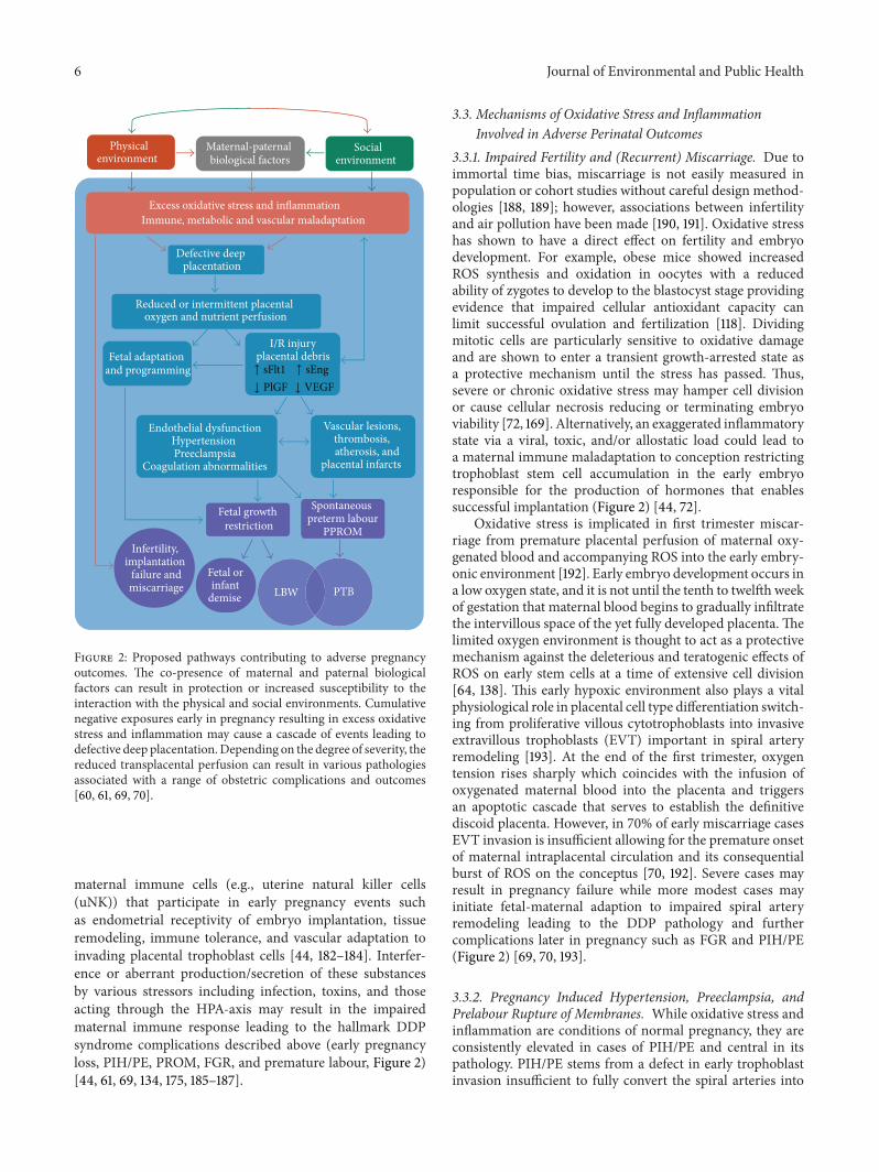
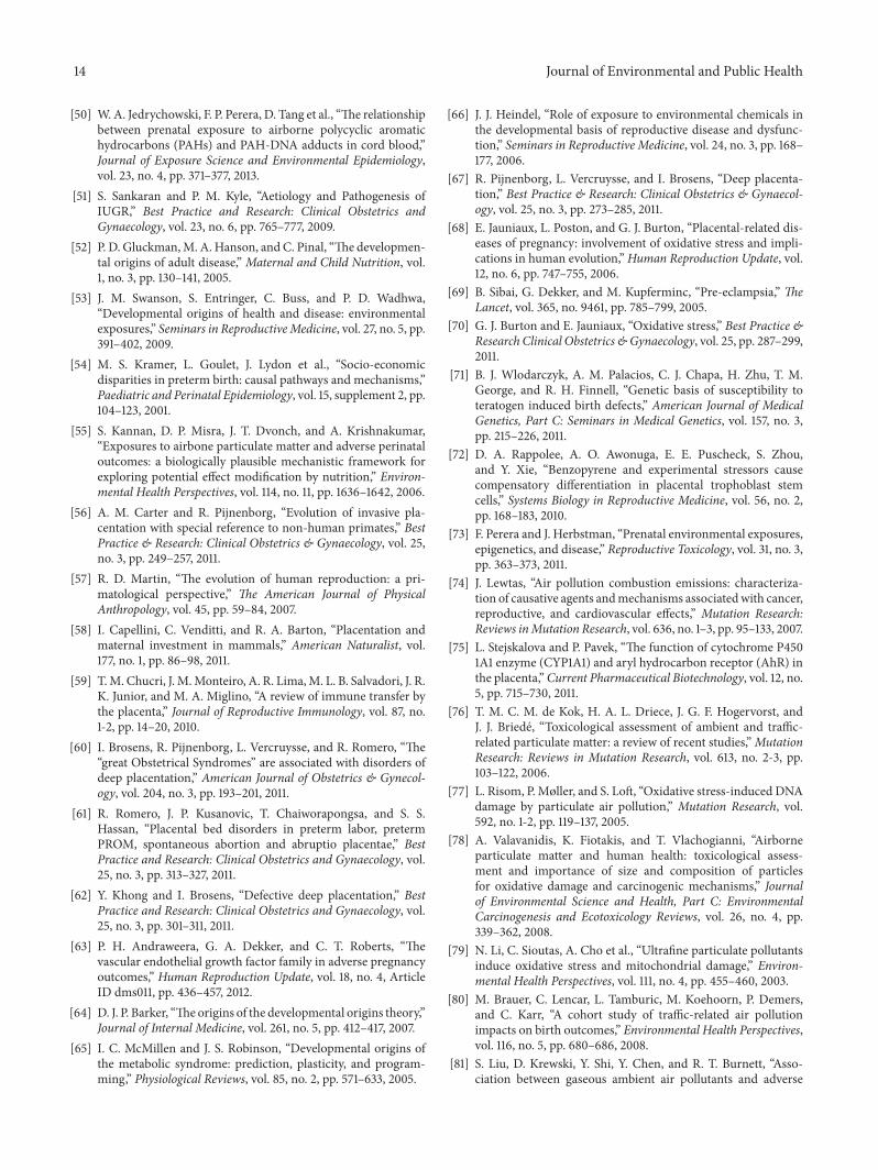
Figure 1 illustrates the interconnectedness between par-ticulate air pollution (PM/PAH) and SES on how they mayact discretely or in a combined manner to yield APOs.Using Figure 1 as a guide, the following text will review thetwo major mechanisms (oxidative stress and inflammation)through which the physical and social environments arebelieved to negatively affect the fetal-placental unit and howtheymay combine/interact to lead to themultifactorial natureof APOs.
3. Biological Mechanisms Leading toAdverse Pregnancy Outcomes
3.1. Oxidative Stress. Aptly known as “The Oxygen Paradox,”oxygen is both essential and toxic to the multicellular aerobicorganisms whose very evolution was dependent on lever-aging this anaerobic waste by-product into a higher energyproducing advantage [167]. Observed in all mammals, a steepoxygen tension gradient from 20% in our atmosphere to 3-4%oxygen concentration in most internal tissues is the primarydefense against oxidative damage. Secondary and tertiarylayers of protection include antioxidant defenses as well asdamage removal, repair, and apoptotic response systems [168,169]. These genetically adaptive responses are upregulatedin the presence of reactive oxygen species (ROS) generatedas natural by-products of cellular aerobic metabolism andexposure to various toxins. Oxidative stress occurs whenthere is an imbalance between pro- and antioxidant capacity.
For example, superoxide is the most common intracellu-lar ROS in mammals. It is produced by the mitochondria as ametabolic by-product but also from the metabolism of vari-ous growth factors, drugs, and toxins by oxidizing enzymessuch as NADPH-oxidase and cytochrome P450 (CYP450).Superoxide is reduced by superoxide dismutase (SOD) into

Journal of Environmental and Public Health 5
Social environmentMaternal dietSmoking/ETSChronic stress
Abnormal placentation and adverse pregnancy outcomes
Physical environment
PM10, PM2.5, UFP, PAHs, and metals
Oxidative stress
Inflammation
Figure 1: A conceptual framework of the shared mechanisms of socioeconomic determinants and particulate air pollution exposurecontributing to adverse pregnancy outcomes. The physical environment (orange) consisting of particulate air pollution and the socialenvironment (green) consisting of community and individual-level social factors/stressors converge to affect the fetal-placental environment(blue) via oxidative stress and inflammatory mechanisms potentially leading to adverse pregnancy outcomes.
hydrogen peroxide (H2O2) which is then further reduced into
water by glutathione peroxidase (GPx) and catalase. Undernormal physiological conditions H
2O2acts as intracellular
secondary messengers; however, it’s accumulation along withsuperoxide can react with free iron ions or nitric oxide toform highly toxic hydroxyl (OH∙) or peroxynitrite (ONOO−)ions, respectively [70, 168]. Free iron is a common metalfound absorbed to PM, and the antioxidant heme oxygenase-1 (HO-1) facilitates its conjugation and removal throughthe increased availability of ferritin thereby preventing theformation of reactive hydroxyl molecules [92, 170–172]. Defi-ciencies inHO-1 have been associatedwith severalAPOs suchas recurrent miscarriage, FGR, and preeclampsia [171, 172].
Common antioxidants include enzymatic (e.g., SOD,GPx, catalase, and HO-1) and nonenzymatic compounds(e.g., vitamin C and E, glutathione, 𝛽-carotene, and ubiquin-one) [118]. Genetic polymorphisms and/or micronutrientdeficiencies in antioxidant enzymes precursors can impairantioxidant capacity, while chronic exposure to toxicants,psychosocial stress, bacteria, viruses, and other inducersof inflammation can foster prooxidant burden [70, 77, 118,172]. Oxidative stress is unavoidable; however, under optimalconditions the presence of ROS leads to homeostatic adap-tation and are safely removed. Failure to effectively manageoxidative stress can result in altered cellular function as excessROS degrade lipids, proteins, and DNA potentially initiatingpathological processes. Refer to [168] for an extensive reviewon the role of cellular ROS in pregnancy outcomes.
3.2. Inflammation and Immunologic Alterations. It is wellrecognized that the maternal immune system plays a centralrole throughout the entire pregnancy, from preimplanta-tion to parturition, and is influenced by the inflammatoryresponse of the mother to her environment as well as toher partner. Alternative to previously hypothesized [173], thematernal immune system is not passive or suppressed duringimplantation and development of the semiallogeneic placentaand fetus. Rather, it exerts executive influence on the estab-lishment and progression of the pregnancy as an immune-mediated quality control mechanism to maximize maternal
and offspring health [44, 173]. This is achieved by favour-ing pro- or anti-inflammatory environments at differenttimes during pregnancy for different purposes. For instance,implantation, initial placentation, and parturition are charac-terized by a proinflammatory environment whereas an anti-inflammatory state prevails for most of midgestation [174].The favoured localized immunological response howeveris highly modified by the infectious, inflammatory, stress,nutritional, and metabolic status of the individual and thuscan be influenced by environmental agents such as PM [175–177] and/or available coping, social, and nutritional resources[44, 128, 164, 178]. Therefore, inflammation is believed to beone pathway involved in both PM and SES-mediated APOs.
Chronic and acute inflammation is a complex responseprocess mediated by a real or perceived attack from for-eign substances. The innate immune response is the rapidautomatic response to externally originating (exogenous)substances such as pathogens, but also from internal (endoge-nous) danger signals including products of trauma, ischemia,necrosis, or oxidative stress [179]. The response includes therelease of proinflammatory signaling cytokine proteins suchas interleukins IL-1𝛽, IL-6, and tumour necrosis factor (TNF-𝛼) which serve to recruit neutrophils to affected tissues. How-ever, the recruited neutrophils release ROS and hydrolyticproinflammatory enzymes (inducible nitric oxide synthase(iNOS), cyclooxygenase (COX-2), and prostaglandins (PG-E2)) which disturb normal cells in addition to affected
tissues. This in turn leads to increased ROS and oxidativestress [180, 181]. The placenta plays a role at the maternal-fetal interface as the main producer of endocrine steroidand protein hormones as well as the immunologic barrierbetween mother and fetus which positively interact for thesuccess of the pregnancy [44, 173]. This is achieved througha nonlinear series of positive and negative feedback path-ways with the stimulation or suppression of molecules withpro- and anti-immunosuppressant properties (interleukins,galectins, placental growth factor, and human chorionicgonadotropin (hCG)) [182–184]. The production of thesecytokines, chemokines, and other immune-regulatory agentsmediates the coordination,migration, and function of several
6 Journal of Environmental and Public Health
Fetal or infant
demise LBW PTB
I/R injuryplacental debris
Fetal growth restriction
Spontaneous preterm labour
PPROM
Vascular lesions, thrombosis, atherosis, and
placental infarcts
Endothelial dysfunctionHypertension Preeclampsia
Coagulation abnormalities
Defective deep placentation
Reduced or intermittent placental oxygen and nutrient perfusion
Fetal adaptation and programming
Infertility, implantation
failure andmiscarriage
Excess oxidative stress and inflammationImmune, metabolic and vascular maladaptation
Social environment
Maternal-paternal biological factors
Physical environment
↑ sFlt1 ↑ sEng↓ PlGF ↓ VEGF
Figure 2: Proposed pathways contributing to adverse pregnancyoutcomes. The co-presence of maternal and paternal biologicalfactors can result in protection or increased susceptibility to theinteraction with the physical and social environments. Cumulativenegative exposures early in pregnancy resulting in excess oxidativestress and inflammation may cause a cascade of events leading todefective deep placentation.Depending on the degree of severity, thereduced transplacental perfusion can result in various pathologiesassociated with a range of obstetric complications and outcomes[60, 61, 69, 70].
maternal immune cells (e.g., uterine natural killer cells(uNK)) that participate in early pregnancy events suchas endometrial receptivity of embryo implantation, tissueremodeling, immune tolerance, and vascular adaptation toinvading placental trophoblast cells [44, 182–184]. Interfer-ence or aberrant production/secretion of these substancesby various stressors including infection, toxins, and thoseacting through the HPA-axis may result in the impairedmaternal immune response leading to the hallmark DDPsyndrome complications described above (early pregnancyloss, PIH/PE, PROM, FGR, and premature labour, Figure 2)[44, 61, 69, 134, 175, 185–187].
3.3. Mechanisms of Oxidative Stress and InflammationInvolved in Adverse Perinatal Outcomes
3.3.1. Impaired Fertility and (Recurrent) Miscarriage. Due toimmortal time bias, miscarriage is not easily measured inpopulation or cohort studies without careful design method-ologies [188, 189]; however, associations between infertilityand air pollution have been made [190, 191]. Oxidative stresshas shown to have a direct effect on fertility and embryodevelopment. For example, obese mice showed increasedROS synthesis and oxidation in oocytes with a reducedability of zygotes to develop to the blastocyst stage providingevidence that impaired cellular antioxidant capacity canlimit successful ovulation and fertilization [118]. Dividingmitotic cells are particularly sensitive to oxidative damageand are shown to enter a transient growth-arrested state asa protective mechanism until the stress has passed. Thus,severe or chronic oxidative stress may hamper cell divisionor cause cellular necrosis reducing or terminating embryoviability [72, 169]. Alternatively, an exaggerated inflammatorystate via a viral, toxic, and/or allostatic load could lead toa maternal immune maladaptation to conception restrictingtrophoblast stem cell accumulation in the early embryoresponsible for the production of hormones that enablessuccessful implantation (Figure 2) [44, 72].
Oxidative stress is implicated in first trimester miscar-riage from premature placental perfusion of maternal oxy-genated blood and accompanying ROS into the early embry-onic environment [192]. Early embryo development occurs ina low oxygen state, and it is not until the tenth to twelfth weekof gestation that maternal blood begins to gradually infiltratethe intervillous space of the yet fully developed placenta. Thelimited oxygen environment is thought to act as a protectivemechanism against the deleterious and teratogenic effects ofROS on early stem cells at a time of extensive cell division[64, 138]. This early hypoxic environment also plays a vitalphysiological role in placental cell type differentiation switch-ing from proliferative villous cytotrophoblasts into invasiveextravillous trophoblasts (EVT) important in spiral arteryremodeling [193]. At the end of the first trimester, oxygentension rises sharply which coincides with the infusion ofoxygenated maternal blood into the placenta and triggersan apoptotic cascade that serves to establish the definitivediscoid placenta. However, in 70% of early miscarriage casesEVT invasion is insufficient allowing for the premature onsetof maternal intraplacental circulation and its consequentialburst of ROS on the conceptus [70, 192]. Severe cases mayresult in pregnancy failure while more modest cases mayinitiate fetal-maternal adaption to impaired spiral arteryremodeling leading to the DDP pathology and furthercomplications later in pregnancy such as FGR and PIH/PE(Figure 2) [69, 70, 193].
3.3.2. Pregnancy Induced Hypertension, Preeclampsia, andPrelabour Rupture of Membranes. While oxidative stress andinflammation are conditions of normal pregnancy, they areconsistently elevated in cases of PIH/PE and central in itspathology. PIH/PE stems from a defect in early trophoblastinvasion insufficient to fully convert the spiral arteries into
Journal of Environmental and Public Health 7
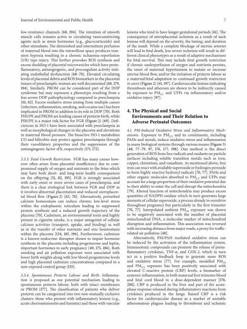
low-resistance channels [68, 194]. The retention of smoothmuscle cells remains active to circulating vasoconstrictingagents such as stress hormones (e.g., glucocorticoids) andother stimulants. The diminished and intermittent perfusionof maternal blood into the intravillous space produces tran-sient hypoxia resulting in a chronic ischaemia-reperfusion(I/R) type injury. This further provokes ROS synthesis andexcess shedding of placental microvesicles which have proin-flammatory, antiangiogenic, and procoagulant activity initi-ating endothelial dysfunction [68–70]. Elevated circulatinglevels of placental debris and ROS biomarkers in the placentaltissues of preeclamptic women are well documented [68, 179,194]. Similarly, PROM can be considered part of the DDPsyndrome but may represent a phenotype resulting from aless severe DDP pathophysiology compared to preeclampsia[61, 62]. Excess oxidative stress arising from multiple causes(infection, inflammation, smoking, and cocaine use) has beenimplicated in PROM in addition to its role in DDP [70]. BothPIH/PE and PROMare leading causes of preterm birth, whilePIH/PE is a major risk factor for FGR (Figure 2) [69]. Defi-ciencies in HO-1 have been associated with preeclampsia aswell as morphological changes in the placenta and elevationsin maternal blood pressure. The bioactive HO-1 metabolitesCO and bilirubin may protect against preeclampsia throughtheir vasodilatory properties and the suppression of theantiangiogenic factor sFlt, respectively [171, 172].
3.3.3. Fetal Growth Restriction. FGR has many causes how-ever often arises from placental insufficiency due to com-promised supply of oxygen and nutrients to the fetus whichmay have both short- and long-term health consequenceson the offspring [51, 82, 195]. FGR is strongly associatedwith early onset or more severe cases of preeclampsia, andthere is a clear etiological link between FGR and DDP asit involves abnormal placentation and reduced uteroplacen-tal blood flow (Figure 2) [62, 70]. Alternatively, perturbedcalcium homeostasis can induce chronic low-level stresswithin the endoplasmic reticulum leading to suppressedprotein synthesis and a reduced growth trajectory of theplacenta [70]. Cadmium, an environmental toxin and highlypresent in cigarette smoke, is a major antagonist of cellularcalcium activities (transport, uptake, and binding) as wellas in the transfer of other nutrients and zinc homeostasiswithin the placenta [134, 185, 196]. Furthermore, cadmiumis a known endocrine disruptor shown to impair hormonesynthesis in the placenta including progesterone and leptin,important hormones in early pregnancy [49, 175, 186]. Bothsmoking and air pollution exposure were associated withlower birth weights along with low blood progesterone levelsand high placental cadmium concentrations compared to anon-exposed control group [135].
3.3.4. Spontaneous Preterm Labour and Birth. Inflamma-tion is proposed as one potential mechanism leading tospontaneous preterm labour, both with intact membranesor PROM [177]. The classification of patients who deliverpreterm can be categorized into two non-mutually exclusiveclusters: those who present with inflammatory lesions (e.g.,acute chorioamnionitis and funisitis) and those with vascular
lesions who tend to have longer gestational periods [61]. Theconsequence of uteroplacental ischemia as a result of suchlesions will depend on the severity, the timing, and durationof the insult. While a complete blockage of uterine arterieswill lead to fetal death, less severe ischemia will result in dif-ferent clinical phenotypes as a result of adaptive mechanismsfor fetal survival. This may include fetal growth restrictionif chronic underperfusion of oxygen and nutrients persists,the onset of maternal hypertension to sustain or increaseuterine blood flow, and/or the initiation of preterm labour asa maternal/fetal adaptation to continued growth restrictionin utero (Figure 2) [61, 197]. Cardiovascular lesions indicatingthrombosis and atherosis are shown to be indirectly causedby exposure to PM
2.5and UFPs via inflammatory and/or
oxidative injury [97].
4. The Physical and SocialEnvironments and Their Relation toAdverse Perinatal Outcomes
4.1. PM-Induced Oxidative Stress and Inflammatory Mech-anisms. Exposure to PM
2.5and its constituents, including
PAHs and metals, induce oxidative stress and inflammationin many biological systems through various means (Figure 3)[48, 77–79, 97, 176, 177, 198]. One method is the directgeneration of ROS from free radicals and oxidants on particlesurfaces including soluble transition metals such as iron,copper, chromium, and vanadium. As mentioned above, freeiron can react with available superoxide or hydrogen peroxideto form highly reactive hydroxyl radicals [70, 77]. PAHs andother organic molecules absorbed to PM
2.5and UFPs may
account for a large proportion of their oxidative potential dueto their ability to enter the cell and disrupt the mitochondria[79]. Altered function of mitochondria may produce excessquantities of NADPH-oxidase which in turn generates largeamounts of cellular superoxide, a process already in overdrivethroughout pregnancy but particularly in the first trimester[70, 77]. Interpolated ambient PM
10exposure was shown
to be negatively associated with the number of placentalmitochondrial DNA, a molecular marker of mitochondrialdisruption and inflammation. This association was reversedwith increasing distance frommajor roads, a proxy for traffic-related air pollution [48].
Alternatively, PM/PAH mediated oxidative stress canbe induced by the activation of the inflammation system.Immunotoxic compounds can promote the release of proin-flammatory cytokines, TNF-𝛼, and COX-2, which in turnact in a positive feedback loop to generate more ROSand oxidative stress [77]. For example, modelled PM
10
and PM2.5
exposure has been positively associated withelevated C-reactive protein (CRP) levels, a biomarker ofsystemic inflammation, in bothmaternal first trimester bloodand fetal cord blood in a dose-dependent manner [176,200]. CRP is produced in the liver and part of the acute-phase response released during inflammatory reactions fromcytokines produced in the lungs. Raised CRP is a riskfactor for cardiovascular disease as a marker of unstableatheromatous plagues leading to thrombosis and ischemic

8 Journal of Environmental and Public Health
Particulate air pollution
Local and regional sourcesTraffic-related
emissionsDiesel
emissionIndustrial emissions
Tobacco smoke exposure
CYP1A1 phase I enzymes
activity
High reactive
metabolites Low reactive metabolites
CYP1A1 phase II enzymes
PAHsInhaled into alveolae
Translocate in blood
Cardiovascular effects
Autonomic effects
Pulmonary inflammation
Spill-over of cytokines
Movement of activated
inflammatory cells
SNPs or low micronutrient intake/status
Excess placentaloxidative stress
and inflammation
Vascular endothelial dysfunction
Transition metals
Abnormal placentation,impaired placental perfusion, and
adverse pregnancy outcomes
↑ AhR
↑ Placental
↑ Placental
PM10 , PM2.5 , and UFPs
Figure 3: Proposed pathways of particulate air pollution contributing to oxidative stress and inflammation leading to adverse pregnancyoutcomes. Exposure to PM and its associated constituents of transition metals, PAHs, and other organic molecules affect the cardiovascularand metabolic systems which are highly active throughout pregnancy. For example, detoxification of PAHs and other organic toxins activateAhR signalling resulting in additional oxidative stress if antioxidant defenses are limited or impaired [55, 79, 98, 108, 109, 199].
events [97]. Exposure to diesel exhaust in healthy humanvolunteers resulted in pulmonary inflammation in additionto systemic inflammation, prothrombotic changes, and othercardiovascular effects consequent of proinflammatory events[99, 201]. This hyper proinflammatory state, along withoxidative stress, is hypothesized to contribute to several APOs[69, 70, 174, 181, 202].
Indirectly, the cellular detoxification of PAHs can induceoxidative stress and cytotoxicity by forming potent ROSmetabolite by-products. Specifically, PAHs and other organicxenobiotics (notably PCBs and dioxins) are detoxified by thecytochrome P-450 (CYP) superfamily of Phase I and PhaseII metabolizing enzymes. The expression of these enzymesis highly modulated by genetic polymorphisms, steroid/sexhormones such as glucocorticoids, insulin, estrogens, andprogesterone, and micronutrient/dietary deficiencies [74,75, 128, 203, 204]. Furthermore, hypoxia, infection, andinflammation are shown, in general, to downregulate CYPenzymes which may affect the clearance and bioavailabilityof growth factors, hormones, drugs, and toxins [203, 205].CYP has numerous isoforms which are expressed in many
tissues especially the liver. CYP1A1 is the only isoform alsosignificantly expressed in the placenta throughout pregnancyresponsible for metabolizing steroid/sex hormones, growthfactors, and fatty acids in addition to toxins [75]. Theseexogenous and endogenous substances act as ligands toactivate the aryl hydrocarbon receptor (AhR), a transcriptionfactor that mediates the biotransformation of such ligands(PAHs, estradiol, etc.) into more polar and bioavailablemetabolites by upregulating CYP enzymes. However, certainmetabolites of PAHs (e.g., o-quinones, arene oxide, and diolepoxide) bind to DNA, RNA, and protein macromoleculesto form toxic adducts that disrupt DNA replication andare considered mutagenic [72, 75]. Such DNA adducts havebeen found in newborn cord-blood positively correlated withmaternal exposure to PAHs [50]. PAHs have also shown tosignificantly decrease the accumulation of trophoblast stemcells in the early placenta thereby limiting their differentiationinto other cell types vital for hormone synthesis and ongoingplacental development, a process that could contribute toDDP [72]. Direct prenatal exposure to airborne PAHs hasbeen associated with FGRwith an increased exposure-related
Journal of Environmental and Public Health 9
risk in the first trimester [206, 207]. Secondary (PhaseII) metabolizing enzymes are required to further detoxifyreactive PAH-metabolites in which their inefficient clearanceresults in prolonged exposure leading to sustained cytotoxic-ity andmutagenicity. Phase II enzymes include glutathione s-transferases (GSTs), UDP-glucuronosyltransferases (UGTs),NAD(P)H-dependent quinone oxidoreductase-1 (NQO1),and aldehyde dehydrogenase-3 (ALDH3) [75, 205].
4.2. Maternal Diet and Micronutrient Intake. Adequate dietandmicronutrient status provides resilience against oxidativestress and inflammation caused by various exposures includ-ing air pollution, allostatic stress, infection, and smoking(Figure 4) [55, 118, 128, 129, 131, 143]. Many micronutrientssuch as essential trace metals are vital cofactors in severalantioxidant enzyme systems. For example, copper and zincare necessary in the production of SOD. Similarly, seleniumand its incorporation into the amino acid selenocysteine arerequired for the functionality of all selenoenzymes, includingGPx and GST. Thus, selenium is essential in several aspectsof human health, particularly conditions involving oxidativestress and inflammation such as CVD, immune function,cancer, and reproduction, but also thyroid regulation andbrain diseases [208, 209].
ROSmayhave direct effects on oocyte quality and appearsto be modulated by dietary antioxidant supplements [118].Women who are obese tend to have higher rates of infer-tility that correlate with increased levels of oxidative stressbiomarkers in their blood as excess glucose availability leadsto higher mitochondrial ROS synthesis [70, 118]. Seleniumdeficiency and corresponding reduced GPx activity has beendocumented in cases of recurrent miscarriage and sponta-neous abortions [210–212] and has also been associated withpreeclampsia and pretermbirth [213, 214].However, given thesupposed role of oxidative stress in preeclampsia, treatmentwith certain antioxidants (notably vitamins C and E) hasnot produced reliable preventative results in experimentaltrials [69]. One hypothesis is that inappropriate antioxidantregiment and/or administration too late in gestation areresponsible and new therapeutic candidates include mela-tonin and selenium [118]. Interestingly, national programs inFinland andNew Zealand fortifying food with selenium havebeen associated with a significant reduction in the rate ofpreeclampsia [215].
Oxidative stress negatively affects the placental transportof amino acids and glucose [45]. Furthermore, fatty acidsand low density lipid (LDL) cholesterols necessary for theplacental synthesis of oestrogens and progesterone are par-ticularly vulnerable to oxidative injury [216]. Regulation ofplacental nutrient transport is controlled by several differentmechanism, including imprinted genes, placental signalingpathways, various cytokines, and hormones such as insulin,leptin, glucocorticoids, and oestrogens (for review see [45]).The major placental transfer mechanisms include simplediffusion of lipophilic substances (e.g., oxygen, CO
2, fatty
acids, steroids, fat soluble vitamins, and anesthetic gases),restricted diffusion of hydrophilic substances, facilitateddiffusion via a membrane bound carrier (e.g., glucose andother carbohydrates), and active transport which requires
Social environment
Unhealthy behaviours
SNPs
Low micronutrient intake/status
Excess glucocorticoid
exposure
Inhibits
HPA axis activation
Adverse psychosocial
factors
Community-level factorsPoverty
marginalizationcrime
Low social supportFood insecurity
Inadequate diet
Tobacco smoke exposure
Abnormal placentation,impaired placental perfusion, and
adverse pregnancy outcomes
Excess placentaloxidative stress
and inflammation
(PAHs and Cd2+)
11𝛽-HSD2↓ Protein
↓ Caloric intake
↑ CRH
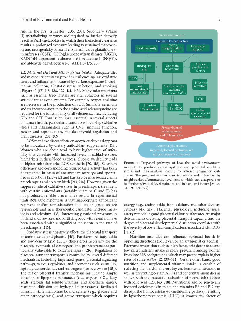
Figure 4: Proposed pathways of how the social environmentinteracts to produce excess systemic and placental oxidativestress and inflammation leading to adverse pregnancy out-comes. The pregnant woman is nested within and influenced byneighbourhood/community-level factors which can exasperate orbuffer the individual-level biological and behavioural factors [24, 26,54, 128, 224, 225].
energy (e.g., amino acids, iron, calcium, and other divalentcations) [45, 217]. Placental physiology, including spiralartery remodeling and placental villous surface area aremajordeterminants dictating placental transport capacity, and thedegree of placental developmental disruption correlates withthe severity of obstetrical complications associated with DDP[51, 62].
Nutrition and diet can influence perinatal health inopposing directions (i.e., it can be an antagonist or agonist).Poor/undernutrition such as high fat/calorie dense food andlow micronutrient intake is more prevalent among womenfrom low SES backgrounds which may partly explain higherrates of some APOs [12, 139–142]. On the other hand, goodnutrition and supplemental vitamin intake is capable ofreducing the toxicity of everyday environmental stressors aswell as preventing certain APOs and congenital anomalies asshown with the successful reduction of neural tube defectswith folic acid [128, 143, 218]. Nutritional and/or geneticallyinduced deficiencies in folate and vitamins B6 and B12 candisrupt the homocysteine-to-methionine pathway resultingin hyperhomocysteinemia (HHC), a known risk factor of
10 Journal of Environmental and Public Health
cardiovascularmorbidities (thrombosis, lesions, and infarcts)and markers of oxidative stress [54, 119, 219, 220]. HHC maysimilarly affect the highly vascularized placenta and has beenassociated with decidual vasculopathy and preterm birth[54, 120]. Omega-3 fatty acids abundant from eating salmonwere shown to improve markers of oxidative stress [221],which may impart neurodevelopmental resilience againststressors [222, 223]. Dietary phytophenols from fruits, veg-etables, herbs, and spices have shown to have antioxidant andanti-inflammatory properties capable of reducing infection-induced inflammatory and contractile pathways in humangestational tissues [129]. Significant differences in pregnancyoutcomes between Dominicans and African Americans bothexposed to similar levels of PAHs inNewYork city neighbour-hoods were thought to be due to healthful dietary/culturalpractices in the Dominican immigrant population [206].
4.3. Maternal Smoking and Environmental Tobacco Smoke(ETS) Exposure. Maternal smoking during pregnancy andexposure to ETS remain to be twomodifiable risk factors withthe greatest potential for beneficial interventions (Figure 4).Their association with numerous APOs including congenitalanomalies is well documented [127, 144–146], as have theirassociated prevalence with indicators of low SES and othersocially patterned risk factors [147–149]. The mechanismsinvolved leading to APOs have been well reviewed [132,134]; however, it is notable that the two main toxins presentin tobacco smoke can also be absorbed to PM
2.5(PAHs
more so than cadmium). Cadmium (Cd) exposure readilyinterferes with the active transport of essential mineralsto the fetus, particularly zinc and calcium [46, 135, 196,226–228]. Cadmium and lead (Pb) exposure has also beenshown to reduce glycogen concentrations thereby potentiallylimiting available glucose to the fetus [229]. Cadmium hasshown to disrupt placental leptin synthesis, a hormonewith several vital functions including placental angiogenesis,immunomodulation, amino acid and fatty acid transport,as well as fetal pancreatic development important in theregulation of insulin-like growth factors and fetal bodyfat accumulation [49, 51]. Finally, synergistic effects in thegeneration of oxidative hydroxyl radicals have been observedbetween tobacco smoke and both ambient PM
2.5and diesel
exhaust particles specifically [35]. Interestingly, the coun-terintuitive association between smoking and lower risk ofpreeclampsia was recently shown to vary according to thetiming and intensity of smoking [230]. It is possible that theincreased exposure to CO from smoking in late gestationacts as a vasodilator and at the same time inhibits therelease of sFlt-1, a hallmark antiangiogenic factor implicatedin the endothelial dysfunction present in preeclampsia [115,230].
4.4. Allostatic Stress and Glucocorticoid Exposure. Reviewedelsewhere [163], the brain is the primary target andmediatingorgan through which SES-related stress pathways are trans-lated to other body systems via the hypothalamic-pituitary-adrenal (HPA) axis. The HPA-axis is actively involved inseveral biological systems, including the cardiovascular,
metabolic, immunological, and endocrinal effects in bothmother and fetus to promote allostatic adaptation [165, 231].Here, the neuroendocrine hormones of the HPA axis, cor-ticotrophin releasing hormone (CRH), adrenocorticotropichormone (ACTH), and glucocorticoids (GC), respectively,coordinate the biological response via feedback loops. Thehuman placenta is also capable of releasing CRH and otherneuropeptides which interact with the HPA axis to regulatethe maternal stress response as well as other normal preg-nancy functions [47]. Proper levels of in utero glucocorticoidsare essential for successful embryo implantation, fetal organmaturation, and the initiation of labour with glucocorticoidlevels gradually increasing over the course of gestation.Normally, levels of maternal cortisol rise sharply in the thirdtrimester causing the release of placental CRH in a positiveadrenal-placental feedback loop. Placental CRH stimulatesfetal cortisol secretion which in turn suppresses placentalprogesterone and activates the release of prostaglandins andoxytocin to promote uterine contractions [47, 232]. However,early and increased levels of fetal glucocorticoids can impairgrowth and predispose to adult-onset diseases [136, 233,234]. The placental enzyme 11𝛽-hydroxysteroid dehydro-genase type 2 (11𝛽-HSD2) protects the fetus from excessendogenous glucocorticoids by converting active cortisol intoinactive cortisone. 11𝛽-HSD2 is hormonally regulatedmakingit susceptible to endocrine disruption from chemical andnonchemical stressors such as maternal anxiety, inflamma-tion, infection, cadmium exposure, and low caloric intake[136, 138, 224, 235, 236]. Placental hypoxia associated withPIH/PE has been shown to suppress 11𝛽-HSD2 activity whichmay be an adaptive response to counteract compromised fetalgrowth by allowing more cortisol to reach the fetus for organdevelopment. Low concentrations/activities of 11𝛽-HSD2 andhigh levels of cortisol have been associated with PTB andFGR [136, 237, 238], two outcomes also associated with poormaternal psychosocial/mental health [233, 234, 239].
Factors affecting 11𝛽-HSD2 activity that are associatedwith low SES include allostatic overload leading to the excessproduction of glucocorticoids that can overwhelm the fetalprotective mechanism (Figure 4) [136, 231, 240]. Indirectly,allostatic load is capable of disrupting the metabolic systemleading to impaired glucose tolerance, insulin resistance,diabetes, and/or obesity, all of which are risk factors forvarious APOs [138, 165, 231]. General maternal undernutri-tion and/or a low dietary protein intake has been shownto impair placental glucose transport and inhibit 11𝛽-HSD2activity in pregnant rats leading to FGR, indicating a possiblemechanism through poor diet [224, 241]. Additionally, cad-mium has also shown to inhibit 11𝛽-HSD2 activity in bothhuman and rodent placentas [225], and prenatal cadmiumexposure has been shown to increase fetal corticosteroneconcentrations in ratswhich resulted in reduced birthweights[236]. This suggests a possible mechanism from active orpassive tobacco smoke exposure or ambient PM
2.5exposure
[135, 242, 243]. Collectively, it is possible for the cumulativeexposures of PM
2.5, smoking, ETS, poor dietary intake, and
other SES-related factors to interact through the same 11𝛽-HSD2 mechanism to increase the risk of impaired fetalgrowth (Figure 4).

Journal of Environmental and Public Health 11
5. Discussion
The ubiquitous exposure to particulate air pollution andits constituents (e.g., PAHs and metals) is but one class ofenvironmental contaminants that can act through oxidativestress, inflammation, and/or endocrine disruption to pro-mote developmental toxicity and adverse perinatal health[177, 244, 245]. Summarized in Figure 2, a perturbed earlyin utero environment can lead to defective deep placentationresulting in a cascade of fetal-placental adaptive mecha-nisms contributing to a range of pregnancy complicationsand adverse outcomes [60]. Here the underlying biological,social, and physical risk factors likely intersect to produceexcessive or atypical oxidative stress, inflammatory response,and biological antagonism to either initiate the defectivedeep placentation pathology and/or contribute to the sever-ity of its phenotype. Socioeconomic disparities are knownto confound the environmental exposure effects; however,they may also act as potential effect modifiers given theiroverlapping etiological mechanisms with PM
2.5exposure.
While the traditional biomedical paradigm that views popu-lations as a collection of independent individuals has yieldeduseful information regarding risk factors, elucidating theintersecting pathways involved in APOs will require placingindividual biologic and behavioural determinants within thesocial and spatial context [22, 246]. It is now well recognizedthat SES operates at multiple levels of organization, andneighbourhood or community-level factors can work toeither ameliorate or exacerbate certain risk factors [15, 24–26]. The healthy migrant paradox exemplifies these effectsin which home country, education, and neighbourhoodqualities combine to modify the expected perinatal outcomesoften observed with low income households [161, 247].
The SES risk factors that overlap or interact with the PM-mediated mechanisms include smoking, nutrition, and psy-chosocial stress acting through the HPA-axis and allostaticload.Given this knowledge, interventions aimed at ameliorat-ing these factors may be the best way to counteract the nega-tive influences of low SES and air pollution exposure on fetaldevelopment. Maternal smoking continues to be one of themost modifiable risk factors to lower the risk of APOs [134,147]. Furthermore, maternal smoking also tends to interactnegatively with nutrient intake and status [133, 248]. Smokersin general have poorer nutritional profiles than nonsmokerswith both behavioural and biological factors independentlyaccounting for the differences inmicronutrients such as folateand essential vitamins and minerals [133, 248–250]. Whilesmokers tend to have lower dietary nutrient intakes, they alsohave an accelerated requirement for micronutrients due toincreased inflammatory cell turnover caused by the oxidativestress of smoking, an effect more pronounced among heavyand long-time smokers [248]. These interacting effects ofsmoking andnutrition are further compounded by their asso-ciationwith other indicators of low SES such as low educationand income contributing to allostatic load [139, 251]. Nutrientintake may be ameliorative after an insult has occurredas shown in rat models of fetal alcohol syndrome wherean omega-3 fatty acid enriched diet reversed the cellulareffects of prenatal ethanol exposure on the fetal brain [222].
Therefore with respect to policy interventions, nutrition inthe form of improved food security and micronutrient intakemay serve to counteract the negative influences of both lowSES and air pollution exposure [252–257].
The complex mixture of particulate air pollution, espe-cially PM
2.5and UFPs which includes absorbed PAHs and
various metals, also emerges as an important target for riskreduction and management. The deserved scrutiny stemsfrom their ubiquity in the environment, the myriad of emis-sion sources, and their established association with APOs[88, 98, 258]. The pervasiveness of PM
2.5and UFPs in the
environment means that a high proportion of people areexposed resulting in a high etiological fraction. Therefore,even a modest reduction in exposure will have a largepopulation effect with reduction of the societal costs of APOs[259]. Notably, their sources are primarily local, such asvehicle emissions and industrial land-use. This makes themmodifiable risk factors that can be addressed at the municipaland provincial/state level with better urban planning toreduce vehicle traffic, increasing access to green-space andenforcing air quality regulations [260–263].
Not unlike the accumulation of evidence on smoking andhealth outcomes or that of air pollution on cardiovascularand pulmonary health [264], the epidemiological and toxi-cological research over the past two decades has establisheda consistent dose-response association with high biologicalspecificity, temporality, and plausibility [3, 55, 177]. Takenin concert, these characteristics and further corroboratingresearch should lend strength for evidence-based policy forintervention strategies targeting high risk areas in order toreduce the environmental burden of disease attributed toparticulate air pollution [98, 265, 266].
6. Conclusion
Adverse pregnancy outcomes such as fetal growth restrictionand preterm birth are a public health priority of globalimportance. We have brought together the multidisciplinaryliterature on the current state of evidence linking the phys-ical and social environment to specific adverse pregnancyoutcomes. The evidence suggests that various exposures,whether socially or environmentally determined, may beinterpreted by the fetoplacental unit in similar ways resultingin a common pathological foundation for adverse outcomes,namely, deficient deep placentation. Given this background,well planned future epidemiology studies using multilevelmodels exploring various biological effects of the social andphysical environment will have the potential to provide theevidence to establish crucial windows of fetal vulnerabilitywith an aim to identify and mitigate modifiable risk factors.
Acronyms
AhR: Aryl hydrocarbon receptorACTH: Adrenocorticotropic hormoneAPO: Adverse pregnancy/perinatal outcomeCd: Cadmium
12 Journal of Environmental and Public Health
CRH: Corticotropin releasing hormoneCOX-2: Cyclooxygenase-2CO: Carbon monoxideCRP: C-reactive proteinCVD: Cardiovascular diseaseCYP: Cytochrome P450DPP: Defective deep placentationETS: Environmental tobacco smokeEVT: Extravillous trophoblastIUGR or FGR: Intrauterine or fetal growth restrictionHHC: HyperhomocysteinemiaHPA axis: Hypothalamus-pituitary-adrenal axisI/R injury: Ischemic-reperfusion injuryGPx: Glutathione peroxidaseGST: Glutathione-S-transferaseLBW: Low birth weightLDL: Low Density lipoproteinsNO, NO
2: Nitrogen oxide/dioxide
PAH: Polycyclic aromatic hydrocarbonsPAP: Particulate air pollutionPIH/PE: Pregnancy induced
hypertension/preeclampsiaPM: Particulate matterpPROM: (Premature) Prelabour rupture of
membranesPTB: Preterm birthROS: Reactive oxygen speciesSES: Socioeconomic statusSNP: Single nucleotide polymorphismSOD: Superoxide dismutasesEng: Soluble endoglinsFlt: Soluble fms-like tyrosine kinaseUFP: Ultrafine particlesuNK (cells): Uterine natural killer11𝛽-HSD2: 11𝛽-Hydroxysteroid dehydrogenase type 2.
Conflict of InterestsThe authors declare that they have no conflict of interests.
AcknowledgmentsThis research was funded by a Canadian Institute of HealthResearch Grant (IPH-98849). Dr. Arbour is also fundedthrough a Michael Smith Foundation for Health ResearchSalary Award. Funders provided no role in the direction ofthis review paper.
References
[1] R. J. Sram, B. Binkova, J. Dejmek, and M. Bobak, “Ambient airpollution and pregnancy outcomes: a review of the literature,”Environmental Health Perspectives, vol. 113, no. 4, pp. 375–382,2005.
[2] P. S. Shah and T. Balkhair, “Air pollution and birth outcomes: asystematic review,” Environment International, vol. 37, no. 2, pp.498–516, 2011.
[3] C. H. Backes, T. Nelin, M. W. Gorr, and L. E. Wold, “Early lifeexposure to air pollution: how bad is it?” Toxicology Letters, vol.216, no. 1, pp. 47–53, 2013.
[4] M. Maisonet, A. Correa, D. Misra, and J. J. K. Jaakkola, “Areview of the literature on the effects of ambient air pollutionon fetal growth,” Environmental Research, vol. 95, no. 1, pp. 106–115, 2004.
[5] C. Protano, T. Scalise, G. B. Orsi, and M. Vitali, “A systematicreview of benzene exposure during pregnancy and adverseoutcomes on intrauterine development and birth: still far fromscientific evidence,” Annali di Igiene, vol. 24, no. 6, pp. 451–463,2012.
[6] C. Bosetti, M. J. Nieuwenhuijsen, S. Gallus, S. Cipriani, C.La Vecchia, and F. Parazzini, “Ambient particulate matter andpreterm birth or birth weight: a review of the literature,”Archives of Toxicology, vol. 84, no. 6, pp. 447–460, 2010.
[7] M. Lacasana, A. Esplugues, and F. Ballester, “Exposure toambient air pollution and prenatal and early childhood healtheffects,”European Journal of Epidemiology, vol. 20, no. 2, pp. 183–199, 2005.
[8] S. V. Glinianaia, J. Rankin, R. Bell, T. Pless-Mulloli, and D.Howel, “Particulate air pollution and fetal health: a systematicreview of the epidemiologic evidence,” Epidemiology, vol. 15, no.1, pp. 36–45, 2004.
[9] P. Dadvand, J. Parker, M. L. Bell et al., “Maternal exposure toparticulate air pollution and term birth weight: a multi-countryevaluation of effect and heterogeneity,” Environmental HealthPerspectives, vol. 121, no. 3, pp. 267–373, 2013.
[10] J. D. Parker, K. C. Schoendorf, and J. L. Kiely, “Associationsbetween measures of socioeconomic status and low birthweight, small for gestational age, and premature delivery in theUnited States,”Annals of Epidemiology, vol. 4, no. 4, pp. 271–278,1994.
[11] M. A. Wilcox, S. J. Smith, I. R. Johnson, P. V. Maynard, andC. E. Chilvers, “The effect of social deprivation on birthweight,excluding physiological and pathological effects,”British Journalof Obstetrics and Gynaecology, vol. 102, no. 11, pp. 918–924, 1995.
[12] M. S. Kramer, L. Seguin, J. Lydon, and L. Goulet, “Socio-economic disparities in pregnancy outcome: why do the poorfare so poorly?” Paediatric and Perinatal Epidemiology, vol. 14,no. 3, pp. 194–210, 2000.
[13] K. S. Joseph, R. M. Liston, L. Dodds, L. Dahlgren, and A.C. Allen, “Socioeconomic status and perinatal outcomes in asetting with universal access to essential health care services,”CMAJ, vol. 177, no. 6, pp. 583–590, 2007.
[14] Z.-C. Luo, R. Wilkins, and M. S. Kramer, “Effect of neighbour-hood income and maternal education on birth outcomes: apopulation-based study,”CanadianMedical Association Journal,vol. 174, no. 10, pp. 1415–1420, 2006.
[15] P. Blumenshine, S. Egerter, C. J. Barclay, C. Cubbin, and P.Braveman, “Socioeconomic disparities in adverse birth out-comes: a systematic review,”TheAmerican Journal of PreventiveMedicine, vol. 39, no. 3, pp. 263–272, 2010.
[16] C. B. Petersen, L. H. Mortensen, C. S. Morgen et al., “Socio-economic inequality in preterm birth: a comparative studyof the Nordic countries from 1981 to 2000,” Paediatric andPerinatal Epidemiology, vol. 23, no. 1, pp. 66–75, 2009.
[17] B. MacMahon,M. G. Kovar, and J. J. Feldman, “Infant mortalityrates: socioeconomic factors. United States,” Vital and HealthStatistics Series 1: Programs and Collection Procedures, vol. 22,no. 14, pp. 1–68, 1972.
[18] A. Antonovsky, “Social class and the major cardiovasculardiseases,” Journal of Chronic Diseases, vol. 21, no. 2, pp. 65–106,1968.
Journal of Environmental and Public Health 13
[19] J. Cassel, “The contribution of the social environment to hostresistance: the Fourth Wade Hampton Frost Lecture,” TheAmerican Journal of Epidemiology, vol. 104, pp. 107–123, 1976.
[20] A. V. Diez-Roux, “Bringing context back into epidemiology:variables and fallacies in multilevel analysis,” American Journalof Public Health, vol. 88, no. 2, pp. 216–222, 1998.
[21] K. E. Pickett and M. Pearl, “Multilevel analyses of neighbour-hood socioeconomic context and health outcomes: a criticalreview,” Journal of Epidemiology and Community Health, vol. 55,no. 2, pp. 111–122, 2001.
[22] A. J. McMichael, “Prisoners of the proximate: loosening theconstraints on epidemiology in an age of change,” AmericanJournal of Epidemiology, vol. 149, no. 10, pp. 887–897, 1999.
[23] R. J. Sampson, J. D. Morenoff, and T. Gannon-Rowley, “Assess-ing “neighborhood effects”: social processes and new directionsin research,” Annual Review of Sociology, vol. 28, pp. 443–478,2002.
[24] G. Meng, M. E. Thompson, and G. B. Hall, “Pathways ofneighbourhood-level socio-economic determinants of adversebirth outcomes,” International Journal of Health Geographics,vol. 12, article 32, 2013.
[25] A. Metcalfe, P. Lail, W. A. Ghali, and R. S. Sauve, “The asso-ciation between neighbourhoods and adverse birth outcomes:a systematic review and meta-analysis of multi-level studies,”Paediatric and Perinatal Epidemiology, vol. 25, no. 3, pp. 236–245, 2011.
[26] J. D. D. Morenoff, “Neighborhood mechanisms and the spatialdynamics of birth weight,” American Journal of Sociology, vol.108, no. 5, pp. 976–1017, 2003.
[27] J. E. Clougherty, C. A. Rossi, J. Lawrence et al., “Chronic socialstress and susceptibility to concentrated ambient fine particlesin rats,” Environmental Health Perspectives, vol. 118, no. 6, pp.769–775, 2010.
[28] M. C. Fortin, D. A. Cory-Slechta, P. Ohman-Strickland et al.,“Increased lead biomarker levels are associated with changesin hormonal response to stress in occupationally exposed maleparticipants,” Environmental Health Perspectives, vol. 120, no. 2,pp. 278–283, 2012.
[29] D. A. Cory-Slechta, M. B. Virgolini, A. Rossi-George, M.Thiruchelvam, R. Lisek, andD.Weston, “Lifetime consequencesof combined maternal lead and stress,” Basic and ClinicalPharmacology and Toxicology, vol. 102, no. 2, pp. 218–227, 2008.
[30] B. B. Gump, J. Reihman, P. Stewart, E. Lonky, D. A. Granger, andK. A. Matthews, “Blood lead (Pb) levels: further evidence for anenvironmental mechanism explaining the association betweensocioeconomic status and psychophysiological dysregulation inchildren,” Health Psychology, vol. 28, no. 5, pp. 614–620, 2009.
[31] J. L. Bolton, N. C. Huff, S. H. Smith et al., “Maternal stressand effects of prenatal air pollution on offspring mental healthoutcomes in mice,” Environmental Health Perspectives, vol. 121,no. 9, pp. 1075–1082, 2013.
[32] A. Kortenkamp, M. Faust, M. Scholze, and T. Backhaus, “Low-level exposure to multiple chemicals: reason for human healthconcerns?” Environmental Health Perspectives, vol. 115, supple-ment, pp. 106–114, 2007.
[33] D. O. Carpenter, K. Arcaro, and D. C. Spink, “Understandingthe human health effects of chemical mixtures,” EnvironmentalHealth Perspectives, vol. 110, no. 1, pp. 25–42, 2002.
[34] C. V. Rider, J. R. Furr, V. S. Wilson, and L. E. Gray Jr.,“Cumulative effects of in utero administration of mixturesof reproductive toxicants that disrupt common target tissues
via diverse mechanisms of toxicity,” International Journal ofAndrology, vol. 33, no. 2, pp. 443–462, 2010.
[35] A. Valavanidis, T. Vlachogianni, and K. Fiotakis, “Tobaccosmoke: involvement of reactive oxygen species and stable freeradicals in mechanisms of oxidative damage, carcinogenesisand synergistic effects with other respirable particles,” Interna-tional Journal of Environmental Research and Public Health, vol.6, no. 2, pp. 445–462, 2009.
[36] P. L. de Fur, G. W. Evans, E. A. C. Hubal, A. D. Kyle, R.A. Morello-Frosch, and D. R. Williams, “Vulnerability as afunction of individual and group resources in cumulative riskassessment,” Environmental Health Perspectives, vol. 115, no. 5,pp. 817–824, 2007.
[37] R. Morello-Frosch and E. D. Shenassa, “The environmental“Riskscape” and social inequality: implications for explainingmaternal and child health disparities,” Environmental HealthPerspectives, vol. 114, no. 8, pp. 1150–1153, 2006.
[38] J. G. Su, R. Morello-Frosch, B. M. Jesdale, A. D. Kyle, B.Shamasunder, and M. Jerrett, “An index for assessing demo-graphic inequalities in cumulative environmental hazards withapplication to Los Angeles, California,” Environmental Scienceand Technology, vol. 43, no. 20, pp. 7626–7634, 2009.
[39] G. C. Gee and D. C. Payne-Sturges, “Environmental healthdisparities: a framework integrating psychosocial and environ-mental concepts,” Environmental Health Perspectives, vol. 112,no. 17, pp. 1645–1653, 2004.
[40] J. Molitor, J. G. Su, N.-T. Molitor et al., “Identifying vulnerablepopulations through an examination of the association betweenmultipollutant profiles and poverty,” Environmental Science &Technology, vol. 45, no. 18, pp. 7754–7760, 2011.
[41] National Environmental Justice Advisory Council, Nation-ally Consistent Environmental Justice Screening Approaches,National Environmental JusticeAdvisoryCouncil,Washington,DC, USA, 2010.
[42] S. H. Linder and K. Sexton, “Conceptual models for cumulativerisk assessment,”TheAmerican Journal of Public Health, vol. 101,no. 1, pp. S74–S81, 2011.
[43] K. Sexton and S. H. Linder, “Cumulative risk assessment forcombined health effects from chemical and nonchemical stres-sors,” American Journal of Public Health, vol. 101, supplement 1,pp. S81–S88, 2011.
[44] S.A. Robertson, “Immune regulation of conception and embryoimplantation-all about quality control?” Journal of ReproductiveImmunology, vol. 85, no. 1, pp. 51–57, 2010.
[45] S. Lager and T. L. Powell, “Regulation of nutrient transportacross the placenta,” Journal of Pregnancy, vol. 2012, Article ID179827, 14 pages, 2012.
[46] M. F. McAleer and R. S. Tuan, “Cytotoxicant-induced tro-phoblast dysfunction and abnormal pregnancy outcomes: roleof zinc and metallothionein,” Birth Defects Research Part C:Embryo Today: Reviews, vol. 72, no. 4, pp. 361–370, 2004.
[47] M. Weinstock, “The potential influence of maternal stresshormones on development and mental health of the offspring,”Brain, Behavior, and Immunity, vol. 19, no. 4, pp. 296–308, 2005.
[48] B. G. Janssen, E. Munters, N. Pieters et al., “Placental mitochon-drial DNA content and particulate air pollution during in Uterolife,” Environmental Health Perspectives, vol. 120, no. 9, pp. 1346–1352, 2012.
[49] S. Stasenko, E. M. Bradford, M. Piasek et al., “Metals in humanplacenta: focus on the effects of cadmium on steroid hormonesand leptin,” Journal of Applied Toxicology, vol. 30, no. 3, pp. 242–253, 2010.
14 Journal of Environmental and Public Health
[50] W. A. Jedrychowski, F. P. Perera, D. Tang et al., “The relationshipbetween prenatal exposure to airborne polycyclic aromatichydrocarbons (PAHs) and PAH-DNA adducts in cord blood,”Journal of Exposure Science and Environmental Epidemiology,vol. 23, no. 4, pp. 371–377, 2013.
[51] S. Sankaran and P. M. Kyle, “Aetiology and Pathogenesis ofIUGR,” Best Practice and Research: Clinical Obstetrics andGynaecology, vol. 23, no. 6, pp. 765–777, 2009.
[52] P. D. Gluckman,M. A. Hanson, and C. Pinal, “The developmen-tal origins of adult disease,” Maternal and Child Nutrition, vol.1, no. 3, pp. 130–141, 2005.
[53] J. M. Swanson, S. Entringer, C. Buss, and P. D. Wadhwa,“Developmental origins of health and disease: environmentalexposures,” Seminars in ReproductiveMedicine, vol. 27, no. 5, pp.391–402, 2009.
[54] M. S. Kramer, L. Goulet, J. Lydon et al., “Socio-economicdisparities in preterm birth: causal pathways and mechanisms,”Paediatric and Perinatal Epidemiology, vol. 15, supplement 2, pp.104–123, 2001.
[55] S. Kannan, D. P. Misra, J. T. Dvonch, and A. Krishnakumar,“Exposures to airbone particulate matter and adverse perinataloutcomes: a biologically plausible mechanistic framework forexploring potential effect modification by nutrition,” Environ-mental Health Perspectives, vol. 114, no. 11, pp. 1636–1642, 2006.
[56] A. M. Carter and R. Pijnenborg, “Evolution of invasive pla-centation with special reference to non-human primates,” BestPractice & Research: Clinical Obstetrics & Gynaecology, vol. 25,no. 3, pp. 249–257, 2011.
[57] R. D. Martin, “The evolution of human reproduction: a pri-matological perspective,” The American Journal of PhysicalAnthropology, vol. 45, pp. 59–84, 2007.
[58] I. Capellini, C. Venditti, and R. A. Barton, “Placentation andmaternal investment in mammals,” American Naturalist, vol.177, no. 1, pp. 86–98, 2011.
[59] T.M. Chucri, J. M.Monteiro, A. R. Lima,M. L. B. Salvadori, J. R.K. Junior, and M. A. Miglino, “A review of immune transfer bythe placenta,” Journal of Reproductive Immunology, vol. 87, no.1-2, pp. 14–20, 2010.
[60] I. Brosens, R. Pijnenborg, L. Vercruysse, and R. Romero, “The“great Obstetrical Syndromes” are associated with disorders ofdeep placentation,” American Journal of Obstetrics & Gynecol-ogy, vol. 204, no. 3, pp. 193–201, 2011.
[61] R. Romero, J. P. Kusanovic, T. Chaiworapongsa, and S. S.Hassan, “Placental bed disorders in preterm labor, pretermPROM, spontaneous abortion and abruptio placentae,” BestPractice and Research: Clinical Obstetrics and Gynaecology, vol.25, no. 3, pp. 313–327, 2011.
[62] Y. Khong and I. Brosens, “Defective deep placentation,” BestPractice and Research: Clinical Obstetrics and Gynaecology, vol.25, no. 3, pp. 301–311, 2011.
[63] P. H. Andraweera, G. A. Dekker, and C. T. Roberts, “Thevascular endothelial growth factor family in adverse pregnancyoutcomes,” Human Reproduction Update, vol. 18, no. 4, ArticleID dms011, pp. 436–457, 2012.
[64] D. J. P. Barker, “Theorigins of the developmental origins theory,”Journal of Internal Medicine, vol. 261, no. 5, pp. 412–417, 2007.
[65] I. C. McMillen and J. S. Robinson, “Developmental origins ofthe metabolic syndrome: prediction, plasticity, and program-ming,” Physiological Reviews, vol. 85, no. 2, pp. 571–633, 2005.
[66] J. J. Heindel, “Role of exposure to environmental chemicals inthe developmental basis of reproductive disease and dysfunc-tion,” Seminars in Reproductive Medicine, vol. 24, no. 3, pp. 168–177, 2006.
[67] R. Pijnenborg, L. Vercruysse, and I. Brosens, “Deep placenta-tion,” Best Practice & Research: Clinical Obstetrics & Gynaecol-ogy, vol. 25, no. 3, pp. 273–285, 2011.
[68] E. Jauniaux, L. Poston, and G. J. Burton, “Placental-related dis-eases of pregnancy: involvement of oxidative stress and impli-cations in human evolution,”Human Reproduction Update, vol.12, no. 6, pp. 747–755, 2006.
[69] B. Sibai, G. Dekker, and M. Kupferminc, “Pre-eclampsia,” TheLancet, vol. 365, no. 9461, pp. 785–799, 2005.
[70] G. J. Burton and E. Jauniaux, “Oxidative stress,” Best Practice &Research Clinical Obstetrics &Gynaecology, vol. 25, pp. 287–299,2011.
[71] B. J. Wlodarczyk, A. M. Palacios, C. J. Chapa, H. Zhu, T. M.George, and R. H. Finnell, “Genetic basis of susceptibility toteratogen induced birth defects,” American Journal of MedicalGenetics, Part C: Seminars in Medical Genetics, vol. 157, no. 3,pp. 215–226, 2011.
[72] D. A. Rappolee, A. O. Awonuga, E. E. Puscheck, S. Zhou,and Y. Xie, “Benzopyrene and experimental stressors causecompensatory differentiation in placental trophoblast stemcells,” Systems Biology in Reproductive Medicine, vol. 56, no. 2,pp. 168–183, 2010.
[73] F. Perera and J. Herbstman, “Prenatal environmental exposures,epigenetics, and disease,” Reproductive Toxicology, vol. 31, no. 3,pp. 363–373, 2011.
[74] J. Lewtas, “Air pollution combustion emissions: characteriza-tion of causative agents andmechanisms associatedwith cancer,reproductive, and cardiovascular effects,” Mutation Research:Reviews inMutation Research, vol. 636, no. 1–3, pp. 95–133, 2007.
[75] L. Stejskalova and P. Pavek, “The function of cytochrome P4501A1 enzyme (CYP1A1) and aryl hydrocarbon receptor (AhR) inthe placenta,”Current Pharmaceutical Biotechnology, vol. 12, no.5, pp. 715–730, 2011.
[76] T. M. C. M. de Kok, H. A. L. Driece, J. G. F. Hogervorst, andJ. J. Briede, “Toxicological assessment of ambient and traffic-related particulate matter: a review of recent studies,”MutationResearch: Reviews in Mutation Research, vol. 613, no. 2-3, pp.103–122, 2006.
[77] L. Risom, P.Møller, and S. Loft, “Oxidative stress-induced DNAdamage by particulate air pollution,” Mutation Research, vol.592, no. 1-2, pp. 119–137, 2005.
[78] A. Valavanidis, K. Fiotakis, and T. Vlachogianni, “Airborneparticulate matter and human health: toxicological assess-ment and importance of size and composition of particlesfor oxidative damage and carcinogenic mechanisms,” Journalof Environmental Science and Health, Part C: EnvironmentalCarcinogenesis and Ecotoxicology Reviews, vol. 26, no. 4, pp.339–362, 2008.
[79] N. Li, C. Sioutas, A. Cho et al., “Ultrafine particulate pollutantsinduce oxidative stress and mitochondrial damage,” Environ-mental Health Perspectives, vol. 111, no. 4, pp. 455–460, 2003.
[80] M. Brauer, C. Lencar, L. Tamburic, M. Koehoorn, P. Demers,and C. Karr, “A cohort study of traffic-related air pollutionimpacts on birth outcomes,” Environmental Health Perspectives,vol. 116, no. 5, pp. 680–686, 2008.
[81] S. Liu, D. Krewski, Y. Shi, Y. Chen, and R. T. Burnett, “Asso-ciation between gaseous ambient air pollutants and adverse
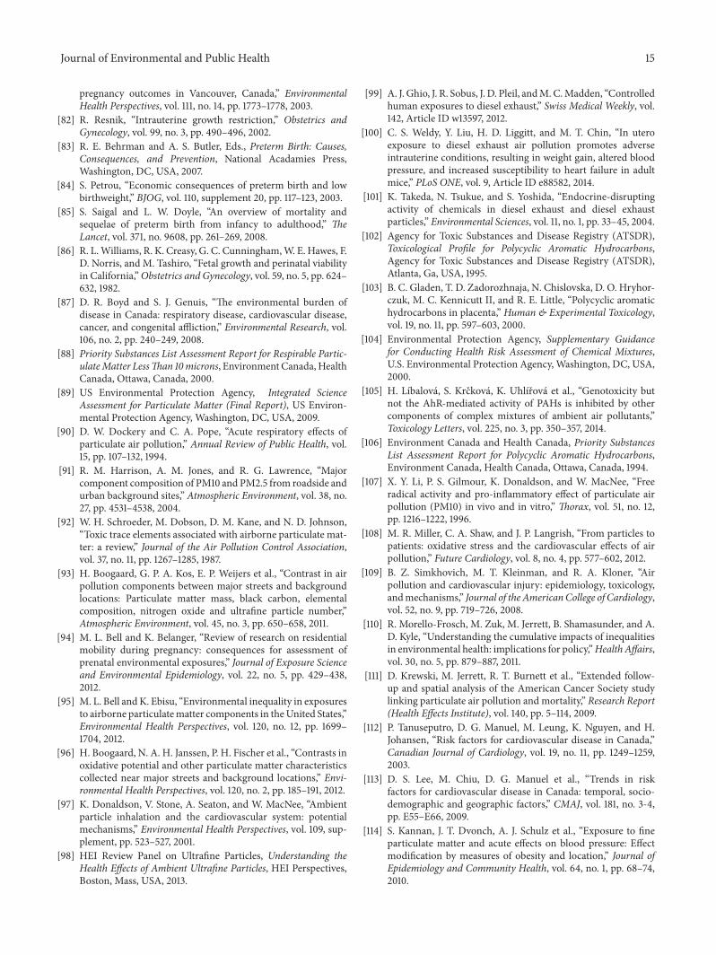
Journal of Environmental and Public Health 15
pregnancy outcomes in Vancouver, Canada,” EnvironmentalHealth Perspectives, vol. 111, no. 14, pp. 1773–1778, 2003.
[82] R. Resnik, “Intrauterine growth restriction,” Obstetrics andGynecology, vol. 99, no. 3, pp. 490–496, 2002.
[83] R. E. Behrman and A. S. Butler, Eds., Preterm Birth: Causes,Consequences, and Prevention, National Acadamies Press,Washington, DC, USA, 2007.
[84] S. Petrou, “Economic consequences of preterm birth and lowbirthweight,” BJOG, vol. 110, supplement 20, pp. 117–123, 2003.
[85] S. Saigal and L. W. Doyle, “An overview of mortality andsequelae of preterm birth from infancy to adulthood,” TheLancet, vol. 371, no. 9608, pp. 261–269, 2008.
[86] R. L.Williams, R. K. Creasy, G. C. Cunningham,W. E. Hawes, F.D. Norris, and M. Tashiro, “Fetal growth and perinatal viabilityin California,”Obstetrics and Gynecology, vol. 59, no. 5, pp. 624–632, 1982.
[87] D. R. Boyd and S. J. Genuis, “The environmental burden ofdisease in Canada: respiratory disease, cardiovascular disease,cancer, and congenital affliction,” Environmental Research, vol.106, no. 2, pp. 240–249, 2008.
[88] Priority Substances List Assessment Report for Respirable Partic-ulateMatter LessThan 10microns, Environment Canada,HealthCanada, Ottawa, Canada, 2000.
[89] US Environmental Protection Agency, Integrated ScienceAssessment for Particulate Matter (Final Report), US Environ-mental Protection Agency, Washington, DC, USA, 2009.
[90] D. W. Dockery and C. A. Pope, “Acute respiratory effects ofparticulate air pollution,” Annual Review of Public Health, vol.15, pp. 107–132, 1994.
[91] R. M. Harrison, A. M. Jones, and R. G. Lawrence, “Majorcomponent composition of PM10 and PM2.5 from roadside andurban background sites,” Atmospheric Environment, vol. 38, no.27, pp. 4531–4538, 2004.
[92] W. H. Schroeder, M. Dobson, D. M. Kane, and N. D. Johnson,“Toxic trace elements associated with airborne particulate mat-ter: a review,” Journal of the Air Pollution Control Association,vol. 37, no. 11, pp. 1267–1285, 1987.
[93] H. Boogaard, G. P. A. Kos, E. P. Weijers et al., “Contrast in airpollution components between major streets and backgroundlocations: Particulate matter mass, black carbon, elementalcomposition, nitrogen oxide and ultrafine particle number,”Atmospheric Environment, vol. 45, no. 3, pp. 650–658, 2011.
[94] M. L. Bell and K. Belanger, “Review of research on residentialmobility during pregnancy: consequences for assessment ofprenatal environmental exposures,” Journal of Exposure Scienceand Environmental Epidemiology, vol. 22, no. 5, pp. 429–438,2012.
[95] M. L. Bell andK. Ebisu, “Environmental inequality in exposuresto airborne particulatematter components in theUnited States,”Environmental Health Perspectives, vol. 120, no. 12, pp. 1699–1704, 2012.
[96] H. Boogaard, N. A. H. Janssen, P. H. Fischer et al., “Contrasts inoxidative potential and other particulate matter characteristicscollected near major streets and background locations,” Envi-ronmental Health Perspectives, vol. 120, no. 2, pp. 185–191, 2012.
[97] K. Donaldson, V. Stone, A. Seaton, and W. MacNee, “Ambientparticle inhalation and the cardiovascular system: potentialmechanisms,” Environmental Health Perspectives, vol. 109, sup-plement, pp. 523–527, 2001.
[98] HEI Review Panel on Ultrafine Particles, Understanding theHealth Effects of Ambient Ultrafine Particles, HEI Perspectives,Boston, Mass, USA, 2013.
[99] A. J. Ghio, J. R. Sobus, J. D. Pleil, andM.C.Madden, “Controlledhuman exposures to diesel exhaust,” Swiss Medical Weekly, vol.142, Article ID w13597, 2012.
[100] C. S. Weldy, Y. Liu, H. D. Liggitt, and M. T. Chin, “In uteroexposure to diesel exhaust air pollution promotes adverseintrauterine conditions, resulting in weight gain, altered bloodpressure, and increased susceptibility to heart failure in adultmice,” PLoS ONE, vol. 9, Article ID e88582, 2014.
[101] K. Takeda, N. Tsukue, and S. Yoshida, “Endocrine-disruptingactivity of chemicals in diesel exhaust and diesel exhaustparticles,” Environmental Sciences, vol. 11, no. 1, pp. 33–45, 2004.
[102] Agency for Toxic Substances and Disease Registry (ATSDR),Toxicological Profile for Polycyclic Aromatic Hydrocarbons,Agency for Toxic Substances and Disease Registry (ATSDR),Atlanta, Ga, USA, 1995.
[103] B. C. Gladen, T. D. Zadorozhnaja, N. Chislovska, D. O. Hryhor-czuk, M. C. Kennicutt II, and R. E. Little, “Polycyclic aromatichydrocarbons in placenta,” Human & Experimental Toxicology,vol. 19, no. 11, pp. 597–603, 2000.
[104] Environmental Protection Agency, Supplementary Guidancefor Conducting Health Risk Assessment of Chemical Mixtures,U.S. Environmental Protection Agency, Washington, DC, USA,2000.
[105] H. Lıbalova, S. Krckova, K. Uhlırova et al., “Genotoxicity butnot the AhR-mediated activity of PAHs is inhibited by othercomponents of complex mixtures of ambient air pollutants,”Toxicology Letters, vol. 225, no. 3, pp. 350–357, 2014.
[106] Environment Canada and Health Canada, Priority SubstancesList Assessment Report for Polycyclic Aromatic Hydrocarbons,Environment Canada, Health Canada, Ottawa, Canada, 1994.
[107] X. Y. Li, P. S. Gilmour, K. Donaldson, and W. MacNee, “Freeradical activity and pro-inflammatory effect of particulate airpollution (PM10) in vivo and in vitro,” Thorax, vol. 51, no. 12,pp. 1216–1222, 1996.
[108] M. R. Miller, C. A. Shaw, and J. P. Langrish, “From particles topatients: oxidative stress and the cardiovascular effects of airpollution,” Future Cardiology, vol. 8, no. 4, pp. 577–602, 2012.
[109] B. Z. Simkhovich, M. T. Kleinman, and R. A. Kloner, “Airpollution and cardiovascular injury: epidemiology, toxicology,andmechanisms,” Journal of the AmericanCollege of Cardiology,vol. 52, no. 9, pp. 719–726, 2008.
[110] R. Morello-Frosch, M. Zuk, M. Jerrett, B. Shamasunder, and A.D. Kyle, “Understanding the cumulative impacts of inequalitiesin environmental health: implications for policy,”Health Affairs,vol. 30, no. 5, pp. 879–887, 2011.
[111] D. Krewski, M. Jerrett, R. T. Burnett et al., “Extended follow-up and spatial analysis of the American Cancer Society studylinking particulate air pollution and mortality,” Research Report(Health Effects Institute), vol. 140, pp. 5–114, 2009.
[112] P. Tanuseputro, D. G. Manuel, M. Leung, K. Nguyen, and H.Johansen, “Risk factors for cardiovascular disease in Canada,”Canadian Journal of Cardiology, vol. 19, no. 11, pp. 1249–1259,2003.
[113] D. S. Lee, M. Chiu, D. G. Manuel et al., “Trends in riskfactors for cardiovascular disease in Canada: temporal, socio-demographic and geographic factors,” CMAJ, vol. 181, no. 3-4,pp. E55–E66, 2009.
[114] S. Kannan, J. T. Dvonch, A. J. Schulz et al., “Exposure to fineparticulate matter and acute effects on blood pressure: Effectmodification by measures of obesity and location,” Journal ofEpidemiology and Community Health, vol. 64, no. 1, pp. 68–74,2010.
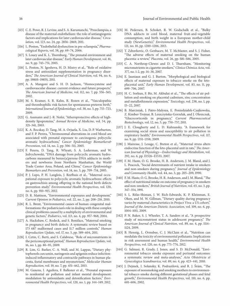
16 Journal of Environmental and Public Health
[115] C. E. Powe, R. J. Levine, and S. A. Karumanchi, “Preeclampsia, adisease of the maternal endothelium: the role of antiangiogenicfactors and implications for later cardiovascular disease,” Circu-lation, vol. 123, no. 24, pp. 2856–2869, 2011.
[116] L. Poston, “Endothelial dysfunction in pre-eclampsia,” Pharma-cological Reports, vol. 58, pp. 69–74, 2006.
[117] S. Louey and K. L. Thornburg, “The prenatal environment andlater cardiovascular disease,” Early HumanDevelopment, vol. 81,no. 9, pp. 745–751, 2005.
[118] L. Poston, N. Igosheva, H. D. Mistry et al., “Role of oxidativestress and antioxidant supplementation in pregnancy disor-ders,”The American Journal of Clinical Nutrition, vol. 94, no. 6,pp. 1980S–1985S, 2011.
[119] A. A. Mangoni and S. H. D. Jackson, “Homocysteine andcardiovascular disease: current evidence and future prospects,”The American Journal of Medicine, vol. 112, no. 7, pp. 556–565,2002.
[120] M. S. Kramer, S. R. Kahn, R. Rozen et al., “Vasculopathicand thrombophilic risk factors for spontaneous preterm birth,”International Journal of Epidemiology, vol. 38, no. 3, pp. 715–723,2009.
[121] G. Assmann and J.-R. Nofer, “Atheroprotective effects of high-density lipoproteins,” Annual Review of Medicine, vol. 54, pp.321–341, 2003.
[122] K. A. Bocskay, D. Tang, M. A. Orjuela, X. Liu, D. P. Warburton,and F. P. Perera, “Chromosomal aberrations in cord blood areassociated with prenatal exposure to carcinogenic polycyclicaromatic hydrocarbons,” Cancer Epidemiology Biomarkers&Prevention, vol. 14, no. 2, pp. 506–511, 2005.
[123] F. Perera, D. Tang, R. Whyatt, S. A. Lederman, and W.Jedrychowski, “DNA damage from polycyclic aromatic hydro-carbons measured by benzo[a]pyrene-DNA adducts in moth-ers and newborns from Northern Manhattan, the WorldTrade Center Area, Poland, and China,” Cancer EpidemiologyBiomarkers and Prevention, vol. 14, no. 3, pp. 709–714, 2005.
[124] P. J. Lupo, P. H. Langlois, J. Reefhuis et al., “Maternal occu-pational exposure to polycyclic aromatic hydrocarbons: effectson gastroschisis among offspring in the national birth defectsprevention study,” Environmental Health Perspectives, vol. 120,no. 6, pp. 910–915, 2012.
[125] D. R. Mattison, “Environmental exposures and development,”Current Opinion in Pediatrics, vol. 22, no. 2, pp. 208–218, 2010.
[126] R. L. Brent, “Environmental causes of human congenital mal-formations: the pediatrician’s role in dealing with these complexclinical problems caused by amultiplicity of environmental andgenetic factors,” Pediatrics, vol. 113, no. 4, pp. 957–968, 2004.
[127] A. Hackshaw, C. Rodeck, and S. Boniface, “Maternal smokingin pregnancy and birth defects: A systematic review based on173 687 malformed cases and 11.7 million controls,” HumanReproduction Update, vol. 17, no. 5, pp. 589–604, 2011.
[128] I. Cetin, C. Berti, and S. Calabrese, “Role of micronutrients inthe periconceptional period,”Human Reproduction Update, vol.16, no. 1, pp. 80–95, 2009.
[129] R. Lim, G. Barker, C. A. Wall, and M. Lappas, “Dietary phy-tophenols curcumin, naringenin and apigenin reduce infection-induced inflammatory and contractile pathways in human pla-centa, foetal membranes and myometrium,”Molecular HumanReproduction, vol. 19, no. 7, pp. 451–462, 2013.
[130] M. Guxens, I. Aguilera, F. Ballester et al., “Prenatal exposureto residential air pollution and infant mental development:modulation by antioxidants and detoxification factors,” Envi-ronmental Health Perspectives, vol. 120, no. 1, pp. 144–149, 2012.
[131] M. Pedersen, B. Schoket, R. W. Godschalk et al., “BulkyDNA adducts in cord blood, maternal fruit-and-vegetableconsumption, and birth weight in a European mother-childstudy (NewGeneris),” Environmental Health Perspectives, vol.121, no. 10, pp. 1200–1206, 2013.
[132] T. Zdravkovic, O. Genbacev, M. T. McMaster, and S. J. Fisher,“The adverse effects of maternal smoking on the humanplacenta: a review,” Placenta, vol. 26, pp. S81–S86, 2005.
[133] C. A. Northrop-Clewes and D. I. Thurnham, “Monitoringmicronutrients in cigarette smokers,” Clinica Chimica Acta, vol.377, no. 1-2, pp. 14–38, 2007.
[134] E. Jauniaux and G. J. Burton, “Morphological and biologicaleffects of maternal exposure to tobacco smoke on the feto-placental unit,” Early Human Development, vol. 83, no. 11, pp.699–706, 2007.
[135] H. C. Sorkun, F. Bir, M. Akbulut et al., “The effects of air pol-lution and smoking on placental cadmium, zinc concentrationand metallothionein expression,” Toxicology, vol. 238, no. 1, pp.15–22, 2007.
[136] B. Marciniak, J. Patro-Malysza, E. Poniedziałek-Czajkowska,Z. Kimber-Trojnar, B. Leszczynska-Gorzelak, and J. Oleszczuk,“Glucocorticoids in pregnancy,” Current PharmaceuticalBiotechnology, vol. 12, no. 5, pp. 750–757, 2011.
[137] J. E. Clougherty and L. D. Kubzansky, “A framework forexamining social stress and susceptibility to air pollution inrespiratory health,” Environmental Health Perspectives, vol. 117,no. 9, pp. 1351–1358, 2009.
[138] J. Mairesse, J. Lesage, C. Breton et al., “Maternal stress altersendocrine function of the feto-placental unit in rats,”TheAmer-ican Journal of Physiology—Endocrinology and Metabolism, vol.292, no. 6, pp. E1526–E1533, 2007.
[139] F. M. Haste, O. G. Brooke, H. R. Anderson, J. M. Bland, and J.L. Peacock, “Social determinants of nutrient intake in smokersand non-smokers during pregnancy,” Journal of Epidemiologyand Community Health, vol. 44, no. 3, pp. 205–209, 1990.
[140] F.M.Haste, O.G. Brooke,H. R. Anderson, and J.M. Bland, “Theeffect of nutritional intake on outcome of pregnancy in smokersand non-smokers,” British Journal of Nutrition, vol. 65, no. 3, pp.347–354, 1991.
[141] S. L. Rifas-Shiman, J. W. Rich-Edwards, K. P. Kleinman, E.Oken, and M. W. Gillman, “Dietary quality during pregnancyvaries by maternal characteristics in Project Viva: a US cohort,”Journal of the American Dietetic Association, vol. 109, no. 6, pp.1004–1011, 2009.
[142] P. N. Baker, S. J. Wheeler, T. A. Sanders et al., “A prospectivestudy of micronutrient status in adolescent pregnancy,” TheAmerican Journal of Clinical Nutrition, vol. 89, no. 4, pp. 1114–1124, 2009.
[143] B. Hennig, L. Ormsbee, C. J. McClain et al., “Nutrition canmodulate the toxicity of environmental pollutants: Implicationsin risk assessment and human health,” Environmental HealthPerspectives, vol. 120, no. 6, pp. 771–774, 2012.
[144] G. Salmasi, R. Grady, J. Jones, and S. D. McDonald, “Envi-ronmental tobacco smoke exposure and perinatal outcomes:a systematic review and meta-analyses,” Acta Obstetricia etGynecologica Scandinavica, vol. 89, no. 4, pp. 423–441, 2010.
[145] J. Dejmek, I. Solansky, K. Podrazilova, and R. J. Sram, “Theexposure of nonsmoking and smokingmothers to environmen-tal tobacco smoke during different gestational phases and fetalgrowth,” Environmental Health Perspectives, vol. 110, no. 6, pp.601–606, 2002.
Journal of Environmental and Public Health 17
[146] A. Castles, E. K. Adams, C. L. Melvin, C. Kelsch, and M.L. Boulton, “Effects of smoking during pregnancy: five meta-analyses,” American Journal of Preventive Medicine, vol. 16, no.3, pp. 208–215, 1999.
[147] A. C. Erickson and L. T. Arbour, “Heavy smoking duringpregnancy as amarker for other risk factors of adverse birth out-comes: a population-based study in British Columbia, Canada,”BMC Public Health, vol. 12, no. 1, article 102, 2012.
[148] M. Mohsin and A. E. Bauman, “Socio-demographic factorsassociatedwith smoking and smoking cessation among 426,344pregnant women in New South Wales, Australia,” BMC PublicHealth, vol. 5, article 138, 2005.
[149] S. Schneider, H. Maul, N. Freerksen, and M. Potschke-Langer,“Who smokes during pregnancy? An analysis of the GermanPerinatal Quality Survey 2005,” Public Health, vol. 122, no. 11,pp. 1210–1216, 2008.
[150] S. Krishnan, Y. C. Cozier, L. Rosenberg, and J. R. Palmer,“Socioeconomic status and incidence of type 2 diabetes: resultsfrom the black women’s health study,” American Journal ofEpidemiology, vol. 171, no. 5, pp. 564–570, 2010.
[151] S. D.Dubowsky,H. Suh, J. Schwartz, B. A. Coull, andD. R. Gold,“Diabetes, obesity, and hypertension may enhance associationsbetween air pollution and markers of systemic inflammation,”Environmental Health Perspectives, vol. 114, no. 7, pp. 992–998,2006.
[152] J. C. Chen, J. M. Cavallari, P. H. Stone, and D. C. Christiani,“Obesity is a modifier of automatic cardiac responses to finemetal particulates,” Environmental Health Perspectives, vol. 115,no. 7, pp. 1002–1006, 2007.
[153] E. Malmqvist, K. Jakobsson, H. Tinnerberg, A. Rignell-Hydbom, and L. Rylander, “Gestational diabetes andpreeclampsia in association with air pollution at levelsbelow current air quality guidelines,” Environmental HealthPerspectives, vol. 121, no. 4, pp. 488–493, 2013.
[154] G. Pereira, F. Haggar, A. W. Shand, C. Bower, A. Cook, and N.Nassar, “Association between pre-eclampsia and locally derivedtraffic-related air pollution: a retrospective cohort study,” Jour-nal of Epidemiology and Community Health, vol. 67, no. 2, pp.147–152, 2013.
[155] P. Dadvand, F. Figueras, X. Basagana et al., “Ambient airpollution and preeclampsia: a spatiotemporal analysis,” Envi-ronmental Health Perspectives, vol. 121, no. 11-12, pp. 1365–1371,2013.
[156] J. Wu, C. Ren, R. J. Delfino, J. Chung, M. Wilhelm, and B. Ritz,“Association between local traffic-generated air pollution andpreeclampsia and preterm delivery in the South Coast Air Basinof california,” Environmental Health Perspectives, vol. 117, no. 11,pp. 1773–1779, 2009.
[157] T. L. Gruenewald, A. S. Karlamangla, P. Hu et al., “History ofsocioeconomic disadvantage and allostatic load in later life,”Social Science and Medicine, vol. 74, no. 1, pp. 75–83, 2012.
[158] T. E. Seeman, B. S. McEwen, J. W. Rowe, and B. H. Singer,“Allostatic load as a marker of cumulative biological risk:MacArthur studies of successful aging,” Proceedings of theNational Academy of Sciences of the United States of America,vol. 98, no. 8, pp. 4770–4775, 2001.
[159] C. E. Bird, T. Seeman, J. J. Escarce et al., “Neighbourhoodsocioeconomic status and biological “wear and tear” in a nation-ally representative sample of US adults,” Journal of Epidemiologyand Community Health, vol. 64, no. 10, pp. 860–865, 2010.
[160] A. V. D. Roux, “Investigating neighborhood and area effects onhealth,” American Journal of Public Health, vol. 91, no. 11, pp.1783–1789, 2001.
[161] N. Auger, J. Giraud, and M. Daniel, “The joint influence of areaincome, income inequality, and immigrant density on adversebirth outcomes: a population-based study,” BMC Public Health,vol. 9, article 237, 2009.
[162] M. Tegethoff, N. Greene, J. Olsen, E. Schaffner, and G.Meinlschmidt, “Stress during pregnancy and offspring pediatricdisease: a national cohort study,” Environmental Health Perspec-tives, vol. 119, no. 11, pp. 1647–1652, 2011.
[163] B. S. Mcewen and P. J. Gianaros, “Central role of the brain instress and adaptation: links to socioeconomic status, health, anddisease,” Annals of the New York Academy of Sciences, vol. 1186,pp. 190–222, 2010.
[164] B. S.McEwen, “Brain on stress: how the social environment getsunder the skin,” Proceedings of the National Academy of Sciencesof the United States of America, vol. 109, no. 2, pp. 17180–17185,2012.
[165] I. N. Karatsoreos and B. S. McEwen, “Psychobiological allosta-sis: Resistance, resilience and vulnerability,” Trends in CognitiveSciences, vol. 15, no. 12, pp. 576–584, 2011.
[166] R. M. Sapolsky, L. M. Romero, and A. U. Munck, “How do glu-cocorticoids influence stress responses? Integrating permissive,suppressive, stimulatory, and preparative actions,” EndocrineReviews, vol. 21, no. 1, pp. 55–89, 2000.
[167] K. J. Davies, “Oxidative stress: the paradox of aerobic life,”Biochemical Society Symposium, vol. 61, pp. 1–31, 1995.
[168] K.H. Al-Gubory, P. A. Fowler, andC. Garrel, “The roles of cellu-lar reactive oxygen species, oxidative stress and antioxidants inpregnancy outcomes,”The International Journal of Biochemistry& Cell Biology, vol. 42, no. 10, pp. 1634–1650, 2010.
[169] K. J. Davies, “Oxidative stress, antioxidant defenses, and damageremoval, repair, and replacement systems,” IUBMB Life, vol. 50,no. 4-5, pp. 279–289, 2000.
[170] D. A. Ferenbach, D. C. Kluth, and J. Hughes, “Hemeoxygenase-1 and renal ischaemia-reperfusion injury,” Nephron—Experimental Nephrology, vol. 115, no. 3, pp. e33–e37, 2010.
[171] K. Levytska, J. Kingdom, D. Baczyk, and S. Drewlo, “Hemeoxygenase-1 in placental development and pathology,” Placenta,vol. 34, no. 4, pp. 291–298, 2013.
[172] H. Zhao, R. J. Wong, F. S. Kalish, N. R. Nayak, and D. K.Stevenson, “Effect of heme oxygenase-1 deficiency on placentaldevelopment,” Placenta, vol. 30, no. 10, pp. 861–868, 2009.
[173] A. Munoz-Suano, A. B. Hamilton, and A. G. Betz, “Gimmeshelter: the immune system during pregnancy,” ImmunologicalReviews, vol. 241, no. 1, pp. 20–38, 2011.
[174] J. R. Prins, N. Gomez-Lopez, and S. A. Robertson, “Interleukin-6 in pregnancy and gestational disorders,” Journal of Reproduc-tive Immunology, vol. 95, no. 1-2, pp. 1–14, 2012.
[175] I. Gerhard, S. Waibel, V. Daniel, and B. Runnebaum, “Impactof heavy metals on hormonal and immunological factorsin women with repeated miscarriages,” Human ReproductionUpdate, vol. 4, no. 3, pp. 301–309, 1998.
[176] P.-C. Lee, E. O. Talbott, J. M. Roberts, J. M. Catov, R. K. Sharma,and B. Ritz, “Particulate air pollution exposure and C-reactiveprotein during early pregnancy,” Epidemiology, vol. 22, no. 4, pp.524–531, 2011.
[177] F. Vadillo-Ortega, A. Osornio-Vargas, M. A. Buxton et al.,“Air pollution, inflammation and preterm birth: a potentialmechanistic link,” Medical Hypotheses, vol. 82, no. 2, pp. 219–224, 2014.
18 Journal of Environmental and Public Health
[178] B. S. McEwen and P. Tucker, “Critical biological pathwaysfor chronic psychosocial stress and research opportunities toadvance the consideration of stress in chemical risk assessment,”American Journal of Public Health, vol. 101, supplement 1, pp.S131–S139, 2011.
[179] C.W. G. Redman, D. S. Tannetta, R. A. Dragovic et al., “Review:does size matter? Placental debris and the pathophysiology ofpre-eclampsia,” Placenta, vol. 33, pp. S48–S54, 2012.
[180] J. D. Pleil and L. S. Sheldon, “Adapting concepts from systemsbiology to develop systems exposure event networks for expo-sure science research,” Biomarkers, vol. 16, no. 2, pp. 99–105,2011.
[181] N. G. Than, R. Romero, A. L. Tarca et al., “Mitochondrialmanganese superoxide dismutase mRNA expression in humanchorioamniotic membranes and its association with labor,inflammation, and infection,” Journal of Maternal-Fetal andNeonatal Medicine, vol. 22, no. 11, pp. 1000–1013, 2009.
[182] J. M. Bowen, L. Chamley, J. A. Keelan, and M. D. Mitchell,“Cytokines of the placenta and extra-placental membranes:Roles and regulation during humanpregnancy and parturition,”Placenta, vol. 23, no. 4, pp. 257–273, 2002.
[183] J. M. Bowen, L. Chamley, M. D. Mitchell, and J. A. Keelan,“Cytokines of the placenta and extra-placental membranes:biosynthesis, secretion and roles in establishment of pregnancyin women,” Placenta, vol. 23, no. 4, pp. 239–256, 2002.
[184] N. G.Than, R. Romero, C. J. Kim,M. R.McGowen, Z. Papp, andD. E. Wildman, “Galectins: Guardians of eutherian pregnancyat the maternal-fetal interface,” Trends in Endocrinology andMetabolism, vol. 23, no. 1, pp. 23–31, 2012.
[185] J. Thompson and J. Bannigan, “Cadmium: toxic effects on thereproductive system and the embryo,” Reproductive Toxicology,vol. 25, no. 3, pp. 304–315, 2008.
[186] M. C. Henson and P. J. Chedrese, “Endocrine disruption bycadmium, a common environmental toxicant with paradoxicaleffects on reproduction,” Experimental Biology and Medicine,vol. 229, no. 5, pp. 383–392, 2004.
[187] A. J. Dunn, “The HPA axis and the immune system: a perspec-tive,” in NeuroImmune Biology, A. del Rey, G. P. Chrousos, andH. O. Besedovsky, Eds., vol. 7, pp. 3–15, NeuroImmune Biology,Elsevier, 2007.
[188] S. A. Venners, X. Wang, C. Chen et al., “Paternal smokingand pregnancy loss: a prospective study using a biomarker ofpregnancy,”The American Journal of Epidemiology, vol. 159, no.10, pp. 993–1001, 2004.
[189] A. G. Ronnenberg, S. A. Venners, X. Xu et al., “PreconceptionB-vitamin and homocysteine status, conception, and earlypregnancy loss,” American Journal of Epidemiology, vol. 166, no.3, pp. 304–312, 2007.
[190] R. Slama, S. Bottagisi, I. Solansky, J. Lepeule, L. Giorgis-Allemand, and R. Sram, “Short-term impact of atmosphericpollution on fecundability,” Epidemiology, vol. 24, no. 6, pp. 871–879, 2013.
[191] M. J. Nieuwenhuijsen, X. Basagana, P. Dadvand et al., “Airpollution and human fertility rates,” Environment International,vol. 70, pp. 9–14, 2014.
[192] E. Jauniaux, A. L.Watson, J. Hempstock, Y.-P. Bao, J. N. Skepper,and G. J. Burton, “Onset of maternal arterial blood flow andplacental oxidative stress: a possible factor in human earlypregnancy failure,”The American Journal of Pathology, vol. 157,no. 6, pp. 2111–2122, 2000.
[193] A. E. Wallace, R. Fraser, and J. E. Cartwright, “Extravilloustrophoblast and decidual natural killer cells: a remodelling
partnership,”HumanReproductionUpdate, vol. 18, no. 4, ArticleID dms015, pp. 458–471, 2012.
[194] C. W. G. Redman and I. L. Sargent, “Placental stress and pre-eclampsia: a revised view,” Placenta, vol. 30, pp. 38–42, 2009.
[195] R. Gagnon, “Placental insufficiency and its consequences,”European Journal of Obstetrics Gynecology and ReproductiveBiology, vol. 110, pp. S99–S107, 2003.
[196] F.-J. Lin., J. W. Fitzpatrick, C. A. Iannotti, D. S. Martin, B. D.Mariani, and R. S. Tuan, “Effects of cadmium on trophoblastcalcium transport,” Placenta, vol. 18, no. 4, pp. 341–356, 1997.
[197] T. K. Morgan, J. E. Tolosa, L. Mele et al., “Placental villoushypermaturation is associated with idiopathic preterm birth,”Journal of Maternal-Fetal and Neonatal Medicine, vol. 26, no. 7,pp. 647–653, 2013.
[198] V. Bollati, B. Marinelli, P. Apostoli et al., “Exposure to metal-rich particulate matter modifies the expression of candidateMicroRNAs in peripheral blood leukocytes,” EnvironmentalHealth Perspectives, vol. 118, no. 6, pp. 763–768, 2010.
[199] R. Slama, L. Darrow, J. Parker et al., “Meeting report: atmo-spheric pollution and human reproduction,” EnvironmentalHealth Perspectives, vol. 116, no. 6, pp. 791–798, 2008.
[200] E. H. van den Hooven, Y. de Kluizenaar, F. H. Pierik etal., “Chronic air pollution exposure during pregnancy andmaternal and fetal c-reactive protein levels: the generation Rstudy,” Environmental Health Perspectives, vol. 120, no. 5, pp.746–751, 2012.
[201] S. Salvi, A. Blomberg, B. Rudell et al., “Acute inflammatoryresponses in the airways and peripheral blood after short-term exposure to diesel exhaust in healthy human volunteers,”American Journal of Respiratory and Critical Care Medicine, vol.159, no. 3, pp. 702–709, 1999.
[202] S.-Q. Wei, W. Fraser, and Z.-C. Luo, “Inflammatory cytokinesand spontaneous preterm birth in asymptomatic women: asystematic review,”Obstetrics and Gynecology, vol. 116, no. 2, pp.393–401, 2010.
[203] K. Monostory, J.-M. Pascussi, L. Kobori, and Z. Dvorak,“Hormonal regulation of CYP1A expression,” Drug MetabolismReviews, vol. 41, no. 4, pp. 547–572, 2009.
[204] A. C. Collier, M. D. Tingle, J. W. Paxton, M. D. Mitchell, and J.A. Keelan, “Metabolizing enzyme localization and activities inthe first trimester human placenta: the effect of maternal andgestational age, smoking and alcohol consumption,” HumanReproduction, vol. 17, no. 10, pp. 2564–2572, 2002.
[205] J. Vondracek, L. Umannova, and M. Machala, “Interactions ofthe aryl hydrocarbon receptor with inflammatory mediators:beyond CYP1A regulation,” Current Drug Metabolism, vol. 12,no. 2, pp. 89–103, 2011.
[206] H. Choi, V. Rauh, R. Garfinkel, Y. Tu, and F. P. Perera, “Prenatalexposure to airborne polycyclic aromatic hydrocarbons andrisk of intrauterine growth restriction,” Environmental HealthPerspectives, vol. 116, no. 5, pp. 658–665, 2008.
[207] H. Choi, L. Wang, X. Lin, J. D. Spengler, and F. P. Perera,“Fetal window of vulnerability to airborne polycyclic aromatichydrocarbons on proportional intrauterine growth restriction,”PLoS ONE, vol. 7, no. 4, Article ID e35464, 2012.
[208] M. P. Rayman, “The importance of selenium to human health,”The Lancet, vol. 356, no. 9225, pp. 233–241, 2000.
[209] J. Chen and M. J. Berry, “Selenium and selenoproteins in thebrain and brain diseases,” Journal of Neurochemistry, vol. 86, no.1, pp. 1–12, 2003.

Journal of Environmental and Public Health 19
[210] A. S. Al-Kunani, R. Knight, S. J. Haswell, J. W. Thompson,and S. W. Lindow, “The selenium status of women with ahistory of recurrent miscarriage,” British Journal of Obstetricsand Gynaecology, vol. 108, no. 10, pp. 1094–1097, 2001.
[211] B. A. Zachara, W. Dobrzynski, U. Trafikowska, and W.Szymanski, “Blood selenium and glutathione peroxidases inmiscarriage,” British Journal of Obstetrics and Gynaecology, vol.108, no. 3, pp. 244–247, 2001.
[212] J.M.Howard, S. Davies, andA.Hunnisett, “Red cellmagnesiumand glutathione peroxidase in infertile women—effects of oralsupplementation with magnesium and selenium,” MagnesiumResearch, vol. 7, no. 1, pp. 49–57, 1994.
[213] W. Dobrzynski, U. Trafikowska, A. Trafikowska, A. Pilecki, W.Szymanski, and B. A. Zachara, “Decreased selenium concentra-tion inmaternal and cord blood in preterm comparedwith termdelivery,” Analyst, vol. 123, no. 1, pp. 93–97, 1998.
[214] H. D. Mistry, V. Wilson, M. M. Ramsay, M. E. Symonds, and F.B. Pipkin, “Reduced selenium concentrations and glutathioneperoxidase activity in preeclamptic pregnancies,”Hypertension,vol. 52, no. 5, pp. 881–888, 2008.
[215] J. Vanderlelie andA. V. A. Perkins, “Selenium and preeclampsia:a global perspective,” PregnancyHypertension, vol. 1, no. 3-4, pp.213–224, 2011.
[216] C. Bertram, A. R. Trowern, N. Copin, A. A. Jackson, and C.B. Whorwood, “The maternal diet during pregnancy programsaltered expression of the glucocorticoid receptor and type 211𝛽-hydroxysteroid dehydrogenase: potential molecular mech-anisms underlying the programming of hypertension in utero,”Endocrinology, vol. 142, no. 7, pp. 2841–2853, 2001.
[217] K. Yang, L. Julan, F. Rubio, A. Sharma, andH. Guan, “Cadmiumreduces 11𝛽-hydroxysteroid dehydrogenase type 2 activity andexpression in human placental trophoblast cells,” AmericanJournal of Physiology: Endocrinology and Metabolism, vol. 290,no. 1, pp. E135–E142, 2006.
[218] M. Piasek, M. Blanusa, K. Kostial, and J. W. Laskey, “Placentalcadmium and progesterone concentrations in cigarette smok-ers,” Reproductive Toxicology, vol. 15, no. 6, pp. 673–681, 2001.
[219] D. Rurak, “Placental development and function,” in FetalGrowth and Development, R. Harding and A. D. Bocking, Eds.,Cambridge University Press, Cambridge, Mass, USA, 2001.
[220] M. S. Kramer, S. R. Kahn, R. W. Platt et al., “Antioxidantvitamins, long-chain fatty acids, and spontaneous pretermbirth,” Epidemiology, vol. 20, no. 5, pp. 707–713, 2009.
[221] A. Dragani, A. Falco, F. Santilli et al., “Oxidative stress andplatelet activation in subjects with moderate hyperhomocys-teinaemia due to MTHFR 677 C→T polymorphism,” Throm-bosis and Haemostasis, vol. 108, no. 3, pp. 533–542, 2012.
[222] B. De Chiara, V. Sedda, M. Parolini et al., “Plasma total cysteineand cardiovascular risk burden: action and interaction,” TheScientific World Journal, vol. 2012, Article ID 303654, 7 pages,2012.
[223] C. E. Garcıa-Rodrıguez, M. D. Mesa, J. Olza et al., “Doesconsumption of two portions of salmon per week enhancethe antioxidant defense system in pregnant women?” Antioxi-dants& Redox Signaling, vol. 16, no. 12, pp. 1401–1406, 2012.
[224] A. R. Patten, P. S. Brocardo, and B. R. Christie, “Omega-3 supplementation can restore glutathione levels and preventoxidative damage caused by prenatal ethanol exposure,” Journalof Nutritional Biochemistry, vol. 24, no. 5, pp. 760–769, 2013.
[225] S. Roy, A. Kale, K. Dangat, P. Sable, A. Kulkarni, and S. Joshi,“Maternal micronutrients (folic acid and vitamin B
12) and
omega 3 fatty acids: implications for neurodevelopmental riskin the rat offspring,” Brain and Development, vol. 34, no. 1, pp.64–71, 2012.
[226] A. M. Ronco, F. Garrido, and M. N. Llanos, “Smoking specifi-cally induces metallothionein-2 isoform in human placenta atterm,” Toxicology, vol. 223, no. 1-2, pp. 46–53, 2006.
[227] M. Kippler, A. M. W. Hoque, R. Raqib, H. Ohrvik, E.-C.Ekstrom, and M. Vahter, “Accumulation of cadmium in humanplacenta interacts with the transport of micronutrients to thefetus,” Toxicology Letters, vol. 192, no. 2, pp. 162–168, 2010.
[228] Agency for Toxic Substances andDisease Registry,ToxicologicalProfile for Cadmium, Agency for Toxic Substances and DiseaseRegistry, Atlanta, Ga, USA, 2012.
[229] L. P. Nampoothiri and S. Gupta, “Biochemical effects of ges-tational coexposure to lead and cadmium on reproductiveperformance, placenta, and ovary,” Journal of Biochemical andMolecular Toxicology, vol. 22, no. 5, pp. 337–344, 2008.
[230] S. M. Engel, E. Scher, S. Wallenstein et al., “Maternal active andpassive smoking and hypertensive disorders of pregnancy: Riskwith trimester-specific exposures,” Epidemiology, vol. 24, no. 3,pp. 379–386, 2013.
[231] R. M. Reynolds, J. Labad, C. Buss, P. Ghaemmaghami, and K.Raikkonen, “Transmitting biological effects of stress in utero:implications for mother and offspring,” Psychoneuroendocrinol-ogy, vol. 38, no. 9, pp. 1843–1849, 2013.
[232] R. L. Goldenberg, J. F. Culhane, J. D. Iams, and R. Romero,“Epidemiology and causes of preterm birth,” The Lancet, vol.371, no. 9606, pp. 75–84, 2008.
[233] C. A. Sandman, P. D. Wadhwa, A. Chicz-DeMet, C. Dunkel-Schetter, and M. Porto, “Maternal stress, HPA activity, andfetal/infant outcome,” Annals of the New York Academy ofSciences, vol. 814, pp. 266–275, 1997.
[234] C. A. Sandman, L. Glynn, C. D. Schetter et al., “Elevatedmaternal cortisol early in pregnancy predicts third trimesterlevels of placental corticotropin releasing hormone (CRH):priming the placental clock,” Peptides, vol. 27, no. 6, pp. 1457–1463, 2006.
[235] L. Duthie and R. M. Reynolds, “Changes in the maternalhypothalamic-pituitary-adrenal axis in pregnancy and post-partum: Influences on maternal and fetal outcomes,” Neuroen-docrinology, vol. 98, no. 2, pp. 106–115, 2013.
[236] A. M. Ronco, M. Urrutia, M. Montenegro, and M. N. Llanos,“Cadmium exposure during pregnancy reduces birth weightand increases maternal and foetal glucocorticoids,” ToxicologyLetters, vol. 188, no. 3, pp. 186–191, 2009.
[237] E. Kajantie, L. Dunkel, U. Turpeinen et al., “Placental 11𝛽-hydroxysteroid dehydrogenase-2 and fetal cortisol/cortisoneshuttle in small preterm infants,” Journal of Clinical Endocrinol-ogy and Metabolism, vol. 88, no. 1, pp. 493–500, 2003.
[238] M. Aufdenblatten, M. Baumann, L. Raio et al., “Prematurityis related to high placental cortisol in preeclampsia,” PediatricResearch, vol. 65, no. 2, pp. 198–202, 2009.
[239] B. I. S. Federenko and P. D. Wadhwa, “Women’s mental healthduring pregnancy influences fetal and infant,” CNS Spectrums,vol. 9, pp. 198–206, 2004.
[240] S. L. Szanton, J. M. Gill, and J. K. Allen, “Allostatic load: amechanism of socioeconomic health disparities?” BiologicalResearch for Nursing, vol. 7, no. 1, pp. 7–15, 2005.
[241] J. Lesage, D. Hahn, M. Leonhardt, B. Blondeau, B. Breant, andJ. P. Dupouy, “Maternal undernutrition during late gestation-induced intrauterine growth restriction in the rat is associated

20 Journal of Environmental and Public Health
with impaired placental GLUT3 expression, but does notcorrelate with endogenous corticosterone levels,” Journal ofEndocrinology, vol. 174, no. 1, pp. 37–43, 2002.
[242] C. D. Salpietro, S. Gangemi, P. L. Minciullo et al., “Cadmiumconcentration in maternal and cord blood and infant birthweight: a study on healthy non-smoking women,” Journal ofPerinatal Medicine, vol. 30, no. 5, pp. 395–399, 2002.
[243] M. N. Llanos and A. M. Ronco, “Fetal growth restriction isrelated to placental levels of cadmium, lead and arsenic but notwith antioxidant activities,” Reproductive Toxicology, vol. 27, no.1, pp. 88–92, 2009.
[244] K. H. Al-Gubory, “Environmental pollutants and lifestyle fac-tors induce oxidative stress and poor prenatal development,”Reproductive BioMedicine Online, vol. 29, no. 1, pp. 17–31, 2014.
[245] E. V. Younglai, Y. J. Wu, and W. G. Foster, “Reproductivetoxicology of environmental toxicants: emerging issues andconcerns,” Current Pharmaceutical Design, vol. 13, no. 29, pp.3005–3019, 2007.
[246] A. V. Diez-Roux, “On genes, individuals, society, and epidemi-ology,” American Journal of Epidemiology, vol. 148, no. 11, pp.1027–1032, 1998.
[247] N. Auger, Z.-C. Luo, R. W. Platt, and M. Daniel, “Do mother’seducation and foreign born status interact to influence birthoutcomes? Clarifying the epidemiological paradox and thehealthymigrant effect,” Journal of Epidemiology andCommunityHealth, vol. 62, no. 5, pp. 402–409, 2008.
[248] A. Alberg, “The influence of cigarette smoking on circulatingconcentrations of antioxidant micronutrients,” Toxicology, vol.180, no. 2, pp. 121–137, 2002.
[249] K. Okumura and H. Tsukamoto, “Folate in smokers,” ClinicaChimica Acta, vol. 412, no. 7-8, pp. 521–526, 2011.
[250] D. B. Kim, Y. S. Oh, K. D. Yoo et al., “Passive smoking in never-smokers is associated with increased: plasma homocysteinelevels,” International Heart Journal, vol. 51, no. 3, pp. 183–187,2010.
[251] F. M. Haste, O. G. Brooke, H. R. Anderson et al., “Nutrientintakes during pregnancy: observations on the influence ofsmoking and social class,” The American Journal of ClinicalNutrition, vol. 51, no. 1, pp. 29–36, 1990.
[252] S. B. Astley and D. G. Lindsay, “Conclusions,”Molecular Aspectsof Medicine, vol. 23, no. 1–3, pp. 287–291, 2002.
[253] M. C. Wang, S. Kim, A. A. Gonzalez, K. E. MacLeod, and M.A. Winkleby, “Socioeconomic and food-related physical char-acteristics of the neighbourhood environment are associatedwith bodymass index,” Journal of Epidemiology and CommunityHealth, vol. 61, no. 6, pp. 491–498, 2007.
[254] H. K. Seligman, A. B. Bindman, E. Vittinghoff, A. M. Kanaya,and M. B. Kushel, “Food insecurity is associated with diabetesmellitus: results from the National Health Examination andNutrition Examination Survey (NHANES) 1999–2002,” Journalof General Internal Medicine, vol. 22, no. 7, pp. 1018–1023, 2007.
[255] E. J. Adams, L. Grummer-Strawn, and G. Chavez, “Food inse-curity is associated with increased risk of obesity in Californiawomen,” Journal of Nutrition, vol. 133, no. 4, pp. 1070–1074,2003.
[256] J. VonBraun,H. Bouis, S. Kumar, andR. Pandya-Lorch, Improv-ing Food Security of the Poor: Concept, Policy, and Programs,International Food Policy Research Institute, Washington, DC,USA, 1992.
[257] B. Laraia, E. Epel, and A. M. Siega-Riz, “Food insecurity withpast experience of restrained eating is a recipe for increasedgestational weight gain,” Appetite, vol. 65, pp. 178–184, 2013.
[258] J. D. Parker, D. Q. Rich, S. V. Glinianaia et al., “The internationalcollaboration on air pollution and pregnancy outcomes: initialresults,” Environmental Health Perspectives, vol. 119, no. 7, pp.1023–1028, 2011.
[259] D. Rojas-Rueda, A. de Nazelle, O. Teixido, andM. Nieuwenhui-jsen, “Health impact assessment of increasing public transportand cycling use in Barcelona: a morbidity and burden of diseaseapproach,” Preventive Medicine, vol. 57, no. 5, pp. 573–579, 2013.
[260] P.Dadvand, A. deNazelle,M. Triguero-Mas et al., “Surroundinggreenness and exposure to air pollution during pregnancy: ananalysis of personal monitoring data,” Environmental HealthPerspectives, vol. 120, no. 9, pp. 1286–1290, 2012.
[261] Z. Yao, Y. Zhang, X. Shen, X. Wang, Y. Wu, and K. He, “Impactsof temporary traffic control measures on vehicular emissionsduring the Asian Games in Guangzhou, China,” Journal of theAir & Waste Management Association, vol. 63, no. 1, pp. 11–19,2013.
[262] H. Boogaard,N.A.H. Janssen, P.H. Fischer et al., “Impact of lowemission zones and local trafficpolicies on ambient air pollutionconcentrations,” Science of the Total Environment, vol. 435-436,pp. 132–140, 2012.
[263] L. E. Jackson, “The relationship of urban design to humanhealthand condition,” Landscape and Urban Planning, vol. 64, no. 4,pp. 191–200, 2003.
[264] C. A. Pope III, “Review: epidemiological basis for particulate airpollution health standards,”Aerosol Science and Technology, vol.32, no. 1, pp. 4–14, 2000.
[265] WorldHealthOrganization,Health Effects of ParticulateMatter:Policy Implications for Countries in Eastern Europe, Caucasusand Central Asia, vol. 50, World Health Organization, Copen-hagen, Denmark, 2013.
[266] O. Hanninen, A. B. Knol, M. Jantunen et al., “Environmentalburden of disease in Europe: assessing nine risk factors in sixcountries,” Environmental Health Perspectives, vol. 122, no. 5, pp.439–446, 2014.

Submit your manuscripts athttp://www.hindawi.com
Stem CellsInternational
Hindawi Publishing Corporationhttp://www.hindawi.com Volume 2014
Hindawi Publishing Corporationhttp://www.hindawi.com Volume 2014
MEDIATORSINFLAMMATION
of
Hindawi Publishing Corporationhttp://www.hindawi.com Volume 2014
Behavioural Neurology
EndocrinologyInternational Journal of
Hindawi Publishing Corporationhttp://www.hindawi.com Volume 2014
Hindawi Publishing Corporationhttp://www.hindawi.com Volume 2014
Disease Markers
Hindawi Publishing Corporationhttp://www.hindawi.com Volume 2014
BioMed Research International
OncologyJournal of
Hindawi Publishing Corporationhttp://www.hindawi.com Volume 2014
Hindawi Publishing Corporationhttp://www.hindawi.com Volume 2014
Oxidative Medicine and Cellular Longevity
Hindawi Publishing Corporationhttp://www.hindawi.com Volume 2014
PPAR Research
The Scientific World JournalHindawi Publishing Corporation http://www.hindawi.com Volume 2014
Immunology ResearchHindawi Publishing Corporationhttp://www.hindawi.com Volume 2014
Journal of
ObesityJournal of
Hindawi Publishing Corporationhttp://www.hindawi.com Volume 2014
Hindawi Publishing Corporationhttp://www.hindawi.com Volume 2014
Computational and Mathematical Methods in Medicine
OphthalmologyJournal of
Hindawi Publishing Corporationhttp://www.hindawi.com Volume 2014
Diabetes ResearchJournal of
Hindawi Publishing Corporationhttp://www.hindawi.com Volume 2014
Hindawi Publishing Corporationhttp://www.hindawi.com Volume 2014
Research and TreatmentAIDS
Hindawi Publishing Corporationhttp://www.hindawi.com Volume 2014
Gastroenterology Research and Practice
Hindawi Publishing Corporationhttp://www.hindawi.com Volume 2014
Parkinson’s Disease
Evidence-Based Complementary and Alternative Medicine
Volume 2014Hindawi Publishing Corporationhttp://www.hindawi.com