review article human herpesvirus 6 and...
TRANSCRIPT

Hindawi Publishing CorporationISRN VirologyVolume 2013, Article ID 834890, 11 pageshttp://dx.doi.org/10.5402/2013/834890
Review ArticleHuman Herpesvirus 6 and Neuroinflammation
Joséphine M. Reynaud and Branka Horvat
International Center for Infectiology Research (CIRI), INSERM U1111, CNRS UMR5308, University of Lyon 1,ENS-Lyon, 21 Avenue T. Garnier, 69365 Lyon, France
Correspondence should be addressed to Branka Horvat; [email protected]
Received 21 January 2013; Accepted 13 February 2013
Academic Editors: B. Kim, M. Magnani, and L. Margolis
Copyright © 2013 J. M. Reynaud and B. Horvat.This is an open access article distributed under the Creative Commons AttributionLicense, which permits unrestricted use, distribution, and reproduction in anymedium, provided the originalwork is properly cited.
Human herpesvirus (HHV-) 6A and HHV-6B are two distinct 𝛽-herpesviruses which have been associated with various neurolog-ical diseases, including encephalitis, meningitis, epilepsy, and multiple sclerosis. Although the reactivation of both viruses is rec-ognized as the cause of some neurological complications in conditions of immunosuppression, their involvement in neuroinflam-matory diseases in immunocompetent people is still unclear, and the mechanisms involved have not been completely elucidated.Here, we review the available data providing evidence for the capacity of HHV-6A and -6B to infect the central nervous systemand to induce proinflammatory responses by infected cells. We discuss the potential role of both viruses in neuroinflammatorypathologies and the mechanisms which could explain virus-induced neuropathogenesis.
1. Introduction
Human herpesvirus (HHV-) 6 was first isolated in 1986 bySalahuddin and colleagues [1]. This enveloped DNA virusbelongs to the 𝛽-herpesvirus family and, together with itsclosest homologue HHV-7, forms the roseoloviruses subfam-ily. HHV-6 is widely spread in the population (seroprevalence> 90%) and can establish a persistent and most often asymp-tomatic infection in humans. Based on genetic, epidemiolog-ical, and functional features, the numerous isolated strains ofHHV-6 were initially separated into two variants, HHV-6Aand HHV-6B, which have recently been recognized as twodistinct viruses. HHV-6A and -6B share an overall sequenceidentity of 90%, and several open reading frames are presentin only one of the two viruses [2]. Primary infection withHHV-6B generally happens before the age of two; the virusis transmitted through saliva and close contacts with parents[3] and provokes exanthem subitum (or roseola), a benignfebrile illness with skin rash. HHV-6A infection is thoughtto happen later in life and was not yet clearly identified as thecausative agent for any disease.
To date, the only identified cellular receptor for bothHHV-6A and -6B is the complement-regulatory transmem-brane protein CD46 [4]. This protein is ubiquitouslyexpressed in humans, allowing the viruses to infect a widerange of cells and tissues, including cells from the central
nervous system (CNS). Both viruses have a high tropismtowards T cells, which are the best virus producers in vitro,and can establish a persistent infection in different tissues,including the salivary glands (for HHV-6B only) and periph-eral lymphocytes.
In immunocompromised patients, HHV-6A and -6Boften reactivate and can provoke neurological pathologies.Moreover, many clinical studies have reported an associationbetween HHV-6A and -6B and neuroinflammatory diseases,such as encephalitis or multiple sclerosis (MS), suggestinga role for both viruses in inflammatory processes. Indeed,although HHV-6A and -6B are generally considered asimmunosuppressive agents, allowing them to evade theimmune system, reports showing their proinflammatoryproperties are accumulating. Here, we review the availabledata providing evidence forHHV-6A and -6B infection in thehuman brain and their involvement in neurological diseases,andwe discuss the potentialmechanisms bywhich they couldparticipate in neuroinflammation.
2. HHV-6A and HHV-6B AreNeurotropic Viruses
2.1. Evidence for the Presence of HHV-6A and -6B in the Brain.AlthoughHHV-6 was first identified as a lymphotropic virus,
2 ISRN Virology
Mock
SH-S
Y5Y
(a)
HHV-6A
SH-S
Y5Y
(b)
Mock
U87
(c)
HHV-6A
U87
(d)
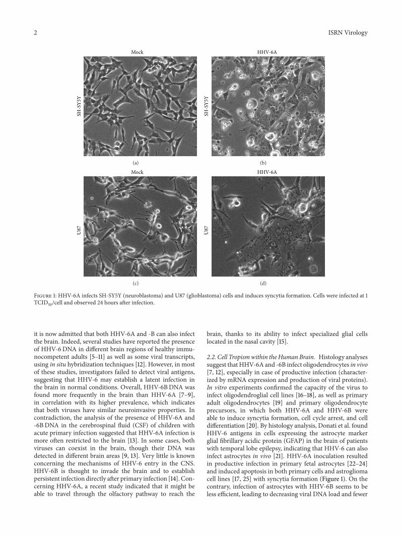
Figure 1: HHV-6A infects SH-SY5Y (neuroblastoma) and U87 (glioblastoma) cells and induces syncytia formation. Cells were infected at 1TCID
50/cell and observed 24 hours after infection.
it is now admitted that both HHV-6A and -B can also infectthe brain. Indeed, several studies have reported the presenceof HHV-6DNA in different brain regions of healthy immu-nocompetent adults [5–11] as well as some viral transcripts,using in situ hybridization techniques [12]. However, in mostof these studies, investigators failed to detect viral antigens,suggesting that HHV-6 may establish a latent infection inthe brain in normal conditions. Overall, HHV-6BDNA wasfound more frequently in the brain than HHV-6A [7–9],in correlation with its higher prevalence, which indicatesthat both viruses have similar neuroinvasive properties. Incontradiction, the analysis of the presence of HHV-6A and-6BDNA in the cerebrospinal fluid (CSF) of children withacute primary infection suggested that HHV-6A infection ismore often restricted to the brain [13]. In some cases, bothviruses can coexist in the brain, though their DNA wasdetected in different brain areas [9, 13]. Very little is knownconcerning the mechanisms of HHV-6 entry in the CNS.HHV-6B is thought to invade the brain and to establishpersistent infection directly after primary infection [14]. Con-cerning HHV-6A, a recent study indicated that it might beable to travel through the olfactory pathway to reach the
brain, thanks to its ability to infect specialized glial cellslocated in the nasal cavity [15].
2.2. Cell Tropismwithin theHumanBrain. Histology analysessuggest that HHV-6A and -6B infect oligodendrocytes in vivo[7, 12], especially in case of productive infection (character-ized by mRNA expression and production of viral proteins).In vitro experiments confirmed the capacity of the virus toinfect oligodendroglial cell lines [16–18], as well as primaryadult oligodendrocytes [19] and primary oligodendrocyteprecursors, in which both HHV-6A and HHV-6B wereable to induce syncytia formation, cell cycle arrest, and celldifferentiation [20]. By histology analysis, Donati et al. foundHHV-6 antigens in cells expressing the astrocyte markerglial fibrillary acidic protein (GFAP) in the brain of patientswith temporal lobe epilepsy, indicating that HHV-6 can alsoinfect astrocytes in vivo [21]. HHV-6A inoculation resultedin productive infection in primary fetal astrocytes [22–24]and induced apoptosis in both primary cells and astrogliomacell lines [17, 25] with syncytia formation (Figure 1). On thecontrary, infection of astrocytes with HHV-6B seems to beless efficient, leading to decreasing viral DNA load and fewer

ISRN Virology 3
Table 1: Modulation of the immune response by HHV-6A and HHV-6B in different cell types.
Cell type HHV-6A HHV-6B References
Lymphoid cells
PBMC ↗ IL-1𝛽b, TNF𝛼b, IFN𝛼b, IL-10, IL-15↘ IL-12, IFN𝛾, IL-2 [29, 41–43]
T cells (primary and cell lines) ↗ IL-18, IL-2R, IFN𝛾R, CCL-2↘ IL-10, IL-14, IL-10R, IL-13R, IL-2
↗ IL-18, IL-2R↘ IL-10, IL-14, IL-10R, IL-13R [29, 44, 45]
NK cells ↗ IL-15, cytotoxicity [46]Monocytes (primary and cell lines) ↗ TNF𝛼, IL-10, IL-12, IL-15 [47, 48, 126]Tonsillar cells (ex vivo) ↗ CCL-5 ↗ CCL-5 [50, 51]
Dendritic cells ↘maturationb, IL-12b ↗ IFN𝜆-1b↘maturationb, IL-12b [40, 49]
Macrophages ↘ IL-12 [39]Brain cells
Astrocytes (primary and cell line)
↗ CCL-5, CCL-2, IL-6, CXCL-2, CCL-3,IL-1𝛽, GM-CSF, CXCL-6, IL-10, TNF𝛼,IL-7R↘ CCR-2
↗ IL-6b, IL-1𝛽b [45, 127]
Other cell typesEndothelial cells ↗ CCL-5, CXCL-8, CCL-2 [52, 53]Hepatoma cells ↗ CXCL-8a [54]↗: enhanced production; ↘: inhibited production.aReplication-dependent effect; breplication-independent effect.
morphological changes, which indicates that the two HHV-6 viruses may have different infection patterns within thebrain. Fewer data concerning the infection of neurons andmicroglial cells are available; yet, some studies suggestedthat both cell types may be susceptible to HHV-6A and/or-6B infection in vitro [16, 17, 19, 24]. HHV-6A seems to beable to induce the formation of syncytia in neuroblastomacell lines (Figure 1), and infected neurons were detectedby immunostaining in patients who succumbed to HHV-6encephalitis [26].
Both HHV-6A and -6B are then able to invade the centralnervous system and to establish a persistent infection. How-ever, HHV-6A seems to infect astrocytes and neurons moreefficiently than HHV-6B, which may lead to the induction ofdifferent CNS pathologies.
3. Evidence for ProinflammatoryEffects of HHV-6
HHV-6 was initially identified as an immunosuppressivevirus. Primary infection with HHV-6B is indeed often asso-ciated with a decrease in leukocyte numbers [3], and bothHHV-6A and -6B preferentially infect T lymphocytes invivo and in vitro, reducing their proliferation [27–29] andinducing their apoptosis [30, 31]. Nevertheless, HHV-6A and-6B have also been demonstrated to exhibit proinflammatoryproperties in different contexts and have been suggested aspotential agents in several inflammatory diseases, such as
hepatitis [32], Sjogren’s syndrome [33, 34], rheumatoid arthri-tis [35, 36], systemic lupus erythematosus [35, 37], and morerecently Hashimoto’s thyroiditis [38]. While these associa-tions remain hypothetical, extensive in vitro studies provideevidence for HHV-6A and -6B proinflammatory effects on avariety of cell types and tissues (summarized in Table 1).
The effects of HHV-6A and -6B on the cytokine expres-sion profile in different types of immune cells have beenwidely investigated. Some studies have suggested that bothviruses can induce a Th2 profile in T cells through the inhi-bition of IL-12 secretion by dendritic cells (DCs) and macro-phages [39, 40] and through the induction of IL-10 produc-tion by peripheral blood mononuclear cells (PBMCs) [41].In contradiction, other reports have shown thatHHV-6 infec-tion upregulates the expression of proinflammatory cyto-kines, including IL-1𝛽, TNF𝛼, and IFN𝛼 in PBMC [42, 43],induces IL-18 and IFN𝛾 receptor, and reduces IL-10 and IL-14expression in T cells [44, 45], thus directing T cells towards aTh1 phenotype.
HHV-6A was also shown to exacerbate the cytotoxicityand IL-15 production in NK cells [46], as well as TNF𝛼and IL-15 expression in monocytes [47, 48]. In plasmacytoidDC, HHV-6B was recently shown to induce type III IFNproduction, which has similar antiviral properties as type IIFN but had no effect on theTh1/Th2 balance [49].
In addition, studies on ex vivo cultures of lymphoidtissue showed that both HHV-6A and -6B can induce thesecretion of chemokines in infected cells. Grivel et al. culturedfreshly excised human tonsils and demonstrated that HHV-6A and -6B productive infection could be achieved, inducing
4 ISRN Virology
an upregulation of CCL-5 and CCL-3 expression [50, 51].Meeuwsen et al. performed a transcriptional microarrayanalysis on infected astrocytes and showed that HHV-6Ainfection increased the expression of many proinflammatorycytokines upon stimulation with TNF𝛼, IL-1𝛽, and IFN𝛾,including several chemokines (e.g., CCL-2, CCL-5, andCXCL-2) [45]. Moreover, HHV-6A was found to up-regulatethe production of chemokines in primary endothelial cells[52, 53] and in a hepatoma cell line [54], indicating that theinfection can promote the recruitment of leukocytes to dif-ferent targeted tissues.
Altogether, these studies indicate that HHV-6A and -6Bboth have diverse proinflammatory effects on a variety of celltypes. Although they could exhibit anti-inflammatory effectson some cell types, they are also able to increase the produc-tion of proinflammatory cytokines by some other cell types(Table 1) and to induce the development of a Th1 phenotypein T cells, thus eliciting the immune response. Moreover,they participate in the establishment of the inflammation ininfected tissues by inducing the production of chemokinesby resident cells. There is an apparent contradiction in theobserved effects of HHV-6 infection, which include boththe induction of immunosuppression and the promotion ofinflammation.These differences may depend on the analyzedcell types or on infection kinetics representing different stagesof infection and would require additional studies to be betterunderstood.
4. HHV-6 and Neurological Diseases
HHV-6A and HHV-6B have both been directly or indirectlyassociated with neurological diseases [55–57], in cases ofprimary infection in immunocompetent young children,reactivation in otherwise healthy adults [3], or in immuno-suppressed patients [58].
4.1. Infection in the “Immunocompetent” Population. HHV-6B was long ago conclusively identified as the etiologic agentfor exanthem subitum (ES), a common infant febrile illnesswith skin rash [59]. Although ES is generally benign, it can beassociated with various neurological complications, includ-ing convulsions, seizures, and encephalitis [60–62], oftenresulting in ataxia and epilepsy [63–66]. The most severeforms of encephalitis associated with ES can even lead to fataloutcome [67, 68].
In immunocompetent adults, evidence for the directimplication of HHV-6A or -6B in neurological diseases ismore difficult to provide. Viral DNA loads in the serum andCSF, as well as IgM levels, are commonly used to detect HHV-6 infection. Based on these data, some cases of probableHHV-6-related encephalitis or meningoencephalitis havebeen reported in otherwise healthy adults and sometimessuccessfully treated with antiviral drugs [69–71]. Further-more, studies of patients with encephalitis of unknownetiology strongly suggested that HHV-6 could be involved indisease establishment in certain cases [72–74].
4.2. Reactivation in Immunosuppressed Patients. As for otherlatent human herpesviruses, immunological defects are ableto triggerHHV-6 reactivation from latency. Indeed,HHV-6Aand -6B have been suggested to reactivate in immunocom-promised patients, which received chemotherapy treatmentsor were diagnosed with AIDS. In hematopoietic stem celltransplant recipients especially, HHV-6DNA (mostly -6B)was detected in the serum or PBMC in around 50% ofthe cases [58, 75, 76], indicating that viral reactivation hasoccurred. In several case reports, where no other possiblecause was found, neurological complications in immunosup-pressed people have been attributed to HHV-6 reactivation[56, 58]. Its involvement in encephalitis development wasgenerally supported by the detection of viral DNA in the CSFand more rarely by the presence of viral proteins in affectedareas of the brain at autopsy [26, 77, 78]. Moreover, severalepidemiological studies have suggested a correlation betweenthe risk of developing neurological symptoms and HHV-6reactivation [79–81].
4.3. Association with Multiple Sclerosis. HHV-6 has longbeen cited as a potential candidate virus for the etiology ofmultiple sclerosis (MS).The importance of this inflammatoryneurological disease, which represents the first cause ofnontraumatic handicap in young adults, particularly inspiredthe research in this area. Abundant clinical studies havehighlighted a correlation between MS and several parame-ters assessing for HHV-6 infection. For instance, the levelsof HHV-6DNA in the serum, which are characteristic ofongoing infection, are significantly increased in MS patientswhen compared to healthy donors or with patients with otherdiseases [82–85]. HHV-6DNA was also detected at higherfrequencies in the CSF and in the peripheral blood mononu-clear cells of MS patients [82, 84, 86]. Moreover, the levels ofHHV-6-specific IgG and IgM in the serum and in the CSFwere reported to be higher in MS patients in several studies[83, 86, 87], although this phenomenon does not appear tobe specific for HHV-6. Indeed, some groups have reportedsimilar increases in the titers of antibodies against otherviruses including Epstein-Barr virus or varicella-zoster virus.Soldan et al. also showed that lymphoproliferative responsesagainst HHV-6 antigens were increased in MS patients [88].The analysis of brain biopsies and postmortem tissues indi-cated that HHV-6DNA was present more frequently in thebrain of MS patients than in control brains, and that it wasalso more frequent in MS lesions than in normal areas of thesame brains. Immunohistochemistry analyses confirmed thepresence of viral proteins in oligodendrocytes and astrocytesin the brain from MS patients, with a higher frequency indemyelinating plaques [7, 12, 87, 89, 90]. Most interestingly,viral loads were detectedmore frequently, and levels of HHV-6-specific IgG were increased in MS patients experiencingdisease exacerbation [84, 91–93], thus suggesting a correla-tion between HHV-6 infection and MS relapses.
As the distinction of HHV-6A and -6B as two differentviruses was only recently adopted, many of the initial studiesdo not discriminate between the two variants. However,based on few reports, it appears that HHV-6A is found more

ISRN Virology 5
frequently than -6B in the serum of MS patients [94].Especially in case of active infection, Alvarez-Lafuente et al.have found only HHV-6A [92]. In contrast, in one study,intrathecal HHV-6B IgG levels were more abundant thanHHV-6A IgG inMS patients, and only HHV-6B-specific IgMlevels were found [95].
The potential association between HHV-6A and HHV-6B infection and MS has often been discussed and remainscontroversial. Some studies provided contradictory results[96–98], raising methodological and technical questions,especially concerning the choice of control groups and theimmunological state of the included patients, who oftenreceive immunosuppressive treatments, that may provokelatent herpesvirus reactivation by itself. Some studies havetaken these matters into account and therefore providesolid data supporting the existence of a correlation betweenHHV-6 infection and MS pathology. Yet, whether HHV-6infection is the etiologic cause, a factor for disease progres-sion, or a consequence of MS remains unclear and wouldneed further investigation.
5. Potential Mechanisms forHHV-6-Induced Neuroinflammation
Although the potential role of HHV-6A and -6B in MS hasnot been completely elucidated, both viruses have been con-clusively involved in some cases of encephalitis in immuno-compromised patients and in neurological complications ofexanthem subitum. Several observations may provide expla-nations on how HHV-6 could trigger or participate in theestablishment of neuroinflammation.
5.1. Molecular Mimicry. Among the mechanisms proposedfor virus-induced autoimmunity, molecular mimicry is oneof the most popular ones. Based on the similarity in peptidesequence between viral proteins and self-proteins, it has beenpostulated that viral infections could activate cross-reactiveT cells, able to recognize both viral and self-antigens, whichcould then trigger an autoimmune response and cause tissuedamage. Several studies suggest that such amechanism couldbe involved in HHV-6-induced neuroinflammation. A firststudy reported that 15%–25% of HHV-6-specific T cell clonesobtained from healthy donors or MS patients were cross-reactive to myelin basic protein (MBP), one of the autoanti-gens implicated in MS pathology [99]. In fact, MBP andthe U24 protein from HHV-6 were later shown to share anidentical amino acid sequence of 7 residues.Moreover, T cellsdirected against an MBP peptide also recognized an HHV-6peptide, both peptides containing the identical sequence.Interestingly, cross-reactive cells were more frequent in MSpatients than in controls [100]. These data were furtherconfirmed by a more recent study, in which the presence ofcross-reactive CD8+ cytotoxic T cells was found [101]. Alto-gether, these studies suggest that HHV-6 infection can acti-vate T cell responses which can simultaneously be directedagainstmyelin sheaths, thus strongly supporting the potentialrole for HHV-6 in autoimmune diseases affecting the CNS(Figure 2(a)).
5.2. Infection of CNS Cells and Creation of a ProinflammatoryEnvironment. As mentioned earlier, HHV-6A and HHV-6Bare able to infect several CNS cell types, both in vitro and invivo, and to trigger proinflammatory responses in a varietyof infected cells. In particular, HHV-6A can infect primaryastrocytes and induce the expression of several proinflam-matory genes, especially when the cells have been pre-treated with proinflammatory cytokines [45]. This suggeststhat HHV-6A could enhance the proinflammatory responseof astrocytes, thus increasing leukocyte infiltration, inpatients who already suffer fromneuroinflammatory diseases(Figure 2(b)).
Recently, one study on dendritic cells demonstrated thatHHV-6B can induce IFN𝜆-1 production via TLR-9 signaling[49]. Moreover, TLR-9 has been shown to be expressed inhuman astrocytes [102]. It is then likely that HHV-6A canalter astrocyte cytokine expression profile through TLR-9signaling.
Other pattern recognition receptors, including TLR-2, -3,and -4, are expressed by human glial cells [102, 103] andneurons [104]. As HHV-6A and -6B are present in the brainof a subset of people, they may bind to these receptors uponreactivation and activate innate immune responses, thuspromoting inflammation in the CNS.
Another consequence of HHV-6 infection of CNS cellscould be the unmasking of autoantigens.HHV-6Awas shownto induce cell death in oligodendrocytes and astrocytes eitherdirectly [25] or indirectly, via the production of solublefactors by productively infected T cells [17, 105]. Therefore,HHV-6A productive infection of CNS cells or the presence ofproductively infected lymphocytes in the brain could provokethe death of glial cells and release previously unrecog-nized self-antigens, thus initiating an autoimmune responsedirected to the brain.
5.3. Leukocyte Chemoattraction via Virokine Expression. Thegenome of HHV-6 encodes two G protein-coupled receptors,U22 and U51, similar to human chemokine receptors [106,107] and one single chemokine-like protein, U83. The U83gene fromHHV-6B encodes a functionally active, highly spe-cific agonist of the chemokine receptor CCR-2 [108, 109],which is expressed on monocytes and macrophages. Simi-larly, the U83 gene from HHV-6A encodes a homologousprotein which can bind with high potency to several recep-tors, including CCR-1, -4, -5, and -8 [110], expressed by avariety of leukocytes. U83 is one of the few genes which arenot present in the genome ofHumanHerpesvirus 7 (HHV-7),the closest homologue ofHHV-6A and -6B. Interestingly, thisother roseolovirus has not yet been associated with neuroin-flammatory diseases.
Therefore, the productive infection of resident cells bybothHHV-6A and -6B and the production of theU83 proteinin the brain could then promote leukocyte infiltration in theCNS by chemoattraction (Figure 2(b)).
5.4. Infection of Endothelial Cells and Recruitment of ImmuneCells to the CNS. Several studies have shown that HHV-6A can infect endothelial cells obtained from different

6 ISRN Virology
(a) Infection in the periphery (b) Infection in the central nervous system
Oligodendrocyte
Microglial cell
Astrocyte
Blood-brainbarrier
Endothelialcell
Brain Brain
HHV-6AHHV-6B
12
3 4
5
Mo
Mo
Blood/periphery Blood/periphery
APC
CD46 IL-17
IL-10 TTT
T
T
B
B
MHC + viral peptide
MHC + self-peptideT-cell receptorB lymphocyteT lymphocyte
Viral infection/internalizationCytokine productionInteraction, ligationMigrationAntigen-presenting cell
Proinflammatory chemokinesViral chemokine (U83)
Chemokine receptorMonocyte/macrophage
Ab
BT
MoAPC
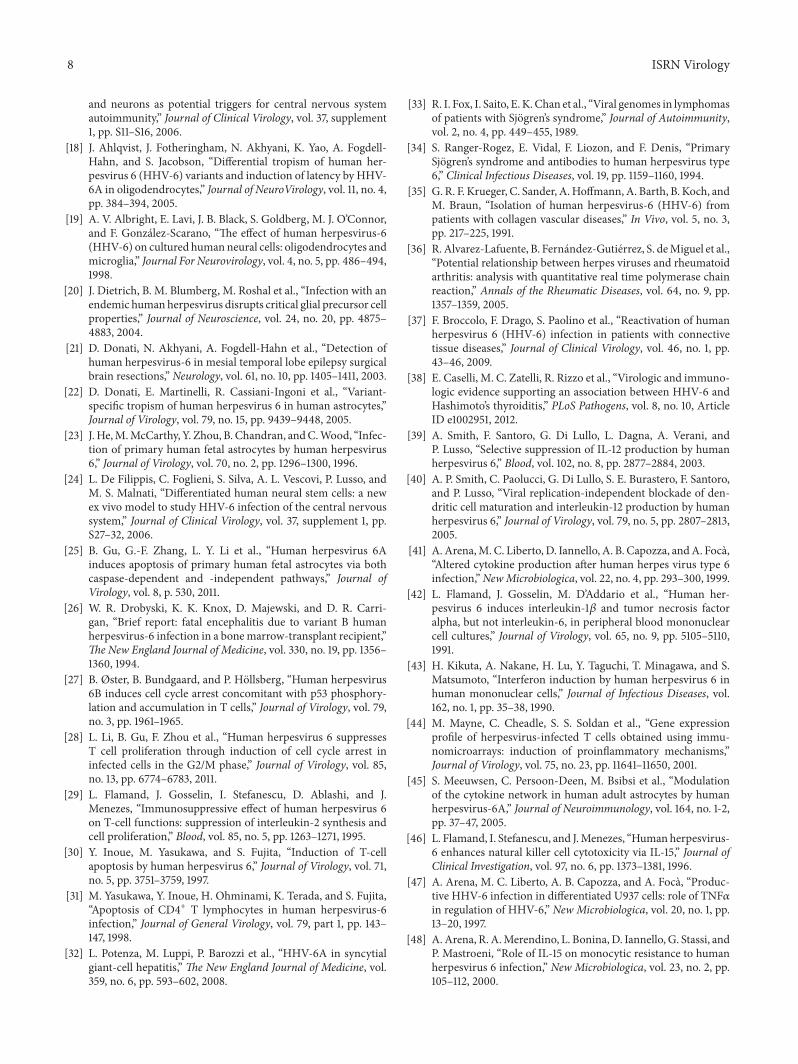
Figure 2: Potential mechanisms for HHV-6-induced neuroinflammation. (a) Based on similarities between viral proteins and brain proteins,HHV-6A or -6B infection in the periphery could lead to the activation of cross-reactive T and B cells, able to recognize both viral antigensand brain antigens, and to the development of an autoimmune response directed to the brain (molecular mimicry). This would promotelymphocyte infiltration in the CNS, where they could have cytotoxic activities against resident cells, especially oligodendrocytes which expressmyelin antigens (1). Peripheral infection could also increase the inflammation by inducing IL-17 and inhibiting IL-10 production by T cellsthrough CD46 binding (2). (b) Infection of astrocytes in the brain can lead to the release of proinflammatory cytokines and chemokines,which promote the infiltration of leukocytes expressing the corresponding chemokine receptor (3). Productive infection of CNS cells canresult in the production of the viral chemokine U83, which can also attract leukocytes to the brain (4). Finally, infection of endothelial cellscan induce the secretion of chemokines, thus attracting circulating leukocytes and facilitating their transmigration through the blood-brainbarrier (5).
organs [53, 111], and that infection induces the productionof proinflammatory chemokines, such as CCL-5, CCL-2, andCXCL-8 [52, 53]. HHV-6A might then be able to infectendothelial cells of the brain vessels and, by increasing CCL-5 secretion, could potentially attract leukocytes to the blood-brain barrier. Moreover, a study reported that, in the contextof liver transplantation, HHV-6 infection was correlated withoverexpression of cell adhesion molecules, such as ICAM-1 and VCAM-1 in the vascular endothelium, and increasednumber of infiltrating lymphoid cells expressing their ligands,LFA-1 and VLA-4 [112]. Therefore, HHV-6 could potentiallyinduce similar upregulation of cell adhesion proteins expres-sion in CNS endothelial cells, thus increasing the blood-brain barrier permeability and facilitating the transfer ofautoreactive lymphocytes in the brain (Figure 2(b)).
5.5. CD46 Engagement. The transmembrane protein CD46 isthe only known entry receptor for both HHV-6A and -6Bentry. This complement regulatory protein also plays animportant role in the adaptive immune response as it canmodulate T cell responses depending on which cytoplasmictail is expressed [113] and can induce CD4+ T cells toward aTr1 phenotype, with high IL-10 production [114]. One couldthen hypothesize that HHV-6A and -6B, by binding to theirreceptor, could modulate its functions. In support to thistheory, a clinical study indicated that increase in HHV-6 viralload was correlated to enhanced CD46 expression in MSpatients [115], and several alterations in CD46 functions weredescribed; the CD46-induced IL-10 secretion by T cells wasstrongly decreased [116], whereas the CD46-dependant IL-23 production by DC and IL-17 expression by T cells were
ISRN Virology 7
enhanced [117, 118]. This suggests that HHV-6 could par-ticipate in neuroinflammation in the context of MS, bypromoting inflammatory processes through CD46 binding(Figure 2(a)).
5.6. Interaction with Other Infectious Agents. In the field ofMS, many different genetic and environmental factors havebeen proposed as potential etiological agents. Yet, if consid-ered separately, none of these candidates could be directlylinked to the onset of the disease. Therefore, efforts are nowfocusing on combinations of factors, including both exoge-nous agents, such as living conditions or viral and bacterialinfections, and endogenous factors, like genetic predisposi-tions. One good example of these potential combinations isthe interaction between herpesvirus infections and humanendogenous retroviruses (HERVs) [119]. HERVs, which rep-resent around 8% of the human genome, have been related toMS pathology since fully mature virions were isolated fromleptomeningeal cells of an MS patient [120]. These viruses,and especially their envelope proteins, have strong inflam-matory properties [121, 122]. HHV-6 infection seems to havedirect transactivating properties on HERV, as it is able toincrease their reverse transcriptase activity [123] and to stim-ulate the transcription of envelope genes [124, 125]. HHV-6infection could then increase neuroinflammation by induc-ing HERV proteins, thus linking exogenous infections toendogenous factors.
6. Conclusion
HHV-6AandHHV-6Bboth exhibit neuroinvasive andproin-flammatory properties. Moreover, both viruses are closelyassociatedwith neurological diseases involving inflammatoryprocesses, which strongly supports the hypothesis that theycan induce neuroinflammation.
The rare cases of encephalitis following primary HHV-6Binfection, in which the virus is the only possible pathogeniccause of disease, provide evidence that HHV-6B has theability to trigger inflammation in the brain. Whether this is adirect or indirect consequence of viral infection and whetherthe virus can induce such complications alone or in synergywith other factors remain to be clarified.
However, in other contexts, it is still difficult to bring solidproof of a decisive role for either HHV-6A or HHV-6B in theestablishment of neuroinflammatory diseases. As HHV-6Aappears to be more neurotropic and was more closely asso-ciated with multiple sclerosis, it may have more importantimplication in neurological diseases in adults. Yet, furtherinvestigations are still needed to better understand how thesetwo viruses may participate in neuroinflammatory processes.The development of new tools, such as more complex in vitrosystems or novel animal models in monkeys and humanizedmice, could be of great help for the research in this field.
Acknowledgments
The work was supported by INSERM and ARSEP, and J. M.Reynaud was supported by a doctoral fellowship from theFrench ministry of research.
References
[1] S. Z. Salahuddin, D. V. Ablashi, P. D. Markham et al., “Isolationof a new virus, HBLV, in patients with lymphoproliferativedisorders,” Science, vol. 234, no. 4776, pp. 596–601, 1986.
[2] G. Dominguez, T. R. Dambaugh, F. R. Stamey, S. Dewhurst,N. Inoue, and P. E. Pellett, “Human herpesvirus 6B genomesequence: coding content and comparison with human her-pesvirus 6A,” Journal of Virology, vol. 73, no. 10, pp. 8040–8052,1999.
[3] K. N. Ward, “The natural history and laboratory diagnosis ofhuman herpesviruses-6 and -7 infections in the immunocom-petent,” Journal of Clinical Virology, vol. 32, no. 3, pp. 183–193,2005.
[4] F. Santoro, P. E. Kennedy, G. Locatelli, M. S. Malnati, E. A.Berger, and P. Lusso, “CD46 is a cellular receptor for humanherpesvirus 6,” Cell, vol. 99, no. 7, pp. 817–827, 1999.
[5] M. Luppi, P. Barozzi, A. Maiorana, R. Marasca, and G. Torelli,“Human herpesvirus 6 infection in normal human brain tissue,”Journal of Infectious Diseases, vol. 169, no. 4, pp. 943–944, 1994.
[6] M. Luppi, P. Barozzi, A. Maiorana et al., “Human herpesvirus-6: a survey of presence and distribution of genomic sequencesin normal brain and neuroglial tumors,” Journal of MedicalVirology, vol. 47, no. 1, pp. 105–111, 1995.
[7] P. B. Challoner, K. T. Smith, J. D. Parker et al., “Plaque-asso-ciated expression of human herpesvirus 6 in multiple sclerosis,”Proceedings of the National Academy of Sciences of the UnitedStates of America, vol. 92, no. 16, pp. 7440–7444, 1995.
[8] P. K. Chan, H. K. Ng, M. Hui, M. Ip, J. L. Cheung, and A.F. Cheng, “Presence of human herpesviruses 6, 7, and 8 DNAsequences in normal brain tissue,” Journal of Medical Virology,vol. 59, no. 4, pp. 491–495, 1999.
[9] P. K. Chan, H. K. Ng, M. Hui, and A. F. Cheng, “Prevalence anddistribution of human herpesvirus 6 variants A and B in adulthuman brain,” Journal of Medical Virology, vol. 64, no. 1, pp. 42–46, 2001.
[10] L. Cuomo, P. Trivedi, M. R. Cardillo et al., “Human herpesvirus6 infection in neoplastic and normal brain tissue,” Journal ofMedical Virology, vol. 63, no. 1, pp. 45–51, 2001.
[11] J. Chi, B. Gu, C. Zhang et al., “Human herpesvirus 6 latent infec-tion in patients with glioma,”The Journal of Infectious Diseases,vol. 206, no. 9, pp. 1394–1398, 2012.
[12] M. L. Opsahl and P. G. E. Kennedy, “Early and late HHV-6 genetranscripts in multiple sclerosis lesions and normal appearingwhite matter,” Brain, vol. 128, no. part 3, pp. 516–527, 2005.
[13] C. B. Hall, M. T. Caserta, K. C. Schnabel et al., “Persistenceof human herpesvirus 6 according to site and variant: possiblegreater neurotropism of variant A,” Clinical Infectious Diseases,vol. 26, no. 1, pp. 132–137, 1998.
[14] M. T. Caserta, C. B. Hall, K. Schnabel et al., “Neuroinvasion andpersistence of human herpesvirus 6 in children,” Journal ofInfectious Diseases, vol. 170, no. 6, pp. 1586–1589, 1994.
[15] E. Harberts, K. Yao, J. E. Wohler et al., “Human herpesvirus-6 entry into the central nervous system through the olfactorypathway,” Proceedings of the National Academy of Sciences of theUnited States of America, vol. 108, no. 33, pp. 13734–13739, 2011.
[16] L. De Bolle, J. Van Loon, E. De Clercq, and L. Naesens, “Quan-titative analysis of human herpesvirus 6 cell tropism,” Journal ofMedical Virology, vol. 75, no. 1, pp. 76–85, 2005.
[17] J. L. Gardell, P. Dazin, J. Islar, T. Menge, C. P. Genain, and P.H. Lalive, “Apoptotic effects of Human Herpesvirus-6A on glia
8 ISRN Virology
and neurons as potential triggers for central nervous systemautoimmunity,” Journal of Clinical Virology, vol. 37, supplement1, pp. S11–S16, 2006.
[18] J. Ahlqvist, J. Fotheringham, N. Akhyani, K. Yao, A. Fogdell-Hahn, and S. Jacobson, “Differential tropism of human her-pesvirus 6 (HHV-6) variants and induction of latency by HHV-6A in oligodendrocytes,” Journal of NeuroVirology, vol. 11, no. 4,pp. 384–394, 2005.
[19] A. V. Albright, E. Lavi, J. B. Black, S. Goldberg, M. J. O’Connor,and F. Gonzalez-Scarano, “The effect of human herpesvirus-6(HHV-6) on cultured humanneural cells: oligodendrocytes andmicroglia,” Journal For Neurovirology, vol. 4, no. 5, pp. 486–494,1998.
[20] J. Dietrich, B. M. Blumberg, M. Roshal et al., “Infection with anendemic human herpesvirus disrupts critical glial precursor cellproperties,” Journal of Neuroscience, vol. 24, no. 20, pp. 4875–4883, 2004.
[21] D. Donati, N. Akhyani, A. Fogdell-Hahn et al., “Detection ofhuman herpesvirus-6 in mesial temporal lobe epilepsy surgicalbrain resections,” Neurology, vol. 61, no. 10, pp. 1405–1411, 2003.
[22] D. Donati, E. Martinelli, R. Cassiani-Ingoni et al., “Variant-specific tropism of human herpesvirus 6 in human astrocytes,”Journal of Virology, vol. 79, no. 15, pp. 9439–9448, 2005.
[23] J. He,M.McCarthy, Y. Zhou, B. Chandran, andC.Wood, “Infec-tion of primary human fetal astrocytes by human herpesvirus6,” Journal of Virology, vol. 70, no. 2, pp. 1296–1300, 1996.
[24] L. De Filippis, C. Foglieni, S. Silva, A. L. Vescovi, P. Lusso, andM. S. Malnati, “Differentiated human neural stem cells: a newex vivo model to study HHV-6 infection of the central nervoussystem,” Journal of Clinical Virology, vol. 37, supplement 1, pp.S27–32, 2006.
[25] B. Gu, G.-F. Zhang, L. Y. Li et al., “Human herpesvirus 6Ainduces apoptosis of primary human fetal astrocytes via bothcaspase-dependent and -independent pathways,” Journal ofVirology, vol. 8, p. 530, 2011.
[26] W. R. Drobyski, K. K. Knox, D. Majewski, and D. R. Carri-gan, “Brief report: fatal encephalitis due to variant B humanherpesvirus-6 infection in a bonemarrow-transplant recipient,”The New England Journal of Medicine, vol. 330, no. 19, pp. 1356–1360, 1994.
[27] B. Øster, B. Bundgaard, and P. Hollsberg, “Human herpesvirus6B induces cell cycle arrest concomitant with p53 phosphory-lation and accumulation in T cells,” Journal of Virology, vol. 79,no. 3, pp. 1961–1965.
[28] L. Li, B. Gu, F. Zhou et al., “Human herpesvirus 6 suppressesT cell proliferation through induction of cell cycle arrest ininfected cells in the G2/M phase,” Journal of Virology, vol. 85,no. 13, pp. 6774–6783, 2011.
[29] L. Flamand, J. Gosselin, I. Stefanescu, D. Ablashi, and J.Menezes, “Immunosuppressive effect of human herpesvirus 6on T-cell functions: suppression of interleukin-2 synthesis andcell proliferation,” Blood, vol. 85, no. 5, pp. 1263–1271, 1995.
[30] Y. Inoue, M. Yasukawa, and S. Fujita, “Induction of T-cellapoptosis by human herpesvirus 6,” Journal of Virology, vol. 71,no. 5, pp. 3751–3759, 1997.
[31] M. Yasukawa, Y. Inoue, H. Ohminami, K. Terada, and S. Fujita,“Apoptosis of CD4+ T lymphocytes in human herpesvirus-6infection,” Journal of General Virology, vol. 79, part 1, pp. 143–147, 1998.
[32] L. Potenza, M. Luppi, P. Barozzi et al., “HHV-6A in syncytialgiant-cell hepatitis,” The New England Journal of Medicine, vol.359, no. 6, pp. 593–602, 2008.
[33] R. I. Fox, I. Saito, E.K.Chan et al., “Viral genomes in lymphomasof patients with Sjogren’s syndrome,” Journal of Autoimmunity,vol. 2, no. 4, pp. 449–455, 1989.
[34] S. Ranger-Rogez, E. Vidal, F. Liozon, and F. Denis, “PrimarySjogren’s syndrome and antibodies to human herpesvirus type6,” Clinical Infectious Diseases, vol. 19, pp. 1159–1160, 1994.
[35] G. R. F. Krueger, C. Sander, A.Hoffmann, A. Barth, B. Koch, andM. Braun, “Isolation of human herpesvirus-6 (HHV-6) frompatients with collagen vascular diseases,” In Vivo, vol. 5, no. 3,pp. 217–225, 1991.
[36] R.Alvarez-Lafuente, B. Fernandez-Gutierrez, S. deMiguel et al.,“Potential relationship between herpes viruses and rheumatoidarthritis: analysis with quantitative real time polymerase chainreaction,” Annals of the Rheumatic Diseases, vol. 64, no. 9, pp.1357–1359, 2005.
[37] F. Broccolo, F. Drago, S. Paolino et al., “Reactivation of humanherpesvirus 6 (HHV-6) infection in patients with connectivetissue diseases,” Journal of Clinical Virology, vol. 46, no. 1, pp.43–46, 2009.
[38] E. Caselli, M. C. Zatelli, R. Rizzo et al., “Virologic and immuno-logic evidence supporting an association between HHV-6 andHashimoto’s thyroiditis,” PLoS Pathogens, vol. 8, no. 10, ArticleID e1002951, 2012.
[39] A. Smith, F. Santoro, G. Di Lullo, L. Dagna, A. Verani, andP. Lusso, “Selective suppression of IL-12 production by humanherpesvirus 6,” Blood, vol. 102, no. 8, pp. 2877–2884, 2003.
[40] A. P. Smith, C. Paolucci, G. Di Lullo, S. E. Burastero, F. Santoro,and P. Lusso, “Viral replication-independent blockade of den-dritic cell maturation and interleukin-12 production by humanherpesvirus 6,” Journal of Virology, vol. 79, no. 5, pp. 2807–2813,2005.
[41] A. Arena,M.C. Liberto, D. Iannello, A. B. Capozza, andA. Foca,“Altered cytokine production after human herpes virus type 6infection,”NewMicrobiologica, vol. 22, no. 4, pp. 293–300, 1999.
[42] L. Flamand, J. Gosselin, M. D’Addario et al., “Human her-pesvirus 6 induces interleukin-1𝛽 and tumor necrosis factoralpha, but not interleukin-6, in peripheral blood mononuclearcell cultures,” Journal of Virology, vol. 65, no. 9, pp. 5105–5110,1991.
[43] H. Kikuta, A. Nakane, H. Lu, Y. Taguchi, T. Minagawa, and S.Matsumoto, “Interferon induction by human herpesvirus 6 inhuman mononuclear cells,” Journal of Infectious Diseases, vol.162, no. 1, pp. 35–38, 1990.
[44] M. Mayne, C. Cheadle, S. S. Soldan et al., “Gene expressionprofile of herpesvirus-infected T cells obtained using immu-nomicroarrays: induction of proinflammatory mechanisms,”Journal of Virology, vol. 75, no. 23, pp. 11641–11650, 2001.
[45] S. Meeuwsen, C. Persoon-Deen, M. Bsibsi et al., “Modulationof the cytokine network in human adult astrocytes by humanherpesvirus-6A,” Journal of Neuroimmunology, vol. 164, no. 1-2,pp. 37–47, 2005.
[46] L. Flamand, I. Stefanescu, and J.Menezes, “Humanherpesvirus-6 enhances natural killer cell cytotoxicity via IL-15,” Journal ofClinical Investigation, vol. 97, no. 6, pp. 1373–1381, 1996.
[47] A. Arena, M. C. Liberto, A. B. Capozza, and A. Foca, “Produc-tive HHV-6 infection in differentiated U937 cells: role of TNF𝛼in regulation of HHV-6,” New Microbiologica, vol. 20, no. 1, pp.13–20, 1997.
[48] A. Arena, R. A.Merendino, L. Bonina, D. Iannello, G. Stassi, andP. Mastroeni, “Role of IL-15 on monocytic resistance to humanherpesvirus 6 infection,” New Microbiologica, vol. 23, no. 2, pp.105–112, 2000.

ISRN Virology 9
[49] I. Nordstrom and K. Eriksson, “HHV-6B induces IFN-lambda1responses in cord plasmacytoid dendritic cells through TLR9,”PLoS ONE, vol. 7, no. 6, Article ID e38683, 2012.
[50] J. C. Grivel, Y. Ito, G. Faga et al., “Suppression of CCR5- but notCXCR4-tropic HIV-1 in lymphoid tissue by human herpesvirus6,” Nature Medicine, vol. 7, no. 11, pp. 1232–1235, 2001.
[51] J.-C. Grivel, F. Santoro, S. Chen et al., “Pathogenic effectsof human herpesvirus 6 in human lymphoid tissue ex vivo,”Journal of Virology, vol. 77, no. 15, pp. 8280–8289, 2003.
[52] A. Caruso, F. Favilli, A. Rotola et al., “Human herpesvirus-6modulates RANTES production in primary human endothelialcell cultures,” Journal of Medical Virology, vol. 70, no. 3, pp. 451–458, 2003.
[53] A. Caruso, A. Rotola, M. Comar et al., “HHV-6 infects humanaortic and heart microvascular endothelial cells, increasingtheir ability to secrete proinflammatory chemokines,” Journal ofMedical Virology, vol. 67, no. 4, pp. 528–533, 2002.
[54] R. Inagi, R. Guntapong, M. Nakao et al., “Human herpesvirus 6induces IL-8 gene expression in human hepatoma cell line, HepG2,” Journal of Medical Virology, vol. 49, no. 1, pp. 34–40, 1996.
[55] D. K. Braun, G. Dominguez, and P. E. Pellett, “Human her-pesvirus 6,”Clinical Microbiology Reviews, vol. 10, no. 3, pp. 521–567, 1997.
[56] K. Yao, J. R. Crawford, A. L. Komaroff, D. V. Ablashi, andS. Jacobson, “Review part 2: human herpesvirus-6 in centralnervous system diseases,” Journal of Medical Virology, vol. 82,no. 10, pp. 1669–1678, 2010.
[57] H. Agut, “Deciphering the clinical impact of acute humanherpesvirus 6 (HHV-6) infections,” Journal of Clinical Virology,vol. 52, no. 3, pp. 164–171, 2011.
[58] D. M. Zerr, “Human herpesvirus 6 and central nervous systemdisease in hematopoietic cell transplantation,” Journal of Clini-cal Virology, vol. 37, upplement 1, pp. S52–S56, 2006.
[59] K. Yamanishi, T. Okuno, K. Shiraki et al., “Identification ofhuman herpesvirus-6 as a causal agent for exanthem subitum,”The Lancet, vol. 1, no. 8594, pp. 1065–1067, 1988.
[60] Y. Asano, T. Yoshikawa, S. Suga et al., “Clinical features ofinfants with primary human herpesvirus 6 infection (exanthemsubitum, roseola infantum),” Pediatrics, vol. 93, no. 1, pp. 104–108, 1994.
[61] C. B. Hall, C. E. Long, K. C. Schnabel et al., “Human her-pesvirus-6 infection in children—a prospective study of com-plications and reactivation,” The New England Journal ofMedicine, vol. 331, no. 7, pp. 432–438, 1994.
[62] S. R. Barone, M. H. Kaplan, and L. R. Krilov, “Humanherpesvirus-6 infection in children with first febrile seizures,”Journal of Pediatrics, vol. 127, no. 1, pp. 95–97, 1995.
[63] Y. Asano, T. Yoshikawa, Y. Kajita et al., “Fatal encephali-tis/encephalopathy in primary human herpesvirus-6 infection,”Archives of Disease in Childhood, vol. 67, no. 12, pp. 1484–1485,1992.
[64] T. Olli-Lahdesmaki, L. Haataja, R. Parkkola, M. Waris, N.Bleyzac, and O. Ruuskanen, “High-dose ganciclovir in HHV-6encephalitis of an immunocompetent child,” Pediatric Neurol-ogy, vol. 43, no. 1, pp. 53–56, 2010.
[65] K. Yanagihara, K. Tanaka-Taya, Y. Itagaki et al., “Human her-pesvirus 6 meningoencephalitis with sequelae,” Pediatric Infec-tious Disease Journal, vol. 14, no. 3, pp. 240–242, 1995.
[66] K. B. Howell, K. Tiedemann, G. Haeusler et al., “Symptomaticgeneralized epilepsy after HHV6 posttransplant acute limbicencephalitis in children,” Epilepsia, vol. 53, no. 7, pp. e122–e126,2012.
[67] T. Yoshikawa, M. Ohashi, F. Miyake et al., “Exanthem subitum-associated encephalitis: nationwide survey in Japan,” PediatricNeurology, vol. 41, no. 5, pp. 353–358, 2009.
[68] H. Matsumoto, D. Hatanaka, Y. Ogura, A. Chida, Y. Nakamura,and S. Nonoyama, “Severe human herpesvirus 6-associatedencephalopathy in three children: analysis of cytokine profilesand the carnitine palmitoyltransferase 2 gene,” The PediatricInfectious Disease Journal, vol. 30, no. 11, pp. 999–1001, 2011.
[69] M. Patnaik and J. B. Peter, “Intrathecal synthesis of antibodiesto human herpesvirus 6 early antigen in patients with menin-gitis/encephalitis,” Clinical Infectious Diseases, vol. 21, no. 3, pp.715–716, 1995.
[70] T. Birnbaum, C. S. Padovan, B. Sporer et al., “Severe menin-goencephalitis caused by human herpesvirus 6 type B in animmunocompetent woman treated with ganciclovir,” ClinicalInfectious Diseases, vol. 40, no. 6, pp. 887–889, 2005.
[71] E. Isaacson, C. A. Glaser, B. Forghani et al., “Evidence of humanherpesvirus 6 infection in 4 immunocompetent patients withencephalitis,”Clinical Infectious Diseases, vol. 40, no. 6, pp. 890–893, 2005.
[72] J. A. McCullers, F. D. Lakeman, and R. J. Whitley, “Humanherpesvirus 6 is associated with focal encephalitis,” ClinicalInfectious Diseases, vol. 21, no. 3, pp. 571–576, 1995.
[73] K. Yao, S. Honarmand, A. Espinosa, N. Akhyani, C. Glaser,and S. Jacobson, “Detection of human herpesvirus-6 in cere-brospinal fluid of patients with encephalitis,” Annals of Neurol-ogy, vol. 65, no. 3, pp. 257–267, 2009.
[74] N. P. Tavakoli, S. Nattanmai, R.Hull et al., “Detection and typingof human herpesvirus 6 by molecular methods in specimensfrom patients diagnosed with encephalitis or meningitis,” Jour-nal of Clinical Microbiology, vol. 45, no. 12, pp. 3972–3978, 2007.
[75] T. Yoshikawa, Y. Asano, M. Ihira et al., “Human herpesvirus 6viremia in bone marrow transplant recipients: clinical featuresand risk factors,” Journal of Infectious Diseases, vol. 185, no. 7, pp.847–853, 2002.
[76] M. Ogata, “Human herpesvirus 6 in hematological malignan-cies,” Journal of Clinical and Experimental Hematopathology,vol. 49, no. 2, pp. 57–67, 2009.
[77] J. Fotheringham, N. Akhyani, A. Vortmeyer et al., “Detection ofactive human herpesvirus-6 infection in the brain: correlationwith polymerase chain reaction detection in cerebrospinalfluid,” Journal of Infectious Diseases, vol. 195, no. 3, pp. 450–454,2007.
[78] F. Forest, S. Duband, S. Pillet et al., “Lethal human herpesvirus-6 encephalitis after cord blood transplant,” Transplant InfectiousDisease, vol. 13, no. 6, pp. 646–649, 2011.
[79] P. Ljungman, F. Z. Wang, D. A. Clark et al., “High levels ofhuman herpesvirus 6 DNA in peripheral blood leucocytes arecorrelated to platelet engraftment and disease in allogeneic stemcell transplant patients,” British Journal of Haematology, vol. 111,no. 3, pp. 774–781, 2000.
[80] D. M. Zerr, T. A. Gooley, L. Yeung et al., “Human herpesvirus6 reactivation and encephalitis in allogeneic bone marrowtransplant recipients,” Clinical Infectious Diseases, vol. 33, no. 6,pp. 763–771, 2001.
[81] M. Ogata, H. Kikuchi, T. Satou et al., “Human herpesvirus 6DNA in plasma after allogeneic stem cell transplantation: inci-dence and clinical significance,” Journal of Infectious Diseases,vol. 193, no. 1, pp. 68–79, 2006.
[82] F. Wilborn, C. A. Schmidt, V. Brinkmann, K. Jendroska, H.Oettle, and W. Siegert, “A potential role for human herpesvirus

10 ISRN Virology
type 6 in nervous system disease,” Journal of Neuroimmunology,vol. 49, no. 1-2, pp. 213–214, 1994.
[83] S. S. Soldan, R. Berti, N. Salem et al., “Association of humanherpes virus 6 (HHV-6) with multiple sclerosis: increased IgMresponse to HHV-6 early antigen and detection of serumHHV-6 DNA,” Nature Medicine, vol. 3, no. 12, pp. 1394–1397, 1997.
[84] S. Chapenko, A. Millers, Z. Nora, I. Logina, R. Kukaine, andM. Murovska, “Correlation between HHV-6 reactivation andmultiple sclerosis disease activity,” Journal of Medical Virology,vol. 69, no. 1, pp. 111–117, 2003.
[85] M. Garcia-Montojo, A. Martinez, V. De Las Heras et al.,“Herpesvirus active replication in multiple sclerosis: a geneticcontrol?” Journal of the Neurological Sciences, vol. 311, no. 1-2,pp. 98–102, 2011.
[86] D. V. Ablashi,W. Lapps, M. Kaplan, J. E.Whitman, J. R. Richert,and G. R. Pearson, “Human Herpesvirus-6 (HHV-6) infectionin multiple sclerosis: a preliminary report,” Multiple Sclerosis,vol. 4, no. 6, pp. 490–496, 1998.
[87] J. E. Friedman, M. J. Lyons, G. Cu et al., “The association of thehuman herpesvirus-6 and MS,” Multiple Sclerosis, vol. 5, no. 5,pp. 355–362, 1999.
[88] S. S. Soldan, T. P. Leist, K. N. Juhng, H. F. McFarland, and S.Jacobson, “Increased lymphoproliferative response to humanherpesvirus type 6A variant in multiple sclerosis patients,”Annals of Neurology, vol. 47, no. 3, pp. 306–313, 2000.
[89] V. J. Sanders, S. Felisan, A. Waddell, and W. W. Tourtellotte,“Detection of Herpesviridae in postmortem multiple sclerosisbrain tissue and controls by polymerase chain reaction,” Journalof NeuroVirology, vol. 2, no. 4, pp. 249–258, 1996.
[90] A. D. Goodman, D. J. Mock, J. M. Powers, J. V. Baker, and B. M.Blumberg, “Human herpesvirus 6 genome and antigen in acutemultiple sclerosis lesions,”The Journal of Infectious Diseases, vol.187, no. 9, pp. 1365–1376, 2003.
[91] R. Berti, M. B. Brennan, S. S. Soldan et al., “Increased detectionof serum HHV-6 DNA sequences during multiple sclerosis(MS) exacerbations and correlation with parameters of MSdisease progression,” Journal of NeuroVirology, vol. 8, no. 3, pp.250–256, 2002.
[92] R. Alvarez-Lafuente, V. De las Heras, M. Bartolome, J. J. Picazo,and R. Arroyo, “Relapsing-remitting multiple sclerosis andhuman herpesvirus 6 active infection,” Archives of Neurology,vol. 61, no. 10, pp. 1523–1527, 2004.
[93] S. Simpson Jr., B. Taylor, D. E. Dwyer et al., “Anti-HHV-6 IgGtiter significantly predicts subsequent relapse risk in multiplesclerosis,”Multiple Sclerosis, vol. 18, no. 6, pp. 799–806, 2012.
[94] N. Akhyani, R. Berti, M. B. Brennan et al., “Tissue distributionand variant characterization of human herpesvirus (HHV)-6: increased prevalence of HHV-6A in patients with multiplesclerosis,” Journal of Infectious Diseases, vol. 182, no. 5, pp. 1321–1325, 2000.
[95] J. Ongradi, C. Rajda, C. L. Marodi, A. Csiszar, and L. Vecsei, “Apilot study on the antibodies to HHV-6 variants and HHV-7 inCSF of MS patients,” Journal For Neurovirology, vol. 5, no. 5, pp.529–532, 1999.
[96] A. M. Fillet, P. Lozeron, H. Agut et al., “HHV-6 and multiplesclerosis,” Nature Medicine, vol. 4, no. 5, pp. 537–538, 1998.
[97] A. R. Coates and J. Bell, “HHV-6 andmultiple sclerosis,”NatureMedicine, vol. 4, no. 5, pp. 537–538, 1998.
[98] K. I. Voumvourakis, D. K. Kitsos, S. Tsiodras, G. Petrikkos, andE. Stamboulis, “Human herpesvirus 6 infection as a trigger ofmultiple sclerosis,” Mayo Clinic Proceedings, vol. 85, no. 11, pp.1023–1030, 2010.
[99] M. Cirone, L. Cuomo, C. Zompetta et al., “Human herpesvirus6 andmultiple sclerosis: a study of T cell cross-reactivity to viraland myelin basic protein antigens,” Journal of Medical Virology,vol. 68, no. 2, pp. 268–272, 2002.
[100] M. V. Tejada-Simon, Y. C. Q. Zang, J. Hong, V. M. Rivera,and J. Z. Zhang, “Cross-reactivity with myelin basic proteinand human herpesvirus-6 in multiple sclerosis,” Annals ofNeurology, vol. 53, no. 2, pp. 189–197, 2003.
[101] W. Cheng, Y.Ma, F. Gong et al., “Cross-reactivity of autoreactiveT cells with MBP and viral antigens in patients with MS,”Frontiers in Bioscience, vol. 17, pp. 1648–1658, 2012.
[102] C. S. Jack, N. Arbour, J. Manusow et al., “TLR signaling tailorsinnate immune responses in human microglia and astrocytes,”Journal of Immunology, vol. 175, no. 7, pp. 4320–4330, 2005.
[103] M. Bsibsi, R. Ravid, D. Gveric, and J. M. Van Noort, “Broadexpression of Toll-like receptors in the human central nervoussystem,” Journal ofNeuropathology andExperimentalNeurology,vol. 61, no. 11, pp. 1013–1021, 2002.
[104] M. Lafon, F. Megret, M. Lafage, and C. Prehaud, “The innateimmune facet of brain: humanneurons express TLR-3 and senseviral dsRNA,” Journal of Molecular Neuroscience, vol. 29, no. 3,pp. 185–194, 2006.
[105] H. Kong, Q. Baerbig, L. Duncan, N. Shepel, and M. Mayne,“Human herpesvirus type 6 indirectly enhances oligodendro-cyte cell death,” Journal of NeuroVirology, vol. 9, no. 5, pp. 539–550, 2003.
[106] Y. Isegawa, Z. Ping, K. Nakano, N. Sugimoto, and K. Yamanishi,“Human herpesvirus 6 open reading frame U12 encodes afunctional 𝛽- chemokine receptor,” Journal of Virology, vol. 72,no. 7, pp. 6104–6112, 1998.
[107] R. S. B. Milne, C. Mattick, L. Nicholson, P. Devaraj, A. Alcami,and U. A. Gompels, “RANTES binding and down-regulation bya novel human herpesvirus-6 𝛽 chemokine receptor,” Journal ofImmunology, vol. 164, no. 5, pp. 2396–2404, 2000.
[108] P. Zou, Y. Isegawa, K. Nakano, M. Haque, Y. Horiguchi, andK. Yamanishi, “Human herpesvirus 6 open reading frame U83encodes a functional chemokine,” Journal of Virology, vol. 73,no. 7, pp. 5926–5933, 1999.
[109] H. R. Luttichau, I. Clark-Lewis, P. Ø. Jensen, C. Moser, J. Ger-stoft, and T. W. Schwartz, “A highly selective CCR2 chemokineagonist encoded by human herpesvirus 6,” The Journal ofBiological Chemistry, vol. 278, no. 13, pp. 10928–10933, 2003.
[110] D. R. Dewin, J. Catusse, and U. A. Gompels, “Identification andcharacterization of U83A viral chemokine, a broad and potent𝛽-chemokine agonist for human CCRs with unique selectivityand inhibition by spliced isoform,” Journal of Immunology, vol.176, no. 1, pp. 544–556, 2006.
[111] C. A.Wu and J. D. Shanley, “Chronic infection of human umbil-ical vein endothelial cells by human herpesvirus-6,” Journal ofGeneral Virology, vol. 79, part 5, pp. 1247–1256, 1998.
[112] I. Lautenschlager, M. Harma, K. Hockerstedt, K. Linnavuori, R.Loginov, and E. Taskinen, “Human herpesvirus-6 infection isassociated with adhesion molecule induction and lymphocyteinfiltration in liver allografts,” Journal of Hepatology, vol. 37, no.5, pp. 648–654, 2002.
[113] J. C. Marie, A. L. Astier, P. Rivailler, C. Rabourdin-Combe, T. F.Wild, and B. Horvat, “Linking innate and acquired immunity:divergent role of CD46 cytoplasmic domains in T cell-inducedinflammation,” Nature Immunology, vol. 3, no. 7, pp. 659–666,2002.
[114] C. Kemper, A. C. Chan, J. M. Green, K. A. Brett, K. M. Murphy,and J. P. Atkinson, “Activation of human CD4+ cells with CD3

ISRN Virology 11
and CD46 induces a T-regulatory cell 1 phenotype,”Nature, vol.421, no. 6921, pp. 388–392, 2003.
[115] R. Alvarez-Lafuente, M. Garcia-Montojo, V. De Las Heras, M.I. Dominguez-Mozo, M. Bartolome, and R. Arroyo, “CD46expression and HHV-6 infection in patients with multiplesclerosis,” Acta Neurologica Scandinavica, vol. 120, no. 4, pp.246–250, 2009.
[116] A. L. Astier, G. Meiffren, S. Freeman, and D. A. Hafler,“Alterations in CD46-mediated Tr1 regulatory T cells in patientswithmultiple sclerosis,” Journal of Clinical Investigation, vol. 116,no. 12, pp. 3252–3257, 2006.
[117] A. Vaknin-Dembinsky, G. Murugaiyan, D. A. Hafler, A. L.Astier, and H. L. Weiner, “Increased IL-23 secretion and alteredchemokine production by dendritic cells upon CD46 activationin patients with multiple sclerosis,” Journal of Neuroimmunol-ogy, vol. 195, no. 1-2, pp. 140–145, 2008.
[118] K. Yao, J. Graham, Y. Akahata, U. Oh, and S. Jacobson,“Mechanism of neuroinflammation: enhanced cytotoxicity andIL-17 production via CD46 binding,” Journal of Hepatology, vol.5, no. 3, pp. 469–478, 2010.
[119] H. Perron, C. Bernard, J. B. Bertrand et al., “Endogenousretroviral genes, herpesviruses and gender inmultiple sclerosis,”Journal of the Neurological Sciences, vol. 286, no. 1-2, pp. 65–72,2009.
[120] H. Perron, J. A. Garson, F. Bedin et al., “Molecular identificationof a novel retrovirus repeatedly isolated from patients withmultiple sclerosis,” Proceedings of the National Academy ofSciences of the United States of America, vol. 94, no. 14, pp. 7583–7588, 1997.
[121] J. M. Antony, G. VanMarle, W. Opii et al., “Human endogenousretrovirus glycoprotein-mediated induction of redox reactantscauses oligodendrocyte death and demyelination,” Nature Neu-roscience, vol. 7, no. 10, pp. 1088–1095, 2004.
[122] A. Rolland, E. Jouvin-Marche, C. Viret, M. Faure, H. Per-ron, and P. N. Marche, “The envelope protein of a humanendogenous retrovirus-W family activates innate immunitythrough CD14/TLR4 and promotesTh1-like responses,” Journalof Immunology, vol. 176, no. 12, pp. 7636–7644, 2006.
[123] T. Brudek, P. Luhdorf, T. Christensen, H. J. Hansen, and A.Møller-Larsen, “Activation of endogenous retrovirus reversetranscriptase in multiple sclerosis patient lymphocytes by inac-tivated HSV-1, HHV-6 and VZV,” Journal of Neuroimmunology,vol. 187, no. 1-2, pp. 147–155, 2007.
[124] A. K. Tai, J. Luka, D. Ablashi, and B. T. Huber, “HHV-6A infec-tion induces expression of HERV-K18-encoded superantigen,”Journal of Clinical Virology, vol. 46, no. 1, pp. 47–48, 2009.
[125] V. L. Turcanova, B. Bundgaard, and P. Hollsberg, “Humanherpesvirus-6B induces expression of the human endogenousretrovirus K18-encoded superantigen,” Journal of Clinical Virol-ogy, vol. 46, no. 1, pp. 15–19, 2009.
[126] C. Li, J. M. Goodrich, and X. Yang, “Interferon-gamma (IFN-𝛾)regulates production of IL-10 and IL-12 in human herpesvirus-6(HHV-6)-infectedmonocyte/macrophage lineage,”Clinical andExperimental Immunology, vol. 109, no. 3, pp. 421–425, 1997.
[127] T. Yoshikawa, Y. Asano, S. Akimoto et al., “Latent infection ofhuman herpesvirus 6 in astrocytoma cell line and alteration ofcytokine synthesis,” Journal of Medical Virology, vol. 66, no. 4,pp. 497–505, 2002.

Submit your manuscripts athttp://www.hindawi.com
Hindawi Publishing Corporationhttp://www.hindawi.com Volume 2014
Anatomy Research International
PeptidesInternational Journal of
Hindawi Publishing Corporationhttp://www.hindawi.com Volume 2014
Hindawi Publishing Corporation http://www.hindawi.com
International Journal of
Volume 2014
Zoology
Hindawi Publishing Corporationhttp://www.hindawi.com Volume 2014
Molecular Biology International
GenomicsInternational Journal of
Hindawi Publishing Corporationhttp://www.hindawi.com Volume 2014
The Scientific World JournalHindawi Publishing Corporation http://www.hindawi.com Volume 2014
Hindawi Publishing Corporationhttp://www.hindawi.com Volume 2014
BioinformaticsAdvances in
Marine BiologyJournal of
Hindawi Publishing Corporationhttp://www.hindawi.com Volume 2014
Hindawi Publishing Corporationhttp://www.hindawi.com Volume 2014
Signal TransductionJournal of
Hindawi Publishing Corporationhttp://www.hindawi.com Volume 2014
BioMed Research International
Evolutionary BiologyInternational Journal of
Hindawi Publishing Corporationhttp://www.hindawi.com Volume 2014
Hindawi Publishing Corporationhttp://www.hindawi.com Volume 2014
Biochemistry Research International
ArchaeaHindawi Publishing Corporationhttp://www.hindawi.com Volume 2014
Hindawi Publishing Corporationhttp://www.hindawi.com Volume 2014
Genetics Research International
Hindawi Publishing Corporationhttp://www.hindawi.com Volume 2014
Advances in
Virolog y
Hindawi Publishing Corporationhttp://www.hindawi.com
Nucleic AcidsJournal of
Volume 2014
Stem CellsInternational
Hindawi Publishing Corporationhttp://www.hindawi.com Volume 2014
Hindawi Publishing Corporationhttp://www.hindawi.com Volume 2014
Enzyme Research
Hindawi Publishing Corporationhttp://www.hindawi.com Volume 2014
International Journal of
Microbiology