resveratrol protects mitochondria against oxidative stress...
TRANSCRIPT
Resveratrol Protects Mitochondria against Oxidative Stressthrough AMP-Activated Protein Kinase-Mediated GlycogenSynthase Kinase-3� Inhibition Downstream of Poly(ADP-ribose)polymerase-LKB1 Pathway
Sang Mi Shin, Il Je Cho, and Sang Geon KimInnovative Drug Research Center for Metabolic and Inflammatory Diseases, College of Pharmacy and Research Institute ofPharmaceutical Sciences, Seoul National University, Seoul, Korea (S.M.S., I.J.C., S.G.K.); and Doping Control Center, KoreaInstitute of Science and Technology, Seoul, Korea (S.M.S.)
Received June 8, 2009; accepted July 20, 2009
ABSTRACTArachidonic acid (AA, a proinflammatory fatty acid) in combi-nation with iron promotes excess reactive oxygen species(ROS) production and exerts a deleterious effect on mitochon-dria. We have shown previously that activation of AMP-acti-vated protein kinase (AMPK) protects hepatocytes from AA �iron-induced apoptosis. Resveratrol, a polyphenol in grapes,has beneficial effects mediated through SIRT1, LKB1, andAMPK. This study investigated the potential of resveratrol toprotect against the mitochondrial impairment induced by AA �iron and the underlying mechanism for this cytoprotection.Resveratrol treatment inhibited apoptosis, ROS production,and glutathione depletion elicited by AA � iron in HepG2 cells.In addition, resveratrol attenuated superoxide generation inmitochondria and inhibited mitochondrial dysfunction inducedby AA � iron. Overall, AMPK activation by resveratrol contrib-uted to cell survival, as supported by the reversal of its resto-ration of mitochondrial membrane potential by either overex-
pression of a dominant-negative mutant of AMPK� orcompound C treatment. Resveratrol increased inhibitory phos-phorylation of glycogen synthase kinase-3� (GSK3�) down-stream of AMPK, which contributed to mitochondrial protectionand cell survival. Likewise, small interfering RNA knockdown ofLKB1, an upstream kinase of AMPK, reduced the ability ofresveratrol to protect cells from mitochondrial dysfunction. Fur-thermore, this LKB1-dependent mitochondrial protection re-sulted from resveratrol’s poly(ADP-ribose)polymerase activa-tion, but not SIRT1 activation, as supported by the experimentusing 3-aminobenzamide, a poly(ADP-ribose)polymerase inhib-itor. Other polyphenols, such as apigenin, genistein, and daid-zein, did not activate AMPK or protect mitochondria against AA� iron. Thus, resveratrol protects cells from AA � iron-inducedROS production and mitochondrial dysfunction through AMPK-mediated inhibitory phosphorylation of GSK3� downstream ofpoly(ADP-ribose)polymerase-LKB1 pathway.
Excess oxidative stress causes cell and tissue injury via themodification of membrane phospholipids (Browning and Hor-ton, 2004). The oxidation of fatty acids in phospholipids by
reactive oxygen species (ROS) or by the response to proin-flammatory cytokines may activate phospholipases (Balboaand Balsinde, 2006). Oxidative modification of fatty acidsand phospholipids triggers inflammatory processes and,thus, exerts detrimental effects on cell signaling. In particu-lar, phospholipase A2 promotes the release of arachidonicacid (AA), a �-6 polyunsaturated fatty acid, from membranephospholipids (Balboa and Balsinde, 2006). AA then contrib-utes to further oxidative stress. In addition, AA in the pres-
This work was supported by the World Class University project funded bythe Korean government (Ministry of Education, Science and Technology De-velopment) [Grant R32-2008-000-10098-0].
Article, publication date, and citation information can be found athttp://molpharm.aspetjournals.org.
doi:10.1124/mol.109.058479.
ABBREVIATIONS: ROS, reactive oxygen species; AA, arachidonic acid; 3-AB, 3-aminobenzamide; ACC, acetyl-CoA carboxylase; AICAR,5-aminoimidazole-4-carboxamide-1-�-D-ribofuranoside; AMPK, AMP-activated protein kinase; CaMKK, calcium/calmodulin-dependent kinasekinase; DCFH-DA, 2�,7�-dichlorofluorescein diacetate; GSK3�, glycogen synthase kinase 3�; MMP, mitochondrial membrane potential; MnTBAP,Mn(III) tetrakis 4-benzoic acid porphyrin; MTT, 3-(4,5-dimethylthiazol-2-yl)-2,5-diphenyl-tetrazolium bromide; PAR, poly(ADP-ribose)polymer;PARP, poly(ADP-ribose)polymerase; PI, propidium iodide; Rh123, rhodamine 123; SOD, superoxide dismutase; GSH, glutathione; siRNA, smallinterfering RNA; L-NAME, L-nitro-arginine-methyl-ester; TUNEL, terminal deoxynucleotidyl transferase dUTP nick-end labeling; DMEM, Dulbecco’smodified Eagle’s medium; FBS, fetal bovine serum; PBS, phosphate-buffered saline; FACS, fluorescence-activated cell sorting; DN, dominantnegative; KM, kinase mutant; Ad, adenovirus; SB216763, 3-(2,4-dichlorophenyl)-4-(1-methyl-1H-indol-3-yl)-1H-pyrrole-2,5-dione; STO-609,7-oxo-7H-benzimidazo[2,1-a]benz[de]isoquinoline-3-carboxylic acid; NOS, nitric-oxide synthase; MMP, mitochrondrial permeability.
0026-895X/09/7604-884–895$20.00MOLECULAR PHARMACOLOGY Vol. 76, No. 4Copyright © 2009 The American Society for Pharmacology and Experimental Therapeutics 58479/3516989Mol Pharmacol 76:884–895, 2009 Printed in U.S.A.
884
at ASPE
T Journals on M
ay 7, 2018m
olpharm.aspetjournals.org
Dow
nloaded from

ence of iron (a catalyst of auto-oxidation) leads cells to pro-duce excess ROS and elicits mitochondrial dysfunction (Shinand Kim, 2009). Therefore, the combinatorial treatment ofAA and iron exerts a negative effect on cell viability, and thistreatment may be used to study potential cytoprotectiveagents targeting mitochondria against severe oxidativestress.
Several lines of evidence indicate that resveratrol (3,4�,5-trihydroxystilbene), a polyphenolic component in grapes andred wine, has diverse beneficial actions, such as protectingcells and tissues against neurodegeneration, cardiovasculardisease, cancer, diabetes, and obesity-related disorders (Baurand Sinclair, 2006), and extending lifespan of organisms(Howitz et al., 2003). This wide range of biological effectsmight be explained in part by resveratrol’s antioxidant prop-erties, including increases in catalase and superoxide dis-mutase (SOD) activity (Rubiolo et al., 2008). In addition, theantioxidant capacity of resveratrol might be mediated by theinduction of phase II enzymes via nuclear factor-E2-relatedfactor-2 (e.g., NADPH-quinone oxidoreductase) (Rubiolo etal., 2008). The antioxidant effect of resveratrol has beenclaimed to be due to the presence of hydroxyl phenolic groupsin its chemical structure (Leonard et al., 2003); however,direct scavenging activity cannot account for all cytoprotec-tive efficacy because of its low bioavailability and weak abil-ity to scavenge ROS (Leonard et al., 2003; Sale et al., 2004).It is more likely that resveratrol may act through specific cellsignaling pathways that lead to activation of the defensiveand cytoprotective systems.
AMP-activated protein kinase (AMPK, an intracellularsensor of energy status) is activated to reserve cellular en-ergy content and serves as a key regulator of cell survival ordeath in response to pathological stress (e.g., oxidativestress, endoplasmic reticulum stress, hypoxia, and osmoticstress) (Hayashi et al., 2000; Terai et al., 2005; Shin and Kim,2009). This regulatory role is supported by increases in cellviability with treatment by the AMPK activators, including5-aminoimidazole-4-carboxamide-1-�-D-ribofuranoside(AICAR) (Ido et al., 2002). Previous work showed that,through AMPK activation, dithiolethiones protect hepato-cytes from AA � iron-induced apoptosis (Shin and Kim,2009). Although the biological effects of resveratrol have beendiversely studied, it is not yet clear whether the cytoprotec-tive effect of resveratrol against ROS is mediated by AMPKactivation. Moreover, the potential cytoprotective effect ofresveratrol against mitochondrial impairment has not beenexplored.
Studies from several laboratories have shown that thebeneficial effects of resveratrol result from the activation ofSIRT1, LKB1, and/or AMPK (Howitz et al., 2003; Hou et al.,2008). In mammalian cells, the upstream kinases of AMPKinclude LKB1, calcium/calmodulin-dependent kinase kinase(CaMKK), and transforming growth factor �-activated ki-nase-1 (Lage et al., 2008). Several signaling pathways includ-ing SIRT1, nitric oxide, protein kinase A, and poly(ADP-ribose)polymerase (PARP) were identified as upstreampathways that regulate LKB1 activity (Alessi et al., 2006;Hou et al., 2008; Huang et al., 2009; Vazquez-Chantada etal., 2009). However, it remains to be elucidated which up-stream signaling pathway regulates the beneficial effect ofresveratrol under conditions of oxidative stress. In addition,it is unclear what molecule or component downstream of
AMPK contributes to mitochondrial protection against oxi-dative stress.
In view of the importance of AMPK in the action of res-veratrol, this study investigated whether resveratrol is capa-ble of protecting mitochondria against the severe oxidativestress induced by AA � iron and, if so, whether this com-pound has the ability to prevent apoptosis. Our work dem-onstrates, for the first time, that resveratrol protects againstAA � iron-induced oxidative stress through the inhibition ofmitochondrial impairment and ROS production: this cytopro-tective effect is mediated by AMPK-dependent GSK3� serinephosphorylation downstream from LKB1. Moreover, we re-vealed that this particular cytoprotective effect of resveratrolagainst oxidative stress is dependent on PARP activation,which leads to LKB1 activation. Additional work comparedthe effect of other polyphenolic compounds on AMPK andtheir efficacy on the mitochondrial dysfunction induced byAA and iron.
Materials and MethodsMaterials. MitoSOX was supplied by Invitrogen (Carlsbad, CA).
Anti-procaspase-3, anti-phospho-acetyl-CoA carboxylase (ACC),anti-phospho-AMPK, anti-AMPK, anti-phospho-LKB1, anti-phospho-GSK3�, and anti-GSK3� antibodies were obtained from Cell Signal-ing Technology (Danvers, MA). Antibodies directed against PARP,Bcl-xl, Bcl-2, and LKB1 were purchased from Santa Cruz Biotech-nology (Santa Cruz, CA). Anti-poly(ADP-ribose)polymer (PAR) anti-body was supplied by BD Bioscience (San Jose, CA). Horseradishperoxidase-conjugated goat anti-rabbit and goat anti-mouse IgGswere purchased from Zymed Laboratories (San Francisco, CA).Mn(III) tetrakis 4-benzoic acid porphyrin (MnTBAP), SB216763,L-nitro-arginine-methyl-ester (L-NAME), and compound C were ob-tained from Calbiochem (Darmstadt, Germany). DeadEnd Colori-metric TUNEL System was supplied from Promega (Madison, WI).Resveratrol, AA, ferric nitrate, nitrilotriacetic acid, AICAR, 3-(4,5-dimethylthiazol-2-yl)-2,5-diphenyl-tetrazolium bromide (MTT), rho-damine 123 (Rh123), propidium iodide (PI), 2�,7�-dichlorofluoresceindiacetate (DCFH-DA), anti-�-actin antibody, trolox, 3-aminobenz-amide (3-AB), and other reagents were purchased from Sigma (St.Louis, MO). The solution of iron-nitrilotriacetic acid complex wasprepared as described previously (Shin and Kim, 2009).
Cell Culture and Treatment. HepG2 cells, a human hepatocyte-derived cell line, were obtained from American Type Culture Collec-tion (Manassas, VA) and maintained in Dulbecco’s modified Eagle’smedium (DMEM) containing 10% fetal bovine serum (FBS), 50 U/mlpenicillin, and 50 �g/ml streptomycin at 37°C in humidified atmo-sphere with 5% CO2. For all experiments, cells (1 � 106) were platedin 10-cm (diameter) plastic dishes for 2 to 3 days (i.e., 80% conflu-ence) and serum-starved for 24 h. Cells were incubated with 10 �MAA for 12 h, followed by additional exposure to 5 �M iron for the timeperiod indicated under Results or in the figure legends. To assess theeffects of resveratrol, the cells were treated with 3 to 60 �M resvera-trol for 1 h before incubation with AA � iron.
MTT Assay. To measure cytotoxicity, HepG2 cells were plated ata density of 5 � 104 cells/well in a 96-well plate. After treatment,viable cells were stained with MTT (0.25 mg/ml, 4 h). The mediawere then removed, and formazan crystals produced in the wellswere dissolved with the addition of 200 �l of dimethyl sulfoxide.Absorbance at 540 nm was measured using an enzyme-linked im-munosorbent assay microplate reader (Tecan, Research TrianglePark, NC). Cell viability was defined relative to untreated control[i.e., viability (% control) � 100 � (absorbance of treated sample)/(absorbance of control)].
TUNEL Assay. TUNEL assay was performed using the DeadEndColorimetric TUNEL System, according to the manufacturer’s in-
PARP-LKB1-Dependent Mitochondrial Protection 885
at ASPE
T Journals on M
ay 7, 2018m
olpharm.aspetjournals.org
Dow
nloaded from

struction. HepG2 cells were fixed with 10% buffered formalin in PBSat room temperature for 30 min and were permeabilized with 0.2%Triton X-100 for 5 min. After washing with PBS, each sample wasincubated with biotinylated nucleotide and terminal deoxynucleoti-dyltransferase in 100 �l of equilibration buffer at 37°C for 1 h. Thereaction was stopped by immersing the samples in 2� saline sodiumcitrate buffer for 15 min. Endogenous peroxidases were blocked byimmersing the samples in 0.3% H2O2 for 5 min. The samples weretreated with 100 �l of horseradish peroxidase-labeled streptavidinsolution (1:500) and incubated for 30 min. Finally, the samples weredeveloped using the diaminobenzidine substrate, chromogen, H2O2,and diaminobenzidine for 10 min. The samples were washed andexamined under light microscope (200�). The counting of TUNEL-positive cells was repeated three times, and the percentage fromeach counting was calculated.
Immunoblot Analysis. Cell lysates were prepared according tomethods published previously (Shin and Kim, 2009). In brief, thecells were centrifuged at 3000g for 3 min and allowed to expandosmotically to the point of lysis after the addition of lysis buffer.Lysates were centrifuged at 10,000g for 10 min to obtain superna-tants and were stored at �70°C until use. Immunoblot analysis wasperformed according to procedures published previously (Shin andKim, 2009). Protein bands of interest were developed using an en-hanced chemiluminescence system (Amersham, Chalfont St. Giles,Buckinghamshire, UK). Equal protein loading was verified by im-munoblotting for �-actin.
Determination of Reduced GSH and Iron. Reduced GSH inthe cells was quantified using a commercial GSH determination kit(Oxis International, Portland, OR). In brief, the GSH-400 method
was a two-step chemical reaction. The first step led to the formationof substitution products (thioethers) between 4-chloro-1-methyl-7-trifluromethyl-quinolinum methylsulfate and all mercaptans present inthe sample. The second step included �-elimination reaction underalkaline conditions. This reaction was mediated by 30% NaOH, whichspecifically transformed the substituted product (thioether) obtainedwith GSH into a chromophoric thione. Analyses of total iron in the cellswere performed on an ICP-AES Optima 4300DV (PerkinElmer Life andAnalytical Sciences, Waltham, MA).
Flow Cytometric Analysis of Mitochondrial Membrane Po-tential. Mitochondrial membrane potential (MMP) was measuredwith Rh123, a membrane-permeable cationic fluorescent dye. Cellswere treated according to the individual experiment, were stainedwith 0.05 �g/ml Rh123 for 1 h, and were harvested by trypsinization.After washing with PBS containing 1% FBS, cells were stained with0.25 �g of PI. The change in MMP was monitored using a BDFACSCalibur flow cytometer (San Jose, CA). In each analysis,15,000 events were recorded.
Measurement of H2O2 Production. DCFH-DA is a cell-perme-able nonfluorescent probe that is cleaved by intracellular esterasesand is turned into the fluorescent dichlorofluorescein upon reactionwith H2O2 and reactive nitrogen species. H2O2 generation was de-termined by the concomitant increase in dichlorofluorescein fluores-cence. After treatment, cells were stained with 10 �M DCFH-DA for1 h at 37°C. Fluorescence intensity in the cells was measured usingFACS. In each analysis, 10,000 events were recorded.
Measurement of Mitochondrial ROS. MitoSOX is a live cell-permeable and mitochondrial localizing superoxide indicator. Aftertreatment of HepG2 cells with AA � iron, the cells were stained with
Fig. 1. Inhibition of AA � iron-in-duced cell death by resveratrol. A, theeffect of resveratrol on cell viability.Light micrographs show the morphol-ogy of the cells incubated with 3 to 60�M resveratrol for 1 h and continu-ously treated with 10 �M AA for 12 h,followed by exposure to 5 �M iron for6 h (magnification, 200�). The dose-response effect of resveratrol on cellviability was assessed using MTT as-says. Data represent the mean � S.E.of four separate experiments. Forgraphs in A and B, the statistical sig-nificance of differences between treat-ments and either the vehicle-treatedcontrol (��, p � 0.01) or cells treatedwith AA � iron (##, p � 0.01) wasdetermined. B, TUNEL assay. Cellswere treated with 30 �M resveratrolfor 1 h, followed by the addition of 10�M AA for 12 h, and finally treatedwith 5 �M iron for 6 h. The percentageof TUNEL-positive cells (dark-brownstaining) was quantified. Data repre-sent the mean � S.E. of four separateexperiments. C, immunoblots for apo-ptotic proteins. Proteins were immu-noblotted from the lysates of cells in-cubated with 30 �M resveratrol for1 h, continuously treated with 10 �MAA for 12 h, and then exposed to 5 �Miron for 1 h. Equal protein loading wasverified by �-actin immunoblotting.Results were confirmed by four sepa-rate experiments.
886 Shin et al.
at ASPE
T Journals on M
ay 7, 2018m
olpharm.aspetjournals.org
Dow
nloaded from
5 �M MitoSOX for 10 min at 37°C. Fluorescence intensity in the cellswas measured using FACS. In each analysis, 10,000 events wererecorded.
Recombinant Adenoviral DN-AMPK� Construct and Plas-mid Transfection. A plasmid encoding a dominant-negative mu-tant of AMPK� (D157A; DN-AMPK�) was kindly provided by Dr. J.Ha (Kyunghee University, Seoul, Korea). To generate a recombinantadenovirus expressing DN-AMPK�, the construct was subcloned intothe attL-containing shuttle plasmid, pENTR-BHRNX (Newgex,Seoul, Korea). Recombinant adenoviral DN-AMPK� was constructedand generated by using the pAd/CMV/V5-DEST gateway plasmid.HepG2 cells were infected with adenovirus diluted in DMEM con-taining 10% FBS at a multiplicity of infection of 50 and incubated for12 h. After removal of the viral suspension, cells were further incu-bated with DMEM containing 10% FBS for 2 days and then weretreated with the indicated reagent. Adenovirus that expresses LacZ(Ad-LacZ) was used as an infection control. Efficiency of infectionwas consistently �90% with this method. The construct encoding fora kinase mutant (KM) form of GSK3� was kindly provided by Dr.J. R. Woodgett (Samuel Lunenfeld Research Institute, Toronto, ON,Canada). Cells were transfected with the plasmids by using Lipo-fectamine 2000 (Invitrogen). The empty plasmid, pCDNA3.1, wasused for the mock transfection.
siRNA Knockdown. To knock down LKB1, cells were transfectedwith either an siRNA directed against human LKB1 (Santa Cruz
Biotechnology) or a nontargeting control siRNA (100 pmol/ml) byusing Lipofectamine 2000 according to the manufacturer’s instruc-tions. After transfection for 24 h, cells were exposed to AA with orwithout resveratrol for 12 h, followed by treatment with 5 �M iron.The resultant samples were analyzed by FACS. LKB1 knockdownwas confirmed by immunoblot analysis.
Data Analysis. Scanning densitometry was performed with anImage Scan and Analysis System (Alpha Innotech, San Leandro,CA). One-way analysis of variance procedures were used to assesssignificant differences among treatment groups. For each treatmentshowing a statistically significant effect, the Newman-Keuls test wasused for comparisons of multiple group means. The criterion forstatistical significance was set at p � 0.05 or � 0.01.
ResultsResveratrol Inhibition of AA � Iron-Induced Apo-
ptosis. A previous study from this laboratory has shown thattreatment of AA (10 �M) and iron (5 �M) elicits synergizedtoxicity in HepG2 cells (Shin and Kim, 2009). First, we ex-amined the effect of resveratrol on cytotoxicity induced by AA� iron. The MTT assay for cell viability indicated that res-veratrol pretreatment (10–60 �M) significantly protectedcells from the injury induced by AA � iron. Maximal cyto-protective effect was observed at 30 �M (Fig. 1A). Given thisresult, 30 �M resveratrol was used in subsequent experi-ments. Morphological examination by light microscope (Fig.1A) and TUNEL assay (Fig. 1B) confirmed the protectiveeffect of resveratrol against the challenge of AA � iron.Resveratrol treatment caused no change in iron contents incells treated with AA � iron (data not shown), excluding thepossibility that resveratrol interferes with iron absorption. Inthe cells treated with AA � iron, the levels of PARP, pro-caspase-3, Bcl-xl, and Bcl-2 levels were lower. Resveratroltreatment completely prevented alterations in the levels ofproteins associated with apoptosis (Fig. 1C). Overall, res-veratrol treatment enables cells to protect against the syn-ergized toxic effects of AA � iron.
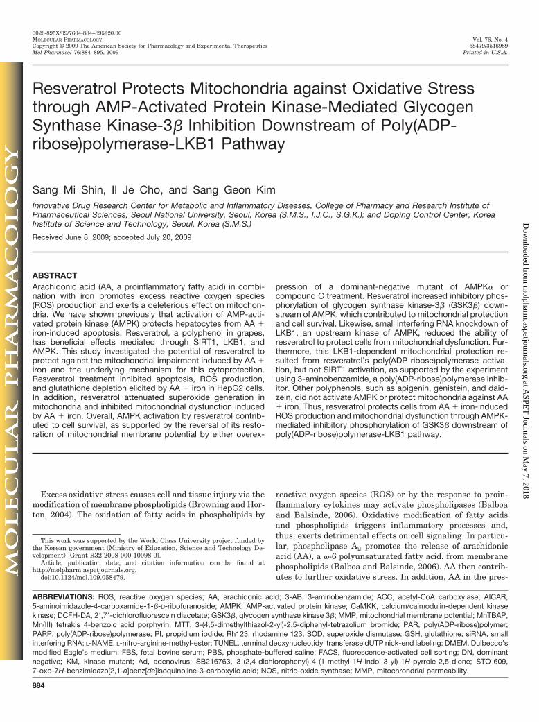
GSH Restoration and Abrogation of ROS. BecauseGSH depletion increases the susceptibility of cells to ROS-induced toxicity (Kode et al., 2008), GSH content was deter-mined in the cells treated with AA � iron in combinationwith resveratrol to test whether resveratrol helps maintainredox homeostasis. AA � iron treatment markedly decreasedlevels of GSH. However, the GSH content was maintained incells exposed to AA � iron after resveratrol treatment (Fig.2A). In addition, a flow cytometric assay using DCFH-DAindicated that resveratrol treatment effectively abrogatedincreases in H2O2 production caused by AA � iron (Fig. 2B).Moreover, trolox (vitamin E analog, 100 �M) treatment for14 h completely attenuated ROS production, confirming thatoxidant species were involved in the process. Both the main-tenance of GSH content and the decrease in H2O2 productionby resveratrol support the hypothesis that the cytoprotectiveeffect of resveratrol results from its antioxidative capacity.
Inhibition of Mitochondrial Dysfunction. AA re-presses mitochondrial respiratory activity by selective inhi-bition of complexes I and III, thereby promoting ROS gener-ation (Cocco et al., 1999). In addition, AA treatment may leadto cell death through mitochondrial permeability transitionin hepatocytes such as HepG2 and MH1C1 cells (Scorrano etal., 2001; Shin and Kim, 2009). To examine the effect ofresveratrol on mitochondria, mitochondrial ROS production
Fig. 2. Inhibition of decrease in GSH content and ROS production byresveratrol A, GSH content. The GSH content was assessed in HepG2cells that had been treated as described in the legend to Fig. 1C. Datarepresent the mean � S.E. of four separate experiments. The statisticalsignificance of differences between treatments and either the vehicle-treated control (��, p � 0.01) or cells treated with AA � iron was deter-mined. B, ROS production. Cells were incubated with resveratrol for 1 h,followed by the addition of AA (12 h) � iron (1 h). Results were confirmedby repeated experiments. Resveratrol or trolox treatment attenuated AA� iron-induced ROS production, as shown by the decrease in DCF fluo-rescence.
PARP-LKB1-Dependent Mitochondrial Protection 887
at ASPE
T Journals on M
ay 7, 2018m
olpharm.aspetjournals.org
Dow
nloaded from
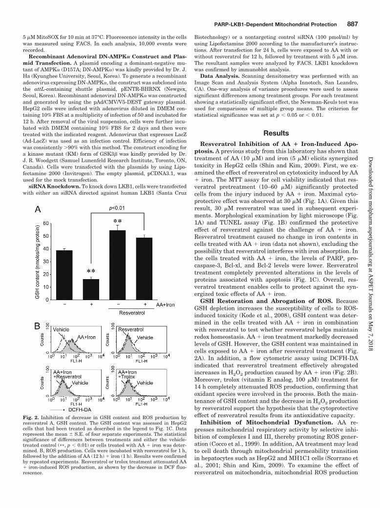
was analyzed using MitoSOX, a live cell-permeable and mi-tochondrial localizing superoxide indicator. AA � iron treat-ment markedly increased mitochondrial MitoSOX fluores-cence, whereas resveratrol treatment (14 h) completelyabolished superoxide production in mitochondria (Fig. 3A).Moreover, MnTBAP (novel superoxide dismutase mimetic, 20�M, for 14 h) enabled cells to scavenge the mitochondrialROS generated by AA � iron treatment, providing furtherevidence that the ROS included superoxide (Fig. 3A). Tocorrelate AA-induced apoptosis with alteration in mitochon-drial function, MMP was measured using FACS after stain-ing of the cells with Rh123. Rh123 as a membrane-permeablecationic fluorescent is used as a sensitive probe of mitochon-drial membrane potential in populations of apoptotic cellsbecause this agent binds to the mitochondrial membranes(Kwon et al., 2009). Low staining intensity of Rh123 repre-sents mitochondrial damage and dysfunction. AA � iron
treatment increased the population of Rh123-negative andPI-negative cells (lower left quadrant), which represents vi-able cells with mitochondria damage (Fig. 3B). The fractionof apoptotic cells in the Rh123-negative and PI-positive field(upper left quadrant) was also increased (Fig. 3B). Therefore,AA � iron treatment induces mitochondrial dysfunction,which consequently leads to cell death. Resveratrol inhibitedthe change in MMP induced by AA � iron, supporting ourconclusion that the cytoprotective effect of resveratrol is as-sociated with the protection of mitochondria.
The Role of AMPK Activation in MitochondrialFunction. Because resveratrol lowered the contents of he-patic lipids in mice through AMPK activation (Baur et al.,2006; Zang et al., 2006), the effects of resveratrol on the timeresponses of ACC and AMPK phosphorylations were mea-sured. Resveratrol treatment increased the phosphorylationof ACC in HepG2 cells, which represents cellular AMPK
Fig. 3. Abrogation of AA � iron-induced mitochondrial ROS production and dysfunction by resveratrol A, ROS production in mitochondria. Cells wereincubated with 30 �M resveratrol for 1 h, treated with 10 �M AA (12 h) followed by incubation with 5 �M iron for 1 h, and then stained with MitoSOX.Increase in MitoSOX fluorescence indicates the production of mitochondrial superoxide. Treatment with either resveratrol or MnTBAP attenuated theability of AA � iron to induce ROS production in mitochondria, as shown by a decrease in MitoSOX fluorescence. B, measurement of MMP. Cells weretreated with 30 �M resveratrol for 1 h, followed by the addition of AA (12 h) � iron (1 h). After staining with Rh123, the cells were harvested andstained with PI. Treatment of AA � iron increased both the subpopulation of Rh123-negative and PI-negative cells (bottom left quadrant), whichrepresents viable cells with mitochondrial damage, and the fraction of apoptotic cells in the Rh123-negative and PI-positive field (top left quadrant).Data represent the mean � S.E. of four replicates. The statistical significance of differences between treatments and either the vehicle-treated control(��, p � 0.01) or cells treated with AA � iron was determined.
888 Shin et al.
at ASPE
T Journals on M
ay 7, 2018m
olpharm.aspetjournals.org
Dow
nloaded from

Fig. 4. AMPK-mediated inhibitory phosphorylation of GSK3� by resveratrol. A, AMPK activation by resveratrol. Immunoblot analyses wereperformed on lysates of cells that had been treated with resveratrol for the indicated time period. Results were confirmed by three separateexperiments. Relative phosphorylated ACC band intensities of immunoblot data were quantified (right). The statistical significance of differencesbetween treatments and the vehicle-treated control was determined (�, p � 0.05, ��, p � 0.01). B, reversal by Ad-DN-AMPK� of the ability ofresveratrol to restore MMP. After Ad-LacZ or Ad-DN-AMPK� infection (48 h), HepG2 cells were incubated with resveratrol for 1 h and continuouslyexposed to AA (12 h) � iron (1 h). MMP was analyzed as described in the legend to Fig. 3B. Data represent the mean � S.E. of four replicates. Thestatistical significance of differences between treatments and either the vehicle-treated control (��, p � 0.01), cells treated with AA � iron (##, p �0.01), or cells treated with AA � iron after Ad-DN-AMPK� infection (N.S., no significance) was determined. Inset, immunoblot analyses forhemagglutinin (HA) confirmed DN-AMPK� overexpression. C, reversal by compound C of resveratrol’s restoration of MMP. After compound Ctreatment (3 �M, 30 min), cells were incubated with resveratrol for 30 min, followed by the addition of AA (12 h) � iron (1 h). Data represent themean � S.E. of four replicates. The statistical significance of differences between treatments and either the vehicle-treated control (��, p � 0.01), cellstreated with AA � iron (##p � 0.01), or cells treated with AA � iron after compound C treatment (N.S., no significance) was determined. D,AMPK-dependent inhibitory phosphorylation of GSK3� by resveratrol. Immunoblot analyses against GSK3� phosphorylated at serine 9 wereperformed on lysates of cells that had been treated with resveratrol for the indicated time period. Cells were treated with compound C as describedin C and then were exposed to resveratrol for 3 h. Equal protein loading was verified by immunoblotting for GSK3�. Results were confirmed byrepeated experiments. E, the effect of GSK3� inhibition on MMP or cell viability. HepG2 cells were incubated with AA (12 h) � iron (1 h) afterSB216763 treatment (10 �M) for 1 h or GSK3�-KM overexpression. MMP was analyzed as described in the legend to Fig. 3B, whereas cell viabilitywas analyzed by MTT assay.
PARP-LKB1-Dependent Mitochondrial Protection 889
at ASPE
T Journals on M
ay 7, 2018m
olpharm.aspetjournals.org
Dow
nloaded from

activity (Fig. 4A). To assess the role of resveratrol’s activa-tion of AMPK in protecting mitochondria and cells, theAMPK-inhibitory effect on rhodamine-negative cell popula-tions was then measured. The recovery of rhodamine-positivecells elicited by resveratrol was significantly reversed byDN-AMPK� overexpression (Fig. 4B). The beneficial effect ofresveratrol on mitochondria was consistently antagonized bysimultaneous treatment with compound C (an AMPK inhib-itor) (Fig. 4C). DN-AMPK� overexpression and the inhibitoryeffect of compound C for AMPK were confirmed previously bythe decreased ACC phosphorylation (Kwon et al., 2009).Therefore, the protection of mitochondria and cells by res-veratrol might be associated at least in part with AMPKactivation.
Resveratrol increases inhibitory phosphorylation of GSK3�,which may be associated with cell survival (Xi et al., 2009). Inan effort to identify downstream molecule of AMPK, we nextexamined the effect of AMPK on GSK3� phosphorylation and
its role in mitochondrial protection. Resveratrol treatment (1 or3 h) increased the phosphorylation of GSK3� at serine 9 resi-due, and this increase was prevented by compound C pretreat-ment (Fig. 4D). Moreover, either SB216763 (a GSK3� inhibitor)treatment or GSK3�-KM (a kinase mutant form of GSK3�)overexpression similarly inhibited the changes in MMP or celldeath induced by AA � iron (Fig. 4E). Overall, our resultsdemonstrate that the cytoprotective effect of resveratrol may beassociated with inhibitory phosphorylation of GSK3� down-stream of AMPK.
The Role of LKB1 Activation in Mitochondrial Pro-tection. To identify the upstream signal of AMPK activationby resveratrol, the time course effect of resveratrol on LKB1phosphorylation was assessed. Resveratrol treatment re-sulted in a notable increase in the phosphorylation of LKB1(Fig. 5A). To test the role of LKB1 in protecting mitochon-drial function against AA � iron, the effect of siRNA knock-down of LKB1 on MMP change was determined. The protec-
Fig. 5. The role of LKB1 activation by resveratrol in mitochondrial function. A, phosphorylation of LKB1. Immunoblot analyses were performed onthe lysates of cells that had been treated with resveratrol for the indicated time period. Results were confirmed by three replicates. Relativephosphorylated LKB1 band intensities of immunoblot data were quantified (right). The statistical significance of differences between treatments andthe vehicle-treated control (��, p � 0.01) was determined. B, reversal by LKB1 knockdown of resveratrol’s effect to restore MMP. HepG2 cells weretransfected with siRNAs directed against LKB1 or nontargeting control siRNA, continuously incubated with resveratrol for 1 h, and then exposed toAA � iron. MMP was analyzed as described in the legend to Fig. 3B. Inset, immunoblot analyses for LKB1 confirmed specific knockdown of LKB1.Data represent the mean � S.E. of four replicates. The statistical significance of differences between treatments and either the vehicle-treated control(��, p � 0.01), cells treated with AA � iron (##, p � 0.01), or cells treated with AA � iron after siLKB1 transfection (N.S., no significance) wasdetermined. C, the effect of a CaMKK inhibitor on the recovery of MMP by resveratrol. After STO-609 treatment (1 �g/ml, for 30 min), cells wereincubated with resveratrol for 30 min, followed by the addition of AA � iron. Results were confirmed by three replicates. MMP was analyzed asdescribed in the legend to Fig. 3B. Data represent the mean � S.E. of four replicates. The statistical significance of differences between treatmentsand either the respective vehicle-treated control (��, p � 0.01) or cells treated with respective AA � iron (##, p � 0.01) was determined.
890 Shin et al.
at ASPE
T Journals on M
ay 7, 2018m
olpharm.aspetjournals.org
Dow
nloaded from
tive effect of resveratrol against mitochondrial dysfunctionelicited by AA � iron was reversed by siRNA knockdown ofLKB1 (Fig. 5B). In contrast, treatment with STO-609 (1�g/ml), an inhibitor of CaMKK, failed to antagonize the ben-eficial effect of resveratrol as indicated by no change in MMP(Fig. 5C). Therefore, the cytoprotective effect of resveratrolagainst mitochondrial dysfunction may depend on LKB1 butnot CaMKK.
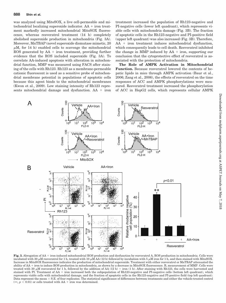
The Effect of PARP Activation on MitochondrialFunction. It has been shown that several signaling path-ways such as SIRT1, nitric oxide, and PARP may regulateLKB1 (Alessi et al., 2006; Hou et al., 2008; Huang et al.,2009; Vazquez-Chantada et al., 2009). Pharmacological in-hibitors were used to assess the upstream signaling of LKB1phosphorylation by resveratrol. Treatment with either nico-tinamide (10 mM, 14 h) or sirtinol (10 �M, 14 h), which hasbeen shown to inhibit SIRT1 activity (Dasgupta and Mil-brandt, 2007), failed to change the protective effect of res-veratrol on MMP in cells treated with AA � iron (Fig. 6A).Likewise, this protective effect of resveratrol was not re-versed by L-NAME [a nitric-oxide synthase (NOS) inhibitor]
pretreatment. In contrast, treatment with 3-AB (a PARPinhibitor) significantly reversed the recovery of rhodamine-positive cells elicited by resveratrol (Fig. 6A). Consistent withthis result, resveratrol increased PARP activity, as supportedby increases in PAR production (Fig. 6B). Moreover, 3-ABtreatment completely inhibited LKB1 phosphorylation in-duced by resveratrol (Fig. 6C). Nicotinamide, sirtinol, or L-NAME failed to reverse the LKB1 phosphorylation (data notshown). These results showed that LKB1-dependent mito-chondrial protection by resveratrol might be associated withPARP activation.
The Effects of Other Polyphenols on AMPK and Mi-tochondrial Protection. Given the current finding thatresveratrol increased cell viability through LKB1-AMPK-de-pendent protection of mitochondrial function, other polyphe-nols including apigenin, genistein, and daidzein (Fig. 7A)were used in an effort to find relationship between AMPKactivation and mitochondrial protective effects. All of thepolyphenols used except for resveratrol had no AMPK-acti-vating effect (Fig. 7B). Moreover, the polyphenolic com-pounds failed to enable cells to restore MMP in response to a
Fig. 6. Resveratrol activation of PARP and itsrole in LKB1 phosphorylation. A, effects of phar-macological inhibitors on the ability of resvera-trol to restore MMP. HepG2 cells were incubatedwith resveratrol for 30 min after treatment with10 mM nicotinamide (NAM), 10 �M sirtinol(Sirt), 10 �M L-NAME, or 10 mM 3-AB for 30min, and the cells were then continuously ex-posed to AA (12 h) � iron (1 h). MMP was ana-lyzed as described in the legend to Fig. 3B. Datarepresent the mean � S.E. of four replicates. Thestatistical significance of differences betweentreatments and either the respective vehicle-treated control (�, p �0.05; ��, p � 0.01) or cellstreated with respective AA � iron (#, p � 0.05;##, p � 0.01) was determined. B, increase in PARproduction by resveratrol. Immunoblot analyseswere performed on the lysates of cells that hadbeen treated with 30 �M resveratrol for the in-dicated time period. Results were confirmed byrepeated experiments. C, inhibition of resvera-trol-induced LKB1 phosphorylation by PARP in-hibitor. Cells were pretreated with 3-AB (10 mM,1 h) and then continuously exposed with 30 �Mresveratrol for 1 h. Results were confirmed bythree separate experiments. The statistical sig-nificance of differences between treatments andeither the vehicle-treated control (��, p � 0.01) orcells treated with resveratrol (##, p � 0.01) wasdetermined.
PARP-LKB1-Dependent Mitochondrial Protection 891
at ASPE
T Journals on M
ay 7, 2018m
olpharm.aspetjournals.org
Dow
nloaded from

challenge by AA � iron (Fig. 7C). All of these results demon-strate that resveratrol has a cytoprotective effect againstsevere oxidative stress induced by AA � iron and the abilityto protect mitochondria, mediated through LKB1-dependentAMPK activation.
DiscussionExcess iron deposition causes injury to and dysfunction of
organs, including the liver. Hepatic iron accumulation mayresult from increases in plasma iron as a result of long-termalcoholism, chronic hepatitis, liver cirrhosis, hemolysis, in-flammatory syndrome, or diabetes. Acquired iron-overloaddisease is characterized by macrophage activation (Flemingand Bacon, 2005). With time, iron is redistributed towardsurrounding hepatocytes. Moreover, the production of proin-flammatory mediators including AA is increased under iron-overload conditions (Galaris and Pantopoulos, 2008). Indeed,we found an increase in plasma AA levels after iron deposi-tion in an in vivo model, in which phenylhydrazine was usedas a hemolytic agent that indirectly creates hepatic ironoverload and promotes oxidative damage (Ferrali et al., 1997;Kim et al., 2009). AA treatment alone promotes ROS produc-tion, causes MMP changes, and thereby leads to cell death
(Kwon et al., 2009). Excess iron catalyzes the release of AA bymodifying membrane phospholipids (Tadolini and Hakim,1996). Moreover, AA synergizes the ability of iron to promoteoxidative stress and cytotoxicity (Shin and Kim, 2009), whichreflects the clinical situations accompanying iron overload.Hence, the combinatorial treatment model may be useful forthe study of drug candidates active in the prevention and/ortreatment of tissue injury caused by iron accumulation.
The mitochondrion serves as the organelle determining acritical point of the apoptotic process because it functions asa stress sensor (Browning and Horton, 2004). Within themitochondrion, the respiratory chain is one of the main sitesof ROS production (Browning and Horton, 2004). The AAgenerated from cell membranes undergoing oxidative stressexerts a direct effect on mitochondria and promotes ROSproduction (Cocco et al., 1999; Scorrano et al., 2001). Like-wise, treatment of cells with AA � iron promotes ROS pro-duction (superoxide) in mitochondria, as shown by theMitoSOX results. Excess production of ROS in mitochondriamay cause apoptosis: combinatorial treatment of AA and ironpromotes cell death, as well as the induction of ROS produc-tion and mitochondrial dysfunction. The prevention of AA �iron-induced mitochondrial dysfunction by cyclosporin A, an
Fig. 7. The effects of polyphenols on mitochondrial function. A, bond-line chemical structures of polyphenols. B, the effects of other polyphenols onAMPK activation. Immunoblot analyses were performed in the lysates of HepG2 cells that had been treated with 30 �M resveratrol, genistein, anddaidzein or 10 �M apigenin for 3 h. Results were confirmed by three replicates. Relative phosphorylated ACC band intensities of immunoblot data werequantified (bottom). The statistical significance of differences between treatments and the vehicle-treated control was determined (��, p � 0.01). C,changes in MMP. Cells were treated with resveratrol, apigenin, genistein, or daidzein for 1 h, followed by additional incubation with AA (12 h) � iron(1 h). Because apigenin at the concentration of 30 �M increased the population of Rh123-negative cells (i.e., possible cytotoxicity), 10 �M apigenin wasused in this experiment. Results were confirmed by three replicates. MMP was analyzed as described in the legend to Fig. 3B. The statisticalsignificance of differences between treatments and either the vehicle-treated control (��, p � 0.01) or cells treated with AA � iron (##, p � 0.01) wasdetermined.
892 Shin et al.
at ASPE
T Journals on M
ay 7, 2018m
olpharm.aspetjournals.org
Dow
nloaded from
inhibitor of permeability transition pore formation, furthersuggests that mitochondrial dysfunction and the consequentROS overproduction might be responsible for cell death (Shinand Kim, 2009). Because polyethylene glycol-SOD, polyeth-ylene glycol-catalase, trolox, or N-acetyl-L-cysteine preventedthe ability of AA � iron to induce both ROS and apoptosis,this evidence provides additional support for the importantrole of ROS in mitochondrial dysfunction and apoptosis (Shinand Kim, 2009).
Resveratrol, a natural polyphenolic compound, has the ca-pacity to inhibit both mitochondrial ROS production andpermeability transition, thereby protecting the key intracel-lular organelle against the oxidative stress promoted by AA� iron. In addition, resveratrol decreases cellular ROS andthus maintains GSH content in cells: these results are con-sistent with previous findings of resveratrol’s potent antiox-idant and anti-inflammatory activity in various organs (Baurand Sinclair, 2006). Our work reveals that the mitochondrialprotection and the antioxidant effect of resveratrol increasesthe viability of hepatocytes against AA � iron. Moreover,resveratrol has a cytoprotective effect against iron-catalyzedoxidative burst through mitochondrial protection, as illus-trated by the inhibition of apoptosis and alterations in apo-ptotic markers.
AMPK is a heterotrimeric protein consisting of one cata-lytic subunit (�) and two noncatalytic subunits (� and �).When AMP binds to the �-subunit, AMPK activation is pro-moted by stimulating phosphorylation at the threonine resi-due within the kinase domain. AMPK monitors cellular en-ergy status by responding to changes in the AMP/ATP ratio.Treatment with an AMPK activator increases the expressionof peroxisome proliferators-activated response-coactivator-1�and manganese-SOD mRNAs, which may inhibit ROS produc-tion in mitochondria (Kukidome et al., 2006). Previous work inour laboratory showed that 1,2-dithiole-3-thiones protect cellsfrom AA � iron-induced ROS production and mitochondrialdysfunction via AMPK activation (Shin and Kim, 2009). Hence,AMPK is a potential target that protects cells from mitochon-drial injury. In this study, the cytoprotective effect of resvera-trol against AA � iron depended on AMPK activation, as evi-denced by the antagonism of AMPK� overexpression orcompound C treatment on the capacity of resveratrol to recoverMMP against AA � iron. The crucial role of AMPK in protectingcells and mitochondria was verified because AICAR exhibited asimilar protective effect (Shin and Kim, 2009).
Oxidative stress activates GSK3� and leads GSK3� totranslocate into the mitochondria. Activated GSK3� in mito-chondria then binds to and phosphorylates the components ofmitochondrial membrane pore (e.g., VDAC and ANT), andthereby induces MMP transition. Thus, GSK3 acts as thepivotal kinase for the regulation of MMP transition (Ju-haszova et al., 2004; Xi et al., 2009). In the present study,resveratrol promoted inhibitory phosphorylation of GSK3� atserine 9. Another important finding of this study is thatresveratrol-induced activation of AMPK is responsible forthis inhibitory phosphorylation. Our data are in line with thefinding that AICAR treatment decreases GSK3 activity indi-rectly through Raf1/ERK mitogen-activated protein kinases/p90 kDa ribosomal S6 kinase (Wang et al., 2008). The resultsof this study demonstrate that the AMPK-dependent mito-chondrial protection of resveratrol against oxidative stress
may be associated with the downstream inhibitory phosphor-ylation of GSK3�.
Although the mechanism of resveratrol’s cytoprotectioninvolves AMPK activation, resveratrol does not directly acti-vate AMPK in vitro (Baur et al., 2006). Therefore, proteinsand/or components that lie upstream of AMPK might be thetarget(s) of resveratrol. LKB1 and CaMKK are the currentlyknown upstream kinases of AMPK (Lage et al., 2008). Ourresults demonstrate that resveratrol activates LKB1, whichpromotes mitochondrial protection in HepG2 cells. By con-trast, CaMKK inhibition fails to reverse this beneficial effect.Hence, resveratrol activates AMPK specifically throughLKB1, but not CaMKK, which parallels the previous findingthat LKB1 was responsible for inhibiting apoptosis in mouseembryonic fibroblast cells under the condition of energystress (Shaw et al., 2004). The upstream signaling pathwaysthat activate LKB1 might include SIRT1, NOS, and proteinkinase A (Alessi et al., 2006; Hou et al., 2008; Vazquez-Chantada et al., 2009). PARP was found as another possiblesignaling pathway of LKB1 activation (Huang et al., 2009).PARP belongs to a group of nuclear enzymes that play acritical role in DNA damage repair through PAR production.It has been known that PAR formation is an energeticallyexpensive process, causing failure in cellular ATP produc-tion, rapid depletion of NAD�, and eventually cell death(Huang et al., 2009). On the contrary, PARP plays a protec-tive role against ROS-induced cell death through LKB1-AMPK-mediated autophagy activation (Huang et al., 2009). Inthe present study, we showed for the first time that resveratrolactivated PARP, which led to the phosphorylation of LKB1 formitochondrial protection.
Resveratrol activates SIRT1 and its target peroxisome pro-liferator-activated response-coactivator-1�, which increasesmitochondrial number and function (Lagouge et al., 2006).SIRT1 acts as a NAD�-dependent deacetylase for numerousprotein targets, thereby modulating cell viability (Vaziri etal., 2001). In addition, SIRT1 is involved in regulating lipidmetabolism through LKB1-dependent AMPK activation(Hou et al., 2008). In the present study, SIRT1 activation byresveratrol was not responsible for mitochondrial protection,
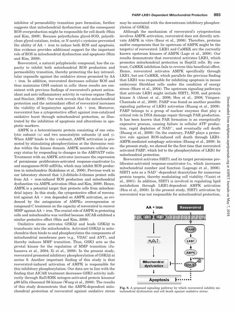
Fig. 8. A proposed signaling pathway by which resveratrol inhibits mi-tochondrial dysfunction and cell death against oxidative stress.
PARP-LKB1-Dependent Mitochondrial Protection 893
at ASPE
T Journals on M
ay 7, 2018m
olpharm.aspetjournals.org
Dow
nloaded from
consistent with the observation that resveratrol stimulatesAMPK in neurons via LKB1, but not SIRT1 (Dasgupta andMilbrandt, 2007). By the same token, the neuroprotectiveeffect of resveratrol was independent of SIRT1 activity (Al-vira et al., 2007).
Phenolic compounds—mostly derivatives and/or isomers offlavones, isoflavones, flavonols, catechins, tocopherols, andphenolic acids—are abundant in fruits, vegetables, tea, andwine. Growing evidences support a role for polyphenols inpreventing cardiovascular disease, cancer, neurodegenera-tive disease, or diabetes (Mahn et al., 2005; Rahman et al.,2006; Zang et al., 2006). In particular, dietary soy isofla-vones, such as genistein and daidzein, increase antioxidantand endothelial NOS gene expression and improve endothe-lial function (Mahn et al., 2005). Our work used cytoprotec-tive polyphenols (i.e., apigenin, genistein, and daidzein) tofurther examine the role of AMPK in protecting mitochon-dria. None of these polyphenols activated AMPK or enhancedmitochondrial function, supporting our hypothesis thatAMPK activation contributes to mitochondrial protection.Apigenin consistently did not increase AMPK and ACC phos-phorylations in HepG2 cells (Zang et al., 2006). Genisteinprotects cells at 0.1 to 40 �M (Kajta et al., 2007; Kim et al.,2007), but the compound activates AMPK in adipocytes at 50to 200 �M (Hwang et al., 2005). Hence, its beneficial effectmay not result from AMPK activation.
Demonstrated for the first time, resveratrol protects cellsfrom AA � iron-induced ROS production and mitochondrialdysfunction; a cytoprotective effect may be mediated byAMPK-mediated inhibitory phosphorylation of GSK3� down-stream of poly(ADP-ribose)polymerase-LKB1 pathway (Fig.8). Thus, resveratrol has the potential to pharmacologicallydefend mitochondria against an iron-catalyzed burst of oxi-dative stress through the AMPK pathway. Resveratrol scav-enges ROS in vitro, which may be mediated by the phenolichydroxyl groups in its molecular structure (Leonard et al.,2003). Although the cytoprotective effect of resveratrol pos-sibly results from ROS scavenging, a relatively high concen-tration is required for radical scavenging (Leonard et al.,2003). Therefore, the radical scavenging effect is less likely tocontribute to resveratrol’s beneficial action. More likely, theantioxidant effect of resveratrol might relate to the activationof a specific signaling pathway that enables cells to promotethe defense system. This concept parallels findings that res-veratrol increases GSH synthesis (Kode et al., 2008) andactivates antioxidant enzymes, catalase, SOD, and NADPH-quinone oxidoreductase (Rubiolo et al., 2008).
Acknowledgments
We are grateful to Dr. Janie Brooks for her comments on thismanuscript.
ReferencesAlessi DR, Sakamoto K, and Bayascas JR (2006) LKB1-dependent signaling path-
ways. Annu Rev Biochem 75:137–163.Alvira D, Yeste-Velasco M, Folch J, Verdaguer E, Canudas AM, Pallas M, and
Camins A (2007) Comparative analysis of the effects of resveratrol in two apoptoticmodels: inhibition of complex I and potassium deprivation in cerebellar neurons.Neuroscience 147:746–756.
Balboa MA and Balsinde J (2006) Oxidative stress and arachidonic acid mobilization.Biochim Biophys Acta 1761:385–391.
Baur JA, Pearson KJ, Price NL, Jamieson HA, Lerin C, Kalra A, Prabhu VV, AllardJS, Lopez-Lluch G, Lewis K, et al. (2006) Resveratrol improves health and survivalof mice on a high-calorie diet. Nature 444:337–342.
Baur JA and Sinclair DA (2006) Therapeutic potential of resveratrol: the in vivoevidence. Nat Rev Drug Discov 5:493–506.
Browning JD and Horton JD (2004) Molecular mediators of hepatic steatosis andliver injury. J Clin Invest 114:147–152.
Cocco T, Di Paola M, Papa S, and Lorusso M (1999) Arachidonic acid interaction withthe mitochondrial electron transport chain promotes reactive oxygen species gen-eration. Free Radic Biol Med 27:51–59.
Dasgupta B and Milbrandt J (2007) Resveratrol stimulates AMP kinase activity inneurons. Proc Natl Acad Sci U S A 104:7217–7222.
Ferrali M, Signorini C, Sugherini L, Pompella A, Lodovici M, Caciotti B, Ciccoli L,and Comporti M (1997) Release of free, redox-active iron in the liver and DNAoxidative damage following phenylhydrazine intoxication. Biochem Pharmacol53:1743–1751.
Fleming RE and Bacon BR (2005) Orchestration of iron homeostasis. N Engl J Med352:1741–1744.
Galaris D and Pantopoulos K (2008) Oxidative stress and iron homeostasis: mecha-nistic and health aspects. Crit Rev Clin Lab Sci 45:1–23.
Hayashi T, Hirshman MF, Fujii N, Habinowski SA, Witters LA, and Goodyear LJ(2000) Metabolic stress and altered glucose transport: activation of AMP-activatedprotein kinase as a unifying coupling mechanism. Diabetes 49:527–531.
Hou X, Xu S, Maitland-Toolan KA, Sato K, Jiang B, Ido Y, Lan F, Walsh K,Wierzbicki M, Verbeuren TJ, et al. (2008) SIRT1 regulates hepatocyte lipid me-tabolism through activating AMP-activated protein kinase. J Biol Chem 283:20015–20026.
Howitz KT, Bitterman KJ, Cohen HY, Lamming DW, Lavu S, Wood JG, Zipkin RE,Chung P, Kisielewski A, Zhang LL, et al. (2003) Small molecule activators ofsirtuins extend Saccharomyces cerevisiae lifespan. Nature 425:191–196.
Huang Q, Wu YT, Tan HL, Ong CN, and Shen HM (2009) A novel function ofpoly(ADP-ribose) polymerase-1 in modulation of autophagy and necrosis underoxidative stress. Cell Death Differ 16:264–277.
Hwang JT, Park IJ, Shin JI, Lee YK, Lee SK, Baik HW, Ha J, and Park OJ (2005)Genistein, EGCG, and capsaicin inhibit adipocyte differentiation process via acti-vating AMP-activated protein kinase. Biochem Biophys Res Commun 338:694–699.
Ido Y, Carling D, and Ruderman N (2002) Hyperglycemia-induced apoptosis inhuman umbilical vein endothelial cells: inhibition by the AMP-activated proteinkinase activation. Diabetes 51:159–167.
Juhaszova M, Zorov DB, Kim SH, Pepe S, Fu Q, Fishbein KW, Ziman BD, Wang S,Ytrehus K, Antos CL, et al. (2004) Glycogen synthase kinase-3beta mediatesconvergence of protection signaling to inhibit the mitochondrial permeability tran-sition pore. J Clin Invest 113:1535–1549.
Kajta M, Domin H, Grynkiewicz G, and Lason W (2007) Genistein inhibits gluta-mate-induced apoptotic processes in primary neuronal cell cultures: an involve-ment of aryl hydrocarbon receptor and estrogen receptor/glycogen synthase ki-nase-3beta intracellular signaling pathway. Neuroscience 145:592–604.
Kim EK, Kwon KB, Song MY, Seo SW, Park SJ, Ka SO, Na L, Kim KA, Ryu DG, SoHS, et al. (2007) Genistein protects pancreatic beta cells against cytokine-mediated toxicity. Mol Cell Endocrinol 278:18–28.
Kim YW, Lee SM, Shin SM, Hwang SJ, Brooks JS, Kang HE, Lee MG, Kim SC, andKim SG (2009) Efficacy of sauchinone as a novel AMPK-activating lignan forpreventing iron-induced oxidative stress and liver injury. Free Radic Biol Med doi:10.1016/j.freeradbiomed.2009.07.018.
Kode A, Rajendrasozhan S, Caito S, Yang SR, Megson IL, and Rahman I (2008)Resveratrol induces glutathione synthesis by activation of Nrf2 and protectsagainst cigarette smoke-mediated oxidative stress in human lung epithelial cells.Am J Physiol Lung Cell Mol Physiol 294:L478–L488.
Kukidome D, Nishikawa T, Sonoda K, Imoto K, Fujisawa K, Yano M, Motoshima H,Taguchi T, Matsumura T, and Araki E (2006) Activation of AMP-activated proteinkinase reduces hyperglycemia-induced mitochondrial reactive oxygen species pro-duction and promotes mitochondrial biogenesis in human umbilical vein endothe-lial cells. Diabetes 55:120–127.
Kwon YN, Shin SM, Cho IJ, and Kim SG (2009) Oxidized metabolites of oltiprazexert cytoprotective effects against arachidonic acid through AMP-activated pro-tein kinase-dependent cellular antioxidant effect and mitochondrial protection.Drug Metab Dispos 37:1187–1197.
Lage R, Dieguez C, Vidal-Puig A, and Lopez M (2008) AMPK: a metabolic gaugeregulating whole-body energy homeostasis. Trends Mol Med 14:539–549.
Lagouge M, Argmann C, Gerhart-Hines Z, Meziane H, Lerin C, Daussin F, MessadeqN, Milne J, Lambert P, Elliott P, et al. (2006) Resveratrol improves mitochondrialfunction and protects against metabolic disease by activating SIRT1 and PGC-1alpha. Cell 127:1109–1122.
Leonard SS, Xia C, Jiang BH, Stinefelt B, Klandorf H, Harris GK, and Shi X (2003)Resveratrol scavenges reactive oxygen species and effects radical-induced cellularresponses. Biochem Biophys Res Commun 309:1017–1026.
Mahn K, Borras C, Knock GA, Taylor P, Khan IY, Sugden D, Poston L, Ward JP,Sharpe RM, Vina J, et al. (2005) Dietary soy isoflavone induced increases inantioxidant and eNOS gene expression lead to improved endothelial function andreduced blood pressure in vivo. FASEB J 19:1755–1757.
Rahman I, Biswas SK, and Kirkham PA (2006) Regulation of inflammation andredox signaling by dietary polyphenols. Biochem Pharmacol 72:1439–1452.
Rubiolo JA, Mithieux G, and Vega FV (2008) Resveratrol protects primary rathepatocytes against oxidative stress damage: activation of the Nrf2 transcriptionfactor and augmented activities of antioxidant enzymes. Eur J Pharmacol 591:66–72.
Sale S, Verschoyle RD, Boocock D, Jones DJ, Wilsher N, Ruparelia KC, Potter GA,Farmer PB, Steward WP, and Gescher AJ (2004) Pharmacokinetics in mice andgrowth-inhibitory properties of the putative cancer chemopreventive agent res-veratrol and the synthetic analogue trans 3,4,5,4�-tetramethoxystilbene. Br JCancer 90:736–744.
Scorrano L, Penzo D, Petronilli V, Pagano F, and Bernardi P (2001) Arachidonic acidcauses cell death through the mitochondrial permeability transition. Implicationsfor tumor necrosis factor-alpha apoptotic signaling. J Biol Chem 276:12035–12040.
894 Shin et al.
at ASPE
T Journals on M
ay 7, 2018m
olpharm.aspetjournals.org
Dow
nloaded from

Shaw RJ, Kosmatka M, Bardeesy N, Hurley RL, Witters LA, DePinho RA, andCantley LC (2004) The tumor suppressor LKB1 kinase directly activates AMP-activated kinase and regulates apoptosis in response to energy stress. Proc NatlAcad Sci U S A 101:3329–3335.
Shin SM and Kim SG (2009) Inhibition of arachidonic acid and iron-induced mito-chondrial dysfunction and apoptosis by oltipraz and novel 1,2-dithiole-3-thionecongeners. Mol Pharmacol 75:242–253.
Tadolini B and Hakim G (1996) The mechanism of iron (III) stimulation of lipidperoxidation. Free Radic Res 25:221–227.
Terai K, Hiramoto Y, Masaki M, Sugiyama S, Kuroda T, Hori M, Kawase I, andHirota H (2005) AMP-activated protein kinase protects cardiomyocytes againsthypoxic injury through attenuation of endoplasmic reticulum stress. Mol Cell Biol25:9554–9575.
Vaziri H, Dessain SK, Ng Eaton E, Imai SI, Frye RA, Pandita TK, Guarente L, andWeinberg RA (2001) hSIR2(SIRT1) functions as an NAD-dependent p53 deacety-lase. Cell 107:149–159.
Vazquez-Chantada M, Ariz U, Varela-Rey M, Embade N, Martínez-Lopez N, Fer-nandez-Ramos D, Gomez-Santos L, Lamas S, Lu SC, Martínez-Chantar ML, et al.(2009) Evidence for LKB1/AMP-activated protein kinase/ endothelial nitric oxide
synthase cascade regulated by hepatocyte growth factor, S-adenosylmethionine,and nitric oxide in hepatocyte proliferation. Hepatology 49:608–617.
Wang HM, Mehta S, Bansode R, Huang W, and Mehta KD (2008) AICAR positivelyregulate glycogen synthase activity and LDL receptor expression through Raf-1/MEK/p42/44MAPK/p90RSK/GSK-3 signaling cascade. Biochem Pharmacol 75:457–467.
Xi J, Wang H, Mueller RA, Norfleet EA, and Xu Z (2009) Mechanism for resveratrol-induced cardioprotection against reperfusion injury involves glycogen synthasekinase 3beta and mitochondrial permeability transition pore. Eur J Pharmacol604:111–116.
Zang M, Xu S, Maitland-Toolan KA, Zuccollo A, Hou X, Jiang B, Wierzbicki M,Verbeuren TJ, and Cohen RA (2006) Polyphenols stimulate AMP-activated proteinkinase, lower lipids, and inhibit accelerated atherosclerosis in diabetic LDL recep-tor-deficient mice. Diabetes 55:2180–2191.
Address correspondence to: Dr. Sang Geon Kim, College of Pharmacy,Seoul National University, Sillim-dong, Kwanak-gu, Seoul 151-742, Korea.E-mail: [email protected]
PARP-LKB1-Dependent Mitochondrial Protection 895
at ASPE
T Journals on M
ay 7, 2018m
olpharm.aspetjournals.org
Dow
nloaded from