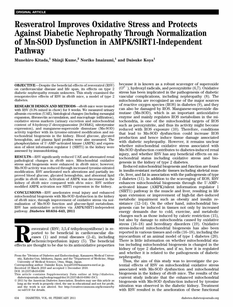
original article resveratrol improves oxidative stress and...
TRANSCRIPT
Resveratrol Improves Oxidative Stress and ProtectsAgainst Diabetic Nephropathy Through Normalizationof Mn-SOD Dysfunction in AMPK/SIRT1-IndependentPathwayMunehiro Kitada,
1Shinji Kume,
2Noriko Imaizumi,
1and Daisuke Koya
1
OBJECTIVE—Despite the beneficial effects of resveratrol (RSV)on cardiovascular disease and life span, its effects on type 2diabetic nephropathy remain unknown. This study examined therenoprotective effects of RSV in db/db mice, a model of type 2diabetes.
RESEARCHDESIGNANDMETHODS—db/dbmice were treatedwith RSV (0.3% mixed in chow) for 8 weeks. We measured urinaryalbumin excretion (UAE), histological changes (including mesangialexpansion, fibronectin accumulation, and macrophage infiltration),oxidative stress markers (urinary excretion and mitochondrialcontent of 8-hydroxy-2’-deoxyguanosine [8-OHdG], nitrotyrosineexpression), and manganese-superoxide dismutase (Mn-SOD)activity together with its tyrosine-nitrated modification and mi-tochondrial biogenesis in the kidney. Blood glucose, glycatedhemoglobin, and plasma lipid profiles were also measured. Thephosphorylation of 59-AMP–activated kinase (AMPK) and expres-sion of silent information regulator 1 (SIRT1) in the kidney wereassessed by immunoblotting.
RESULTS—RSV significantly reduced UAE and attenuated renalpathological changes in db/db mice. Mitochondrial oxidativestress and biogenesis were enhanced in db/db mice; however,Mn-SOD activity was reduced through increased tyrosine-nitratedmodification. RSV ameliorated such alterations and partially im-proved blood glucose, glycated hemoglobin, and abnormal lipidprofile in db/db mice. Activation of AMPK was decreased in thekidney of db/db mice compared with db/m mice. RSV neithermodified AMPK activation nor SIRT1 expression in the kidney.
CONCLUSIONS—RSV ameliorates renal injury and enhancedmitochondrial biogenesis with Mn-SOD dysfunction in the kidneyof db/db mice, through improvement of oxidative stress via nor-malization of Mn-SOD function and glucose-lipid metabolism.RSV has antioxidative activities via AMPK/SIRT1-independentpathway. Diabetes 60:634–643, 2011
Resveratrol (RSV; 3,5,4’-trihydroxystilbene) is re-ported to be beneficial in cardiovascular dis-eases (1) and renal diseases (2–4), includingischemic/reperfusion injury (5). The beneficial
effects are thought to be due to its antioxidative properties
because it is known as a robust scavenger of superoxide(O22), hydroxyl radicals, and peroxynitrite (6,7). Oxidativestress has been implicated in the pathogenesis of diabeticvascular complications, including nephropathy (8). Themitochondria are recognized as one of the major sourcesof reactive oxygen species (ROS) in diabetes (9), and theycan also be damaged by ROS. Manganese-superoxide dis-mutase (Mn-SOD), which is an important antioxidativeenzyme and mainly regulates ROS metabolism in the mi-tochondria, is one of the mitochondrial targets of ROSsuch as peroxynitrite, and thus its activity might becomereduced with ROS exposure (10). Therefore, conditionsthat lead to Mn-SOD dysfunction could increase ROSproduction and hence induce tissue damage associatedwith diabetic nephropathy. However, it remains unclearwhether mitochondrial oxidative stress associated withMn-SOD dysfunction contributes to diabetes-induced renalinjury, and whether RSV has any beneficial effects on mi-tochondrial status including oxidative stress and bio-genesis in the kidney of type 2 diabetes.
Reduced mitochondrial biogenesis and function are foundin insulin-resistant metabolic tissues including skeletal mus-cle, liver, and fat in association with the pathogenesis of type2 diabetes (11). In addition to the scavenging of ROS, RSVenhances mitochondrial biogenesis through the 59-AMP–activated kinase (AMPK)/silent information regulator 1(SIRT1) pathway in the muscle and liver, resulting in lifespan extension or improvement of high-fat diet–inducedmetabolic impairment such as obesity and insulin re-sistance (12–14). On the other hand, mitochondrial bio-genesis can be induced in tissues not only by increasedenergy demands due to cold, exercise, and metabolicchanges such as those induced by caloric restriction (15),but also by damage to mitochondria caused by oxidativestress (16–18) and hereditary disorders (19). Oxidativestress-induced mitochondrial biogenesis has also beenreported in various tissues and cells (16–18), including themyocardium of an animal model of type 1 diabetes (20).There is little information on whether mitochondrial sta-tus including mitochondrial biogenesis is changed in thekidney of type 2 diabetes, and if so, how it is regulatedand whether it is related to the pathogenesis of diabeticnephropathy.
Thus, the aim of this study was to investigate the po-tential effects of RSV on mitochondrial oxidative stressassociated with Mn-SOD dysfunction and mitochondrialbiogenesis in the kidney of db/db mice. The results of thecurrent study indicate that the enhanced mitochondrialbiogenesis with Mn-SOD dysfunction induced by tyrosinenitration was observed in the diabetic kidney. Treatmentwith RSV resulted in the amelioration of these functional
From the 1Division of Diabetes and Endocrinology, Kanazawa Medical Univer-sity, Kahoku-Gun, Ishikawa, Japan; and the 2Department of Medicine, ShigaUniversity of Medical Science, Otsu, Shiga, Japan.
Corresponding author: Daisuke Koya, [email protected] 19 March 2010 and accepted 1 December 2010.DOI: 10.2337/db10-0386This article contains Supplementary Data online at http://diabetes.
diabetesjournals.org/lookup/suppl/doi:10.2337/db10-0386/-/DC1.� 2011 by the American Diabetes Association. Readers may use this article as
long as the work is properly cited, the use is educational and not for profit,and the work is not altered. See http://creativecommons.org/licenses/by-nc-nd/3.0/ for details.
634 DIABETES, VOL. 60, FEBRUARY 2011 diabetes.diabetesjournals.org
ORIGINAL ARTICLE
and histological abnormalities and mitochondrial biogenesisin the diabetic kidney, possibly by the attenuation of oxi-dative stress through scavenging of ROS, normalizationof Mn-SOD dysfunction in an AMPK/SIRT1-independentmechanism, and partial improvement of glucose-lipidmetabolism.
RESEARCH DESIGN AND METHODS
Materials and antibodies. Details of the materials and antibodies used in thecurrent study are available in the Supplementary Data.Animals. Male db/db mice and age-matched db/m mice were purchased fromClea Japan (Tokyo, Japan). At 9 weeks of age, mice were divided into fourgroups: db/m mice, db/db mice, db/m mice treated with RSV, and db/db micetreated with RSV. RSV was mixed (0.3%) with chow and administered orally.Body weight, blood glucose level, food consumption, and blood pressure weremeasured every 2 weeks in all animals. The blood pressure of conscious micewas measured at steady state by a programmable tail-cuff sphygmomanometer(BP98-A; Softron, Tokyo, Japan). At 17 weeks of age, individual mice wereplaced in metabolic cages for 24-h urine collection. The urine samples werestored at 280°C until analysis. Mice were anesthetized by intraperitoneal in-jection of pentobarbital sodium, and then the right kidneys were removed andstored at –80°C for experiments as described below. After collection of bloodsamples from the left cardiac ventricle, the left kidney was perfused with ice-cold phosphate-buffered saline (PBS) and 10% neutral buffered formalin andthen removed. The Research Center for Animal Life Science of KanazawaMedical University approved all experiments.Morphological analysis and immunohistochemistry. To assess the mesan-gial expansion, thirty glomeruli, cut at the vascular pole, randomly selected fromeach mouse were measured the periodic acid/Schiff (PAS)-positive materialin the mesangial area and glomerular tuft area by computer-assisted colorimage analysis (Micro Analyzer; Japan Poladigital, Tokyo, Japan) as previouslydescribed (21).
For semiquantitative evaluation of the fibronectin, F4/80 and nitrotyrosinescores, 20 randomly selected glomerulus or tubulointerstitial areas per mousewere graded in a double-blind manner, as reported previously (21–23), withminor modifications.8-OHdG levels in mitochondrial DNA and quantification of mitochondrial
DNA deletionmutation. The mitochondrial DNA (mtDNA) was extracted fromthe kidney using the mtDNA Extractor CT kit. The 8-OHdG levels in DNase I-digestedmtDNAwere determined by ELISA using a kit (8-OHdG Check, Institutefor the Control of Aging, Shizuoka, Japan) (24). We determined. the deletionmutation, D-17, as reported previously (23). The sequences of primers are listedin Supplementary Table 1.Detection of O
22formation in renal isolated mitochondria. The mito-
chondria were isolated from the renal cortex of all groups of mice using themitochondria isolation kit, according to the manufacturer’s instructions.O22 production from the mitochondria was measured by L-012 chemiluminescence
(CL) dye (25). Mitochondrial suspensions were diluted to a final proteinconcentration of 0.1 mg/mL in 0.2 mL of PBS buffer containing 100 mmol/LL-012. O22 from mitochondria was detected after stimulation with 4 mmol/Lsuccinate and 20 mg/mL antimycin A by L-012. The CL registered at intervals of30 s over 5 min with a chemiluminometer, and the signal was expressed ascounts of CL/min/100 mg protein at 5 min. In ex vivo study, similar experi-ments were performed in isolated mitochondria from the renal cortex of db/dbmice at various concentrations (1023
–100 mmol/L) of RSV or 100 U/mL SOD. Inaddition, O22 from the reaction of hypoxanthine and xanthine oxidase wasalso measured. L-012 (100 mM) was incubated for 5 min in PBS buffer con-taining 100 mmol/L diethylenetriamine pentaacetic acid (DTPA) and 1 mmol/Lhypoxanthine at room temperature, and then the basal (background) signalwas determined in a chemiluminometer at intervals of 30 s for 5 min. The CLwas counted after the addition of 10 mU/mL xanthine oxidase at variousconcentrations of RSV (1023
–100 mmol/L) or 100 U/mL SOD at intervals of 30 sover 5 min.Mn-SOD activity. The whole kidney was homogenized in 2 mL of 50 mmol/LTris HCl buffer containing 0.1 mmol/L ethylenediaminetetraacetic acid (EDTA)at pH 7.0. After centrifugation at 15,000g for 15 min, the supernatant was re-moved and total protein concentration was measured using a protein assay kit.The Mn-SOD activity was measured by inhibiting extracellular and cytosolicCu/Zn SOD activity with KCN (1 mmol/L) using an SOD assay kit (WST-1) (26).One unit of SOD activity was defined as the amount causing 50% inhibition ofthe initial rate of reduction of WST-1 (2-(4-Iodophenyl)-3-(4-nitrophenyl)-5-(2, 4-disulfophenyl)-2H-tetrazolium, monosodium salt), a highly water-solubletetrazolium salt. Mn-SOD activity was calculated in terms of protein content(U/mg) and expressed as a fold increase relative to that found in db/m mice.Immunoprecipitation and Western blot analysis. The whole kidney washomogenized in ice-cold radioimmunoprecipitation assay (RIPA) buffer. Solu-bilized protein (1 mg) was used for immunoprecipitation with rabbit polyclonalanti–Mn-SOD antibody (10 mg/mL) using protein A-Sepharose, and then Westernblot analysis was performed with mouse monoclonal antinitrotyrosine antibody(1:1000) or rabbit polyclonal anti–Mn-SOD antibody (1:1000). Samples of pro-tein solutions from the kidney were used for Western blotting with antiphospho-AMPKa (Thr172) antibody, anti-AMPKa (23A3) antibody (1:1000), or anti-SIRT1antibody (1:1000).Quantitative real-time PCR. Isolation of total RNA from kidney, and de-termination of complementary DNA synthesis by reverse transcription andquantitative real-time PCR were performed as described previously (23). PCRprimer sets are listed (Supplementary Table 1).Cell culture. Murine proximal tubular cells (mProx) (derivative, patentWO9927363, Japan, U.S., European Union), kindly provided by CMIC Co., werecultured as described previously (27).Retroviral infection. The pSUPERretro and pSUPERretro-SIRT1 RNA in-terference vectors were kind gifts from Dr. L. Guarente (Massachusetts In-stitute of Technology, Cambridge, MA). Human embryonic kidney 293T cellswere transfected with pSUPERretro or pSUPERretro-SIRT1 RNAi by usingLipofectamine reagent. At 48 h after transfection, the media containing theretroviruses were collected, centrifuged, and transferred to mProx treated by
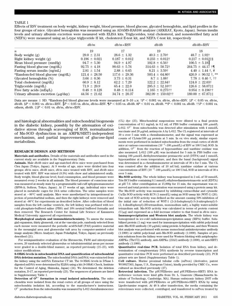
TABLE 1Effects of RSV treatment on body weight, kidney weight, blood pressure, blood glucose, glycated hemoglobin, and lipid profiles in thefour groups of mice. Glycated hemoglobin was measured using an ADAMS-HA8106 analyzer (ARKRAY, Kyoto, Japan). Serum insulinlevels and urinary albumin excretion were measured with ELISA kits. Triglycerides, total cholesterol, and nonesterified fatty acid(NEFA) were measured using an L-type triglyceride H kit, cholesterol E-test kit, and NEFA C-test kit, respectively
db/m db/m+RSV db/db db/db+RSV
n 19 17 19 18Body weight (g) 29.0 6 2.31 28.6 6 1.32 49.3 6 2.70† 48.7 6 1.92†Right kidney weight (g) 0.196 6 0.021 0.187 6 0.012 0.233 6 0.012† 0.217 6 0.012‡§Mean blood pressure (mmHg) 94.7 6 5.30 94.9 6 4.97 102.8 6 6.56† 100.5 6 5.18║Fasting blood glucose (mg/dL) 92.75 6 4.18 90.63 6 5.76 314.63 6 50.72† 264.75 6 44.17 †¶Fasting serum insulin (ng/mL) 2.81 6 1.54 2.88 6 0.65 8.21 6 3.59† 4.40 6 1.44 †, #*Random-fed blood glucose (mg/dL) 121.4 6 28.50 117.6 6 29.36 503.4 6 64.80† 426.9 6 90.52 †, **Glycated hemoglobin (%) 3.66 6 0.36 3.73 6 0.31 8.7 6 1.08† 7.78 6 0.46 †, ††Total cholesterol (mg/dL) 60.9 6 8.12 62.2 6 7.29 122.2 6 22.84† 124.0 6 22.03†Triglyceride (mg/dL) 71.9 6 20.9 65.4 6 22.0 205.3 6 52.10†† 126.6 6 58.07‡‡Free fatty acids (mEq/L) 0.48 6 0.128 0.48 6 0.114 1.165 6 0.275†† 0.954 6 0.196††Urinary albumin excretion (mg/day) 44.56 6 21.62 34.74 6 20.37 382.99 6 159.02†† 189.00 6 67.87‡‡
Data are means 6 SD. *Random-fed blood glucose levels were measured at 9–10 A.M. †P , 0.001 vs. db/m, db/m+RSV. ‡P , 0.05 vs. db/m,db/db. §P , 0.001 vs. db/m+RSV. ║P , 0.05 vs. db/m, db/m+RSV. ¶P , 0.05 vs. db/db. #P , 0.01 vs. db/db. **P , 0.001 vs. db/db. ††P , 0.001 vs.others, db/db. ‡‡P , 0.01 vs. db/m, db/m+RSV.
M. KITADA AND ASSOCIATES
diabetes.diabetesjournals.org DIABETES, VOL. 60, FEBRUARY 2011 635
polybrene (1 g/mL). The infected cells were selected by treatment with pu-romycin (2.5 mg/mL) for several days as described previously (28).Infection of adenoviral dominant negative-AMPK. Adenoviruses con-taining green fluorescent protein (Ad-GFP) or dominant-negative AMPK(DN-AMPK) were added to subconfluent mProx at a concentration of 50 mul-tiplicity of infection for 1 h at 37°C in serum-free Dulbecco’s modified Eagle’smedium (29).Detection of ROS in mProx. To determine the effect of RSV on oxidativestress in mProx, cells were incubated with RSV (10 mmol/L) for 180 min at 37°Cbefore exposure to H2O2 (10 mmol/L). After incubation, the levels of intracellularROS were measured with the fluoroprobe 2.7-dichlorodihydrofluorescein diac-etate. The intracellular ROS was evaluated as the fluorescence intensity (anexcitation wavelength 488 nm, an emission wavelength 525 nm) of dichloro-fluorescein by Infinite M200 microplate reader (Tecan Japan Co., Kanagawa,Japan). The results of intracellular ROS are expressed in arbitrary units.mtDNA content.Well-conserved nuclear and other mitochondrial genes wereselected to quantify mtDNA copy number per nuclear genome. Cytochrome coxidase subunit 2 was used as a marker for mtDNA and uncoupling protein 2
for nuclear DNA. Renal DNA was extracted from frozen kidney tissues of allanimals using the DNeasy tissue kit. Total DNA concentration was determinedusing a PicoGreen DNA quantitation kit. Specific mouse primer sequences areprovided in the Supplementary Table 1. mtDNA per nuclear genome was calcu-lated as the ratio of cytochrome c oxidase subunit 2 DNA to uncoupling protein 2DNA (30) and expressed as the fold increase relative to that found in db/m mice.Citrate synthase activity. Citrate synthase activity in the kidney was de-termined using the citrate synthase activity measurement kit, according to themanufacturer’s instructions.Electron microscopy. The mitochondria in proximal tubular cells were ob-served by electron microscopy as previously described (23). Mitochondrialarea and number were estimated in 15–18 micrographs, which were taken forrenal proximal tubular cells of three animals of each group (20). The mito-chondrial area was measured using the ImageJ software, and the number ofmitochondria per cell was counted manually by a blind observer.Statistical analysis. Data are expressed as means 6 SD. The Tukey multiple-comparison test was used to determine the significance of pairwise differ-ences among three or more groups. P , 0.05 was considered significant.
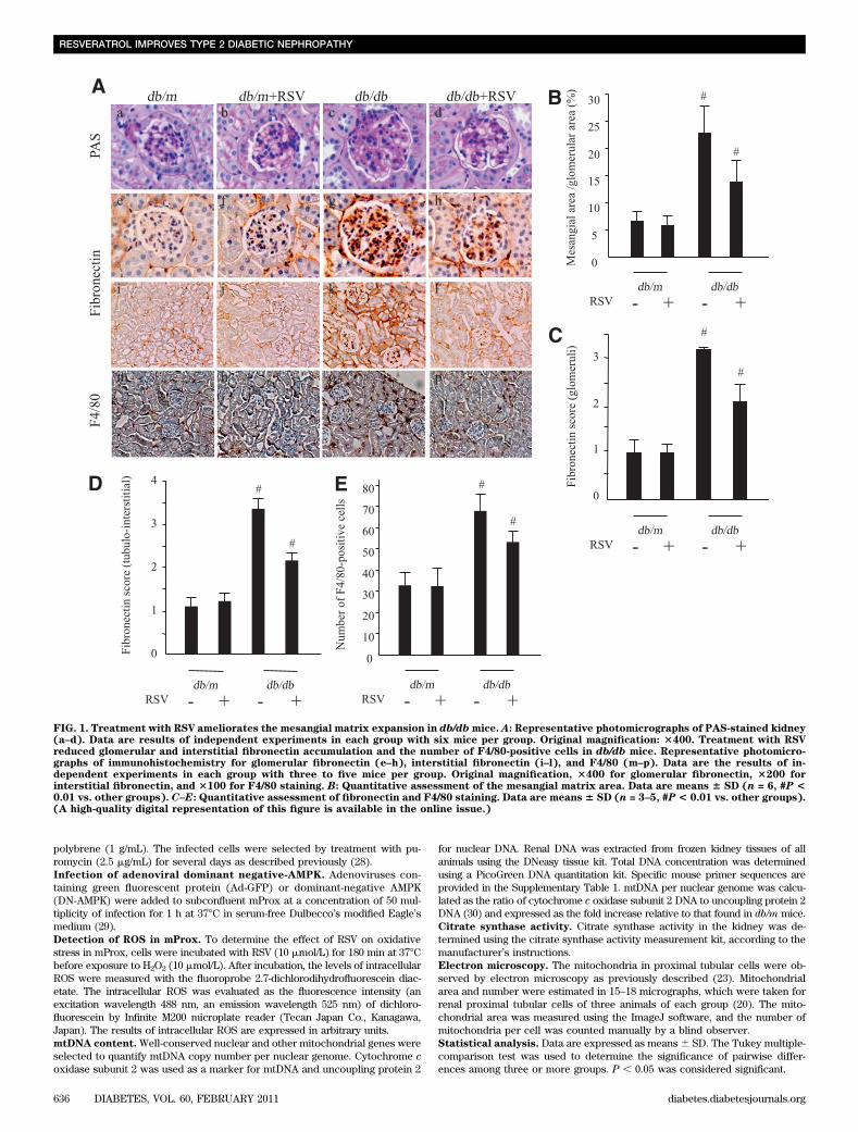
FIG. 1. Treatment with RSV ameliorates the mesangial matrix expansion in db/dbmice. A: Representative photomicrographs of PAS-stained kidney(a–d). Data are results of independent experiments in each group with six mice per group. Original magnification: 3400. Treatment with RSVreduced glomerular and interstitial fibronectin accumulation and the number of F4/80-positive cells in db/db mice. Representative photomicro-graphs of immunohistochemistry for glomerular fibronectin (e–h), interstitial fibronectin (i–l), and F4/80 (m–p). Data are the results of in-dependent experiments in each group with three to five mice per group. Original magnification, 3400 for glomerular fibronectin, 3200 forinterstitial fibronectin, and 3100 for F4/80 staining. B: Quantitative assessment of the mesangial matrix area. Data are means 6 SD (n = 6, #P <0.01 vs. other groups). C–E: Quantitative assessment of fibronectin and F4/80 staining. Data are means 6 SD (n = 3–5, #P < 0.01 vs. other groups).(A high-quality digital representation of this figure is available in the online issue.)
RESVERATROL IMPROVES TYPE 2 DIABETIC NEPHROPATHY
636 DIABETES, VOL. 60, FEBRUARY 2011 diabetes.diabetesjournals.org
RESULTS
Characteristics of experimental mice. Table 1 detailsthe characteristics of four groups of mice at the end of theexperimental period. The whole body and right kidneyweights were significantly higher in db/db mice comparedwith db/m mice. The mean blood pressure (MBP) wassignificantly higher in db/db mice than in db/m mice. RSVdid not affect changes in MBP. db/db mice exhibitedmarkedly elevated blood glucose levels compared with db/mmice throughout the entire experiment. In db/db mice,treatment with RSV induced a partial improvement inblood glucose levels and glycated hemoglobin by the end
of the experiments. Serum lipid profiles including totalcholesterol, triglyceride, and free fatty acid levels werealso significantly elevated in db/db mice compared withdb/mmice; the increases in triglycerides and free fatty acidswere partially rescued by RSV (Table 1). In addition, animpairment of glucose and insulin tolerance was evident indb/db mice relative to db/m mice (Supplementary Fig. 1),and high levels of fasting glucose and insulin (Table 1)indicating insulin resistance were also observed in db/dbmice. Such alteration of insulin resistance in db/dbmice waspartially improved by treatment with RSV. There were nodifferences in body weight and food consumption between
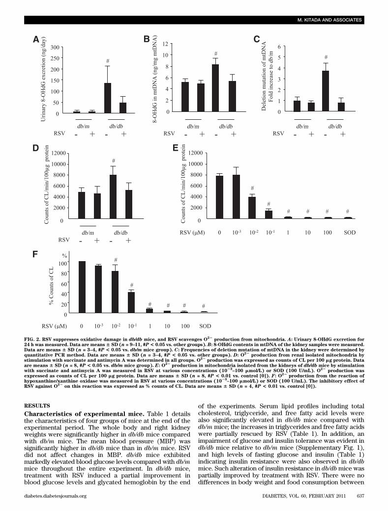
FIG. 2. RSV suppresses oxidative damage in db/db mice, and RSV scavenges O22
production from mitochondria. A: Urinary 8-OHdG excretion for24 h was measured. Data are means6 SD (n = 9–11, #P< 0.05 vs. other groups). B: 8-OHdG contents in mtDNA of the kidney samples were measured.Data are means 6 SD (n = 3–4, #P < 0.05 vs. db/m mice group). C: Frequencies of deletion mutation of mtDNA in the kidney were determined byquantitative PCR method. Data are means 6 SD (n = 3–4, #P < 0.05 vs. other groups). D: O
22production from renal isolated mitochondria by
stimulation with succinate and antimycin A was determined in all groups. O22
production was expressed as counts of CL per 100 mg protein. Dataare means 6 SD (n = 8, #P < 0.05 vs. db/m mice group). E: O
22production in mitochondria isolated from the kidneys of db/db mice by stimulation
with succinate and antimycin A was measured in RSV at various concentrations (1023
–100 mmol/L) or SOD (100 U/mL). O22
production wasexpressed as counts of CL per 100 mg protein. Data are means 6 SD (n = 8, #P < 0.01 vs. control [0]). F: O22
production from the reaction ofhypoxanthine/xanthine oxidase was measured in RSV at various concentrations (10
23–100 mmol/L) or SOD (100 U/mL). The inhibitory effect of
RSV against O22
on this reaction was expressed as % counts of CL. Data are means 6 SD (n = 4, #P < 0.01 vs. control [0]).
M. KITADA AND ASSOCIATES
diabetes.diabetesjournals.org DIABETES, VOL. 60, FEBRUARY 2011 637
untreated and RSV-treated db/db mice. The 24-h urinevolume was significantly larger in db/db mice comparedwith db/m mice, and this was partially reduced by treat-ment with RSV (data not shown).Changes in urinary albumin excretion. To evaluate theeffects of RSV on functional abnormalities in db/db mice,we measured the urinary albumin excretion. Values weremarkedly higher in db/db mice and RSV treatment signifi-cantly reduced urinary albumin excretion (Table 1), in-dicating that RSV ameliorates the functional abnormalityof diabetic nephropathy in db/db mice.Changes in kidney morphology. Figure 1A a–d showsrepresentative photomicrographs of mesangial matrix ac-cumulation in the PAS-stained kidneys of the four groups.The mesangial matrix was more extensive in the glomeruliof db/db mice than in db/m mice, and treatment with RSVreduced such expansion. Figure 1B shows the results ofquantitative analysis of mesangial matrix expansion in allgroups. Although the ratio of mesangial matrix/glomerulararea was markedly larger in db/db mice than in db/m mice,treatment with RSV significantly reduced this expansion.
Immunohistochemistry for fibronectin (Fig. 1A e–h andi–l) also showed a significantly higher score for renal glo-merular and tubulointerstitial expression in db/db micethan in db/m mice (Fig. 1C and D). Treatment with RSVreduced the score for fibronectin in db/db mice but had noeffect on db/m (Fig. 1C and D).
The number of cells positive for F4/80 (a macrophagemarker) in the renal interstitial lesion was significantlyhigher in db/db mice than in db/m mice (Fig. 1A m–p), butthis pattern was not found in the glomeruli (data not shown).
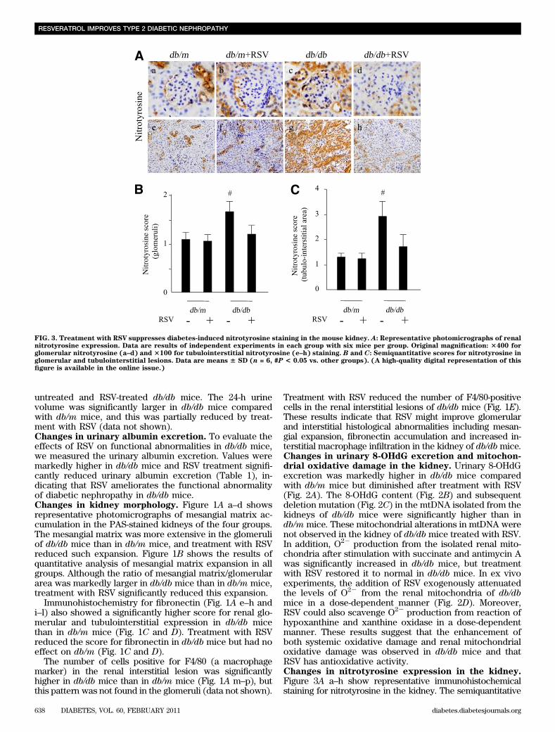
Treatment with RSV reduced the number of F4/80-positivecells in the renal interstitial lesions of db/db mice (Fig. 1E).These results indicate that RSV might improve glomerularand interstitial histological abnormalities including mesan-gial expansion, fibronectin accumulation and increased in-terstitial macrophage infiltration in the kidney of db/dbmice.Changes in urinary 8-OHdG excretion and mitochon-drial oxidative damage in the kidney. Urinary 8-OHdGexcretion was markedly higher in db/db mice comparedwith db/m mice but diminished after treatment with RSV(Fig. 2A). The 8-OHdG content (Fig. 2B) and subsequentdeletion mutation (Fig. 2C) in the mtDNA isolated from thekidneys of db/db mice were significantly higher than indb/m mice. These mitochondrial alterations in mtDNA werenot observed in the kidney of db/db mice treated with RSV.In addition, O22 production from the isolated renal mito-chondria after stimulation with succinate and antimycin Awas significantly increased in db/db mice, but treatmentwith RSV restored it to normal in db/db mice. In ex vivoexperiments, the addition of RSV exogenously attenuatedthe levels of O22 from the renal mitochondria of db/dbmice in a dose-dependent manner (Fig. 2D). Moreover,RSV could also scavenge O22 production from reaction ofhypoxanthine and xanthine oxidase in a dose-dependentmanner. These results suggest that the enhancement ofboth systemic oxidative damage and renal mitochondrialoxidative damage was observed in db/db mice and thatRSV has antioxidative activity.Changes in nitrotyrosine expression in the kidney.Figure 3A a–h show representative immunohistochemicalstaining for nitrotyrosine in the kidney. The semiquantitative
Nitr
otyr
osin
e
Nitr
otyr
osin
e
FIG. 3. Treatment with RSV suppresses diabetes-induced nitrotyrosine staining in the mouse kidney. A: Representative photomicrographs of renalnitrotyrosine expression. Data are results of independent experiments in each group with six mice per group. Original magnification: 3400 forglomerular nitrotyrosine (a–d) and 3100 for tubulointerstitial nitrotyrosine (e–h) staining. B and C: Semiquantitative scores for nitrotyrosine inglomerular and tubulointerstitial lesions. Data are means 6 SD (n = 6, #P < 0.05 vs. other groups). (A high-quality digital representation of thisfigure is available in the online issue.)
RESVERATROL IMPROVES TYPE 2 DIABETIC NEPHROPATHY
638 DIABETES, VOL. 60, FEBRUARY 2011 diabetes.diabetesjournals.org

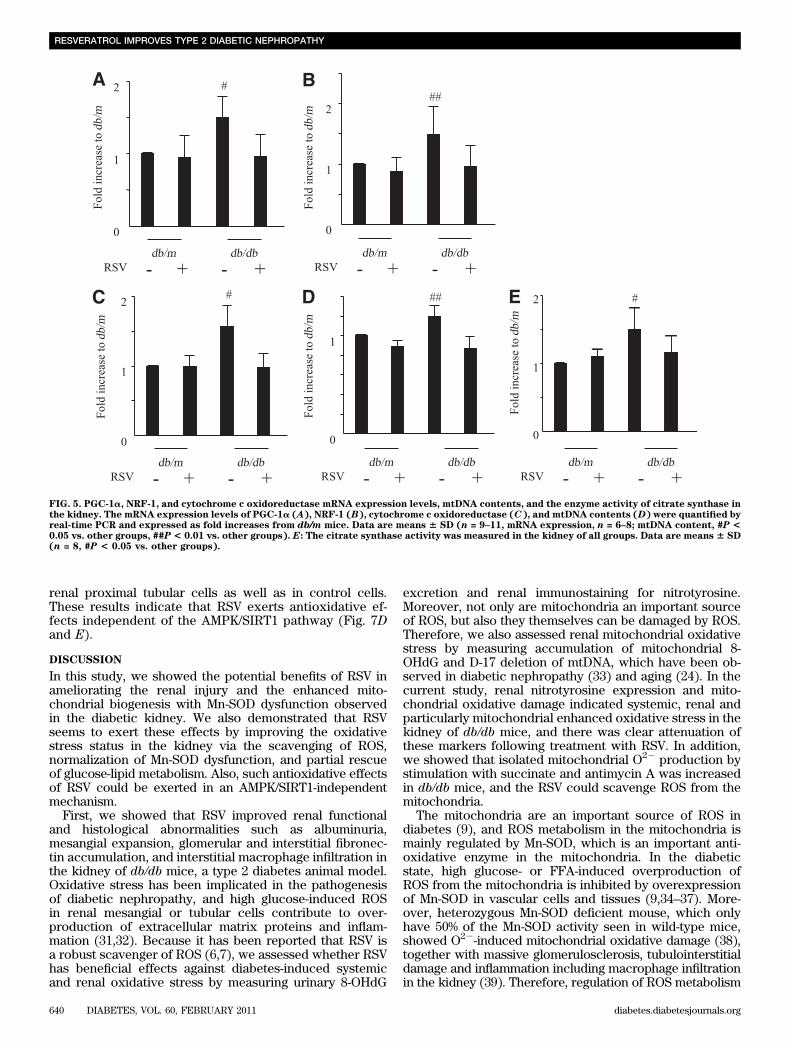
scores for nitrotyrosine in both the glomerular and thetubulointerstitial lesions of the renal cortex were in-creased in db/db mice compared with those of db/m mice(Fig. 3B and C). Treatment with RSV reduced the scores inthe glomeruli and tubulointerstitium of db/db mice but hadno effect on the lesions of db/m mice (Fig. 3B and C).These results indicate that tyrosine nitration of proteins isenhanced in the kidney of db/db mice and that RSV seemsto attenuate the effect.Changes in Mn-SOD expression and activity in thekidney. The mRNA and protein expression of Mn-SODwas higher in db/db mice than in db/m mice (Fig. 4A–C).The Mn-SOD activity was significantly lower in the kidneyof db/db mice compared with that of db/m mice (Fig. 4D).Nitrotyrosine immunoreactivity was significantly higher inMn-SOD immunoprecipitates from the kidney of db/dbmice than in that from db/m mice, and the increase inimmunoreactivity was attenuated by treatment with RSV indb/db but not db/m mice (Fig. 4E and F). These findingssuggest that Mn-SOD activity could be reduced by modi-fying tyrosine nitration in the kidneys of db/db mice.mRNA levels of mitochondrial biogenesis–relatedgenes and the enzyme activity of citrate synthase.To evaluate mitochondrial biogenesis, we assessed peroxi-some proliferator-activated receptor g coactivator (PGC)-1a, nuclear respiratory factor (NRF)-1, and cytochrome
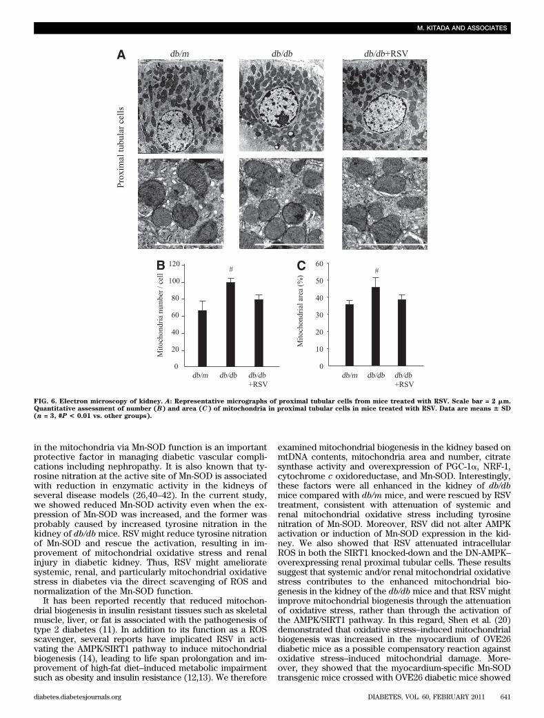
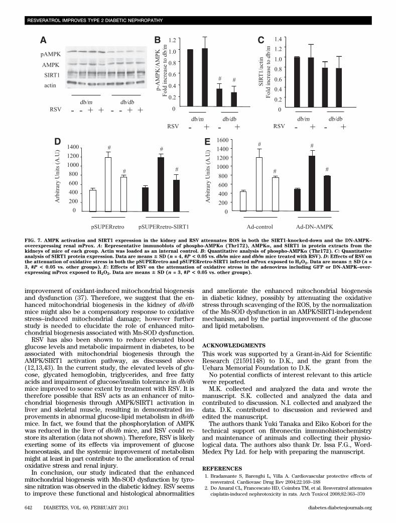
c oxidoreductase mRNA expression levels and mtDNAcontents in the kidney. The citrate synthase activity wasalso measured in the kidney. All these were significantlyhigher in the kidneys of db/db mice than in that of db/mmice (Fig. 5A–E). These changes improved followingtreatment with RSV, consistent with the observed normali-zation of Mn-SOD activity and oxidative stress.Electron microscopy. To confirm the beneficial effects ofRSV on mitochondrial biogenesis, we examined the renalmorphology in more detail by electron microscopy. Thenumber and area of the mitochondria were significantlylarger in renal proximal tubular cells of db/db mice com-pared with db/m mice, but treatment with RSV rescuedthese differences in db/db mice (Fig. 6A–C).Changes in AMPK activation and SIRT 1 expressionin the kidney. We assessed whether RSV activates AMPKin the kidney using immunoblotting for phosphorylation ofAMPK. Activation of AMPK was significantly reduced inthe kidney of db/db mice compared with db/m mice, andRSV did not alter AMPK activation in the kidney (Fig. 7Aand B). In addition, SIRT1 expression was no differentamong the groups (Fig. 7A and C).RSV exerts antioxidative effects in AMPK/SIRT1-independent mechanism in cultured renal proximaltubular cells. RSV attenuated intracellular ROS in boththe SIRT1-knocked-down and DN-AMPK–overexpressing
FIG. 4. Mn-SOD expression and activity in the kidney. A: Mn-SOD mRNA expression in kidneys was quantified by real-time PCR and expressed asfold increase relative to db/mmice. Data are means6 SD (n = 9–11, #P< 0.05 vs. other groups). B: Mn-SOD protein expression in the kidney shownby representative immunoblots of Mn-SOD in protein extracts from the kidneys of mice of each group. Actin was loaded as an internal control. C:Quantitative analysis of Mn-SOD protein expression in the kidneys. Data are means6 SD (n = 4, #P< 0.05 vs. other groups). D: Mn-SOD activity inthe kidney homogenate is expressed as fold increase compared with db/m mice. Data are means 6 SD (n = 9–11, #P < 0.05 vs. other groups).Immunoreactivity to nitrotyrosine for immunoprecipitated Mn-SOD. Proteins from the kidneys of each group were immunoprecipitated with anti–Mn-SOD antibodies and blotted for nitrotyrosine antibodies. Representative results of Western blotting are shown (E), and the ratio to immu-noprecipitated Mn-SOD is shown (F). Data are means 6 SD (n = 6, #P < 0.05 vs. other groups).
M. KITADA AND ASSOCIATES
diabetes.diabetesjournals.org DIABETES, VOL. 60, FEBRUARY 2011 639
renal proximal tubular cells as well as in control cells.These results indicate that RSV exerts antioxidative ef-fects independent of the AMPK/SIRT1 pathway (Fig. 7Dand E).
DISCUSSION
In this study, we showed the potential benefits of RSV inameliorating the renal injury and the enhanced mito-chondrial biogenesis with Mn-SOD dysfunction observedin the diabetic kidney. We also demonstrated that RSVseems to exert these effects by improving the oxidativestress status in the kidney via the scavenging of ROS,normalization of Mn-SOD dysfunction, and partial rescueof glucose-lipid metabolism. Also, such antioxidative effectsof RSV could be exerted in an AMPK/SIRT1-independentmechanism.
First, we showed that RSV improved renal functionaland histological abnormalities such as albuminuria,mesangial expansion, glomerular and interstitial fibronec-tin accumulation, and interstitial macrophage infiltration inthe kidney of db/db mice, a type 2 diabetes animal model.Oxidative stress has been implicated in the pathogenesisof diabetic nephropathy, and high glucose-induced ROSin renal mesangial or tubular cells contribute to over-production of extracellular matrix proteins and inflam-mation (31,32). Because it has been reported that RSV isa robust scavenger of ROS (6,7), we assessed whether RSVhas beneficial effects against diabetes-induced systemicand renal oxidative stress by measuring urinary 8-OHdG
excretion and renal immunostaining for nitrotyrosine.Moreover, not only are mitochondria an important sourceof ROS, but also they themselves can be damaged by ROS.Therefore, we also assessed renal mitochondrial oxidativestress by measuring accumulation of mitochondrial 8-OHdG and D-17 deletion of mtDNA, which have been ob-served in diabetic nephropathy (33) and aging (24). In thecurrent study, renal nitrotyrosine expression and mito-chondrial oxidative damage indicated systemic, renal andparticularly mitochondrial enhanced oxidative stress in thekidney of db/db mice, and there was clear attenuation ofthese markers following treatment with RSV. In addition,we showed that isolated mitochondrial O22 production bystimulation with succinate and antimycin A was increasedin db/db mice, and the RSV could scavenge ROS from themitochondria.
The mitochondria are an important source of ROS indiabetes (9), and ROS metabolism in the mitochondria ismainly regulated by Mn-SOD, which is an important anti-oxidative enzyme in the mitochondria. In the diabeticstate, high glucose- or FFA-induced overproduction ofROS from the mitochondria is inhibited by overexpressionof Mn-SOD in vascular cells and tissues (9,34–37). More-over, heterozygous Mn-SOD deficient mouse, which onlyhave 50% of the Mn-SOD activity seen in wild-type mice,showed O22-induced mitochondrial oxidative damage (38),together with massive glomerulosclerosis, tubulointerstitialdamage and inflammation including macrophage infiltrationin the kidney (39). Therefore, regulation of ROS metabolism
FIG. 5. PGC-1a, NRF-1, and cytochrome c oxidoreductase mRNA expression levels, mtDNA contents, and the enzyme activity of citrate synthase inthe kidney. The mRNA expression levels of PGC-1a (A), NRF-1 (B), cytochrome c oxidoreductase (C), and mtDNA contents (D) were quantified byreal-time PCR and expressed as fold increases from db/m mice. Data are means 6 SD (n = 9–11, mRNA expression, n = 6–8; mtDNA content, #P <0.05 vs. other groups, ##P < 0.01 vs. other groups). E: The citrate synthase activity was measured in the kidney of all groups. Data are means 6 SD(n = 8, #P < 0.05 vs. other groups).
RESVERATROL IMPROVES TYPE 2 DIABETIC NEPHROPATHY
640 DIABETES, VOL. 60, FEBRUARY 2011 diabetes.diabetesjournals.org
in the mitochondria via Mn-SOD function is an importantprotective factor in managing diabetic vascular compli-cations including nephropathy. It is also known that ty-rosine nitration at the active site of Mn-SOD is associatedwith reduction in enzymatic activity in the kidneys ofseveral disease models (26,40–42). In the current study,we showed reduced Mn-SOD activity even when the ex-pression of Mn-SOD was increased, and the former wasprobably caused by increased tyrosine nitration in thekidney of db/db mice. RSV might reduce tyrosine nitrationof Mn-SOD and rescue the activation, resulting in im-provement of mitochondrial oxidative stress and renalinjury in diabetic kidney. Thus, RSV might amelioratesystemic, renal, and particularly mitochondrial oxidativestress in diabetes via the direct scavenging of ROS andnormalization of the Mn-SOD function.
It has been reported recently that reduced mitochon-drial biogenesis in insulin resistant tissues such as skeletalmuscle, liver, or fat is associated with the pathogenesis oftype 2 diabetes (11). In addition to its function as a ROSscavenger, several reports have implicated RSV in acti-vating the AMPK/SIRT1 pathway to induce mitochondrialbiogenesis (14), leading to life span prolongation and im-provement of high-fat diet–induced metabolic impairmentsuch as obesity and insulin resistance (12,13). We therefore
examined mitochondrial biogenesis in the kidney based onmtDNA contents, mitochondria area and number, citratesynthase activity and overexpression of PGC-1a, NRF-1,cytochrome c oxidoreductase, and Mn-SOD. Interestingly,these factors were all enhanced in the kidney of db/dbmice compared with db/m mice, and were rescued by RSVtreatment, consistent with attenuation of systemic andrenal mitochondrial oxidative stress including tyrosinenitration of Mn-SOD. Moreover, RSV did not alter AMPKactivation or induction of Mn-SOD expression in the kid-ney. We also showed that RSV attenuated intracellularROS in both the SIRT1 knocked-down and the DN-AMPK–overexpressing renal proximal tubular cells. These resultssuggest that systemic and/or renal mitochondrial oxidativestress contributes to the enhanced mitochondrial bio-genesis in the kidney of the db/db mice and that RSV mightimprove mitochondrial biogenesis through the attenuationof oxidative stress, rather than through the activation ofthe AMPK/SIRT1 pathway. In this regard, Shen et al. (20)demonstrated that oxidative stress–induced mitochondrialbiogenesis was increased in the myocardium of OVE26diabetic mice as a possible compensatory reaction againstoxidative stress–induced mitochondrial damage. More-over, they showed that the myocardium-specific Mn-SODtransgenic mice crossed with OVE26 diabetic mice showed
FIG. 6. Electron microscopy of kidney. A: Representative micrographs of proximal tubular cells from mice treated with RSV. Scale bar = 2 mm.Quantitative assessment of number (B) and area (C) of mitochondria in proximal tubular cells in mice treated with RSV. Data are means 6 SD(n = 3, #P < 0.01 vs. other groups).
M. KITADA AND ASSOCIATES
diabetes.diabetesjournals.org DIABETES, VOL. 60, FEBRUARY 2011 641
improvement of oxidant-induced mitochondrial biogenesisand dysfunction (37). Therefore, we suggest that the en-hanced mitochondrial biogenesis in the kidney of db/dbmice might also be a compensatory response to oxidativestress–induced mitochondrial damage; however furtherstudy is needed to elucidate the role of enhanced mito-chondrial biogenesis associated with Mn-SOD dysfunction.
RSV has also been shown to reduce elevated bloodglucose levels and metabolic impairment in diabetes, to beassociated with mitochondrial biogenesis through theAMPK/SIRT1 activation pathway, as discussed above(12,13,43). In the current study, the elevated levels of glu-cose, glycated hemoglobin, triglycerides, and free fattyacids and impairment of glucose/insulin tolerance in db/dbmice improved to some extent by treatment with RSV. It istherefore possible that RSV acts as an enhancer of mito-chondrial biogenesis through AMPK/SIRT1 activation inliver and skeletal muscle, resulting in demonstrated im-provements in abnormal glucose-lipid metabolism in db/dbmice. In fact, we found that the phosphorylation of AMPKwas reduced in the liver of db/db mice, and RSV could re-store its alteration (data not shown). Therefore, RSV is likelyexerting some of its effects via improvement of glucosehomeostasis, and the systemic improvement of metabolismmight at least in part contribute to the amelioration of renaloxidative stress and renal injury.
In conclusion, our study indicated that the enhancedmitochondrial biogenesis with Mn-SOD dysfunction by tyro-sine nitration was observed in the diabetic kidney. RSV seemsto improve these functional and histological abnormalities
and ameliorate the enhanced mitochondrial biogenesisin diabetic kidney, possibly by attenuating the oxidativestress through scavenging of the ROS, by the normalizationof the Mn-SOD dysfunction in an AMPK/SIRT1-independentmechanism, and by the partial improvement of the glucoseand lipid metabolism.
ACKNOWLEDGMENTS
This work was supported by a Grant-in-Aid for ScientificResearch (21591148) to D.K., and the grant from theUehara Memorial Foundation to D.K.
No potential conflicts of interest relevant to this articlewere reported.
M.K. collected and analyzed the data and wrote themanuscript. S.K. collected and analyzed the data andcontributed to discussion. N.I. collected and analyzed thedata. D.K. contributed to discussion and reviewed andedited the manuscript.
The authors thank Yuki Tanaka and Eiko Kobori for thetechnical support on fibronectin immunohistochemistryand maintenance of animals and collecting their physio-logical data. The authors also thank Dr. Issa F.G., Word-Medex Pty Ltd. for help with preparing the manuscript.
REFERENCES
1. Bradamante S, Barenghi L, Villa A. Cardiovascular protective effects ofresveratrol. Cardiovasc Drug Rev 2004;22:169–188
2. Do Amaral CL, Francescato HD, Coimbra TM, et al. Resveratrol attenuatescisplatin-induced nephrotoxicity in rats. Arch Toxicol 2008;82:363–370
FIG. 7. AMPK activation and SIRT1 expression in the kidney and RSV attenuates ROS in both the SIRT1-knocked-down and the DN-AMPK–overexpressing renal mProx. A: Representative immunoblots of phospho-AMPKa (Thr172), AMPKa, and SIRT1 in protein extracts from thekidneys of mice of each group. Actin was loaded as an internal control. B: Quantitative analysis of phospho-AMPKa (Thr172). C: Quantitativeanalysis of SIRT1 protein expression. Data are means 6 SD (n = 4, #P < 0.05 vs. db/m mice and db/m mice treated with RSV). D: Effects of RSV onthe attenuation of oxidative stress in both the pSUPERretro and pSUPERretro-SIRT1 infected mProx exposed to H2O2. Data are means 6 SD (n =3, #P < 0.05 vs. other groups). E: Effects of RSV on the attenuation of oxidative stress in the adenovirus including GFP or DN-AMPK–over-expressing mProx exposed to H2O2. Data are means 6 SD (n = 3, #P < 0.05 vs. other groups).
RESVERATROL IMPROVES TYPE 2 DIABETIC NEPHROPATHY
642 DIABETES, VOL. 60, FEBRUARY 2011 diabetes.diabetesjournals.org

3. Nihei T, Miura Y, Yagasaki K. Inhibitory effect of resveratrol on pro-teinuria, hypoalbuminemia and hyperlipidemia in nephritic rats. Life Sci2001;68:2845–2852
4. Sharma S, Anjaneyulu M, Kulkarni SK, Chopra K. Resveratrol, a poly-phenolic phytoalexin, attenuates diabetic nephropathy in rats. Pharma-cology 2006;76:69–75
5. Sener G, Tu�gtepe H, Yüksel M, Cetinel S, Gedik N, Ye�gen BC. Resveratrolimproves ischemia/reperfusion-induced oxidative renal injury in rats. ArchMed Res 2006;37:822–829
6. Leonard SS, Xia C, Jiang BH, et al. Resveratrol scavenges reactive oxygenspecies and effects radical-induced cellular responses. Biochem BiophysRes Commun 2003;309:1017–1026
7. Pervaiz S, Holme AL. Resveratrol: its biologic targets and functional ac-tivity. Antioxid Redox Signal 2009;11:2851–2897
8. Ceriello A. New insights on oxidative stress and diabetic complicationsmay lead to a “causal” antioxidant therapy. Diabetes Care 2003;26:1589–1596
9. Nishikawa T, Edelstein D, Du XL, et al. Normalizing mitochondrial su-peroxide production blocks three pathways of hyperglycaemic damage.Nature 2000;404:787–790
10. MacMillan-Crow LA, Thompson JA. Tyrosine modifications and in-activation of active site manganese superoxide dismutase mutant (Y34F)by peroxynitrite. Arch Biochem Biophys 1999;366:82–88
11. Kim JA, Wei Y, Sowers JR. Role of mitochondrial dysfunction in insulinresistance. Circ Res 2008;102:401–414
12. Baur JA, Pearson KJ, Price NL, et al. Resveratrol improves health andsurvival of mice on a high-calorie diet. Nature 2006;444:337–342
13. Lagouge M, Argmann C, Gerhart-Hines Z, et al. Resveratrol improves mi-tochondrial function and protects against metabolic disease by activatingSIRT1 and PGC-1alpha. Cell 2006;127:1109–1122
14. Um JH, Park SJ, Kang H, et al. AMP-activated protein kinase-deficient miceare resistant to the metabolic effects of resveratrol. Diabetes 2010;59:554–563
15. Liang F, Kume S, Koya D. SIRT1 and insulin resistance. Nat Rev Endo-crinol 2009;5:367–373
16. Perez-de-Arce K, Foncea R, Leighton F. Reactive oxygen species mediateshomocysteine-induced mitochondrial biogenesis in human endothelialcells: modulation by antioxidants. Biochem Biophys Res Commun 2005;338:1103–1109
17. Suematsu N, Tsutsui H, Wen J, et al. Oxidative stress mediates tumornecrosis factor-alpha-induced mitochondrial DNA damage and dysfunctionin cardiac myocytes. Circulation 2003;107:1418–1423
18. Liu CS, Tsai CS, Kuo CL, et al. Oxidative stress-related alteration of thecopy number of mitochondrial DNA in human leukocytes. Free Radic Res2003;37:1307–1317
19. Heddi A, Stepien G, Benke PJ, Wallace DC. Coordinate induction of energygene expression in tissues of mitochondrial disease patients. J Biol Chem1999;274:22968–22976
20. Shen X, Zheng S, Thongboonkerd V, et al. Cardiac mitochondrial damageand biogenesis in a chronic model of type 1 diabetes. Am J Physiol En-docrinol Metab 2004;287:E896–E905
21. Koya D, Haneda M, Nakagawa H, et al. Amelioration of accelerated di-abetic mesangial expansion by treatment with a PKC beta inhibitor in di-abetic db/db mice, a rodent model for type 2 diabetes. FASEB J 2000;14:439–447
22. Deji N, Kume S, Araki S, et al. Structural and functional changes in thekidneys of high-fat diet-induced obese mice. Am J Physiol Renal Physiol2009;296:F118–F126
23. Kume S, Uzu T, Horiike K, et al. Calorie restriction enhances cell adap-tation to hypoxia through Sirt1-dependent mitochondrial autophagy inmouse aged kidney. J Clin Invest 2010;120:1043–1055
24. Kakoki M, Kizer CM, Yi X, et al. Senescence-associated phenotypes inAkita diabetic mice are enhanced by absence of bradykinin B2 receptors.J Clin Invest 2006;116:1302–1309
25. Daiber A, Oelze M, August M, et al. Detection of superoxide and perox-ynitrite in model systems and mitochondria by the luminol analogue L-012.Free Radic Res 2004;38:259–269
26. Guo W, Adachi T, Matsui R, et al. Quantitative assessment of tyrosine ni-tration of manganese superoxide dismutase in angiotensin II-infused ratkidney. Am J Physiol Heart Circ Physiol 2003;285:H1396–H1403
27. Takaya K, Koya D, Isono M, et al. Involvement of ERK pathway in albumin-induced MCP-1 expression in mouse proximal tubular cells. Am J PhysiolRenal Physiol 2003;284:F1037–F1045
28. Kume S, Haneda M, Kanasaki K, et al. SIRT1 inhibits transforming growthfactor beta-induced apoptosis in glomerular mesangial cells via Smad7deacetylation. J Biol Chem 2007;282:151–158
29. Kim SY, Jeoung NH, Oh CJ, et al. Activation of NAD(P)H:quinone oxido-reductase 1 prevents arterial restenosis by suppressing vascular smoothmuscle cell proliferation. Circ Res 2009;104:842–850
30. Civitarese AE, Ukropcova B, Carling S, et al. Role of adiponectin in humanskeletal muscle bioenergetics. Cell Metab 2006;4:75–87
31. Ha H, Lee HB. Reactive oxygen species as glucose signaling molecules inmesangial cells cultured under high glucose. Kidney Int Suppl 2000;77:S19–S25
32. Iglesias-De La Cruz MC, Ruiz-Torres P, Alcamí J, et al. Hydrogen peroxideincreases extracellular matrix mRNA through TGF-beta in human me-sangial cells. Kidney Int 2001;59:87–95
33. Kakimoto M, Inoguchi T, Sonta T, et al. Accumulation of 8-hydroxy-29-deoxyguanosine and mitochondrial DNA deletion in kidney of diabeticrats. Diabetes 2002;51:1588–1595
34. Kiritoshi S, Nishikawa T, Sonoda K, et al. Reactive oxygen species frommitochondria induce cyclooxygenase-2 gene expression in human me-sangial cells: potential role in diabetic nephropathy. Diabetes 2003;52:2570–2577
35. Munusamy S, MacMillan-Crow LA. Mitochondrial superoxide plays a cru-cial role in the development of mitochondrial dysfunction during highglucose exposure in rat renal proximal tubular cells. Free Radic Biol Med2009;46:1149–1157
36. Kowluru RA, Atasi L, Ho YS. Role of mitochondrial superoxide dismutasein the development of diabetic retinopathy. Invest Ophthalmol Vis Sci2006;47:1594–1599
37. Shen X, Zheng S, Metreveli NS, Epstein PN. Protection of cardiac mito-chondria by overexpression of MnSOD reduces diabetic cardiomyopathy.Diabetes 2006;55:798–805
38. Williams MD, Van Remmen H, Conrad CC, Huang TT, Epstein CJ,Richardson A. Increased oxidative damage is correlated to altered mito-chondrial function in heterozygous manganese superoxide dismutaseknockout mice. J Biol Chem 1998;273:28510–28515
39. Rodriguez-Iturbe B, Sepassi L, Quiroz Y, Ni Z, Wallace DC, Vaziri ND.Association of mitochondrial SOD deficiency with salt-sensitive hyper-tension and accelerated renal senescence. J Appl Physiol 2007;102:255–260
40. van der Loo B, Labugger R, Skepper JN, et al. Enhanced peroxynitrite for-mation is associated with vascular aging. J Exp Med 2000;192:1731–1744
41. Cruthirds DL, Novak L, Akhi KM, Sanders PW, Thompson JA, MacMillan-Crow LA. Mitochondrial targets of oxidative stress during renal ischemia/reperfusion. Arch Biochem Biophys 2003;412:27–33
42. MacMillan-Crow LA, Crow JP, Kerby JD, Beckman JS, Thompson JA. Ni-tration and inactivation of manganese superoxide dismutase in chronicrejection of human renal allografts. Proc Natl Acad Sci USA 1996;93:11853–11858
43. Milne JC, Lambert PD, Schenk S, et al. Small molecule activators of SIRT1 astherapeutics for the treatment of type 2 diabetes. Nature 2007;450:712–716
M. KITADA AND ASSOCIATES
diabetes.diabetesjournals.org DIABETES, VOL. 60, FEBRUARY 2011 643