respiratory care of children with rare diseases -...
TRANSCRIPT

Respiratory Care of Children
with Rare Diseases
Sharon McGrath-Morrow, M.D., M.B.A.
Professor of Pediatrics
Eudowood Division of Pediatric Respiratory
Division of Pediatric Pulmonary
Department of Pediatric
Johns Hopkins School of Medicine

No disclosures or COIs

Objectives
• Swallowing dysfunction, aspiration and recurrent
respiratory symptoms
• Impaired cough and mucociliary clearance
• Sleep disordered breathing associated with airway
abnormalities, hypotonia, obesity and control of
breathing abnormalities
• Neuromuscular disease and restrictive lung disease

Understanding respiratory morbidity in children with
Mobius syndrome
• Mobius syndrome is a rare disease
• No prospective studies have been done on natural
history of lung function and lung decline in people with
Mobius syndrome
• Great variability exists regarding respiratory symptoms in
children with Mobius syndrome exist
• Lessens learned from other diseases with similar
respiratory co-morbidities

Adapted from Lefton-Greif and McGrath-Morrow,
Seminars in Speech and Language, 2007
Host Characteristics
• Co-morbidities
• Swallowing dysfunction
• Neuromuscular weakness
• Upper airway abnormalities
Age/Timing of Exposure
• Growth and Development
• Susceptibility to injury
Environmental and Social Factors
• Viral illnesses/ exposure to secondhand smoke
• Health care access and management
• Exposure to other environmental stressors
Factors that may influence respiratory
disease and severity

Respiratory symptoms associated with chronic
diseases of childhood
• Impaired swallowing (dysphagia) and recurrent
aspiration
• Poor cough and clearance of airway secretions
• Poor nutrition and prolonged respiratory
symptoms with viral infections
• Upper airway obstruction, neuromuscular
weakness, hypotonia

Extent of cranial nerve involvement can influence
respiratory symptoms
• Impairment of cranial nerves, 9,10,11 and 12 in addition to 6
and 7
Swallowing and chewing difficulties
Tongue movement abnormalities may increase risk of aspiration
and cause failure to thrive
Nasal regurgitation
Trouble swallowing liquids
Dysphonia and dysarthria
Impaired gag reflex

The developing lung and aspiration

Recurrent aspiration may impair
alveolar growth in infants/children
Adapted from Thebaud and Abman, AJRCCM, 2007
http://embryology.med.unsw.edu.au/embryology/index.php?title=Lecture_-
_Respiratory_Development

Adapted from Thurlbeck W.M. Thorax 37:564-571, 1982
Alv
eo
lar
nu
mb
er
(millio
n)
The majority of postnatal alveolar growth
occurs during the first two years of life
Aspiration
Alv
eo
lar
nu
mb
er
(millio
ns)
Normal alveolar growth

Symptoms of dysphagia and aspiration in infants
and young children
• Chronic wheezing and/or cough that does not or only
partially responds to medications
• Failure of disease resolution
– Chronic chest xray changes that persist including:
• Interstitial changes, atelectasis or hyperinflation
• Poor weight gain, including food refusal

Symptoms of dysphagia and aspiration in infants
and young children
• Oxygen desaturation with feeds, exertion and sleep
• Coughing and tachypnea (fast breathing)with feeds
• Increased severity of respiratory symptoms with common
respiratory viruses- such as RSV or rhinovirus
– Increased respiratory rate and work of breathing may
exacerbate swallowing dysfunction or aspiration

Common tests/strategies used to detect dysphagia
with aspiration in infants/children
• * Videofluoroscopic swallow study
• * Flexible endoscopic evaluation of swallowing (FEES)
• * Bronchoscopy
– Fat-ladened macrophages and airway inflammation- not specific
• * Clinical response
– Enteral feeds to assess clinical response
– Transpyloric feeds, continuous GT feeds

Laryngeal penetration increases risk of aspiration

Aspiration into right upper lobe and
bronchus intermedius

Other factors that may influence severity of
respiratory symptoms in children with aspiration
– Gastroesophageal reflux
– Airway abnormalities- interfere with normal function
• Undetected tracheal clefts
• Vocal fold abnormalities
• Impaired mucociliary clearance and chronic infection
– Neurological impairment
• Abnormal tone and inability to coordinate swallowing
• Impaired protective airway responses- such as silent aspiration

Other factors that can influence severity of
respiratory symptoms in children with aspiration
– Age of swallowing dysfunction and duration
• May have greater impact on lungs during the first two years
of life
– Severity of lung disease
• Some infants/children become symptomatic with minimal
aspiration while others can tolerate aspiration without
significant lung injury

Treatment options for infants and children with
swallowing dysfunction and respiratory symptoms
• Avoidance of textures that cause aspiration based on
videofluoroscopic swallow study
• Supplement with enteral gastric tube feedings
– Many children with aspiration and swallowing dysfunction can take a limited
oral diet until swallow improves
• Gastroesophageal reflux with swallowing dysfunction and
aspiration
– Can try antireflux medications- H2 blockers or proton blockers
– Severe GER with aspiration often requires surgical intervention
• Nissen fundoplication and gastric tube
• Gastro-jujenal tube placement

Also remember -respiratory viruses may temporally
worsen swallowing and aspiration in infants
Khoshoo and Edell, Pediatrics 1999
Respiratory viruses
Chronic
respiratory
symptoms
Worsen
swallowing
dysfunction
In vulnerable children

Lung function and mucociliary clearance

Bulbar dysfunction, neuromuscular weakness
recurrent aspiration can impair lung function
• Peak cough flows may be decreased in individuals
with bulbar dysfunction
• Reduced airway flows and decreased expiratory
muscle strength may impair the ability to clear
airway secretions
– Increasing risk and severity of lower airway illnesses with
respiratory viruses
– Need for annual influenza vaccine and pneumovax
Winck et.al., Chest, 2004

Therapies to improve mucociliary clearance
• Chest physiotherapy
– Not expensive but requires a person trained in doing it
• Therapy vest
– Tolerated well, simple to use but expensive
• Exercise
• Acapella
– Required good respiratory strength and a good mouth seal
• Cough-assist device
– Requires training and some coordination

Therapy vest

Cough assist device

Spirometry to measure lung function
• Assess stability of lung
function with age
• Assess response to therapy
• Children as young as 6yrs
can perform spirometry

Force vital capacity (FVC) and FEV1
FVC can drop with declining respiratory muscle strength
Decreased FVC associated with restrictive lung
disease and worsening respiratory muscle strength
Low FEV1% predicted can be associated with
obstructive lung disease
Decreased FEV1/FVC ratio- seen in children with
asthma and other airway diseases (recurrent
aspiration)

Adjustments may be necessary to monitor lung
function by spirometry
Optimizing head position and mouth-seal

Sleep disordered breathing and abnormalities
in ventilation
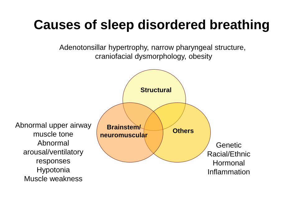
Adenotonsillar hypertrophy, narrow pharyngeal structure,
craniofacial dysmorphology, obesity
Genetic
Racial/Ethnic
Hormonal
Inflammation
Structural
OthersBrainstem/
neuromuscular
Abnormal upper airway
muscle tone
Abnormal
arousal/ventilatory
responses
Hypotonia
Muscle weakness
Causes of sleep disordered breathing

Abnormalities in gas exchange
• Craniofacial, neuromuscular and brainstem
abnormalities-
– Can lead to upper airway obstruction, hypoxia and hypercarbia
with sleep and during illnesses and stress
– Central apneas and oxygen desaturations with brainstem
abnormalities and decreased pulmonary reserve
– Gas exchange abnormalities can be present at birth or develop
with increasing age

Abnormalities in control of breathing
• Micrognathia may increase risk of upper airway obstruction
• Obesity and hypotonia may increase risk of obstructive sleep apnea
• Neuromuscular weakness may increase risk of partial obstructions
• Decreased pulmonary reserve from aspiration or recurrent lower
airway illnesses may cause oxygen desaturations
• Brainstem abnormalities may increase risk of central apneas
• Combinations of the above

Structural airway abnormalities can cause
obstructive apnea awake and asleep
Micrognathia
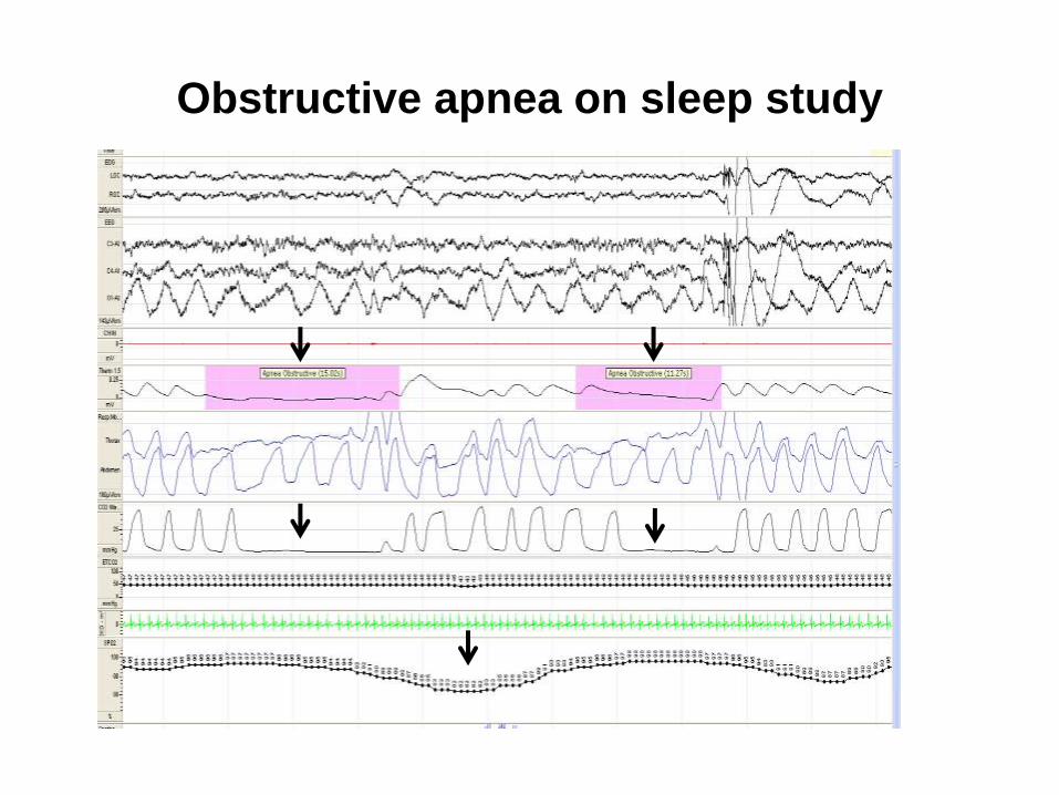
Obstructive apnea on sleep study

Obesity can cause obstructive apnea

Obstructive apnea on sleep study from obesity
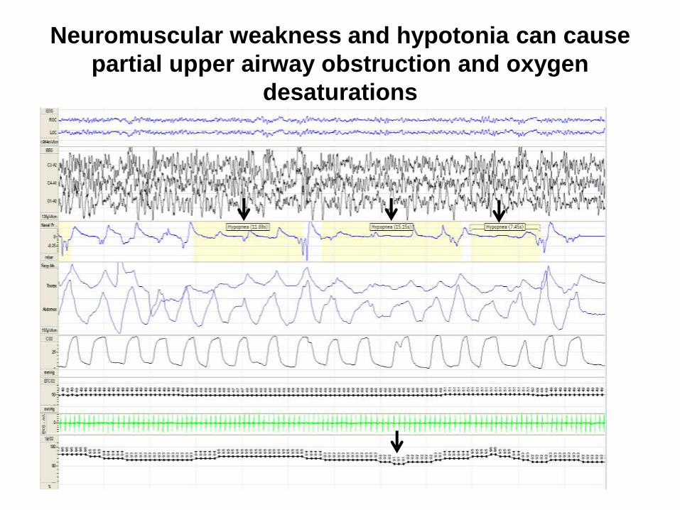
Neuromuscular weakness and hypotonia can cause
partial upper airway obstruction and oxygen
desaturations

Brainstem abnormalities can be associated
with central apneas

EEG
Nasal
flow
O2 sat
EKG
Thor
Abd
CA (5.2 sec) CA (5.3 sec)
86% 84%98% 96%
Decreased pulmonary reserve from lower
respiratory tract disease

Interventions/treatment used for control of
breathing problems
• Micrognathia
– ENT evaluation
• Jaw distraction, tracheostomy, positioning, non-invasive
ventilation (cpap/bipap)
• Obesity
– ENT evaluation of tonsils, adenoid or other upper airway
abnormality
– CPAP
• Neuromuscular weakness and hypotonia
– Non-invasive ventilation
• Central apneas due to brain stem abnormalities
– Positive pressure ventilation and tracheostomy
– Non-invasive ventilation

Nasal mask and CPAP device

Nasal pillows for non-invasive ventilation

Respiratory management of children with
Mobius syndrome
• Monitor respiratory symptoms closely
• Respiratory symptoms can be worse in the very young, with illnesses and in
children with co-morbidities
• Annual flu shots and consider pneumovax every 5-7 years in children at risk for
more severe respiratory compromise
• Sleep studies to rule out obstructive or central apneas as indicated
• Swallow studies in child with symptoms of swallowing disorder or chronic
respiratory symptoms
• Monitor lung function with age
• Aggressive chest physiotherapy and mucociliary clearance techniques as
indicated