resilient living materials built by printing bacterial spores · living materials, where cells are...
TRANSCRIPT
1
ResilientLivingMaterialsBuiltByPrintingBacterialSpores
LinaM.González1andChristopherA.Voigt1*
1SyntheticBiologyCenter,DepartmentofBiologicalEngineering,MassachusettsInstituteofTechnology,
Cambridge,MA,USA
*CorrespondingandrequestformaterialsshouldbeaddressedtoC.A.V.([email protected])
Keywords: SyntheticBiology,EngineeredLivingMaterial,GeneticCircuit,AdditiveManufacturing,3D
Printing
certified by peer review) is the author/funder. All rights reserved. No reuse allowed without permission. The copyright holder for this preprint (which was notthis version posted February 2, 2019. . https://doi.org/10.1101/537571doi: bioRxiv preprint
2
Abstract:
Aroutetoadvancedmultifunctionalmaterialsistoembedthemwithlivingcellsthatcanperformsensing,
chemicalproduction,energyscavenging,andactuation.Achallengeinrealizingthispotentialisthatthe
conditions for keeping cells alive are not conducive tomaterials processing and require a continuous
sourceofwaterandnutrients.Here,wepresenta3Dprinterthatcanmixmaterialandcellstreamsina
novelprintheadandbuild3Dobjects(upto2.5cmby1cmby1cm). Hydrogelsareprintedusing5%
agarose,whichhasalowmeltingtemperature(65oC)consistentwiththermophiliccells,arigidstorage
modulus(G’=6.5x10⁴),exhibitsshearthinning,andcanberapidlyhardeneduponcoolingtopreserve
structural features.SporesofB. subtilis areprintedwithin thematerialandgerminateon itsexterior,
including spontaneously in cracks and new surfaces exposed by tears. By introducing genetically
engineeredbacteria,thematerialscansensechemicals(IPTG,xylose,orvanillicacid).Further,weshow
thatthesporesareresilienttoextremeenvironmentalstresses,includingdesiccation,solvents(ethanol),
highosmolarity(1.5mMNaCl),365nmUVlight,andg-radiation(2.6kGy).Theconstructionof3Dprinted
materialscontainingsporesenablesthelivingfunctionstobeusedforapplicationsthatrequirelong-term
storage,in-fieldfunctionality,orexposuretouncertainenvironmentalstresses.
certified by peer review) is the author/funder. All rights reserved. No reuse allowed without permission. The copyright holder for this preprint (which was notthis version posted February 2, 2019. . https://doi.org/10.1101/537571doi: bioRxiv preprint
3
Introduction
Living materials, where cells are embedded within a structural scaffold, pervade the natural world,
examplesbeingwood,bone,andskin.Thecellsprovidedynamicfunctions,includingenergyharvesting,
repair, sensing, and actuation. Natural living materials can survive for years, even millennia, under
fluctuating and stressful conditions. There has been an effort to design so-called engineered living
materials, to artificially combine structural components with cells, including those that have been
genetically engineered1,2. Biomedical applications include the differentiation of human cells to grow
artificialtissueortoserveinlivingmedicaldevices3,4,5,6.Thepotential,however,ismuchgreateracross
many applications, where cells are harnessed for their sensing7, chemical/material production8 self-
healing9, self-powering10, self-cleaning8, and self-gluing11, 12 abilities. Natural microbes have been
harnessedtobuildconcreteandfixcracks9,13,buildpackagingmaterials11,12,andtocontrolbreathability
intextilesbyopeningventsinresponsetosweat14.Engineeredbacteriahavebeendemonstratedtobuild
metalwireswithinelectronics15,16,actasapressuresensorwithinadevice16,anddegradepollutants17.A
keychallengeforlivingmaterialsismaintainingviablecellsforlongtimesoutsideofthelaboratoryunder
extreme,unpredictableconditions.
Livingmaterialsrequiretheorganizationofcellswithinastructuralscaffold.Whileitispossible
toenvisioncellsgrowingtheirownscaffoldovermacroscopiclengthscales,forexamplethegrowthofa
tree,theseprocessesareslowandcurrentlydifficulttocontrolgenetically.Analternativeistouseadditive
manufacturing, also referred to as 3D printing, to organize cellswithin a scaffoldingmaterial at sub-
millimeter resolution over macroscopic length scales. Additive manufacturing has revolutionized
industries,fromarchitecturetoaerospace18-23.Therangeofmaterialsthatcanbeprintedhasexpanded
toincludeceramics24,metals25,26,nylon27,silk28,cellulose29,30,andwoodpolymercomposites31.Thesize
oftheobjectscreatedspanfromnanometerstohouses18.
Incorporating living cells into existing 3D printing platforms is challenging because of toxic
materialsandconditions,suchashightemperaturestoextrudeplasticsorUVlightforcuring32-34.Several
approacheshavebeentakentoaddresstheseissues.Anobjectcanbemadeusingaconventionalprinter
first,afterwhichcellsareintroducedonthesurfaceasabiofilmordiffusedintoahydrogel35,36.Another
approachistosuspendbacteriaevenlythroughoutahydrogelandthenusemultiphotonlithographyto
crosslink barriers to entrap the bacteria in 3D geometries37. These approaches can be used to create
intricatephysicalstructures,butdonotprintthebacteriawithinthematerial.Tothisend,“bioinks”have
beendevelopedthatcombinebacteriaandtheir requirednutrientswith theprecursors that formthe
structuralsupport.Zhaoandco-workersdevelopedabioinkbasedonEscherichiacoliandmicellesthat
certified by peer review) is the author/funder. All rights reserved. No reuse allowed without permission. The copyright holder for this preprint (which was notthis version posted February 2, 2019. . https://doi.org/10.1101/537571doi: bioRxiv preprint
4
canbeUV-crosslinkedafterprinting38.Usingthis,theyconstructedhigh-resolution3Dstructures(upto3
cm) anddemonstrated that living cells canperform sensing and computing functions. Another bioink
basedonE.coliwasdevelopedthatutilizestheformationofahydrogelbyalginatewhenitcomesinto
contactwithcalciumchlorideontheprintsurface39.LivingmaterialsbasedonE.coliremainfunctionalfor
severaldaysafterprinting.Studartandco-workersdesignedabioinkbasedonashear-thinninghydrogel
(hyaluronicacid,k-carrageenan,and fumedsilica) thatwasshowntobeable toprintBacillus subtilis,
PseudomonasputidaandAcetobacterxylinum40.AbioinkbasedonB.subtilishasalsobeendeveloped
thatutilizesitsnaturalbiofilmamyloidsfiberstoproduce2Dshapesthatcansurviveat4oCfor5weeks41.
Alloftheseapproachesrequirethatthecellsbemaintainedinahydrogelwithampleaccesstowaterand
replenishednutrientstomaintainafunctionallivingmaterial.
Some bacteria survive under adverse conditions by forming endospores – small spherical
structuresthataredormantandtough.Thecellmembranearchitecture iscomprisedofseveral layers
that include an almost impermeable innermembrane, a germ cell membrane, a thick peptidoglycan
cortex,theoutermembrane,abasementlayer,theinnercoat,theoutercoatandthecrust42.TheDNAis
protected by its tight packing by specialized proteins43-45. Spores are able to survive extreme
environmentalinsults,includinghightemperature,freezing,oxidizingagents,acidandalkalinesolutions,
genotoxic agents, solvents, highpressure,X-rays,g-radiation, andUV-light 46,47. They canalso survive
desiccationbyreplacingwaterwithdipicolinicacidandadoptingawrinkledshapetowithstandosmotic
stress44, 45. Spores can lie dormant indefinitely and purportedly formillions of years48. Spore-forming
bacteria have been used in biotechnology as vaccines, radiation detectors, insecticides, hygrovoltaic
generators,andbio-cements9,10,49-51.
Here, we modify a 3D printer (MakerBot Replicator) that builds objects by extruding plastic
throughahigh-temperaturenozzle(Figure1a).Thenozzleisredesignedtomixtwostreamstoformthe
bioink just prior to printing: one a polymer that is maintained at high temperature and a lower
temperaturemixtureof cellsandmedia. Agaroseexhibits shear thinningandweshowthat it canbe
printedwithvariousthermophilicBacilli species.Usingonlypurifiedspores inthebioink improvesthe
fractionofviablecellsandtheirdistribution in the inprintedstructure.Thesporesremaindistributed
throughout thematerial and provide a constant source of germinated cells at the surface, including
spontaneously in new cracks or tears. There, they can perform their programmed function, such as
respondingtochemicalsdetectedbygenetically-encodedchemicalsensors(shownforIPTG,vanillicacid,
andxylose).Further,thespore-containingmaterialcanbedesiccatedandstoredatroomtemperature
indefinitely.Uponrehydration, theprintedshapereconstitutes, thecellsgerminate,and theyperform
certified by peer review) is the author/funder. All rights reserved. No reuse allowed without permission. The copyright holder for this preprint (which was notthis version posted February 2, 2019. . https://doi.org/10.1101/537571doi: bioRxiv preprint

5
their programmed function. When various extreme stresses are applied to the materials, including
ethanol,highosmolarity,UVlight,andradiation,thesporesareabletosurviveandquicklygerminate.
Thisworkdemonstratesaroutebywhichmaterialscanretainthefunctionsprovidedbyembeddedliving
cellslongaftertheyarecreated.
Results
Repurposingofa3DPrinter
TheMakerBot Replicatorwas selected as a simple, inexpensive fused depositionmodeling (FDM) 3D
printer.Theprinterhadtobeextensivelyre-engineeredtoprintlivingcellswithinastructuralmaterial
(Figure1b,SupplementaryFigures1-3).Anovelprinthead(#1inFigure1b)wasdesignedtofacilitatethe
blendingofthestructuralmaterialsandthecellsfromseparatestreamsandtheextrusionofthemixture
througha400µmdiameternozzle.Thestreamsarepropelledusingamodifiedpressurizedtankforthe
material(#2inFigure1b)andaliquidDCpumpforthemediacontainingthecells(#4inFigure1b).The
chamberwhereprintingoccursiscooledtoacceleratematerialhardening.Detailedphotos,schematics,
andelectronicdiagramsforthesedesignsareprovidedintheMethodsandSupplementaryFigures4-11.
TheoriginalMakerBotprintheadhasasingleinputthattakesinasolidplasticfilamentandheats
itto220oC;andalternativecommercialprintheadsweredeemedinappropriateforhandlingcells52,53.We
developedanovelprintheadthatisabletoefficientlymixthetwostreamswhilemaintainingaconstant
temperature (Tchamber) (Figure 1b, Supplementary Figure 4). The extrusion rate from the nozzle is
maintainedclosetotherateatwhichtheMakerBotreelsinfilament.WhentheMakerBotprints,itsmotor
turns in the forward direction. Our printhead detects this motion with an optical sensor and it
subsequentlytriggerstheopeningofsolenoidvalvesthatallowsthecontrolofboththematerialandcell
streams.Thestreamispropelledbypneumaticpressure(0.75-1psi)appliedtothematerialstoragetank
thatiscontrolledbyadigitalelectronicreliefvalve(SupplementaryFigures7-8andTable4).Thisflowis
discontinued when the temperature at the top of the printhead reaches ~63oC and cells have been
introduced (see calibration in Supplementary Figure 9). A second optical sensor detects when the
MakerBot’s motor moves via a trigger and then turns on the cell stream with a 15 ms delay
(SupplementaryFigures10-11).Topromotemixingintheprinthead,thecellinputstreamentersata30°
angleandthecellflowissettobehigh(3.2mL/s).Bubbleformationduringmixing(andpresentinthe
agarose)waseliminatedbyaddinga1mmholeatthetopmostpartoftheprintheadforairtoescape(see
SupplementaryFigure4).
certified by peer review) is the author/funder. All rights reserved. No reuse allowed without permission. The copyright holder for this preprint (which was notthis version posted February 2, 2019. . https://doi.org/10.1101/537571doi: bioRxiv preprint

6
TheMakerBotstopsthefilamentfromexitingthenozzlebyturningthemotorbackward;inour
system,this isdetectedbyathirdopticalsensor,which leadstothestoppageofthematerialandcell
streams.Earlydesignsexperiencedproblemswithclogging(notshown).Toavoidthis,atthesametime
thatasolenoidvalvestopstheflowofagaroseneartheprinthead,thereliefvalvereleasespressurein
theagarosetank.
Itiscriticaltomaintainthetemperatureinthelinescarryingagarosetoandattheprintheadso
thatitissufficientlyhightofacilitatetheflowofthematerialstream,yetislowenoughtonotkillthecells.
Forthespecificmaterialandcellcombinationweuse(nextsection)thisbalancewasachievedat75oC
(SupplementaryFigures12-13).Thetemperatureismonitorednearthenozzleoutput(T1),thecenterof
theprinthead(T2)andneartheinputstreams(T3)(Tchamber=<T1,T2>).Tchamberismonitoredusingafeedback
controlsystem(aPIDcontroller,seeSupplementaryFigures14-17).T3isusedtomonitoranddetectwhen
theagaroseisrunninglowandallowagaroseflowviaopeningthesolenoidvalve(s).Thecellsarestored
atT=22oCandexposedtohightemperatureforlessthan20min.Themixtureiscooledimmediatelyafter
exitingthenozzlebymaintainingtheprintchamber(#3inFigure1b)at16oCusingthermoelectriccooling
(peltierelements)(SupplementaryFigures18and19).Thiscoolingsignificantlyimprovesthestructural
detailsintheprintedobject(SupplementaryFigure20).
Toprintanobject,itisdrawnusingSolidWorks,savedinastandardformat(e.g.,asaSTLfile)and
loadedintothesoftwareprovidedbyMakerbot.Thegcodeparametersaremodifiedsothatthenozzle
temperatureis72oC,platformtemperatureis22oC,andtheinfillis100%.Fordualprinting,wemerged
twoSTLfilesusingthemergetoolinReplicatorX(seepictureofthesysteminSupplementaryFigure5).
Mixing is initiated by filling the printhead with a pre-mix of material, running the temperature PID
controllercodetobringthetemperatureintheprintheadto75oC,thenaddingcells(Methods).Afterthis,
theobjectedisprintedfollowingthesameprotocolastheobjectsproducedbytheunmodifiedMakerBot.
MaterialandStrainSelection
Storingtheliquidscontainingthematerialandcellsseparately,asopposedtoasinglebioink,allowsthem
to be independently varied and stored under different optimized conditions. Still, they must be
compatiblewitheachotherastheyaremixedintheprinthead.Thecriticalparameter inourdesign is
temperature,whereamaterialneedstobeselectedthatmeltsatatemperaturethatdoesnotkillthe
cells.Similarly,morethermophilicstrainswillbecompatiblewithawiderrangeofmaterialsthatmeltat
highertemperatures.Aftertestingdifferentmaterialsandbacterialspecies,wefoundthatagaroseandB.
subtilissporesproducedthemostconsistentstructureswiththehighestviabilityofprintedcells.
certified by peer review) is the author/funder. All rights reserved. No reuse allowed without permission. The copyright holder for this preprint (which was notthis version posted February 2, 2019. . https://doi.org/10.1101/537571doi: bioRxiv preprint
7
Agarose is a hydrogel, derived from seaweed, consisting of linear polymers of alternating D-
galactoseand3,6-anhydro-L-galactopyranosesubunits.Itisusedcommonlyinbiologyandbeenshown
tosupportembeddedcells54.Agarosewasselectedbecauseitexhibitsshearthinning,istransparent,has
highwatercontent,isstrongafterprinting,andrapidlysolidifieswithouttheneedforchemical/physical
inputsorUV-curing.Whileagarosemeltsat65oC(Figure1c),wefoundthat72oC(Tnozzle) isoptimalfor
obtainingbondingbetweenprintedlayersandavoiddelamination.
Ahigherconcentrationofagaroseleadstoamorerigidstructure,butismoredifficulttoprint.
Wefoundtheidealconcentrationtobe4%(weight/volume).At3%,theagaroseisnotabletoharden
into thedesiredgeometry,evenafter cooling thechamber (SupplementaryFigure21).Above6%,we
started to encounter problemswith line clogging. To balance these effects, thematerial streamwas
chosentobe5%agarosesothatitcanbemixedwiththecellstreamintheprintheadandremain>4%in
the printed object. The storagemodulus (G’) of 5% agarose is» 105, consistentwith the stiffness of
previously published 3Dprinted hyrdrogels17, 55 (Figure 1d and Supplementary Figure 22). The rate at
whichthematerialhardensafterexitingthenozzleiscapturedbytheinitialchangeinG’uponshifting
from70oCto16oC(Figure1d).Inourhands,arate>5000Pa/s(>4%agarose)isimportantforaccurately
printingthedesiredstructure(Figure1dandSupplementaryFigure23).Agaroseisanon-Newtonianfluid
thatexhibitsshearthinning,whichaidsitsextrusionthroughthesmallnozzle(SupplementaryFigure24).
Due to the temperature requirements of themodified printer (Tchamber = 75oC),weneeded to
identify a bacterial species that can survive in the material stream. Mesophiles and thermophiles
representative of different phyla were tested (Anoxybacillus flavithermus, Bacillus amyloquifaciens,
Bacillus licheniformis, Bacillus megaterium, Cupriavidus metallidurans, Escherichia coli, Geobacillus
stearothermophilus, Geobacillus thermoglycosidasius, Gluconacetobacter xylinus, Gluconacetobacter
hansenii, Halobacterium salinarum, Lactobacillus lactis, and Sporosarcina pasteurii) (Supplementary
Figures25-27).Experimentsweredesignedtotestsurvivalofthecellsbyexposingthemto75oCfor20
minutes(Methods)(Figure1e,SupplementaryFigures26-27).Ofthespeciestested,onlyspore-forming
organismswereable tosurvivewith littleday-to-dayvariability.WhenB.subtilisPY79 (aderivativeof
168)56isfirstgrowninspore-inducingmedia(DSM),thisenhancessurvivalandallowsforcellstomore
rapidly recover heat shock (Figure 1e blue lines). Furthermore, we exposed the spores to higher
temperaturefor20min(>75oCandupto100oC)andfoundthatsporescansurviveat100oC,butgrowth
isnotdetecteduntilafter8h(SupplementaryFigure28).
The5%agaroseandB.subtilisPY79inDSMwereselectedasthetwocomponentsofthebioink.
Note that other spore-forming organisms would also work as the cell component of the ink
certified by peer review) is the author/funder. All rights reserved. No reuse allowed without permission. The copyright holder for this preprint (which was notthis version posted February 2, 2019. . https://doi.org/10.1101/537571doi: bioRxiv preprint
8
(SupplementaryFigure27).Asimplebarwasprintedconsistingof7layers(2by3by25mm)(Figure1f).
Toaidimaging,anexpressioncassettewheregreenfluorescentprotein(GFP)isconstitutivelyexpressed
wasintroducedintotheB.subtilisgenomeattheamyE locus(Methods).Figure1fshowscomparisons
between the structuresproducedwhenB. subtilis is grownunder conditions favoring vegetative cells
versusspores.Theformershowspunctategrowthdistributedunevenlythroughtheagarosestructure.
Whensporesareprinted,theyareevenlydistributedthroughoutthestructure.
3DPrintingFunctionalLivingMaterials
Figure2showsexamplesofgeometriesthatcanbeprintedbythematerialstreamaloneandtogether
with the cell stream. The minimum feature size is consistent with that obtained by the MakerBot
Replicatorwhenprintingplastics(100µm),whichisdictatedbythe400µmnozzlediameter.Thehydrogel
canbeprintedatascaletheutilizesthemaximumsize inthex-andy-dimensions(10cmby20cm).
Simpleshapescanbeprintedupto1cminthez-dimension,requiringtheprintingof~37layers,each300
µm(atarateof3layers/min).InFigure2i,wecomparetheprintedobjectswiththoseobtainedfroma
high-resolutioncommercially-availableprinter(ProJet6000)andtheshapeandresolutionissimilar.
Therearesomeapplicationswhereitisdesirabletoprintcellsatspecificlocations,ratherthan
throughout the structure. To do this, a second printhead is added intowhich thematerial stream is
injectedviaateeconnector,butforwhichthereisnocellstreaminput(SupplementaryFigure5).TheSTL
filesarepreparedfortheportionscontainingandlackingcellsandthenmerged.Thisgeneratesstructures
withsharpboundariesbetweentheregionscontainingcells(Figure2h).Itisstraightforwardtoexpand
thisapproachtoprintmultiplematerialsandcelltypes.
The spores are distributed evenly throughout the material in which they are printed. They
germinatepreferentiallyatthemedia-exposedsurfaceofthehydrogelduetotheavailabilityofoxygen
andnutrients(Figure3a-candSupplementaryFigure29).Germinationoccursuptoadepthof0.85±0.16
mm,determinedbyimagingcellsconstitutivelyexpressingGFP.Whenthestructureistornandincubated
inLBmedia for10hours, this induces thegerminationofsporesat thesurfaceexposedto themedia
(Figure3c). Thisdemonstratesthattheembeddingofspores inthematerialprovides,undertheright
conditions,asourceofnewgerminatedcellsthatcanperformtheirfunction.
Thegerminatedcellscanfunctionasbiosensors. Todemonstratethis,we implementedthree
genetically-encodedsensorsinthegenomeofB.subtilisthatrespondtosmallmolecules.Thesensors
wereeither taken from the literatureorbuiltdenovo andoptimized forB. subtilis tomaximize their
dynamicrange,suchthattheycanbeimagedinthematerial.AnisopropylB-D-1-thiogalactopyranoside
certified by peer review) is the author/funder. All rights reserved. No reuse allowed without permission. The copyright holder for this preprint (which was notthis version posted February 2, 2019. . https://doi.org/10.1101/537571doi: bioRxiv preprint
9
(IPTG)-induciblesystemwasused thathadbeenpreviously reported tohaveadynamic rangeof276-
fold57,58(Figure4aandSupplementaryFigure30).Asugarsensor(xylose)wasmodifiedtofunctioninLB
media by removing the catabolite-responsive element (Figure 4c, Supplementary Figure 31,
SupplementaryTable7)59.Finally,asensorforaplantrootexudate(vanillicacid)wasdesignedbasedon
anoptimizedVanRrepressorandcorrespondingoperator thatwas inserted intothe -10/-35regionof
Pspank(hy)tocreatePspank(V)(Figure4d,SupplementaryTable7)7,60.Thissensorgeneratesa60-folddynamic
range(SupplementaryFigure32).
Materialswereprintedwithsporesofbacteriacontainingthesensors(Figure4).Afterprinting,
thematerialwasincubatedat37oCinmediaeithercontainingorlackingthesmallmolecule.Forallthe
sensors,someinductioncanbeseenat6hourswithfullinductionat12hours.
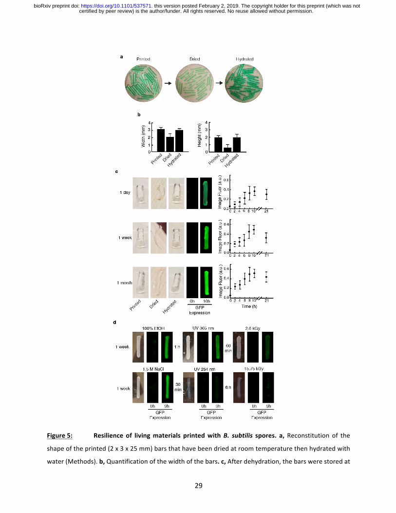
SporeSurvivalin3DPrintedMaterials
Thesensitivityoflivingcellsandtheircontinuousrequirementfornutrientsandwater,makeitchallenging
to embed themwithinmaterials and function over long times. First,we evaluated the ability for the
printedmaterialstosurvivedesiccation.The5%agarosematerialcontains92%water(Methods).Afterit
isprinted,thewatercanberemovedbydryingandstoredforlongtimes(Figure5a).Whenrehydrated,it
returnstoitsoriginalprintedshape(Figure5b).WhensporesofGFP-expressingB.subtilisareprinted,
theysurvivefor1month(themaximumtested)andgerminatewhenrehydrated.GFPexpressionafter
germinationandgrowthdoesnotgodownforlongerstoragetimes(Figure5c,SupplementaryFigure33).
Given the known longevity of spores61, it is expected that they would be able to regerminate after
indefinitestorageundertheseconditions.
Sporescansurviveundermoreextremeenvironmentalstresses.Alcoholisknowntonothave
sporicidalactivity.Whenprintedobjectscontainingthesporesareexposedto100%ethanolforone
week,thecellssurvive(Figure5dandSupplementaryFigure34).Highosmolarityhasbeenshownto
haveaneffectonsporeviability62.Again,afteroneweek,thesporesremainviable.Notethat,forboth
ofthestresses,someofthisprotectionmaybebeingconferredbytheagarosematerial.
ThematerialscontainingsporeswerethensubjectedtoUVlight,X-rays,andg-radiation(Figure
5dandSupplementaryFigures35-37).Thesporessurvive365nmUVlighttreatment,buttheydonot
survivewhenexposedto254nmlight.Thisisexpectedas254nmUVlightisknowntobeeffectiveat
exterminatingspores63.ThesporesalsosurvivewhenthematerialisexposedtoX-raysfor10min(8.469
R/min).Whenexposedtog-radiation,thesporessurviveafter1hour(2.6kGy)exposure,butnot6hour
(15.7kGy)or12hour (31.5kGy) treatments. Note thatg-radiationwithadoseof25kGy isused for
certified by peer review) is the author/funder. All rights reserved. No reuse allowed without permission. The copyright holder for this preprint (which was notthis version posted February 2, 2019. . https://doi.org/10.1101/537571doi: bioRxiv preprint

10
sterilization64.Inaddition,after6hourstheagarosehydrogelweakensandthebarsstarttodeteriorate
duetoshakingduringthegrowthassay(Methods).
Conclusion
Thisworkdemonstratesthatembeddingspores,asopposedtovegetativecells,offersadvantageswhen
producinglivingmaterials.Usingsporessimplifiesthepreparationandstorageofthecellcomponentof
thebioinkandgeneratesmorereliableprintswithouthavingtocarefullycontroltheviabilityorgrowth
phaseofthecellsinthereservoir.Whileweprintthesporeswithinanagarosehydrogel,theyareableto
surviveextremedesiccationandstorageunderambientconditionswithoutacontinuousnutrientsupply.
Whentheygerminate, theyareable toperformtheirprogrammed function,whether itbe tosensea
chemical or turn on a biosynthetic pathway. Further, the spores selectively germinate on the object
surface. Thisallowsthespores inthecenterofthematerialtoprovideacontinuoussourceoffreshly
germinatedcells. Whenthematerial istorn,thesporesgerminateandgrowonthenewsurface.One
couldimagineharnessingthiscapability,alongwiththeabilitytoproducebiopolymers65,tocreateself-
healing materials. While we utilize the natural signals that induce germination, methods have been
developedtogeneticallyengineersporestogerminateunderdefinedconditions,suchasthepresenceof
chemicals,includingneurotransmittersandspecificDNAsequences66.Therecalcitrantnatureofspores
makesthisapproachpotentiallycompatiblewithawiderangeof3Dprintingtechnologybasedonharsher
conditions,suchaslasersinteringofceramics/metals(highT).
Manyapplicationsofsyntheticbiologyrequirethatengineeredcellssurviveandperformtheir
function in fluctuating and stressful environmental conditions67. UV protection will be required for
bacteriathatfunctionintheopenenvironment,forexampleonplantsurfacesinagriculture,68inopen
pondsforbio-production69,orpollutionremediation70.Theabilityofsporestosurvivewithoutwaterand
nutrientsandunderharshconditionscanaid long termstorage.Harnessing the recalcitrantnatureof
spores,stabilizedwithina3Dprintedhydrogelisasteptowardsrealizingtheseapplications.
certified by peer review) is the author/funder. All rights reserved. No reuse allowed without permission. The copyright holder for this preprint (which was notthis version posted February 2, 2019. . https://doi.org/10.1101/537571doi: bioRxiv preprint
11
Methods:
Modificationstothe3DPrinter
Wemade extensivemodifications to aMakerBot Replicator 2X, entailing five separate subsystems: a
printhead,anagarosepumpingsubsystem,acellpumpingsubsystem,andaheatingsubsystemanda
coolingsubsystem(SupplementaryFigure1).Thecellsandagarosearepropelledtotheprintheadusing
theagarosepumpingandthecellinputsubsystems.Attheprinthead,thetemperatureisregulatedusing
theheatingsubsystem.Attheotherextreme,thecoolingsubsystemmaintainsthechamberattheproper
temperaturefortheagarosetoretain its filamentaryshapewhen itexits thenozzle.Details regarding
thesemodifications,includingphotos,schematics,partnumbers,filesfor(traditional)3Dprintingofnovel
parts,andArduinoprogrammingdiagramsareprovidedintheSupplementalInformation.
Printhead and optical sensors subsystem. The originalMakerBot printhead has a steppermotor that
extrudestheplasticfilament,drawninasasolidfilamentfromasingleinlet.Ourprinthead’sbarrelhas
twoinlets,oneforthematerialandtheotherforthecells.Italsohasthreethermistorsensorstomeasure
thetemperatureatthetop,middleandbottomoftheprinthead(T1,T2,T3)(SupplementaryFig.4aand
SupplementaryFigure4b).TheaverageofT1andT2isusedtomaintainthetemperatureintheprinthead
at75°Ctoensuremixingoftheagaroseandspores(seecellinputandheatingsubsystemsections).Adual
printingconfigurationisusedbyalternatingbetweentwosolenoidvalvestohavetwodifferentprinthead
printingeitherthematerialaloneorwiththecells(SupplementaryFigure4c).Opticalsensorsareusedto
detectwhichmotormoves and thus,which extruder is active at a given time.Using a pair of optical
interrupters,wecoupledthesteppermotor(fromtheMakerBot)andtheagarosepumpingsystem.An
encoder(10spokes)monitorsthesteppermotor’smovement.Whenamovementchangeisdetectedand
thetemperatureis~63°C(attheverytopoftheprinthead),thesolenoidvalveisopensothatagarose
flowfreelyintotheprinthead.AslotopticalinterrupterisaU-shapedsensorwithaninfraredemitteron
onesideandthecorrespondingdetectorontheoppositeside.Whenanobjectbreakstheinfraredbeam,
thiscanbedetectedthroughabreakinthecircuit.Two8mmwideslotsensorswereplacedintandem
(Digi-KeyElectronicspartno.EE-SX1070).Thesearealsousedtodetectwhenthemotorreversedirection
usingaquadratureencodersignal.Briefly, ifthetwochannelsare90°outofphaseandifpin2seesa
changebeforepin3,thenthemotorismovingclockwise;otherwise,itismovingcounterclockwise(see
code).Asingleopticalinterrupter(Digi-KeyElectronicspartno.EE-SX1042)wasusedtoturnonthecell
streamforduringthetimeittakesfortworevolutionstooccur(seecellinputsubsystemformoredetails).
Ahole(1mmindiameter)wasintroducedatthetopoftheprintheadforexcessairtoescapeafterpriming
certified by peer review) is the author/funder. All rights reserved. No reuse allowed without permission. The copyright holder for this preprint (which was notthis version posted February 2, 2019. . https://doi.org/10.1101/537571doi: bioRxiv preprint

12
andwhileprinting(airfromtheagarosecabinetisconstantlypurgedintotheprinthead).Thismodification
relievedtheproblemwithhavingbubblesinsidetheprintedobjectthatdistortedthematerialandledto
non-uniformprints. In SupplementaryFigure6,we showa schematicof theelectronic circuitused to
controlthethreeslotsensors.Eachofthetwoprintheadscontainsthreeopticalsensors.
Agarosepumpsubsystem.Ahermeticallysealeddesiccatorcabinet(FisherScientific,Model#33060)was
repurposedasapneumaticpump(SupplementaryFigure7). Thiscabinethasa¾”thickwallmadeof
polymethylmethacrylate (PMMA).Thevacuumgaugewas replacedwithapressuregauge (McMaster-
Carr,partno.4000K721)witha rangeof0-15psi tomonitorandmaintainaconstantpressure in the
cabinet.Oneorificeinthecabinetwasusedasaninletforhouseairtoenterandtheotherorificewas
used as an outlet for agarose to leave using positive pressure (Supplementary Figure 7a and b). To
maintainagaroseflow,amagnetwire(AppliedMagnets,AWG21)waswoundaroundtheplastictubing
suchthatapassingcurrentreleasesheat(Jouleheating).Tubingconsistedofhigh-temperaturesilicone
rubber.Athickertubing(OD=3/4”,ID=½”,McMaster-Carr,partno.5236K47)wasusedtofittheagarose
outletandastraightreducerbarbedfitting(McMaster-Carr,partno.5463K635)toconnecttoathinner
tubing(OD=5/16”, ID=3/16”,McMasterr-Carr,partno.5236K841)thatfitsthesolenoidvalves.The
lengthofthetubeis27”longtoprovidesomeslackfortheprintheadtomove.Thecurrentinthepower
supplywassetto5.0Aandthevoltageis~12V(BKPrecision1901BSwitchingModePowerSupply).In
addition,thecabinethasanelectricaloutletusedtoplugaheatingtapeformaintainingtheagarosein
thereservoirasaliquid.Thetubingthatcarriesagarosefromthereservoirtotheagaroseoutletonthe
liddoesnothaveanywirewindingsanditstubingissmaller(OD=1/4”,ID=1/8”,McMasterr-Carr,partno.
5236K83).Thereisano-ring(size114)intheinletandoutletofthelidofthecabinettopreventleakage.
Inaddition,acheckvalve(McMaster-Carr,partno.6079T58)preventstheflowofthisviscousliquidback
intothereservoir.Anarrowinreliefonthecheckvalvewassandeddownandthetwolayersofthemagnet
wirewaswoundaroundthisvalve.A1polepush-insignalpowerconnector (McMaster-Carr,partno.
9193T11)wasinstalledformaintenanceassometimesthisvalvegetscloggedandneedstobecleaned.
Epoxywasusedtopermanentlyfixthemagnetwiretothecheckvalves.Thischeckvalveaddsabout1psi
ofresistancetotheline;morecheckvalvesledtoseverecloggingandcleaningdifficulty.Anelectronic
reliefvalvewasinstalledinthecabinettoimmediately(withinafewms)relievethepressurewhenitis
notneededtoactuateflowtotheprinthead.Whenstartingaprint,thelineisinitiallyprimedwithmolten
agarose,whichkeepsthisreliefvalveclosed.Whenthemixingchamber’stemperaturereaches~63°C,
thisindicatesitisfull,andthevalverelievesthepressureinthetank(SupplementaryTable4).Whenthe
certified by peer review) is the author/funder. All rights reserved. No reuse allowed without permission. The copyright holder for this preprint (which was notthis version posted February 2, 2019. . https://doi.org/10.1101/537571doi: bioRxiv preprint
13
moltenagaroseispumbedfromthecabinet,thepressureisincreasedandpulsedonandoffsothatit
remainsbetween0.75and1psi.Apairofsolenoidsisplacedbetweenthecheckvalveandtheprinthead
(SupplementaryFigure7aandSupplementaryFigure5)toexertmorecontrolovertheagaroseflowand
to facilitate dual printing (see section on Printhead and slot sensors subsystem for more details).
SupplementaryFigure8showsaschematicoftheelectroniccircuitusedtocontrolthepumpingofthe
agarose.
Cellinputsubsystem. A DC liquid pump (Trossen Robotics, part no. HW-WVALVE) (Supplementary
Figure9a)isusedtopropelthecellstreamintotheprinthead.Theflowismodulatedbyasolenoidvalve
thatiscontrolledbyanArduinomicrocontroller.Toreducethepumpflowrate,8checkvalvesareplaced
intandem,includingoneclosetotheentrypointtotheprinthead(eachcontributing~1psiofresistance)
(SupplementaryFigure9b).Wecalibratedthepumpwiththetimethatthepumpisenergizedoropen
anddeterminetheamountofliquidtoaddforcontinuouspumpingandtokeeptheagarosepercentage
above4%(SupplementaryFigure9c).Aftertheinitialagaroseprimingoftheprinthead,itcontainsabout
5mLofagaroseandweinput600µLofcellstokeeptheagarosepercentageabove4%.Toadd600µLof
cells,weopenedtheDCliquidpump(andsolenoidvalve)for90ms(seeDCliquidpumpcalibrationin
Supplementary Figure 9c). For continuous and subsequent input of the cells, we did a second set of
calibrationstodeterminehowmuchcellstoaddper100µLofagaroseandchose15msforthetimethe
DCliquidpumpandsolenoidvalvesareopened(SupplementaryFigure11).
Heatingsubsystem.Onepowersupply(Newark,partno.78Y9404)iskeptataconstantvoltage/current
andisusedtopowertheheatingofthematerialslinetokeeptheagarosemoltenpriortoreachingthe
printhead.Tomixcellandagarosestreams,thetemperatureintheprintheadneedstobemaintained75
°C(SupplementaryFigure13).Todothis,asecondpowersupply(Newark,partno.34T4663)iscontrolled
remotelyandweimplementaPIDcontrollertomaintainthisconstanttemperatureintheprinthead.The
PIDalgorithm(SupplementaryFigure14a)comparesthesetpointandmeasuredtemperaturesandused
theirdifference(theerror)tocontrolthecurrentfromthepowersupplybasedontheequation
𝑜𝑢𝑡𝑝𝑢𝑡 = 𝐾'𝑒 𝑡 + 𝐾+ 𝑒 𝑡 𝑑𝑡 + 𝐾--(/(0)-0
(1)
wheretheoutputisthesignalcontrollingthecurrent,tisthetime,Kp,KiandKdarethecoefficientsfor
theproportional, the integraland thederivative, respectively. It is implementedusinganArduinoPID
library and the tuningparameters are adjustedusing an autotune function (SetTuning). The feedback
controlisimplementedusingtheremote-controlledpowersupplywithan8pinplugadapter(onlypin1
certified by peer review) is the author/funder. All rights reserved. No reuse allowed without permission. The copyright holder for this preprint (which was notthis version posted February 2, 2019. . https://doi.org/10.1101/537571doi: bioRxiv preprint
14
(5V),pin2(input)andpin3(ground)wereused),magnetwire,thermistorsandanArduinomicrocontroller
(SupplementaryFigure14b).SupplementaryFigure15showsaschematicofthecircuitwiring.Weverified
thatthesystemandcodeworkatroomtemperature(SupplementaryFigure16)andwhilecoolingthe
chamber(SupplementaryFigure17).Thetemperatureofthenozzleisindependentoftheprintheadand
wecontrolleditthroughthegcodeintheoriginalMakerBot.Wescreenedforthebesttemperatureto
keep the nozzle and we found it to be 72°C (Tnozzle + Tchamber + Tambient) which leads to an average
temperatureof~54°C.
Cooling subsystem. The rapid gelation of the agarose/cell mixture after leaving the printhead nozzle
requiredthecoolingoftheprintchamberto14-17°Cbyblowingcoldairintoit.AStyrofoamboxwithan
aluminumpanwasplacedbehindtheprinter(SupplementaryFigure18).Twometalbarsthatspanacross
thepanwereattachedtopeltierplates(SupplementaryFigure19).Afanblowsairintothecoolingsystem
andtwoaluminumelbowpipesmovethecoldairintotheprintingchamber.Metalfinswereattachedto
thebarstoincreasethesurfaceareaandfacilitateheattransferasairpassesthroughthesefins.Priorto
usingtheprinter,1Lofwater isaddedtothealuminumpan, it iscooledforonehour,andthen ice is
addedtothepan.
Preparationandstorageofagarose.TheSeaPlaqueagarose(Lonza,catno.50100)powderisdissolvedin
300mLofwater(tomakea5%w/v),autoclaveandstoredatroomtemperature.Rightbeforeaddingit
to the material reservoirs (a 600 mL beaker with a heating tape wrapped around it) the agarose is
microwavedfor3.5minoruntilitcompletelymelts.Wecoverthebeakerwithapieceofaluminumto
preventexcessiveevaporationinsidethedesiccatorcabinet.
Embeddingandscaffoldingmaterialscharacterization. Usingarheometer(AR2000,TAInstruments),we
characterized the gelation time, the melting temperature, and the shear thinning properties of the
agarose.Allofthemeasurementsweretakenusingapeltierplate(withelementsforrapidcoolingand
heating)(itcanrapidlycoolandheat).Toobtainadequaterheometermeasurementsofthehydrogels,a
cross-hatchedgeometry(543337.001)wasused.Thisprovidesaroughsurfacetogriponthehydrogels.
Toavoidslippingofthehydrogelonthemetalsurfaceofthepeltierplateandprovidesometexture,a
self-adhesivesandpaper(320grit,cat.McMaster-Carr,no.4647A13)wasused(SupplementaryFigure
22a).Topreventdryingofthehydrogels,weusedanevaporationsolventtrap(SupplementaryFigure22;
inset).The%strainwasset to0.1and the frequencyat1Hz forallmeasurements.Thesampleswere
certified by peer review) is the author/funder. All rights reserved. No reuse allowed without permission. The copyright holder for this preprint (which was notthis version posted February 2, 2019. . https://doi.org/10.1101/537571doi: bioRxiv preprint

15
preparedasintheprevioussection,butinasmallervolume(5mL).Torunthesamplesontherheometer,
about1mLispipettedontotheself-adhesivesandpaper,thegeometryisloweredtoabout1050µmfrom
thesurfaceandletsolidify.Theexcessmaterialisthenremovedusingaspatulasothatcross-sectional
areamatchesthatofthegeometry(490mm2)andthisgeometryisthenlowerto1000µm(orspecified
gapinthefile).
MolecularBiologyandGeneticEngineering
Strainsandcultureconditions.ForacompletelistofstrainsusedinthisstudyseeSupplementaryTable5.
Chemically-competentEscherichiacoliDH10Bcellswereusedforallroutinecloning(NewEnglandBiolabs,
Ipwich,MA).TheE.coliMG1655(inFigure1e)andthevariousBacillusstrainwereobtainedfromthelab
stockoriginallyobtainedfromeitherATCCorBacillusGeneticStockCenter(BGSC).AllE.coliandBacillus
strains were grown at 37oC in LB-Miller media (BD, cat no. 244620) unless indicated. Anoxybacillus
flavithermusstrainy.d.(DSM2641)cellsweregrownat55oCinNutrientBroth(5g/Lofpeptone(BD,cat
no.211677),3g/Lofbeefextract(BD,catno.212303),pHadjustedto7.0)andAnoxybacillusflavithermus
strainWK1(DSMZ,Germany)cellsweregrownat55oCinCasoBroth(15g/Lofpeptonefromcasein(BD,
catno.211921),5g/Lofpeptonefromsoybean(Sigma-Aldrich70178-100G),5g/mLofNaCl).Geobacillus
thermoglucosidasiusM5EXG(ATCCBAA-1069)weregrownat55oC inTrypticSoyBroth(40g/L,BDcat.
211825).Lactococcuslactissubsp.cremoris(Mobitech,MG1363),Lactococcuslactissubsp.lactis(ATCC
11454)weregrowninBrainHeartInfusion(BHI)Broth(BD,237500).Gluconacetobacterxylinus(ATCC
700178)andGluconacetobacterhasenii(ATCC23769)weregrowninHestrinandShramm(HS)medium
at30°C.HScontains2%(w/v)ofglucose(FisherScientific,C6H1206)0.5%(w/v)ofyeastextract(BD,cat
no.210929),0.5%(w/v)peptone(BD,catno.211677),0.27%(w/v)ofsodiumphosphasediabasic(Fisher
Scientific,S375-500),0.15%(w/v)ofcitricacid(FisherScientific,A940-500)and0.1%(w/v)ofcellulase
(Sigma-Aldrich,catno.C2730-50ML).Theglucoseandthecellulasewerefiltersterilizedusinga0.2µm
Acodisc syringe filter (Pall, cat no. PN 4612). Cupriavidus metallidurans CH34 (ATCC 43123) and a
domesticatedstrainderivedfromSporosarcinapasteurii(ATCC11859)weregrowinNutrientBroth(BD
cat.234000)at30°C.TogrowtheS.pasteurii,thepHoftheNutrientBrothwasadjustedto8.5before
autoclaving.HalobacteriumsalinarumNRC-1(ATCC700922)weregrownin2185HalobacteriumNRC-1
mediumat30°C.The2185HalobacteriumNRC-1mediumismadewithaBasalmediumandtracemetals,
asdescribed in theATCCprotocol.MinimalS750mediumcontains the following ingredients ina total
volumeof100mL:10mLof10XS750salts,1mLof100Xmetals,2mLof50%(w/v)arabinose(Sigma-
Aldrich,catno.A3256-100G),1mL10%glutamate(FisherScientific,catno.A125-100).The10XS750salts
certified by peer review) is the author/funder. All rights reserved. No reuse allowed without permission. The copyright holder for this preprint (which was notthis version posted February 2, 2019. . https://doi.org/10.1101/537571doi: bioRxiv preprint
16
consistof(filtersterilized):0.5MMOPS(freeacid)(FisherScientific,catno.ICN19483725),100mMof
ammonium sulfate (NH4)2SO4 (Millipore Sigma, cat no. AX1385-1), 50 mM of potassium phosphate
monobasic (KH2PO4) (Sigma Aldrich, cat no. 795488-500G), 8g of potassium hydroxide (KOH) (Sigma-
Aldrich, cat no. 221473-500G) (finely adjusted to pH 7 with 1M KOH). The 100X metals consist the
followingmetals(filtersterilized):0.2Mofmagnesiumchloride(MgCl2)(Sigma-Aldrich,catno.M8266-
100g),70mMofcalciumchloride(CaCl2)(Sigma-Aldrich,catno.C1016-500G),5mMofmanganese(II)
chloride(MnCl2)(Sigma-Aldrich,catno.244589),0.1mMofzincchloride(ZnCl2)(Sigma-Aldrich,catno.
Z0173),100mgmL-1ofthiamine-HCL(Sigma-Aldrich,catno.T4625-25G),2mMofHCL(MilliporeSigma,
catno.HX0603-3),and0.5mMofiron(II)chloride(FeCl3)(Sigma-Aldrich,catno.701122-1G).ForE.coli,
thefollowingantibioticconcentrationsareused(bothplatesandinculture):100μg/mlampicillin(Ap,
GoldBio;CAS#69-52-3);50μg/mlkanamycin(Kn,GoldBio;CAS#25389-94-0),100μg/mlspectinomycin
(Sp, GoldBio; CAS#22189-32-8) and 35 μg/ml chloramphenicol (Cm, AlfaAesar; #25-75-7). For B.
subtilis,thefollowingconcentrationwereused:5μg/mlCm,100μg/mlSp,5μg/mlkn.
Plasmidsandstrainconstruction.AllplasmidswereconstructedusingTypeIISassembly.DNAsequences
wereinspectedandmodifiedtoeliminatetherecognitionsitesfortherestrictionenzymes(BsaIorBbsI
orBsmbI)asnecessary.AbsaIsitewasremovedfromtheoriginalgfpmut2anditwasnamedgfpmut2x.
DNAwasamplifiedusingthepolymeraseKapaHiFiDNApolymerase(KapaBiosystems,catno.KK2602)
andgelpurifiedusingSeakemGTGagarose (fornucleicacidrecovery) (Lonza,catno.50070),agarose
dissolvingbuffer(ABD)(ZymoResearch,catno.D4001-1-50),Zymo-SpinI(ZymoResearch,catno.C1003-
50)andcollectiontubes(ZymoResearch,catno.C1001-50).Allgenetically-modifiedB.subtilisstrainsare
based on Bacillus subtilis PY79 (obtained from the lab stock). All DNA designed for B. subtilis were
integrated into the genome at the amyE site. Genomic integrationwas verified by polymerase chain
reaction(PCR).
PreparationofB.subtilisspores.ToinduceB.subtilissporulation,weusedtheDifcoSporulationMedium
(DSM)containing8g/LofDifcoNutrientBroth(BD,catno.234000),1g/Lofpotassiumchloride(Sigma-
Aldrich,catno.P5405-500g),0.25g/Lofmagnesiumsulfateheptahydrate(MgSO4×7H20)(Sigma-Aldrich,
catno.230391-500).ThefollowingsolutionswereaddedtoDSMafterautoclaving:1Mcalciumnitrate
hydrate (Ca(NO3)2) (Sigma-Aldrich, cat no. 202967-10G), 0.01MMnCl2(Sigma-Aldrich, cat no. 244589-
500G)and1mMiron(II)sulfate(FeSO4)(Sigma-Aldrich,215422-5G)(to1L,1mLofeach).B.subtilisstrains
werestreakedonLBplates(withcorrespondingantibiotics)andsinglecoloniesweregrownintwoculture
certified by peer review) is the author/funder. All rights reserved. No reuse allowed without permission. The copyright holder for this preprint (which was notthis version posted February 2, 2019. . https://doi.org/10.1101/537571doi: bioRxiv preprint

17
tubes(in3mLofDSM)for3hpriortotransferringthisinoculumto500mLin2800-LErlenmeyerflask.
Thesecellsweregrownfor2days,shakingat250rpm,at37°C,andinaNewBrunswickScientificInnova
44 incubator.TheOD600wasmeasuredtobe~1.Weverifiedthepresenceofspores inthecultureby
microscopy.Thecellswerestoreat4°Cpriortouse intheprinter.Thismediawastransferredtoa1L
Nalgenecentrifugebottles(FisherScientific,05562-25)forloadingintotheprinter.Thelidofthesebottles
weremodifiedbydrillingahole(0.25”indiameter)tofitatubing(McMaster-Carr,catno5236K83).The
otherendofthetubingwastightlyfitintotheinletoftheDCliquidpump.
Heatshockandoutgrowthexperiments.Athermocycler(Bio-RadC1000Touch)wasusetoheatshock
thecellsfor20minat75°C.Cellswerestreakedinplatesthensinglecolonieswerepickedandgrownin
theirstrain-specificmediaandtemperaturepriortodilution.Thecellswereheatshockedin50µL(OD600
=0.04)andtransferredtoplates(Nunc96-WellOptical-BottomPlate)containing150µLoftheirrespective
medium(finalofOD600=0.01).Thecellsweregrowninamulti-modemicroplatereader(Biotek,Synergy
H1)for20hourswithcontinuousorbitalshakingandattherespectivestraintemperature.
MeasurementofGFPin3DPrintedParts.Theprintedobjectsweregrownin5mLofLBandincubated
withshaking(250rpm)at37°C.Thepartswerewashedwith12mLof1Xphosphate-buffered
saline(PBS)toreducetheautofluorescenceoftheLBmedia.TheexcessPBSwasgentlyremoved
usingKimwipes(Kimberly-Clark™Professional34120).TheobjectswerethenimagedwithaChemiDoc
MPImagingSystem(BioRad,Hercules,CA)usingtheAlexa488filteranda0.5secofexposuretime.
TheplasmidpLG166wasusedtomakethe+GFPB.subtilisstrainLMG09(SupplementaryFigure33a).
Thegfpmut2xgeneisdrivenconstitutivelywiththepromoterPpenandaspectinomycincassettewas
usedforselection.TheplasmidpLG173wasusedtomakethe-GFPB.subtilisstrainLMG16
(SupplementaryFigure33b).ToquantifyandtodotheimageanalysisofthedatashowninFigure4and
5,weloadedtherawimagesintoaroutinemacroforImageJ(availableuponrequest).Thereported
meanfluorescencevaluesarethe+Gfpcells.
Dryingandrehydrationexperiments.Thebareagarosematerialswereprintedanddriedovernightat
room temperature. A green dye was used to color the agarose (Figure 5a). The dried object was
hydratedwithdeionize autoclavedwater. Thewidth and theheightweremeasuredusing a digital
caliper(Global,item#WG534250).Forthelongevityexperiments,wedriedthematerialsovernightin
petri dishes, then wrapped them with 2” parafilm (VWR, cat no. 52858) and stored at room
certified by peer review) is the author/funder. All rights reserved. No reuse allowed without permission. The copyright holder for this preprint (which was notthis version posted February 2, 2019. . https://doi.org/10.1101/537571doi: bioRxiv preprint

18
temperaturefor1day,1weekand1month.Forthehydrationpartoftheexperiment,wecompletely
submergedthe3Dprintedpartsin1Xphosphate-bufferedsaline(PBS)for2hourspriortotransferring
the3Dobjectstotheroutineculturingconditionsasdenotedintheprevioussection(Measurementof
GFPin3DPrintedParts).Tocalculatethewatercontent(72%),theabsolutevalueofthedifferenceinthe
wetweightandthedryweight(afterdryingfor24hoursatroomtemperature)ofthehydrogelisdivided
bythewetweightmultipliedby100%.
Inducible systems characterization in culture. Chemical inducers were used in the following
concentrationsunlessindicated:1mMofisopropyl-β-D-thiogalactoside(IPTG)(GoldBio;CAS67-93-1),
1mM of vanillic acid (Sigma CAT 94770) and 1% (w/v) xylose. Prior to using the instrument, single
colonieswereinoculatedin2mLofLB(withtherespectiveantibiotic)andgrownat37°Cwith250rpm
shakingforapproximately3h.ThecellswerebackdilutedtoOD600of0.001andgrownfor1hour(oruntil
earlylogphase)inplates(CostarAssay96-wellplateclearroundbottom)usinganElmishakerat1000
rpmat37°C.After this timeelapsed the corresponding inducerswereaddedand then the cellswere
allowedtogrowfor1.5h.
Inducible systems characterization in material. The same culture conditions as described in the
MeasurementofGFPin3DPrintedPartssectionwasused.Theonlydifferenceisthattheinducerswere
addedatt=0hr.Theconcentrationofinducersaddedforeachsensorwasthesameasinculture(see
Inducible systemscharacterization in culture). The strainsused in this setofexperimentswere theB.
subtilis LMG04 (IPTG inducible system),B. subtilis LMG117 (the vanillic acid inducible system), andB.
subtilisLMG125(thexyloseinduciblesystem).
Sporesinthecoreofthematerials.Blockswithdimensionof6x6x25mmwereprintedusingthespore
oftheB.subtilisstrainLMG09(gfpconstitutivelyactive)andtheB.subtilisstrainLMG16.Thecellswere
grownasindicatedinthesectionMeasurementofGFPin3DPrintedParts,except,thatthemediawas
replacedevery3handthecellswereincubatedforatotalof10hours.Theblockswerecutinhalfusing
disposablescalpel(ElectronMicroscopyScience,catno.72042-21)priortoincubation.Thecontrolgroup
weretheintactblocks(nocutinhalf).Atthe10htheblockswerewashedwith1XPBS.A1mmthinslide
wascutfromeitherthecenterortheedgeoftheblocks.Fortheexperimentalgroup,a1mmthinslide
fromtheedgewascutandforthecontrolgroupa1mmslidewascutfromthecenteroftheblock.For
theimageanalysis,weusedthestraightlineandplotprofiletoolsinImageJ(usingcommandK).Thegray
certified by peer review) is the author/funder. All rights reserved. No reuse allowed without permission. The copyright holder for this preprint (which was notthis version posted February 2, 2019. . https://doi.org/10.1101/537571doi: bioRxiv preprint
19
scalevalueobtainedisnormalizedbydividingthevaluesby255(denotedas‘Intensity’inFigure3).
Flowcytometry.CytometrywasperformedwithaLSRIIFortessa(BDBiosciences,SanJose,CA).20µLof
B.subtiliscellsweretransferredto180µL1XPBSwith34µg/mLofchloramphenicol.Foreachsample,at
least20,000eventswerecollected.ThedatawereanalyzedusingFlowJo(TreeStar, Inc.,Ashland,OR).
Thepopulationsweregatedbasedonforwardandsidescatterandthegeometricmeanwasrecorded.
Whitecells(cellsnotproducingGfp,butcontainingaSpcassette)wererunduringeachexperimentto
subtractthecellularautofluorescence.
EthanolandNaClexposureexperiments.Theprintedbarscontainingsporeswereexposedthemto100%
ethanoland1.5MNaCl.Weadded25mL(oruntilsubmerged)ofeithertopetridishescontainingthe
printedpartsandcoveredthemforthedurationindicated(SupplementaryFigure34).Afterwhich,we
washedtheobjectswith12mLof1XPBSpriortoculturingthecellsasdescribedinMeasurementofGFP
in3DPrintedParts.
ExperimentswiththeUltraviolet(UV)light,X-rayandtheGammaIrradiators(SupplementaryFigure34-
37). An Ultraviolet (UV) lamp (Model: ENF-240C; Spectroline™,WestBury, NY) with dual wavelength
(365nmand254nm)wasusedfortheUVexperiments.The lampwasheldwithastand(Spectroline™
SE140)at60mmfromtheplatewiththeparts.TheX-raymachineusedtodothisexperiment isare-
purposedhome-builtsystem(MITRPPX-raymachine).Theoperationalrangeisfrom75-250kVandthe
irradiateddiameteris~6inches.Thefilterusedtoperformtheseexperimentswasa0.2mmCufilterand
wemeasuredthedoseratetobe8.282R/minusinganionbeam(Cat.10X6,RadCal,Monrovia,California).
To do the g-radiation experiments, two gamma irradiators were used: GC-40E (dose rate was 93.16
rad/min)andGC-220E(doseratewas4375rad/mininthemonthofDecember2018).Thetimecourse
was initiated immediately following the exposure period and then the experiments described in
MeasurementofGFPin3DPrintedPartssectionwereperformed(excepttheLBmediawasreplacedand
imagetakenevery3h).
certified by peer review) is the author/funder. All rights reserved. No reuse allowed without permission. The copyright holder for this preprint (which was notthis version posted February 2, 2019. . https://doi.org/10.1101/537571doi: bioRxiv preprint

20
Acknowledgments. LMG and CAVwere supported by the Vannevar Bush Faculty Fellowship program
sponsoredbytheBasicResearchOfficeoftheAssistantSecretaryofDefenseforResearchandEngineering
andfundedbytheOfficeofNavalResearchthroughgrantnumberN00014-16-1-2509.Thisresearchwas
alsofundedbytheInstituteforCollaborativeBiotechnologies(ICB)throughcontractnumberW911NF-
09-0001withtheU.S.ArmyResearchOffice.
Printfiles.ThenameoftheSTLfilesoftheprintdesignsareprovidedintheSupplementalFiles.
Dataavailability.Thedatathatsupportsthefindingsofthisstudyareavailablefromthecorresponding
authoruponrequest.
References:
1. Nguyen,P.Q.,Courchesne,N.-M.D.,Duraj-Thatte,A.,Praveschotinunt,P.&Joshi,N.S.EngineeredLivingMaterials:ProspectsandChallengesforUsingBiologicalSystemstoDirecttheAssemblyofSmartMaterials.AdvancedMaterials30,1704847(2018).
2. Chen,A.Y.,Zhong,C.&Lu,T.K.EngineeringLivingFunctionalMaterials.ACSSyntheticBiology4,8-11(2015).
3. Kolesky,D.B.,Homan,K.A.,Skylar-Scott,M.A.&Lewis,J.A.Three-dimensionalbioprintingofthickvascularizedtissues.ProceedingsoftheNationalAcademyofSciences113,3179(2016).
4. Markstedt,K.etal.3DBioprintingHumanChondrocyteswithNanocellulose–AlginateBioinkforCartilageTissueEngineeringApplications.Biomacromolecules16,1489-1496(2015).
5. Yanagawa,F.,Sugiura,S.&Kanamori,T.Hydrogelmicrofabricationtechnologytowardthreedimensionaltissueengineering.RegenerativeTherapy3,45-57(2016).
6. Mao,A.S.&Mooney,D.J.Regenerativemedicine:Currenttherapiesandfuturedirections.ProceedingsoftheNationalAcademyofSciencesoftheUnitedStatesofAmerica112,14452-14459(2015).
7. Meyer,A.J.,Segall-Shapiro,T.H.,Glassey,E.,Zhang,J.&Voigt,C.A.Escherichiacoli“Marionette”strainswith12highlyoptimizedsmall-moleculesensors.NatureChemicalBiology(2018).
8. Gerber,L.C.,Koehler,F.M.,Grass,R.N.&Stark,W.J.Incorporatingmicroorganismsintopolymerlayersprovidesbioinspiredfunctionallivingmaterials.ProceedingsoftheNationalAcademyofSciences109,90-94(2012).
9. Jonkers,H.M.,Thijssen,A.,Muyzer,G.,Copuroglu,O.&Schlangen,E.Applicationofbacteriaasself-healingagentforthedevelopmentofsustainableconcrete.EcologicalEngineering36,230-235(2010).
10. Chen,X.,Mahadevan,L.,Driks,A.&Sahin,O.Bacillussporesasbuildingblocksforstimuli-responsivematerialsandnanogenerators.NatNano9,137-141(2014).
11. Zeller,P.&Zocher,D.Ecovative’sBreakthroughBiomaterials.FungiMagazine5,51-56(2012).12. Haneef,M.etal.AdvancedMaterialsFromFungalMycelium:FabricationandTuningofPhysical
Properties.ScientificReports7,41292(2017).
certified by peer review) is the author/funder. All rights reserved. No reuse allowed without permission. The copyright holder for this preprint (which was notthis version posted February 2, 2019. . https://doi.org/10.1101/537571doi: bioRxiv preprint
21
13. Tarczewski,R.Formationofsustainableinfrastructureusingmicrobialmethodsandhumanizationofman-madeenvironment.ProcediaManufacturing3,1704-1711(2015).
14. Wang,W.etal.Harnessingthehygroscopicandbiofluorescentbehaviorsofgeneticallytractablemicrobialcellstodesignbiohybridwearables.ScienceAdvances3,e1601984(2017).
15. Chen,A.Y.etal.Synthesisandpatterningoftunablemultiscalematerialswithengineeredcells.NatMater13,515-523(2014).
16. Cao,Y.etal.Programmableassemblyofpressuresensorsusingpattern-formingbacteria.NatureBiotechnology35,1087(2017).
17. Schaffner,M.,Rühs,P.A.,Coulter,F.,Kilcher,S.&Studart,A.R.3Dprintingofbacteriaintofunctionalcomplexmaterials.ScienceAdvances3,eaao6804(2017).
18. Gosselin,C.etal.Large-scale3Dprintingofultra-highperformanceconcrete–anewprocessingrouteforarchitectsandbuilders.Materials&Design100,102-109(2016).
19. Conner,B.P.etal.Makingsenseof3-Dprinting:Creatingamapofadditivemanufacturingproductsandservices.AdditiveManufacturing1-4,64-76(2014).
20. Joshi,S.C.&Sheikh,A.A.3Dprintinginaerospaceanditslong-termsustainability.VirtualandPhysicalPrototyping10,175-185(2015).
21. Ventola,C.L.MedicalApplicationsfor3DPrinting:CurrentandProjectedUses.P&T:apeer-reviewedjournalforformularymanagement39,704-711(2014).
22. Murphy,S.V.&Atala,A.3Dbioprintingoftissuesandorgans.NatureBiotechnology32,773(2014).
23. Dawood,A.,Marti,B.M.,Sauret-Jackson,V.&Darwood,A.3Dprintingindentistry.Bdj219,521(2015).
24. Feilden,E.etal.3DPrintingBioinspiredCeramicComposites.ScientificReports7,13759(2017).25. Wang,Y.M.etal.Additivelymanufacturedhierarchicalstainlesssteelswithhighstrengthand
ductility.NatureMaterials17,63(2017).26. Martin,J.H.etal.3Dprintingofhigh-strengthaluminiumalloys.Nature549,365(2017).27. Mostafa,K.G.,Montemagno,C.&Qureshi,A.J.StrengthtocostratioanalysisofFDMNylon12
3DPrintedParts.ProcediaManufacturing26,753-762(2018).28. Dickerson,M.B.etal.3DPrintingofRegeneratedSilkFibroinandAntibody-Containing
MicrostructuresviaMultiphotonLithography.ACSBiomaterialsScience&Engineering3,2064-2075(2017).
29. SydneyGladman,A.,Matsumoto,E.A.,Nuzzo,R.G.,Mahadevan,L.&Lewis,J.A.Biomimetic4Dprinting.NatureMaterials15,413(2016).
30. Pattinson,S.W.&Hart,A.J.AdditiveManufacturingofCellulosicMaterialswithRobustMechanicsandAntimicrobialFunctionality.AdvancedMaterialsTechnologies2,1600084(2017).
31. LeDuigou,A.,Castro,M.,Bevan,R.&Martin,N.3Dprintingofwoodfibrebiocomposites:Frommechanicaltoactuationfunctionality.Materials&Design96,106-114(2016).
32. Cazón,A.,Morer,P.&Matey,L.PolyJettechnologyforproductprototyping:Tensilestrengthandsurfaceroughnessproperties.ProceedingsoftheInstitutionofMechanicalEngineers,PartB:JournalofEngineeringManufacture228,1664-1675(2014).
33. Jacobs,P.F.Rapidprototyping&manufacturing:fundamentalsofstereolithography.(SocietyofManufacturingEngineers,1992).
34. Tumbleston,J.R.etal.Continuousliquidinterfaceproductionof3Dobjects.Science347,1349(2015).
35. Keating,S.J.etal.3DPrintedMultimaterialMicrofluidicValve.PloSone11,e0160624-e0160624(2016).
36. Schmieden,D.T.etal.PrintingofPatterned,EngineeredE.coliBiofilmswithaLow-Cost3DPrinter.ACSSyntheticBiology7,1328-1337(2018).
certified by peer review) is the author/funder. All rights reserved. No reuse allowed without permission. The copyright holder for this preprint (which was notthis version posted February 2, 2019. . https://doi.org/10.1101/537571doi: bioRxiv preprint

22
37. Connell,J.L.,Ritschdorff,E.T.,Whiteley,M.&Shear,J.B.3Dprintingofmicroscopicbacterialcommunities.ProceedingsoftheNationalAcademyofSciences110,18380(2013).
38. Liu,X.etal.3DPrintingofLivingResponsiveMaterialsandDevices.AdvancedMaterials30,1704821(2017).
39. Lehner,B.A.E.,Schmieden,D.T.&Meyer,A.S.AStraightforwardApproachfor3DBacterialPrinting.ACSSyntheticBiology6,1124-1130(2017).
40. Schaffner,M.,Rühs,P.A.,Coulter,F.,Kilcher,S.&Studart,A.R.3Dprintingofbacteriaintofunctionalcomplexmaterials.ScienceAdvances3(2017).
41. Huang,J.etal.ProgrammableandprintableBacillussubtilisbiofilmsasengineeredlivingmaterials.NatureChemicalBiology15,34-41(2019).
42. McKenney,P.T.,Driks,A.&Eichenberger,P.TheBacillussubtilisendospore:assemblyandfunctionsofthemultilayeredcoat.NatureReviewsMicrobiology11,33(2012).
43. Setlow,B.&Setlow,P.Bindingofsmall,acid-solublesporeproteinstoDNAplaysasignificantroleintheresistanceofBacillussubtilissporestohydrogenperoxide.AppliedandEnvironmentalMicrobiology59,3418(1993).
44. Setlow,B.,Atluri,S.,Kitchel,R.,Koziol-Dube,K.&Setlow,P.RoleofdipicolinicacidinresistanceandstabilityofsporesofBacillussubtiliswithorwithoutDNA-protectivealpha/beta-typesmallacid-solubleproteins.Journalofbacteriology188,3740-3747(2006).
45. Sahin,O.,Yong,E.H.,Driks,A.&Mahadevan,L.PhysicalbasisfortheadaptiveflexibilityofBacillussporecoats.JournaloftheRoyalSociety,Interface9,3156-3160(2012).
46. Setlow,P.SporesofBacillussubtilis:theirresistancetoandkillingbyradiation,heatandchemicals.JournalofAppliedMicrobiology101,514-525(2006).
47. Moeller,R.etal.RolesoftheMajor,Small,Acid-SolubleSporeProteinsandSpore-SpecificandUniversalDNARepairMechanismsinResistanceofBacillussubtilisSporestoIonizingRadiationfromXRaysandHigh-EnergyCharged-ParticleBombardment.JournalofBacteriology190,1134(2008).
48. Cano,R.J.&Borucki,M.K.Revivalandidentificationofbacterialsporesin25-to40-million-year-oldDominicanamber.Science268,1060(1995).
49. Cutting,S.M.,Hong,H.A.,Baccigalupi,L.&Ricca,E.OralVaccineDeliverybyRecombinantSporeProbiotics.InternationalReviewsofImmunology28,487-505(2009).
50. Yung,P.T.&Ponce,A.Faststerilityassessmentbygerminable-endosporebiodosimetry.Appliedandenvironmentalmicrobiology74,7669-7674(2008).
51. Adler,J.&Templeton,B.TheEffectofEnvironmentalConditionsontheMotilityofEscherichiacoli.JournalofGeneralMicrobiology46,175-184(1967).
52. N.Turner,B.,Strong,R.&A.Gold,S.Areviewofmeltextrusionadditivemanufacturingprocesses:I.Processdesignandmodeling.RapidPrototypingJournal20,192-204(2014).
53. Hongxi,Y.,Ming,Q.,Haiyan,Z.&YeChan,L.3DPrintingandBuildings:ATechnologyReviewandFutureOutlook.Technology|Architecture+Design2,94-111(2018).
54. Levskaya,A.,Chevalier,A.A.,Tabor,J.J.,Simpson,Z.B.&Lavery,L.A.Syntheticbiology:engineeringEscherichiacolitoseelight.Nature438(2005).
55. Compton,B.G.&Lewis,J.A.3D-PrintingofLightweightCellularComposites.AdvancedMaterials26,5930-5935(2014).
56. McLoon,A.L.,Guttenplan,S.B.,Kearns,D.B.,Kolter,R.&Losick,R.Tracingthedomesticationofabiofilm-formingbacterium.Journalofbacteriology193,2027-2034(2011).
57. Brophy,J.A.N.etal.EngineeredintegrativeandconjugativeelementsforefficientandinducibleDNAtransfertoundomesticatedbacteria.NatureMicrobiology3,1043-1053(2018).
58. Bose,B.&Grossman,A.D.RegulationofHorizontalGeneTransferinBacillussubtilisbyActivationofaConservedSite-SpecificProtease.JournalofBacteriology193,22(2011).
certified by peer review) is the author/funder. All rights reserved. No reuse allowed without permission. The copyright holder for this preprint (which was notthis version posted February 2, 2019. . https://doi.org/10.1101/537571doi: bioRxiv preprint

23
59. Miwa,Y.,Nakata,A.,Ogiwara,A.,Yamamoto,M.&Fujita,Y.Evaluationandcharacterizationofcatabolite-responsiveelements(cre)ofBacillussubtilis.Nucleicacidsresearch28,1206-1210(2000).
60. Kunjapur,A.M.&Prather,K.L.J.DevelopmentofavanillatebiosensorforthevanillinbiosynthesispathwayinE.coli.bioRxiv,375287(2018).
61. Ulrich,N.etal.ExperimentalstudiesaddressingthelongevityofBacillussubtilisspores–Thefirstdatafroma500-yearexperiment.PloSone13,e0208425(2018).
62. Nagler,K.,Setlow,P.,Li,Y.-Q.&Moeller,R.HighSalinityAlterstheGerminationBehaviorofBacillussubtilisSporeswithNutrientandNonnutrientGerminants.AppliedandEnvironmentalMicrobiology80,1314(2014).
63. Nicholson,W.L.,Munakata,N.,Horneck,G.,Melosh,H.J.&Setlow,P.ResistanceofBacillusEndosporestoExtremeTerrestrialandExtraterrestrialEnvironments.MicrobiologyandMolecularBiologyReviews64,548(2000).
64. Harrell,C.R.,Djonov,V.,Fellabaum,C.&Volarevic,V.RisksofUsingSterilizationbyGammaRadiation:TheOtherSideoftheCoin.Internationaljournalofmedicalsciences15,274-279(2018).
65. Kreyenschulte,D.,Krull,R.&Margaritis,A.RecentAdvancesinMicrobialBiopolymerProductionandPurification.CriticalReviewsinBiotechnology34,1-15(2014).
66. Bayer,T.S.&Samson,J.A.(GooglePatents,2015).67. Arkin,A.P.Awiseconsistency:engineeringbiologyforconformity,reliability,predictability.
CurrentOpinioninChemicalBiology17,893-901(2013).68. Mus,F.etal.SymbioticNitrogenFixationandtheChallengestoItsExtensiontoNonlegumes.
Appliedandenvironmentalmicrobiology82,3698-3710(2016).69. Dismukes,G.C.,Carrieri,D.,Bennette,N.,Ananyev,G.M.&Posewitz,M.C.Aquaticphototrophs:
efficientalternativestoland-basedcropsforbiofuels.Currentopinioninbiotechnology19,235-240(2008).
70. Sajid,M.,Nazal,M.K.,Ihsanullah,Baig,N.&Osman,A.M.Removalofheavymetalsandorganicpollutantsfromwaterusingdendriticpolymersbasedadsorbents:Acriticalreview.SeparationandPurificationTechnology191,400-423(2018).
certified by peer review) is the author/funder. All rights reserved. No reuse allowed without permission. The copyright holder for this preprint (which was notthis version posted February 2, 2019. . https://doi.org/10.1101/537571doi: bioRxiv preprint
24
Figures:
Figure1: 3D printer design and development of the bioink components. a, The printhead
combinesthematerialandcellstreams;thisschematicshowsthecombinationofagaroseandB.subtilis
spores(bluecircles).Thenozzletemperatureandplatetemperaturesareheldconstantandtheambient
temperatureoftheprintchamberiscooled.Onceprinted,thesporescanbegerminated(greenovals)to
performtheirengineeredfunction.b,ThemodifiedMakerBotReplicator2Xhighlighting:1.theprinthead
design,2.modifiedpressurizedtank,3.coolingsubsystem,and4.electroniccircuitboard.Thelocations
ofthetemperaturesmeasuredintheprintheadareshown.DetailsareprovidedintheSupplementary
Information. c, Themelting temperature of 5% agarose is shown. The points represent data from 3
certified by peer review) is the author/funder. All rights reserved. No reuse allowed without permission. The copyright holder for this preprint (which was notthis version posted February 2, 2019. . https://doi.org/10.1101/537571doi: bioRxiv preprint
25
independent replicates.d, The hardening (storagemodulus) of 5% agarosewhen the temperature is
shiftedfrom70°C(Tnozzle)to16°C(cooledprintchamber).Thepointsrepresentdatafrom3independent
replicates.Theinitialrateofgelation(dG’/dtast®0)iscalculatedandtheinsetshowsitsdependence
onthe%agarose.e,Theresponseofdifferentbacterialspeciesto20minutesofheatshockat75°C(red
lines). At t = 0, the bacteria are resuspended in fresh media and grown at their optimal growth
temperature (Methods). Each line corresponds toonegrowthexperiment,performed ingroupsover
threedays.BluelinesshowwhenB.subtilis168isfirstgrowninspore-inducingmedia(DSM)priortoheat
shock. Black linesshowgrowthwhenthere isnoheatshock.f,Comparisonofmaterialsproducedby
printingvegetativecellsorsporesofB.subtilis168constitutivelyexpressingGFP.Theblankbardoesnot
haveanycells.Scalebar,1mm.
certified by peer review) is the author/funder. All rights reserved. No reuse allowed without permission. The copyright holder for this preprint (which was notthis version posted February 2, 2019. . https://doi.org/10.1101/537571doi: bioRxiv preprint

26
Figure2: 3Dprintingoflivingmaterials.a-d,Examplesofprintedobjectsareshownwhenthe5%
agarosematerialstreamisprintedalone. (e,f,g)ObjectsareshownwheresporesofGFP-expressingB.
subtilis168areprintedwiththeagarose.GFPisimagedbyilluminatingtheobjectwithbluelightfroma
SafeImager(Invitrogen,Carlsbad,CA).h,Anobjectwherethebottomhalfisprintedwithcellsandthe
top half without using two printheads (Methods). i, Comparison of printed parts with that of a
commerciallyavailableprinter(ProJet6000).Scalebars,5mm.
certified by peer review) is the author/funder. All rights reserved. No reuse allowed without permission. The copyright holder for this preprint (which was notthis version posted February 2, 2019. . https://doi.org/10.1101/537571doi: bioRxiv preprint
27
Figure3: Cuttingtheobjectinducesgerminationofspores.a,Objectswereprintedwithsporesof
B.subtilisPY79constitutivelyexpressingGFP.b,Whenimagedimmediatelyaftercutting,germinatedcells
areonlypresentattheedge(Methods).c,Afterincubationfor10hoursinLBmedia,germinatedcellsare
presentonthenewsurface.Imagesrepresentativeofthreeexperimentsperformedondifferentdaysare
shown.Thegrayscalevalueobtainedisnormalizedbydividingthevaluesby255.Scalebars,3mm.
certified by peer review) is the author/funder. All rights reserved. No reuse allowed without permission. The copyright holder for this preprint (which was notthis version posted February 2, 2019. . https://doi.org/10.1101/537571doi: bioRxiv preprint

28
Figure4: Inductionofgenome-encodedsensorsintheprintedmaterial.Sensorsweredesigned
forB.subtilisthatrespondto:(a)IPTG,(b)VanillicAcidand(c)Xylose(SupplementaryFigures30-32).The
bargraphs(left)showtheinductionofcells(1mMIPTG,1mMVanand1%Xylose,respectively)for90
minutesinLBmedia,measuredbyflowcytometry(Methods).Barswereprinted(2by3by25mm)with
sporesofB.subtiliscontainingthesensors.Thebarfluorescencewasimagedbeforeandafterinduction
for12hoursinLB.Thetimecoursesquantifythefluorescencesofthebars(right)asquantifiedfromthe
Images(Methods).Themeansandstandarddeviationsofthreeexperimentsareshown,performedon
differentdays.
certified by peer review) is the author/funder. All rights reserved. No reuse allowed without permission. The copyright holder for this preprint (which was notthis version posted February 2, 2019. . https://doi.org/10.1101/537571doi: bioRxiv preprint
29
Figure5: Resilience of livingmaterials printedwithB. subtilis spores. a, Reconstitution of the
shapeoftheprinted(2x3x25mm)barsthathavebeendriedatroomtemperaturethenhydratedwith
water(Methods).b,Quantificationofthewidthofthebars.c,Afterdehydration,thebarswerestoredat
certified by peer review) is the author/funder. All rights reserved. No reuse allowed without permission. The copyright holder for this preprint (which was notthis version posted February 2, 2019. . https://doi.org/10.1101/537571doi: bioRxiv preprint

30
ambienttemperatureonalabbenchfortheindicatedtimesandthenrehydrated.Thesporesinthebars
weregerminatedbyplacingtheminLBmedia.d,Barexposedtootherchallengingconditionssuchas
100%ethanol,highosmolarity(1.5MNaCl),UVlight(365and254nm)andg-radiation(2.6and15.75
kGy).ImagesoftheexpressionofGFPfromaconstitutivepromoterareshown(Methods).Themeansand
standarddeviationsofthreeexperimentsareshown,performedondifferentdays.Additionaltimepoints
andcontrolsareshowninSupplementaryFigure34-37.
certified by peer review) is the author/funder. All rights reserved. No reuse allowed without permission. The copyright holder for this preprint (which was notthis version posted February 2, 2019. . https://doi.org/10.1101/537571doi: bioRxiv preprint