hybrid living materials: digital design and fabrication of
TRANSCRIPT
1
Hybrid Living Materials: Digital Design and Fabrication of 3D Multi-material Structures with Programmable Biohybrid Surfaces Rachel Soo Hoo Smith 1,†, Christoph Bader 1,†, Sunanda Sharma 1,†, Dominik Kolb 1, Tzu-
Chieh Tang 1,4, Ahmed Hosny 3, Felix Moser 4, James C. Weaver 2, Christopher A. Voigt 4, and
Neri Oxman 1,*
† These authors contributed equally to this work
* Corresponding Author
1Media Lab, Massachusetts Institute of Technology, Cambridge, Massachusetts, USA.
2Wyss Institute, Harvard University, Boston, Massachusetts, USA.
3Dana-Farber Cancer Institute, Harvard Medical School, Cambridge, Massachusetts, USA.
4Synthetic Biology Center, Massachusetts Institute of Technology, Cambridge, Massachusetts,
USA.
Correspondence should be addressed to Prof. N.O.
MIT Media Lab, 77 Massachusetts Ave, Cambridge, MA 02139. Email: [email protected]
KEYWORDS
3D printing, synthetic biology, computer-aided design (CAD), chemical patterning,
engineered living material (ELM)

2
ABSTRACT
Significant efforts exist to develop living composite materials—known as biohybrids—
that can support and control the functionality of biological agents. To enable the production of
broadly applicable biohybrid materials, new tools are required to improve reproducibility,
scalability, and control. Here, we present the Hybrid Living Material (HLM) fabrication
platform, which integrates computational design, additive manufacturing, and synthetic
biology to achieve replicable fabrication and control of biohybrids. The approach involves
modification of multi-material 3D print descriptions to control distribution of chemical signals
within printed objects, and subsequent introduction of hydrogel to object surfaces to
immobilize engineered E. coli and facilitate material-driven chemical signaling. As a result, the
platform demonstrates predictable, reproducible spatial control of protein expression across
the surfaces of 3D-printed objects. Custom-developed orthogonal signaling resins and
synthetic gene circuits enable multiplexed expression patterns. The platform also demonstrates
a computational model of interaction between digitally-controlled material distribution and
genetic regulatory responses across 3D surfaces, providing a digital tool for HLM design and
validation. Thus, the HLM approach produces biohybrid materials of wearable-scale, self-
supporting 3D structure, and programmable biological surfaces that are replicable and
customizable, thereby unlocking paths to apply industrial modeling and fabrication methods
towards the design of living materials.

3
MAIN TEXT
1. Introduction
Living cells host a diverse and extensive repertoire of processes that, if functionalized,
would have extraordinary value as synthetic tools.[1–4] Yet, beyond the confines of industrial
bioreactors, there are limited examples of how bioengineers can utilize the functionalities of
living cells reliably, at macroscopic length scales, or outside of cells’ natural environments. At
present, the field of biohybrid materials combines living and non-living components with the
objective of harnessing the capabilities of biological systems within structural materials[5].
Living cells integrated into biohybrid walls,[6–9] biohybrid fibers,[10–12] and bio-bots[13–16]
provide early examples of how such fabrications can enable a new class of uniquely responsive
and multifunctional products. However, to enable biohybrid materials to be used like industrial
material counterparts, we must build new tools to solve problems related to replicability,
scalability, and standardized control. In this paper, we introduce Hybrid Living Materials
(HLMs) as a subfield that aims to interface genetic engineering with material fabrication
platforms to expand the ways in which living cells can be functionalized to generate new
material properties, while also solving the shortcomings of controllability found in existing
biohybrid materials.
Existing approaches for biohybrid control largely originate in techniques found in rapid
fabrication, tissue engineering, and implantable or drug-eluting biomaterials. Examples
include the direct printing of cells (i.e., bioprinting),[17,18] seeding cells into scaffolds or
media,[19–21] and engineering cells to endogenously grow the structural materials that they
4
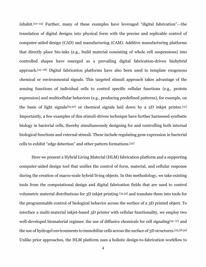
inhabit.[22–24] Further, many of these examples have leveraged “digital fabrication”—the
translation of digital designs into physical form with the precise and replicable control of
computer-aided design (CAD) and manufacturing (CAM). Additive manufacturing platforms
that directly place bio-inks (e.g., build material consisting of whole cell suspensions) into
controlled shapes have emerged as a prevailing digital fabrication-driven biohybrid
approach.[25–28] Digital fabrication platforms have also been used to template exogenous
chemical or environmental signals. This targeted stimuli approach takes advantage of the
sensing functions of individual cells to control specific cellular functions (e.g., protein
expression) and multicellular behaviors (e.g., producing predefined patterns), for example, on
the basis of light signals[29,30] or chemical signals laid down by a 2D inkjet printer.[31]
Importantly, a few examples of this stimuli-driven technique have further harnessed synthetic
biology in bacterial cells, thereby simultaneously designing for and controlling both internal
biological functions and external stimuli. These include regulating gene expression in bacterial
cells to exhibit “edge detection” and other pattern formations.[32]
Here we present a Hybrid Living Material (HLM) fabrication platform and a supporting
computer-aided design tool that unifies the control of form, material, and cellular response
during the creation of macro-scale hybrid living objects. In this methodology, we take existing
tools from the computational design and digital fabrication fields that are used to control
volumetric material distributions for 3D inkjet printing [33,34] and translate them into tools for
the programmable control of biological behavior across the surface of a 3D printed object. To
interface a multi-material inkjet-based 3D printer with cellular functionality, we employ two
well-developed biomaterial regimes: the use of diffusive chemicals for cell signaling[35–37] and
the use of hydrogel environments to immobilize cells across the surface of 3D structures.[25,38,39]
Unlike prior approaches, the HLM platform uses a holistic design-to-fabrication workflow to
5
digitally model and control the gene-regulated function of engineered bacteria in response to
targeted chemical signals programmed into the 3D object. The resulting outputs can be human-
scale objects with programmable biological surfaces which are customized, replicable, and
made on-demand.
In this paper, we establish a replicable, scalable system for controlling and modeling the
gene-regulated function of engineered bacteria in response to chemical signals on a 3D-
material surface. The Methods section describes the development of the HLM digital
fabrication platform, detailing the chemical templating strategy developed by exploiting the
digital material descriptions of a multi-material printer and the properties of the print
materials and the hydrogel. The Results section addresses the HLM outputs achieved by this
platform. First, we show the degree of spatial regulation over bacterial gene expression that can
be achieved on 3D surfaces using digital material descriptions. We demonstrate the design
freedom available for multifunctional HLMs by producing artifacts that exhibit a diversity of
3D geometries, varying mechanical rigidities (flexible, elastic, soft), and different opacities.
Second, we show that augmented 3D printer resins enable the patterning of multiple chemical
signal channels in a multi-material 3D printing process. This feature enables multiplexed
pattern formation when used in combination with artificial biological circuits (i.e., multi-input
pattern drivers). Finally, we establish a computational model of chemical diffusion dynamics
and biological response across arbitrary 3D surfaces, in relation to 3D material distributions,
which is applicable to the CAD environment in which HLMs are designed. In summary, our
HLM platform provides a new tool for designers, engineers, and scientists to control bacterial
functionalities with material technology to perform custom biological and material functions
across a broad range of applications.

6
2. Methods
2.1 Platform Overview
The platform overview presents the integrated framework for the controlled fabrication
of HLMs, comprising computational design, digital fabrication, and genetic engineering
techniques (Figure 1). The biohybrid face mask featured in this example was digitally
modelled to custom fit a human face and produce a prescribed biological response (i.e. colored
patterning indicating locally tunable gene-regulated protein expression), demonstrating a
potential use as an applicator.[40–44]
In this process, the planning of HLM objects begins in a computer-aided design
environment (Figure 1a-c). A volumetric material description represents a 3D object as
a set of voxels (3D-pixels) arranged in a regular grid. Unlike stereolithographic (STL) file
formats, which assign one material per mesh bounded object[33], voxel-based descriptions are
able to assign a specific material composition to each voxel, independent of neighboring voxels,
to represent both an overall geometry of an object (Figure 1a) and a heterogeneous
distribution of materials throughout its volume (Figure 1b). In our method, digital modelling
tools are extended, first, to designate how chemical signals are distributed in the object, and
second, to estimate the biological response in relation to the geometry and concentration of
signal in the object (Figure 1c).
The digital object file is next prepared for digital fabrication on a photopolymeric 3D
printer (Figure 1d-f). A digital material is defined here as a mixing ratio of resins that the
inkjets are able to combine “on-the-fly” during the printing process.[45] The printer receives

7
the volumetric material description and translates it into droplet deposition instructions, to
build up a heterogeneous and continuously varying material composite (Figure 1d). Novel to
our approach, the use of support material mixtures in combination with build materials enables
the 3D printer to fabricate custom digital materials that possess relevant properties for the
absorption and retention of chemical signal. Further, we tailor chemical signal additives for
resins (Figure 1e) to enable the direct distribution of both chemical signals and chemical-
retentive matrices into the permanent structure of the print (Figure 1f).
To realize a programmed biohybrid function, the 3D-printed object is inoculated with
engineered cells (e.g., E. coli) that are adhered across the object’s surface via a thin, aerosolized
hydrogel coating (Figure 1g). The biological behavior of the HLM is mediated by the diffusion
of chemical signals through the hydrogel layer, and the capacity of the cells to sense chemical
signals predictably and respond in accordance to a genetically engineered rule set (Figure 1h).
Final outputs demonstrate that this methodology may be used to design and fabricate HLMs
with predictable and replicable spatiotemporal functions and biological templating (Figure
1i).
Due to the replicability of a digitally-controlled biological response, experimental data
collected from cell-material interactions aids the development and refinement of a simulation
of chemical signal diffusion and engineered genetic response on HLMs (Figure 1j-k).
Integrating experimental data into the CAD-environment thus yields an informed design tool
for programing interactions between 3D manufactured materials and engineered bacteria. This
modelling tool feeds back into the initial CAD step, thus creating a virtuous cycle for refining
predictions of spatially-templated biological behavior and function for subsequent HLMs. The
following subsections of the Methods detail the materials studies and processes leading to the
establishment of the HLM fabrication platform.

8
2.2 Digital-to-Physical Object Description
The Objet Connex500 (Stratasys, Rehovot, Israel) multi-material inkjet-based 3D
printer was the main digital fabrication tool for the HLM framework due to its high resolution,
unique control of multiple material jetting, and inherent ability to create complex self-
supporting structures.[46] Photopolymeric inkjet-based 3D printers print a wide range of
materials properties by using an large array of ejection nozzles to deliver droplets of
photopolymer resins to targeted positions within a macroscale build space (40 x 50 x 20 cm),
and further blend the loaded materials on-the-fly to create an expansive range of digital
material combinations.
To tailor the printer’s capabilities for chemical signal printing, we operated the printer
using a bitmap-based printing[47] or voxel printing[48] approach. Using a recently developed
Data-Driven Material Modeling (DDMM) approach,[33,34] a digital file of a 3D object was
converted into a set of Z-slices with a slice-thickness set by the native height resolution of the
printer (32 µm). For each resin type, the XY-dimension of each Z-slice was represented in as a
bitmap file, in which each pixel represents an individually addressable binary command for
resin droplet deposition. Unique to HLM fabrication, this approach to material assignment was
used to instruct the printer to make new resin combinations for the creation of absorptive
materials that immobilize aqueous chemical signals (see Section 2.3). The capacity to deposit
consistent droplet volumes (12 pL) of material at a high level of spatial accuracy was
additionally leveraged to precisely distribute chemical signals throughout the build volume of
3D structures (see Section 2.7), thus allowing for the production of complex 3D geometries

9
with excellent digital controllability over position of absorptive material properties and
concentrations of chemical signals.
2.3 Digital Material Discovery for Signal-Releasing and
Biocompatible Substrates
The Objet Polyjet system provides a collection of UV-curable acrylate-based polymer
resins that range in composition and cured material behavior, and have been characterized
previously.[47,49–51] In this study, three print resins: two traditional “build materials”, rigid
VeroClear (RGD810)[52] and flexible Tango (FLX930),[53] and one “support material”
(SUP705)[54]—conventionally a sacrificial material used to print overhangs—were used to
create a panel of digital material combinations for a series of experiments to evaluate their
potential use as bioactive templating materials and viable substrates for living cells.
Wettability and hygroscopic behavior, which are both associated with a polymer’s
internal crosslinking density,[55,56] were used to identify digital material compositions capable
of encapsulating and releasing aqueous chemical signal solutions. Wetting behavior was
characterized as a contact angle measured with the Sessile Drop method,[57] and hygroscopic
swelling was measured by change in weight and volume of each cured polymer over a 24-hour
soak in water (Figure S1). Polymer mixes containing SUP705 exhibited up to a tenfold
decrease in wetting angle and a 2-fold greater swelling equilibrium by weight than their build
material counterparts. They are thus well suited for the absorption and release of liquids, and
represent candidate digital materials for embedding chemical signal solutions into 3D-printed
objects.

10
To test biocompatibility, printed polymer samples were introduced to early log-phase E.
coli in liquid culture and incubated for 48 hours. A LIVE/DEAD assay performed on the cells
and quantified through fluorescence activated cell sorting (FACS, Figure S2)[58] verified E.
coli viability (<50% of population is dead/injured) for all material compositions, but showed
better compatibility (<15% of population is dead/injured) for SUP705-build material
intermediates than pure SUP705. These findings suggest that polymer intermediates created
by unconventionally blending SUP705 into build materials provided suitable substrates for cell
culture, while creating an absorptive matrix to store and release chemical signals for the
purpose of inducing gene expression. Namely, by modulating the concentration of SUP705 in
a digital material, the printer platform achieved control of the amount of chemical signal stored
per location.
2.4 Chemical Signal Preparation
Chemical signals were introduced to 3D-printed parts in one of two ways. For initial
experiments, a method was established that introduced chemical signals after an object was 3D
printed, since SUP705 containing materials absorb chemical solutions. Objects were 3D-
printed and then soaked in a H2O/DMSO (50/50 v/v) solution containing the specific chemical
signal of interest for 12 hours. Experiments using Isopropyl β-D-1-thiogalactopyranoside
(IPTG, 50 mM) and the colorless reagent 5-bromo-chloro-3-indoyl-β-D-galactopyranoside (X-
gal, 24mM) employed the use of E. coli cells transformed with pUC19 plasmids. When the 3D-
printed object was removed from the bath, only areas containing SUP705 retained the chemical
signals. Chemical signals were later incorporated directly into the objects during the printing
process using custom resins (see Section 2.7), eliminating the soaking step and allowing for

11
multiple chemicals to be placed simultaneously. In this case, the patterning of SUP705-
intermediate materials at the surface of the object was still key to the release profile of chemical
signals from the cured structure.
2.5 Cell Cloning
The E. coli strains used in this study proliferate rapidly, are relatively hardy, and are
highly tractable for genetic engineering applications. These cells represent the agents
performing local logic functions in response to the local chemical signal environments
programmed into the 3D-printed objects. Figure S3 and S4 map the gene constructs
engineered to produce the signal-gated visual outputs (colorimetric, fluorescent) used to
monitor the spatial and temporal protein expression levels on HLMs. For instance, E. coli (K12-
derivative, NEB Turbo) with the pUC19 plasmid performed IPTG-gated β-galactosidase (β-gal)
expression to catalyze insoluble color production (e.g., blue, magenta) on the surface of HLM
objects. Experiments requiring a direct reporter of protein expression level (as opposed to an
enzyme-substrate assay) used strains with fluorescent protein outputs. A library of
transcriptional regulator constructs for one- and two-input logic functions—including
IPTG.AHL��������������/GFP and IPTG.AHL/GFP, equated to NAND and AND functions, respectively—was
utilized for experiments in multi-signal pattern generation and computational model
development for the HLM platform. Details of these constructs are described in
Supplemental Methods.
12
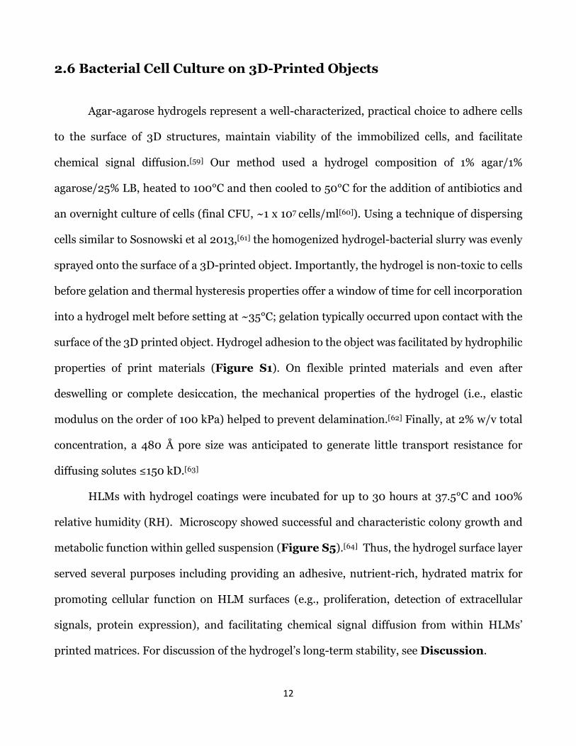
2.6 Bacterial Cell Culture on 3D-Printed Objects
Agar-agarose hydrogels represent a well-characterized, practical choice to adhere cells
to the surface of 3D structures, maintain viability of the immobilized cells, and facilitate
chemical signal diffusion.[59] Our method used a hydrogel composition of 1% agar/1%
agarose/25% LB, heated to 100°C and then cooled to 50°C for the addition of antibiotics and
an overnight culture of cells (final CFU, ~1 x 10⁷ cells/ml[60]). Using a technique of dispersing
cells similar to Sosnowski et al 2013,[61] the homogenized hydrogel-bacterial slurry was evenly
sprayed onto the surface of a 3D-printed object. Importantly, the hydrogel is non-toxic to cells
before gelation and thermal hysteresis properties offer a window of time for cell incorporation
into a hydrogel melt before setting at ~35°C; gelation typically occurred upon contact with the
surface of the 3D printed object. Hydrogel adhesion to the object was facilitated by hydrophilic
properties of print materials (Figure S1). On flexible printed materials and even after
deswelling or complete desiccation, the mechanical properties of the hydrogel (i.e., elastic
modulus on the order of 100 kPa) helped to prevent delamination.[62] Finally, at 2% w/v total
concentration, a 480 Å pore size was anticipated to generate little transport resistance for
diffusing solutes ≤150 kD.[63]
HLMs with hydrogel coatings were incubated for up to 30 hours at 37.5°C and 100%
relative humidity (RH). Microscopy showed successful and characteristic colony growth and
metabolic function within gelled suspension (Figure S5).[64] Thus, the hydrogel surface layer
served several purposes including providing an adhesive, nutrient-rich, hydrated matrix for
promoting cellular function on HLM surfaces (e.g., proliferation, detection of extracellular
signals, protein expression), and facilitating chemical signal diffusion from within HLMs’
printed matrices. For discussion of the hydrogel’s long-term stability, see Discussion.

13
2.7 Modifications for Chemical Signaling Resins
As a final physical adaptation for facilitating the HLM platform, custom resins were
developed to enable multiple chemical signals to be independently controlled by print heads
and directly embedded into the 3D-printed objects. To accomplish this, a panel of three
chemicals—IPTG, N-acyl homoserine lactone (AHL), and Rhamnose (RHA)—and cognate signal-
gated GFP-reporting cell constructs were confirmed to have acceptable signal orthogonality on
HLMs (Figure S6), and then were used to assess the chemicals for resin candidacy by
investigating their compatibility with print head function and the conservation of their
biological function after printing.
Each chemical additive was fully dissolved in H2O/DMSO (50/50 v/v) before mixing
with the SUP705 resin, to ensure that no solid particulates damaged the print head. However,
the addition of solvents to resin is known to alter a resin’s viscoelastic shear properties, and
thus droplet ejection.[65] Hence, a rotational rheometer (Discovery HR-3) was employed to
define the upper limit of solvent addition. Using a concentric cylinder geometry, we measured
the relative shearing behavior of candidate resin mixtures (Figure S6c). Solvent mixes of 1%
v/v or less exhibited no significant changes (p<.05) in shear properties in comparison to the
original SUP705 resin, and thus defined the tolerance for the addition of solvent in custom
resins.
Next, we screened for chemical signal degradation, post-incorporation into resin and
exposure to a UV-light dose representative of the printing process. The efficacy of candidate
chemical signals embedded in cured resin was evaluated as the level of fluorescent induction
from cognate bacterial strains. Figure S6d shows that UV-cured IPTG (2.0 M) and AHL (20
µM) resins were still able to generate a robust induction in respective E. coli strains (IPTG/GFP,

14
AHL/GFP). However, RHA (2.0 M) failed to elicit a strong biological response from an RHA-
inducible strain (RHA/GFP) within the 1% v/v chemical addition limit and was thus eliminated
from subsequent investigations. Collectively, this series of validation tests created a
methodology to rapidly generate new custom chemical signaling resins that are safe and
effective for Objet Polyjet printing. The resin mixtures comprising of IPTG (2.0 M, 0.1-0.05%
v/v) and AHL (50 mM, 0.04% v/v) were used for subsequent direct printing experiments.
Collectively, the Methods prepares the digitally controllable printing platform to
interface with synthetic gene circuits for the goal of a replicable biohybrid fabrication process
within user-designed control. In the Results section, we explore what can be produced via this
approach.
3. Results
The work of developing the HLM platform addressed three major objectives. Primarily,
the results demonstrated the feasibility of using digital material descriptions to guide bacterial
gene expression across the surface of a 3D-printed object. Prototypes demonstrated that
printed material distributions allowed for tunable release of chemical signals, in addition to the
ability to generate complex, free-standing, 3D, multi-material, multifunctional hybrid living
objects (Section 3.1). Second, the results showed that augmented resins enabled the 3D
printer to pattern multiple chemical signal channels. Using those materials to regulate multi-
input synthetic gene constructs, we demonstrated the robustness of the platform to generate
spatial patterning based on multiplexed gene expression (Section 3.2). Finally, the results
established a computational model to simulate chemical diffusion dynamics and biological

15
response given a volumetric material distribution, thereby providing a predictive design tool
for HLM fabrication (Section 3.3).
3.1 Spatiotemporal Control of Gene Expression across 3D-Printed
Structures
Initial experiments with the HLM platform aimed to characterize the controllability of
spatiotemporal gene induction produced by printed objects. We hypothesized that the platform
would enable a replicable response in engineered bacteria across 3D surfaces, including
producing an expected result from variables, such as diffusion, that are not within the printer’s
direct control. To define the relationship between printer-mediated chemical signal placement
and cellular response, we printed a set of multi-material test templates: Vero disks (50mm-
diameter, 3mm height) with 10mm center regions of incremental digital material compositions
(SUP705/RGD810 ratio, 0–1.0, with 0.1 steps), termed “active regions” (Figure 2b).
Following the HLM fabrication process, disks were soaked in an IPTG/X-Gal bath and
incubated with a pUC19 E. coli hydrogel layer. Resulting LacZ gene expression patterns (blue)
were observed on the objects’ surfaces via locally induced β-gal activity (Figure 2a).
Results presented in Figure 2c show bacterial response to templates of varied digital
materials and plot the relative colorimetric intensity per radial distance from the active region.
Importantly, line thickness represents one standard deviation of the average of four
experimental replicates, and hence, the consistency in outputs. Gene expression on the HLM
objects correlated positively to the ratio of SUP705 within the active region, indicating that
bacteria were responding proportionally to the amount of chemical-releasing material in the
printed structure. Active regions composed of no SUP705 had negligible expression.

16
Expression patterns were radially symmetrical from the active region, denoting that the
hydrogel facilitated even chemical signal and substrate dispersion from the 3D-printed
structure. However, at higher SUP705-ratios (>0.7), a ring of decreased expression appeared
around the active region (see Discussion). Importantly, the spatial response of HLM objects,
consisting of living cells and multi-material 3D-printed components, was repeatable and
tunable to the SUP705 material ratios defined by the print description.
In Figure 2d, CPRG was used in place of X-gal for another set of HLM test disks to
observe the temporal aspects of HLM response. Time-lapse image capture of the incubation of
HLMs (n=4) showed a period of CPRG diffusion from the active region (yellow, 0-18 hr). At
hour 18, the β-gal-catalyzed colorimetric conversion of CPRG to CPR (magenta) first became
visible and continued to intensify and propagate (18-35 hr). From these observations, we
presume that the rate of signal release does change over time, and eventually attenuates due to
the finite amount of chemical stored. Yet signal release time is relatively well matched to
bacterial growth and expression for a robust and consistent final output. These results
demonstrated that the spatial and temporal behavior of the HLM templates relied on both the
diffusion profile of chemical signal and the response profile of engineered cells, which was used
to inform a representative framework for the model for HLM behavior (see Section 3.3).
We next produced HLMs to illustrate digitally-driven templating based on reproducible
behavior. Figure S7 shows the bacteria response of HLM generated from a continuous linear
material gradient. The recorded pattern was profiled across ten cross-sections and then used
to guide the redistribution of digital material in the design of a second HLM template. On the
subsequent HLM, an expected oscillating pattern was achieved. Thus, enabled by a repeatable
HLM response to digital material, the original template served as a standard on which to base
subsequent designs. In Figure S8 a resolution template was prepared. While output resolution

17
is reliant on factors outside of printer control (e.g., diffusion, cell sensitivity), a template can
qualify the effect that print parameters (e.g., geometry, digital material composition) have on
the lower-limit of IPTG/X-gal signal-response. On this template, the lower limit of visible
response (≥10% relative saturation) was described as a SUP705 surface feature of at least a 24
voxel cross-sectional area (0.08 mm2), or at least a 0.45 SUP705-ratio for any feature above
120 voxel cross-sectional area (0.4 mm2). Importantly, each ruleset derived from an observed
interaction between the 3D-printed material and a bacteria-signal coupling are accessible via
digital parameters afforded to a designer using the HLM platform.
Finally, the HLM framework was used to produce combinatorial physical property
distributions within biohybrid objects, including stiffness and opacity. High-resolution photo
polymeric inkjet printing has been shown to generate objects with heterogeneous physical
properties widely used in industrial production. Thus, our method enables functional products
with augmented biological functionality. Figure 3 shows biologically and materially patterned
HLM objects: optically-patterned wearable masks and soft bandage-like patches with
programmed bacterial surface activity.
In Figure 3a-b, the masks produced on the HLM platform feature distributions of rigid
clear (RGD810), rigid opaque (RGD835), and chemical signaling materials, for the
simultaneous digital control of optical properties (i.e., transparency) and bioactivity (i.e., local
colorimetric protein expression). With the mask, we also contrasted the total build envelope
(49 x 39 x 19 cm) with the smallest feature size (12 pL, ~1.2 mm3) to demonstrate the
sophistication of internal material distributions and print control across twelve orders of
magnitude (Figure 3c). In the production of soft devices, a conformable bandage-like
prototype was fabricated using distributions of rigid (RGD810), rubber-like (FLX930), and
chemical signaling materials to exhibit site-specific flexibility and bioactivity (Figure 3d). The

18
resulting construct produced a programmed bacterial response across a variable flexible-to-
rigid substrate, designed to twist along one axis in a manner that would be applicable to
ergonomic support or local conformation to the body (e.g., for biomedical splints, sockets, or
bedrests).[66,67]
Overall, the biohybrid artifacts produced illustrate the unique potential of the HLM
framework to deploy tunable levels of bacterial expression, in a site-specific manner, across
complex 3D objects. The production of self-supporting HLM objects at the wearable scale,
featuring combinatorial physical and chemical signaling properties, served to prototype a
diversity of unprecedented hybrid living devices that exhibit bio-templating as well as
mechanical or ergonomic functionality.
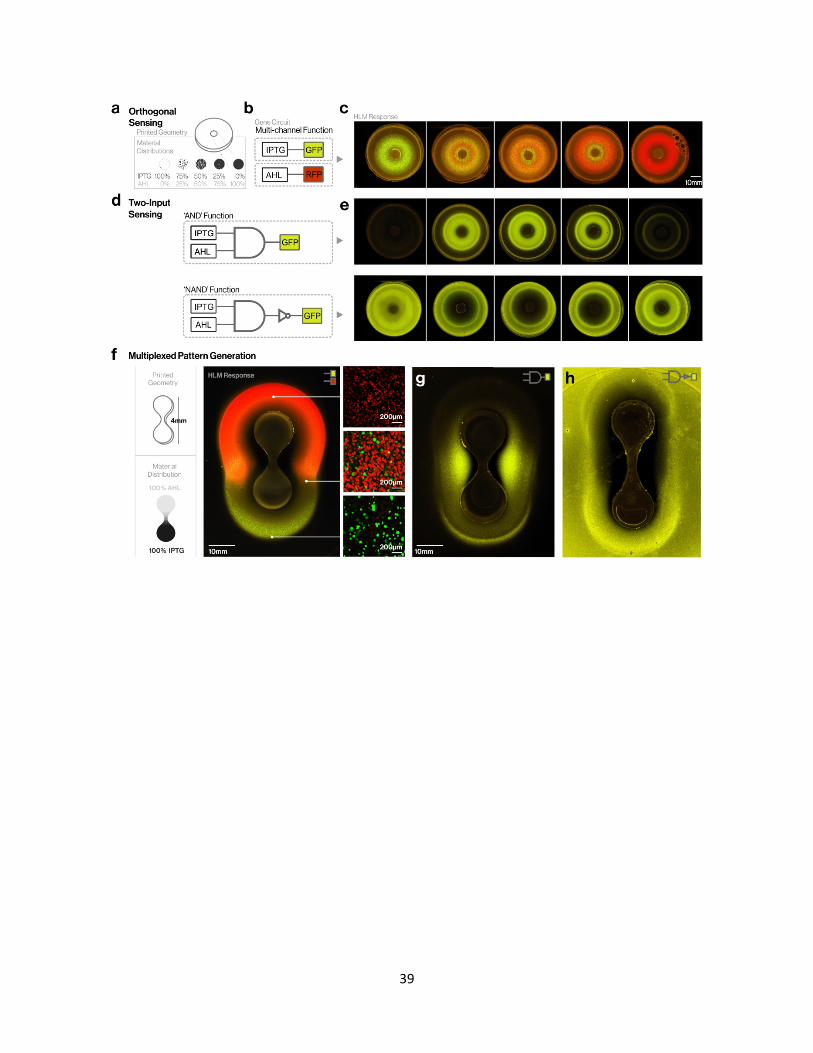
3.2 Multi-Signal Channel Control of Synthetic Genes for Pattern
Regulation
We further modified the HLM platform to print multi-chemical environments (e.g.,
multiplexing), for the objective of controlling cell systems with multi-input gene regulatory
constructs across 3D objects. In synthetic biology, gene-based logic gates are commonly
constructed to introduce computation-like, “rational” or synthetically tractable regulatory
behavior into cells.[35,36,68] Thus, we hypothesized that using the printer’s polyjetting
technology towards the control of multiple chemical signals may allow engineered cells to
produce “decision based” outputs not available to chemical diffusion patterns alone.
IPTG and AHL chemical signal additives were incorporated into the UV-curable
photopolymer resins, in a process described in Section 2.7. The chemical signaling resins were
loaded into printer cartridges to enable their direct deposition via inkjets at the native
19
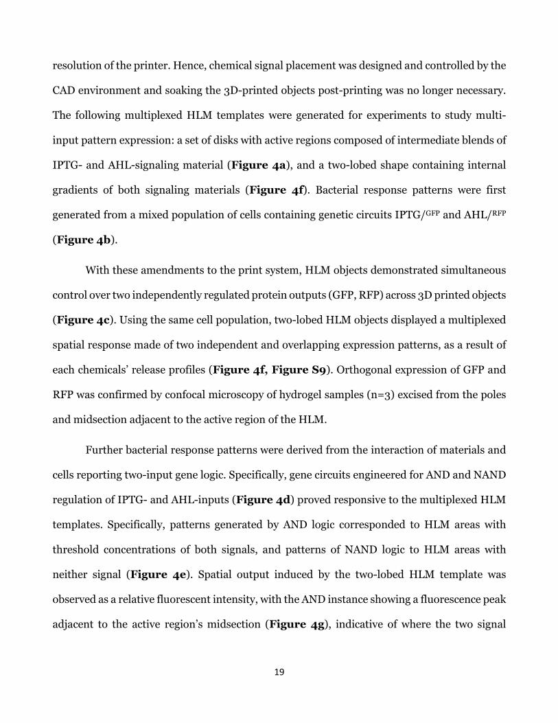
resolution of the printer. Hence, chemical signal placement was designed and controlled by the
CAD environment and soaking the 3D-printed objects post-printing was no longer necessary.
The following multiplexed HLM templates were generated for experiments to study multi-
input pattern expression: a set of disks with active regions composed of intermediate blends of
IPTG- and AHL-signaling material (Figure 4a), and a two-lobed shape containing internal
gradients of both signaling materials (Figure 4f). Bacterial response patterns were first
generated from a mixed population of cells containing genetic circuits IPTG/GFP and AHL/RFP
(Figure 4b).
With these amendments to the print system, HLM objects demonstrated simultaneous
control over two independently regulated protein outputs (GFP, RFP) across 3D printed objects
(Figure 4c). Using the same cell population, two-lobed HLM objects displayed a multiplexed
spatial response made of two independent and overlapping expression patterns, as a result of
each chemicals’ release profiles (Figure 4f, Figure S9). Orthogonal expression of GFP and
RFP was confirmed by confocal microscopy of hydrogel samples (n=3) excised from the poles
and midsection adjacent to the active region of the HLM.
Further bacterial response patterns were derived from the interaction of materials and
cells reporting two-input gene logic. Specifically, gene circuits engineered for AND and NAND
regulation of IPTG- and AHL-inputs (Figure 4d) proved responsive to the multiplexed HLM
templates. Specifically, patterns generated by AND logic corresponded to HLM areas with
threshold concentrations of both signals, and patterns of NAND logic to HLM areas with
neither signal (Figure 4e). Spatial output induced by the two-lobed HLM template was
observed as a relative fluorescent intensity, with the AND instance showing a fluorescence peak
adjacent to the active region’s midsection (Figure 4g), indicative of where the two signal

20
release profiles overlap. The NAND instance demonstrated that the inversion of this pattern
was also possible (Figure 4h).
Thus, the HLM platform enables multiplexed chemical signaling in 3D printed objects.
The resulting HLMs demonstrated that the digital printing platform can be paired with gene
constructs that act as multi-input pattern-drivers to generate orthogonal, AND, and NAND
computed protein expression patterns on various templates. The allotted control over chemical
distributions and engineered cell circuits allows for the systematic design of output patterns.
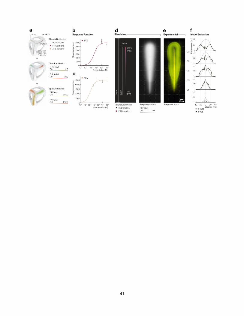
3.3 Computational Model for HLM Behavior
As a final objective, we created a computational model to predict the biological outcomes
of HLM artifacts based on a digital material description in a CAD environment, thereby
providing a virtual design tool analogous to other 3D material modeling software.[69,70] Our
framework for modeling HLM bacterial response was derived from two underlying processes:
the hydrogel-mediated diffusion of chemical signals from a 3D object and the resulting
bacterial response. Further, in contrast to existing models,[37,71] the HLM model needed to
account for the 3D geometric complexity made possible by the HLM print platform. Thus, we
developed a quantitative model, usable in a CAD environment, to account for each of these
factors (see Supplemental Methods).
First, to simulate hydrogel-facilitated chemical signal diffusion (Figure 5a), the model
is given a digital print description. Based upon the volumetric material distribution data, the
model derives an initial chemical concentration value per location. Concentration is calculated
form the chemical signaling resin composition, or, for soaked templates, the swelling
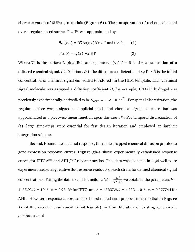
21
characterization of SUP705-materials (Figure S1). The transportation of a chemical signal
over a regular closed surface Γ ⊂ ℝ3 was approximated by
𝜕𝜕𝑡𝑡𝑐𝑐(𝑥𝑥, 𝑡𝑡) = D∇Γ2𝑐𝑐(𝑥𝑥, 𝑡𝑡) ∀𝑥𝑥 ∈ Γ and t > 0, (1)
𝑐𝑐(𝑥𝑥, 0) = 𝑐𝑐0(𝑥𝑥) ∀𝑥𝑥 ∈ Γ (2)
Where ∇Γ2 is the surface Laplace-Beltrami operator, 𝑐𝑐(⋅, 𝑡𝑡): Γ → ℝ is the concentration of a
diffused chemical signal, 𝑡𝑡 ≥ 0 is time, 𝐷𝐷 is the diffusion coefficient, and 𝑐𝑐0: Γ → ℝ is the initial
concentration of chemical signal embedded (or stored) in the HLM template. Each chemical
signal molecule was assigned a diffusion coefficient D; for example, IPTG in hydrogel was
previously experimentally-derived[72] to be 𝐷𝐷𝐼𝐼𝐼𝐼𝐼𝐼𝐼𝐼 = 3 × 10⁻¹⁰𝑚𝑚2𝑠𝑠 . For spatial discretization, the
regular surface was assigned a simplicial mesh and chemical signal concentration was
approximated as a piecewise linear function upon this mesh[73]. For temporal discretization of
(1), large time-steps were essential for fast design iteration and employed an implicit
integration scheme.
Second, to simulate bacterial response, the model mapped chemical diffusion profiles to
gene expression response curves. Figure 5b-c shows experimentally established response
curves for IPTG/GFP and AHL/GFP reporter strains. This data was collected in a 96-well plate
experiment measuring relative fluorescence readouts of each strain for defined chemical signal
concentrations. Fitting the data to a hill-function ℎ(𝑐𝑐) = 𝑏𝑏𝑐𝑐𝑛𝑛
𝑘𝑘𝑛𝑛+𝑐𝑐𝑛𝑛 we obtained the parameters 𝑏𝑏 =
4485.93,𝑘𝑘 = 10−3, 𝑛𝑛 = 0.95489 for IPTG, and 𝑏𝑏 = 45837.9,𝑘𝑘 = 4.833 ⋅ 10−6, 𝑛𝑛 = 0.877744 for
AHL. However, response curves can also be estimated via a process similar to that in Figure
2c (if fluorescent measurement is not feasible), or from literature or existing gene circuit
databases.[74,75]
22
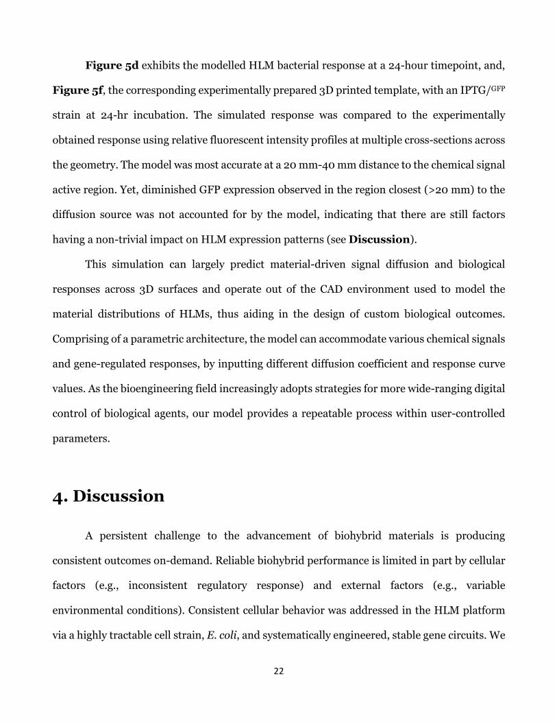
Figure 5d exhibits the modelled HLM bacterial response at a 24-hour timepoint, and,
Figure 5f, the corresponding experimentally prepared 3D printed template, with an IPTG/GFP
strain at 24-hr incubation. The simulated response was compared to the experimentally
obtained response using relative fluorescent intensity profiles at multiple cross-sections across
the geometry. The model was most accurate at a 20 mm-40 mm distance to the chemical signal
active region. Yet, diminished GFP expression observed in the region closest (>20 mm) to the
diffusion source was not accounted for by the model, indicating that there are still factors
having a non-trivial impact on HLM expression patterns (see Discussion).
This simulation can largely predict material-driven signal diffusion and biological
responses across 3D surfaces and operate out of the CAD environment used to model the
material distributions of HLMs, thus aiding in the design of custom biological outcomes.
Comprising of a parametric architecture, the model can accommodate various chemical signals
and gene-regulated responses, by inputting different diffusion coefficient and response curve
values. As the bioengineering field increasingly adopts strategies for more wide-ranging digital
control of biological agents, our model provides a repeatable process within user-controlled
parameters.
4. Discussion
A persistent challenge to the advancement of biohybrid materials is producing
consistent outcomes on-demand. Reliable biohybrid performance is limited in part by cellular
factors (e.g., inconsistent regulatory response) and external factors (e.g., variable
environmental conditions). Consistent cellular behavior was addressed in the HLM platform
via a highly tractable cell strain, E. coli, and systematically engineered, stable gene circuits. We
23
discuss the future utility of this or similar model chasses for efficient synthetic gene design.
Further, as an invitation for future consideration, we discuss the efficacy of the materials used
in this platform (i.e., hydrogels, photopolymers) in preventing external factors from disrupting
HLM performance.
The HLM platform employed E. coli to capitalize on the broad basis of knowledge and
most recent advancements in the synthetic functionalization of bacterial cells. We
demonstrated that proteins can be directly genetically encoded for expression; yet, in the
broader context of bioproduction, the HLM platform can be extended to template diverse
metabolic outputs, such as functional chemical syntheses[1] across 3D structures (e.g., point-of-
use drug production) or material surface treatments[76] (e.g., mineralization or enzymatic
digestion). In the context of gene regulation, synthetic gene circuits designed to drive
increasingly sophisticated forms of biological computation have the potential to exhibit new
phenomena when distributed across chemical-templated surfaces. For instance, HLMs could
enable spatial forms of memory across 3D objects,[77] coordinated dynamic or oscillatory (e.g.,
quorum) surface expression patterns,[78–80] or incorporation of external cues (e.g.,
physiological signals from a device wearer) into localized decisions.[81,82] Moreover, the union
of computational modelling and digital print platform achieved here creates a tractable
experimental and simulation space to aid the study of synthetic morphogenesis.[83]
While E. coli was a practical starting point for the HLM platform, many other cell lines
are increasingly easy to culture or genetically engineer, and thus could provide interesting next
steps (e.g., desiccation-resistant Bacillus subtilis, tumor cells, insect cells, etc.).[84–86]
Advantageously, the modular nature of the HLM framework permits the components—both
hydrogel and cell strain—to be interchanged in a straightforward fashion without impacting the
24
print regime. Thus, it is feasible for other compositions of hydrogels or cells to be selected on
the basis of the intended application.
Regarding external factors impacting biohybrid performance, the integrity of the
hydrogel was essential for reliable HLM outcomes. HLM activity, with respect to signal
diffusion and cell growth, occurred over an 18-35 hour period at 100% RH (Figure 2d). If
removed from a high-humidity environment, the hydrogel experienced water loss, which
interfered with signal diffusivity and cell function. However, hydrogels did adhere to surfaces
throughout desiccation, and all stable compounds previously produced (e.g., pigments) would
remain laminated to the 3D object’s surface once the hydrogel dried. Thus, one can choose from
applications intended to have active cell function (an “active-life”, ~24 hours)[44,87] or processes
that allow signals and cells to desiccate once the intended surface properties are obtained.
Future work may leverage recent advancements in natural and synthetic hydrogels to tune the
timeframe of HLM activity, and create more robust, desiccation-resilient constructs.[71,88] Of
additional note, ongoing observations of the IPTG and AHL signaling materials developed for
the HLM platform show promise of a considerable ambient shelf life (>6 months), implying
that HLMs can be digitally fabricated and stored prior to being “activated” by the addition of
hydrogel.
Finally, in some instances, our platform experienced unexpected outputs due to
photopolymeric compositions. Specifically, surface regions adjacent to some digital materials
(SUP705/RGD810 ratios > 0.7, in Figures 2c, 4c, 5e) exhibited diminished expression—a
possible artifact of cell interaction with a material composition that was later demonstrated to
lower the pH when exposed to aqueous environments. This was not represented by our
computational model, yet could be incorporated into future models or design considerations.

25
5. Conclusion
In conclusion, the HLM fabrication platform provides a new technology centered on
bridging computational design, material fabrication, and synthetic biology to control living
gene expression patterns across the surface of geometrically complex, mechanically robust,
multi-material 3D structures. Specifically, our process modifies the digital commands and
materials of a 3D printing platform to fabricate structured photopolymers that can retain
precise spatial distributions of chemical regulatory signals within their cured architectures.
Subsequently, our method immobilizes engineered E. coli on the surface of these objects, to
facilitate designed interactions between engineered gene constructs and chemical signaling
profiles. Thus, the HLMs presented herein are designed to produce programmed gene-
regulated responses across the surface of 3D objects in reproducible and predictive ways.
In contribution to the field of biohybrid materials, our platform demonstrates self-
supporting HLM constructions of up to half a meter in length, with freeform shape generation,
and the incorporation of site-specific mechanical and optical properties. Because engineered
bacterial response was defined in terms of a digital print description, the platform achieved a
programmable methodology for producing controlled spatial interactions between 3D-printed
digital materials and genetic regulatory circuits. Thus, our platform generated objects with
various protein expression patterns from single-input gene circuits and—with the development
of multiplexed chemical signal printing—multi-input logic constructs (e.g., AND and NAND
regulatory operations). Furthermore, we linked the design-to-output workflow for HLMs by
integrating a computational model of signal diffusion and bacterial response across 3D surfaces
into a CAD environment. The HLM fabrication platform’s capacity to simultaneously direct
complex 3D structures, bacterial functions, and material property distributions enables

26
unprecedented control of functional outputs. This work advances biohybrid materials towards
applications ranging from wearable therapeutics or monitoring devices to customizable
consumer products.[89–91]
27
ACKNOWLEDGMENTS
This work was supported by the Robert Wood Johnson Foundation (grant no. 74479),
GETTYLAB, DARPA Engineered Living Materials (ELM) agreement W911NF-17-2-0077, and
National Security Science and Engineering Faculty Fellowship (NSSEFF) N00014-16-1-
2509. The authors thank GETTYLAB and the Robert Wood Johnson Foundation for their
generous support of our scientific research into living devices, N. Kaempfer (creative director
of art, fashion, and design) and B. Belocon at Stratasys Ltd. for enabling the production of some
of the models shown herein, the W.M. Keck Microscopy Facility at the MIT Whitehead
Institute, and N. Jakimo for helpful discussions regarding bacterial color production.
COMPETING INTERSTS
All authors declare that they have no competing interests. It is noted that C.B., R.S.S., S.S., D.K.,
and N.O. are authors on a patent application filed by the Massachusetts Institute of Technology
that describes methods similar to those described in this work (US application no. 16295502).
Received: ((will be filled in by the editorial staff))
Revised: ((will be filled in by the editorial staff))
Published online: ((will be filled in by the editorial staff))

28
REFERENCES
[1] M. J. Smanski, H. Zhou, J. Claesen, B. Shen, M. A. Fischbach, C. A. Voigt, Nat. Rev. Microbiol. 2016, 14, 135.
[2] D. Jullesson, F. David, B. Pfleger, J. Nielsen, Biotechnol. Adv. 2015, 33, 1395.
[3] V. Libis, B. Delépine, J. L. Faulon, Sensing new chemicals with bacterial transcription factors. Curr. Opin. Microbiol. 2016, 33, 105–112.
[4] Z. Ma, F. E. Jacobsen, D. P. Giedroc, Chem. Rev. 2009, 109, 4644.
[5] P. Q. Nguyen, N. M. D. Courchesne, A. Duraj-Thatte, P. Praveschotinunt, N. S. Joshi, Engineered Living Materials: Prospects and Challenges for Using Biological Systems to Direct the Assembly of Smart Materials. Adv. Mater. 2018, 30.
[6] M. Haneef, L. Ceseracciu, C. Canale, I. S. Bayer, J. A. Heredia-Guerrero, A. Athanassiou, Sci. Rep. 2017, 7.
[7] L. Valentini, S. Bittolo Bon, S. Signetti, N. M. Pugno, ACS Appl. Mater. Interfaces 2016, 8, 7607.
[8] F. Moser, M. Trautz, A.-L. Beger, M. Löwer, G. Jacobs, F. Hillringhaus, A. Wormit, B. Usadel, J. Reimer, Proc. IASS Annu. Symp. 2017.
[9] L. C. Gerber, F. M. Koehler, R. N. Grass, W. J. Stark, Proc. Natl. Acad. Sci. U. S. A. 2012, 109, 90.
[10] Y. Liu, M. H. Rafailovich, R. Malal, D. Cohn, D. Chidambaram, Proc. Natl. Acad. Sci. U. S. A. 2009, 106, 14201.
[11] M. Akbari, A. Tamayol, V. Laforte, N. Annabi, A. H. Najafabadi, A. Khademhosseini, D. Juncker, Adv. Funct. Mater. 2014, 24, 4060.
[12] S. Xie, S. Tai, H. Song, X. Luo, H. Zhang, X. Li, J. Mater. Chem. B 2016, 4, 6820.
[13] R. Raman, C. Cvetkovic, S. G. M. Uzel, R. J. Platt, P. Sengupta, R. D. Kamm, R. Bashir, Proc. Natl. Acad. Sci. U. S. A. 2016, 113, 3497.
[14] J. Bastos-Arrieta, A. Revilla-Guarinos, W. E. Uspal, J. Simmchen, Bacterial biohybrid microswimmers. Front. Robot. AI 2018, 5.
[15] L. Ricotti, B. Trimmer, A. W. Feinberg, R. Raman, K. K. Parker, R. Bashir, M. Sitti, S. Martel, P. Dario, A. Menciassi, Biohybrid actuators for robotics: A review of devices actuated by living cells. Sci. Robot. 2017, 2.
[16] J. C. Nawroth, H. Lee, A. W. Feinberg, C. M. Ripplinger, M. L. McCain, A. Grosberg, J. O. Dabiri, K. K. Parker, Nat. Biotechnol. 2012, 30.
[17] S. Moon, I. L. Fritz, Z. S. Singer, T. Danino, 3D Print. Addit. Manuf. 2016, 3, 195.
[18] A. L. Rutz, K. E. Hyland, A. E. Jakus, W. R. Burghardt, R. N. Shah, Adv. Mater. 2015, 27, 1607.
[19] R. G. Wylie, S. Ahsan, Y. Aizawa, K. L. Maxwell, C. M. Morshead, M. S. Shoichet, Nat. Mater. 2011, 10, 799.
[20] T. Lu, Y. Li, T. Chen, Techniques for fabrication and construction of three-dimensional scaffolds for tissue engineering. Int. J. Nanomedicine 2013, 8, 337–350.

29
[21] S. A. L. De Koster, R. M. Mors, H. W. Nugteren, H. M. Jonkers, G. M. H. Meesters, J. R. Van Ommen, In Procedia Engineering; Elsevier Ltd, 2015; Vol. 102, pp. 475–484.
[22] A. Y. Chen, Z. Deng, A. N. Billings, U. O. S. Seker, M. Y. Lu, R. J. Citorik, B. Zakeri, T. K. Lu, Nat. Mater. 2014, 13.
[23] M. Florea, H. Hagemann, G. Santosa, J. Abbott, C. N. Micklem, X. Spencer-Milnes, L. de Arroyo Garcia, D. Paschou, C. Lazenbatt, D. Kong, H. Chughtai, K. Jensen, P. S. Freemont, R. Kitney, B. Reeve, T. Ellis, Proc. Natl. Acad. Sci. U. S. A. 2016, 113, E3431.
[24] M. Mukherjee, Y. Hu, C. H. Tan, S. A. Rice, B. Cao, Sci. Adv. 2018, 4.
[25] M. Schaffner, P. A. Rühs, F. Coulter, S. Kilcher, A. R. Studart, Sci. Adv. 2017, 3.
[26] X. Liu, H. Yuk, S. Lin, G. A. Parada, T.-C. Tang, E. Tham, C. de la Fuente-Nunez, T. K. Lu, X. Zhao, Adv. Mater. 2018, 30, 1704821.
[27] B. A. E. Lehner, D. T. Schmieden, A. S. Meyer, ACS Synth. Biol. 2017, acssynbio.6b00395.
[28] S. V Murphy, A. Atala, Nat. Biotechnol. 2014, 32, 773.
[29] F. Moser, E. Tham, L. M. González, T. K. Lu, C. A. Voigt, Adv. Funct. Mater. 2019, 29, 1901788.
[30] A. Levskaya, A. A. Chevalier, J. J. Tabor, Z. B. Simpson, L. A. Lavery, M. Levy, E. A. Davidson, A. Scouras, A. D. Ellington, E. M. Marcotte, C. A. Voigt, Nature 2005, 438, 441.
[31] D. J. Cohen, R. C. Morfino, M. M. Maharbiz, W.-S. Chen, J. Axelrod, PLoS One 2009, 4, e7086.
[32] J. J. Tabor, H. M. Salis, Z. B. Simpson, A. A. Chevalier, A. Levskaya, E. M. Marcotte, C. A. Voigt, A. D. Ellington, Cell 2009, 137, 1272.
[33] C. Bader, D. Kolb, J. C. Weaver, S. Sharma, A. Hosny, J. Costa, N. Oxman, Sci. Adv. 2018, 4, eaas8652.
[34] C. Bader, D. Kolb, J. C. Weaver, N. Oxman, 3D Print. Addit. Manuf. 2016, 3, 71.
[35] A. Tamsir, J. J. Tabor, C. A. Voigt, Nature 2011, 469.
[36] S. Basu, Y. Gerchman, C. H. Collins, F. H. Arnold, R. Weiss, Nature 2005, 434, 1130.
[37] M. Weitz, A. Mückl, K. Kapsner, R. Berg, A. Meyer, F. C. Simmel, J. Am. Chem. Soc. 2014, 136, 72.
[38] R. Suntivich, I. Drachuk, R. Calabrese, D. L. Kaplan, V. V. Tsukruk, Biomacromolecules 2014, 15, 1428.
[39] P. Chen, S. Wang, F. Inci, S. Güven, S. Tasoglu, U. Demirci, 2016; pp. 327–356.
[40] A. Goyanes, U. Det-Amornrat, J. Wang, A. W. Basit, S. Gaisford, J. Control. Release 2016, 234, 41.
[41] Alary, (No Title).
[42] X. G. Cheng, J. J. Yoo, R. G. Hale, Biomask for skin regeneration. Regen. Med. 2014, 9, 245–248.
[43] Y. Wei, Y. Wang, M. Zhang, G. Yan, S. Wu, W. Liu, G. Ji, C. W. P. Li-Tsang, Burns 2018, 44, 453.
[44] L. C. Gerber, F. M. Koehler, R. N. Grass, W. J. Stark, Angew. Chemie - Int. Ed. 2012, 51, 11293.

30
[45] J.-Y. Lee, J. An, C. K. Chua, Appl. Mater. Today 2017, 7, 120.
[46] B. Bhushan, M. Caspers, Microsyst. Technol. 2017, 23, 1117.
[47] C. Bader, W. G. Patrick, D. Kolb, S. G. Hays, S. Keating, S. Sharma, D. Dikovsky, B. Belocon, J. C. Weaver, P. A. Silver, N. Oxman, 3D Print. Addit. Manuf. 2016, 3, 79.
[48] E. L. Doubrovski, E. Y. Tsai, D. Dikovsky, J. M. P. Geraedts, H. Herr, N. Oxman, Comput. Des. 2015, 60, 3.
[49] PolyJet Materials Data Sheet 2014.
[50] K. K. Reichl, D. J. Inman, Top. Modal Anal. Test. 2016, 10, 191.
[51] J. Mańkowski, J. Lipnicki, Int. J. Appl. Mech. Eng. 2017, 22, 601.
[52] Stratasys, VEROCLEAR RGD810 Safety Data Sheet; 2016.
[53] Stratasys, OBJET TANGOPLUS FLX930 Safety Data Sheet; 2014.
[54] Stratasys, OBJET SUPPORT SUP705 Safety Data Sheet; 2014.
[55] K.-J. Kim, S.-B. Lee, N. W. Han, Polym. J. 1993, 25, 1295.
[56] Y. Wu, S. Joseph, N. R. Aluru, J. Phys. Chem. B 2009, 113, 3512.
[57] Y. Yuan, T. R. Lee, Bracco, G. Holst, B., Eds., Surf. Sci. Tech. Springer Ser. Surf. Sci. 2013, 51, 3.
[58] M. Berney, F. Hammes, F. Bosshard, H.-U. Weilenmann, T. Egli, Appl. Environ. Microbiol. 2007, 73, 3283.
[59] E. M. Ahmed, Hydrogel: Preparation, characterization, and applications: A review. J. Adv. Res. 2015, 6, 105–121.
[60] G. Sezonov, D. Joseleau-Petit, R. D’Ari, J. Bacteriol. 2007, 189, 8746.
[61] T. R. Sosnowski, A. Kurowska, B. Butruk, K. Jabłczyńska, Chem. Eng. Trans. 2013, 32, 2257.
[62] V. Normand, D. L. Lootens, E. Amici, K. P. Plucknett, P. Aymard, Biomacromolecules 2000, 1, 730.
[63] R. H. Li, D. H. Altreuter, F. T. Gentile, Biotechnol. Bioeng. 1996, 50, 365.
[64] K. Boons, E. Noriega, R. Van den Broeck, C. C. David, J. Hofkens, J. F. Van Impe, Appl. Environ. Microbiol. 2014, 80, 5330.
[65] B.-J. de Gans, P. C. Duineveld, U. S. Schubert, Adv. Mater. 2004, 16, 203.
[66] D. M. Sengeh, H. Herr, JPO J. Prosthetics Orthot. 2013, 25, 129.
[67] A. M. Paterson, R. Bibb, R. I. Campbell, G. Bingham, Rapid Prototyp. J. 2015, 21, 230.
[68] F. Moser, A. Espah Borujeni, A. N. Ghodasara, E. Cameron, Y. Park, C. A. Voigt, Mol. Syst. Biol. 2018, 14, e8605.
[69] W. Regli, J. Rossignac, V. Shapiro, V. Srinivasan, The new frontiers in computational modeling of material structures. CAD Comput. Aided Des. 2016, 77, 73–85.
[70] M. M. Francois, A. Sun, W. E. King, N. J. Henson, D. Tourret, C. A. Bronkhorst, N. N. Carlson, C. K. Newman, T. Haut, J. Bakosi, J. W. Gibbs, V. Livescu, S. A. Vander Wiel, A. J. Clarke, M.

31
W. Schraad, T. Blacker, H. Lim, T. Rodgers, S. Owen, F. Abdeljawad, J. Madison, A. T. Anderson, J. L. Fattebert, R. M. Ferencz, N. E. Hodge, S. A. Khairallah, O. Walton, Curr. Opin. Solid State Mater. Sci. 2017, 21, 198.
[71] X. Liu, T.-C. Tang, E. Tham, H. Yuk, S. Lin, T. K. Lu, X. Zhao, Proc. Natl. Acad. Sci. U. S. A. 2017, 114, 2200.
[72] S. Lin, H. Yuk, T. Zhang, G. A. Parada, H. Koo, C. Yu, X. Zhao, Adv. Mater. 2016, 28, 4497.
[73] M. Botsch, L. Kobbelt, M. Pauly, P. Alliez, L. Bruno, Polygon Mesh Processing; 2010.
[74] A. J. Meyer, T. H. Segall-Shapiro, C. A. Voigt, bioRxiv [Pre-print] 2018.
[75] A. a K. Nielsen, B. S. Der, J. Shin, P. Vaidyanathan, V. Paralanov, E. a Strychalski, D. Ross, D. Densmore, C. a. Voigt, Science 2016, 352, aac7341.
[76] S. Douglas, T. J. Beveridge, FEMS Microbiol. Ecol. 1998, 26, 79.
[77] N. Roquet, A. P. Soleimany, A. C. Ferris, S. Aaronson, T. K. Lu, Science (80-. ). 2016, 353.
[78] O. Mondragón-Palomino, T. Danino, J. Selimkhanov, L. Tsimring, J. Hasty, Science 2011, 333, 1315.
[79] M. O. Din, T. Danino, A. Prindle, M. Skalak, J. Selimkhanov, K. Allen, E. Julio, E. Atolia, L. S. Tsimring, S. N. Bhatia, J. Hasty, Nat. Publ. Gr. 2016, 536.
[80] F. Moser, A. Espah Borujeni, A. N. Ghodasara, E. Cameron, Y. Park, C. A. Voigt, Mol. Syst. Biol. 2018, 14, e8605.
[81] T. S. Moon, C. Lou, A. Tamsir, B. C. Stanton, C. A. Voigt, Nature 2012, 491, 249.
[82] D. T. Riglar, T. W. Giessen, M. Baym, S. J. Kerns, M. J. Niederhuber, R. T. Bronson, J. W. Kotula, G. K. Gerber, J. C. Way, P. A. Silver, Nat. Biotechnol. 2017, 35, 653.
[83] B. P. Teague, P. Guye, R. Weiss, Cold Spring Harb. Perspect. Biol. 2016, 8.
[84] T. L. Deans, A. Singh, M. Gibson, J. H. Elisseeff, Proc. Natl. Acad. Sci. U. S. A. 2012, 109, 15217.
[85] N. R. Rubio, K. D. Fish, B. A. Trimmer, D. L. Kaplan, ACS Biomater. Sci. Eng. 2019, 5, 1071.
[86] J. Huang, S. Liu, C. Zhang, X. Wang, J. Pu, F. Ba, S. Xue, H. Ye, T. Zhao, K. Li, Y. Wang, J. Zhang, L. Wang, C. Fan, T. K. Lu, C. Zhong, Nat. Chem. Biol. 2019, 15, 34.
[87] M. Kurečič, T. Rijavec, S. Hribernik, A. Lapanje, K. S. Kleinschek, U. Maver, Nanomedicine 2018, 13, 1583.
[88] H. Yuk, T. Zhang, G. A. Parada, X. Liu, X. Zhao, Nat. Commun. 2016, 7.
[89] Y.-F. Zhang, N. Zhang, H. Hingorani, N. Ding, D. Wang, C. Yuan, B. Zhang, G. Gu, Q. Ge, Adv. Funct. Mater. 2019, 1806698.
[90] A. Nuseir, M. M. Hatamleh, A. Alnazzawi, M. Al-Rabab’ah, B. Kamel, E. Jaradat, J. Prosthodont. 2019, 28, 10.
[91] J. Holländer, N. Genina, H. Jukarainen, M. Khajeheian, A. Rosling, E. Mäkilä, N. Sandler, J. Pharm. Sci. 2016, 105, 2665.

32
FIGURES
33

34
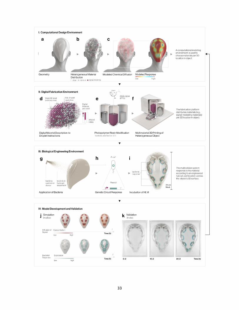
Figure 1 An Iterative Framework for the Design and Fabrication of Hybrid Living Objects. The
framework encompasses components from (I) Computational Design, (II) Digital Fabrication, (III)
Biological Engineering and (IV) Simulation. (a-c) Computational Design: (a) The computational design
process for HLM objects defines surface geometry (b) and internal material distributions of the desired
object—for instance, a 25 cm custom biohybrid mask, fit to a human face. (c) Material distributions
specify the placement and concentration of chemical signals throughout the part and can be used in a
model of signal diffusion and biological response. (d-f) Digital Fabrication: (d) For production on a
multi-material inkjet-based 3D printing platform, first, material distributions are decoded to droplet
deposition instructions for the creation of tunable digital materials, including compositions that enable
chemical signal retention. (e) With respect to the chemical signal incorporation method (see Section
2.4), custom chemical signaling resins can be developed to enable the platform to deposit signal(s)
directly into the 3D-printed architecture, during the printing process. (f) The platform outputs a
heterogeneous material composite with local control of signal retaining matrices, and/or multiple
embedded chemical signals. (g-i) Biological engineering: (g) Bacteria within a hydrogel suspension
are applied and immobilized, via a thin aerosolized coating, to the 3D-printed template. (h) Engineered
cell constructs represent genetic regulatory rule sets, able to respond to cognate diffusing signals at
any point across the HLM surface. (i) Photograph: The HLM, consisting of living and non-living
components, is incubated for 18-24 hours. By hosting and mediating an engineered cellular system, the
template material is programmed to generate specified biological response patterning (e.g., color
protein expression) at any point across the object surface. (j) Simulation: A computational model for
the biological response of HLM objects is developed using finite modeling of signal diffusion across 3D
geometries and experimental dose-response data per gene construct. (k) Validation: Image time-lapse
of the incubation (37.5°C, 100% RH) of a 3D-printed wearable HLM mask that gradually yields blue
color patterned in prescribed areas, shown at 0, 10, and 20 hours, provides one example of platform
output in vivo. Image analysis can be used to validate the in silico model of spatial biological response,
creating an iterative, self-reinforcing design process for hybrid-living material systems.
35

36
Figure 2 Characterization of Chemical Signal-Retaining Digital Materials. (a) Schematic of the Lac-
regulated expression in pUC19 E. coli constructs and resulting chromogenic β-gal activity. (b) 50 mm
diameter RGD810 disk templates contained 10 mm diameter active regions, which consisted of varying
SUP705/RGD810 digital materials; the dither pattern represents droplet instructions for blended
intermediates (1 pixel = 100x100 µm). (c) HLM disks were 3D-printed, soaked in an IPTG-X-gal solution,
and coated with a hydrogel suspension of pUC19 cells. After a 30 hr incubation, the chromogenic
response (blue) derived from β-gal activity was observed. Relative chromogenic saturation (a.u.) is
shown as a function of radial distance outward from the edge of the active region (mm), for the average
of four replicates of the experiment (n=4). Line-thickness represent one standard deviation. The material
ratio of SUP705 per template correlates the spatial response of the system. (d) The indicator CPRG
(yellow) is used as a spatiotemporal visualization of the chemical release profile; β-gal conversion of
CPRG to the CPR product (magenta) is used as a visualization of bacterial response. For the average
of four replicate experiments run in parallel (n=4), saturation of each color indicator was measured over
radial distance from the edge of the active region (mm) and time (hours), collected via time-lapse image
data.

37

38
Figure 3 Large-scale HLM Device Prototypes with Graded Physical Properties. (a) A wearable
scale object (mask) with designed heterogeneous distributions of opacity and chemical signal
concentration demonstrates size and material complexity of the objects achieved through our
framework. (b) Time-lapse of the mask’s surface, showing bacterial response (e.g., chromogenisis)
develop according to programmed material distribution during hours incubated (h). (c) x2.5 magnified
view of a wearable mask surface, showing the sub-millimeter resolution of opaque-transparent internal
material distributions and feature size. An image of the actual HLM object (top), converted to a B/W
grayscale (middle), and is compared to a computational render of the digital material description
(bottom). (d) A schematic design of an object with graded material distribution of chemical signal (red
to grey) and flexibility (brown to white) (top), and the 3D printed HLM object before and after incubation,
demonstrating templated bacterial response and flexibility (bottom).
39

40
Figure 4 Multiplexed HLM Templates Generate Multi-Signal Patterning and Spatial Logic. (a) The
schematic design of disk-shaped templates for testing the 3D printing of multiplexed chemical signaling
objects comprised of active regions with intermediate ratios of the two SUP705-based signal resins
(IPTG-SUP705/AHL-SUP705 ratio, n=4). (b) Engineered gene constructs, IPTG/GFP and AHL/RFP, used
to demonstrate orthogonal multi-output patterning. (c) Resulting HLM disks demonstrated tunable
control and orthogonality between simultaneous protein expression (GFP, RFP) channels. (d) Gene
constructs engineered for two-input “logic-gates”: a AND function (top), and the inverse, a NAND
function (bottom). (e) Bacterial AND (top) and NAND (bottom) response on HLM disks, demonstrating
that two-input logic functions generate spatial protein expression patterns in relation to regions of two-
signal overlap, or lack thereof. (f) The schematic design of second template, a two-lobed shape 3D
printed with an internal gradation of two signaling materials (IPTG, AHL) was incubated in contact with
an evenly distributed IPTG/GFP and AHL/RFP constructs. The HLM output pattern demonstrated
orthogonal regulatory computation for two inputs, per location on the HLM surface, in macro (left), and
confirmed by way of confocal fluorescent microscopy (10x) in insets (right). (g-h) The two-lobed shaped
template was incubated with AND and NAND constructs. The spatial pattern generated demonstrates
the relationship of the rule-set (e.g, logic gate) to the HLM template geometry and material distribution.
41

42
Figure 5 A Computational Model of HLM-programmed Biological Response. (a) The modeling
process to predict HLM outcome, applied to an arbitrary 3D object with multiple signals: first, the material
distribution created within the HLM framework is imported (top); the model performs finite analysis of
hydrogel-mediated chemical diffusion across the object surface, over time (middle); signal concentration
is translated to a spatial response, by way of a defined biological dose-response curve, for the resulting
HLM (bottom). (b-c) Dose-response curves for IPTG/GFP and AHL/RFP were experimentally obtained
through fluorescence measurements (Ex 485nm, Em 528 nm) of cell response to respective inducer
concentrations in growth conditions matched to immobilization on HLM object surfaces (24 hrs). Plot
represents the average of four wells per condition (n=4); error bars represent one standard deviation.
The response was fit to a Hill-Function (plotted line) to define the parameters for the response curves
of IPTG/GFP and AHL/RFP, respectively. (d) For validation, the computational model is given a material
description of a template presenting a linear material gradient of IPTG-SUP705 and RGD810 (left). In
silico, the model simulates diffusion of chemical signal concentration on the object over time, and maps
a bacterial response, in accordance with the experimental IPTG/GFP response curve of (b), for the time
point: 24 hr (right). (e) In vivo, an experimentally generated spatial bacterial response is created by
printing the template and incubating with IPTG/GFP for 24 hr. (f) Comparison of in silico and in vivo
outputs, as cross-section profiles of bacterial response as a function of relative fluorescent signal (a.u.)
at multiple positions along the object.

43
TOC
Hybrid Living Materials (HLMs) are a class of multifunctional materials that harness the
responsive capabilities of engineered bacteria in combination with established digital
fabrication processes (e.g., computer-aided design and additive manufacturing) of structural
materials. The programmable biological control enabled by the multi-material HLM
framework creates new possibilities for large-scale, structurally complex, and diversely
functional hybrid living devices.
Keyword: Hybrid Materials
Rachel Soo Hoo Smith1,†, Christoph Bader1,†, Sunanda Sharma1,†, Dominik Kolb1, Tzu-Chieh
Tang1,4, Ahmed Hosny3, Felix Moser4, James C. Weaver2, Christopher A. Voigt4, and Neri
Oxman1,*
Multi-material 3D Printing of Hybrid Living Materials