researcharticle … · thatvariation betweenthetime ofanalysis,classification ofih, resolution...
TRANSCRIPT

RESEARCH ARTICLE
The Role of Shear Stress in ArteriovenousFistula Maturation and Failure: A SystematicReviewLeonard D. Browne1, Khalid Bashar2, Philip Griffin1, Eamon G. Kavanagh2, StewartR. Walsh2,3, Michael T. Walsh1*
1 Centre for Applied Biomedical Engineering Research (CABER), Department of Mechanical, Aeronauticaland Biomedical Engineering, Materials and Surface Science Institute, The Health Research Institute,University of Limerick, Limerick, Ireland, 2 Department of Vascular Surgery, Limerick University Hospital,Dooradoyle, Limerick, Ireland, 3 Department of Surgery, National University of Ireland, Galway, Ireland
Abstract
Introduction
Non-maturation and post-maturation venous stenosis are the primary causes of failure
within arteriovenous fistulae (AVFs). Although the exact mechanisms triggering failure
remain unclear, abnormal hemodynamic profiles are thought to mediate vascular remodel-
ling and can adversely impact on fistula patency.
Aim
The review aims to clarify the role of shear stress on outward remodelling during maturation
and evaluate the evidence supporting theories related to the localisation and development
of intimal hyperplasia within AVFs.
Methods
A systematic review of studies comparing remodelling data with hemodynamic data
obtained from computational fluid dynamics of AVFs during and after maturation was
conducted.
Results
Outward remodelling occurred to reduce or normalise the level of shear stress over time in
fistulae with a large radius of curvature (curved) whereas shear stress was found to aug-
ment over time in fistulae with a small radius of curvature (straight) coinciding with minimal
to no increases in lumen area. Although this review highlighted that there is a growing body
of evidence suggesting low and oscillating shear stress may stimulate the initiation and
development of intimal medial thickening within AVFs. Further lines of evidence are needed
to support the disturbed flow theory and outward remodelling findings before surgical config-
urations and treatment strategies are optimised to conform to them. This review highlighted
PLOSONE | DOI:10.1371/journal.pone.0145795 December 30, 2015 1 / 24
OPEN ACCESS
Citation: Browne LD, Bashar K, Griffin P, KavanaghEG, Walsh SR, Walsh MT (2015) The Role of ShearStress in Arteriovenous Fistula Maturation andFailure: A Systematic Review. PLoS ONE 10(12):e0145795. doi:10.1371/journal.pone.0145795
Editor: Alberto Aliseda, University of Washington,UNITED STATES
Received: June 8, 2015
Accepted: December 8, 2015
Published: December 30, 2015
Copyright: © 2015 Browne et al. This is an openaccess article distributed under the terms of theCreative Commons Attribution License, which permitsunrestricted use, distribution, and reproduction in anymedium, provided the original author and source arecredited.
Data Availability Statement: All relevant data arewithin the paper and its Supporting Information files.
Funding: LB received funding from The IrishResearch Council for Science Engineering andTechnology (IRCSET). LB and MW received fundingfrom The European Union’s Seventh FrameworkProgramme for research, technological developmentand demonstration under grant agreement no.324487.
Competing Interests: The authors have declaredthat no competing interests exist.

that variation between the time of analysis, classification of IH, resolution of simulations,
data processing techniques and omission of various shear stress metrics prevented forming
pooling of data amongst studies.
Conclusion
Standardised measurements and data processing techniques are needed to comprehen-
sively evaluate the relationship between shear stress and intimal medial thickening.
Advances in image acquisition and flow quantifications coupled with the increasing preva-
lence of longitudinal studies commencing from fistula creation offer viable techniques and
strategies to robustly evaluate the relationship between shear stress and remodelling during
maturation and thereafter.
IntroductionHemodialysis is the treatment modality of choice for patients with end stage renal disease(ESRD). Adequate and efficient hemodialysis requires a reliable vascular access which is easilyaccessible and provides consistently high flow rates greater than 600 ml/min [1,2]. Arteriove-nous fistulae (AVFs) are the preferred access choice due to lower infection and stenosis rates.However, they are prone to complications during remodelling and have a high incidence of pri-mary failure during this process [2]. Thrombotic occlusion arising from aggressive intimalhyperplasia (IH) and impaired remodelling leading to reduced flow rates at the access site, arethe two major causes of patency loss [2–5]. Complex AVF hemodynamics are believed to pro-vide a stimulus for remodelling and IH related failure [6,7]. Computational fluid dynamics(CFD) allows for the study of hemodynamics within multiple vasculatures as it can approxi-mate analytically complex flow fields. CFD can also calculate hemodynamic parametersderived from the flow field such as shear stress. Owing to the improving resolution of medicalimaging and the ability to decompose these images into CFD models, modelling hemodynam-ics of realistic patient geometries is possible [8].
The review aims to clarify the role of shear stress on outward remodelling during matura-tion and evaluate the evidence supporting theories related to the localisation and developmentof intimal hyperplasia within AVFs.
Methods
2.1 Search strategyA systematic search was conducted on the PubMed and Google scholar database for articlespublished online before 1/1/2015 whose title/abstract contained the following sequence of key-words as outlined in Table 1.
2.2 Eligibility criteriaThe titles and abstracts of all potentially suitable studies were inspected, articles meeting theinclusion criteria were retrieved and reviewed by review authors (LB, MW).The results of theprocess are illustrated in [Fig 1]. Included in the review were:
• Studies which analysed blood flow in one or more vascular access geometries which areanatomically realistic and were acquired by a relevant imaging modality
Role of Shear Stress in AVF Maturation and Failure
PLOS ONE | DOI:10.1371/journal.pone.0145795 December 30, 2015 2 / 24
• Studies in which the localisation of stenotic lesions or adaptive responses were based on thepresence of an established marker of disease or remodelling
• Studies in which results were compared against lesion data from a companion paper fromthe same group, or from a cited article from a different research group for the same species
• Studies which discussed the relationship between blood flow or shear stress on vascularaccess remodelling or the formation of intimal hyperplasia
Excluded from the review were:
• Studies which did not attempt to compare hemodynamic results in detail with the localisa-tion of lesions or adaptive responses in the relevant species
• Studies in which geometries were acquired after interventional revisions
• Studies which superimposed stenosis by ligation or other means to alter flow as these studiesmay not replicate the normal initiation of the disease
• Studies which utilised idealised geometries
2.3 Data extractionData from each article was extracted by two review authors (LB, MW) and were compared forconsistency of data extraction; any disagreement was discussed with an additional author. Thefollowing information regarding AVF characteristics were recorded for maturation studies:Time post creation, image modality, radius of curvature, species, site location, variation ofshear stress, flow rate, cross sectional area (CSA) and intima-media thickness (IMT). For IHstudies the following were extracted: Time post creation, patterns of shear stress related param-eters, biological markers, image modality, experimental modality and IMT.
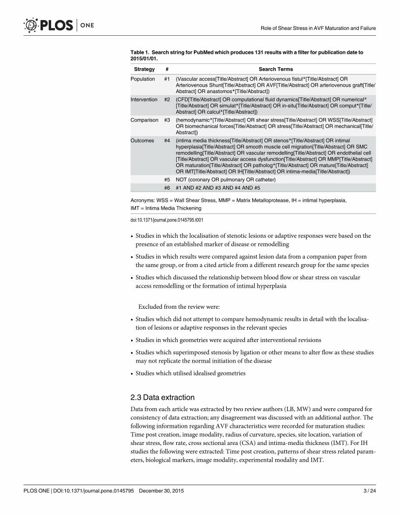
Table 1. Search string for PubMed which produces 131 results with a filter for publication date to2015/01/01.
Strategy # Search Terms
Population #1 (Vascular access[Title/Abstract] OR Arteriovenous fistul*[Title/Abstract] ORArteriovenous Shunt[Title/Abstract] OR AVF[Title/Abstract] OR arteriovenous graft[Title/Abstract] OR anastomos*[Title/Abstract])
Intervention #2 (CFD[Title/Abstract] OR computational fluid dynamics[Title/Abstract] OR numerical*[Title/Abstract] OR simulat*[Title/Abstract] OR in-situ[Title/Abstract] OR comput*[Title/Abstract] OR calcul*[Title/Abstract])
Comparison #3 (hemodynamic*[Title/Abstract] OR shear stress[Title/Abstract] OR WSS[Title/Abstract]OR biomechanical forces[Title/Abstract] OR stress[Title/Abstract] OR mechanical[Title/Abstract])
Outcomes #4 (intima media thickness[Title/Abstract] OR stenos*[Title/Abstract] OR intimalhyperplasia[Title/Abstract] OR smooth muscle cell migration[Title/Abstract] OR SMCremodelling[Title/Abstract] OR vascular remodelling[Title/Abstract] OR endothelial cell[Title/Abstract] OR vascular access dysfunction[Title/Abstract] OR MMP[Title/Abstract]OR maturation[Title/Abstract] OR patholog*[Title/Abstract] OR mature[Title/Abstract]OR IMT[Title/Abstract] OR IH[Title/Abstract] OR intima-media[Title/Abstract])
#5 NOT (coronary OR pulmonary OR catheter)
#6 #1 AND #2 AND #3 AND #4 AND #5
Acronyms: WSS = Wall Shear Stress, MMP = Matrix Metalloprotease, IH = intimal hyperplasia,
IMT = Intima Media Thickening
doi:10.1371/journal.pone.0145795.t001
Role of Shear Stress in AVF Maturation and Failure
PLOS ONE | DOI:10.1371/journal.pone.0145795 December 30, 2015 3 / 24

2.4 Selected literatureFifteen articles were selected from an original set of 436. Removing duplicates, reviews and arti-cles that did not relate to hemodynamics within AVFs left 70 articles. A further 25 were dis-carded because they did not compare their hemodynamic results with the localisation oflesions or adaptive responses in relevant species. 27 articles were removed as they did not pro-vide shear stress computed data. An additional 2 articles were removed as they were conductedon geometries pre and post interventional treatment. Finally, 1 article was removed as it waslikely a duplicate conference preceding. After screening, 15 articles which compared remodel-ling or IH data with shear stress metrics remained as outlined in Table 2.
Eight articles focused on the role or distribution of shear stress within maturing or matureAVFs. Five articles focused on the roles of shear stress metrics or hemodynamic metrics in rela-tion to IH formation. Two articles focused on the influence of distensibility on the shear stressdistribution and its influence on IH. The selected articles originated from 7 institutions andstudied human & porcine models to assess maturation and intimal hyperplasia formation withthe addition of a canine model for the later. The brachiocephalic, radiocephalic and brachioba-sillic fistula were constructed in humans. Anastomoses between the femoral artery and veinand the aorta and iliac vein were the configurations created in porcine models.
Fig 1. PRISMA diagram: Flow chart of the strategy used to select articles for review.
doi:10.1371/journal.pone.0145795.g001
Role of Shear Stress in AVF Maturation and Failure
PLOS ONE | DOI:10.1371/journal.pone.0145795 December 30, 2015 4 / 24

MaturationMaturation is the remodelling process whereby a fistula becomes suitable for cannulation.AVFs are assessed for non-maturation after 4–6 weeks; mature AVFs should have a diameterof 6mm, be less than 6mm below the skin surface and have a flow rate greater than 600 ml/min[9].
Endothelial cells (ECs) which line the internal surface of a blood vessel are important medi-ators of maturation. ECs are continually exposed to shear stress, compression via blood pres-sure and to tension from strain in the extracellular matrix (ECM) as shown in [Fig 2]. Each ofthese factors is known to modulate intracellular signalling pathways and gene expression. Vari-ations of these mechanical factors from the normal level can alter EC function and stimulateremodelling [10–12]. Exposure of a vein to the arterial environment during AVF creation is aparadigm of such.
AVF formation involves directly connecting a high pressure pulsatile flow conduit (artery)to a low pressure steady flow conduit (vein). The resulting pressure gradient results in an
Table 2. Reviewed articles in chronological order, with the affiliation of the corresponding author, species and AVF configuration.
REF Title Year Affiliation ofCorresponding Author
Species Fistula
8 Measurement of hemodynamic and anatomic parameters in a swinearteriovenous fistula model
2008 University of Cincinnati Porcine Femoral artery and femoralvein AVF
15 Longitudinal assessment of hemodynamic endpoints in predictingarteriovenous fistula maturation
2013 University of Cincinnati Porcine Femoral artery and femoralvein AVF
16 Influence of temporal variation in wall shear stress on intima-mediathickening in arteriovenous fistulae
2012 University of Cincinnati Porcine Femoral artery and femoralvein AVF
17 Vascular remodeling in autogenous arterio-venous fistulas by MRIand CFD
2013 University of California Human Brachiocephalic (n = 2)Brachiobasillic (n = 1)
18 Serial analysis of lumen geometry and hemodynamics in humanarteriovenous fistula for hemodialysis using magnetic resonanceimaging and computational fluid dynamics
2012 University of Utah Human Brachiocephalic
19 Hemodynamic wall shear stress profiles influence the magnitude andpattern of stenosis in a pig AV fistula
2008 University of Cincinnati Porcine Femoral artery and femoralvein AVF
20 Numerical and experimental study of blood flow through a patient-specific arteriovenous fistula used for hemodialysis
2010 Universite deTechnologie deCompiegne
Human Brachiocephalic
21 Investigations into the relationship between hemodynamics andvascular alterations in an established arteriovenous fistula
2007 Universite deTechnologie deCompiegne
Human Brachiocephalic
22 Incomplete restoration of homeostatic shear stress withinarteriovenous fistulae
2013 University of Washington Human Radiocephalic (n = 2)Brachiocephalic(n = 2)
23 Wall shear stresses remain elevated in mature arteriovenous fistulas:a case study
2011 University of Limerick Human Radiocephalic AVF
31 Realistic temporal variations of shear stress modulate MMP-2 andMCP-1 expression in arteriovenous vascular access
2009 University of Limerick Human Radiocephalic
32 New Techniques for Determining the Longitudinal Effects of LocalHemodynamics on the Intima‐Media Thickness in ArteriovenousFistulae in an Animal Model
2013 University of Cincinnati Porcine Femoral artery and femoralvein AVF
41 Transitional flow at the venous anastomosis of an arteriovenous graft:potential activation of the ERK1/2 mechanotransduction pathway
2003 The University of Illinoisat Chicago
Canine Femoral artery to Femoralvein graft
52 Numerical simulation of the fluid structure interactions in a compliantpatient‐specific arteriovenous fistula
2014 Universite deTechnologie deCompiegne
Human Radiocephalic
53 Effects of wall distensibility in hemodynamic simulations of anarteriovenous fistula
2013 University of Washington Human Radiocephalic
doi:10.1371/journal.pone.0145795.t002
Role of Shear Stress in AVF Maturation and Failure
PLOS ONE | DOI:10.1371/journal.pone.0145795 December 30, 2015 5 / 24

immediate increase in flow in both the artery and vein and the resulting hemodynamics initi-ates a vascular remodelling response within both vessels. Pressure in the venous segment risesupon AVF creation and remains relatively constant during the time course of remodelling andthereafter. The increase in pressure is known to stimulate vascular smooth muscle cell (VSMC)proliferation and induce moderate medial thickening over time [13]. Remodelling in maturingAVFs is primarily characterised by eccentric medial hypertrophy resulting from increased cir-cumferential tension due to flow mediated dilation rather than the elevated pressure alone[14]. Dilation is induced by high levels of shear stress, resulting from elevated flow. The level ofshear stress is also known to regulate the intimal layer with a well-established inverse correla-tion between intimal thickness and shear stress observed during venous and arterial adaption[13]. One can conjecture that the balance between dilation and intimal medial thickening willdetermine whether a favourable outward hypertrophic remodelling or unfavourable inwardhypertrophic remodelling response occurs [6], these remodelling responses are illustrated in[Fig 3].
Endothelial cells are finely primed to sense variations in shear stress and respond accord-ingly. Therefore, knowledge of shear stress patterns within AVFs as they progress to maturitycould help predict the molecular and structural responses that occur during remodelling,thereby identifying critical linkages and targets for therapeutic strategies and intervention.
3.1 Longitudinal studiesA longitudinal study is a correlational research study that involves repeated observations of thesame variables over long periods of time. A number of studies adopted this approach to moni-tor shear stress and structural changes within an AVF during its maturation period. For thisreview AVFs were categorised as either straight or curved based on a qualitative assessment ofthe radius of curvature of the swing segment to assess the variation of flow rate and shear stresson outward remodelling. Curved AVFs were categorised by a large radius of curvature,
Fig 2. A section of an artery wall shows the endothelial cells that form the inner lining and alignlongitudinally in the direction of the flow. Pressure (P) acts normal to the vessel wall, which results incircumferential stretching of the vessel wall. Shear stress (τ) is parallel to the vessel wall and is exertedlongitudinally in the direction of blood flow. The intima, media and adventitia layers of an artery and vein areshown. Vascular smooth muscle cells form the outer layers and align circumferentially. IMT refers to intimamedia thickness.
doi:10.1371/journal.pone.0145795.g002
Role of Shear Stress in AVF Maturation and Failure
PLOS ONE | DOI:10.1371/journal.pone.0145795 December 30, 2015 6 / 24

identified by the gradual bend of the fistulas swing segment. Whereas, straight AVFs were cate-gorised by a small radius of curvature, identified by an abrupt bend of the swing segment.
For curved AVFs the flow rate within the venous segment increases during the first 6 weeks[Table 3]. Cross sectional area increases to reduce the level of shear stress to its physiologicalrange. Intimal medial thickening increases progressively during this period. The increase inarea varies along the length of the venous segment due to the non-uniform distribution of
Fig 3. Typical geometry of an arteriovenous fistula is shownwith the swing segment highlighted; the dashed blue line highlights a cross sectionof the vein for which various vascular remodelling responses within the venous segment of an AVF are shown for a healthy vein and an ESRDvein.
doi:10.1371/journal.pone.0145795.g003
Table 3. Variation of experimental conditions and outcomes for AVF with curved configurations i.e. time of analysis, variation of flow rate, shearstress, lumen cross sectional area (CSA) and intimamedia thickness (IMT).
Stage Author Methodology Species & (location) Time postformation
Flowrate
Shearstress level
CSA IMT
<6wks.
Rajabi-Jagahrghet al [15,16].
CT angiography Ultrasound CFDHistological analysis
Porcine (Femoral artery andvein) n = 3
2 days - - - -
7 days " # " "28 days " # " "
Sigovan et al [17]. Magnetic resonance angiogramCFD
Human (Brachiocephalic)n = 2
5 days - - - -
1 mo. " # " X
>6wks.
3 mo. # # " X
He et al [18]. Magnetic resonance angiogramCFD
Human (Brachiocephalic)n = 1
4 mo. - - - -
5 mo. # # " X
7 mo. # # " X
X no data available; " Increase; # Decrease;—Initial time point of analysis
doi:10.1371/journal.pone.0145795.t003
Role of Shear Stress in AVF Maturation and Failure
PLOS ONE | DOI:10.1371/journal.pone.0145795 December 30, 2015 7 / 24

shear stress in the segment. After 6 weeks flow rate decreases and cross sectional area continuesto increase to reduce the level of shear stress. This trend may fluctuate about this time point ina larger cohort due to higher levels of heterogeneity. Nonetheless, high shear stresses are foundto persist near the anastomosis. Further away from the anastomosis shear stress is found todecrease towards the physiological range.
For straight AVFs the flow rate was found to increase during the first 6 weeks, cross sec-tional area did not significantly change during this period and the level of shear stress wasfound to increase [Table 4]. Intimal medial thickness was also found to progressively increaseduring this period. High shear stresses were found to extend into and occupy a larger propor-tion of the venous segment compared to curved configurations.
Rajabi-Jagahrgh et al [15] found that intima media thickness increases progressively forboth configurations with a significant difference between the two configurations at 28 dayswith greater thickening within the curved configuration. An earlier study by Krishnamoorthyet al [19] found an inverse correlation between shear stress and IMT. Hence, as lower levels ofshear stress occupy the curved configuration greater thickening may be induced.
3.2 Single time point studiesA number of computational studies were conducted at single time points in mature patent fis-tulae in an attempt to characterize the hemodynamics of these patent accesses. All of whichfound that shear stress was not homogeneously distributed over the vessel with high nonhomeostatic shear stresses persisting at the arterial curvatures feeding the anastomosis, withinthe anastomosis junction and within the swing segment of the vein just after the anastomosisdue to the jet velocity striking the wall. Further away from the anastomosis shear stresses werewithin the physiological range [Table 5].
Intimal HyperplasiaIntimal hyperplasia (IH) has been cited as the underlying cause of stenotic lesion formationwithin AVFs. Unlike atherosclerosis which is a chronic, inflammatory, fibroproliferative dis-ease of the vascular wall that gradually occurs in time [24], IH is a rapid adaptive response toinjury of the endothelium by surgical, hemodynamic, immune or metabolic stresses. Theevents leading to IH can be divided into an (i) inflammatory (ii) proliferation and (iii) remodel-ling phase as outlined in Table 6. IH is characterised by VSMC proliferation, followed byVSMC and intimal growth which can diminish the vascular lumen [4,25]. IH primarily
Table 4. Variation of experimental outcomes for AVF with straight configurations.i.e. time of analysis, variation of flow rate, shear stress, lumencross sectional area (CSA) and intimamedia thickness (IMT).
Stage Author Methodology Species & (location) Time postformation
Flowrate
Shearstress level
CSA IMT
<6 wks. Rajabi-Jagahrghet al [15,16].
CT angiography UltrasoundCFD Histological analysis
Porcine (Femoral arteryand vein) n = 3
2 days - - - -
7 days " " $ "28 days " " $ "
Sigovan et al [17]. Magnetic resonance angiogramCFD
Human (Brachiobasillic)n = 1
5 days - - - -
1 mo. " " $ X
>6 wks. 3 mo. " " % X
X no data available; " Increase; # Decrease;—Initial time point of analysis; $ no significant change; % minor increase)
doi:10.1371/journal.pone.0145795.t004
Role of Shear Stress in AVF Maturation and Failure
PLOS ONE | DOI:10.1371/journal.pone.0145795 December 30, 2015 8 / 24

comprises of α-smooth muscle actin (α-SMA) positive cells, extracellular matrix proteins andcytokines such as platelet-derived growth factor, transforming growth factor-β and endothelinwithin the intima and media of the vein [26,27]. The majority of the α-SMA positive cells inthe intimal lesions exhibit a myofibroblast or synthetic VSMC phenotype [28–30]. The sourceand action of the main cytokines, proteinases and growth factors involved in IH formation aresummarised in Table 7.
4.1 TheoriesThere are a number of theories related to the influence of shear stress parameters on the devel-opment of IH within AVFs. Table 8 outlines the findings from the selected articles supportingmagnitude and gradient based shear stress parameters. Definitions for these parameters areprovided within S1 Table. All configurations were included to establish the role of variousshear stress metrics on inward remodelling.
4.1.1. High shear stress. Carroll et al [23,31] suggested that high shear stress could inducethe upregulation of MCP-1 and MMP-2 and initiate IH in regions of high shear which coulddenude ECs and expose a thrombogenic surface. However, high shear stresses have been foundto persist in mature patent fistulae’s ranging from 2 to 20 yrs. post creation. [20–22]. Whetheran unfavourable threshold of high shear stress exists has yet to be quantified.
Table 6. Events leading to intimal hyperplasia.
(i) Endothelium injury
#Platelet adhesion
#Aggregation and activation of platelets and inflammatory cells at the site of endothelial injury
#(ii) VSMC proliferation and migration to intima
#Re-endothelialisation of the injured site
#(iii) Intimal thickening via secretion of ECM composed of elastin, collagens, glycoproteins and
proteoglycans
#Adventitial fibroblasts migrate into intima and differentiate into myofibroblasts
doi:10.1371/journal.pone.0145795.t006
Table 5. Species and location and shear stress distribution of mature AVFs assessed at single time points.
Author Methodology Species & (location) Time post formation Shear stress level
AJ SS V
Kharboutly et al [20,21]. CT angiography CFD Human (Brachiocephalic) n = 1 20 yrs. High High Normal
McGah et al [22]. Ultrasound CFD Human (Radiocephalic) n = 2 7.6 yrs. High High Normal
2.0 yrs. High High Normal
(Brachiocephalic)n = 2 3.3 yrs. High High Normal
2.2 yrs. High High Normal
Carrol et al [23]. MRI Ultrasound CFD Human (Radiocephalic) n = 1 >1yr. High High Normal
AJ Anastomotic Junction; SS Swing Segment; V Vein
doi:10.1371/journal.pone.0145795.t005
Role of Shear Stress in AVF Maturation and Failure
PLOS ONE | DOI:10.1371/journal.pone.0145795 December 30, 2015 9 / 24

4.1.2. Low and Oscillatory shear stress. Disturbed flow accounts for a pattern of flow thatis non-uniform and irregular; including recirculation eddies and changes in direction withtime and space [33]. The combination of low and oscillatory shear stress has been utilised as anindicator of disturbed flow within AVFs. The low shear stress results were paired against inti-mal medial thickening. Krishnamoorthy et al [19] compared circumferential averages of IMTat selective locations against averaged shear stress and reported an inverse correlation betweenshear stress and IMT. Rajabi-Jagahrgh et al [32] compared the circumferential distribution ofshear stress and IMT at selective locations. Sites of low shear and high oscillating shear index at
Table 7. Main proteinases, cytokines and growth factors involved in IH formation.
Source Action
GrowthFactor
PDGF Platelets, ECs, VSMCs VSMC proliferation and migration
TGF-B ECs, VSMCs VSMC proliferation
IGF-1 VSMCs
bFGF VSMCs
VEGF ECs Endothelisation
Cytokines MCP-1 Macrophages, VSMCs, ECs, Fibroblasts Monocyte recruitment
IL-1, IL-6 Leucocytes, macrophages, VSMCs, ECs,Fibroblasts
Neutrophil and monocyte recruitment
Proteinases MMP-2MMP-9
ECs, VSMCs, Macrophages ECM degradation and reorganisation VSMC proliferation and migrationFibroblast migration
TIMPs ECs, VSMCs, Macrophages Reduced proliferation and migration
PDGF, platelet-derived growth factor; bFGF, basic fibroblast growth factor; IGF, insulin-like growth factor; TGF, transforming growth factor; VEGF,
vascular endothelial growth factor; IL, interleukin; MCP-1 monocyte chemoattractant protein 1 MMPs; Matrix metalloproteinases, TIMPs; tissue inhibitors
of MMPs
doi:10.1371/journal.pone.0145795.t007
Table 8. Overview of the different theories reviewed on IH within AVFs and their experimental conditions, i.e. time of analysis, shear parametermeasured and effect on biological markers and intimamedia thickness.
Theory Author Methodology Time postformation
ShearParameter
Biological markers IMT
High Shear Carroll et al [31]. CFD Cone & plate 1.5 hr. High WSS "MMP-2 "MCP-1 X
12 hr.
Carroll et al [23]. CFD X "WSS "WSSG X X
Low shear Krishnamoorthy et al[19].
CFD Histological analysis CTangiography Micro MRI
42 days Low WSS X MaxIMT
42 days High WSS X MinIMT
Low shear & OSI Rajabi-Jagahrgh et al[32].
CFD * Histological analysis 2 days Low WSS &high OSI
28 days X MaxIMT
Kharboutly et al [21]. CFD CT angiography 20 yrs. OSI No association to calcifiedplaque
X
Temporal WSSgradient
Kharboutly et al [21]. CFD CT angiography 20 yrs. High TWSSG Strongest association tocalcified plaque
X
X no data available; " Increase; # Decrease.
* CFD results at 2 days compared against Histological analysis at 28 days.
doi:10.1371/journal.pone.0145795.t008
Role of Shear Stress in AVF Maturation and Failure
PLOS ONE | DOI:10.1371/journal.pone.0145795 December 30, 2015 10 / 24
early time points correlated to maximum IMT at later time points. However, IMT was alsoobserved in a location downstream in a region of higher shear and low OSI. This suggests thatmultiple aspects of shear stress may stimulate intima-media thickening or that multidirectionalaspects of disturbed flow might be described more accurately with metrics such as transversewall shear stress (transWSS) rather than OSI alone. Nonetheless, the low and oscillating shearstress theory associated with IMT may not be as a robust as previously taught.
4.1.3. Temporal gradient of shear stress. The study by Kharboutly et al [20,21] reported astrong association with high temporal wall shear stress gradient (TWSSG) and locations of cal-cified plaque compared to lowWSS and OSI and concluded that this parameter may be animportant determinant of endothelial cell function and plaque formation.
4.1.4. Turbulence. Transitional to turbulent flow has been found to cause EC elongationsimilar to laminar flow but a loss of EC alignment due to the fluctuating shear stress compo-nent [34,35]. A significant reduction in nitric oxide (NO) production, an important inhibitorof inflammation and VSMC proliferation, has also been reported in-vitro on EC cultured com-pliant tubes [36]. Therefore, the presence of transitional flow can disrupt normal function ofECs within AVFs.
Early studies by Fillinger et al [37,38] reported a direct correlation with Reynolds numberand IH and hypothesised that turbulence leads to IH via the transfer of kinetic energy to thevessel wall in the form of vein wall vibration within AVFs. However, subsequent studies wereunable to validate the theory. This was accredited to computational assumptions such as steadyflow, rigid walls and flow split which could bias results. The latter was found to contribute sig-nificantly to promoting a transitional regime [39,40].
Loth et al [41] reported that fluctuating shear stress overlapped with areas of vein wall vibra-tion coinciding with regions of elevated extracellular regulatory kinases (ERK1/2) which arelinked to the upregulation of PDGF and TGF following vessel injury. None of the previousstudies which quantitatively compared shear stress parameters and IH noted a transitionalregime. Most determined that peak Reynolds numbers did not reach the critical value for tur-bulence in a straight pipe (Re>2000) and therefore the laminar flow model was likely to be ade-quate. However, unstable physiological flow has been simulated by a number of studiesattempting to resolve characteristics of the flow field within an AVF [22,42,43]. Ene-Iordacheet al [43] have highlighted that transitional to turbulent flow can lead to fluctuations and multi-directional disturbed flow; aspects which have been obscured by under-resolved simulations.The presence of these instabilities could account for the inconsistency between patterns of IMTand hemodynamic metrics observed by Rajabi-Jagahrgh et al [32].
Hemodynamics of AVFsWere Assessed at Different Sites andStages of Maturation
5.1 Maturation studiesRemodelling alters lumen geometry and significantly alters the distribution and level of shearstress. Hence, the time points at which studies were conducted will impact the geometry andpattern of shear stress. Therefore, the validity of inter-study means for area, flow, Reynoldsnumber and shear stress depends on the time of analysis, location of access site, surgical config-uration of the AVF and the accuracy of its reconstruction. [Fig 4] outlines the various recon-structions of patient specific brachiocephalic fistulae and radiocephalic fistulae against thetimes at which resulting hemodynamics were assessed. The majority of AVFs had an end-sideconfiguration with two AVFs configured in a side to side manner. From visual comparisons itis evident that adequate dilation is a precursor of a mature fistula.
Role of Shear Stress in AVF Maturation and Failure
PLOS ONE | DOI:10.1371/journal.pone.0145795 December 30, 2015 11 / 24

5.2 IH studiesDuring the initial six week period following AVF formation it is expected that high shear stresswill promote dilation and subsequent remodelling in the form of expansion and medial hyper-trophy. Elementary factors such as MMPs involved in outward remodelling are also involvedin IH formation [44–47]. The early expression of MMPs may be mediated by nitric oxide pro-duction in response to increased flow and shear stress, the subsequent vasodilation responseincreases circumferential stretch and thickens the medial layer [11,48]. Therefore, increases inIMT will be attributed in part to medial hypertrophy and will account for the expression of cer-tain genes and proteases which are involved in both hypertrophic remodelling and intimalhyperplasia.
Fig 4. Reconstructions of curved brachiocephalic & radiocephalic fistulae at different longitudinal time points. * indicates a side to side configurationall other AVFs were configured in an end to side manner.
doi:10.1371/journal.pone.0145795.g004
Role of Shear Stress in AVF Maturation and Failure
PLOS ONE | DOI:10.1371/journal.pone.0145795 December 30, 2015 12 / 24

Species Varied Amongst StudiesPorcine and human models were included in the review as it is known that these models havesimilar vascular responses to injury with a direct correlation between the magnitude of vascularinjury and the amount of intimal hyperplasia. Porcine models can adequately develop intimalhyperplasia over a short period, in contrast to canine or murine models [28]. Intimal thicken-ing within porcine models conformed to the low and oscillatory shear stress theory. Whetherthis theory is transferrable to ESRD patients remains to be determined as lesion patterns areknown to vary amongst species and at different age grades.
Intimal Hyperplasia Was Classified by Various Modalities andCharacterised at Different Pathological StagesThe progression of IH was determined by the identification of one or more proteases, genes orgrowth factors that lead to its development. Measurements of geometrical characteristics of thevessel wall were also undertaken and IH was also identified by contrast thresholds.
7.1 Histological analysisCarroll et al [31] utilised a cone and plate bioreactor and real time reverse-transcriptase PCR toassess the expression of proteases and cytokines following the onset of flow similar to an AVFenvironment. The overlap of certain genes and growth factors involved in IH and outwardremodelling make it difficult to determine which pathological response will be initiated whenthese factors are expressed following the onset of flow.
A number of studies conducted histological analysis on porcine models over a longitudinalperiod. This allowed IMT to be assessed through the use of Hematoxylin and eosin (H&E).This staining makes it difficult to identify the interface between the intimal and medial layers.The subsequent use of Verhoeff’s van Gieson (VVG), PCNA and α-SMA stain have beenshown to define these layers [49,50].
7.2 Geometrical characteristicsThree studies [15,16,32] utilised IMT to assess pathological change in response to hemody-namic conditions. The technique involves measuring the length or thickness between the outerboundary of the vessel and the intimal layer. This technique makes it difficult to delineatebetween the intimal and medial layers and subsequent amount of thickening. The time point ofanalysis will be important as studies conducted within six weeks of formation which assed IHby such means will account for progressive changes that are in part due to outwardremodelling.
Therefore, the data processing technique chosen must be quantitatively robust to distin-guish layers and capture the initiation or development of IH. The method adapted by Rajabi-Jagahrgh et al [32] in which the circumferential distribution of shear stress and IMT were com-pared is a viable option. A radiopaque marker was sutured along the fistula providing a fixedlocal reference point to orient cross-sectional slices. This marker is defined as zero degreesfrom the luminal centre. Shear stress metrics and IMT are calculated and compared from0–360 degrees along the circumference of the slice to provide a point by point comparison. Asthe suture is a fixed local reference point, results between different time points can also be com-pared. High resolution ultrasound and Micro MRI may offer valid non-invasive imagingapproaches to distinguish between the layers but are currently unable to identify its composi-tion [32,51].
Role of Shear Stress in AVF Maturation and Failure
PLOS ONE | DOI:10.1371/journal.pone.0145795 December 30, 2015 13 / 24

7.3 Contrast thresholdsKharboutly et al [20,21] utilised CT angiography to visualise and reconstruct the lumen andcalcified plaques within a patent 20 yr. old mature AVF. Calcification zones were identified bytheir high contrast value within the vascular lumen. Virtual removal of the plaques was carriedout and the localisation of calcified plaque against shear stress derived parameters was ana-lysed. The authors acknowledged that the imaging technique made it difficult to differentiatebetween IH regions and atherosclerotic plaque regions.
Data ReductionA number of data processing methods were employed to assess the influence of shear stressrelated parameters on maturation or IH lesion formation [Fig 5]. The choice of technique uti-lised to compare hemodynamic maps with pathological remodelling can potentially limit thefidelity of conclusions drawn.
8.1 Circumferential averaging
• Rajabi-Jagahrgh et al [15] circumferentially averaged TAWSS, IMT and lumen area at 20 dif-ferent cross sections along the venous segment and attained an overall average of each. Theyreported that the reduction in shear stress over time was found to have a statistically signifi-cant effect on the increase of lumen area over time (r = -0.77, p<0.05).
• He et al [18] circumferentially averaged TAWSS, WSSG and OSI and lumen area at 1 mmintervals; a significant increase in lumen area between each time point was recorded, noother statistical relationships were presented.
• Krishnamoorthy et al [19] circumferentially averaged shear stress at 4 different locations andreported an inverse correlation between WSS and luminal stenosis.
The lumen does not encounter uniform levels of shear stress especially in areas of curvaturesuch as the swing segment of an AVF. Therefore, circumferentially averaging obscures theinfluence of vessel curvature on IMT and shear stress
8.2 Axial AveragingAxial averages of TAWSS were acquired at 5 mm intervals by McGah et al [22] to compare theresults with the value of shear stress determined by Pouseuille’s flow equation which was foundto underestimate shear stress. Sigovan et al [17] also utilised a similar technique by averagingthe TAWSS on the surface of the venous outflow between two planes from the suture line tothe venous outlet. The distribution of shear stresses was found to decrease over time for bothbrachiocephalic patients. No correlation or significance probability was reported for the axialaveraging technique.
8.3 Selective analysisA number of studies limited their analysis to a subset of known lesion prone sites or conditionsof shear stress.
• Kharboutly et al [20,21] divided an AVF volume into seven sub-volumes by defining the sec-tions perpendicular to the vascular centreline axis. A set of antipodal points were createdalong the interior and exterior wall. A selective point comparison analysis of shear stressderived parameters and calcification was performed. A strong association between highTWSSG zones and the presence of the calcification plaques was recorded.
Role of Shear Stress in AVF Maturation and Failure
PLOS ONE | DOI:10.1371/journal.pone.0145795 December 30, 2015 14 / 24

• Rajabi-Jagahrgh et al [32] compared the circumferential distribution of shear stress to a mapof IMT at two selective locations of high and low shear stress to find a relationship betweenlow and oscillating shear with maximum IMT.
Both techniques are quantitatively robust, however, limiting their analysis to subsets of datamay distort important aspects of the relationship between shear related stimulators of patho-logical remodelling.
Fig 5. Schematic overview of the various data-processingmethods employed in the reviewed articles.
doi:10.1371/journal.pone.0145795.g005
Role of Shear Stress in AVF Maturation and Failure
PLOS ONE | DOI:10.1371/journal.pone.0145795 December 30, 2015 15 / 24

8.4 SummaryThe circumferential averaging and axial averaging approaches undertaken by researchers toassess lumen changes were quantitatively similar with the later having finer spatial resolutionin the axial direction of the vessel. Such quantitative approaches may be apt to quantify thevasodilator induced changes of shear stress but are ill-defined when identifying sites of patho-logical remodelling particularly IH and its relationship to shear stress. Point by point compari-sons or other techniques which directly compare hemodynamic maps with stained or contrastmaps will offer a more rigorous analysis for this interaction.
Limitations and Assumptions of Numerical Simulations
9.1 Flow regime and rheologyDetermining whether a reconstructed model is realistically reproducing the in-vivo hemody-namic environment is difficult to ascertain as there is no benchmark technique for measuringvelocities in-vivo. The flow within AVFs is characteristically unstable, particularly within theanastomosis and may fall within a transitional to turbulent flow regime. Hence, whether theuse of a laminar solution and low resolution meshes are adequate to resolve the flow fieldneeds to be determined.
In all the longitudinal studies the viscosity of blood was kept constant, in-vivo it is knownthat the viscosity of blood decreases during remodelling to promote a return of shear stress tohomeostatic values [7]. Only one of the selected articles characterised blood as a non-Newto-nian fluid. Despite the high flow environment, regions of lowWSS exist within AVFs thereforethe influence of blood rheology should be assessed [52].
9.2 Boundary conditionsPatient specific reconstructions of the domain of interest need to be coupled with realisticboundary conditions. All studies investigated prescribed a transient flow rate at the arterialinlet with the vast majority applying parabolic velocity profiles, flat or Wormsley profiles. Moststudies prescribed a targeted flow rate at the venous outlet and the distal artery outlet based onin-vivomeasurements. The exceptions prescribed a desired pressure at the venous outlet andrecorded a simulated arterial pressure to be within 10% of the mean pressure measured in-vivo[8,19]. McGah et al [53] utilised a Windkessel model to set the desired pressure at the venousoutlet and recorded venous flows rates to be within 10% of the in vivomeasurements. However,this boundary condition resulted in a significantly lower pressure drop compared to recent in-vivo and computational measurements [54,55].
9.3 DistensibilityIt is difficult to discern whether the incorporation of distensible wall boundary conditions willimprove model accuracy. A recent study by McGah et al [53] found that distensible walls didnot reduce very high wall shear stress in AVFs to “normal” levels and concluded that rigid-walled analyses as acceptable for identifying trends in AVFs to establish risk but not acceptablefor understanding physical phenomena such as perivascular vibrations (thrills) and bruitswithin AVFs. The impact of distensibility on the physical phenomena of remodelling ratherthan the shear stimulus is of more significance as it can potentially limit the level of dilation.
9.4 Image reconstruction and mesh sensitivityDespite the increasing resolution of imaging techniques such as MRI, IVUS and high resolu-tion ultrasound, geometric errors may still persist in the region of 100 μm or more which will
Role of Shear Stress in AVF Maturation and Failure
PLOS ONE | DOI:10.1371/journal.pone.0145795 December 30, 2015 16 / 24

affect the accuracy of the absolute WSS values rather than the WSS distribution. The recon-struction of the geometry is also influenced by the level of surface smoothing Excessivesmoothing may result in minor shrinkage which would affect the accuracy of absolute WSS val-ues however this level of error is favourable compared to the error resulting from the presenceof these imaging artefacts [56].
The spatial and temporal resolution of meshes is another factor which significantly influ-ences the calculation of shear stress and overall accuracy of the solution. A lack of mesh refine-ment in areas such as the anastomotic junction will result in either under or overestimates theflow field and the absolute values of WSS. Most of the selected articles utilised surface averageWSS to determine the spatial and temporal resolution of meshes. A difference of less than 5%between the nominal mesh and finest mesh was deemed to be acceptable by most authors.However, a more robust approach is likely needed to reduce the level of uncertainty.
9.5 SummaryThe assumption of a Newtonian fluid and rigid wall analyses may overestimate the magnitudeof shear stress. Iterative and local refinement of the mesh chosen is necessary to limit theseerrors. These analyses may overestimate the magnitude of shear stress but won’t skew the vari-ation in the level of shear stress over time. Therefore, such analysis coupled with histological orpatient characteristic data are acceptable for determining patient risk of non-maturation. Theaccuracy of future simulations will depend on the acquisition and reconstruction of patientspecific lumen geometry, inlet profiles, flow regime and blood rheology prior to the incorpo-ration of elastic properties of surrounding tissue.
Vascular PathologyThe endothelium is not only influenced by hemodynamic factors, molecular influences includ-ing hypoxia and availability of various soluble factors also influence remodelling. Therefore, itis important to note that the pathology of the vascular vessels may also have a detrimentalimpact on fistula patency. Johansson et al [51] have shown that radial arteries of ESRD patientshave thicker intima and media layers compared to healthy subjects. Some calcification in theintima-media layer has been found in arteries of ESRD patients with uremic toxins cited ascontributing factors [57]. Allon et al [58] found that arterial micro-calcification was associatedwith non-maturing AVFs and reported that arterial or venous intimal hyperplasia was notfound in any patients at the time of AVF creation but developed de novo after AVF creation.Lee et al [59] have shown extensive calcification in the intima and media of venous segmentsthat were harvested at the time of vascular access surgery. The presence of calcified plaquewithin the intima-media layer will influence the compliance of the vessel which may limit out-ward remodelling. Vessel distensibility and elasticity have previously been shown to be betterpredictors of AVF maturation, compared to preoperative arterial or venous diameter [60–62].
Discussion
11.1 MaturationIt had been generally accepted that remodelling during the maturation process aims to restoreshear stress to homeostatic values. However, there is only a partial restoration of shear stressafter the maturation process. Shear stress within a mature AVF is not homogeneously distrib-uted over the vessel. High levels of shear stress beyond the physiological range are found to per-sist in arterial curvatures feeding the anastomosis and within the swing segment of the vein justafter the anastomosis due to the velocity jet phenomenon. This results in a non-uniform
Role of Shear Stress in AVF Maturation and Failure
PLOS ONE | DOI:10.1371/journal.pone.0145795 December 30, 2015 17 / 24

distribution of high shear stress between the inner and outer walls of this segment. Furtheraway from the anastomosis shear stress levels are found to return to homeostatic values. Signif-icant remodelling occurs within 4–6 weeks post formation with increases in flow and lumenarea, after this time point flow rate reduces until remodelling becomes quiescent which maytake 3–8 months post formation. From the review it is evident that adequate dilation is a pre-cursor to successful maturation. The creation of the fistulae results in an increase in flow andshear stress within both the artery and vein. The increase or variation of shear stress from thenormal level stimulates responsive remodelling. High shear stress tends to stimulate dilationwith some elongation and tortuosity.
The increase in lumen area varies along the length of the venous segment due to the non-uniform distribution of shear stress in the segment. In general the increase in lumen area leadsto a reduction in shear stress. If the level of shear stress is still beyond the normal level then fur-ther dilation is stimulated. When the level of shear stress is within or varies below the physio-logical range intimal thickening will be primarily triggered to normalise the shear stress level[16–19].
In the segment of the vein nearer the anastomosis where it is anchored by suturing theincrease in lumen is partially limited [18]. The increase in lumen area reduces the shear stresslevel but due to the velocity jet phenomenon in this region the level remains high and still var-ies beyond the physiological range. Dilation and the increase in pressure augment circumferen-tial strain and stimulates medial thickening, the high shear stress present inhibits themagnitude of intimal thickening [16–19]. The balance between vessel thickening and moderateincreases in lumen area preserves the lumen calibre of the vessel in this segment. The combina-tion of increased lumen area and intimal and medial thickening over time throughout the ves-sel is indicative of outward hypertrophic remodelling and successful remodelling. The patternsof shear and remodelling previously described are characteristic of mature fistulae in whichthere was a large radius of curvature of the venous swing segment. For straight fistulae the pat-terns are notably different and the outcomes were unfavourable. High shear stresses werefound to occupy a large portion of the swing segment. The arterial response may have contrib-uted to impair remodelling of the venous segment. Substantial increases in arterial lumen areain response to high shear stress weakened the impact of venous remodelling as increases inflow resulted in elevated high shear stresses throughout the venous segment [Fig 6].
Supraphysiological shear stress may hinder the beneficial effects of high shear stress as anaugmentation in intimal medial thickening is noted. The imbalance between moderate changesin lumen area and substantial IMT reduces the calibre of the vessel and is indicative of inwardhypertrophic remodelling which is unfavourable. For straight configurations a greater surfacearea of the venous segment is subjected to elevated high shear stresses which could denude theendothelium and expose a thrombogenic surface.
11.2 Shear stress and Intimal hyperplasiaThe remodelling process can alter the lumen geometry at any stage during maturation. Minorchanges to geometry can result in acute or chronic changes in shears stress which is an impor-tant mediator of vasodilation and vessel remodelling. The occurrence of IH cannot be linked tofailure as the severity cannot be quantified. Seemingly intimal thickening attempts to augmentshear stress as a part of the remodelling response. If the level of shear is normalised then IHwill cease due to re-endothelisation and secretion of atheroprotective genes.
It is evident that the vessel is susceptible to pro-atherogenic factors during the maturationperiod; what remains to be identified is what switch alters the normal intimal phenotype to onewhich promotes intimal hyperplasia. A quantitative inverse correlation between shear stress
Role of Shear Stress in AVF Maturation and Failure
PLOS ONE | DOI:10.1371/journal.pone.0145795 December 30, 2015 18 / 24

and intimal thickening has been observed within the review with maximal thickening occurringat sites of oscillatory flow. The presence of low shear stress promotes endothelial gene and proteinexpression of VSMCmitogens which lead to IH. If an unfavourable balance between lumenarea and IMT occurs it can lead to a stenosis and would be indicative of impaired remodelling.Despite this quantitative finding and those drawn for other shear related parameters implicatedin IH, their interpretations are most likely limited. The variation in experimental conditionsamongst studies i.e. time of analysis, classification of IH and variation in data processing makescomparison between theories and findings difficult to establish. Species type and subject age willimpact findings as the nominal range of shear and vessel calibres are known to vary. Each ofthese aspects will need to be addressed and accounted for within future studies.
The rapid aggressive growth of a stenotic lesion in-vivo which leads to lumen loss has yet tobe accounted by IH alone and has yet to be recreated within these studies. This aggressive remod-elling is characterised by a combination of IH and the deposition of platelets and fibrin with aninflux and adhesion of inflammatory cells and molecules which result in thrombus remodelling.Therefore, inflammatory markers should be assessed and tracked for future studies.
11.3 Prospective workFuture analyses should encompass as many shear based parameters as possible when assessingtheir influence on endothelial cell function and pathological remodelling. The sensitivity andpositive predictive value of each parameter to IMT, IH and thrombus formation should bedetermined. It is necessary to identify a normal control hence knowledge of the normal func-tional hemodynamics within maturing AVFs coupled with high resolution imaging or histolog-ical data are needed. Quantitative measures of calcification and distensibility will be required toassess the vessel wall and establish normal controls. A number of issues will need to be resolvedby future studies prior to this.
Fig 6. A fistula between the femoral artery and vein in a porcine model and a brachiobasillic fistula ina human patient is shown. For straight configurations there was a larger increase in arterial lumen areacompared to venous lumen area at early time points.
doi:10.1371/journal.pone.0145795.g006
Role of Shear Stress in AVF Maturation and Failure
PLOS ONE | DOI:10.1371/journal.pone.0145795 December 30, 2015 19 / 24

11.3.1. Data processing. It is necessary to establish standardised data processing tech-niques and statistical methods to quantify pathological changes of the vessel wall. The overalllevel of data reduction through the use of quantitative techniques should be minimal andaccount for a large subset of data. Visual maps of hemodynamic parameters along with sites ormarkers of disease should be presented prior to decomposing the data further to find statisti-cally significant relationships. All aspects of hemodynamic parameters should be consider notjust mean values as cells are exposed to maximum and minimum values of shear stress derivedparameters throughout the cardiac cycle.
11.3.2. Energy conversion. The pressure drop across an AVF may be attributed to energylosses resulting from the presence of turbulent flow which increases resistance. Further in-vivodata is needed to confirm such relationships. The presence of turbulent flow may be an impor-tant factor in AVF patency, like its nature its role in relation to pathological remodelling mayfluctuate. A large pressure drop is needed for enhanced flow and expansive remodelling, how-ever, increased turbulent intensity may disrupt endothelial function and adversely alters itsphenotype. The influence of turbulence on endothelial function needs to be delineated furtherand sites of peak and dissipating turbulent kinetic energy need to be identified to assess energyloss and ensure CFD simulations are depicting the in-vivo environment.
ConclusionThe creation of an AVF initiates a complex cascade of structural remodelling resulting fromperturbations in the flow field which generates a non-uniform distribution of shear stress. Vari-ation of shear stress from normal levels will initiate remodelling. The findings of this reviewsuggest that AVF configuration, the reduction of the level of shear stress over time and the bal-ance between dilation and the degree of intimal medial thickening may determine maturation.The later factors are transient aspects of maturation that are not reflected by the single timepoint measures of flow rate and diameter currently used to assess maturation status.
The various processing techniques used amongst studies reduced the range of shear stressbased parameter values over which correlations were sought with the localisation or develop-ment of IH. Therefore, the level of data reduction rendered studies incompatible, making it dif-ficult to interpret the pathological response and fully establish the low and oscillatory shearstress theory of intimal hyperplasia development.
If effective diagnostic, therapeutic and surgical strategies are to be developed to promotematuration then standardised models and assessments techniques will need to be adopted byfuture longitudinal studies to unequivocally clarify the role of shear stress during remodelling.Future analyses should encompass as many shear based parameters as possible, the sensitivityand positive predictive value of each parameter to vasodilation and intimal-medial thickeningshould be assessed. Robust point or spatial comparison techniques should be employed andcategorical factors such as configuration type and vessel pathology should also be accountedfor.
Supporting InformationS1 PRISMA Checklist. PRSIMA Checklist.(DOC)
S1 Table. Wall Shear Stress derived parameters TAWSS, OSI, transWSS, WSSG andTWSSG. (τ!w represents the WSS vector and T represents the period of the cardiac cycle)(DOCX)
Role of Shear Stress in AVF Maturation and Failure
PLOS ONE | DOI:10.1371/journal.pone.0145795 December 30, 2015 20 / 24

Author ContributionsConceived and designed the experiments: LB KB PG EK SWMW. Performed the experiments:LB KB. Analyzed the data: LB KB PG EK SWMW. Contributed reagents/materials/analysistools: PG EK SWMW.Wrote the paper: LB KB. Edited the manuscript: PG EK SWMW.
References1. Shenoy S. Innovative surgical approaches to maximize arteriovenous fistula creation. Semin Vasc
Surg. 2007; 20: 141–7. doi: 10.1053/j.semvascsurg.2007.07.005 PMID: 17884614
2. Tordoir J, Canaud B, Haage P, Konner K, Basci A, Fouque D, et al. EBPG on Vascular Access. NephrolDial Transplant. 2007; 22 Suppl 2: ii88–117. doi: 10.1093/ndt/gfm021 PMID: 17507428
3. Gelbfish GA. Clinical surveillance and monitoring of arteriovenous access for hemodialysis. Tech VascInterv Radiol. Elsevier Inc.; 2008; 11: 156–66. doi: 10.1053/j.tvir.2008.09.002 PMID: 19100944
4. Mangrum A, Okusa M. Mechanisms underlying vascular access dysfunction. Drug Discov Today DisMech. 2007; 4: 147–151. doi: 10.1016/j.ddmec.2008.02.007
5. Ravani P, Spergel LM, Asif A, Roy-Chaudhury P, Besarab A. Clinical epidemiology of arteriovenous fis-tula in 2007. J Nephrol. 2007; 20: 141–9. Available: http://www.ncbi.nlm.nih.gov/pubmed/17514618PMID: 17514618
6. Rothuizen TC, Wong C, Quax PHA, van Zonneveld AJ, Rabelink TJ, Rotmans JI. Arteriovenous accessfailure: more than just intimal hyperplasia? Nephrol Dial Transplant. 2013; 28: 1085–92. doi: 10.1093/ndt/gft068 PMID: 23543595
7. Dixon BS. Why don’t fistulas mature? Kidney Int. 2006; 70: 1413–22. doi: 10.1038/sj.ki.5001747 PMID:16883317
8. Krishnamoorthy M, Roy-Chaudhury P, Wang Y, Sinha Roy A, Zhang J, Khoury S, et al. Measurementof hemodynamic and anatomic parameters in a swine arteriovenous fistula model. J Vasc Access.2008; 9: 28–34. Available: http://www.ncbi.nlm.nih.gov/pubmed/18379977 PMID: 18379977
9. Shenoy S. Surgical anatomy of upper arm: what is needed for AVF planning. J Vasc Access. 2009; 10:223–32. Available: http://www.ncbi.nlm.nih.gov/pubmed/19916160 PMID: 19916160
10. Sho M, Sho E, Singh TM, Komatsu M, Sugita A, Xu C, et al. Subnormal shear stress-induced intimalthickening requires medial smooth muscle cell proliferation and migration. Exp Mol Pathol. 2002; 72:150–60. doi: 10.1006/exmp.2002.2426 PMID: 11890724
11. Sho E, Sho M, Singh TM, Nanjo H, Komatsu M, Xu C, et al. Arterial enlargement in response to highflow requires early expression of matrix metalloproteinases to degrade extracellular matrix. Exp MolPathol. 2002; 73: 142–153. doi: 10.1006/exmp.2002.2457 PMID: 12231217
12. Gusic RJ, Myung R, Petko M, Gaynor JW, Gooch KJ. Shear stress and pressure modulate saphenousvein remodeling ex vivo. J Biomech. 2005; 38: 1760–9. doi: 10.1016/j.jbiomech.2004.10.030 PMID:16023463
13. Gusic RJ, Myung R, Petko M, Gaynor JW, Gooch KJ. Shear stress and pressure modulate saphenous veinremodeling ex vivo. J Biomech. 2005; 38: 1760–9. doi: 10.1016/j.jbiomech.2004.10.030 PMID: 16023463
14. Corpataux J- M, Haesler E, Silacci P, Ris HB, Hayoz D. Low-pressure environment and remodelling ofthe forearm vein in Brescia-Cimino haemodialysis access. Nephrol Dial Transplant. 2002; 17: 1057–1062. doi: 10.1093/ndt/17.6.1057 PMID: 12032197
15. Rajabi-Jagahrgh E, Krishnamoorthy MK, Roy-Chaudhury P, Succop P, Wang Y, Choe A, et al. Longitu-dinal assessment of hemodynamic endpoints in predicting arteriovenous fistula maturation. Semin Dial.2013; 26: 208–15. doi: 10.1111/j.1525-139X.2012.01112.x PMID: 22892020
16. Rajabi-Jagahrgh E, Krishnamoorthy MK, Wang Y, Choe A, Roy-Chaudhury P, Banerjee RK. Influenceof temporal variation in wall shear stress on intima-media thickening in arteriovenous fistulae. SeminDial. 2013; 26: 511–519. doi: 10.1111/sdi.12045 PMID: 23278290
17. Sigovan M, Rayz V, Gasper W, Alley HF, Owens CD, Saloner D. Vascular remodeling in autogenousarterio-venous fistulas by MRI and CFD. Ann Biomed Eng. 2013; 41: 657–668. doi: 10.1007/s10439-012-0703-4 PMID: 23188560
18. He Y, Terry CM, Nguyen C, Berceli S a, Shiu YTE, Cheung AK. Serial analysis of lumen geometry andhemodynamics in human arteriovenous fistula for hemodialysis using magnetic resonance imagingand computational fluid dynamics. J Biomech. Elsevier; 2013; 46: 165–169. doi: 10.1016/j.jbiomech.2012.09.005 PMID: 23122945
19. Krishnamoorthy MK, Banerjee RK, Wang Y, Zhang J, Roy AS, Khoury SF, et al. Hemodynamic wallshear stress profiles influence the magnitude and pattern of stenosis in a pig AV fistula. Kidney Int.2008; 74: 1410–9. doi: 10.1038/ki.2008.379 PMID: 18818686
Role of Shear Stress in AVF Maturation and Failure
PLOS ONE | DOI:10.1371/journal.pone.0145795 December 30, 2015 21 / 24

20. Kharboutly Z, Deplano V, Bertrand E, Legallais C. Numerical and experimental study of blood flowthrough a patient-specific arteriovenous fistula used for hemodialysis. Med Eng Phys. Institute of Phys-ics and Engineering in Medicine; 2010; 32: 111–8. doi: 10.1016/j.medengphy.2009.10.013 PMID:19962337
21. Kharboutly Z, Fenech M, Treutenaere JM, Claude I, Legallais C. Investigations into the relationshipbetween hemodynamics and vascular alterations in an established arteriovenous fistula. Med EngPhys. 2007; 29: 999–1007. doi: 10.1016/j.medengphy.2006.10.018 PMID: 17137826
22. McGah PM, Leotta DF, Beach KW, Eugene Zierler R, Aliseda A. Incomplete restoration of homeostaticshear stress within arteriovenous fistulae. J Biomech Eng. 2013; 135: 011005. doi: 10.1115/1.4023133PMID: 23363216
23. Carroll GT, McGloughlin TM, Burke PE, Egan M, Wallis F, Walsh MT. Wall shear stresses remain ele-vated in mature arteriovenous fistulas: a case study. J Biomech Eng. 2011; 133: 021003. doi: 10.1115/1.4003310 PMID: 21280875
24. Chatzizisis YS, Coskun AU, Jonas M, Edelman ER, Feldman CL, Stone PH. Role of endothelial shearstress in the natural history of coronary atherosclerosis and vascular remodeling: molecular, cellular,and vascular behavior. J Am Coll Cardiol. 2007; 49: 2379–93. doi: 10.1016/j.jacc.2007.02.059 PMID:17599600
25. Mills B. Commentary on: intimal hyperplasia: slow but deadly. Perfusion. 2012; 27: 529. doi: 10.1177/0267659112465807 PMID: 23118127
26. Newby AC, Zaltsman AB. Molecular mechanisms in intimal hyperplasia. J Pathol. 2000; 190: 300–9.doi: 10.1002/(SICI)1096-9896(200002)190:3<300::AID-PATH596>3.0.CO;2-I PMID: 10685064
27. Weiss MF, Scivittaro V, Anderson JM. Oxidative stress and increased expression of growth factors inlesions of failed hemodialysis access. Am J Kidney Dis. 2001; 37: 970–80. doi:S0272-6386(05)80013-7 [pii] PMID: 11325679
28. Wang Y, Krishnamoorthy M, Banerjee R, Zhang J, Rudich S, Holland C, et al. Venous stenosis in a pigarteriovenous fistula model—anatomy, mechanisms and cellular phenotypes. Nephrol Dial Transplant.2008; 23: 525–33. doi: 10.1093/ndt/gfm547 PMID: 18037619
29. Roy-Chaudhury P, Wang Y, Krishnamoorthy M, Zhang J, Banerjee R, Munda R, et al. Cellular pheno-types in human stenotic lesions from haemodialysis vascular access. Nephrol Dial Transplant. 2009;24: 2786–2791. doi: 10.1093/ndt/gfn708 PMID: 19377054
30. Lee T, Wang Y, Arend L, Cornea V, Campos B, Munda R, et al. Comparative analysis of cellular pheno-types within the neointima from vein segments collected prior to vascular access surgery and stenoticarteriovenous dialysis accesses. Semin Dial. 2014; 27: 303–309. doi: 10.1111/sdi.12172 PMID:24341903
31. Carroll GT, McGloughlin TM, O’Keeffe LM, Callanan A, Walsh MT. Realistic Temporal Variations ofShear Stress Modulate MMP-2 and MCP-1 Expression in Arteriovenous Vascular Access. Cell MolBioeng. 2009; 2: 591–605. doi: 10.1007/s12195-009-0089-z
32. Rajabi-Jagahrgh E, Roy-Chaudhury P, Wang Y, Al-Rjoub M, Campos-Naciff B, Choe A, et al. Newtechniques for determining the longitudinal effects of local hemodynamics on the intima-media thick-ness in arteriovenous fistulae in an animal model. Semin Dial. 2013; 27: 424–35. doi: 10.1111/sdi.12162 PMID: 24261988
33. Chiu J-J, Chien S. Effects of disturbed flow on vascular endothelium: pathophysiological basis and clini-cal perspectives. Physiol Rev. 2011; 91: 327–387. doi: 10.1152/physrev.00047.2009 PMID: 21248169
34. McCormick SM, Seil JT, Smith DS, Tan F, Loth F. Transitional Flow in a Cylindrical Flow Chamber forStudies at the Cellular Level. Cardiovasc Eng Technol. 2012; 3: 439–449. doi: 10.1007/s13239-012-0107-5 PMID: 23205152
35. Unnikrishnan S, Huynh TN, Brott BC, Ito Y, Cheng CH, Shih a. M, et al. Turbulent flow evaluation of thevenous needle during hemodialysis. J Biomech Eng. 2005; 127: 1141–6. doi: 10.1115/1.2112927PMID: 16502656
36. Huynh TN, Chacko BK, Teng X, Brott BC, Allon M, Kelpke SS, et al. Effects of venous needle turbu-lence during ex vivo hemodialysis on endothelial morphology and nitric oxide formation. J Biomech.2007; 40: 2158–66. doi: 10.1016/j.jbiomech.2006.10.028 PMID: 17161843
37. Fillinger MF, Reinitz ER, Schwartz RA, Resetarits DE, Paskanik AM, Bredenberg CE. Beneficial effectsof banding on venous intimal-medial hyperplasia in arteriovenous loop grafts. Am J Surg. 1989; 158:87–94. doi: 10.1016/0002-9610(89)90353-X PMID: 2757149
38. Fillinger MF, Reinitz ER, Schwartz R a, Resetarits DE, Paskanik a M, Bruch D, et al. Graft geometryand venous intimal-medial hyperplasia in arteriovenous loop grafts. J Vasc Surg. 1990; 11: 556–66.doi: 10.1067/mva.1990.18322 PMID: 2182916
Role of Shear Stress in AVF Maturation and Failure
PLOS ONE | DOI:10.1371/journal.pone.0145795 December 30, 2015 22 / 24

39. Lee S, Fischer P, Loth F, Royston T, Grogan J, Bassiouny H. Flow-induced vein-wall vibration in anarteriovenous graft. J Fluids Struct. 2005; 20: 837–852. doi: 10.1016/j.jfluidstructs.2005.04.006
40. Lee S-W, Smith DS, Loth F, Fischer PF, Bassiouny HS. Importance of flow division on transition to tur-bulence within an arteriovenous graft. J Biomech. 2007; 40: 981–92. doi: 10.1016/j.jbiomech.2006.03.024 PMID: 16904678
41. Loth F, Fischer PF, Arslan N, Bertram CD, Lee SE, Royston TJ, et al. Transitional flow at the venousanastomosis of an arteriovenous graft: potential activation of the ERK1/2 mechanotransduction path-way. J Biomech Eng. 2003; 125: 49–61. doi: 10.1115/1.1537737 PMID: 12661196
42. Browne LD, Walsh MT, Griffin P. Experimental and Numerical Analysis of the Bulk Flow ParametersWithin an Arteriovenous Fistula. Cardiovasc Eng Technol. 2015; 6: 450–462. doi: 10.1007/s13239-015-0246-6 PMID: 26577478
43. Ene-Iordache B, Semperboni C, Dubini G, Remuzzi A. Disturbed flow in a patient-specific arteriove-nous fistula for hemodialysis: Multidirectional and reciprocating near-wall flow patterns. J Biomech.2015; doi: 10.1016/j.jbiomech.2015.04.013
44. Lee ES, Shen Q, Pitts RL, Guo M, Wu MH, Sun SC, et al. Serummetalloproteinases MMP-2, MMP-9,and metalloproteinase tissue inhibitors in patients are associated with arteriovenous fistula maturation.J Vasc Surg. 2011; 54: 454–460. doi: 10.1016/j.jvs.2011.02.056 PMID: 21620625
45. Chan CY, Chen YS, Ma MC, Chen CF. Remodeling of experimental arteriovenous fistula withincreased matrix metalloproteinase expression in rats. J Vasc Surg. 2007; 45: 804–811. doi: 10.1016/j.jvs.2006.12.063 PMID: 17398390
46. Chang CJ, Ko YS, Ko PJ, Hsu LA, Chen CF, Yang CW, et al. Thrombosed arteriovenous fistula forhemodialysis access is characterized by a marked inflammatory activity. Kidney Int. 2005; 68: 1312–1319. doi: 10.1111/j.1523-1755.2005.00529.x PMID: 16105066
47. Misra S, Fu AA, Anderson JL, Sethi S, Glockner JF, McKusick MA, et al. The Rat Femoral Arteriove-nous Fistula Model: Increased Expression of Matrix Metalloproteinase-2 and -9 at the Venous Stenosis.J Vasc Interv Radiol. 2008; 19: 587–594. doi: 10.1016/j.jvir.2008.01.005 PMID: 18375305
48. Tronc F, Mallat Z, Lehoux S, Wassef M, Esposito B, Tedgui A. Role of matrix metalloproteinases inblood flow-induced arterial enlargement: interaction with NO. Arterioscler Thromb Vasc Biol. 2000; 20:E120–E126. doi: 10.1161/01.ATV.20.12.e120 PMID: 11116076
49. Misra S, Fu A a, Puggioni A, Karimi KM, Mandrekar JN, Glockner JF, et al. Increased shear stress withupregulation of VEGF-A and its receptors and MMP-2, MMP-9, and TIMP-1 in venous stenosis ofhemodialysis grafts. Am J Physiol Heart Circ Physiol. 2008; 294: H2219–30. doi: 10.1152/ajpheart.00650.2007 PMID: 18326810
50. Misra S, Doherty MG, Woodrum D, Homburger J, Mandrekar JN, Elkouri S, et al. Adventitial remodelingwith increased matrix metalloproteinase-2 activity in a porcine arteriovenous polytetrafluoroethylenegrafts. Kidney Int. 2005; 68: 2890–900. doi: 10.1111/j.1523-1755.2005.00763.x PMID: 16316367
51. Johansson M, Myredal A, Friberg P, Gan LM. High-resolution ultrasound showing increased intima andmedia thickness of the radial artery in patients with end-stage renal disease. Atherosclerosis. 2010;211: 159–163. Available: http://www.ncbi.nlm.nih.gov/pubmed/20163795 doi: 10.1016/j.atherosclerosis.2010.01.031 PMID: 20163795
52. Decorato I, Kharboutly Z, Vassallo T, Penrose J, Legallais C, Salsac A. Numerical simulation of thefluid structure interactions in a compliant patient-specific arteriovenous fistula. Int j numer methodbiomed eng. 2014; 30: 143–59. doi: 10.1002/cnm.2595 PMID: 24493402
53. McGah PM, Leotta DF, Beach KW, Aliseda A. Effects of wall distensibility in hemodynamic simulationsof an arteriovenous fistula. Biomech Model Mechanobiol. 2014; 13: 679–95. doi: 10.1007/s10237-013-0527-7 PMID: 24037281
54. Browne LD, Griffin P, Bashar K, Walsh SR, Kavanagh EG, Walsh MT. In vivo validation of the in silicopredicted pressure drop across an arteriovenous fistula. Ann Biomed Eng. 2015; 43: 1275–86. doi: 10.1007/s10439-015-1295-6 PMID: 25753016
55. Botti L, Canneyt K, Kaminsky R, Claessens T, Planken RN, Verdonck P, et al. Numerical Evaluationand Experimental Validation of Pressure Drops Across a Patient-Specific Model of Vascular Access forHemodialysis. Cardiovasc Eng Technol. 2013; 4: 485–499. doi: 10.1007/s13239-013-0162-6
56. Moore J a, Rutt BK, Karlik SJ, Yin K, Ethier CR. Computational blood flow modeling based on in vivomeasurements. Ann Biomed Eng. 1999; 27: 627–40. Available: http://www.ncbi.nlm.nih.gov/pubmed/10548332 PMID: 10548332
57. Moe SM, Chen NX. Pathophysiology of vascular calcification in chronic kidney disease. Circ Res.2004; 95: 560–7. doi: 10.1161/01.RES.0000141775.67189.98 PMID: 15375022
Role of Shear Stress in AVF Maturation and Failure
PLOS ONE | DOI:10.1371/journal.pone.0145795 December 30, 2015 23 / 24

58. Allon M, Litovsky S, Young CJ, Deierhoi MH, Goodman J, Hanaway M, et al. Medial fibrosis, vascularcalcification, intimal hyperplasia, and arteriovenous fistula maturation. Am J Kidney Dis. 2011; 58: 437–43. doi: 10.1053/j.ajkd.2011.04.018 PMID: 21719173
59. Lee T, Safdar N, Mistry MJ, Wang Y, Chauhan V, Campos B, et al. Preexisting venous calcificationprior to dialysis vascular access surgery. Semin Dial. 2012; 25: 592–5. doi: 10.1111/j.1525-139X.2012.01063.x PMID: 22452638
60. Kheda MF, Brenner LE, Patel MJ, Wynn JJ, White JJ, Prisant LM, et al. Influence of arterial elasticityand vessel dilatation on arteriovenous fistula maturation: a prospective cohort study. Nephrol DialTransplant. 2010; 25: 525–31. doi: 10.1093/ndt/gfp462 PMID: 19755475
61. Van der Linden J, Lameris TW, van den Meiracker AH, de Smet AAEA, Blankestijn PJ, van den DorpelMA. Forearm Venous Distensibility Predicts Successful Arteriovenous Fistula. Am J Kidney Dis. 2006;47: 1013–1019. doi: 10.1053/j.ajkd.2006.01.033 PMID: 16731296
62. Korten E, Spronk S, Hoedt MTC, de Jong GMT, Tutein Nolthenius RP. Distensibility of Forearm Veinsin Haemodialysis Patients on Duplex Ultrasound Testing Using Three Provocation Methods. Eur JVasc Endovasc Surg. 2009; 38: 375–380. doi: 10.1016/j.ejvs.2009.03.029 PMID: 19464202
Role of Shear Stress in AVF Maturation and Failure
PLOS ONE | DOI:10.1371/journal.pone.0145795 December 30, 2015 24 / 24