researcharticle …€¦ ·...
TRANSCRIPT

RESEARCH ARTICLE
Changes in Knee Laxity and Relaxin ReceptorIsoforms Expression (RXFP1/RXFP2) in theKnee throughout Estrous Cycle Phases inRodentsFirouzeh Dehghan1,6*, Rahman Soori1, Parvin Dehghan2, Khadijeh Gholami3,Sekaran Muniandy4, Mohammad Ali Azarbayjani5, Ashril Yusof6
1 Department of Exercise Physiology, Faculty of Physical Education and Sport Sciences, University ofTehran, Tehran, Iran, 2 Health Deputy, Babol University of Medical Sciences, Babol, Iran, 3 Department ofPhysiology, Faculty of Medicine, University of Malaya, Kuala Lumpur, 50603, Malaysia, 4 Department ofMolecular Medicine, Faculty of Medicine, University of Malaya, Kuala Lumpur, 50603, Malaysia,5 Department of Physical Education and Sports Sciences, Central Tehran Branch, Islamic Azad University,Tehran, Iran, 6 Department of exercise science, Sports Center, University of Malaya, Kuala Lumpur, 50603,Malaysia
AbstractThe changes in knee laxity and relaxin receptor expression at different phases of rodent
estrous cycle are not known. Here, changes in the parameter were investigated in rats at dif-
ferent phases of the estrous cycle. Estrous cycle phases of intact female rats were deter-
mined by cytological examination of the vaginal smear. Following phase identification,
blood was collected for serum hormone analyses. Knee passive range of motion (ROM)
was determined by using a digital miniature goniometer. The animals were then sacrificed
and patellar tendon, collateral ligaments and hamstring muscles were harvested for relaxin/
insulin-like family peptide receptor 1 and 2 (RXFP1/RXFP2) analyses. Knee passive ROM
was the highest at proestrus followed by diestrus and the lowest at estrus. Estrogen level
was the highest at proestrus while progesterone and relaxin levels were the highest at dies-
trus. A strong correlation was observed between relaxin and progesterone levels. At proes-
trus, expression of RXFP1 and RXFP2 proteins and mRNAs were the highest at proestrus
followed by diestrus and estrus. The finding shows that higher level of progesterone and
relaxin in diestrus might be responsible for higher laxity of knee joint in rats.
IntroductionThe female joint laxity has been reported to be influenced by hormones. Relaxin, a polypeptidehormone produced by the corpus luteum, is known to reduce the pelvic joint laxity in guineapigs, mice and bats during pregnancy [1]. Administration of relaxin in rats decreases thestrength and organization of the periodontal ligament [2]. In human females, the serum relaxin
PLOSONE | DOI:10.1371/journal.pone.0160984 August 11, 2016 1 / 14
a11111
OPEN ACCESS
Citation: Dehghan F, Soori R, Dehghan P, GholamiK, Muniandy S, Azarbayjani MA, et al. (2016)Changes in Knee Laxity and Relaxin ReceptorIsoforms Expression (RXFP1/RXFP2) in the Kneethroughout Estrous Cycle Phases in Rodents. PLoSONE 11(8): e0160984. doi:10.1371/journal.pone.0160984
Editor: Hubert Vaudry, Universite de Rouen,FRANCE
Received: August 21, 2015
Accepted: July 28, 2016
Published: August 11, 2016
Copyright: © 2016 Dehghan et al. This is an openaccess article distributed under the terms of theCreative Commons Attribution License, which permitsunrestricted use, distribution, and reproduction in anymedium, provided the original author and source arecredited.
Data Availability Statement: All relevant data arewithin the paper.
Funding: This work was supported by High ImpactResearch grant (HIR; grant UM.C/HIR/MOHE/MED/11) from the University of Malaya, Kuala Lumpur,Malaysia. The funders had no role in study design,data collection and analysis, decision to publish, orpreparation of the manuscript.
Competing Interests: The authors have declaredthat no competing interests exist.

levels were reported to correlate positively with the incidence of anterior cruciate ligament(ACL) tear, suggesting of the influence of relaxin on knee laxities [3]. The expression of relaxinreceptors has been reported in humans ACL [4] and in rats, both relaxin receptor isoforms,RXFP1(relaxin family peptide receptor 1) and RXFP2 (specific ligand to insulin-like peptide 3(INSL3) were found to be expressed in the collateral ligaments and patellar tendon [5].
There were evidences which indicate the involvement of female sex hormones in modulat-ing the knee joint laxity. Knee joint contracture created by prolonged immobilization wasslightly reduced in the pregnant ratsas compared to non-pregnant rats, suggesting that thefemale reproductive hormones during pregnancy play a therapeutic role [6]. A pregnancy-associated increase in the laxity of medial collateral ligament was also observed in rabbits [7].Meanwhile, in humans, higher prevalence of back pain during pregnancy was associated withthe increase in the pelvic ligament laxity under progesterone influence [8]. Administration of aphysiological dose of estrogen to the ovariectomized sheep however has no effect on the ACLand medial collateral ligaments (MCL) laxities [9]. On the other hand, knee laxity in femalewas also found to be increased during the ovulatory or post-ovulatory phases of the menstrualcycle [10, 11]. Furthermore, knee laxity was reported to be high under the progesterone influ-ence during the luteal phase of the menstrual cycle [12]. These findings suggested that inhumans steroid hormones could cause an increase in knee laxity.
Although changes in the knee laxity have been reported throughout phases of the humanmenstrual cycle, the laxity change throughout the rat estrous cycle phases is unknown. Sincesex-steroids were reported to modulate the expression of relaxin receptor isoforms [5], wehypothesized that changes in these isoforms expression in the knee and hamstring muscleswhich controls the knee joint movement might affect the knee laxity throughout phases of theestrous cycle. This study was aimed to investigate the changes in knee passive range of motion(ROM), steroid hormone levels, and the expression of relaxin receptor isoforms, RXFP1 andRXFP2 in the patellar tendon, collateral ligaments and hamstring muscles in rodents, whichcould explain changes in the laxity at different phases of the estrous cycle.
Materials and Methods
AnimalsAdult WKY (Wistar-Kyoto) female rats (8–10 weeks of age, 180–200 g of weight) were pro-vided by the Animal Experimental Unit, University of Malaya. All procedures involving animalexperiments were carried out in strict accordance with the recommendations in the Guide forthe Care and Use of Laboratory Animals of the United States Institute of Animal Researchguidelines [13]. The protocol was approved by the Committee on the Ethics of Animal Experi-ments of the University of Malaya with ethics number: FIS/22/11/2011/FD(R). All surgery wasperformed under ketamine & xylazine (80 + 8 mg/kg) anaesthesia, and all effort was weremade to minimize suffering. The animals were housed in a clean and well-ventilated standardenvironment of 12:12 h light: dark cycle with controlled temperature and humidity (n = 6 ratsper cage). The animals had free access to soy-free diet (Gold Coin Pellet) and tap water adlibitum.
Estrous phase identificationVaginal secretions were collected by using a plastic pipette filled with 10 μL of normal saline(NaCl 0.9%). The tip of the pipette was inserted into the rat vagina, but not deeply to avoid cer-vical stimulation. Unstained material was placed onto a slide and was observed under a lightmicroscope. The proportion of different cells was used to determine the estrous cycle phases, inwhich round and nucleated cells are the epithelial cells which define proestrus; irregular shape
Laxity and Relaxin Receptor Expression of the Rat Knee
PLOS ONE | DOI:10.1371/journal.pone.0160984 August 11, 2016 2 / 14
cells without nuclei are the cornified cells observed during estrus; little round cells are the leu-kocytes that characterized diestrus. At metestrus, however, three different types of cells couldbe identified [14].
The animals were divided into four groups based on their estrous cycle phases (proestrus,estrus, metestrus and diestrus). Following estrous cycle phases identification, the rats wereanesthetized and knee passive ROM was determined using a digital miniature goniometer [15].The blood was then collected via a heart puncture for the serum hormones analyses. Rats werethen sacrificed by cervical dislocation. Knee patellar tendon, collateral ligament and hamstringmuscles were harvested and placed into RNA-Later solution for real-time PCR or snapped fro-zen in the liquid nitrogen for Western blot analyses. The animals were divided into four groupsbased on their estrous cycle phases (proestrus, estrus, metestrus and diestrus).
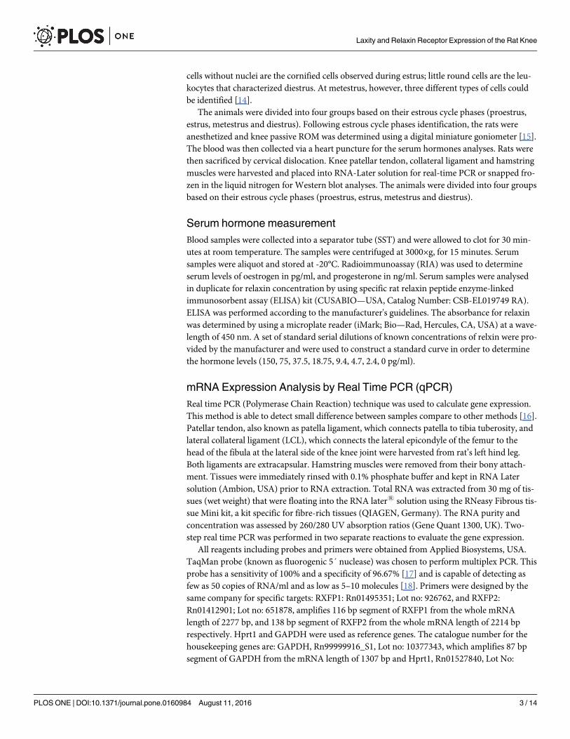
Serum hormone measurementBlood samples were collected into a separator tube (SST) and were allowed to clot for 30 min-utes at room temperature. The samples were centrifuged at 3000×g, for 15 minutes. Serumsamples were aliquot and stored at -20°C. Radioimmunoassay (RIA) was used to determineserum levels of oestrogen in pg/ml, and progesterone in ng/ml. Serum samples were analysedin duplicate for relaxin concentration by using specific rat relaxin peptide enzyme-linkedimmunosorbent assay (ELISA) kit (CUSABIO—USA, Catalog Number: CSB-EL019749 RA).ELISA was performed according to the manufacturer's guidelines. The absorbance for relaxinwas determined by using a microplate reader (iMark; Bio—Rad, Hercules, CA, USA) at a wave-length of 450 nm. A set of standard serial dilutions of known concentrations of relxin were pro-vided by the manufacturer and were used to construct a standard curve in order to determinethe hormone levels (150, 75, 37.5, 18.75, 9.4, 4.7, 2.4, 0 pg/ml).
mRNA Expression Analysis by Real Time PCR (qPCR)Real time PCR (Polymerase Chain Reaction) technique was used to calculate gene expression.This method is able to detect small difference between samples compare to other methods [16].Patellar tendon, also known as patella ligament, which connects patella to tibia tuberosity, andlateral collateral ligament (LCL), which connects the lateral epicondyle of the femur to thehead of the fibula at the lateral side of the knee joint were harvested from rat’s left hind leg.Both ligaments are extracapsular. Hamstring muscles were removed from their bony attach-ment. Tissues were immediately rinsed with 0.1% phosphate buffer and kept in RNA Latersolution (Ambion, USA) prior to RNA extraction. Total RNA was extracted from 30 mg of tis-sues (wet weight) that were floating into the RNA later1 solution using the RNeasy Fibrous tis-sue Mini kit, a kit specific for fibre-rich tissues (QIAGEN, Germany). The RNA purity andconcentration was assessed by 260/280 UV absorption ratios (Gene Quant 1300, UK). Two-step real time PCR was performed in two separate reactions to evaluate the gene expression.
All reagents including probes and primers were obtained from Applied Biosystems, USA.TaqMan probe (known as fluorogenic 5´ nuclease) was chosen to perform multiplex PCR. Thisprobe has a sensitivity of 100% and a specificity of 96.67% [17] and is capable of detecting asfew as 50 copies of RNA/ml and as low as 5–10 molecules [18]. Primers were designed by thesame company for specific targets: RXFP1: Rn01495351; Lot no: 926762, and RXFP2:Rn01412901; Lot no: 651878, amplifies 116 bp segment of RXFP1 from the whole mRNAlength of 2277 bp, and 138 bp segment of RXFP2 from the whole mRNA length of 2214 bprespectively. Hprt1 and GAPDH were used as reference genes. The catalogue number for thehousekeeping genes are: GAPDH, Rn99999916_S1, Lot no: 10377343, which amplifies 87 bpsegment of GAPDH from the mRNA length of 1307 bp and Hprt1, Rn01527840, Lot No:
Laxity and Relaxin Receptor Expression of the Rat Knee
PLOS ONE | DOI:10.1371/journal.pone.0160984 August 11, 2016 3 / 14

1118680, which amplifies 67 bp, segment from the mRNA length of 1260 bp. The target assaywas validated in-silico using whole rat genome sequences and in-vitro by the using whole ratcDNA sequences to ensure target sequences were detected (Applied Biosystems, USA).
All amplification experiments were done in 3 biological replicates. Amplification programinclude 15 minutes at 48°C (reverse transcriptase), 10 minutes at 95°C activation of ampli Taqgold DNA polymerase, denaturation at 95°C for 15 second and annealing at 60°C for 1 minute.Denaturation and annealing steps were performed for 40 cycles. Step One Plus real time PCRmachine, TaqMan Fast Advanced Master Mix and assays were purchased from Applied Biosys-tems, USA. The assay used (TaqMan1-Rxfp1: Rn01495351; Lot No: 926762 and Rxfp2:Rn01412901; Lot No: 651878) amplifies a 116 bp segment of Rxfp1 from the whole mRNAlength of 2277 and 138 bp segment of Rxfp2 from the whole mRNA length of 2214 bp. The cat-alogue number for the housekeeping genes were: Gapdh, Rn99999916_S1, Lot No: 10377343which amplifies a 87 bp segment of Gapdh from the mRNA length of 1307 bp and Hprt1,Rn01527840, Lot No: 1118680 which amplifies a 67 bp segment from the mRNA length of1260 bp.
Selection of stable reference genes for normalization of qPCR data analysis was performedby initial screening of six commonly used genes. Then two most stable and suitable genes(Hprt1 and GAPDH) were selected for further qPCR experiments, in which geometric meansof chosen reference genes used for analysis [19]. Data was analysed according to the Compara-tive Ct (2-ΔΔCt) method [20], where amplification of the target and of the reference genes weremeasured in the samples and reference. Measurements were normalized using Gen Ex soft-ware. The relative quantity of target was determined by comparing the normalized target quan-tity in each sample to the normalized target quantity in the reference. Data Assist v3 softwarefrom Applied Biosystems, USA was used to calculate the RNA folds changes.
Protein expression analysis by Western blottingPatellar tendon, hamstring muscles and collateral ligaments were removed from their attach-ment of the rat left hind leg. These tissues were then snapped frozen in the liquid nitrogen andstored at -80°C prior to protein extraction. Total amount of protein was extracted from 50mgtissue (wet weight). Following total protein extraction with PRO-PREP (Intron, UK), equalamount of protein from each tissue lysate were mixed with a loading dye, and separated bySDS-PAGE 12%. The protein was then transferred onto a PVDF membrane (BIORAD, UK)and blocked with 5% BSA for 90 minutes at a room temperature. The membrane was exposedto rabbit polyclonal primary RXFP-1/LGR7 antibody (Abcam, UK), mouse polyclonal RXFP2/LGR8 (Abcam, UK) and rabbit anti-mouse beta actin (Abcam, UK) diluted at 1:1000 in PBScontaining 1% BSA and tween-20 for 90 minutes. Blots were washed three times with each last-ing for five minutes, and were then incubated with anti-rabbit or anti-mouse horseradish per-oxidase conjugated secondary antibodies (Abcam, UK) at a dilution of 1:2000, for 1 hour. Themembrane was then washed and subjected to Opti-4CN™ Substrate (Bio-Rad, USA) to visualizethe protein bands. Photos of each blot were captured using a gel documentation system anddensity of each band was determined using Image J software. Ratio of each target band/β actinwas calculated and was considered as the expression level of the target proteins.
Antibody validationThe primary antibodies for RXFP1 and RXFP2 proteins were validated using western blot inrat tissue. Total proteins concentration of 5, 2.5, 1.25, and 0.5 μg were loaded from a grosslydissected rat tissue, measured by BCA protein assay (Thermo Fischer Scientific, Waltham, MA,USA).
Laxity and Relaxin Receptor Expression of the Rat Knee
PLOS ONE | DOI:10.1371/journal.pone.0160984 August 11, 2016 4 / 14

Measurement of Knee Passive ROMRat’s knee ROM was determined using a digital miniature goniometer [15]. Following estrousphase identification, the rats were anesthetized using ketamine and xylazine (80 + 8 mg/kg).The depth of anesthesia was confirmed from the lack of response to a painful stimuli, whichwas applied to the plantar surface [21]. Maintenance of animals in deep anesthesia is importantas to prevent active muscle contraction in response to a painful stimuli, which will then resultin the increased resistance towards the passive traction. Hip and knee joints were fixed andrested on the sensor. Meanwhile, the lower leg (knee up to ankle) was tied in-situ onto thedevice arm. Knee passive ROM was measured by pulling the device arm in a clockwise direc-tion at a minimum constant force of 12 ± 1 Newton (N) using a mini digital Newton meter(American Weight, Loiusville, KY, USA; model: AMW-SR-1KG). The changes in value offorce can be observed from the screen of the newton meter. Once the force exceeded 13 N, trac-tion was immediately terminated and the angle obtained was recorded which represents thepassive knee extension. Determination of the angle was made in different study groups. Anglewas analyzed by using Torque principle. Torque is defined as the tendency of a force to rotatean object around a fixed axis which is given by “τ = r Fsinθ” formula [22]. r: rat leg length (r);F: force (A); θ: angle between the applied force and rat’s ROM.
Statistical analysisAll data were presented as mean ± standard error of mean (SEM). Shapiro-Wilk test wasapplied to evaluate data normality and homogeneity distribution. One way ANOVA, withTukey’s post-hoc test was used to determine pair wise difference, and the level of significancewas set at p<0.05. Pearson and Spearman correlation coefficients were applied to determinecorrelation between knee range of motion and hormones levels. Density of each band in West-ern blot was analyzed by using Image J software, and the results were presented as the ratio oftarget proteins to β-actin. SPSS 18.0 statistical package was used in this study.
Results
Knee Passive ROMFig 1 shows knee passive ROM in intact female rats at different phases of estrous cycle. Thepassive knee ROM was significantly higher at proestrus and diestrus compared to estrus(p<0.05).
RXFP1 & RXFP2 expressions in hamstring muscleFig 2 shows (A) RXFP1 mRNA, (B) RXFP2 mRNA and (C) RXFP1 and RXFP2 over β-actinprotein expression in the hamstring muscles of intact rats at different phases of the estrouscycle. Our findings indicate that RXFP1 and RXFP2 mRNA and protein levels were signifi-cantly higher at proestrus and diestrus compared to estrus.
RXFP1 & RXFP2 expression in patellar tendonFig 3 shows (A) RXFP1 mRNA, (B) RXFP2 mRNA and (C) RXFP1 and RXFP2 over β-actinprotein expression in the patellar tendon of intact rats at different phases of the estrous cycle.Our findings indicate that RXFP1 and RXFP2 mRNA and protein levels were higher at proes-trus and diestrus compared to estrus. No significant differences in the expression levels ofRXFP1 were noted between proestrus and diestrus phases of the estrous cycle.
Laxity and Relaxin Receptor Expression of the Rat Knee
PLOS ONE | DOI:10.1371/journal.pone.0160984 August 11, 2016 5 / 14

Fig 1. Knee passive ROM at different phases of the estrous cycle. The knee ROMwas the highest atproestrus and diestrus and the lowest at estrus. Ps- proestrus; Es-estrus; Ms; metestrus; Ds; diestrus. Datawere expressed as mean ± SEM and n = 6 per study group. *p<0.05 as compared to estrus.
doi:10.1371/journal.pone.0160984.g001
Fig 2. RXFP1& RXFP2mRNA and protein expressions in the hamstring muscles. The expression of (A) RXFP1mRNA (B) RXFP2 mRNA and (C) the ratio of RXFP1& RXFP2/ β-actin and Western blot images of these proteins in thehamstring muscle homogenates. The highest RXFP1 and RXFP2 mRNA and protein expressions were observed atproestrus followed by diestrus. *p<0.05 as compared to estrus for the respective isoforms. Ps-proestrus; Es-estrus; Ms;metestrus; Ds; diestrus. Data were expressed as mean ± SEM and n = 6 per study group.
doi:10.1371/journal.pone.0160984.g002
Laxity and Relaxin Receptor Expression of the Rat Knee
PLOS ONE | DOI:10.1371/journal.pone.0160984 August 11, 2016 6 / 14

RXFP1 & RXFP2 expression in collateral ligamentFig 4 shows (A) RXFP1 mRNA, (B) RXFP2 mRNA and (C) RXFP1 and RXFP2 over β-actinprotein expression in collateral ligaments of intact rats at different phases of estrous cycle. Ourfindings indicate that RXFP1 and RXFP2 mRNA and protein levels were significantly higher atproestrus and diestrus compared to estrus.
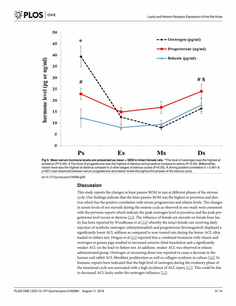
Mean serum hormones levels in intact female ratsTable 1 shows serum levels of oestrogen, progesterone and relaxin at different phases of estrouscycle in non ovariectomized rats. The level of oestrogen was the highest at proestrus compareto 3 other stages of estrous cycles (P<0.05). The level of progesterone was the highest at dies-trus compare to estrus (P<0.05), however there was no significant in progesterone level differ-ence between proestrus and diestrus. Meanwhile, relaxin level was the highest at diestruscompare to 3 other stages of estrous cycles (P<0.05), whereas no remarkable differencesobserved in relaxin level at proestrus, estrus and metstrus stages (Fig 5). A strong positive cor-relation (r = 0.901) was observed between serum progesterone and relaxin levels throughoutthe phases of the estrous cycle (Fig 5).
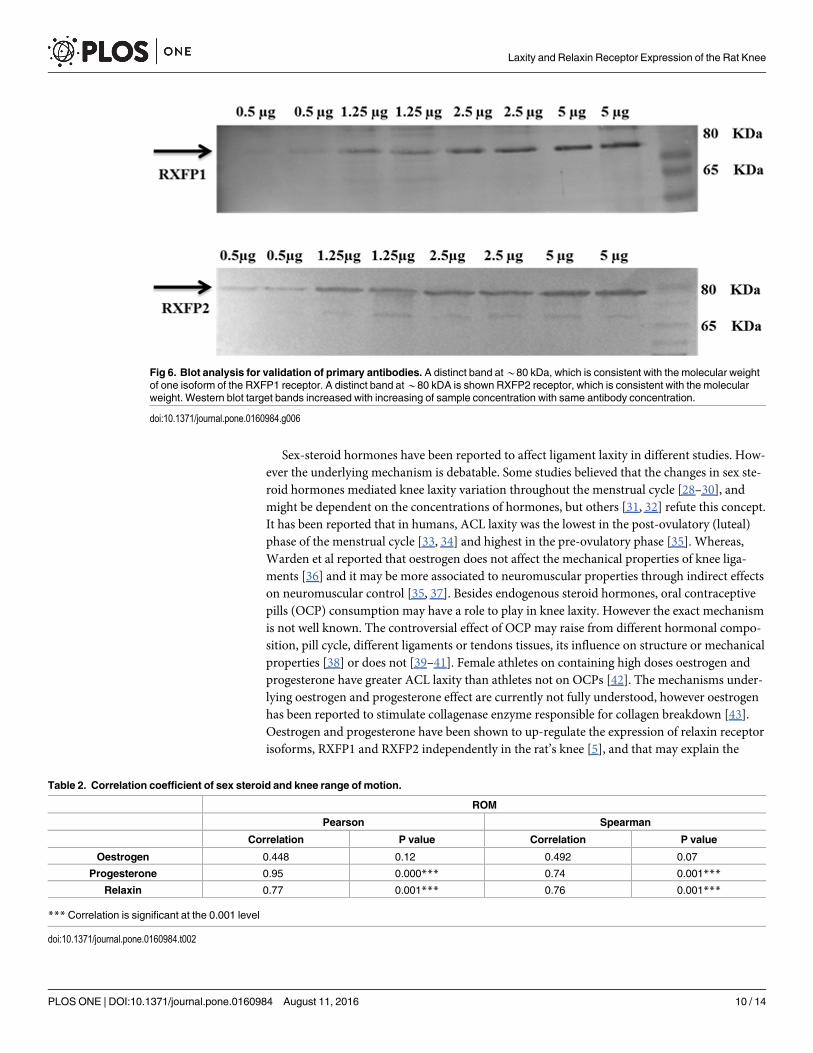
Validation of the AntibodiesThe result shows that the western blot target bands are increased with increasing of proteinsconcentration with same antibody concentration (Fig 6).
Fig 3. RXFP1& RXFP2mRNA and protein expressions in the patellar tendon. The expression of (A) RXFP1mRNA(B) RXFP2mRNA and (C) ratio of RXFP1& RXFP2/ β-actin andWestern blot bands of these proteins in the patellartendon homogenates. The mRNA and protein expression were the highest at proestrus and diestrus phases. *p<0.05as compared to estrus and metestrus for the respective isoforms. Ps-prosterous; Es-estrus; Ms; metestrus; Ds; diestrus.Data were expressed as mean ± SEM and n = 6 per study group.
doi:10.1371/journal.pone.0160984.g003
Laxity and Relaxin Receptor Expression of the Rat Knee
PLOS ONE | DOI:10.1371/journal.pone.0160984 August 11, 2016 7 / 14

Correlation between sex-steroid levels and knee ROMTable 2 shows correlation between sex-steroid levels and knee ROM throughout the estrouscycle in the rats. There was no significant correlations between oestrogen and ROM whereasstrong positive correlations was observed between progesterone/relaxin levels and ROM.
Fig 4. RXFP1& RXFP2mRNA and protein expression levels in the collateral ligament. The expression of (A)RXFP1 mRNA (B) RXFP2 mRNA and (C) RXFP1 & RXFP2/β-actin andWestern blot bands in the collateralligament homogenates. The expression of mRNA and protein were the highest at proestrus followed by diestrus.*p<0.05 as compared to estrus and metestrus for the respective isoforms. Ps-proestrus; Es- estrus; Ms; metestrus;Ds; diestrus. Data were expressed as mean ± SEM and n = 6 per study group.
doi:10.1371/journal.pone.0160984.g004
Table 1. Serum hormone level in non ovariectomized rat at different phases of the estrous cycle.
Hormones Estrous cycle Hormone level
(Mean ± SEM)
Oestrogen Proestrus 39.38 ± 4.56 pg/ml
Estrus 12.64 ± 2.98 pg/ml
Metestrus 8.03 ± 1.09 pg/ml
Diestrus 16.49 ± 2.32 pg/ml
Progesterone Proestrus 22.87 ± 2.65 ng/ml
Estrus 14.92 ± 2.05 ng/ml
Metestrus 16.96 ± 2.73 ng/ml
Diestrus 24.03 ± 3.08 ng/ml
Relaxin Proestrus 12.34 ± 3.96 pg/ml
Estrus 8.04 ± 1.17 pg/ml
Metestrus 9.48 ± 1.75 pg/ml
Diestrus 18.03 ± 2.85 pg/ml
doi:10.1371/journal.pone.0160984.t001
Laxity and Relaxin Receptor Expression of the Rat Knee
PLOS ONE | DOI:10.1371/journal.pone.0160984 August 11, 2016 8 / 14
DiscussionThis study reports the changes in knee passive ROM in rats at different phases of the estrouscycle. Our findings indicate that the knee passive ROM was the highest at proestrus and dies-trus which has the positive correlation with serum progesterone and relaxin levels. The changesin serum levels of sex-steroids during the oestrus cycle as observed in our study were consistentwith the previous reports which indicate the peak oestrogen level at proestrus and the peak pro-gesterone level occurs at diestrus [23]. The influence of female sex steroids on female knee lax-ity has been reported by Woodhouse et al [24] whereby the intact female rats receiving dailyinjection of synthetic oestrogen (ethinylestradiol) and progesterone (levonogestrel) displayed asignificantly lower ACL stiffness as compared to non-treated rats during the femur-ACL-tibialoaded-to-failure test. Dragoo et al [25] reported that a combined treatment with relaxin andoestrogen to guinea pigs resulted in increased anterior tibial translation and a significantlyweaker ACL on the load-to-failure test. In addition, weaker ACL was observed in relaxinadministrated group. Oestrogen at increasing doses was reported to cause a decrease in thehuman and rabbit ACL fibroblast proliferation as well as collagen synthesis in culture [26]. Inhumans, reports have indicated that the high level of oestrogen during the ovulatory phase ofthe menstrual cycle was associated with a high incidence of ACL injury [27]. This could be dueto decreased ACL laxity under the oestrogen influence [12].
Fig 5. Mean serum hormone levels are presented as mean + SEM in intact female rats. *The level of oestrogen was the highest atproestrus (P<0.05). # The level of progesterone was the highest at diestrus and proestrus compare to estrus (P<0.05). $Meanwhile,relaxin level was the highest at diestrus compare to 3 other stages of estrous cycles (P<0.05). A strong positive correlation (r = 0.901 &p<001) was observed between serum progesterone and relaxin levels throughout the phases of the estrous cycle.
doi:10.1371/journal.pone.0160984.g005
Laxity and Relaxin Receptor Expression of the Rat Knee
PLOS ONE | DOI:10.1371/journal.pone.0160984 August 11, 2016 9 / 14
Sex-steroid hormones have been reported to affect ligament laxity in different studies. How-ever the underlying mechanism is debatable. Some studies believed that the changes in sex ste-roid hormones mediated knee laxity variation throughout the menstrual cycle [28–30], andmight be dependent on the concentrations of hormones, but others [31, 32] refute this concept.It has been reported that in humans, ACL laxity was the lowest in the post-ovulatory (luteal)phase of the menstrual cycle [33, 34] and highest in the pre-ovulatory phase [35]. Whereas,Warden et al reported that oestrogen does not affect the mechanical properties of knee liga-ments [36] and it may be more associated to neuromuscular properties through indirect effectson neuromuscular control [35, 37]. Besides endogenous steroid hormones, oral contraceptivepills (OCP) consumption may have a role to play in knee laxity. However the exact mechanismis not well known. The controversial effect of OCP may raise from different hormonal compo-sition, pill cycle, different ligaments or tendons tissues, its influence on structure or mechanicalproperties [38] or does not [39–41]. Female athletes on containing high doses oestrogen andprogesterone have greater ACL laxity than athletes not on OCPs [42]. The mechanisms under-lying oestrogen and progesterone effect are currently not fully understood, however oestrogenhas been reported to stimulate collagenase enzyme responsible for collagen breakdown [43].Oestrogen and progesterone have been shown to up-regulate the expression of relaxin receptorisoforms, RXFP1 and RXFP2 independently in the rat’s knee [5], and that may explain the
Fig 6. Blot analysis for validation of primary antibodies. A distinct band at*80 kDa, which is consistent with the molecular weightof one isoform of the RXFP1 receptor. A distinct band at*80 kDA is shown RXFP2 receptor, which is consistent with the molecularweight. Western blot target bands increased with increasing of sample concentration with same antibody concentration.
doi:10.1371/journal.pone.0160984.g006
Table 2. Correlation coefficient of sex steroid and knee range of motion.
ROM
Pearson Spearman
Correlation P value Correlation P value
Oestrogen 0.448 0.12 0.492 0.07
Progesterone 0.95 0.000*** 0.74 0.001***
Relaxin 0.77 0.001*** 0.76 0.001***
*** Correlation is significant at the 0.001 level
doi:10.1371/journal.pone.0160984.t002
Laxity and Relaxin Receptor Expression of the Rat Knee
PLOS ONE | DOI:10.1371/journal.pone.0160984 August 11, 2016 10 / 14

increased laxity under the influence of both hormones. However, both hormones have alsobeen shown to inhibit the collagen synthesis in the tendon and skeletal muscles [44] whichmight also contribute towards a decrease in knee laxity. Therefore, variation of the reportedeffect of oestrogen and progesterone in human or animal model might be related to interactionbetween these two hormones.
Our findings have shown that the serum levels of relaxin were the highest at diestrus whichwere consistent with the reported increase in relaxin synthesis by the corpus luteum formedafter ovulation and was maintained throughout diestrus phase of the estrous cycle [45]. In spe-cies such as rodents, relaxin plays an important role in modulating joint laxity [1]. However, inhumans, its role is still debatable. The latter was supported by an observation, where a relation-ship was observed between peripheral joint laxity and serum relaxin levels in the females dur-ing the pregnancy period [46]. Although relaxin might not play important role in determiningthe ligament laxity in humans, the expression of relaxin receptors has been reported in humanACL [4] and human ACL fibroblast [47]. In rats however, relaxin most likely plays an impor-tant role in modulating knee laxity in view that its receptor expression has been reported bothin the patellar tendon and collateral ligaments [15]. Additionally, relaxin has been reported tostimulate the activity of matrix metalloproteinases (MMPs) through induction of collagenase-1and stromelysin-1, which are involved in the collagen breakdown [48, 49]. We postulated thatthe highest level of relaxin and progesterone during diestrus phase and its moderately highlevel during proestrus phase might contribute towards the increase in knee passive ROM inrats. With regards to the role of progesterone in knee passive ROM in our previous study [50],which was suppressed by its antagonist (mifepristone), so progesterone may act in parallel withrelaxin since this is positive correlation between these two hormones throughout phases of theestrous cycle.
Our findings have further shown the changes in the expression of two main relaxin receptorisoforms, RXFP1 and RXFP2 in the hamstring muscles, patellar tendon and collateral liga-ments which could affect knee laxity. The expression of RXFP1 and RXFP2 were the highestduring proestrus followed by diestrus phase. In the hamstring muscles and patellar tendon,RXFP1 is the main isoform while in the collateral ligament, RXFP2 expression exceeds RXFP1.The up-regulation of both relaxin receptor isoforms may contribute towards increased in kneelaxity both at proestrus and diestrus phases of the cycle. Changes in these isoforms expressionin the hamstring muscle is important as this could affect knee extension.
ConclusionTo summary, we have demonstrated that changes in knee ROM in rats is fluctuates with theostrous cycle. Furthermore, the increased knee laxity at proestrus and diestrus is correlatedwith the high levels of serum progesterone, relaxin, and relaxin receptors expression in the ratknee tissues. The results can be implicated to understand the non-traumatic knee injury associ-ated with the level of sex-steroid hormones. In view of this, more studies are warranted toinvestigate changes in knee laxity in relation to combination of sex steroid hormones or indi-vidual in cellular and molecular levels with respect to different tissues, gender, subjects.
Author Contributions
Conceptualization: FD AY SM.
Funding acquisition: SM.
Investigation: AY RS MAA.
Laxity and Relaxin Receptor Expression of the Rat Knee
PLOS ONE | DOI:10.1371/journal.pone.0160984 August 11, 2016 11 / 14

Methodology: FD RS KG.
Project administration: FD.
Resources: AY SM.
Software: FD PDMAA.
Writing - original draft: FD PD KGMAA.
Writing - review & editing: FD AY SM.
References1. Sherwood O, Downing S, Guico-LammM, Hwang J, O'Day-Bowman M, Fields P. The physiological
effects of relaxin during pregnancy: studies in rats and pigs. Oxford reviews of reproductive biology.1992; 15:143–89.
2. Madan MS, Liu ZJ, Gu GM, King GJ. Effects of human relaxin on orthodontic tooth movement and peri-odontal ligaments in rats. American Journal of Orthodontics and Dentofacial Orthopedics. 2007; 131(1):8.e1–8.e10.
3. Dragoo JL, Castillo TN, Braun HJ, Ridley BA, Kennedy AC, Golish SR. Prospective CorrelationBetween Serum Relaxin Concentration and Anterior Cruciate Ligament Tears Among Elite CollegiateFemale Athletes. The American Journal of Sports Medicine. 2011; 39(10):2175–80. doi: 10.1177/0363546511413378 PMID: 21737831
4. Dragoo JL, Lee RS, Benhaim P, Finerman GA, Hame SL. Relaxin receptors in the human female ante-rior cruciate ligament. The American journal of sports medicine. 2003; 31(4):577–84. PMID: 12860548
5. Dehghan F, Muniandy S, Yusof A, Salleh N. Sex-steroid regulation of relaxin receptor isoforms (RXFP1& RXFP2) expression in the patellar tendon and lateral collateral ligament of female WKY rats. Int JMed Sci. 2014a 11(2):180–91. doi: 10.7150/ijms.6283
6. Ohtera K, Zobitz ME, Luo Z-P, Morrey BF, O'Driscoll SW, Ramin KD, et al. Effect of pregnancy on jointcontracture in the rat knee. Journal of Applied Physiology. 2002; 92(4):1494–8. PMID: 11896015
7. Hart DA, Reno C, Frank CB, Shrive NG. Pregnancy affects cellular activity, but not tissue mechanicalproperties, in the healing rabbit medial collateral ligament. Journal of Orthopaedic Research. 2000; 18(3):462–71. PMID: 10937635
8. Aldabe D, Ribeiro DC, Milosavljevic S, Bussey MD. Pregnancy-related pelvic girdle pain and its rela-tionship with relaxin levels during pregnancy: a systematic review. European Spine Journal. 2012; 21(9):1769–76. PMID: 22310881
9. Strickland SM, Belknap TW, Turner SA, Wright TM, Hannafin JA. Lack of hormonal influences onmechanical properties of sheep knee ligaments. The American journal of sports medicine. 2003; 31(2):210–5. PMID: 12642254
10. Zazulak BT, Paterno M, Myer GD, Romani WA, Hewett TE. The effects of the menstrual cycle on ante-rior knee laxity. Sports medicine. 2006; 36(10):847–62. PMID: 17004848
11. Park S-K, Stefanyshyn DJ, Loitz-Ramage B, Hart DA, Ronsky JL. Changing hormone levels during themenstrual cycle affect knee laxity and stiffness in healthy female subjects. The American journal ofsports medicine. 2009; 37(3):588–98. doi: 10.1177/0363546508326713 PMID: 19174550
12. Shultz S, Sander T, Kirk S, Perrin D. Sex differences in knee joint laxity change across the female men-strual cycle. J Sports Med Phys Fitness. 2005; 45(4):594. PMID: 16446695
13. Garber JC, Barbee R, Bielitzki JT, Clayton L, Donovan J, Hendriksen C, et al. Guide for the care anduse of laboratory animals2010. 220 p.
14. Marcondes FK, Bianchi FJ, Tanno AP. Determination of the estrous cycle phases of rats: some helpfulconsiderations. Braz J Biol. 2002; 62(4A):609–14. Epub 2003/03/28. PMID: 12659010.
15. Dehghan F, Muniandy S, Yusof A, Salleh N. Testosterone Reduces Knee Passive Range of Motionand Expression of Relaxin Receptor Isoforms via 5α-Dihydrotestosterone and Androgen ReceptorBinding. International journal of molecular sciences. 2014; 15(3):4619–34. doi: 10.3390/ijms15034619PMID: 24642882
16. WongML, Medrano JF. Real-time PCR for mRNA quantitation. Biotechniques. 2005; 39(1):75. PMID:16060372
17. Tsai Y-L, Wang H-TT, Chang H-FG, Tsai C-F, Lin C-K, Teng P-H, et al. Development of TaqManProbe-Based Insulated Isothermal PCR (iiPCR) for Sensitive and Specific On-Site Pathogen Detection.PloS one. 2012; 7(9):e45278. doi: 10.1371/journal.pone.0045278 PMID: 23049781
Laxity and Relaxin Receptor Expression of the Rat Knee
PLOS ONE | DOI:10.1371/journal.pone.0160984 August 11, 2016 12 / 14

18. Hofmann-Lehmann R, Williams AL, Swenerton RK, Li PL, Rasmussen RA, Chenine AL, et al. Quantita-tion of simian cytokine and beta-chemokine mRNAs, using real-time reverse transcriptase-polymerasechain reaction: variations in expression during chronic primate lentivirus infection. AIDS research andhuman retroviruses. 2002; 18(9):627–39. PMID: 12079558
19. Vandesompele J, De Preter K, Pattyn F, Poppe B, Van Roy N, De Paepe A, et al. Accurate normaliza-tion of real-time quantitative RT-PCR data by geometric averaging of multiple internal control genes.Genome biology. 2002; 3(7):Research0034. Epub 2002/08/20. PMID: 12184808; PubMed CentralPMCID: PMCPMC126239.
20. WongML, Medrano JF. Real-time PCR for mRNA quantitation. Biotechniques. 2005; 39:1–10.
21. Flecknell P. Laboratory animal anaesthesia: Academic Press; 2009.
22. Chabay RW. Matter and Interactions: Modern Mechanics / Electric and Magnetic Interactions: JohnWiley & Sons Incorporated; 2015.
23. Staley K, Scharfman H. A woman's prerogative. Nature neuroscience. 2005; 8(6):697–9. PMID:15917829
24. Woodhouse E, Schmale GA, Simonian P, Tencer A, Huber P, Seidel K. Reproductive hormone effectson strength of the rat anterior cruciate ligament. Knee Surgery, Sports Traumatology, Arthroscopy.2007; 15(4):453–60. PMID: 17187283
25. Dragoo JL, Padrez K, Workman R, Lindsey DP. The effect of relaxin on the female anterior cruciate lig-ament: Analysis of mechanical properties in an animal model. The Knee. 2009; 16(1):69–72. doi: 10.1016/j.knee.2008.09.005 PMID: 18964043
26. Thornton GM, Bailey SJ, Shao X, Morck D, Hart DA, Achari Y, editors. Influence of Early Ovariohyster-ectomy on the Mechanical Properties of Rabbit Medial Collateral Ligament. ASME 2007 Summer Bio-engineering Conference; 2007: American Society of Mechanical Engineers.
27. Beynnon BD, Johnson RJ, Braun S, Sargent M, Bernstein IM, Skelly JM, et al. The RelationshipBetween Menstrual Cycle Phase and Anterior Cruciate Ligament Injury: A Case-Control Study of Rec-reational Alpine Skiers. The American journal of sports medicine. 2006; 34(5):757–64. doi: 10.1177/0363546505282624 PMID: 16436538
28. Shultz SJ, Kirk SE, Johnson ML, Sander TC, Perrin DH. Relationship between sex hormones and ante-rior knee laxity across the menstrual cycle. Medicine and science in sports and exercise. 2004; 36(7):1165–74. Epub 2004/07/06. PMID: 15235320; PubMed Central PMCID: PMCPMC1993893.
29. Deie M, Sakamaki Y, Sumen Y, Urabe Y, Ikuta Y. Anterior knee laxity in young women varies with theirmenstrual cycle. International orthopaedics. 2002; 26(3):154–6. PMID: 12073107
30. Romani W, Patrie J, Curl LA, Flaws JA. The correlations between estradiol, estrone, estriol, progester-one, and sex hormone-binding globulin and anterior cruciate ligament stiffness in healthy, activefemales. Journal of women's health (2002). 2003; 12(3):287–98. Epub 2003/06/14. doi: 10.1089/154099903321667627 PMID: 12804359.
31. Beynnon BD, Bernstein IM, Belisle A, Brattbakk B, Devanny P, Risinger R, et al. The effect of estradioland progesterone on knee and ankle joint laxity. The American journal of sports medicine. 2005; 33(9):1298–304. PMID: 16002485
32. Karageanes SJ, Blackburn K, Vangelos ZA. The association of the menstrual cycle with the laxity of theanterior cruciate ligament in adolescent female athletes. Clinical Journal of Sport Medicine. 2000; 10(3):162–8. PMID: 10959925
33. Romani W, Patrie J, Curl LA, Flaws JA. The correlations between estradiol, estrone, estriol, progester-one, and sex hormone-binding globulin and anterior cruciate ligament stiffness in healthy, activefemales. Journal of Women's Health. 2003; 12(3):287–98. PMID: 12804359
34. Park S-K, Stefanyshyn DJ, Ramage B, Hart DA, Ronsky JL. Relationship between knee joint laxity andknee joint mechanics during the menstrual cycle. Br J Sports Med. 2009; 43(3):174–9. doi: 10.1136/bjsm.2008.049270 PMID: 18728055
35. Hewett TE, Zazulak BT, Myer GD. Effects of the menstrual cycle on anterior cruciate ligament injuryrisk: a systematic review. Am J Sports Med. 2007; 35(4):659–68. Epub 2007/02/13. doi: 10.1177/0363546506295699 PMID: 17293469.
36. Warden SJ, Saxon LK, Castillo AB, Turner CH. Knee ligament mechanical properties are not influencedby estrogen or its receptors. American journal of physiology Endocrinology and metabolism. 2006; 290(5):E1034–40. Epub 2005/12/01. doi: 10.1152/ajpendo.00367.2005 PMID: 16317027.
37. Hewett TE. Neuromuscular and hormonal factors associated with knee injuries in female athletes.Strategies for intervention. Sports medicine. 2000; 29(5):313–27. Epub 2000/06/07. PMID: 10840866.
38. Martineau PA, Al-Jassir F, Lenczner E, Burman ML. Effect of the oral contraceptive pill on ligamentouslaxity. Clinical journal of sport medicine: official journal of the Canadian Academy of Sport Medicine.2004; 14(5):281–6. Epub 2004/09/21. PMID: 15377967.
Laxity and Relaxin Receptor Expression of the Rat Knee
PLOS ONE | DOI:10.1371/journal.pone.0160984 August 11, 2016 13 / 14

39. Lee H, Petrofsky JS, Daher N, Berk L, Laymon M. Differences in anterior cruciate ligament elasticityand force for knee flexion in women: oral contraceptive users versus non-oral contraceptive users. EurJ Appl Physiol. 2014; 114(2):285–94. Epub 2013/11/19. doi: 10.1007/s00421-013-2771-z PMID:24240566.
40. Hansen M, Couppe C, Hansen CS, Skovgaard D, Kovanen V, Larsen JO, et al. Impact of oral con-traceptive use and menstrual phases on patellar tendon morphology, biochemical composition, andbiomechanical properties in female athletes. J Appl Physiol. 2013 114(8):998–1008. doi: 10.1152/japplphysiol.01255.2012 PMID: 23429870
41. Pokorny MJ, Smith TD, Calus SA, Dennison EA. Self-reported oral contraceptive use and peripheraljoint laxity. The Journal of orthopaedic and sports physical therapy. 2000; 30(11):683–92. Epub 2000/12/05. doi: 10.2519/jospt.2000.30.11.683 PMID: 11104379.
42. Lee H, Petrofsky JS, Daher N, Berk L, Laymon M. Differences in anterior cruciate ligament elasticityand force for knee flexion in women: oral contraceptive users versus non-oral contraceptive users.European journal of applied physiology. 2014; 114(2):285–94. doi: 10.1007/s00421-013-2771-z PMID:24240566
43. Rajabi M, Solomon Samuel, Poole Robin. Hormonal Regulation of Interstitial Collagenase in the Uter-ine Cervix of the Pregnant Guinea Pig*. Endocrinology. 1991; 128(2):863–71. PMID: 1846591
44. Hansen M, Miller BF, Holm L, Doessing S, Petersen SG, Skovgaard D, et al. Effect of administration oforal contraceptives in vivo on collagen synthesis in tendon and muscle connective tissue in youngwomen. Journal of Applied Physiology. 2009; 106(4):1435–43. doi: 10.1152/japplphysiol.90933.2008PMID: 18845777
45. Fields PA, Fields MJ. Ultrastructural localization of relaxin in the corpus luteum of the nonpregnant,pseudopregnant, and pregnant pig. Biology of reproduction. 1985; 32(5):1169–79. PMID: 3893554
46. Schauberger CW, Rooney BL, Goldsmith L, Shenton D, Silva PD, Schaper A. Peripheral joint laxityincreases in pregnancy but does not correlate with serum relaxin levels. American journal of obstetricsand gynecology. 1996; 174(2):667–71. PMID: 8623804
47. Faryniarz DA, Bhargava M, Lajam C, Attia ET, Hannafin JA. Quantitation of estrogen receptors andrelaxin binding in human anterior cruciate ligament fibroblasts. In Vitro Cellular & Developmental Biol-ogy-Animal. 2006; 42(7):176–81.
48. HashemG, Zhang Q, Hayami T, Chen J, WangW, Kapila S. Relaxin and β-estradiol modulate targetedmatrix degradation in specific synovial joint fibrocartilages: progesterone prevents matrix loss. Arthritisresearch & therapy. 2006; 8(4):R98.
49. Naqvi T, Duong TT, HashemG, Shiga M, Zhang Q, Kapila S. Relaxin’s induction of metalloproteinasesis associated with the loss of collagen and glycosaminoglycans in synovial joint fibrocartilaginousexplants. Arthritis Res Ther. 2005; 7(1):R1–11. PMID: 15642129
50. Dehghan F, Yusof A, Muniandy S, Salleh N. Estrogen receptor (ER)-α, β and progesterone receptor(PR) mediates changes in relaxin receptor (RXFP1 and RXFP2) expression and passive range ofmotion of rats’ knee. Environmental Toxicology and Pharmacology. 2015; 40(3):785–91. doi: 10.1016/j.etap.2015.09.004 PMID: 26447688
Laxity and Relaxin Receptor Expression of the Rat Knee
PLOS ONE | DOI:10.1371/journal.pone.0160984 August 11, 2016 14 / 14