original article: rosiglitazone diminishes the high ... · 3 mushroom research centre, university...
TRANSCRIPT

EXCLI Journal 2018;17:186-199 – ISSN 1611-2156 Received: December 27, 2017, accepted: January 22, 2018, published: February 06, 2018
186
Original article:
ROSIGLITAZONE DIMINISHES THE HIGH-GLUCOSE-INDUCED MODULATION OF 5-FLUOROURACIL CYTOTOXICITY IN
COLORECTAL CANCER CELLS
Meng-Fei Lau1,3, Shalini Vellasamy1, Kek-Heng Chua1,3, Vikineswary Sabaratnam2,3, Umah Rani Kuppusamy1,3* 1 Department of Biomedical Science, Faculty of Medicine, University of Malaya, 50603
Kuala Lumpur, Malaysia 2 Institute of Biological Science, Faculty of Science, University of Malaya, 50603 Kuala
Lumpur, Malaysia 3 Mushroom Research Centre, University of Malaya, 50603 Kuala Lumpur, Malaysia * Corresponding author: Prof. Dr. Umah Rani Kuppusamy, Department of Biomedical
Science, Faculty of Medicine, University of Malaya, 50603 Kuala Lumpur, Malaysia. Tel: +603 7967 6616/+603 7967 4900. Fax: +603 7967 6600. E-mail: [email protected]
http://dx.doi.org/10.17179/excli2018-1011
This is an Open Access article distributed under the terms of the Creative Commons Attribution License (http://creativecommons.org/licenses/by/4.0/).
ABSTRACT
Colorectal cancer (CRC) is the third most leading cause of morbidity and mortality throughout the world. 5-fluorouracil (5-FU), which is often administrated to disrupt carcinogenesis, was found to elevate blood glucose level among CRC patients. Thus, this study was conducted to evaluate the influence of rosiglitazone on antipro-liferative effect of 5-FU using cellular model. Two human colonic carcinoma cell lines (HCT 116 and HT 29) were cultured in the presence of 5-FU, rosiglitazone or in combination under normal and high glucose concentra-tion. The drug cytotoxicity was evaluated using the MTT assay whereas the assessment of cell cycle was carried out using the flow cytometry technique. Combination index (CI) method was used to determine the drug interac-tion between rosiglitazone and 5-FU. High glucose diminished the cytotoxic effect of 5-FU but at a high drug dosage, this effect could be overcome. Cell cycle analysis demonstrated that 5-FU and rosiglitazone caused G1-phase arrest and S-phase arrest, respectively. CI values indicated that rosiglitazone exerted synergistic effect on 5-FU regardless of glucose levels. This study is the first to demonstrate the influence of rosiglitazone on cytotox-icity of 5-FU under normal or high glucose level. Rosiglitazone may be a promising drug for enhancing the effi-cacy of 5-FU in the treatment of CRC associated with hyperglycemia. Keywords: rosiglitazone, 5-fluorouracil, high glucose, MTT, cell cycle, colorectal cancer cell
INTRODUCTION
Colorectal cancer (CRC) is the third most leading cause of morbidity and mortality throughout the world (Ferlay et al., 2015). It is estimated that over one million new cases are diagnosed per year, which accounts for around 10 % of all cancer incidences in both sexes.
Chemotherapy with curative intent is of-ten applied as a first-line treatment to disrupt carcinogenesis. In this context, 5-fluorouracil (5-FU) plus leucovorin is the main option for CRC patients (Goodwin and Asmis, 2009). Other active agents such as irinotecan, cape-citabine, oxaliplatin, bevacizumab, cetuxi-mab or panitumumab are also adopted in
EXCLI Journal 2018;17:186-199 – ISSN 1611-2156 Received: December 27, 2017, accepted: January 22, 2018, published: February 06, 2018
187
fluorouracil-based therapy to improve over-all survival and reduce the risk of disease recurrence. Unfortunately, the clinical effi-cacy is highly dose-dependent due to phar-macokinetic variability and it causes severe toxicities in some CRC patients despite the administration of standard drug protocol (André et al., 2004).
Based on previous epidemiological stud-ies, diabetes mellitus is suggested to be an independent risk factor for colorectal cancer. Although the relationship is not entirely un-derstood, most findings concur with a posi-tive association between diabetes and colo-rectal cancer (de Bruijn et al., 2013; Deng et al., 2012; Krämer et al., 2012; Larsson et al., 2005; Luo et al., 2012; Mills et al., 2013). Giovannucci et al. (2010) reported that plau-sible biological mechanisms underlying this association can be attributed to the effect of hyperglycemia, hyperinsulinemia or inflam-mation on cancer aetiology and progression. An improved glucose control should be an important therapeutic approach for CRC pa-tients.
Besides metformin, thiazolidinediones including rosiglitazone and pioglitazone are another class of oral antidiabetic drugs which help to protect against hyperglycemia (Yki-Järvinen, 2004). Thiazolidinediones increase insulin sensitivity by activating one or more peroxisome proliferator-activated receptors (PPARs) to regulate glucose utilization and production. Several in vitro studies indicated that rosiglitazone acts as a PPAR-gamma agonist which suppresses cell proliferation, inhibits cell invasiveness, arrests cell cycle and induces apoptosis in cancer cell lines (Cao et al., 2009, 2015; Han and Roman, 2006; He et al., 2008; Lin et al., 2007; Zhang et al., 2008a). These anticancer properties provoke the use of rosiglitazone on individu-als who developed diabetic complications during or after fluorouracil-based regimen.
A recent meta-analysis showed a de-creased risk of colorectal cancer incidence when rosiglitazone was administered to dia-betic patients (Monami et al., 2014). While high glucose can modulate cytotoxicity of 5-
FU (Ma et al., 2014), the efficacy of rosig-litazone is possibly affected by the level of hyperglycemic condition. In this study, the influence of rosiglitazone on 5-FU pretreated human colon cancer cell lines was evaluated at high glucose level. The drug interaction between rosiglitazone and 5-FU was also determined.
MATERIAL AND METHODS
Reagents Blank glucose Dulbecco's Modified Ea-
gle Medium (DMEM) (Cat. no.: 11966) and normal glucose DMEM (Cat. no: 11885) were purchased from ThermoFisher. High glucose DMEM (D5671 Sigma) was com-plemented with 4.0 mM L-glutamine, 1.0 mM sodium pyruvate, 10 % fetal bovine serum, 1 % penicillin streptomycin and 1 % amphoterin B. The stock solutions of 5-FU (F6627 Sigma) and rosiglitazone (R2408 Sigma) were prepared in phosphate-buffered saline (PBS) and dimethyl sulfoxide (DMSO) with a concentration of 5 mg/ml and 1 mg/ml respectively. MTT formazan powder (M5655 Sigma) was dissolved in PBS and diluted to 5 mg/ml. Propidium io-dide (PI, Cat. no: P1304MP) and RNAse A (12091-021) were obtained from Thermo-Fisher.
Cell culture
Two human colonic carcinoma cell lines, namely HCT 116 and HT 29, were cultured in normal glucose DMEM (Cat No: 11885) supplemented with 10 % fetal bovine serum, 1 % penicillin streptomycin and 1% am-photerin B. A human normal colon cell line (CCD-18Co) was cultured in normal glucose Minimum Essential Medium Eagle (MEME, D2279 Sigma) complemented with 4.0 mM L-glutamine, 1.0 mM sodium pyruvate, 20 % fetal bovine serum, 1 % non-essential amino acid and 1 % penicillin streptomycin. All cell lines were maintained in humidified atmos-phere of 5 % CO2 at 37±2 °C.
EXCLI Journal 2018;17:186-199 – ISSN 1611-2156 Received: December 27, 2017, accepted: January 22, 2018, published: February 06, 2018
188
Cell proliferation assay HCT 116 cells, HT 29 cells and CCD-
18Co cells were seeded into 96-well plates at a density of 2000 cells/100 µl, 2500 cells/100 µl and 3500 cells/100 µl per well respectively. After incubation at 37±2 °C for 24 h, the cells were then treated with 5-FU or rosiglitazone at various concentrations for 48 h. Next, 10 µl of MTT was added and the plates were incubated in the dark at 37±2 °C for 3 h. After solubilizing the formazan crys-tals in 100 µl DMSO, absorbance was meas-ured at 560 nm. The cell proliferation was calculated as follows: {[absorbance of treat-ed group-absorbance of blank] ⁄ [absorbance of control group-absorbance of blank]} × 100. The IC20 and IC50 were determined through linear regression analysis. Cell cycle assay
For the cell cycle assay, drug treatment was carried out in 12-well culture plates with a density of 40, 000 cells/ml/well (HCT 116) and 55, 000 cells/ml/well (HT 29) at 37±2°C for 48 h. The cells were harvested and fixed in 2 ml of cold 70 % (v/v) absolute ethanol and stored overnight at -20 °C. Then, the fixed cells were washed twice with PBS. Cell staining was done by sequentially add-ing 450 µl PBS, 25 µl RNAse A (1 mg/ml) and 50 µl PI (0.1 mg/ml). The stained cells were incubated in the dark at room tempera-ture (25±2 °C) for 30 min and further exam-ined by a flow cytometer (BD FACScanto II) integrated with BD FACSDiva Software. Modfit LT 2.0 was used to analyze DNA content histograms. Experimental design
First, the cell proliferation of HCT 116 and HT 29 under normal glucose (NG, 5.5mM) and high glucose (HG, 25mM) cul-ture conditions for 48 h was carried out. The complete blank glucose DMEM containing 1.0 mM sodium pyruvate, 10 % fetal bovine serum, 1 % penicillin streptomycin and 1 % amphoterin B served as a negative control whereas NG DMEM+19.5 mM D-mannitol and blank glucose DMEM+25 mM D-
mannitol were the osmotic controls. The cell proliferation of CCD-18Co cells was evalu-ated at different glucose concentrations with NG MEME+19.5 mM D-mannitol as an os-motic control.
To study the effect of glucose on drug cytotoxicity, both cancer and normal cells lines were pre-treated with 0.2 µg/ml 5-FU for 24 h. Then, spent culture media was aspi-rated and replaced with the following exper-imental media: (i)NG medium+0.2 µg/ml 5-FU; (ii)NG medium+2 µg/ml 5-FU; (iii)HG medium+0.2 µg/ml 5FU; (iv)HG medium+2 µg/ml 5-FU. The control group was 5-FU pre-treated cells in NG medium without any drug treatment for subsequent 48 h incuba-tion. The same experiment was repeated with treatments of rosiglitazone alone and a com-bination of 5-FU and rosiglitazone. In this study, low dose was defined as a concentra-tion close to IC20 of the drug (Kashif et al., 2015). Statistical analysis
SPSS Statistics 17.0 software, ANOVA followed by Duncan Multiple Range Test was performed to analyse the data. Value of p< 0.05 was considered statistically signifi-cant. All results were expressed as mean±SEM from three independent experi-ments of at least three replicates.
RESULTS
Effect of 5-FU and rosiglitazone on cell proliferation
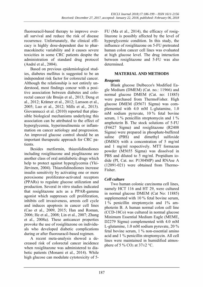
Figure 1 shows the cell proliferation of HCT 116 and HT 29 cells upon treatment with increasing concentrations of 5-FU or rosiglitazone. Both cell lines showed a dose-dependent decrease in cell proliferation. HCT 116 responded to 5-FU (Figure 1A1) at IC50= 4.77±0.55 µg/ml and rosiglitazone (Figure 1B1) at IC50= 11.61±3.49 µg/ml. The respective drugs resulted in lower IC50 values of 1.86±0.18 µg/ml (5-FU, Figure 1A2) and 1.04±0.14 µg/ml (rosiglitazone, Figure 1B2) on HT 29. Since the IC20 of both drugs was comparable between HCT 116 and HT 29, a low dose of 5-FU at 0.2 µg/ml and rosiglita-
EXCLI Journal 2018;17:186-199 – ISSN 1611-2156 Received: December 27, 2017, accepted: January 22, 2018, published: February 06, 2018
189
zone at 0.5 µg/ml were selected. 5-FU and rosiglitazone did not exert any significant effect at the low dose range but showed 30-
40 % inhibition at extremely high concentra-tion (>10 µg/ml) on CCD-18Co cells (Fig-ures 1A3 and 1B3).
Figure 1: Effect of drug treatment on the cell proliferations. Cells were treated with (A) 5-FU or (B) rosiglitazone at various concentrations for 48 h. The control group were untreated cells in normal glu-cose medium. *Compared to control, p <0.05.
EXCLI Journal 2018;17:186-199 – ISSN 1611-2156 Received: December 27, 2017, accepted: January 22, 2018, published: February 06, 2018
190
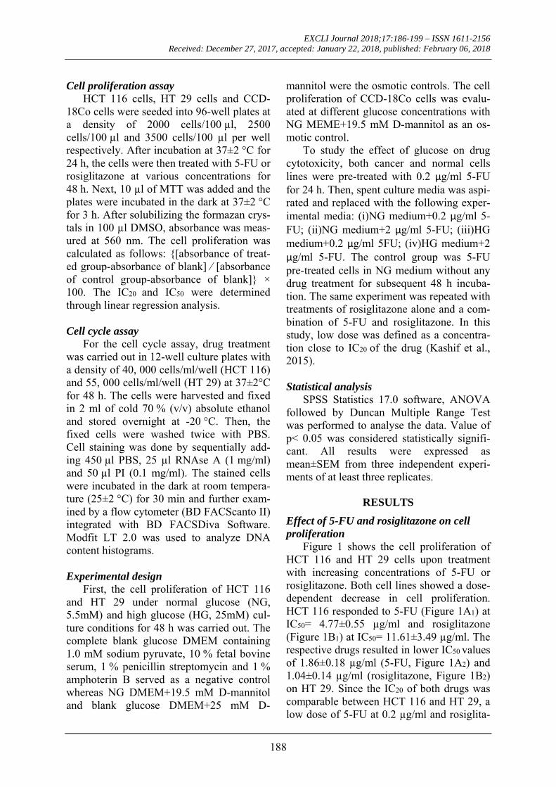
In HG culture, the cell proliferation was stimulated significantly 0.7 fold (HCT116, Figure 2A1) and 1.6 fold (HT29, Figure 2A2)
compared with NG culture. Subsequent in-troduction of high glucose (25 mM) even reduced the inhibitory effect of 5-FU.
Figure 2: Effect of glucose on drug treatments. (A) HCT 116 and HT29 cells were treated in NG or HG culture for 48 h. The control group were cells incubated in blank medium. Two media containing mannitol with a concentration of iso-osmolar to HG was used as osmotic controls. CCD-18Co cells were cultured at different glucose concentrations. (B-C) 5-FU pretreated cells with subsequent low dose or high dose of drug treatments in blank (absent on CCD-18Co), NG and HG media for 48 h. The control group were 5-FU pre-treated cells in NG culture without any subsequent drug treatments. *Compared to control, p <0.05. **Compared to NG group, p <0.05. NG, normal glucose (5.5 mM); HG, high glucose (25 mM) Panel B: 5-FU, low dose =0.2 µg/ml; high dose =2 µg/ml, Panel C: Rosiglita-zone, low dose =0.5 µg/ml; high dose =5 µg/ml

EXCLI Journal 2018;17:186-199 – ISSN 1611-2156 Received: December 27, 2017, accepted: January 22, 2018, published: February 06, 2018
191
At low dose of 5-FU (0.2 µg/ml), HG culture significantly (p< 0.05) increased the cell proliferation of HCT 116 up to 124 % (Figure 2B1). The cell proliferation of HT 29 was approximately 105 % in HG culture and it was significantly (p< 0.05) higher than that in NG culture (80 %) (Figure 2B2). At low dose of rosiglitazone (0.5 µg/ml), a signifi-cant difference (p< 0.05) in cell prolifera-tions between HG culture (HCT 116= 100%; HT 29= 99 %) and NG culture (HCT 116= 80 %; HT 29= 85 %) was evident (Figures 2C1 and 2C2). The viability of both cell lines was less than 30 %, either in HG or NG cul-ture, when treated with a high dose of rosig-litazone (5 µg/ml). Glucose level had no sig-nificant effect on CCD-18Co cells regardless of drug treatment (Figures 2B3 and 2C3).
Figures 3A and 3B show the cell prolif-erations of HCT 116 and HT 29 respectively when 5-FU and rosiglitazone were used in combination. At low dose of 5-FU, an addi-tion of low dose rosiglitazone significantly (p< 0.05) decreased the cell proliferation of HCT 116 from 118 % to 96 % in HG culture while the same treatment had no effect on HT 29. At high dose of 5-FU, an addition of low dose rosiglitazone in HG culture trig-gered the cells to proliferate at an equivalent rate to those treated with single dose of 5-FU. The two cell lines maintained viability below 20 % with combined drugs containing high dose of rosiglitazone in NG and HG cultures. Similarly, glucose level had no sig-nificant effect on CCD-18Co cells regardless of drug treatment (Figure 3C). Effect of 5-FU and rosiglitazone on cell cycle
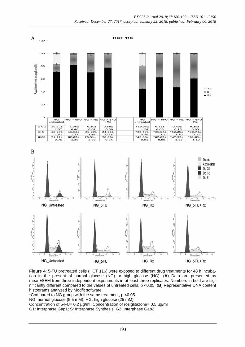
Exposure to low dose 5-FU caused G1 phase arrest on HCT 116 cells cultured in NG (82.02±1.35 %) and HG (62.83±0.99 %) conditions (Figure 4). However, low dose rosiglitazone arrested the cells in S phase in HG culture (52.25±1.70 %). In the presence of both 5-FU and rosiglitazone, G1 phase
arrest was observed in NG (78.06±0.74 %) with S-phase cells significantly (p< 0.05) increased from 12.27±1.57 % to 21.26±0.73 % and HG (61.01±1.17 %) cul-tures. Under the similar treatments, HG cul-ture significantly increased the cell propor-tion in S phase when compared to those in NG culture.
In NG culture, HT 29 cells treated with low dose 5-FU were arrested in S phase (50.62±0.94 %) as shown in Figure 5. Low dose of rosiglitazone induced G1 phase ar-rest but significantly (p< 0.05) increased the proportion of S phase-cells from 30.37±1.66 % to 37.97±1.84 %. Treatment with 5-FU or rosiglitazone had no effect on the cell cycle in HG culture. The significant (p< 0.05) increase in cell proportion, 43.16±1.52 % (NG) and 30.32±2.46 % (HG), was observed in S phase when treated with both drugs together. Regardless of drug treatment, HG culture significantly (p< 0.05) increased the cell proportion in G2 phase when compared with NG culture.
DISCUSSION
Warburg hypothesis postulates that the origin of carcinogenesis is a change of me-tabolism which is often associated with en-hanced glucose uptake and consumption in cancer cells (vander Heiden et al., 2009). In this respect, hyperglycemia can be a primary glucose source to meet the metabolic de-mand of fast growing cancerous cells. Sever-al in vitro studies have demonstrated that high glucose levels promote the proliferation of human colorectal carcinomas (Ma et al., 2014; Masur et al., 2011; Tomas et al., 2012) which concur with the result obtained in this study. The increase in cell proliferation could be independent of osmotic stress as it was reproduced by D-mannitol with a signif-icant difference against normal glucose. It was differed to normal cells which had no respond to glucose level (Figure 2A3).

EXCLI Journal 2018;17:186-199 – ISSN 1611-2156 Received: December 27, 2017, accepted: January 22, 2018, published: February 06, 2018
192
Figure 3: Effect of combined drugs on cell proliferation under NG or HG culture. 5-FU pretreated cells were further exposed to 5-FU alone or in combination with rosiglitazone for 48 h incubation. The con-trol group were 5-FU pre-treated cells in NG culture without any subsequent drug treatments. *Compared to control, p <0.05. **Compared to single dose 5-FU, p <0.05. NG, normal glucose (5.5 mM); HG, high glucose (25 mM) a0.2 µg/ml as low dose 5-FU; 2 µg/ml as high dose 5-FU LRz, low dose rosiglitazone= 0.5 µg/ml HRz, high dose rosiglitazone= 5 µg/ml
EXCLI Journal 2018;17:186-199 – ISSN 1611-2156 Received: December 27, 2017, accepted: January 22, 2018, published: February 06, 2018
193
Figure 4: 5-FU pretreated cells (HCT 116) were exposed to different drug treatments for 48 h incuba-tion in the present of normal glucose (NG) or high glucose (HG). (A) Data are presented as mean±SEM from three independent experiments in at least three replicates. Numbers in bold are sig-nificantly different compared to the values of untreated cells, p <0.05. (B) Representative DNA content histograms analyzed by Modfit software. *Compared to NG group with the same treatment, p <0.05. NG, normal glucose (5.5 mM); HG, high glucose (25 mM) Concentration of 5-FU= 0.2 µg/ml; Concentration of rosiglitazone= 0.5 µg/ml G1: Interphase Gap1; S: Interphase Synthesis; G2: Interphase Gap2

EXCLI Journal 2018;17:186-199 – ISSN 1611-2156 Received: December 27, 2017, accepted: January 22, 2018, published: February 06, 2018
194
Figure 5: 5-FU pretreated cells (HT 29) were exposed to different drug treatments for 48 h incubation in the present of normal glucose (NG) or high glucose (HG). (A) Data are presented as mean±SEM from three independent experiments in at least three replicates. Numbers in bold are significantly dif-ferent compared to the values of untreated cells, p <0.05. (B) Representative DNA content histograms analyzed by Modfit software. *Compared to NG group with the same treatment, p <0.05. NG, normal glucose (5.5 mM); HG, high glucose (25 mM) Concentration of 5-FU= 0.2 µg/ml; Concentration of rosiglitazone= 0.5 µg/ml G1: Interphase Gap1; S: Interphase Synthesis; G2: Interphase Gap2
EXCLI Journal 2018;17:186-199 – ISSN 1611-2156 Received: December 27, 2017, accepted: January 22, 2018, published: February 06, 2018
195
5-FU is widely used to treat metastatic colorectal cancer. It is a uracil analog and principally works through irreversible inhibi-tion of thymidylate synthase (TS), an en-zyme responsible for the conversion of de-oxyuridylate (dUMP) to deoxythymidylate (dTMP) under normal physiological condi-tions (Zhang et al., 2008b). 5-FU is convert-ed to fluorodeoxyuridine monophosphate (FdUMP) which competes with dUMP in binding to TS and thus limiting dTMP pro-duction. The dTMP depletion can lead to cytotoxicity causing cell death via thy-mineless death (Longley et al., 2003). Alt-hough cytotoxicity of 5-FU in colorectal cell lines have been extensively determined (Failli et al., 2011, 2013; Flis and Spławiński, 2009; Wiebke et al., 2003), the role of glucose concentrations in altering the drug cytotoxicity still need to be clarified. In the present study, we showed that 5-FU and high glucose affected the cancer cell prolif-eration antagonistically.
As demonstrated in the study, the cyto-toxicity of 5-FU in colorectal cancer cells was diminished by HG treatment, a concen-tration equivalent to the serum glucose level in diabetic individuals (glucose level >200 mg/dl). Conversely, proliferative effect of high glucose could be overcome when dosage of the drug was high enough. Ma et al. (2014) speculated that a higher admin-istration dosage is required to sustain the therapeutic effect of 5-FU for CRC patients with hyperglycemia. Unfortunately, cases of 5-FU toxicity have been reported on diabetic patients and the severity was directly related to the degree of hyperglycemia (Sadoff, 1998). It implicated that administration of 5-FU can pose a threat to developing drug tox-icity if blood glucose was poorly managed. Past clinical investigations have proposed that deficiency of, either dihydropyrimidine dehydrogenese or dihydropyrimidinase, is a pharmacogenetic disorder associated with 5-FU toxicity (Milano et al., 1999; van Kui-lenburg, 2004; van Kuilenburg et al., 2003), but the activities of these 5-FU catabolic
enzymes in the cancer patients with diabetes are still not fully understood.
As the mainstay of chemotherapy for colorectal cancer, some common adverse events induced by 5-FU have been reported, namely mucositis, diarrhea and myelosup-pression (Vincenzi et al., 2008). 5-FU was also found to elevate blood glucose level in CRC patients (Köhne et al., 1997; Tayek and Chlebowski, 1992). A recent study even concluded that hyperglycemia is a potent complication due to 5FU-based regimen (Feng et al., 2013). To address this issue, groups of antidiabetic agents including insu-lin analogs, insulin sensitizers, secretagogues and incretin mimitics are currently being considered. Based on a review by García-Jiménez et al. (2016), agents that improve insulin sensitivity may reduce cancer risk rather than those that increase circulating insulin. Therefore, rosiglitazone as an insulin sensitizer should be appropriately applied in combination with 5-FU when taking cancer risk factors into consideration.
Ample evidences showed that rosiglita-zone, a PPAR-gamma agonist suppressed proliferation of various cancerous cells at concentrations varying from 0.1 µmol/L to 100 µmol/L (Cao et al., 2009, 2015; Han and Roman, 2006; He et al., 2008; Lin et al., 2007; Zhang et al., 2008a). Similarly, in the present study, 5-FU pretreatment followed by a comparable dosage of rosiglitazone in-hibited cell growth of HCT 116 and HT 29. While only one glucose concentration (16.67 mM or 11.11 mM) was used in the previous related studies (Lin et al., 2007; Miao et al., 2011; Zhang et al., 2007), this study demonstrated that the inhibitory effect of rosiglitazone was modulated by glucose levels. Activation of PPAR-gamma by rosig-litazone stimulates the expression of phos-phate and tension homolog (PTEN) in hu-man carcinoma cell lines (Han and Roman, 2006; Cao et al., 2009). When glucose level is high, the PTEN expression is decreased leading to an increased Akt activity (Liu et al., 2012; Mahimainathan et al., 2006). The deregulation of Akt signaling allows cell
EXCLI Journal 2018;17:186-199 – ISSN 1611-2156 Received: December 27, 2017, accepted: January 22, 2018, published: February 06, 2018
196
survival and cell growth which may explain why the inhibitory effect of rosiglitazone was significantly (p< 0.05) reduced in HG culture (Figures 2C1-C2). There are other possible downstream regulations via a PPAR-gamma dependent signal pathway such as COX-2, MMP-7 and TIMP-1 (Miao et al., 2011).
5-FU treatment causes DNA damage due to misincorparation of FdUTP into DNA (Longley et al., 2003). While cell cycle pro-gression is regulated by checkpoint control in the G1 or G2 phase, cycle arrests in G1 and G2 phases allow DNA repair prior to replication and mitosis respectively. In the present study, low dose of 5-FU appeared to trigger cytostasis via G1 phase arrest on HCT 116 cells but S phase arrest on HT 29 cells in NG culture. The different cell cycle response might be due to the variance in mu-tation status of HCT 116 (MMR-deficient/p53-proficient) and HT 29 (MMR-proficient/p53-deficient). Admittedly, MMR-deficient cell lines confer less sensitivity to 5-FU (Adamsen et al., 2011) thus, HG could diminish G1 phase arrest on 5FU-treated HCT 116 cells. The result was consistent with the increase of cell proliferation at high glucose level (Figure 2B1). On the other hand, Hawn et al. (1995) suggested that any agent that induces DNA mispairs will lead to G2 arrest in MMR-proficient cells. The in-crease of HT 29 cells in G2 phase corre-sponded to the increase of cell proliferation (Figure 2B2) indicating that the mismatch repair (MMR) system interacted with G2 checkpoint in response to 5-FU, especially at high glucose level for DNA repair which allow cell replication. Unlike 5-FU, low dose rosiglitazone induced S-phase arrest and this differed to the effects whereby G1-phase arrest was evident on gastric cancer (He et al., 2008), breast cancer (Zhang et al., 2008a), and liver cancer (Yu et al., 2010). In fact, rosiglitazone was also reported to cause G1-phase arrest in colorectal cancers at higher concentrations specifically 10 µmol/L (HT 29) (Lin et al., 2007) and 25 µmol/L (HCT 15) (Miao et al., 2011). These conflict-
ing results suggested that low dose of rosig-litazone might trigger the cells to experience genotoxic stress during DNA replication and delay their progression in a transient manner through activation of intra-S-phase check-point (Bartek et al., 2004). Proteins that may be involved in the intra-S-phase checkpoint are kinase Chk1, MSH2 and MCH 1. It is possible to enhance the efficacy of 5-FU if DNA synthesis can be inhibited by regulat-ing the TS levels during the S phase of cell cycle (Subbarayan et al., 2010). The down-regulation of TS induces p53 protein expres-sion (Liu et al., 2002) and initiates cell apop-tosis consequently.
To determine the drug interaction be-tween rosiglitazone and 5-FU, the cytotoxici-ty outcomes from individual and combined drug treatments were further analyzed using combination index (CI) method (Chou and Talalay, 1983, 1984). Based on CI values generated by CompuSyn software version 1.0 (data not shown), the present study re-vealed that high dose of rosiglitazone exerted synergistic effect on 5-FU treatment regard-less of glucose levels. Indeed, it was far more effective than 5FU/metformin combi-nation whereby the cell growth suppression occurred at a higher dosage of both drugs (Zhang et al., 2013). Addition of low dose rosiglitazone was antagonistic to 5-FU, meaning that the combined drugs had an overall effect that was less than the sum of their individual effect. The cell cycle analy-sis reflected a similar trend between 5-FU treatment and combined drug treatment, whereby the highest cell proportion occurred in G1 phase followed by S phase and G2 phase under NG or HG culture.
CONCLUSION
This study is the first attempt to demon-strate the influence of rosiglitazone on cyto-toxicity of 5-FU under normal or high glu-cose condition. The present results showed that rosiglitazone had an antiproliferative effect on colorectal cancer cells via G1 or S phase arrest. Moreover, the antiproliferative effect was synergistic to 5-FU drug in the

EXCLI Journal 2018;17:186-199 – ISSN 1611-2156 Received: December 27, 2017, accepted: January 22, 2018, published: February 06, 2018
197
presence of high glucose. Thus, combining rosiglitazone in 5-FU regimen may improve the therapeutic effect. Taken together, the present finding provides a better insight for the management of hyperglycemic CRC pa-tients on 5-FU chemotherapy. Acknowledgement
This work was supported by University of Malaya Research Grant BK025-2014.
Declaration of interest
The authors disclose no conflicts of in-terest.
REFERENCES
Adamsen BL, Kravik KL, de Angelis PM. DNA dam-age signaling in response to 5-fluorouracil in three colorectal cancer cell lines with different mismatch repair and TP53 status. Int J Oncol. 2011;39:673-82.
André T, Boni C, Mounedji-Boudiaf L, Navarro M, Tabernero J, Hickish T, et al. Oxaliplatin, fluoroura-cil, and leucovorin as adjuvant treatment for colon cancer. N Engl J Med. 2004;350:2343-51.
Bartek J, Lukas C, Lukas J. Checking on DNA dam-age in S phase. Nat Rev Mol Cell Biol. 2004;5:792-804.
Cao L, Wang X, Wang Q, Xue P, Jiao X, Peng H, et al. Rosiglitazone sensitizes hepatocellular carcinoma cell lines to 5-fluorouracil antitumor activity through activation of the PPARγ signaling pathway. Acta Pharmacol Sin. 2009;30:1316-22.
Cao X, He L, Li Y. Effects of PPARγ agonistro-siglitazone on human retinoblastoma cell in vitro and in vivo. Int J Clin Exp Pathol. 2015;8:12549-56.
Chou TC, Talalay P. Analysis of combined drug ef-fects: a new look at a very old problem. Trends Phar-macol Sci. 1983;4:450-4.
Chou TC, Talalay P. Quantitative analysis of dose-effect relationships: the combined effects of multiple drugs or enzyme inhibitors. Adv Enzyme Regul. 1984; 22:27-55.
de Bruijn KMJ, Arends LR, Hansen BE, Leeflang S, Ruiter R, van Eijck CHJ. Systematic review and me-ta-analysis of the association between diabetes melli-tus and incidence and mortality in breast and colorec-tal cancer. Br J Surg. 2013;100:1421-9.
Deng L, Gui Z, Zhao L, Wang J, Shen L. Diabetes mellitus and the incidence of colorectal cancer: an updated systematic review and meta-analysis. Dig Dis Sci. 2012;57:1576-85.
Failli A, Consolini R, Legitimo A, Orsini G, Romani-ni A, Spisni R, et al. Evaluation of in vitro cytotoxici-ty of oxaliplatin and 5-fluorouracil in human colon cancer cell lines: combination versus sequential expo-sure. J Biol Regul Homeost Agents. 2011;25:575-88.
Failli A, Legitimo A, Orsini G, Castagna M, Spisni R, Miccoli P, et al. Antiproliferative effects of 5-fluorouracil and oxaliplatin in colon cancer cell lines: comparison of three different cytotoxicity assays. J Biol Regul Homeost Agents. 2013;27:275-84.
Feng JP, Yuan XL, Li M, Fang J, Xie T, Zhou Y, et al. Secondary diabetes associated with 5-fluorouracil-based chemotherapy regimens in non-diabetic patients with colorectal cancer: results from a single-centre cohort study. Color Dis. 2013;15:27-33.
Ferlay J, Soerjomataram I, Dikshit R, Eser S, Mathers C, Rebelo M, et al. Cancer incidence and mortality worldwide: sources, methods and major patterns in GLOBOCAN 2012. Int J Cancer. 2015;136:E359-86.
Flis S, Spławiński J. Inhibitory effects of 5-fluorouracil and oxaliplatin on human colorectal can-cer cell survival are synergistically enhanced by su-lindac sulfide. Anticancer Res. 2009;29:435-41.
García-Jiménez C, Gutiérrez-Salmerón M, Chocarro-Calvo A, García-Martinez JM, Castaño A, de la Vieja A. From obesity to diabetes and cancer: epidemiol-ogical links and role of therapies. Br J Cancer. 2016; 114:716-22.
Giovannucci E, Harlan DM, Archer MC, Bergenstal RM, Gapstur SM, Habel LA, et al. Diabetes and can-cer: a consensus report. Diabetes care. 2010;33: 1674-85.
Goodwin RA, Asmis TR. Overview of systemic ther-apy for colorectal cancer. Clin Colon Rectal Surg. 2009;22:251-6.
Han S, Roman J. Rosiglitazone suppresses human lung carcinoma cell growth through PPARgamma-dependent and PPARgamma-independent signal path-ways. Mol Cancer Ther. 2006;5:430-7.
Hawn MT, Umar A, Carethers JM, Marra G, Kunkel TA, Boland CR, et al. Evidence for a connection between the mismatch repair system and the G2 cell cycle checkpoint. Cancer Res. 1995;55:3721-5.

EXCLI Journal 2018;17:186-199 – ISSN 1611-2156 Received: December 27, 2017, accepted: January 22, 2018, published: February 06, 2018
198
He Q, Pang R, Song X, Chen J, Chen H, Chen B, et al. Rosiglitazone suppresses the growth and invasive-ness of SGC-7901 gastric cancer cells and angiogene-sis in vitro via PPARγ dependent and independent mechanisms. PPAR Res. 2008;2008:649808.
Kashif M, Andersson C, Hassan S, Karlsson H, Senkowski W, Fryknäs M, et al. In vitro discovery of promising anti-cancer drug combinations using itera-tive maximisation of a therapeutic index. Sci Rep. 2015;5:14118.
Köhne CH, Harstrick A, Hiddemann W, Schöffski P, Wilke H, Bokemeyer C, et al. Modulation of 5-fluorouracil with methotrexate and low-dose N-(phosphonacetyl)-L-aspartate in patients with ad-vanced colorectal cancer. Results of a phase II study. Eur J Cancer. 1997;33:1896-9.
Krämer HU, Schöttker B, Raum E, Brenner H. Type 2 diabetes mellitus and colorectal cancer: meta-analysis on sex-specific differences. Eur J Cancer. 2012;48: 1269-82.
Larsson SC, Orsini N, Wolk A. Diabetes mellitus and risk of colorectal cancer: A meta-analysis. J Natl Cancer Inst. 2005;97:1679-87.
Lin MS, Chen WC, Bai X, Wang YD. Activation of peroxisome proliferator-activated receptor gamma inhibits cell growth via apoptosis and arrest of the cell cycle in human colorectal cancer. J Dig Dis. 2007;8: 82-8.
Liu J, Schmitz JC, Lin X, Tai N, Yan W, Farrell M, et al. Thymidylate synthase as a translational regulator of cellular gene expression. Biochim Biophys Acta-Mol Basis Dis. 2002;1587:174-82.
Liu L, Hu X, Cai GY, Lv Y, Zhuo L, Gao JJ, et al. High glucose-induced hypertrophy of mesangial cells is reversed by connexin43 overexpression via PTEN/Akt/ mTOR signaling. Nephrol Dial Trans-plant. 2012;27: 90-100.
Longley DB, Harkin DP, Johnston PG. 5-fluorouracil: mechanisms of action and clinical strategies. Nat Rev Cancer. 2003;3:330-8.
Luo W, Cao Y, Liao C, Gao F. Diabetes mellitus and the incidence and mortality of colorectal cancer: a meta-analysis of 24 cohort studies. Color Dis. 2012;14: 1307-12.
Ma YS, Yang IP, Tsai HL, Huang CW, Juo SHH, Wang JY. High glucose modulates antiproliferative effect and cytotoxicity of 5-fluorouracil in human colon cancer cells. DNA Cell Biol. 2014;33:64-72.
Mahimainathan L, Das F, Venkatesan B, Choudhury GG. Mesangial cell hypertrophy by high glucose is mediated by downregulation of the tumor suppressor PTEN. Diabetes. 2006;55:2115-25.
Masur K, Vetter C, Hinz A, Tomas N, Henrich H, Niggemann B, et al. Diabetogenic glucose and insulin concentrations modulate transcriptom and protein levels involved in tumour cell migration, adhesion and proliferation. Br J Cancer. 2011;104:345-52.
Miao R, Xu TAO, Liu L, Wang M, Jiang Y, Li J, et al. Rosiglitazone and retinoic acid inhibit proliferation and induce apoptosis in the HCT-15 human colorectal cancer cell line. Exp Ther Med. 2011;2:413-7.
Milano G, Etienne MC, Pierrefite V, Barberi-Heyob M, Deporte-Fety R, Renee N. Dihydropyrimidine dehydrogenase deficiency and fluorouracil-related toxicity. Br J Cancer. 1999;79:627-30.
Mills KT, Bellows CF, Hoffman AE, Kelly TN, Gagliardi G. Diabetes mellitus and colorectal cancer prognosis: a meta-analysis. Dis Colon Rectum. 2013; 56:1304-19.
Monami M, Dicembrini I, Mannucci E. Thiazoli-dinediones and cancer: results of a meta-analysis of randomized clinical trials. Acta Diabetol. 2014;51:91-101.
Sadoff L. Overwhelming 5-fluorouracil toxicity in patients whose diabetes is poorly controlled. Am J Clin Oncol. 1998;21:605-7.
Subbarayan PR, Lee K, Ardalan B. Arsenic trioxide suppresses thymidylate synthase in 5-FU-resistant colorectal cancer cell line HT29 in vitro re-sensitizing cells to 5-FU. Anticancer Res. 2010;30:1157-62.
Tayek JA, Chlebowski RT. Metabolic response to chemotherapy in colon cancer patients. JPEN J Parenter Enter Nutr. 1992;16:65S-71S.
Tomas NM, Masur K, Piecha JC, Niggemann B, Zänker KS. Akt and phospholipase Cγ are involved in the regulation of growth and migration of MDA-MB-468 breast cancer and SW480 colon cancer cells when cultured with diabetogenic levels of glucose and insu-lin. BMC Res Notes. 2012;5:1-7.
van Kuilenburg ABP. Dihydropyrimidine dehydro-genase and the efficacy and toxicity of 5-fluorouracil. Eur J Cancer. 2004;40:939-50.
van Kuilenburg ABP, Meinsma R, Zonnenberg BA, Zoetekouw L, Baas F, Matsuda K, et al. Dihydropy-rimidinase deficiency and severe 5-fluorouracil toxici-ty. Clin Cancer Res. 2003;9:4363-7.
EXCLI Journal 2018;17:186-199 – ISSN 1611-2156 Received: December 27, 2017, accepted: January 22, 2018, published: February 06, 2018
199
vander Heiden MG, Cantley LC, Thompson CB. Understanding the Warburg effect: the metabolic requirements of cell proliferation. Science. 2009;324: 1029-33.
Vincenzi B, Schiavon G, Pantano F, Santini D, Tonini G. Predictive factors for chemotherapy-related toxic effects in patients with colorectal cancer. Nat Clin Pr Oncol. 2008;5:455-65.
Wiebke EA, Grieshop NA, Loehrer PJ, Eckert GJ, Sidner RA. Antitumor effects of 5-fluorouracil on human colon cancer cell lines: antagonism by levami-sole. J Surg Res. 2003;111:63-9.
Yki-Järvinen H. Thiazolidinediones. N Engl J Med. 2004;351:1106-18.
Yu J, Shen B, Chu ES, Teoh N, Cheung KF, Wu CW, et al. Inhibitory role of peroxisome proliferator-activated receptor gamma in hepatocarcinogenesis in mice and in vitro. Hepatology. 2010;51:2008-19.
Zhang YQ, Tang XQ, Sun L, Dong L, Qin Y, Liu HQ, et al. Rosiglitazone enhances fluorouracil-induced apoptosis of HT-29 cells by activating peroxisome proliferator-activated receptor gamma. World J Gas-troenterol. 2007;13:1534-40.
Zhang T, Zhang Q, Chen D, Jiang J, Zhou Q. Growth inhibiton of human breast cancer cell line MDA-MB-231 by rosiglitazone through activation of PPARγ. Chinese J Clin Oncol. 2008a;5:407-12.
Zhang N, Yin Y, Xu SJ, Chen WS. 5-fluorouracil: mechanisms of resistance and reversal strategies. Molecules. 2008b;13:1551-69.
Zhang Y, Guan M, Zheng Z, Zhang Q, Gao F, Xue Y. Effects of metformin on CD133+ colorectal cancer cells in diabetic patients. PLoS One. 2013;8:e81264.