research article embryonic heart morphogenesis from...
TRANSCRIPT

Hindawi Publishing CorporationComputational and Mathematical Methods in MedicineVolume 2013 Article ID 293069 7 pageshttpdxdoiorg1011552013293069
Research ArticleEmbryonic Heart Morphogenesis from Confocal MicroscopyImaging and Automatic Segmentation
Hongda Mao1 Megan Gribble2 Arkady M Pertsov2 and Pengcheng Shi1
1 Computational Biomedicine Laboratory Rochester Institute of Technology Rochester NY 14623 USA2Department of Pharmacology SUNY Upstate Medical University Syracuse NY 13210 USA
Correspondence should be addressed to Hongda Mao hongdamaomailritedu
Received 10 October 2013 Accepted 14 November 2013
Academic Editor Heye Zhang
Copyright copy 2013 Hongda Mao et al This is an open access article distributed under the Creative Commons Attribution Licensewhich permits unrestricted use distribution and reproduction in any medium provided the original work is properly cited
Embryonic heart morphogenesis (EHM) is a complex and dynamic process where the heart transforms from a single tube into afour-chambered pump This process is of great biological and clinical interest but is still poorly understood for two main reasonsOn the one hand the existing imaging modalities for investigating EHM suffered from either limited penetration depth or limitedspatial resolution On the other hand current works typically adopted manual segmentation which was tedious subjective andtime consuming considering the complexity of developing heart geometry and the large size of images In this paper we proposeto utilize confocal microscopy imaging with tissue optical immersion clearing technique to image the heart at different stages ofdevelopment for EHM study The imaging method is able to produce high spatial resolution images and achieve large penetrationdepth at the same time Furthermore we propose a novel convex active contour model for automatic image segmentation Themodel has the ability to deal with intensity fall-off in depth which is characterized by confocal microscopy images We acquired theimages of embryonic quail hearts from day 6 to day 14 of incubation for EHM study The experimental results were promising andprovided us with an insight view of early heart growth pattern and also paved the road for data-driven heart growth modeling
1 Introduction
The heart is the first functioning organ in the embryoAlthough the morphology of the heart changes dramaticallyduring development where it transforms from a single tubeinto a four-chambered pump the heart functions withoutinterruption to serve themetabolic needs of the rapidly grow-ing embryo [1] Embryonic heart morphogenesis (EHM) iscritically important for long-time survival and any defectsin the developmental mechanism during embryogenesis mayresult in congenital cardiac anomalies In fact congenitalheart disease is relatively frequent which affects from 19 to 75per 1000 births worldwide and has been an important causeof childhood morbidity and mortality [2] UnderstandingEHM in normal and malformed hearts therefore has beenof considerable clinical and biological interest
Despite a large body of research in the last decades [3ndash8] EHM is still poorly understood mainly because of thecomplexity of the growing geometry and extremely small sizeof the developing heart Thanks to the rapid development
of imaging techniques 3D reconstruction of embryonichearts from biomedical images has dramatically improvedour ability to visualize EHM Several imaging modalitieshave been proposed for the study of EHM however eachof them has its own limitations Histological sectioning wasone of the most widely used approaches for rendering 3Dstructure of the developing heart [6] Nevertheless it neededsophisticated manual alignment of all the sections whichwas difficult and labor-intensive and therefore left it onlyfor lab researchers Optical scanning techniques were alsoused for rendering 3D4D volumes of embryonic hearts[7 9] but low penetration depth limits their applicationin imaging late stages of embryonic heart development[8] There were also other imaging modalities used forunderstanding EHM which unfortunately provided verylimited spatial resolution [10] Recently micro-CT techniquewas used to image the chambers of embryonic hearts [8]However the sophisticated polymerization process ignoredthe structure of the peripheral luminal space which is actuallyvery important for EHM understanding Last but not least
2 Computational and Mathematical Methods in Medicine
all the aforementioned works adopted manual segmentationfor 3D heart segmentation due to the lack of appropriateautomatic segmentation approaches Nevertheless manualsegmentation is tedious subjective and time consumingconsidering the complexity of the developing heart and highresolution images Thus an automatic image segmentationmethod is badly needed
In view of the problems we propose a new imagingapproach for studying EHM utilizing tissue optical immer-sion clearing and 3D confocal microscopy imaging whichcan produce high spatial resolution images and achievelarge penetration depth at the same time Furthermoreconsidering the intensity fall-off in depth nature of confocalmicroscopy images we propose a convex active contourmodel with image depth information for automatic imagesegmentation A recently proposed Split Bregman methodwas used to minimize the objective function of the model[11 12] Embryonic quail hearts at different stages of devel-opment were scanned and segmented Initial heart growthpattern was found through comparison of the structure ofhearts at different stages of development We also quantifiedthe volume change of the whole heart and luminal spacefrom day 6 to day 14 of incubation to provide an insightview of embryonic heart development Furthermore realisticcontinuous growth modeling of the living organs from datasparsely distributed in time has been an emerging field inbiomedical field [13 14] with potential applications to theanalysis and prediction of evolving pathologic structuresThus this work can be also considered as the first step fordata-driven heart growth modeling
2 Methodology
The human heart becomes a four-chambered organ byapproximately week 8 which is almost the same time theembryo can be visualized through ultrasound a point that istoo late to visualize EHM [15]We chose quail embryos as ourmodel to study EHM because of their rapid development andshort embryonic gestation period (a hatch time of 165 daysof incubation) Except for the time scale the development ofthe quail heart parallels that of the human heart
21 Image Acquisition Optical imaging method has beenwidely used as a tool for clinical functional imaging owingto its unique informative features simplicity safety and lowcost compared to conventional X-ray MRI and ultrasoundimaging However the main limitations of optical imagingtechniques including confocal microscopy are low contrastand spatial resolution as well as a small probing depth dueto strong light scattering in tissue layers [16] To utilizeits strengths and overcome its weaknesses we combinedconfocal microscopy imaging with tissue optical immersionclearing Optical clearing technique has been used in manyareas [16] However to the best of our knowledge this is thefirst time to use optical clearing with confocal microscopyimaging for EHM study
In this paper all the experiments conformed to theGuidefor the Care and Use of Laboratory Animals (NIH publication
no 85-23 revised 1996) Embryonic hearts were obtainedafter incubation of Coturnix japonica (GQF ManufacturingCo Savannah GA) or Japanese quail eggs to differentstages of development The hearts were stained with di-4-ANBDQBS which was voltage sensitive fluorescent dyesand then were dehydrated by a graded ethanol series Afterdehydration the hearts were cleared using a 1 2 benzylalcohol to benzyl benzoate mixture The cleared heart whichappeared virtually transparent was stored in the clearingsolution until imaging For image acquisition the clearedhearts were mounted in a special cuvette and scanned bya Zeiss LSM 510 confocal microscope with its numericalaperture being equal to 05 and the radius of back-projectedpinhole equal to 253 nm The dye was excited at wavelengthof 543 nm and fluorescence recorded the wave length above560 nm using a long-pass filter For more details of heartpreparation and imaging we refer the readers to [17]
High spatial resolution images were obtained after heartscanning which had an intraslice pixel size of 175 120583 mtimes 175120583m and interslice pixel size 129000120583m In Figure 1we present 3D view of three hearts at days 6 8 and 14respectively From the images the evolution of the luminalspace of the heart from spongy structure to well-separatedchamber structure can be clearly observed
22 Image Formation In a confocal microscope a pinholeis used to reject most out-of-focus light Thus the amountof light reaching the detector is low and the noise statisticscan be well described by a Poisson process [18] A generalimage formation model can be represented as the followingequation
1198680
(119909) = 119899 ([ℎ lowast 119868] (119909)) (1)
where 119909 isin Ω is a point in the image domain 1198680is an observed
image 119868 is an ideal image ℎ is a point spread function(PSH) lowast means convolution operation 119899 models the noisedistribution Based on our imaging setting the PSF of themicroscope is very small compared to our voxel size andtherefore the effect of convolution by PSF can be ignoredIn the work we used median filter to smooth the observedimage and assume the noise in the smoothed image can beconsidered as additive zeromeanGaussian distributionThusthe final image formation can be represented as the followingequation
1198680
asymp 119868 + n (2)
where 1198680and n represent smoothed image and image noise
respectively
23 Image Segmentation Thepurpose of image segmentationis to find a partition120595(Ω) of the image domainΩ and recoverthe ideal image 119868 as well Under the assumption that theintensity distribution of the ideal image is piecewise constantChan-Vese (CV) model with level set implementation wasproposed and has been widely used for image segmentation[19] Later on this model was further extended to globalCV (G-CV) model by transforming it into a global convex
Computational and Mathematical Methods in Medicine 3
(a) (b) (c)
Figure 1 3D view of embryonic quail hearts From left to right day 6 day 8 and day 14
optimization problem [20] However confocal microscopyimages are characterized by intensity fall-off in depth whichmakes G-CVmodel unsuitable for this purpose To solve thisproblem we further assume the ideal image can be describedas the multiplication of an intensity piecewise constant image119888 and a depth-related bias field 119887
119868 =
119873
sum
119894=1
(119888119894sdot 119906119894) sdot 119887119894 (3)
where 119873 is number of regions in the image and 119906119894is an image
partition function Under this assumption we developed anew convex active contourmodel for automatic segmentationof confocalmicroscopy imagesThemodel can be representedas the following equation
min119906isin[01]
119864 (1198881 1198882 119906) = int
Ω
|nabla119906 (119909)| 119889119909
+ 120582 int
Ω
(1198680 (119909) minus 119868
1 (119909))2
119906 (119909) 119889119909
+ 120582 int
Ω
(1198680
(119909) minus 1198682
(119909))2
(1 minus 119906 (119909)) 119889119909
(4)
The first term on the right side of (4) is a L1 total variation(TV) norm which is used for smoothing the variable 119906 Thesecond and third terms are data fidelity terms which keepthe intensity distribution of the ideal images close to theoriginal image Here 119906 is a partition variable and 120582 is aweighting constant to keep the balance among the three termson the right side of (4) 119868
1(119909) = 119888
1sdot 120574119911(119909) and 119868
2(119909) =
1198882
sdot 120574119911(119909) are two ideal images that represent the intensity
on two different subregions 120574119911(119909) is a depth-dependent bias
field to characterize the intensity fall-off in depth propertyof the images where 119911 is the position of the point 119909 in
119911-direction and 120574 is an experimentally determined decreasingconstant
There are total three unknown variables in our model 1198881
1198882 and 119906 By using first variation with respect to 119888
1and 1198882 we
can obtain
1198881
=
int119909isinΩ
119906 (119909) 1198680 (119909) 120574
119911(119909)119889119909
int119909isinΩ
119906 (119909) 1205742119911(119909)
119889119909
1198882
=
int119909isinΩ
(1 minus 119906 (119909)) 1198680
(119909) 120574119911(119909)
119889119909
int119909isinΩ
(1 minus 119906 (119909)) 1205742119911(119909)
119889119909
(5)
To minimize (4) with respect to 119906 we adopt the fastand efficient Split Bregman method proposed in [11 12]Split Bregman method does not require regularization con-tinuation or the enforcement of inequality constraints andit is very efficient for solving L1-regularized optimizationproblems like (4) For easier description of Split Bregmanmethod we rewrite the form of (4)
min119906isin[01]
119864 = int
Ω
|nabla119906 (119909)| + 120582119890119903(119909) 119906 (119909) 119889119909 (6)
where 119890119903(119909) = (119868
0(119909) minus 119868
1(119909))2
minus (1198680(119909) minus 119868
2(119909))2 Here the
term 120582 intΩ
(1198680(119909) minus 119868
2(119909))2119889119909 has been ignored because it does
not include the variable 119906To minimize (6) with respect to 119906 we introduce an
auxiliary variable 119889 such that 119889 = nabla119906 Thus the problem ofminimizing the energy function of (6) becomes to minimizethe following energy function
min119906isin[01]119889
int
Ω
|119889| + 120582119890119903(119909) 119906 (119909) 119889119909 with 119889 = nabla119906 (7)
4 Computational and Mathematical Methods in Medicine
To solve the constrained problem in (7) we use Split Breg-man method The problem becomes to solve the followingsequence of optimization problems
(119906119896+1
119889119896+1
) = arg min119906isin[01]119889
int
Ω
|119889| + 120582119890119903(119909) 119906 (119909)
+120583
2
10038161003816100381610038161003816119889 minus nabla119906 minus 119887
11989610038161003816100381610038161003816
2
119889119909
(8)
119887119896+1
= 119887119896
+ nabla119906119896+1
minus 119889119896+1
(9)
Here 119896 = 0 1 2 the third term on the right side of (8) isused to enforcing the constraint 119889 = nabla119906 119887
119896 is the Bregmanvector120583 and 120582 are two constant weighting parameters to keepa balance of two terms To solve (8) we adopt the alternatingminimization scheme First we consider the minimizationof (8) with respect to 119906 The minimizing solution 119906
119896+1 ischaracterized by the optimality condition
120582nabla119906 = 120582119890119903
+ 120583 div (119887119896
minus 119889119896) 119906 isin [0 1] (10)
By usingGauss-Seidel iterative scheme we can get an approx-imate solution for a 3D variable 119906
119896+1 (119894 = 0 1 2 )
120577119897119898119899
= 119889119909119896
119897minus1119898119899minus 119889119909119896
119897119898119899minus 119887119909119896
119897minus1119898119899+ 119887119909119896
119897119898119899
+ 119889119910119896
119897minus1119898119899minus 119889119910119896
119897119898119899minus 119887119910119896
119897minus1119898119899+ 119887119910119896
119897119898119899
+ 119889119911119896
119897minus1119898119899minus 119889119911119896
119897119898119899minus 119887119911119896
119897minus1119898119899+ 119887119911119896
119897119898119899
120601119897119898119899
=1
6(119906119896+1119894
119897minus1119898119899+ 119906119896+1119894
119897+1119898119899+ 119906119896+1119894
119897119898minus1119899+ 119906119896+1119894
119897119898+1119899
+119906119896+1119894
119897119898119899minus1+ 119906119896+1119894
119897119898119899+1+ 120577119897119898119899
minus120582
120583119890119903(119897119898119899)
)
119906119896+1119894+1
119897119898119899= max min 120601
119897119898119899 1 0
(11)
where 119894 is the iteration index for Gauss-Seidel iterativemethod and 119897 119898 119899 are the indices of the voxel in axes 119909 119910and 119911 respectively One has 119906
119896+10
119897119898119899= 119906119896
119897119898119899
After calculating an approximate 119906119896+1 we can obtain 119889
119896+1
by minimizing (8) with respect to 119889
119889119896+1
=nabla119906119896+1
+ 119887119896
1003816100381610038161003816nabla119906119896+1
+ 1198871198961003816100381610038161003816
max (10038161003816100381610038161003816nabla119906119896+1
+ 11988711989610038161003816100381610038161003816
minus1
120582 0) (12)
Once 119906119896+1 and 119889
119896+1 are available the Bregman vector 119887119896 can
be updated according to (9) For more details of 2D SplitBregman method we refer the readers to [11]
As a summary the procedures of using Split Bregmanmethod to solve (7) contain the following steps
(1) Initialization 1198870 1198890 and 119906
0(2) Fix 119906 calculate 119888
1and 1198882according to (5) and further
calculate 119890119903
(3) Update 119906119896+1 by solving (11)
(4) Update 119889119896+1 by solving (12)
(5) Update 119887119896+1 by solving (9)
(6) Convergence test test whether a stable solution 119906 hasreached If not go to step (2)
(7) The objects are detected by thresholding Σ = 119909
119906(119909) gt 120572 where 120572 isin [0 1] In this paper we choose120572 = 05
3 Experimental Results
31 Data In this study we selected three groups of quailhearts Each group had five embryonic quail hearts at thedevelopment stage of days 6 7 8 9 and 14 respectively All thehearts were processed and imaged according to Section 21The image size varied from 768 times 768 times 112 (day 6) to3075 times 2560 times 478 (day 14) To build a database of manualsegmentation for reference we invited two biologists toindependently segment the heartsmanually with the softwareITK-SNAP [21] Due to the large size of the image and thecomplexity of the heart geometry it typically took a biologistmore than one week to finish one heart segmentation Forthis reason we currently only selected one group for manualsegmentation
32 Evaluation of Automatic Segmentation Figure 2 showsone 3D and three 2D slice views of the quail heart atday 14 with two manual segmentations and one automaticsegmentation Although the image exhibits severe intensityinhomogeneity visual inspection of the results shows thatautomatic segmentation can correctly capture most of thestructures of the heart as manual segmentation The mostdifference between manual and automatic segmentationoccurs at the regions above the atrioventricular valve whichcan be observed in both 3D and 2D views Because thecontrast is very low at this region automatic segmentationalgorithm only uses image information can not detect theboundary precisely while biologists using their knowledgecan manually locate the boundary What is more we havealso observed the presence of small objects within the heartchambers only detected by automatic segmentation Thesesmall objects could be papillarymuscle that can be consideredas either a part of the myocardium or a part of the bloodpool As a result both manual and automatic segmentationfor these small objects are acceptable
We also quantitatively evaluate our algorithm by mea-suring the overlap of automatic segmentation and manualsegmentation by using Dicersquos similarity coefficient (DSC)For two segmentations 119878
1and 119878
2 the DSC value is defined
as 2|1198781
cap 1198782|(|1198781| + |119878
2|) The DSC value is normalized
where 0 indicates complete dissimilarity and 1 indicates com-plete agreement The overlap values reflecting the variabilitybetween the manual segmentations by two biologists arelisted in the second row of Table 1 Except for days 6 and7 all the DSC values are greater than 085 which meansthere is sufficient level of reliability for the two manualsegmentations The reason of low DSC values at days 6and 7 is that the geometry of the hearts at these days isvery complex as shown in Figure 3 and thus it is difficultfor the biologists to achieve high agreement The overlap
Computational and Mathematical Methods in Medicine 5
(a) (b) (c) (d)
Figure 2 Visual comparison between manual segmentation and automatic segmentation (a) Original 3D image and three slices indifferent views (b) Manual segmentation done by the first biologist (c) Manual segmentation done by the second biologist (d) Automaticsegmentation
(a) (b) (c) (d) (e)
Figure 3 3D segmentation of one group of the hearts Columns from left to right are the heart at days 6 7 8 9 and 14 For visualizationpurpose the outer boundary is rendered as transparent (L left ventricle R right ventricle)
comparison between automatic and manual segmentationsis listed in the third and fourth rows of Table 1 Similarlywe can find the DSC values are low at days 6 and 7 andhigh at the rest Overall the overlap measures show thatthe automatic segmentation method has similar level ofvariability to the manual segmentation which means ourautomatic segmentation method is applicable to EHM study
33 EHM Study Figure 3 shows automatic segmentationresults of one group of hearts As we can see an obviousphenomenon of early heart development is the morphology
evolution of the ventricles The left and right ventriclesare merged together and exhibit sponge network structureat days 6 and 7 as the result of cardiac looping [4] Theinterventricular septum starts to grow between day 7 and day8 as the ventricles are partially separated at day 8 At day 9the interventricular septum eventually forms and divides theventricles into the left and right ventricle However the twoventricles still present some sponge structure at day 9 Theshape of the ventricles eventually becomes mature at day 14
We also quantify the average volume of the whole heartand the luminal space at different stages of developmentbased on the segmentation results We use the open source
6 Computational and Mathematical Methods in Medicine
Table 1 DSC values that measure the overlap between the two manual segmentations the first manual segmentation against automaticsegmentation and the second manual segmentation against automatic segmentation
Day 6 Day 7 Day 8 Day 9 Day 14Biologist 1 versus biologist 2 075 079 085 087 093Automatic versus biologist 1 065 072 078 085 088Automatic versus biologist 2 068 075 079 081 091
Table 2 Average volume of the whole heart and the luminal space at different stages of development (mm3)
Day 6 Day 7 Day 8 Day 9 Day 14Total heart 26 34 60 102 775Luminal space 041 062 075 174 202
VTK library (httpwwwvtkorg) to calculate the averagevolume and list the average volume values in Table 2 We findthat the average volume of the whole heart and the luminalspace increased from 26mm3 to 775mm3 and 041mm3to 202mm3 respectively which is nearly two orders ofmagnitude increase in an incubation period of approximately10 days This finding is similar in range to the findings in[8] Furthermore we also find that the average volume of thewhole heart grows faster than the luminal space whichmeansthat the myocardium grows towards both inside and outside
4 Discussion
The technique outlined in this paper provided the frame-work of imaging and automatic segmentation of developinghearts for EHM study By combining confocal microscopyimaging with optical clearing our method was able toachieve penetration depth over 6mm that enabled us toacquire volumetric images of the developing heart throughthe whole incubation period We believe this imaging datacan help biologists to understand more details of early heartdevelopment and investigate events that lead to congenitalheart defects
Image segmentation is always a headache for researchersin this field because of the complexity of the developinggeometry The convex active contour model proposed in thispaper was a first step towards automatic segmentation inEHM study and showed promising results One significantchallenge in developing heart segmentation is the lack ofa gold standard Due to the expensive labor cost to labelthe images we provided limited validation in the paperIn the future we will build a larger manually segmenteddatabase for segmentation algorithm validation What ismore this database could also be used for training and testingparameter-free machine learning algorithms
The ultimate goal of this work will be heart growthmodeling Due to the complexity of developing heart currentheart growth modeling mainly focuses on very early stages ofEHM [22] To the best of our knowledge there exist no workson modeling the whole EHM process from the single tubeshape to four-chambered shape With EHM knowledge fromEHM study we will work towards data-driven heart growthmodeling
5 Conclusion
We proposed an imaging approach and a novel automaticsegmentation method for EHM study We demonstratedthe applicability of our imaging method to capture the 3Dstructure of embryonic quail hearts and also proved theefficiency of our segmentation algorithm for EHM study inboth visual inspection and quantitative analysis Based onthe findings from EHM study we believe this work couldhelp us to further understand the fundamental mechanismsof embryonic heart development
References
[1] J Icardo and F Manasek ldquoCardiogenesis development mech-anisms and embryologyrdquo in The Heart and CardiovascularSystem pp 1563ndash1586 1992
[2] World Health Organization Global Atlas on CardiovascularDisease Prevention and Control Expert Review of MedicalDevices 2011
[3] J Lacktis and F Manasek ldquoAn analysis of deformation during anormal morphogenic eventrdquo Morphogenesis and Malformationof the Cardiovascular System vol 17 pp 205ndash227 1978
[4] L A Taber B B Keller and E B Clark ldquoCardiac mechanics inthe stage-16 chick embryordquo Journal of Biomechanical Engineer-ing vol 114 no 4 pp 427ndash434 1992
[5] M Liebling J Vermot and S E Fraser ldquoDouble time-scaleimage reconstruction of the beating and developing embryoniczebrafish heartrdquo in Proceedings of the 5th IEEE InternationalSymposium on Biomedical Imaging pp 855ndash858 May 2008
[6] B C W Groenendijk B P Hierck J Vrolijk et al ldquoChanges inshear stress-related gene expression after experimentally alteredvenous return in the chicken embryordquoCirculation Research vol96 no 12 pp 1291ndash1298 2005
[7] M Liebling A S Forouhar R Wolleschensky et al ldquoRapidthree-dimensional imaging and analysis of the beating embry-onic heart reveals functional changes during developmentrdquoDevelopmental Dynamics vol 235 no 11 pp 2940ndash2948 2006
[8] J T Butcher D Sedmera R E Guldberg and R R MarkwaldldquoQuantitative volumetric analysis of cardiac morphogenesisassessed through micro-computed tomographyrdquo Developmen-tal Dynamics vol 236 no 3 pp 802ndash809 2007
[9] MW Jenkins F Rothenberg D Roy et al ldquo4D embryonic car-diography using gated optical coherence tomographyrdquo OpticsExpress vol 14 no 2 pp 736ndash748 2006
Computational and Mathematical Methods in Medicine 7
[10] B Smith ldquoMagnetic resonance microscopy in cardiac develop-mentrdquoMicroscopy Research and Technique vol 52 pp 323ndash3302001
[11] T Goldstein and S Osher ldquoThe split Bregman method for L1regularized problemsrdquo SIAM Journal on Imaging Sciences vol2 pp 323ndash343 2009
[12] H Mao H Liu and P Shi ldquoA convex neighbor-constrainedactive contour model for image segmentationrdquo in Proceedingsof the 17th IEEE International Conference on Image Processing(ICIP rsquo10) pp 793ndash796 September 2010
[13] J FishbaughM Prastawa S Durrleman J Piven andG GerigldquoAnalysis of longitudinal shape variability via subject specificgrowth modelingrdquo inMedical Image Computing and ComputerAssisted Intervention pp 731ndash738 2012
[14] S Durrleman X Pennec A Trouve G Gerig and N AyacheldquoSpatiotemporal atlas estimation for developmental delay detec-tion in longitudinal datasetsrdquo inMedical Image Computing andComputer Assisted Intervention pp 297ndash304 2009
[15] K W Fong A Toi S Salem et al ldquoDetection of fetal structuralabnormalities with US during early pregnancyrdquo Radiographicsvol 24 no 1 pp 157ndash174 2004
[16] E A Genina A N Bashkatov and V V Tuchin ldquoTissue opticalimmersion clearingrdquo Expert Review of Medical Devices vol 7no 6 pp 825ndash842 2010
[17] R M Smith A Matiukas C W Zemlin and A M PertsovldquoNondestructive optical determination of fiber organization inintactmyocardial wallrdquoMicroscopy Research and Technique vol71 no 7 pp 510ndash516 2008
[18] N Dey L Blanc-Feraud C Zimmer et al ldquoRichardson-Lucyalgorithm with total variation regularization for 3D confocalmicroscope deconvolutionrdquo Microscopy Research and Tech-nique vol 69 no 4 pp 260ndash266 2006
[19] T F Chan and L A Vese ldquoActive contours without edgesrdquo IEEETransactions on Image Processing vol 10 no 2 pp 266ndash2772001
[20] X Bresson S Esedoglu P Vandergheynst J-P Thiran and SOsher ldquoFast global minimization of the active contoursnakemodelrdquo Journal of Mathematical Imaging and Vision vol 28 no2 pp 151ndash167 2007
[21] P A Yushkevich J Piven H C Hazlett et al ldquoUser-guided 3Dactive contour segmentation of anatomical structures signifi-cantly improved efficiency and reliabilityrdquo NeuroImage vol 31no 3 pp 1116ndash1128 2006
[22] S Goenezen M Rennie and S Rugonyi ldquoBiomechanics ofearly cardiac developmentrdquo Biomechanics and Modeling inMechanobiology vol 11 pp 1187ndash1204 2012
Submit your manuscripts athttpwwwhindawicom
Stem CellsInternational
Hindawi Publishing Corporationhttpwwwhindawicom Volume 2014
Hindawi Publishing Corporationhttpwwwhindawicom Volume 2014
MEDIATORSINFLAMMATION
of
Hindawi Publishing Corporationhttpwwwhindawicom Volume 2014
Behavioural Neurology
EndocrinologyInternational Journal of
Hindawi Publishing Corporationhttpwwwhindawicom Volume 2014
Hindawi Publishing Corporationhttpwwwhindawicom Volume 2014
Disease Markers
Hindawi Publishing Corporationhttpwwwhindawicom Volume 2014
BioMed Research International
OncologyJournal of
Hindawi Publishing Corporationhttpwwwhindawicom Volume 2014
Hindawi Publishing Corporationhttpwwwhindawicom Volume 2014
Oxidative Medicine and Cellular Longevity
Hindawi Publishing Corporationhttpwwwhindawicom Volume 2014
PPAR Research
The Scientific World JournalHindawi Publishing Corporation httpwwwhindawicom Volume 2014
Immunology ResearchHindawi Publishing Corporationhttpwwwhindawicom Volume 2014
Journal of
ObesityJournal of
Hindawi Publishing Corporationhttpwwwhindawicom Volume 2014
Hindawi Publishing Corporationhttpwwwhindawicom Volume 2014
Computational and Mathematical Methods in Medicine
OphthalmologyJournal of
Hindawi Publishing Corporationhttpwwwhindawicom Volume 2014
Diabetes ResearchJournal of
Hindawi Publishing Corporationhttpwwwhindawicom Volume 2014
Hindawi Publishing Corporationhttpwwwhindawicom Volume 2014
Research and TreatmentAIDS
Hindawi Publishing Corporationhttpwwwhindawicom Volume 2014
Gastroenterology Research and Practice
Hindawi Publishing Corporationhttpwwwhindawicom Volume 2014
Parkinsonrsquos Disease
Evidence-Based Complementary and Alternative Medicine
Volume 2014Hindawi Publishing Corporationhttpwwwhindawicom
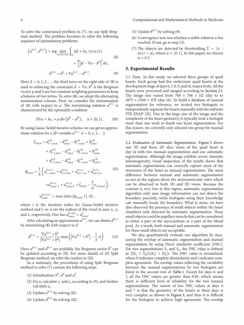