how to make a heart valve: from embryonic development to
TRANSCRIPT
How to Make a Heart Valve: From EmbryonicDevelopment to Bioengineering of Living ValveSubstitutes
Donal MacGrogan1, Guillermo Luxan1, Anita Driessen-Mol2, Carlijn Bouten2,Frank Baaijens2, and Jose Luis de la Pompa1
1Program of Cardiovascular Developmental Biology, Department of Cardiovascular Developmentand Repair, Centro Nacional de Investigaciones Cardiovasculares (CNIC), 28029 Madrid, Spain
2Biomedical Engineering/Eindhoven University of Technology, 5600 MB Eindhoven,The Netherlands
Correspondence: [email protected]
Cardiac valve disease is a significant cause of ill health and death worldwide, and valvereplacement remains one of the most common cardiac interventions in high-income econo-mies. Despite major advances in surgical treatment, long-term therapy remains inadequatebecause none of the current valve substitutes have the potential for remodeling, regeneration,and growth of native structures. Valve development is coordinated by a complex interplay ofsignaling pathways and environmental cues that cause disease when perturbed. Cardiacvalves develop from endocardial cushions that become populated by valve precursor mesen-chymeformedbyanepithelial–mesenchymal transition(EMT).Themesenchymalprecursors,subsequently, undergo directed growth, characterized by cellular compartmentalization andlayering of a structured extracellular matrix (ECM). Knowledge gained from research into thedevelopment of cardiac valves is driving exploration into valve biomechanics and tissue engi-neering directed at creating novel valve substitutes endowed with native form and function.
Cardiac valves maintain unidirectional bloodflow during the cardiac cycle. As the heart
contracts and relaxes, passive opening and clos-ing of the valves, caused by a transvalvular pres-sure gradient, results in alternate blood flowfrom the atria to the ventricles and from theventricles to the great vessels (Fig. 1A) (Yogana-than et al. 2000; Sacks and Yoganathan 2007).When the ventricles contract, the inlet or atrio-ventricular (AV) valves (the tricuspid and mitral
valves) prevent backflow from the ventriclesto the atria; and when they relax, the outlet orsemilunar (SL) valves (the pulmonary and aor-tic valves) prevent reverse flow from the arteriesto the ventricles. Valve malfunction occurs whenthe valve fails to open properly (stenosis) ordoes not shut completely (regurgitation).
Heart valve disease, which primarily affectsthe aortic and miral valves, is a health-care prob-lem of epidemic proportions because of the in-
Editors: Margaret Buckingham, Christine L. Mummery, and Kenneth R. Chien
Additional Perspectives on The Biology of Heart Disease available at www.perspectivesinmedicine.org
Copyright # 2014 Cold Spring Harbor Laboratory Press; all rights reserved; doi: 10.1101/cshperspect.a013912
Cite this article as Cold Spring Harb Perspect Med 2014;4:a013912
1
ww
w.p
ersp
ecti
vesi
nm
edic
ine.
org
on March 21, 2022 - Published by Cold Spring Harbor Laboratory Press http://perspectivesinmedicine.cshlp.org/Downloaded from
DC
F
S
V
BA
Leftcoronary
cusp
Noncoronarycusp
Rightcoronary
cuspSinus
Aorta
MV
AoV
LA
LV
RV
PV
TV
Vc
RA
AoVc PA
Anteriormitral valve
leaflet
Chordaetendinae
Papillarymuscle
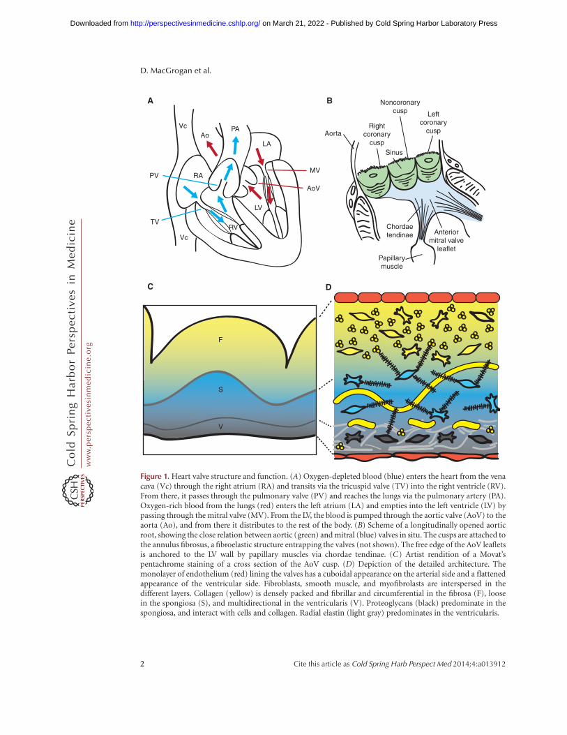
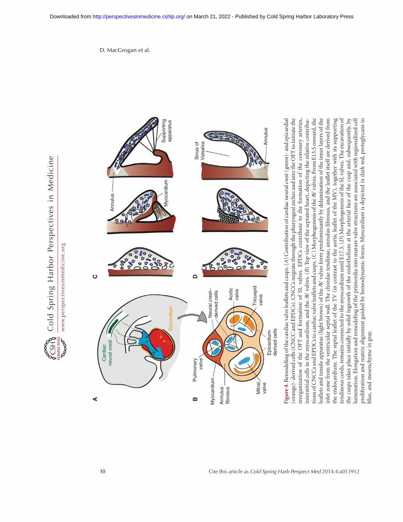
Figure 1. Heart valve structure and function. (A) Oxygen-depleted blood (blue) enters the heart from the venacava (Vc) through the right atrium (RA) and transits via the tricuspid valve (TV) into the right ventricle (RV).From there, it passes through the pulmonary valve (PV) and reaches the lungs via the pulmonary artery (PA).Oxygen-rich blood from the lungs (red) enters the left atrium (LA) and empties into the left ventricle (LV) bypassing through the mitral valve (MV). From the LV, the blood is pumped through the aortic valve (AoV) to theaorta (Ao), and from there it distributes to the rest of the body. (B) Scheme of a longitudinally opened aorticroot, showing the close relation between aortic (green) and mitral (blue) valves in situ. The cusps are attached tothe annulus fibrosus, a fibroelastic structure entrapping the valves (not shown). The free edge of the AoV leafletsis anchored to the LV wall by papillary muscles via chordae tendinae. (C) Artist rendition of a Movat’spentachrome staining of a cross section of the AoV cusp. (D) Depiction of the detailed architecture. Themonolayer of endothelium (red) lining the valves has a cuboidal appearance on the arterial side and a flattenedappearance of the ventricular side. Fibroblasts, smooth muscle, and myofibrolasts are interspersed in thedifferent layers. Collagen (yellow) is densely packed and fibrillar and circumferential in the fibrosa (F), loosein the spongiosa (S), and multidirectional in the ventricularis (V). Proteoglycans (black) predominate in thespongiosa, and interact with cells and collagen. Radial elastin (light gray) predominates in the ventricularis.
D. MacGrogan et al.
2 Cite this article as Cold Spring Harb Perspect Med 2014;4:a013912
ww
w.p
ersp
ecti
vesi
nm
edic
ine.
org
on March 21, 2022 - Published by Cold Spring Harbor Laboratory Press http://perspectivesinmedicine.cshlp.org/Downloaded from
creasing burden of elderly patients with degen-erative heart valve disease and a growing popu-lation of young adults with congenital heartdisease, involving complex valve anomalies. Thegold standard treatment for advanced heartvalve disease is surgical replacement, but noneof the currently available mechanical and bio-logical heart valve substitutes are ideal solutions.Thus, patients fitted with mechanical valves facethe burden of lifelong treatment with anticoag-ulants, whereas patients with biological valvesface the prospect of reoperation because of thelimited durability of biological valve substitutes(Schoen and Levy 2005). Tissue engineeringseeks to overcome these drawbacks by exploitingliving cells to develop a living valve replacementthat has the capacity to remodel in response tofunctional demand and repair inflicted damage.In principle, the living tissue-engineered heartvalve (TEHV) could have the capacity to grow,but once it has matured, it might achieve thesame functional and durability properties asthe native valve. The superiority of a living heartvalve replacement is underscored by the out-standing results achieved using living autograftscompared with nonliving homografts for aorticroot replacement (El-Hamamsy et al. 2010).
Translating knowledge gained from study-ing the mechanisms that drive valve develop-ment to engineering living valve replacementsis an ongoing challenge for biomedical engi-neers. This article describes the anatomy andstructure of native adult cardiac valves, summa-rizes how the endocardial cushions (ECs) formby epithelial-mesenchyme transition (EMT) andextracardiac mesenchyme contribution, andreviews the processes of valve morphogenesisand the ensuing cellular and matrix remodel-ing that give rise to the mature cusps and leaflets.The article closes by outlining how this knowl-edge is applied in state-of-the-art of tissue-engi-neering approaches to generate functional andlong-lasting cardiac valve replacements.
CARDIAC VALVE ANATOMY
Cardiac valves have a complex anatomy (Yoga-nathan et al. 2000; Sacks and Yoganathan 2007;Schoen 2008). The central component of each
valve consists of three leaflets, except the mitralvalve (MV), which has only twoleaflets (Fig. 1B).The AV valves are attached to the annulus fibro-sus, a fibroelastic tissue that encircles the AV ca-nal and outflow tract (OFT) and provides struc-tural support to the valves while also separatingthe ventricular and atrial myocardium. The freeedges of the AV valves are tethered to the base ofthe ventricular walls by tendon-like cords, calledchordae tendinae, via papillary muscles (Fig.1B). This “subvalvular apparatus” maintainsthe AV leaflets within the ventricular chamber,preventing their prolapse into the atria duringvalve closure or ventricular contraction. Systolicbillowing of one or both leaflets, as seen in MVprolapse, can be caused by myxomatous degen-eration, rupture of the chordae tendinae, or, incases of infarction or ventricular hypertrophy,displacement of the papillary muscles (Guyand Hill 2012). The SL valves do not require asupporting apparatus. Instead, the aortic valve(AoV) cusps are self-supporting and attach tocrown-shaped arterial roots via the annulus fi-brosus, whereas the pulmonary valve (PV) cuspsinsert primarily through a freestanding muscularsleeve called the right ventricular infindibulum.AV (or PV) insufficency is caused by impropercoaptation of the valve cusps and results in themovement of blood down its pressure gradientback into the ventricles (Prodromo et al. 2012).
STRUCTURAL BASIS OF VALVE FUNCTION
The composition and distribution of specializedvalvular connective tissue types is essential formaintaining normal valve function through-out life (Hinton and Yutzey 2011). The designof replacements that faithfully reproduce nativevalves depends on elucidating the role played bythe extracellular matrix (ECM) in valve devel-opment and maintenance.
THE ECM
Traditionally, the cardiac valves have been de-scribed as having a trilamellar architecture,which incorporates cellular and ECM compo-nents (Fig. 1C) (Latif et al. 2005; Schoen 2008).The layer closest to the sinus side (or atrial side
How to Make a Heart Valve
Cite this article as Cold Spring Harb Perspect Med 2014;4:a013912 3
ww
w.p
ersp
ecti
vesi
nm
edic
ine.
org
on March 21, 2022 - Published by Cold Spring Harbor Laboratory Press http://perspectivesinmedicine.cshlp.org/Downloaded from
for AV valves), called fibrosa, is composed ofdensely packed fibrillar collagens whose circum-ferential orientation provides tensile strengthwhen the valve is closed. The expression of col-lagens (mainly type I, and some type II and V)is not restricted to the outflow layer, but suffusesall the valve strata to form a network of thinfibrils, enmeshing and interacting with the oth-er ECM and cellular components. (Fig. 1D) Theintervening spongiosa is composed primarilyof glycosamino- and proteoglycans and servesas a buffer zone for the bending and stretchingimposed by leaflet opening and closing (Fig.1C,D). Glycoaminoglycans are found through-out the valves and consist of hyaluronic acid(HA), a nonproteoglycan polysaccharide, chon-droitin sulfates 4 and 6, and decorin (Latif etal. 2005). Proteoglycans are necessary for thestable assembly of the ECM and functional cel-l–ECM interactions, whereas decorin and bi-glycan interact specifically with type I collagenfibrils, modulating the kinetics of fibril for-mation and the distance between adjacent col-lagen fibrils. Facing blood flow is a thin ventri-cularis (or atrialis in the AV valves) composed ofelastin and collagen, which provides elasticitythrough mostly radially oriented elastic fibers(Fig. 1C,D).
ECM Functions
The valve ECM does not merely afford a physi-cal support for cellular growth, it is also a bio-logically active structure that provides cells withinstructional cues and signals (mechanical andhumoral), which determine many cellular func-tions. Valve cells bind to the ECM either throughfocal adhesions, which connect to actin fila-ments in the cell, or hemidesmosomes, whichconnect to intermediate filaments, such as ker-atin. These cell–matrix interactions are regulat-ed by integrins, which interact with the ECMthrough associations of their extracellular do-mains with glycosaminoglycans and lamininsbound to fibronectin. This sequence initiates in-tracellular signaling pathways and associationswith the cellular cytoskeleton via adaptor mole-cules, such as actin. Moreover, collagen-bindingglycans, including HA and decorin, can regulate
cell–matrix interactions and growth-factor sig-nal output by sequestering growth factors andreceptors, thereby regulating the release and dif-fusion of signaling effectors (reviewed in Latifet al. 2005).
CARDIAC VALVE CELL TYPES
Cardiac valve cells sense the local environmentthrough poorly characterized mechanotrans-duction pathways. These pathways mediate con-nective tissue repair through the synthesis, deg-radation, and remodeling of the ECM, enablingthe cells to maintain homeostasis through con-tinuous adaptation to dynamic strain and shearstress states (Fig. 1C).
Valvular Endothelial Cells
Cardiac valve cusps and leaflets are covered bya monolayer of endothelial cells (valvular en-dothelial cells [VECs]), which are continuouswith the endocardium and endothelium of theaortic and pulmonary arteries (Tao et al. 2012).The VEC population regulates multiple aspectsof valve physiology, including platelet aggrega-tion, inflammation, myofibroblast contractionand migration, and valve mechanics (Butcherand Markwald 2007; Sacks and Yoganathan2007; Schoen 2008; El-Hamamsy et al. 2009).The importance of VECs for these processesis underscored by the fact that VEC dysfunc-tion is strongly associated with valvular dys-function (Leask et al. 2003). In response to in-jury or disease, VECs can replenish the pool ofvalve interstitial cells through a process of EMT(Bischoff and Aikawa 2011). Valve cusp endo-thelium is morphologically different from vas-cular wall endothelium and responds differentlyto fluid shear stress. Vascular endothelial cellsrealign to be parallel with flow in response tocyclic shear stress, whereas VECs always alignperpendicularly to flow, regardless of the under-lying matrix orientation (Butcher and Nerem2004). Transcriptional profiling indicates that,although mechanical stress activates a commonset of genes in the endothelia of valves and ves-sels, each cell type also activates its own uniquegene expression program when exposed to the
D. MacGrogan et al.
4 Cite this article as Cold Spring Harb Perspect Med 2014;4:a013912
ww
w.p
ersp
ecti
vesi
nm
edic
ine.
org
on March 21, 2022 - Published by Cold Spring Harbor Laboratory Press http://perspectivesinmedicine.cshlp.org/Downloaded from
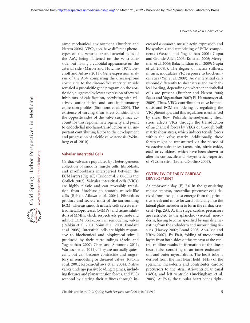
same mechanical environment (Butcher andNerem 2006). VECs, too, have different pheno-types on the ventricular and arterial sides ofthe AoV, being flattened on the ventricularside, but having a cuboidal appearance on thearterial side (Maron and Hutchins 1974; Bis-choff and Aikawa 2011). Gene expression anal-ysis of the AoV comparing the disease-proneaortic side to the disease-free ventricular siderevealed a procalcific gene program on the aor-tic side, suggested by lower expression of severalinhibitors of calcification, coexisting with rel-atively antioxidative and anti-inflammatoryexpression profiles (Simmons et al. 2005). Theexistence of varying shear stress conditions onthe opposite sides of the valve cusps may ac-count for this regional heterogeneity and pointto endothelial mechanotransduction as an im-portant contributing factor to the developmentand progression of calcific valve stenosis (Wein-berg et al. 2010).
Valvular Interstitial Cells
Cardiac valves are populated by a heterogeneouscollection of smooth muscle cells, fibroblasts,and myofibroblasts interspersed between theECM layers (Fig. 1C) (Taylor et al. 2003; Liu andGotlieb 2007). Valvular interstitial cells (VICs)are highly plastic and can reversibly transi-tion from fibroblast to smooth muscle-likecells (Rabkin-Aikawa et al. 2004). Fibroblastsproduce and secrete most of the surroundingECM, whereas smooth muscle cells secrete ma-trix metalloproteases (MMPs) and tissue inhib-itors of MMPs, which, respectively, promote andinhibit ECM breakdown in remodeling valves(Rabkin et al. 2001; Soini et al. 2001; Fondardet al. 2005). Interstitial cells are highly respon-sive to biochemical and biophysical stimuliproduced by their surroundings (Sacks andYoganathan 2007; Chen and Simmons 2011;Warnock et al. 2011). They are normally quies-cent, but can become contractile and migra-tory in remodeling or diseased valves (Rabkinet al. 2001; Rabkin-Aikawa et al. 2004). Nativevalves undergo passive loading regimes, includ-ing flexures and planar tension forces, and VICsrespond by altering their stiffness through in-
creased a-smooth muscle actin expression andbiosynthesis and remodeling of ECM compo-nents (Weston and Yoganathan 2001; Guptaand Grande-Allen 2006; Ku et al. 2006; Merry-man et al. 2006; Balachandran et al. 2009; Guptaet al. 2009b). The degree of matrix stiffness,in turn, modulates VIC response to biochemi-cal cues (Yip et al. 2009). AoV interstitial cellsrespond differently to shear stress and mechan-ical loading, depending on whether endothelialcells are present (Butcher and Nerem 2006;Sacks and Yoganathan 2007; El-Hamamsy et al.2009). Thus, VECs contribute to valve homeo-stasis and ECM remodeling by regulating theVIC phenotype, and this regulation is enhancedby shear flow. Pulsatile hemodynamic shearstress affects VICs through the transductionof mechanical forces by VECs or through bulkmatrix shear stress, which induces tensile forceswithin the valve matrix. Additionally, theseforces might be transmitted via the release ofvasoactive substances (serotonin, nitric oxide,etc.) or cytokines, which have been shown toalter the contractile and biosynthetic propertiesof VICs in vitro (Liu and Gotlieb 2007).
OVERVIEW OF EARLY CARDIACDEVELOPMENT
At embryonic day (E) 7.0 in the gastrulatingmouse embryo, precardiac precursor cells de-rived from the epiblast emerge from the primi-tive streak and move forward bilaterally into thelateral plate mesoderm to form the cardiac cres-cent (Fig. 2A). At this stage, cardiac precursorsare restricted to the splanchic (visceral) meso-derm, having become specified by signals ema-nating from the endoderm and surrounding tis-sues (Harvey 2002; Brand 2003; Abu-Issa andKirby 2007). By E8.0, folding of mesodermallayers from both sides of the embryo at the ven-tral midline results in formation of the linearheart tube, consisting of an inner endocardi-um and outer myocardium. The heart tube isderived from the first heart field (FHF) of thesplanchic mesoderm and contributes cardiacprecursors to the atria, atrioventricular canal(AVC), and left ventricle (Buckingham et al.2005). At E9.0, the tubular heart bends right-
How to Make a Heart Valve
Cite this article as Cold Spring Harb Perspect Med 2014;4:a013912 5
ww
w.p
ersp
ecti
vesi
nm
edic
ine.
org
on March 21, 2022 - Published by Cold Spring Harbor Laboratory Press http://perspectivesinmedicine.cshlp.org/Downloaded from
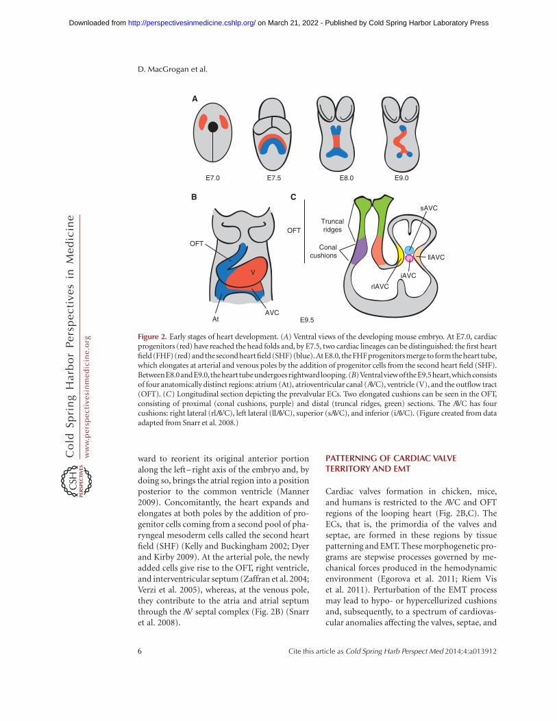
ward to reorient its original anterior portionalong the left–right axis of the embryo and, bydoing so, brings the atrial region into a positionposterior to the common ventricle (Manner2009). Concomitantly, the heart expands andelongates at both poles by the addition of pro-genitor cells coming from a second pool of pha-ryngeal mesoderm cells called the second heartfield (SHF) (Kelly and Buckingham 2002; Dyerand Kirby 2009). At the arterial pole, the newlyadded cells give rise to the OFT, right ventricle,and interventricular septum (Zaffran et al. 2004;Verzi et al. 2005), whereas, at the venous pole,they contribute to the atria and atrial septumthrough the AV septal complex (Fig. 2B) (Snarret al. 2008).
PATTERNING OF CARDIAC VALVETERRITORY AND EMT
Cardiac valves formation in chicken, mice,and humans is restricted to the AVC and OFTregions of the looping heart (Fig. 2B,C). TheECs, that is, the primordia of the valves andseptae, are formed in these regions by tissuepatterning and EMT. These morphogenetic pro-grams are stepwise processes governed by me-chanical forces produced in the hemodynamicenvironment (Egorova et al. 2011; Riem Viset al. 2011). Perturbation of the EMT processmay lead to hypo- or hypercellurized cushionsand, subsequently, to a spectrum of cardiovas-cular anomalies affecting the valves, septae, and
E9.0
A
B
E9.5
Truncalridges
Conalcushions
iAVC
sAVC
rlAVC
llAVC
C
OFT
V
AtAVC
OFT
E7.0 E7.5 E8.0
Figure 2. Early stages of heart development. (A) Ventral views of the developing mouse embryo. At E7.0, cardiacprogenitors (red) have reached the head folds and, by E7.5, two cardiac lineages can be distinguished: the first heartfield(FHF) (red) and the second heart field(SHF)(blue).At E8.0, the FHFprogenitorsmerge toformthe heart tube,which elongates at arterial and venous poles by the addition of progenitor cells from the second heart field (SHF).BetweenE8.0andE9.0, thehearttubeundergoesrightwardlooping.(B)VentralviewoftheE9.5heart,whichconsistsof four anatomically distinct regions: atrium (At), atrioventricular canal (AVC), ventricle (V), and the outflow tract(OFT). (C) Longitudinal section depicting the prevalvular ECs. Two elongated cushions can be seen in the OFT,consisting of proximal (conal cushions, purple) and distal (truncal ridges, green) sections. The AVC has fourcushions: right lateral (rlAVC), left lateral (llAVC), superior (sAVC), and inferior (iAVC). (Figure created from dataadapted from Snarr et al. 2008.)
D. MacGrogan et al.
6 Cite this article as Cold Spring Harb Perspect Med 2014;4:a013912
ww
w.p
ersp
ecti
vesi
nm
edic
ine.
org
on March 21, 2022 - Published by Cold Spring Harbor Laboratory Press http://perspectivesinmedicine.cshlp.org/Downloaded from
heart chambers. Endocardial tissue patterningis achieved by establishing developmental do-mains permissive for valve formation (de laPompa and Epstein 2012). Specification of theheart-valve-forming region requires signalingfrom myocardial bone morphogenetic protein2 (Bmp2) to activate Tbx2 (Yamada et al. 2000;Ma et al. 2005). Outside this territory, cardio-genic signals activate expression of Tbx20,which drives expression of Hey1 and Hey2 andrepresses Tbx2 (Singh et al. 2005; Stennard et al.2005; Kokubo et al. 2007). Tbx20 and Hey1,2,thus, restrict Bmp2 and Tbx2 to valve territory.In the endocardium, Notch1 represses Bmp2 viaHey1, Hey2, and HeyL rendering AVC endocar-dial endothelium competent to form heart-valve mesenchyme (Luna-Zurita et al. 2010).
The first signs of EC formation occur at E9.5when swellings of proteoglycan-rich ECM se-creted by the myocardium appear at the AV andOFT junctions (Eisenberg and Markwald 1995;Person et al. 2005). Over the following day, asubset of endocardial cells lining the AV andOFT cushions undergo EMT (Eisenberg andMarkwald 1995; Armstrong and Bischoff 2004;Person et al. 2005). These transforming cells hy-pertrophy, lose apicobasal polarity, extend fili-podia, and migrate into the cardiac jelly (Eisen-berg and Markwald 1995; Person et al. 2005).Snail1 and Slug/Snail2 are crucial transcriptionfactors for EMT because they down-regulate theexpression of vascular endothelial cadherin, anadhesion molecule that maintains intercellularjunctions in endothelial tissue (Romano andRunyan 1999; Timmerman et al. 2004; Niessenet al. 2008). Interestingly, in zebrafish, AV valveleaflets form directly through a process of invag-ination, during which the endocardium doesnot transform, but instead remains as a singlesheet of cells (Scherz et al. 2008).
The signaling pathways that control endocar-dial EMT have been recently reviewed in depth(Lim and Thiery 2012; von Gise and Pu 2012).EMTinductionisregulatedbyanetworkintegrat-ing Bmp, transforming growth factor (TGF)-b,and Notch signaling (Yamagishi et al. 2009; Len-cinas et al. 2011; de la Pompa and Epstein 2012;Kruithof et al. 2012). Downstream from TGF-b,b-catenin acts in the AVC endocardium to pro-
mote the acquisition of a mesenchymal pheno-type (Gessert and Kuhl 2010). The precisespatiotemporal pattern and levels of vascular en-dothelial growth factor (VEGF) expression arecritical for both the onset and resolution ofEMT (Dor et al. 2001; Chang et al. 2004; Stanku-nas et al. 2010). Nfatc1 is required downstreamfrom VEGF to regulate the extent of EMT andsustain endocardial proliferation during EMTand post-EMT valve elongation (Chang et al.2004; Wu et al. 2011; Lin et al. 2012). TheErbB/SHP-2/NF-1/Ras signaling axis pro-motes mesenchymal migration into the cardiacjelly and proliferation and expansion of cushionmesenchyme (Yutzey et al. 2005; Iwamoto andMekada 2006; Sanchez-Soria and Camenisch2010).
The ECM is a critical regulator of EMT(Schroederet al. 2003; Lockhart et al. 2011). Dis-ruption of hyaluronan synthase-2 (Has2) abro-gates normal cardiac morphogenesis and EMTmediated by HA (Camenisch et al. 2002). Highmolecular weight HA interacts with and acti-vates ErbB2-ErbB3 receptors, initially, to pro-mote EMTand, subsequently, in its depolymer-ized form to limit the extent of endocardial celldelamination and transformation (Camenischet al. 2002; Rodgers et al. 2006). Moreover, asimilar cardiac phenotype to the HA-deficientembryos occurs in zebrafish harboring a mu-tated uridine 50-diphosphate (UDP)-glucosedehydrogenase (ugdh) gene ( jekyll mutant). Thisenzyme produces UDP-glucuronate, which isnecessary for Has2 synthesis of HA, suggestingthat Ugdh may function in valve formationthrough its requirement for HA synthesis (Walshand Stainier 2001).
CARDIAC VALVE MORPHOGENESIS
Valvular morphogenesis is intimately tied todevelopmental processes that lead to heart-chamber septation and connection to pulmo-nary and systemic circuitries. Multiple typesof progenitor cells, originating from both in-and outside the heart orchestrate valve morpho-genesis through highly conserved signaling net-works. Once again, mistiming or malfunctionof the unfolding events cause valvuloseptal de-
How to Make a Heart Valve
Cite this article as Cold Spring Harb Perspect Med 2014;4:a013912 7
ww
w.p
ersp
ecti
vesi
nm
edic
ine.
org
on March 21, 2022 - Published by Cold Spring Harbor Laboratory Press http://perspectivesinmedicine.cshlp.org/Downloaded from

fects in patients with congenital heart diseaseand in experimental animal models (Srivastava2006; Joziasse et al. 2008; Lin et al. 2012).
Prefusion of Mesenchyme Structures
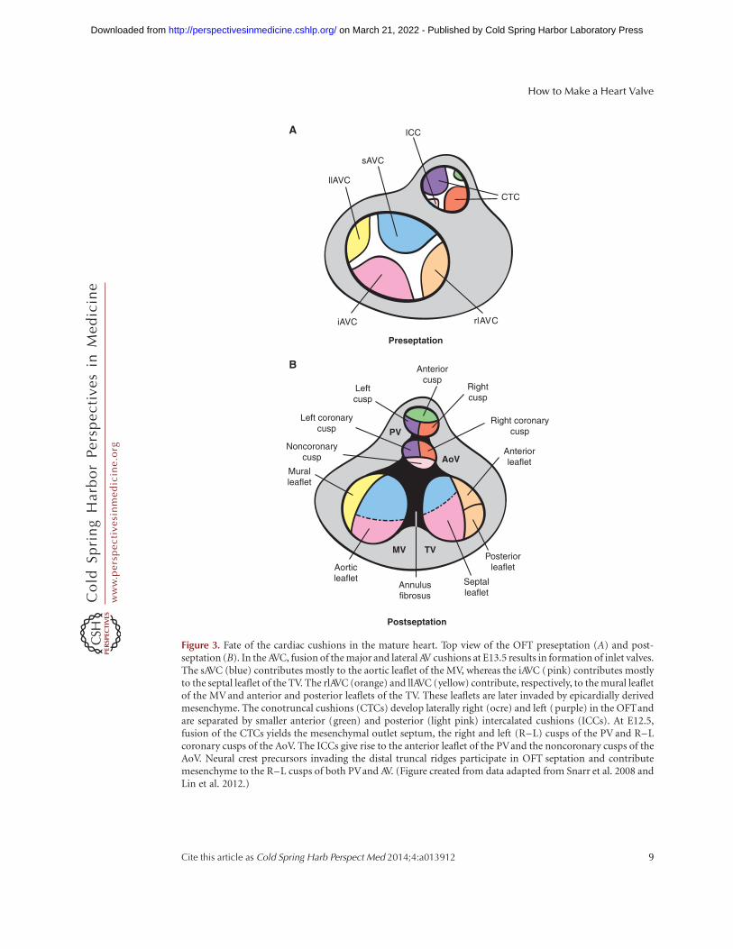
By E10.5, EMT subsides in the AVC. The major(inferior and superior) AV cushions constitutethe bulk of mesenchyme occupying the AVClumen (Figs. 2B and 3A). Smaller lateral AV cush-ions start to develop at this stage (Fig. 3A) (Wes-sels and Sedmera 2003; Snarr et al. 2008). Themajor cushions take part in the formation of theAV mesenchymal complex and contribute to theformation of the aortic leaflet of the MV andseptal leaflet of the tricuspid valve (TV) (Snarret al. 2008). The lateral AV cushions do not fuse,nor do they participate in AV complex forma-tion. Instead, the right cushion gives rise to theanterosuperior and -posterior leaflets of the TV,whereas the left cushion gives rise to the muralleaflet of the MV (Fig. 3A,B) (Snarr et al. 2008).
By E11.5, the OFT is occupied by spiralingand elongated mesenchymal cushions, referredto asseptal and parietal ridges, andtwo less prom-inent intercalating ridges (Snarr et al. 2008). Theboundary between the proximal conal and distaltruncal cushions is marked by the outer curva-ture of the OFT (i.e., the cono-truncal curva-ture) and determines the site for SL valve forma-tion (Fig. 2B) (Lin et al. 2012). By E12.5, thefused larger cushions and nonfused Immunocy-tochemistry (ICC) have given rise to the threearterial valve cusps for each SL valve (Fig. 3A,B).The major cushions participate in forming theaortico-pulmonary septum and contribute tothe R–L coronary cusps of the AoV and R–Lcusps of the PV. The intercalating cushions donot fuse; instead, the right lateral cushion givesrise to the noncoronary (NC) cusp of the AoV,whereas the left lateral cushion gives rise to theanterior leaflet of the PV. Eventually, the coro-nary arteries connect to the AoV sinuses imme-diately proximal to the R–L cusp precursors.
Lineage Tracing of Valve Tissues
Atrioventricular Valves
Labeling of endothelial and endocardial line-ages using the Tie2-Cre; ROSA26R mouse line
shows that the early mesenchyme forming themajor cushions is derived almost entirely fromendocardial EMT (Rivera-Feliciano et al. 2006;Snarr et al. 2008). b-galactosidase (b-gal) stain-ing is detected in the AV fibrous continuity, valveleaflets, and the chordae tendinae at later gesta-tional stages and postnatally, consistent withthese structures being derived from the endo-cardium (de Lange et al. 2004; Lincoln et al.2004).
Contributions to AV valve formation by theepicardium and cardiac neural crest also requirean EMT process (Fig. 4B) (Lim and Thiery2012; von Gise and Pu 2012). After undergoingEMT, epicardium-derived cells (EPDCs) giverise to the subepicardial mesenchyme and, sub-sequently, yield fibroblasts in the myocardialwall and smooth muscle cells of the media incoronary arteries (Lie-Venema et al. 2007; vonGise and Pu 2012). Quail-chick chimera analy-sis indicates that EPDCs populate the mesen-chyme of developing AV valves and contributeto the fibrous heart skeleton (Fig. 4B) (Gitten-berger-de Groot et al. 1998; Snarr et al. 2008).EPDC fate analysis using the mouse inducibleWt1-CreERT2; ROSA26 Cre reporter line showsan important contribution to the AV sulcusand annulus fibrosus, a fibrous continuity that,in addition to separating atrial and ventricularmyocardium, supports the mature valve leaflets(Zhou et al. 2010). At around E12.0, EPDCsmigrate through the AV junction to populatethe AV cushions. Using the Wt1/IRES/GFP-Cre; ROSA26 Cre reporter line, b-gal positivecells can be readily detected in the remodeledleaflets derived from the postfusion lateral cush-ions, but there is little contribution to the majorAV cushions (Wessels et al. 2012). In the muralMV and TV leaflets, the EPDCs eventually re-place the endocardially derived cells to makeup most of the mesenchyme. Remarkably, thesesame leaflets are often implicated in congenitaland acquired valve abnormalities, including Eb-stein’s anomaly, AV valve insufficiency or pro-lapse, and mitral/tricuspid stenosis (Fig. 4B)(Snarr et al. 2008).
A third source of AV leaflet mesenchyme is apopulation of cranial neural crest cells (CNCCs)(Fig. 4B). Beginning at E9.5, after undergoing
D. MacGrogan et al.
8 Cite this article as Cold Spring Harb Perspect Med 2014;4:a013912
ww
w.p
ersp
ecti
vesi
nm
edic
ine.
org
on March 21, 2022 - Published by Cold Spring Harbor Laboratory Press http://perspectivesinmedicine.cshlp.org/Downloaded from
Preseptation
llAVC
ICC
CTC
rlAVCiAVC
sAVC
A
Postseptation
Septalleaflet
Annulusfibrosus
B
PV
AoV
MV TV
Aorticleaflet
Muralleaflet
Anteriorleaflet
Posteriorleaflet
Anteriorcusp
Leftcusp
Rightcusp
Left coronarycusp
Right coronarycusp
Noncoronarycusp
Figure 3. Fate of the cardiac cushions in the mature heart. Top view of the OFT preseptation (A) and post-septation (B). In the AVC, fusion of the major and lateral AV cushions at E13.5 results in formation of inlet valves.The sAVC (blue) contributes mostly to the aortic leaflet of the MV, whereas the iAVC (pink) contributes mostlyto the septal leaflet of the TV. The rlAVC (orange) and llAVC (yellow) contribute, respectively, to the mural leafletof the MV and anterior and posterior leaflets of the TV. These leaflets are later invaded by epicardially derivedmesenchyme. The conotruncal cushions (CTCs) develop laterally right (ocre) and left (purple) in the OFTandare separated by smaller anterior (green) and posterior (light pink) intercalated cushions (ICCs). At E12.5,fusion of the CTCs yields the mesenchymal outlet septum, the right and left (R–L) cusps of the PV and R–Lcoronary cusps of the AoV. The ICCs give rise to the anterior leaflet of the PVand the noncoronary cusps of theAoV. Neural crest precursors invading the distal truncal ridges participate in OFT septation and contributemesenchyme to the R–L cusps of both PVand AV. (Figure created from data adapted from Snarr et al. 2008 andLin et al. 2012.)
How to Make a Heart Valve
Cite this article as Cold Spring Harb Perspect Med 2014;4:a013912 9
ww
w.p
ersp
ecti
vesi
nm
edic
ine.
org
on March 21, 2022 - Published by Cold Spring Harbor Laboratory Press http://perspectivesinmedicine.cshlp.org/Downloaded from
Pul
mon
ary
valv
e
Myo
card
ium
Ann
ulus
fibro
sus
Mitr
alva
lve
Epi
card
ium
-de
rived
cel
ls
Tric
uspi
dva
lve
Aor
ticva
lve
Neu
ral c
rest
–de
rived
cel
ls
Epi
card
ium
Car
diac
neur
al c
rest
Ann
ulus
Sup
port
ing
appa
ratu
s
Sin
us o
fV
alsa
lva Ann
ulus
Myo
card
ium
A B
C D
Figu
re4.
Rem
od
elin
go
fth
eca
rdia
cva
lve
leafl
ets
and
cusp
s.(A
)C
on
trib
uti
on
ofc
ard
iac
neu
ralc
rest
(gre
en)-
and
epic
ard
ial
(ora
nge
)-d
eriv
edce
lls
(CN
CC
and
EP
DC
s).C
NC
Cs
mig
rate
thro
ugh
the
ph
aryn
geal
arch
esan
din
toth
eO
FT
toin
itia
teth
ere
org
aniz
atio
no
fth
eO
FT
and
form
atio
no
fSL
valv
es.
EP
DC
sco
ntr
ibu
teto
the
form
atio
no
fth
eco
ron
ary
arte
ries
,in
ters
titi
alce
lls
inth
em
yoca
rdiu
m,a
nd
the
AVva
lves
.(B
)To
pvi
ewo
fth
ese
pta
ted
hea
rt,d
epic
tin
gth
ere
lati
veco
ntr
ibu
-ti
on
so
fCN
CC
san
dE
PD
Cst
oca
rdia
cva
lve
leafl
ets
and
cusp
s.(C
)M
orp
ho
gen
esis
oft
he
AVva
lves
.Fro
mE
13.5
on
war
d,t
he
leafl
ets
and
ten
sile
app
arat
us
(lig
ht
bro
wn
)o
fth
eAV
valv
esfo
rmp
red
om
inan
tly
by
del
amin
atio
no
fth
ein
ner
laye
rso
fth
ein
let
zon
efr
om
the
ven
tric
ula
rse
pta
lwal
l.T
he
cho
rdae
ten
din
ae,
ann
ulu
sfi
bro
sus,
and
the
leafl
etit
self
are
der
ived
fro
mth
een
do
card
ium
.T
he
sep
tal
leafl
eto
fth
eT
V(i
nco
ntr
ast
toth
eao
rtic
leafl
eto
fth
eM
V),
toge
ther
wit
hit
ssu
pp
ort
ing
ten
din
ou
sco
rds,
rem
ain
sco
nn
ecte
dto
the
myo
card
ium
un
tilE
17.5
.(D
)M
orp
ho
gen
esis
oft
he
SLva
lves
.Th
eex
cava
tio
no
fth
ecu
sps
take
sp
lace
init
iall
yb
yso
lid
ingr
owth
of
the
end
oth
eliu
mat
the
arte
rial
face
of
the
cusp
and
,su
bse
qu
entl
y,b
ylu
men
atio
n.E
lon
gati
on
and
rem
od
elin
go
fth
ep
rim
ord
iain
tom
atu
reva
lve
stru
ctu
res
are
asso
ciat
edw
ith
regi
on
aliz
edce
llp
roli
fera
tio
nan
dm
atri
xal
ign
men
tgu
ided
by
hem
od
ynam
icfo
rces
.Myo
card
ium
isd
epic
ted
ind
ark
red
,pro
tegl
ycan
sin
blu
e,an
dm
esen
chym
ein
gray
.
D. MacGrogan et al.
10 Cite this article as Cold Spring Harb Perspect Med 2014;4:a013912
ww
w.p
ersp
ecti
vesi
nm
edic
ine.
org
on March 21, 2022 - Published by Cold Spring Harbor Laboratory Press http://perspectivesinmedicine.cshlp.org/Downloaded from
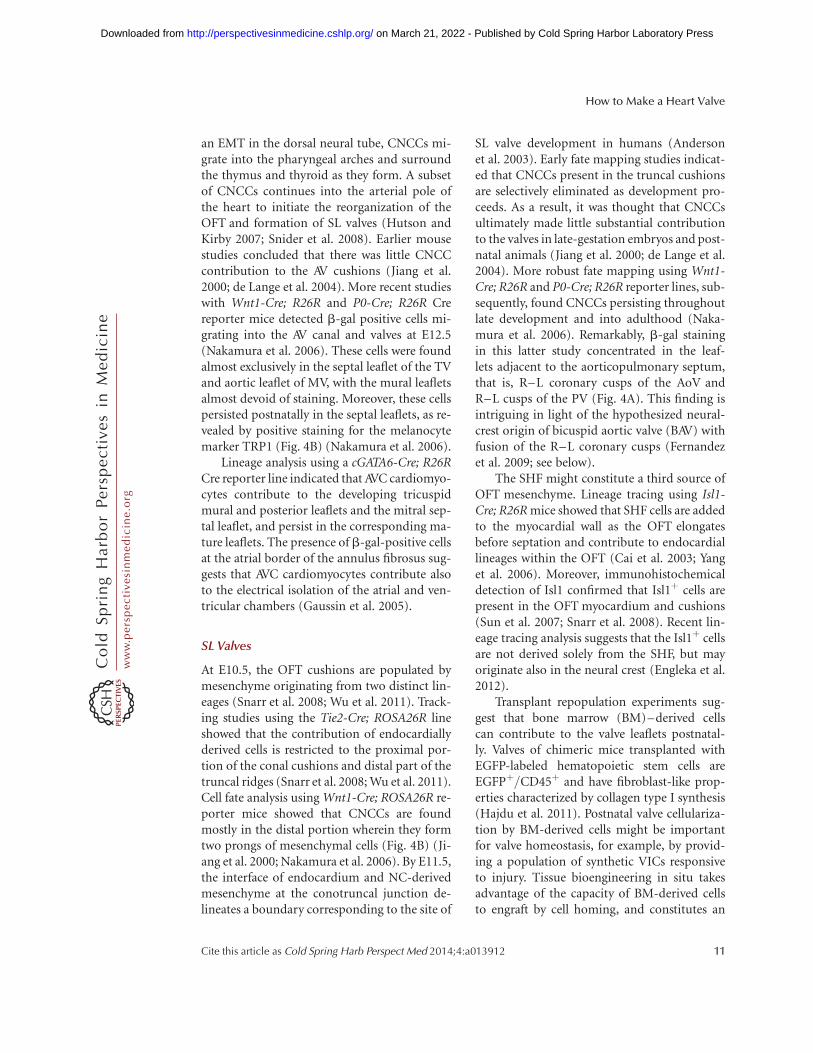
an EMT in the dorsal neural tube, CNCCs mi-grate into the pharyngeal arches and surroundthe thymus and thyroid as they form. A subsetof CNCCs continues into the arterial pole ofthe heart to initiate the reorganization of theOFT and formation of SL valves (Hutson andKirby 2007; Snider et al. 2008). Earlier mousestudies concluded that there was little CNCCcontribution to the AV cushions (Jiang et al.2000; de Lange et al. 2004). More recent studieswith Wnt1-Cre; R26R and P0-Cre; R26R Crereporter mice detected b-gal positive cells mi-grating into the AV canal and valves at E12.5(Nakamura et al. 2006). These cells were foundalmost exclusively in the septal leaflet of the TVand aortic leaflet of MV, with the mural leafletsalmost devoid of staining. Moreover, these cellspersisted postnatally in the septal leaflets, as re-vealed by positive staining for the melanocytemarker TRP1 (Fig. 4B) (Nakamura et al. 2006).
Lineage analysis using a cGATA6-Cre; R26RCre reporter line indicated that AVC cardiomyo-cytes contribute to the developing tricuspidmural and posterior leaflets and the mitral sep-tal leaflet, and persist in the corresponding ma-ture leaflets. The presence of b-gal-positive cellsat the atrial border of the annulus fibrosus sug-gests that AVC cardiomyocytes contribute alsoto the electrical isolation of the atrial and ven-tricular chambers (Gaussin et al. 2005).
SL Valves
At E10.5, the OFT cushions are populated bymesenchyme originating from two distinct lin-eages (Snarr et al. 2008; Wu et al. 2011). Track-ing studies using the Tie2-Cre; ROSA26R lineshowed that the contribution of endocardiallyderived cells is restricted to the proximal por-tion of the conal cushions and distal part of thetruncal ridges (Snarr et al. 2008; Wu et al. 2011).Cell fate analysis using Wnt1-Cre; ROSA26R re-porter mice showed that CNCCs are foundmostly in the distal portion wherein they formtwo prongs of mesenchymal cells (Fig. 4B) (Ji-ang et al. 2000; Nakamura et al. 2006). By E11.5,the interface of endocardium and NC-derivedmesenchyme at the conotruncal junction de-lineates a boundary corresponding to the site of
SL valve development in humans (Andersonet al. 2003). Early fate mapping studies indicat-ed that CNCCs present in the truncal cushionsare selectively eliminated as development pro-ceeds. As a result, it was thought that CNCCsultimately made little substantial contributionto the valves in late-gestation embryos and post-natal animals (Jiang et al. 2000; de Lange et al.2004). More robust fate mapping using Wnt1-Cre; R26R and P0-Cre; R26R reporter lines, sub-sequently, found CNCCs persisting throughoutlate development and into adulthood (Naka-mura et al. 2006). Remarkably, b-gal stainingin this latter study concentrated in the leaf-lets adjacent to the aorticopulmonary septum,that is, R–L coronary cusps of the AoV andR–L cusps of the PV (Fig. 4A). This finding isintriguing in light of the hypothesized neural-crest origin of bicuspid aortic valve (BAV) withfusion of the R–L coronary cusps (Fernandezet al. 2009; see below).
The SHF might constitute a third source ofOFT mesenchyme. Lineage tracing using Isl1-Cre; R26R mice showed that SHF cells are addedto the myocardial wall as the OFT elongatesbefore septation and contribute to endocardiallineages within the OFT (Cai et al. 2003; Yanget al. 2006). Moreover, immunohistochemicaldetection of Isl1 confirmed that Isl1þ cells arepresent in the OFT myocardium and cushions(Sun et al. 2007; Snarr et al. 2008). Recent lin-eage tracing analysis suggests that the Isl1þ cellsare not derived solely from the SHF, but mayoriginate also in the neural crest (Engleka et al.2012).
Transplant repopulation experiments sug-gest that bone marrow (BM)–derived cellscan contribute to the valve leaflets postnatal-ly. Valves of chimeric mice transplanted withEGFP-labeled hematopoietic stem cells areEGFPþ/CD45þ and have fibroblast-like prop-erties characterized by collagen type I synthesis(Hajdu et al. 2011). Postnatal valve cellulariza-tion by BM-derived cells might be importantfor valve homeostasis, for example, by provid-ing a population of synthetic VICs responsiveto injury. Tissue bioengineering in situ takesadvantage of the capacity of BM-derived cellsto engraft by cell homing, and constitutes an
How to Make a Heart Valve
Cite this article as Cold Spring Harb Perspect Med 2014;4:a013912 11
ww
w.p
ersp
ecti
vesi
nm
edic
ine.
org
on March 21, 2022 - Published by Cold Spring Harbor Laboratory Press http://perspectivesinmedicine.cshlp.org/Downloaded from
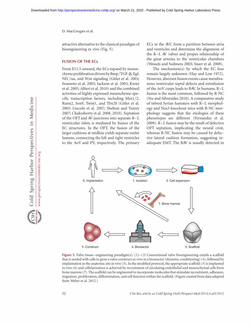
attractive alternative to the classical paradigm ofbioengineering ex vivo (Fig. 5).
FUSION OF THE ECs
From E11.5 onward, the ECs expand by mesen-chyme proliferation driven by Bmp/TGF-b, Egf,Nf1/ras, and Wnt signaling (Gitler et al. 2003;Iwamoto et al. 2003; Jackson et al. 2003; Krenzet al. 2005; Alfieri et al. 2010) and the combinedactivities of highly expressed mesenchyme-spe-cific transcription factors, including Msx1/2,Runx2, Sox9, Twist1, and Tbx20 (Gitler et al.2003; Lincoln et al. 2007; Shelton and Yutzey2007; Chakraborty et al. 2008, 2010). Septationof the OFT and AV junctions into separate R–Lventricular inlets is mediated by fusion of theEC structures. In the OFT, the fusion of thelarger cushions at midline yields separate outletlumens, connecting the left and right ventriclesto the AoV and PV, respectively. The primary
ECs in the AVC form a partition between atriaand ventricles and determine the alignment ofthe R–L AV valves and proper relationship ofthe great arteries to the ventricular chambers(Wessels and Sedmera 2003; Snarr et al. 2008).
The mechanism(s) by which the EC fuseremain largely unknown (Hay and Low 1972).However, aberrant fusion events cause membra-nous ventricular septal defects and extrafusionof the AoV cusps leads to BAV. In humans, R–Lfusion is the most common, followed by R-NC(Siu and Silversides 2010). A comparative studyof inbred Syrian hamsters with R–L morphol-ogy and Nos3-knockout mice with R-NC mor-phology suggests that the etiologies of thesephenotypes are different (Fernandez et al.2009). R–L fusion may be the result of defectiveOFT septation, implicating the neural crest,whereas R-NC fusion may be caused by defec-tive lateral cushion formation, suggesting in-adequate EMT. The BAV is usually detected in
4. Bioreactor5. Construct 3. Scaffold
2. Cell expansion1. Isolation6. Implantation
7. Bone marrow
Figure 5. Valve tissue–engineering paradigm(s). (1)–(3) Conventional valve bioengineering entails a scaffoldthat is seeded with cells to grow a valve construct ex vivo in a bioreactor (dynamic conditioning) (4), followed byimplantation in the anatomic site in vivo (5). In the modified protocol, the appropriate scaffold (3) is implantedin vivo (6) and cellularization is achieved by recruitment of circulating endothelial and mesenchymal cells frombone marrow (7). The scaffold can be engineered to incorporate molecules that stimulate recruitment, adhesion,migration, proliferation, differentiation, and cell function within the scaffold. (Figure created from data adaptedfrom Weber et al. 2012.)
D. MacGrogan et al.
12 Cite this article as Cold Spring Harb Perspect Med 2014;4:a013912
ww
w.p
ersp
ecti
vesi
nm
edic
ine.
org
on March 21, 2022 - Published by Cold Spring Harbor Laboratory Press http://perspectivesinmedicine.cshlp.org/Downloaded from

isolation, but can also coexist with other cardio-vascular malformations, suggesting a multigen-ic etiology (Siu and Silversides 2010). Consistentwith this notion, family-based linkage analysessuggest linkage to loci on chromosome 18q, 5q,and 13q, but the genes within these regions re-main to be identified (Martin et al. 2007). Todate, only mutated NOTCH1 alleles on chromo-some band 9q34-35 have been found to be caus-ative in familial BAV in the context of calcificAoV disease (Garg et al. 2005).
POSTFUSION MORPHOGENESIS
During late AV valvulogenesis, the septal leafletof the TV remains in contact with the septumuntil it delaminates at E17.5 (Fig. 4C) (Lamerset al. 1995; de Lange et al. 2004; Gaussin et al.2005). The mural leaflets of both AV valves aresupported by AV myocardium at their ventric-ular side. In comparison, the mitral septal leafletis never supported by myocardium, but is incontact with the AV-myocardium-derived mi-tral gully at its anterior and posterior margins(de Lange et al. 2004; Gaussin et al. 2005). As theprimordial leaflets distend into the lumen, thinstrands of elongated muscle remain attached tothe valve leaflet until E17.5. Programmed celldeath yields a mobile leaflet and remnants thatcontribute to the chordae tendinae and papil-lary muscles (de Lange et al. 2004; Lincoln et al.2004). Postfusion SL valve morphogenesis is lesswell characterized. SL valves undergo progres-sive excavation from E12.5 until E15 when theyachieve their typical morphology (Fig. 4D).This process is driven by “selective endothelialgrowth” of the free edges of the cusps on theirarterial face, producing an epithelial ridge orgroove between the emerging cusps and the ar-terial wall. The groove eventually becomes lumi-nated to yield the sinus of Valsalva (Hurle 1979;Hurle et al. 1980).
CARDIAC VALVE REMODELING
The later phases of valve development are char-acterized by the gradual transition from undif-ferentiated mesenchyme to specialized VICs.Remodeling of the primitive ECM into a highly
organized and stratified ECM is strongly in-fluenced by hemodynamic stimuli. The devel-opmental mechanisms that coordinate VICspecialization and ECM organization duringvalvulogenesis remain uncertain (de Vlaminget al. 2012).
Lineage Diversification
Cell proliferation, density, and turnover, sub-stantial in early valvulogenesis, become lesspronounced in remodeling valves (Aikawa etal. 2006; Hinton et al. 2006). Apoptosis in theOFT cushions is substantial at later fetal stages,consistent with ongoing remodeling processes(Poelmann and Gittenberger-de Groot 2005;Aikawa et al. 2006; Jain et al. 2011). Early over-lapping gene expression patterns become re-stricted (Lincoln et al. 2004; Chakraborty et al.2008). The transition from EC growth to re-modeling requires calcineurin/NFATc1 signal-ing in the endocardium (de la Pompa et al.1998; Ranger et al. 1998; Chang et al. 2004),notably through the regulation of RANKLand Cathepsin K expression (Lange and Yutzey2006). Interstitial cell progenitors show tran-scriptional profiles normally associated withcartilage and tendon lineages (Lincoln et al.2006b; Chakraborty et al. 2008). Expression ofSox9 is necessary for early interstitial cell pro-liferation and, subsequently, expression of car-tilage matrix proteins (Akiyama et al. 2004;Lincoln et al. 2007), and scleraxis is requiredfor tendinous cord specification (Levay et al.2008). In the AV valves of avian embryos, Sox9and aggrecan are predominantly expressed inthe leaflets, whereas scleraxis and tenascin Care expressed in the supporting AV structures,including the chordae tendineae (Lincoln et al.2004). In contrast, SL valves express genes asso-ciated with both lineages and diversify intocusps with an internal supporting apparatus(Zhao et al. 2007). A delicate balance of BMPand fibroblast growth factors (FGF) signals isrequired for lineage diversification. In culturedearly valve precusors, Sox9 and aggrecan are in-duced by BMP2, whereas scleraxis and tenascinare regulated by FGF-4, but it is unclear howthese activities coordinate spatiotemporally to
How to Make a Heart Valve
Cite this article as Cold Spring Harb Perspect Med 2014;4:a013912 13
ww
w.p
ersp
ecti
vesi
nm
edic
ine.
org
on March 21, 2022 - Published by Cold Spring Harbor Laboratory Press http://perspectivesinmedicine.cshlp.org/Downloaded from
direct VIC diversification (Lincoln et al. 2006a;Zhao et al. 2007).
Remodeling of the ECM
The elongation of the valve leaflets and stratifi-cation of the ECM begins late in gestation andcontinues postnatally (Aikawa et al. 2006; Hin-ton et al. 2006; Kruithof et al. 2007; Peacocket al. 2008; Stephens et al. 2010). Initial ECMpatterning along the AV leaflet axis occurs be-tween E15.5 and E18.5 through a transient in-crease in cell density called condensation (Krui-thof et al. 2007). In mature valves, the ECM isfound in alignment with blood flow, suggestingthat stratification and remodeling are driven byhemodynamic forces acting via the endotheli-um (Combs and Yutzey 2009). Postnatally, ad-ditional leaflet elongation is thought to occur byphysical pulling of the tendinous cord attach-ments by the rapidly growing ventricles.
As remodeling procedes, collagen fibrilsbecome densely packed locally at the outflowside of the leaflet and loosely arranged and in-terweaving elsewhere (Aikawa et al. 2006; Hin-ton et al. 2006; Peacock et al. 2008; Tan et al.2011). Fibrillogenesis is promoted by periostin,a multifunctional fascilin-domain-containingprotein (Norris et al. 2009). Periostin promotesthe differentiation of endothelial and epicar-dially derived mesenchyme while blocking oth-er cell types, in particular, cardiomyocytes, andis required for fibrous maturation of the AVleaflets and their supporting apparatus (Norriset al. 2008; Snider et al. 2008). Valve remodelingand organization is dependent on the coordi-nated actions of MMPs and a disintegrin andmetalloproteinase with thrombospondin mo-tifs (ADAMTS) families of zinc metalloprotei-nases (Lockhart et al. 2011). These pericellularsecreted proteases target the ECM and medi-ate structural changes required for cell migra-tion and proliferation. For example, proteolyticcleavage of versican, a chondroitin sulfate pro-teoglycan, by ADAMTS1 plays a significant rolein the maturation of the AV cushions and re-modeling of the OFT by facilitating the densepacking of mesenchyme in the elongating valves(Kern et al. 2006).
Dysregulation of the ECM appears to bea general feature of valve disease regardless ofetiology; for example, BAVs from pediatric pa-tients have increased collagen and proteoglycancontent, whereas myxomatous MVs have loosecollagen, increased proteoglycan, and reducedelastin content with altered fiber orientation inall layers (Rabkin et al. 2001; Hinton et al. 2006;Gupta et al. 2009a). These structural changesare associated with the aberrant reexpressionof early valve mesenchymal and chondrogenicprogenitor markers and may be related to thereawakening of fetal transcriptional programs(Wirrig et al. 2011; Cheek et al. 2012).
HEART-VALVE TISSUE ENGINEERING
General Overview
In the classical tissue-engineering paradigm,cells are harvested from a donor (which can bethe recipient) and are subsequently expandedto obtain a sufficiently large number of cellsfor seeding on a biodegradable starter matrix(scaffold) (Fig. 5). The porous starter matrix isshaped like a heart valve and supports the for-mation of neotissue (Hoerstrup et al. 2000). Inthe bioreactor, the construct is subjected tobiochemical and mechanical stimuli to enhancethe production of ECM proteins in an effort tocreate a valve that has sufficient strength anddurability to meet in vivo hemodynamic re-quirements (Mol et al. 2005a; Kortsmit et al.2009). Particular attention has been focused onreplicating the anisotropic architecture of thenative leaflet (Mol et al. 2006; Neidert and Tran-quillo 2006; Balguid et al. 2007; Rubbens et al.2009; Cox et al. 2010; Sander et al. 2011).
An alternative approach is autologous tis-sue formation, initiated by intraperitoneal im-plantation of a heart valve–shaped scaffold (DeVisscher et al. 2007) or mold (Hayashida et al.2007; Yamanami et al. 2010). The mold willelicit a foreign body response causing deposi-tion of autologous ECM on the mold surface.The resulting construct is then transplanted tothe heart as a valve replacement. However, thevolume fraction of cellular phenotypes current-ly achieved with this approach is unbalanced,
D. MacGrogan et al.
14 Cite this article as Cold Spring Harb Perspect Med 2014;4:a013912
ww
w.p
ersp
ecti
vesi
nm
edic
ine.
org
on March 21, 2022 - Published by Cold Spring Harbor Laboratory Press http://perspectivesinmedicine.cshlp.org/Downloaded from

which may result in inadequate remodeling ofthe valves (De Visscher et al. 2007).
Cell Sources for In Vitro Heart-ValveTissue Engineering
A large variety of cell types has been investi-gated for in vitro seeding of synthetic or biolog-ical scaffolds. Early experiments comparing thein vivo response to allogenic and autologouscells showed that seeded cells from an autolo-gous source were desirable to minimize the im-mune response (Shinoka et al. 1995). However,autologous cells were recently shown to provokean immune response caused by proinflamma-tory cytokines originating from cell death, ordamage-associated molecular pattern (DAMP)molecules released by dying cells within the au-tologous tissue (Badylak and Gilbert 2008).
Vascular-derived myofibroblasts and endo-thelial cells are the gold standard in heart-valvetissue engineering because of their excellentECM production capacity. These cells can beharvested from the recipient’s saphenous vein.Alternative sources are progenitor cells derivedfrom bone marrow, adipose tissue or umbilicalcord blood, and circulating endothelial progen-itor cells, all of which have shown potential toprovide interstitial and endothelial function andhave been used to generate heart valves in vitro(Hoerstrup et al. 2002; Schmidt et al. 2007; Saleset al. 2010). One of the advantages of progenitorcells is that, unlike vascular-derived cells, theycan be harvested without additional surgical in-tervention. In this regard, bone marrow–derivedmesenchymal stem cells (MSCs) are attractivecandidates and have been used to seed decel-lularized matrices (Vincentelli et al. 2007; Iopet al. 2009) and synthetic scaffolds (Hoerstrupet al. 2002; Sutherland et al. 2005). MSCs areremarkably similar to VICs (Latif et al. 2007),and have antithrombogenic (Hashi et al. 2007)and immunosuppressive properties (Uccelli etal. 2006). MSCs are able to differentiate into en-dothelial cells, fibroblasts or myofibroblasts, andsmooth muscle cells (Iop et al. 2009). Their ac-cessibility, ease of handling, and potential forallogenic applications make MSCs suitable forroutine clinical use (Pittenger and Martin 2004).
In addition to supporting proliferation, differ-entiation, and ECM production ex vivo, MSCscan induce the homing and differentiation ofautologous host cells through paracrine signal-ing, involving an array of cytokines and growthfactors (Roh et al. 2010). Despite the encourag-ing results obtained with preseeded scaffolds insheep and baboons (Vincentelli et al. 2007; We-beret al. 2011), the antithrombogenic propertiesof MSCs (Hashi et al. 2007) and their ability tostimulate in vivo endothelialization (Mirza et al.2008) should preclude future requirements forpreseeding with endothelial cells.
Scaffolds for Heart-Valve Tissue Engineering
Different types of scaffold materials, or so-calledstarter matrices, have been used for heart-valvetissue engineering. These include biodegradablesynthetic polymers, natural materials, such asfibrin and collagen, and xenogenic or allogen-ic decellularized heart valves. A decellularizedxenograft or homograft is an obvious choiceof scaffold material because these grafts closelyresemble native human valve geometry andstructure and have excellent mechanical and he-modynamic properties. The microstructure ofthe allograft favors proliferation, differentia-tion, and survival of reseeded cells (Mirza etal. 2008). The use of allografts, however, is lim-ited by donor availability. Because of its ana-tomic similarity to human valves, the porcineheart valve makes an attractive alternative. Clin-ical application of decellularized xenografts has,however, resulted in early failure (Simon et al.2003; Roh et al. 2010; Ruffer et al. 2010; Hibinoet al. 2011). A major concern with xenogenicmaterials is infection with endogenous porcineretroviruses, prions, or other zoonotic vectors.Improved decellularization techniques recentlyallowed the preparation of nonimmunogenicdecellularized xenograft valves (Bloch et al.2011). Complete removal of cells from the tissueis crucial, as residual cells and cell remnantswithin the matrix might lead to calcification(Human and Zilla 2001). It is also importantto minimize structural alteration of the ECMto preserve the biomechanical characteristicsof the native heart valve. A promising alternative
How to Make a Heart Valve
Cite this article as Cold Spring Harb Perspect Med 2014;4:a013912 15
ww
w.p
ersp
ecti
vesi
nm
edic
ine.
org
on March 21, 2022 - Published by Cold Spring Harbor Laboratory Press http://perspectivesinmedicine.cshlp.org/Downloaded from
is the use of decellularized TEHVs, yielding off-the-shelf available homologous heart valves(Dijkman et al. 2012).
Biodegradable synthetic materials have beenwidely used as scaffold material for tissue engi-neering (Bouten et al. 2011). Commonly usedbiomaterials are different copolymers and de-rivatives thereof, whereas, more recently, elasto-meric materials have gained attention (Court-ney et al. 2006; Sales et al. 2007; Stella et al.2010). Compared with decellularized xenogenicand allogenic matrices, these materials avoidthe risk of disease transfer and immunologicalcomplications. In addition, synthetic materialshave the advantage of unlimited supply andtheir biological, mechanical, and degradationproperties can be tailored to the specific appli-cation. Synthetic scaffold materials have alsobeen combined with natural substrates, suchas ECM components (e.g., collagen) and fibrin.Fibrin, in particular, has been frequently usedas scaffold material (Ye et al. 2000; Syedain et al.2008; Flanagan et al. 2009), either alone or incombination with a synthetic scaffold (Molet al. 2005b). After seeding the scaffold withthe selected cell source, the construct is subse-quently exposed to biochemical and mechanicalcues in a bioreactor to stimulate ECM forma-tion. When seeded with autologous cells andthen cultured in vitro, these materials havebeen shown to be suitable for heart-valve tissueengineering, with functionality shown in vitroand in vivo (Hoerstrup et al. 2000; Sutherlandet al. 2005; Flanagan et al. 2009; Gottlieb et al.2010; Schmidt et al. 2010).
In Situ Tissue Engineering UsingSynthetic Scaffolds
An emerging alternative is tissue engineering insitu using synthetic biodegradable scaffolds andendogenous cells (Roh et al. 2010), either re-cruited from the bloodstream or harvested onthe fly (Weber et al. 2011). This approach is sup-ported by recent findings that dramatically alterthe view of the role of the seeded cells in relationto in vivo cell recruitment. These findings showthat bone marrow–derived mononuclear cells(BM-MNCs) seeded onto a biodegradable scaf-
fold are rapidly replaced by host cells whenimplanted either as a vascular graft in severecombined immunodeficiency (SCID)/beige(bg) mice (Roh et al. 2010) or a heart valve in anonhuman primate model (Weber et al. 2011).The evidence suggests that the seeded BM-MNCs play a paracrine-signaling role duringin vivo tissue formation. This further suggeststhat, even without preseeding, mature tissuemight be obtained via an inflammation-medi-ated process in which infiltration of circulatingmonocytes into the scaffold is stimulated andcontrolled by the release of specific cytokines(e.g., monocyte chemotactic protein [MCP]-1). All implanted biomaterials trigger an inflam-matory host response, which is essential for thecolonization of starter matrices by blood-de-rived cells. The nature and differentiation ofthe infiltrating cells are of pivotal importanceto the delicate balance that determines genera-tion of a fibrotic or functional ECM (Roh et al.2010; Hibino et al. 2011). The inflammatoryprocess and subsequent fibrosis or regenerationare tightly controlled, both spatially and tem-porally, by a plethora of cytokines that regulatesecondary cellular homing, infiltrated cell dif-ferentiation, and ensuing ECM production. Animportant cytokine guiding the inflammatoryprocess toward regeneration is MCP-1, a chemo-kine secreted by macrophages to attract addi-tional inflammatory cells, resulting in rapid andhomogenous infiltration of the starter matrixwith blood-derived cells. MCP-1 has, moreover,proved to be an important chemokine in theregulation of macrophage polarization towarda reparative phenotype (Roh et al. 2010). Stro-mal cell–derived factor (SDF)-1a is critical forattracting blood-derived tissue-producing pro-genitor cells and controlling valvular cell pheno-type (De Visscheret al. 2010). TGF-b, during theinitial inflammatory phase, contributes to po-larization of macrophages toward the reparativephenotype, but because of its profibrotic ac-tions, it should be suppressed during later in-flammatory phases (De Visscher et al. 2010).
The mechanical and biological require-ments to the scaffold materials used for tis-sue engineering in situ are quite different fromthose used for the tissue engineering in vitro
D. MacGrogan et al.
16 Cite this article as Cold Spring Harb Perspect Med 2014;4:a013912
ww
w.p
ersp
ecti
vesi
nm
edic
ine.
org
on March 21, 2022 - Published by Cold Spring Harbor Laboratory Press http://perspectivesinmedicine.cshlp.org/Downloaded from
(Bouten et al. 2011, 2012). In addition to meet-ing generic biocompatibility requirements, theimplanted scaffold must be durable and able tocarry the hemodynamic load so that its degra-dation properties align with the time scale of invivo synthesis of ECM proteins.
Challenges in Heart-Valve Tissue Engineering
Preclinical experiments in sheep using in vitroengineered heart valves have highlighted twomain problems, thickening and retraction ofthe leaflets, leading to unacceptable valve regur-gitation (Schmidt et al. 2007; Flanagan et al.2009; Gottlieb et al. 2010; Syedain et al. 2013).Furthermore, both the radial and circumferen-tial length of the leaflets decreased over time.The retraction phenomenon appears to be inde-pendent of the scaffold material and cell sourcebecause it was observed in fibrin-based scaf-fold using fibroblasts (Syedain et al. 2013) ormyofibroblasts (Flanagan et al. 2009), polygly-colic acid (PGA) coated with a poly-4-hydroxy-butyrate (P4HB) scaffold and myofibroblasts(Schmidt et al. 2010), and a PGA-PLLA [PGA-poly(L-lactic acid]) scaffold with BM-MNCs(Gottlieb et al. 2010). Cell-mediated retractionof the leaflets might be compensated for bycreating longer leaflets (Neidert and Tranquillo2006) or adding a slowly degrading additionalsupport scaffold next to the fibrin scaffold (Flan-agan et al. 2009). Attempts have been made toreduce cell-mediated retraction by adding themyosin inhibitor blebbistatin (Syedain et al.2013), but although this reduced cell-mediatedretraction initially, leaflet retraction remainedapparent beyond 4 weeks. The passive and ac-tive contribution of cells to the generated trac-tion forces in tissue-engineered strips have beenidentified independently (van Vlimmeren et al.2012). Active cell traction forces were elimi-nated by treatment with cytochalasin D to dis-rupt the actin cytoskeleton, and inhibition ofthe Rho-associated kinase pathway. The passivecontribution of the cell can be examined bydecellularizing the tissue construct. Cell-medi-ated retraction accounted for 85% of the ob-served retraction, with the remaining 15% at-tributed to residual stresses in the matrix; this
validates the use of decellularization to substan-tially reduce cell-mediated retraction in TEHVs(Dijkman et al. 2012). Subsequently, reseedingof decelluarized valves with bone marrow–derived MSCs does not appear to reintroducecell-mediated retraction in vitro (Dijkman etal. 2012; Syedain et al. 2013).
Decellularized xenografts and homograftsare in clinical use. In animal models, decellular-ized valves recellularize slowly and only partiallyin vivo (Goldstein et al. 2000; Elkins et al. 2001;Leyh et al. 2003; Erdbrugger et al. 2006). In hu-mans, in vivo recellularizaton of xenografts re-mains uncertain (Sayk et al. 2005), and clinicalfailures have been reported (Simon et al. 2003;Ruffer et al. 2010; Lepage et al. 2012), includ-ing complete destruction of a porcine xenograft(Hiemann et al. 2010). Decellularized homo-grafts (Cebotari et al. 2006), because they areless thrombogenic and carry a lower risk of in-fection, may be more appropriate for humanapplication (Rieder et al. 2005). Recellulariza-tion has been observed in a cryopreserved aortichomograft 2 years after implantation (Milleret al. 2006). Freshly decellularized homograftsfor PV replacements showed improved interme-diate performance compared with traditionalbioprosthetic valves and cryopreserved homo-grafts in children (Cebotari et al. 2011). How-ever, limited donor availability may inhibitthe widespread use of the decellularized homo-grafts, in particular, for young patients.
CONCLUSIONS AND PERSPECTIVES
Significant advances in valve development ge-netics over the past decade have helped to deci-pher the underlying causes of valve disease innewborns and adults. The unifying concept thathas emerged is that most, if not all, valve diseasehas its origin during embryogenesis, either asthe manifestation of developmental processesgone awry or the aberrant reexpression of fetalgene programs normally quiescent in adult-hood. The manipulation of these processes intransgenic lines and the availability of the 3Dcollagen explant approach have led to the iden-tification of many signaling pathways criticalfor EMT. However, much remains to be under-
How to Make a Heart Valve
Cite this article as Cold Spring Harb Perspect Med 2014;4:a013912 17
ww
w.p
ersp
ecti
vesi
nm
edic
ine.
org
on March 21, 2022 - Published by Cold Spring Harbor Laboratory Press http://perspectivesinmedicine.cshlp.org/Downloaded from

stood about post-EMT valve morphogenesis.Attributable to their iterative use, most signal-ing pathways critical for EMT are also criticalfor post-EMT developmental processes and re-sult in early embryonic lethality when genetical-ly removed. Lack of adequate in vivo modelsof post-EMT events is related to the absence ofvalve-specific mesenchyme enhancer(s); thecreation of conditional or inducible models spe-cifically affecting valve mesenchyme will gosome way to providing answers.
Progress in heart valve bioengineering re-quires further understanding of the molecularcascades active during cardiac valve formationto enable more efficient ex vivo differentiationof valve progenitor cells. Recent advances instem-cell biology, such as the discovery of in-duced pluripotent cells and improved proto-cols for differentiating endocardial and endo-thelial lineages, may provide solutions for thedesign of constructs that more closely matchthe patients’ genetic makeup. Another impor-tant advance will be the design of biomaterials,which permits control of cell behavior in engi-neered valves in situ, thereby promoting moreefficient recruitment of endogenous cells.
ACKNOWLEDGMENTS
We thank S. Bartlett (Centro Nacional de Inves-tigaciones Cardiovasculares [CNIC]) for Englishediting. We apologize to colleagues for omis-sions because of space limitations. J.L.d.l.P. isfunded by Grants SAF2010-17555, RD12/0042/0005 (RIC), and RD12/0019/0003 (TER-CEL) from the Spanish Ministry of Economyand Competition (MINECO). The CNIC is sup-portedby theMinisteriodeCiencia e Innovacion(MICINN) and the Pro-CNIC Foundation.
REFERENCES
Abu-Issa R, Kirby ML. 2007. Heart field: From mesodermto heart tube. Annu Rev Cell Dev Biol 23: 45–68.
Aikawa E, Whittaker P, Farber M, Mendelson K, Padera RF,Aikawa M, Schoen FJ. 2006. Human semilunar cardiacvalve remodeling by activated cells from fetus to adult:Implications for postnatal adaptation, pathology, andtissue engineering. Circulation 113: 1344–1352.
Akiyama H, Chaboissier MC, Behringer RR, Rowitch DH,Schedl A, Epstein JA, de Crombrugghe B. 2004. Essentialrole of Sox9 in the pathway that controls formation ofcardiac valves and septa. Proc Natl Acad Sci 101: 6502–6507.
Alfieri CM, Cheek J, Chakraborty S, Yutzey KE. 2010. Wntsignaling in heart valve development and osteogenic geneinduction. Dev Biol 338: 127–135.
Anderson RH, Webb S, Brown NA, Lamers W, Moorman A.2003. Development of the heart: (3) Formation of theventricular outflow tracts, arterial valves, and intraperi-cardial arterial trunks. Heart 89: 1110–1118.
Armstrong EJ, Bischoff J. 2004. Heart valve development:Endothelial cell signaling and differentiation. Circ Res 95:459–470.
Badylak SF, Gilbert TW. 2008. Immune response to biologicscaffold materials. Semin Immunol 20: 109–116.
Balachandran K, Sucosky P, Jo H, Yoganathan AP. 2009.Elevated cyclic stretch alters matrix remodeling in aorticvalve cusps: Implications for degenerative aortic valvedisease. Am J Physiol Heart Circ Physiol 296: H756–H764.
Balguid A, Rubbens MP, Mol A, Bank RA, Bogers AJ, vanKats JP, de Mol BA, Baaijens FP, Bouten CV. 2007. Therole of collagen cross-links in biomechanical behavior ofhuman aortic heart valve leaflets—Relevance for tissueengineering. Tissue Eng 13: 1501–1511.
Bischoff J, Aikawa E. 2011. Progenitor cells confer plasticityto cardiac valve endothelium. J Cardiovasc Transl Res 4:710–719.
Bloch O, Golde P, Dohmen PM, Posner S, Konertz W, Erd-brugger W. 2011. Immune response in patients receivinga bioprosthetic heart valve: Lack of response with decel-lularized valves. Tissue Eng Part A 17: 2399–2405.
Bouten CV, Dankers PY, Driessen-Mol A, Pedron S, BrizardAM, Baaijens FP. 2011. Substrates for cardiovascular tis-sue engineering. Adv Drug Deliv Rev 63: 221–241.
Bouten CV, Driessen-Mol A, Baaijens FP. 2012. In situ heartvalve tissue engineering: Simple devices, smart materials,complex knowledge. Exp Rev Med Devices 9: 453–455.
Brand T. 2003. Heart development: Molecular insights intocardiac specification and early morphogenesis. Dev Biol258: 1–19.
Buckingham M, Meilhac S, Zaffran S. 2005. Building themammalian heart from two sources of myocardial cells.Nat Rev Genet 6: 826–835.
Butcher JT, Markwald RR. 2007. Valvulogenesis: The mov-ing target. Philos Trans R Soc Lond B Biol Sci 362: 1489–1503.
Butcher JT, Nerem RM. 2004. Porcine aortic valve interstitialcells in three-dimensional culture: Comparison of phe-notype with aortic smooth muscle cells. J Heart ValveDis 13: 485–476.
Butcher JT, Nerem RM. 2006. Valvular endothelial cells reg-ulate the phenotype of interstitial cells in co-culture: Ef-fects of steady shear stress. Tissue Eng 12: 905–915.
Cai CL, Liang X, Shi Y, Chu PH, Pfaff SL, Chen J, Evans S.2003. Isl1 identifies a cardiac progenitor population thatproliferates prior to differentiation and contributes a ma-jority of cells to the heart. Dev Cell 5: 877–889.
Camenisch TD, Schroeder JA, Bradley J, Klewer SE, Mc-Donald JA. 2002. Heart-valve mesenchyme formation
D. MacGrogan et al.
18 Cite this article as Cold Spring Harb Perspect Med 2014;4:a013912
ww
w.p
ersp
ecti
vesi
nm
edic
ine.
org
on March 21, 2022 - Published by Cold Spring Harbor Laboratory Press http://perspectivesinmedicine.cshlp.org/Downloaded from
is dependent on hyaluronan-augmented activation ofErbB2-ErbB3 receptors. Nat Med 8: 850–855.
Cebotari S, Lichtenberg A, Tudorache I, Hilfiker A, Mertsch-ing H, Leyh R, Breymann T, Kallenbach K, Maniuc L,Batrinac A, et al. 2006. Clinical application of tissue en-gineered human heart valves using autologous progeni-tor cells. Circulation 114: I132–I137.
Cebotari S, Tudorache I, Ciubotaru A, Boethig D, SarikouchS, Goerler A, Lichtenberg A, Cheptanaru E, Barnaciuc S,Cazacu A, et al. 2011. Use of fresh decellularized allograftsfor pulmonary valve replacement may reduce the re-operation rate in children and young adults: Early report.Circulation 124: S115–S123.
Chakraborty S, Cheek J, Sakthivel B, Aronow BJ, Yutzey KE.2008. Shared gene expression profiles in developing heartvalves and osteoblast progenitor cells. Physiol Genomics35: 75–85.
Chakraborty S, Wirrig EE, Hinton RB, Merrill WH, SpicerDB, Yutzey KE. 2010. Twist1 promotes heart valve cellproliferation and extracellular matrix gene expressionduring development in vivo and is expressed in humandiseased aortic valves. Dev Biol 347: 167–179.
Chang CP, Neilson JR, Bayle JH, Gestwicki JE, Kuo A, Stan-kunas K, Graef IA, Crabtree GR. 2004. A field of myocar-dial-endocardial NFAT signaling underlies heart valvemorphogenesis. Cell 118: 649–663.
Cheek JD, Wirrig EE, Alfieri CM, James JF, Yutzey KE. 2012.Differential activation of valvulogenic, chondrogenic,and osteogenic pathways in mouse models of myxoma-tous and calcific aortic valve disease. J Mol Cell Cardiol52: 689–700.
Chen JH, Simmons CA. 2011. Cell-matrix interactions inthe pathobiology of calcific aortic valve disease: Criticalroles for matricellular, matricrine, and matrix mechanicscues. Circ Res 108: 1510–1524.
Combs MD, Yutzey KE. 2009. Heart valve development:Regulatory networks in development and disease. CircRes 105: 408–421.
Courtney T, Sacks MS, Stankus J, Guan J, Wagner WR. 2006.Design and analysis of tissue engineering scaffolds thatmimic soft tissue mechanical anisotropy. Biomaterials27: 3631–3638.
Cox MA, Kortsmit J, Driessen N, Bouten CV, Baaijens FP.2010. Tissue-engineered heart valves develop native-likecollagen fiber architecture. Tissue Eng Part A 16: 1527–1537.
de Lange FJ, Moorman AF, Anderson RH, Manner J, SoufanAT, de Gier-de Vries C, Schneider MD, Webb S, van denHoff MJ, Christoffels VM. 2004. Lineage and morphoge-netic analysis of the cardiac valves. Circ Res 95: 645–654.
de la Pompa JL, Epstein JA. 2012. Coordinating tissue inter-actions: Notch signaling in cardiac development and dis-ease. Dev Cell 22: 244–254.
de la Pompa JL, Timmerman LA, Takimoto H, Yoshida H,Elia AJ, Samper E, Potter J, Wakeham A, Marengere L,Langille BL, et al. 1998. Role of the NF-ATc transcriptionfactor in morphogenesis of cardiac valves and septum.Nature 392: 182–186.
Del Monte G, Grego-Bessa J, Gonzalez-Rajal A, Bolos V, DeLa Pompa JL. 2007. Monitoring Notch1 activity in devel-opment: Evidence for a feedback regulatory loop. DevDyn 236: 2594–2614.
De Visscher G, Vranken I, Lebacq A, Van Kerrebroeck C,Ganame J, Verbeken E, Flameng W. 2007. In vivo cellula-rization of a cross-linked matrix by intraperitoneal im-plantation: A new tool in heart valve tissue engineering.Eur Heart J 28: 1389–1396.
De Visscher G, Lebacq A, Mesure L, Blockx H, Vranken I,Plusquin R, Meuris B, Herregods MC, Van OosterwyckH, Flameng W. 2010. The remodeling of cardiovascularbioprostheses under influence of stem cell homing signalpathways. Biomaterials 31: 20–28.
de Vlaming A, Sauls K, Hajdu Z, Visconti RP, Mehesz AN,Levine RA, Slaugenhaupt SA, Hagege A, Chester AH,Markwald RR, et al. 2012. Atrioventricular valve devel-opment: New perspectives on an old theme. Differentia-tion 84: 103–116.
Dijkman PE, Driessen-Mol A, Frese L, Hoerstrup SP, Baai-jens FP. 2012. Decellularized homologous tissue-engi-neered heart valves as off-the-shelf alternatives to xeno-and homografts. Biomaterials 33: 4545–4554.
Dor Y, Camenisch TD, Itin A, Fishman GI, McDonald JA,Carmeliet P, Keshet E. 2001. A novel role for VEGF in en-docardial cushion formation and its potential contributionto congenital heart defects. Development 128: 1531–1538.
Dyer LA, Kirby ML. 2009. The role of secondary heart fieldin cardiac development. Dev Biol 336: 137–144.
Egorova AD, Khedoe PP, Goumans MJ, Yoder BK, Nauli SM,ten Dijke P, Poelmann RE, Hierck BP. 2011. Lack of pri-mary cilia primes shear-induced endothelial-to-mesen-chymal transition. Circ Res 108: 1093–1101.
Eisenberg LM, Markwald RR. 1995. Molecular regulationof atrioventricular valvuloseptal morphogenesis. CircRes 77: 1–6.
El-Hamamsy I, Balachandran K, Yacoub MH, Stevens LM,Sarathchandra P, Taylor PM, Yoganathan AP, Chester AH.2009. Endothelium-dependent regulation of the me-chanical properties of aortic valve cusps. J Am Coll Car-diol 53: 1448–1455.
El-Hamamsy I, Eryigit Z, Stevens LM, Sarang Z, George R,Clark L, Melina G, Takkenberg JJ, Yacoub MH. 2010.Long-term outcomes after autograft versus homograftaortic root replacement in adults with aortic valve dis-ease: A randomised controlled trial. Lancet 376: 524–531.
Elkins RC, Goldstein S, Hewitt CW, Walsh SP, Dawson PE,Ollerenshaw JD, Black KS, Clarke DR, O’Brien MF. 2001.Recellularization of heart valve grafts by a process of adap-tive remodeling. Semin Thorac Cardiovasc Surg 13: 87–92.
Engleka KA, Manderfield LJ, Brust RD, Li L, Cohen A, Dy-mecki SM, Epstein JA. 2012. Islet1 derivatives in the heartare of both neural crest and second heart field origin. CircRes 110: 922–926.
Erdbrugger W, Konertz W, Dohmen PM, Posner S, EllerbrokH, Brodde OE, Robenek H, Modersohn D, Pruss A, Ho-linski S, et al. 2006. Decellularized xenogenic heart valvesreveal remodeling and growth potential in vivo. TissueEng 12: 2059–2068.
Fernandez B, Duran AC, Fernandez-Gallego T, FernandezMC, Such M, Arque JM, Sans-Coma V. 2009. Bicuspidaortic valves with different spatial orientations of theleaflets are distinct etiological entities. J Am Coll Cardiol54: 2312–2318.
Flanagan TC, Sachweh JS, Frese J, Schnoring H, Gronloh N,Koch S, Tolba RH, Schmitz-Rode T, Jockenhoevel S. 2009.
How to Make a Heart Valve
Cite this article as Cold Spring Harb Perspect Med 2014;4:a013912 19
ww
w.p
ersp
ecti
vesi
nm
edic
ine.
org
on March 21, 2022 - Published by Cold Spring Harbor Laboratory Press http://perspectivesinmedicine.cshlp.org/Downloaded from
In vivo remodeling and structural characterization of fi-brin-based tissue-engineered heart valves in the adultsheep model. Tissue Eng Part A 15: 2965–2976.
Fondard O, Detaint D, Iung B, Choqueux C, Adle-BiassetteH, Jarraya M, Hvass U, Couetil JP, Henin D, Michel JB,et al. 2005. Extracellular matrix remodelling in humanaortic valve disease: The role of matrix metalloproteinasesand their tissue inhibitors. Eur Heart J 26: 1333–1341.
Garg V, Muth AN, Ransom JF, Schluterman MK, Barnes R,King IN, Grossfeld PD, Srivastava D. 2005. Mutations inNOTCH1 cause aortic valve disease. Nature 437: 270–274.
Gaussin V, Morley GE, Cox L, Zwijsen A, Vance KM, EmileL, Tian Y, Liu J, Hong C, Myers D, et al. 2005. Alk3/Bmpr1a receptor is required for development of theatrioventricular canal into valves and annulus fibrosus.Circ Res 97: 219–226.
Gessert S, Kuhl M. 2010. The multiple phases and faces ofwnt signaling during cardiac differentiation and develop-ment. Circ Res 107: 186–199.
Gitler AD, Zhu Y, Ismat FA, Lu MM, Yamauchi Y, Parada LF,Epstein JA. 2003. Nf1 has an essential role in endothelialcells. Nat Genet 33: 75–79.
Gittenberger-de Groot AC, Vrancken Peeters MP, MentinkMM, Gourdie RG, Poelmann RE. 1998. Epicardium-de-rived cells contribute a novel population to the myocar-dial wall and the atrioventricular cushions. Circ Res 82:1043–1052.
Goldstein S, Clarke DR, Walsh SP, Black KS, O’Brien MF.2000. Transpecies heart valve transplant: Advanced stud-ies of a bioengineered xeno-autograft. Ann Thorac Surg70: 1962–1969.
Gottlieb D, Kunal T, Emani S, Aikawa E, Brown DW, PowellAJ, Nedder A, Engelmayr GC Jr, Melero-Martin JM,Sacks MS, et al. 2010. In vivo monitoring of function ofautologous engineered pulmonary valve. J Thorac Cardi-ovasc Surg 139: 723–731.
Gupta V, Grande-Allen KJ. 2006. Effects of static and cyclicloading in regulating extracellular matrix synthesis bycardiovascular cells. Cardiovasc Res 72: 375–383.
Gupta V, Barzilla JE, Mendez JS, Stephens EH, Lee EL, Col-lard CD, Laucirica R, Weigel PH, Grande-Allen KJ. 2009a.Abundance and location of proteoglycans and hyalu-ronan within normal and myxomatous mitral valves.Cardiovasc Pathol 18: 191–197.
Gupta V, Tseng H, Lawrence BD, Grande-Allen KJ. 2009b.Effect of cyclic mechanical strain on glycosaminoglycanand proteoglycan synthesis by heart valve cells. Acta Bio-mater 5: 531–540.
Guy TS, Hill AC. 2012. Mitral valve prolapse. Annu Rev Med63: 277–292.
Hajdu Z, Romeo SJ, Fleming PA, Markwald RR, Visconti RP,Drake CJ. 2011. Recruitment of bone marrow-derivedvalve interstitial cells is a normal homeostatic process. JMol Cell Cardiol 51: 955–965.
Harvey RP. 2002. Patterning the vertebrate heart. Nat RevGenet 3: 544–556.
Hashi CK, Zhu Y, Yang GY, Young WL, Hsiao BS, Wang K,Chu B, Li S. 2007. Antithrombogenic property of bonemarrow mesenchymal stem cells in nanofibrous vasculargrafts. Proc Natl Acad Sci 104: 11915–11920.
Hay DA, Low FN. 1972. The fusion of dorsal and ventralendocardial cushions in the embryonic chick heart: Astudy in fine structure. Am J Anat 133: 1–23.
Hayashida K, Kanda K, Yaku H, Ando J, Nakayama Y. 2007.Development of an in vivo tissue-engineered, autologousheart valve (the biovalve): Preparation of a prototypemodel. J Thorac Cardiovasc Surg 134: 152–159.
Hibino N, Yi T, Duncan DR, Rathore A, Dean E, Naito Y,Dardik A, Kyriakides T, Madri J, Pober JS, et al. 2011. Acritical role for macrophages in neovessel formation andthe development of stenosis in tissue-engineered vasculargrafts. FASEB J 25: 4253–4263.
Hiemann NE, Mani M, Huebler M, Meyer R, Hetzer R,Thieme R, Bethge C. 2010. Complete destruction of atissue-engineered porcine xenograft in pulmonary valveposition after the Ross procedure. J Thorac CardiovascSurg 139: e67–e68.
Hinton RB, Yutzey KE. 2011. Heart valve structure andfunction in development and disease. Annu Rev Physiol73: 29–46.
Hinton RB Jr, Lincoln J, Deutsch GH, Osinska H, ManningPB, Benson DW, Yutzey KE. 2006. Extracellular matrixremodeling and organization in developing and diseasedaortic valves. Circ Res 98: 1431–1438.
Hoerstrup SP, Sodian R, Daebritz S, Wang J, Bacha EA,Martin DP, Moran AM, Guleserian KJ, Sperling JS, Kau-shal S, et al. 2000. Functional living trileaflet heart valvesgrown in vitro. Circulation 102: III44–III49.
Hoerstrup SP, Kadner A, Melnitchouk S, Trojan A, Eid K,Tracy J, Sodian R, Visjager JF, Kolb SA, Grunenfelder J,et al. 2002. Tissue engineering of functional trileafletheart valves from human marrow stromal cells. Circula-tion 106: I143–I150.
Human P, Zilla P. 2001. The possible role of immune re-sponses in bioprosthetic heart valve failure. J Heart ValveDis 10: 460–466.
Hurle JM. 1979. Scanning and light microscope studies ofthe development of the chick embryo semilunar heartvalves. Anat Embryol (Berl) 157: 69–80.
Hurle JM, Colvee E, Blanco AM. 1980. Development ofmouse semilunar valves. Anat Embryol (Berl) 160: 83–91.
Hutson MR, Kirby ML. 2007. Model systems for the studyof heart development and disease. Cardiac neural crestand conotruncal malformations. Semin Cell Dev Biol 18:101–110.
Iop L, Renier V, Naso F, Piccoli M, Bonetti A, Gandaglia A,Pozzobon M, Paolin A, Ortolani F, Marchini M, et al.2009. The influence of heart valve leaflet matrix charac-teristics on the interaction between human mesenchymalstem cells and decellularized scaffolds. Biomaterials 30:4104–4116.
Iwamoto R, Mekada E. 2006. ErbB and HB-EGF signalingin heart development and function. Cell Struct Funct 31:1–14.
Iwamoto R, Yamazaki S, Asakura M, Takashima S, HasuwaH, Miyado K, Adachi S, Kitakaze M, Hashimoto K, RaabG, et al. 2003. Heparin-binding EGF-like growth factorand ErbB signaling is essential for heart function. ProcNatl Acad Sci 100: 3221–3226.
Jackson LF, Qiu TH, Sunnarborg SW, Chang A, Zhang C,Patterson C, Lee DC. 2003. Defective valvulogenesis in
D. MacGrogan et al.
20 Cite this article as Cold Spring Harb Perspect Med 2014;4:a013912
ww
w.p
ersp
ecti
vesi
nm
edic
ine.
org
on March 21, 2022 - Published by Cold Spring Harbor Laboratory Press http://perspectivesinmedicine.cshlp.org/Downloaded from
HB-EGF and TACE-null mice is associated with aberrantBMP signaling. EMBO J 22: 2704–2716.
Jain R, Engleka KA, Rentschler SL, Manderfield LJ, Li L,Yuan L, Epstein JA. 2011. Cardiac neural crest orches-trates remodeling and functional maturation of mousesemilunar valves. J Clin Invest 121: 422–430.
Jiang X, Rowitch DH, Soriano P, McMahon AP, Sucov HM.2000. Fate of the mammalian cardiac neural crest. Devel-opment 127: 1607–1616.
Joziasse IC, van de Smagt JJ, Smith K, Bakkers J, SieswerdaGJ, Mulder BJ, Doevendans PA. 2008. Genes in congenitalheart disease: Atrioventricular valve formation. Basic ResCardiol 103: 216–227.
Kelly RG, Buckingham ME. 2002. The anterior heart-form-ing field: Voyage to the arterial pole of the heart. TrendsGenet 18: 210–216.
Kern CB, Twal WO, Mjaatvedt CH, Fairey SE, Toole BP,Iruela-Arispe ML, Argraves WS. 2006. Proteolytic cleav-age of versican during cardiac cushion morphogenesis.Dev Dyn 235: 2238–2247.
Kokubo H, Tomita-Miyagawa S, Hamada Y, Saga Y. 2007.Hesr1 and Hesr2 regulate atrioventricular boundary for-mation in the developing heart through the repression ofTbx2. Development 134: 747–755.
Kortsmit J, Driessen NJ, Rutten MC, Baaijens FP. 2009. Realtime, non-invasive assessment of leaflet deformationin heart valve tissue engineering. Ann Biomed Eng 37:532–541.
Krenz M, Yutzey KE, Robbins J. 2005. Noonan syndromemutation Q79R in Shp2 increases proliferation of valveprimordia mesenchymal cells via extracellular signal-reg-ulated kinase 1/2 signaling. Circ Res 97: 813–820.
Kruithof BP, Krawitz SA, Gaussin V. 2007. Atrioventricularvalve development during late embryonic and postnatalstages involves condensation and extracellular matrix re-modeling. Dev Biol 302: 208–217.
Kruithof BP, Duim SN, Moerkamp AT, Goumans MJ. 2012.TGF-b and BMP signaling in cardiac cushion formation:Lessons from mice and chicken. Differentiation 84: 89–102.
Ku CH, Johnson PH, Batten P, Sarathchandra P, ChambersRC, Taylor PM, Yacoub MH, Chester AH. 2006. Collagensynthesis by mesenchymal stem cells and aortic valveinterstitial cells in response to mechanical stretch. Car-diovasc Res 71: 548–556.
Lamers WH, Viragh S, Wessels A, Moorman AF, AndersonRH. 1995. Formation of the tricuspid valve in the humanheart. Circulation 91: 111–121.
Lange AW, Yutzey KE. 2006. NFATc1 expression in the de-veloping heart valves is responsive to the RANKL path-way and is required for endocardial expression of cathep-sin K. Dev Biol 292: 407–417.
Latif N, Sarathchandra P, Taylor PM, Antoniw J, YacoubMH. 2005. Localization and pattern of expression of ex-tracellular matrix components in human heart valves. JHeart Valve Dis 14: 218–227.
Latif N, Sarathchandra P, Thomas PS, Antoniw J, Batten P,Chester AH, Taylor PM, Yacoub MH. 2007. Characteri-zation of structural and signaling molecules by humanvalve interstitial cells and comparison to human mesen-chymal stem cells. J Heart Valve Dis 16: 56–66.
Leask RL, Jain N, Butany J. 2003. Endothelium and valvulardiseases of the heart. Microsc Res Tech 60: 129–137.
Lencinas A, Tavares AL, Barnett JV, Runyan RB. 2011. Col-lagen gel analysis of epithelial-mesenchymal transition inthe embryo heart: An in vitro model system for the anal-ysis of tissue interaction, signal transduction, and envi-ronmental effects. Birth Defects Res C Embryo Today 93:298–311.
Lepage L, Krapf L, Hekimian G, Duval X, Nataf P, Hvass U,Vahanian A, Messika-Zeitoun D. 2012. Unusual presen-tation of cryolife O’Brien stentless aortic valve biopros-thesis dysfunction mimicking infective endocarditis. EurJ Cardiothorac Surg 41: 800–805.
Levay AK, Peacock JD, Lu Y, Koch M, Hinton RB Jr, KadlerKE, Lincoln J. 2008. Scleraxis is required for cell lineagedifferentiation and extracellular matrix remodeling dur-ing murine heart valve formation in vivo. Circ Res 103:948–956.
Leyh RG, Wilhelmi M, Rebe P, Fischer S, Kofidis T, HaverichA, Mertsching H. 2003. In vivo repopulation of xenoge-neic and allogeneic acellular valve matrix conduits in thepulmonary circulation. Ann Thorac Surg 75: 1457–1463;discussion 1463.
Lie-Venema H, van den Akker NM, Bax NA, Winter EM,Maas S, Kekarainen T, Hoeben RC, deRuiter MC, Poel-mann RE, Gittenberger-de Groot AC. 2007. Origin, fate,and function of epicardium-derived cells (EPDCs) innormal and abnormal cardiac development. ScientificWorld Journal 7: 1777–1798.
Lim J, Thiery JP. 2012. Epithelial-mesenchymal transitions:Insights from development. Development 139: 3471–3486.
Lin CJ, Lin CY, Chen CH, Zhou B, Chang CP. 2012. Parti-tioning the heart: Mechanisms of cardiac septation andvalve development. Development 139: 3277–3299.
Lincoln J, Alfieri CM, Yutzey KE. 2004. Development ofheart valve leaflets and supporting apparatus in chickenand mouse embryos. Dev Dyn 230: 239–250.
Lincoln J, Alfieri CM, Yutzey KE. 2006a. BMP and FGFregulatory pathways control cell lineage diversificationof heart valve precursor cells. Dev Biol 292: 292–302.
Lincoln J, Lange AW, Yutzey KE. 2006b. Hearts and bones:Shared regulatory mechanisms in heart valve, cartilage,tendon, and bone development. Dev Biol 294: 292–302.
Lincoln J, Kist R, Scherer G, Yutzey KE. 2007. Sox9 is re-quired for precursor cell expansion and extracellular ma-trix organization during mouse heart valve development.Dev Biol 305: 120–132.
Liu AC, Gotlieb AI. 2007. Characterization of cell motility insingle heart valve interstitial cells in vitro. Histol Histo-pathol 22: 873–882.
Lockhart M, Wirrig E, Phelps A, Wessels A. 2011. Extracel-lular matrix and heart development. Birth Defects Res AClin Mol Teratol 91: 535–550.
Luna-Zurita L, Prados B, Grego-Bessa J, Luxan G, del MonteG, Benguria A, Adams RH, Perez-Pomares JM, de laPompa JL. 2010. Integration of a Notch-dependent mes-enchymal gene program and Bmp2-driven cell invasive-ness regulates murine cardiac valve formation. J Clin In-vest 120: 3493–3507.
How to Make a Heart Valve
Cite this article as Cold Spring Harb Perspect Med 2014;4:a013912 21
ww
w.p
ersp
ecti
vesi
nm
edic
ine.
org
on March 21, 2022 - Published by Cold Spring Harbor Laboratory Press http://perspectivesinmedicine.cshlp.org/Downloaded from
Ma L, Lu MF, Schwartz RJ, Martin JF. 2005. Bmp2 is essentialfor cardiac cushion epithelial-mesenchymal transitionand myocardial patterning. Development 132: 5601–5611.
Manner J. 2009. The anatomy of cardiac looping: A steptowards the understanding of the morphogenesis ofseveral forms of congenital cardiac malformations. ClinAnat 22: 21–35.
Maron BJ, Hutchins GM. 1974. The development of thesemilunar valves in the human heart. Am J Pathol 74:331–344.
Martin LJ, Ramachandran V, Cripe LH, Hinton RB, Andel-finger G, Tabangin M, Shooner K, Keddache M, BensonDW. 2007. Evidence in favor of linkage to human chro-mosomal regions 18q, 5q and 13q for bicuspid aorticvalve and associated cardiovascular malformations.Hum Genet 121: 275–284.
Merryman WD, Youn I, Lukoff HD, Krueger PM, Guilak F,Hopkins RA, Sacks MS. 2006. Correlation between heartvalve interstitial cell stiffness and transvalvular pressure:Implications for collagen biosynthesis. Am J Physiol HeartCirc Physiol 290: H224–H231.
Miller DV, Edwards WD, Zehr KJ. 2006. Endothelial andsmooth muscle cell populations in a decellularized cryo-preserved aortic homograft (SynerGraft) 2 years afterimplantation. J Thorac Cardiovasc Surg 132: 175–176.
Mirza A, Hyvelin JM, Rochefort GY, Lermusiaux P, Antier D,Awede B, Bonnet P, Domenech J, Eder V. 2008. Undiffer-entiated mesenchymal stem cells seeded on a vascularprosthesis contribute to the restoration of a physiologicvascular wall. J Vasc Surg 47: 1313–1321.
Mol A, Driessen NJ, Rutten MC, Hoerstrup SP, Bouten CV,Baaijens FP. 2005a. Tissue engineering of human heartvalve leaflets: A novel bioreactor for a strain-based con-ditioning approach. Ann Biomed Eng 33: 1778–1788.
Mol A, van Lieshout MI, Dam-de Veen CG, Neuenschwan-der S, Hoerstrup SP, Baaijens FP, Bouten CV. 2005b. Fi-brin as a cell carrier in cardiovascular tissue engineeringapplications. Biomaterials 26: 3113–3121.
Mol A, Rutten MC, Driessen NJ, Bouten CV, Zund G, Baai-jens FP, Hoerstrup SP. 2006. Autologous human tissue-engineered heart valves: Prospects for systemic applica-tion. Circulation 114: I152–I158.
Nakamura T, Colbert MC, Robbins J. 2006. Neural crest cellsretain multipotential characteristics in the developingvalves and label the cardiac conduction system. Circ Res98: 1547–1554.
Neidert MR, Tranquillo RT. 2006. Tissue-engineered valveswith commissural alignment. Tissue Eng 12: 891–903.
Niessen K, Fu Y, Chang L, Hoodless PA, McFadden D, Kar-san A. 2008. Slug is a direct Notch target required forinitiation of cardiac cushion cellularization. J Cell Biol182: 315–325.
Norris RA, Moreno-Rodriguez RA, Sugi Y, Hoffman S,Amos J, Hart MM, Potts JD, Goodwin RL, MarkwaldRR. 2008. Periostin regulates atrioventricular valve mat-uration. Dev Biol 316: 200–213.
Norris RA, Moreno-Rodriguez R, Hoffman S, MarkwaldRR. 2009. The many facets of the matricelluar proteinperiostin during cardiac development, remodeling, andpathophysiology. J Cell Commun Signal 3: 275–286.
Peacock JD, Lu Y, Koch M, Kadler KE, Lincoln J. 2008.Temporal and spatial expression of collagens during mu-rine atrioventricular heart valve development and main-tenance. Dev Dyn 237: 3051–3058.
Person AD, Klewer SE, Runyan RB. 2005. Cell biology ofcardiaccushion development. Int Rev Cytol 243: 287–335.
Pittenger MF, Martin BJ. 2004. Mesenchymal stem cells andtheir potential as cardiac therapeutics. Circ Res 95: 9–20.
Poelmann RE, Gittenberger-de Groot AC. 2005. Apoptosisas an instrument in cardiovascular development. BirthDefects Res C Embryo Today 75: 305–313.
Prodromo J, D’Ancona G, Amaducci A, Pilato M. 2012.Aortic valve repair for aortic insufficiency: A review. JCardiothorac Vasc Anesth 26: 923–932.
Rabkin E, Aikawa M, Stone JR, Fukumoto Y, Libby P, SchoenFJ. 2001. Activated interstitial myofibroblasts express cat-abolic enzymes and mediate matrix remodeling in myx-omatous heart valves. Circulation 104: 2525–2532.
Rabkin-Aikawa E, Farber M, Aikawa M, Schoen FJ. 2004.Dynamic and reversible changes of interstitial cell phe-notype during remodeling of cardiac valves. J Heart ValveDis 13: 841–847.
Ranger AM, Grusby MJ, Hodge MR, Gravallese EM, de laBrousse FC, Hoey T, Mickanin C, Baldwin HS, GlimcherLH. 1998. The transcription factor NF-ATc is essential forcardiac valve formation. Nature 392: 186–190.
Rieder E, Seebacher G, Kasimir MT, Eichmair E, Winter B,Dekan B, Wolner E, Simon P, Weigel G. 2005. Tissue engi-neering of heart valves: Decellularized porcine and humanvalve scaffolds differ importantly in residual potentialto attract monocytic cells. Circulation 111: 2792–2797.
Riem Vis PW, Kluin J, Sluijter JP, van Herwerden LA, BoutenCV. 2011. Environmental regulation of valvulogenesis:Implications for tissue engineering. Eur J CardiothoracSurg 39: 8–17.
Rivera-Feliciano J, Lee KH, Kong SW, Rajagopal S, Ma Q,Springer Z, Izumo S, Tabin CJ, Pu WT. 2006. Develop-ment of heart valves requires Gata4 expression in endo-thelial-derived cells. Development 133: 3607–3618.
Rodgers LS, Lalani S, Hardy KM, Xiang X, Broka D, AntinPB, Camenisch TD. 2006. Depolymerized hyaluronaninduces vascular endothelial growth factor, a negativeregulator of developmental epithelial-to-mesenchymaltransformation. Circ Res 99: 583–589.
Roh JD, Sawh-Martinez R, Brennan MP, Jay SM, Devine L,Rao DA, Yi T, Mirensky TL, Nalbandian A, Udelsman B,et al. 2010. Tissue-engineered vascular grafts transforminto mature blood vessels via an inflammation-mediatedprocess of vascular remodeling. Proc Natl Acad Sci 107:4669–4674.
Romano LA, Runyan RB. 1999. Slug is a mediator of epi-thelial-mesenchymal cell transformation in the develop-ing chicken heart. Dev Biol 212: 243–254.
Rubbens MP, Driessen-Mol A, Boerboom RA, KoppertMM, van Assen HC, TerHaar Romeny BM, Baaijens FP,Bouten CV. 2009. Quantification of the temporal evolu-tion of collagen orientation in mechanically conditionedengineered cardiovascular tissues. Ann Biomed Eng 37:1263–1272.
Ruffer A, Purbojo A, Cicha I, Glockler M, Potapov S, Dit-trich S, Cesnjevar RA. 2010. Early failure of xenogenous
D. MacGrogan et al.
22 Cite this article as Cold Spring Harb Perspect Med 2014;4:a013912
ww
w.p
ersp
ecti
vesi
nm
edic
ine.
org
on March 21, 2022 - Published by Cold Spring Harbor Laboratory Press http://perspectivesinmedicine.cshlp.org/Downloaded from
de-cellularised pulmonary valve conduits—A word ofcaution! Eur J Cardiothorac Surg 38: 78–85.
Sacks MS, Yoganathan AP. 2007. Heart valve function: Abiomechanical perspective. Philos Trans R Soc Lond BBiol Sci 362: 1369–1391.
Sales VL, Engelmayr GC Jr, Johnson JA Jr, Gao J, Wang Y,Sacks MS, Mayer JE Jr. 2007. Protein precoating of elas-tomeric tissue-engineering scaffolds increased cellularity,enhanced extracellular matrix protein production, anddifferentially regulated the phenotypes of circulating en-dothelial progenitor cells. Circulation 116: I55–63.
Sales VL, Mettler BA, Engelmayr GC Jr, Aikawa E, Bischoff J,Martin DP, Exarhopoulos A, Moses MA, Schoen FJ, SacksMS, et al. 2010. Endothelial progenitor cells as a solesource for ex vivo seeding of tissue-engineered heartvalves. Tissue Eng Part A 16: 257–267.
Sanchez-Soria P, Camenisch TD. 2010. ErbB signaling incardiac development and disease. Semin Cell Dev Biol21: 929–935.
Sander EA, Barocas VH, Tranquillo RT. 2011. Initial fiberalignment pattern alters extracellular matrix synthesis infibroblast-populated fibrin gel cruciforms and correlateswith predicted tension. Ann Biomed Eng 39: 714–729.
Sayk F, Bos I, Schubert U, Wedel T, Sievers HH. 2005. His-topathologic findings in a novel decellularized pulmo-nary homograft: An autopsy study. Ann Thorac Surg 79:1755–1758.
Scherz PJ, Huisken J, Sahai-Hernandez P, Stainier DYR.2008. High-speed imaging of developing heart valves re-veals interplay of morphogenesis and function. Develop-ment 135: 1179–1187.
Schmidt D, Achermann J, Odermatt B, Breymann C, Mol A,Genoni M, Zund G, Hoerstrup SP. 2007. Prenatally fab-ricated autologous human living heart valves based onamniotic fluid derived progenitor cells as single cellsource. Circulation 116: I64–I70.
Schmidt D, Dijkman PE, Driessen-Mol A, Stenger R,Mariani C, Puolakka A, Rissanen M, Deichmann T,Odermatt B, Weber B, et al. 2010. Minimally invasiveimplantation of living tissue engineered heart valves: Acomprehensive approach from autologous vascular cellsto stem cells. J Am Coll Cardiol 56: 510–520.
Schoen FJ. 2008. Evolving concepts of cardiac valve dynam-ics: The continuum of development, functional struc-ture, pathobiology, and tissue engineering. Circulation118: 1864–1880.
Schoen FJ, Levy RJ. 2005. Calcification of tissue heart valvesubstitutes: Progress toward understanding and preven-tion. Ann Thorac Surg 79: 1072–1080.
Schroeder JA, Jackson LF, Lee DC, Camenisch TD. 2003.Form and function of developing heart valves: Coordi-nation by extracellular matrix and growth factor signal-ing. J Mol Med (Berl) 81: 392–403.
Shelton EL, Yutzey KE. 2007. Tbx20 regulation of endocar-dial cushion cell proliferation and extracellular matrixgene expression. Dev Biol 302: 376–388.
Shinoka T, Breuer CK, Tanel RE, Zund G, Miura T, Ma PX,Langer R, Vacanti JP, Mayer JE Jr. 1995. Tissue engineer-ing heart valves: Valve leaflet replacement study in a lambmodel. Ann Thorac Surg 60: S513–S516.
Simmons CA, Grant GR, Manduchi E, Davies PF. 2005.Spatial heterogeneity of endothelial phenotypes corre-lates with side-specific vulnerability to calcification innormal porcine aortic valves. Circ Res 96: 792–799.
Simon P, Kasimir MT, Seebacher G, Weigel G, Ullrich R,Salzer-Muhar U, Rieder E, Wolner E. 2003. Early failureof the tissue engineered porcine heart valve SYNER-GRAFT in pediatric patients. Eur J Cardiothorac Surg23: 1002–1006; discussion 1006.
Singh MK, Christoffels VM, Dias JM, Trowe MO, Petry M,Schuster-Gossler K, Burger A, Ericson J, Kispert A. 2005.Tbx20 is essential for cardiac chamber differentiation andrepression of Tbx2. Development 132: 2697–2707.
Siu SC, Silversides CK. 2010. Bicuspid aortic valve disease.J Am Coll Cardiol 55: 2789–2800.
Snarr BS, Kern CB, Wessels A. 2008. Origin and fate ofcardiac mesenchyme. Dev Dyn 237: 2804–2819.
Snider P, Hinton RB, Moreno-Rodriguez RA, Wang J, Rog-ers R, Lindsley A, Li F, Ingram DA, Menick D, Field L,et al. 2008. Periostin is required for maturation and ex-tracellular matrix stabilization of noncardiomyocyte lin-eages of the heart. Circ Res 102: 752–760.
Soini Y, Satta J, Maatta M, Autio-Harmainen H. 2001. Ex-pression of MMP2, MMP9, MT1-MMP, TIMP1, andTIMP2 mRNA in valvular lesions of the heart. J Pathol194: 225–231.
Srivastava D. 2006. Making or breaking the heart: From lin-eage determination to morphogenesis. Cell 126: 1037–1048.
Stankunas K, Ma GK, Kuhnert FJ, Kuo CJ, Chang CP. 2010.VEGF signaling has distinct spatiotemporal roles duringheart valve development. Dev Biol 347: 325–336.
Stella JA, D’Amore A, Wagner WR, Sacks MS. 2010. On thebiomechanical function of scaffolds for engineering load-bearing soft tissues. Acta Biomater 6: 2365–2381.
Stennard FA, Costa MW, Lai D, Biben C, Furtado MB, Sollo-way MJ, McCulley DJ, Leimena C, Preis JI, DunwoodieSL, et al. 2005. Murine T-box transcription factor Tbx20acts as a repressor during heart development, and is es-sential for adult heart integrity, function and adaptation.Development 132: 2451–2462.
Stephens EH, Post AD, Laucirica DR, Grande-Allen KJ.2010. Perinatal changes in mitral and aortic valve struc-ture and composition. Pediatr Dev Pathol 13: 447–458.
Sun Y, Liang X, Najafi N, Cass M, Lin L, Cai CL, Chen J,Evans SM. 2007. Islet 1 is expressed in distinct cardiovas-cular lineages, including pacemaker and coronary vascu-lar cells. Dev Biol 304: 286–296.
Sutherland FW, Perry TE, Yu Y, Sherwood MC, Rabkin E,Masuda Y, Garcia GA, McLellan DL, Engelmayr GC Jr,Sacks MS, et al. 2005. From stem cells to viable autolo-gous semilunar heart valve. Circulation 111: 2783–2791.
Syedain ZH, Weinberg JS, Tranquillo RT. 2008. Cyclic dis-tension of fibrin-based tissue constructs: Evidence of ad-aptation during growth of engineered connective tissue.Proc Natl Acad Sci 105: 6537–6542.
Syedain ZH, Bradee AR, Kren S, Taylor DA, Tranquillo RT.2013. Decellularized tissue-engineered heart valve leafletswith recellularization potential. Tissue Eng Part A 19:759–769.
How to Make a Heart Valve
Cite this article as Cold Spring Harb Perspect Med 2014;4:a013912 23
ww
w.p
ersp
ecti
vesi
nm
edic
ine.
org
on March 21, 2022 - Published by Cold Spring Harbor Laboratory Press http://perspectivesinmedicine.cshlp.org/Downloaded from
Tan H, Junor L, Price RL, Norris RA, Potts JD, Goodwin RL.2011. Expression and deposition of fibrous extracellularmatrix proteins in cardiac valves during chick develop-ment. Microsc Microanal 17: 91–100.
Tao G, Kotick JD, Lincoln J. 2012. Heart valve development,maintenance, and disease: The role of endothelial cells.Curr Top Dev Biol 100: 203–232.
Taylor PM, Batten P, Brand NJ, Thomas PS, Yacoub MH.2003. The cardiac valve interstitial cell. Int J Biochem CellBiol 35: 113–118.
Timmerman LA, Grego-Bessa J, Raya A, Bertran E, Perez-Po-mares JM, Diez J, Aranda S, Palomo S, McCormick F, Izpi-sua-Belmonte JC, et al. 2004. Notch promotes epithelial-mesenchymal transition during cardiac development andoncogenic transformation. Genes Dev 18: 99–115.
Uccelli A, Moretta L, Pistoia V. 2006. Immunoregulatoryfunction of mesenchymal stem cells. Eur J Immunol 36:2566–2573.
van Vlimmeren MA, Driessen-Mol A, Oomens CW, Baai-jens FP. 2012. Passive and active contributions to gener-ated force and retraction in heart valve tissue engineering.Biomech Model Mechanobiol 11: 1015–1027.
Verzi MP, McCulley DJ, De Val S, Dodou E, Black BL. 2005.The right ventricle, outflow tract, and ventricular septumcomprise a restricted expression domain within the sec-ondary/anterior heart field. Dev Biol 287: 134–145.
Vincentelli A, Wautot F, Juthier F, Fouquet O, Corseaux D,Marechaux S, Le Tourneau T, Fabre O, Susen S, Van BelleE, et al. 2007. In vivo autologous recellularization of atissue-engineered heart valve: Are bone marrow mesen-chymal stem cells the best candidates? J Thorac CardiovascSurg 134: 424–432.
von Gise A, Pu WT. 2012. Endocardial and epicardial epi-thelial to mesenchymal transitions in heart developmentand disease. Circ Res 110: 1628–1645.
Walsh EC, Stainier DYR. 2001. UDP-glucose dehydrogenaserequired for cardiac valve formation in zebrafish. Science293: 1670–1673.
Warnock JN, Nanduri B, Pregonero Gamez CA, Tang J, Ko-back D, Muir WM, Burgess SC. 2011. Gene profiling ofaortic valve interstitial cells under elevated pressure con-ditions: Modulation of inflammatory gene networks. IntJ Inflam 2011: 176412.
Weber B, Scherman J, Emmert MY, Gruenenfelder J, VerbeekR, Bracher M, Black M, Kortsmit J, Franz T, SchoenauerR, et al. 2011. Injectable living marrow stromal cell-basedautologous tissue engineered heart valves: First experi-ences with a one-step intervention in primates. Eur HeartJ 32: 2830–2840.
Weber B, Emmert MY, Hoerstrup SP. 2012. Stem cells forheart valve regeneration. Swiss Med Wkly 142: w13622.
Weinberg EJ, Mack PJ, Schoen FJ, Garcia-Cardena G, Kaa-zempur Mofrad MR. 2010. Hemodynamic environmentsfrom opposing sides of human aortic valve leaflets evokedistinct endothelial phenotypes in vitro. Cardiovasc Eng10: 5–11.
Wessels A, Sedmera D. 2003. Developmental anatomy of theheart: A tale of mice and man. Physiol Genom 15: 165–176.
Wessels A, van den Hoff MJ, Adamo RF, Phelps AL, LockhartMM, Sauls K, Briggs LE, Norris RA, van Wijk B, Perez-Pomares JM, et al. 2012. Epicardially derived fibroblasts
preferentially contribute to the parietal leaflets of theatrioventricular valves in the murine heart. Dev Biol366: 111–124.
Weston MW, Yoganathan AP. 2001. Biosynthetic activity inheart valve leaflets in response to in vitro flow environ-ments. Ann Biomed Eng 29: 752–763.
Wirrig EE, Hinton RB, Yutzey KE. 2011. Differential expres-sion of cartilage and bone-related proteins in pediatric andadult diseased aortic valves. J Mol Cell Cardiol 50: 561–569.
Wu B, Wang Y, Lui W, Langworthy M, Tompkins KL, Hat-zopoulos AK, Baldwin HS, Zhou B. 2011. Nfatc1 coor-dinates valve endocardial cell lineage development re-quired for heart valve formation. Circ Res 109: 183–192.
Yacoub MH, Takkenberg JJ. 2005. Will heart valve tissueengineering change the world? Nat Clin Pract CardiovascMed 2: 60–61.
Yamada M, Revelli JP, Eichele G, Barron M, Schwartz RJ.2000. Expression of chick Tbx-2, Tbx-3, and Tbx-5 genesduring early heart development: Evidence for BMP2 in-duction of Tbx2. Dev Biol 228: 95–105.
Yamagishi T, Ando K, Nakamura H. 2009. Roles of TGF-band BMP during valvulo-septal endocardial cushion for-mation. Anat Sci Int 84: 77–87.
Yamanami M, Yahata Y, Uechi M, Fujiwara M, Ishibashi-Ueda H, Kanda K, Watanabe T, Tajikawa T, Ohba K,Yaku H, et al. 2010. Development of a completely autol-ogous valved conduit with the sinus of Valsalva using in-body tissue architecture technology: A pilot study in pul-monary valve replacement in a beagle model. Circulation122: S100–S106.
Yang L, Cai CL, Lin L, Qyang Y, Chung C, Monteiro RM,Mummery CL, Fishman GI, Cogen A, Evans S. 2006.Isl1Cre reveals a common Bmp pathway in heart andlimb development. Development 133: 1575–1585.
Ye Q, Zund G, Benedikt P, Jockenhoevel S, Hoerstrup SP,Sakyama S, Hubbell JA, Turina M. 2000. Fibrin gel as athree dimensional matrix in cardiovascular tissue engi-neering. Eur J Cardiothorac Surg 17: 587–591.
Yip CY, Chen JH, Zhao R, Simmons CA. 2009. Calcificationby valve interstitial cells is regulated by the stiffness of theextracellular matrix. Arterioscler Thromb Vasc Biol 29:936–942.
Yoganathan AP, Lemmon JD, Ellis JT. 2000. Heart valve dy-namics. In The biomedical engineering handbook, 2nd ed.(ed. Bronzino JD). CRC, Boca Raton, FL.
Yutzey KE, Colbert M, Robbins J. 2005. Ras-related signalingpathways in valve development: Ebb and flow. Physiology(Bethesda) 20: 390–397.
Zaffran S, Kelly RG, Meilhac SM, Buckingham ME, BrownNA. 2004. Right ventricular myocardium derives fromthe anterior heart field. Circ Res 95: 261–268.
Zhao B, Etter L, Hinton RB Jr, Benson DW. 2007. BMP andFGF regulatory pathways in semilunar valve precursorcells. Dev Dyn 236: 971–980.
Zhou B, von Gise A, Ma Q, Hu YW, Pu WT. 2010. Geneticfate mapping demonstrates contribution of epicardium-derived cells to the annulus fibrosis of the mammalianheart. Dev Biol 338: 251–261.
Zilla P, Brink J, Human P, Bezuidenhout D. 2008. Prostheticheart valves: Catering for the few. Biomaterials 29: 385–406.
D. MacGrogan et al.
24 Cite this article as Cold Spring Harb Perspect Med 2014;4:a013912
ww
w.p
ersp
ecti
vesi
nm
edic
ine.
org
on March 21, 2022 - Published by Cold Spring Harbor Laboratory Press http://perspectivesinmedicine.cshlp.org/Downloaded from
2014; doi: 10.1101/cshperspect.a013912Cold Spring Harb Perspect Med de la PompaDonal MacGrogan, Guillermo Luxán, Anita Driessen-Mol, Carlijn Bouten, Frank Baaijens and José Luis Bioengineering of Living Valve SubstitutesHow to Make a Heart Valve: From Embryonic Development to
Subject Collection The Biology of Heart Disease
The Genetic Basis of Aortic AneurysmMark E. Lindsay and Harry C. Dietz
Cardiac Cell Lineages that Form the Heart
Blanpain, et al.Sigolène M. Meilhac, Fabienne Lescroart, Cédric
DiseasePersonalized Genomes and Cardiovascular
Kiran Musunuru Cardiovascular Biology and MedicineToward a New Technology Platform for Synthetic Chemically Modified mRNA (modRNA):
Kenneth R. Chien, Lior Zangi and Kathy O. Lui
Congenital Heart DiseaseComplex Genetics and the Etiology of Human
Bruce D. Gelb and Wendy K. Chungvia Genome EditingNext-Generation Models of Human Cardiogenesis
Xiaojun Lian, Jiejia Xu, Jinsong Li, et al.Genetic Networks Governing Heart Development
Romaric Bouveret, et al.Ashley J. Waardenberg, Mirana Ramialison,
SubstitutesDevelopment to Bioengineering of Living Valve How to Make a Heart Valve: From Embryonic
Driessen-Mol, et al.Donal MacGrogan, Guillermo Luxán, Anita
Heart Fields and Cardiac Morphogenesis
Antoon F. MoormanRobert G. Kelly, Margaret E. Buckingham and
Monogenic DisordersonHeart Disease from Human and Murine Studies
Insights into the Genetic Structure of Congenital
PuTerence Prendiville, Patrick Y. Jay and William T.
Discovery and Development ParadigmRegenerative Medicine: Transforming the Drug
Sotirios K. Karathanasisfrom a Research-Based Pharmaceutical CompanyCardiovascular Drug Discovery: A Perspective
G. Gromo, J. Mann and J.D. FitzgeraldMyocardial Tissue Engineering: In Vitro Models
and Christine MummeryGordana Vunjak Novakovic, Thomas Eschenhagen
Genetics and Disease of Ventricular Muscle
G. SeidmanDiane Fatkin, Christine E. Seidman and Jonathan
DiseasePluripotent Stem Cell Models of Human Heart
Dorn, et al.Alessandra Moretti, Karl-Ludwig Laugwitz, Tatjana
Embryonic Heart Progenitors and Cardiogenesis
et al.Thomas Brade, Luna S. Pane, Alessandra Moretti,
http://perspectivesinmedicine.cshlp.org/cgi/collection/ For additional articles in this collection, see
Copyright © 2014 Cold Spring Harbor Laboratory Press; all rights reserved
on March 21, 2022 - Published by Cold Spring Harbor Laboratory Press http://perspectivesinmedicine.cshlp.org/Downloaded from