research article antibacterial activity of...
TRANSCRIPT

Hindawi Publishing CorporationBioMed Research InternationalVolume 2013, Article ID 280512, 6 pageshttp://dx.doi.org/10.1155/2013/280512
Research ArticleAntibacterial Activity of Nanocomposites ofCopper and Cellulose
Ricardo J. B. Pinto,1 Sara Daina,2 Patrizia Sadocco,2
Carlos Pascoal Neto,1 and Tito Trindade1
1 Department of Chemistry; CICECO, University of Aveiro, 3810-193 Aveiro, Portugal2 Innovhub-SSI Divisione Carta, Via Giuseppe Colombo, 83-20133 Milan, Italy
Correspondence should be addressed to Ricardo J. B. Pinto; [email protected]
Received 30 April 2013; Revised 26 September 2013; Accepted 1 October 2013
Academic Editor: Helder A. Santos
Copyright © 2013 Ricardo J. B. Pinto et al. This is an open access article distributed under the Creative Commons AttributionLicense, which permits unrestricted use, distribution, and reproduction in any medium, provided the original work is properlycited.
The design of cheap and safe antibacterial materials for widespread use has been a challenge in materials science.The use of coppernanostructures combinedwith abundant biopolymers such as cellulose offers a potential approach to achieve suchmaterials thoughthis has been less investigated as compared to other composites. Here, nanocomposites comprising copper nanofillers in cellulosematrices have been prepared by in situ and ex situ methods. Two cellulose matrices (vegetable and bacterial) were investigatedtogether with morphological distinct copper particulates (nanoparticles and nanowires). A study on the antibacterial activity ofthese nanocomposites was carried out for Staphylococcus aureus and Klebsiella pneumoniae, as pathogen microorganisms. Theresults showed that the chemical nature and morphology of the nanofillers have great effect on the antibacterial activity, with anincrease in the antibacterial activity with increasing copper content in the composites. The cellulosic matrices also show an effecton the antibacterial efficiency of the nanocomposites, with vegetal cellulose fibers acting as the most effective substrate. Regardingthe results obtained, we anticipate the development of new approaches to prepare cellulose/copper based nanocomposites therebyproducing a wide range of interesting antibacterial materials with potential use in diverse applications such as packaging or papercoatings.
1. Introduction
The overuse of conventional antibiotics has led to new strainsof bacteria with increasing levels of resistance posing poten-tial problems for the public health. There have been effortsfrom diverse scientific fields in order to achieve solutions thatmight contribute to attenuate this problem. In this context,research on new bactericidal materials has become a currentand important goal in materials science [1].The developmentof polymer based nanocomposites with antimicrobial activityoffers interesting possibilities because the polymermatrix canbe varied in order to fulfill not only specific technologicalrequirements but also nanostructures with size- and shape-dependent properties that can be exploited [2].
Various inorganic nanostructures have been used in awide range of matrices that result intomaterials with antibac-terial properties [3, 4]. Among the polymer nanocomposites
investigated so far, those incorporating Ag and Cu nanopar-ticles have been regarded as particularly useful for applica-tions in various fields, including biomedical equipment anddevices, water treatment, and food processing [3, 5]. Silver hasbeen widely investigated regarding its antimicrobial activitydue to its superior effectiveness and strong cytotoxicitytowards a broad range of microorganisms, such as bacteriaand fungi [1]. The efficacy of Ag NPs as antimicrobial agentis well established and silver based materials have been usedin a variety of commercial products. Although there is strongevidence that the antimicrobial activity of silver is associatedwith cationic release, themechanism is not totally understoodposing some concerns about the potential cytotoxicity andgenotoxicity in human cells [6, 7]. In this context, it seemsof interest to search for alternatives that could replace, ifnot totally, at least to some extent silver nanoparticles usedas fillers in some composite materials. Copper is of natural

2 BioMed Research International
occurrence in plant and animal tissues where it participatesin a number of important roles. To certain limits, the humanbody has mechanisms available for protection against coppertoxicity at the cellular, tissue, and organ levels [8, 9]. It hasbeen reported that Cu NPs have bactericidal effects compa-rable to Ag nanoparticles in single strains of E. coli and B.subtilis [10].
The great interest for Cu based composites can be easilyperceived by the number of polymer matrices investigatedin their preparation, both of synthetic and natural origin[11]. Among the biopolymers used, the polysaccharides,cellulose [8, 12], starch [13], and chitosan [14], have receivedspecial attention. These are renewable polymers with poten-tial biocompatibility and biodegradability that can be usedin a variety of formulations depending on the envisagedfunctionality [15].
Following our own interest in developing silver basedantimicrobial materials [16, 17], we report here our first studyon the antibacterial activity of bionanocomposites made ofcopper and bacterial (BC) and vegetable cellulose (VC). Thisresearch follows our recent findings that the chemistry ofCu nanostructures, in ambient conditions and when incor-porated in cellulose matrices, depends on the morphologicalfeatures of the copper particles as well as on the type ofcellulose employed [12]. Although bacterial and vegetablecellulose are identical from a chemical point of view, their dis-tinct microstructures seem to influence the chemical sta-bility of incorporated copper nanostructures, thereby withpotential effects on the antibacterial properties of the cor-responding composites. Hence, this research will have focuson the antibacterial activity of nanocomposites in which thecellulosematrices are different and at the same timematchingCu nanofillers with distinct morphologies (spherical NPs andNWs).
2. Materials and Methods
2.1. Materials. All chemicals were used as received: copper(II) sulphate pentahydrate (p.a., Panreac), copper (II) nitratetrihydrate (p.a., RPE), trisodium citrate (99%, BDH), sodiumborohydride (NaBH
4) (95%, Riedel-de Haen), sodium
hydroxide (98,5%, Acros Organics), ethylenediamine (99%,Aldrich), and hydrazine hydrate (50–60%, Sigma-Aldrich).Wood cellulose fibers (Eucalyptus globulus, ECF bleachedkraft pulp, average length 0.9mm, average width 20 𝜇m)composed essentially of cellulose (∼85%) and glucuronoxylan(∼15%) were supplied by Portucel (Portugal). Wood cellulosewas disintegrated and washed with distilled water beforeuse. Pure bacterial cellulose was produced by Acetobacterxylinum, in the form of a wet 3D network of ribbon-likenanofibril structures (50–100 nm width).
2.2. Preparation of the Cellulose/Copper NPs Nanocomposites.The preparation of the nanocomposites containing Cu NPswas performed by an adaptation of the procedure describedby Loo et al. [18] for Cu hydrosols but by reducing thecopper (II) salt in the presence of vegetable or bacterial cellu-lose fibers. Thus, the fibers were homogeneously mixed with5mL of CuSO
4⋅5H2O (1 × 10⁻²M) and 60mL of a sodium
citrate solution (5 × 10⁻³M), over 1 hour.This suspension waspurged under a N
2stream and then 30mL of NaBH
4(2 ×
10−2M) and 30mL of NaOH (2 × 10⁻²M) were added dropwise, under vigorous stirring. The mixture was then stirredand maintained under N
2atmosphere for 1 hour. The
nanocomposites acquired a red color after complete copper(II) reduction, after which the product was collected byfiltering and thoroughly washed with distilled water. The VCnanocomposites were dried overnight in a desiccator withsilica gel and the BC nanocomposites were lyophilized.
2.3. Preparation of the Cellulose/CopperNWsNanocomposites.The copper NWs were prepared by reduction of copper (II)nitrate with hydrazine in alkaline medium [19]. An aqueoussolution (1mL) of Cu(NO
3)2⋅3H2O (0.1M) was mixed with
20mL of NaOH (15M) aqueous solution. This solution waskept under N
2and then 150 𝜇L of ethylenediamine and 25 𝜇L
of hydrazine were added, by this order, to the reacting mix-ture. The temperature of this mixture was set to 60∘C, withvigorous stirring, for a period of 1 hour.
The cellulose nanocomposites containing the nanowireswere prepared by mixing Cu NWs and cellulose (VC orBC) fibers in 20mL of water. This mixture was kept underconstant stirring, at room temperature, over 3 h.The resultingnanocomposites were collected by filtering and thoroughlywashed with distilled water. The VC nanocomposites weredried and the BC nanocomposites were lyophilized.
2.4. Stock Cultures and Culture Media. All microbial strainscited in the paper were provided by DSMZ, Deutsche Samm-lung von Mikroorganismen und Zellkulturen GmbH (Ger-man Collection of Microorganisms and Cell Cultures). K.pneumoniaeATCC4352 (DSM789) and S. aureusATCC6538(DSM799) were maintained frozen (−80∘C) and transferredmonthly on TSA (Tryptone Soya Agar) made of 15 g/L tryp-tone, 5 g/L soya peptone, 5 g/L NaCl, and 15 g/L neutralizedbacteriological agar.
2.5. Quantitative Assessment of Antibacterial Activity of Cellu-lose/Cu Nanocomposites. All bacterial preinoculum cultureswere grown overnight at 37∘C in 20mL of Nutrient Broth(made of 1 g/L beef extract; 5 g/L neutralized peptone; 2 g/Lyeast extract; 5 g/L NaCl) subjected to horizontal shaking at100 rpm. The nanocomposite samples were placed in contactwith a microbial liquid suspension, subjected to vigorousshaking in order to assure the best contact between bacteriaand sample. At 0 h and 24 h contact times, the bacterialconcentration (CFU/mL) of the microbial suspension wasdetermined by plating serial dilution on Plate Count Agar toobtain the overall number of bacteria (CFU—Colony Form-ing Units). For the antibacterial tests the specific conditionswere as follows:
(i) microbial liquid suspension: 5mL of 5% NutrientBroth in phosphate buffer (0.3mM, pH 7.2) inocu-lated with 10−4–10−5 CFU/mL bacteria;
(ii) total flask volume: 25mL;(iii) sample incubation: 24 hours at 23 ± 1∘C under vigor-
ous shaking;
BioMed Research International 3
(iv) quantity of tested material: 100mg for vegetable cel-lulose based samples and 50mg for bacterial cellulosebased samples. The samples were cut in small piecesand tested in duplicate;
(v) control samples: BC and CV fibers without the addi-tion of Cu were tested as blank reference, while asinternal reference of the method the bacteria growthwas tested on flasks only containing inoculated Brothmedia. All the samples were subjected to sterilizationby autoclave.
The bacteria log reduction of the samples was calculatedas follows: log reduction = log (CFU T
24control sample) −
log (CFU T24nanocomposite). As mentioned in the standard
dynamic shake flask method, at least a 1 log reduction ofbacteria load is required to claim antibacterial property.
2.6. Instrumentation. Scanning electron microscopy (SEM)imageswere obtained using aHitachi SU-70 instrument fittedwith an energy dispersive spectroscopy (EDX) accessory(EDX Detector: Bruker AXS, Software: Quantax). Sampleswere deposited on a glass plate and coated with carbon.
The diameter of the Cu nanoparticles was determined byanalysis of SEM micrographs of the BC/Cu NPs nanocom-posites. In this case, at least 50 NPs were analyzed usingImageJ program and the average value and its standard devi-ation were calculated, respectively.
The optical spectra were recorded using a Jasco V-560UV-Vis spectrophotometer; for solid samples the spectrawere recorded in the diffuse reflectance mode using MgO asthe reference. Inductively Coupled Plasma Optical EmissionSpectrometry (ICP-OES), using a Jobin Yvon 70 Plus equip-ment, was used to determine the copper content. Typically,the samples are digested in a microwave at 160∘C withconcentrated nitric acid before analysis.
3. Results and Discussion
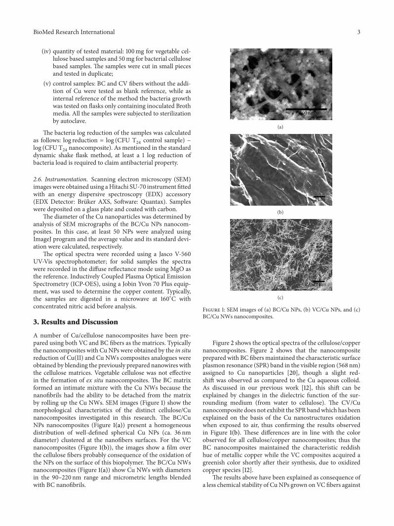
A number of Cu/cellulose nanocomposites have been pre-pared using both VC and BC fibers as the matrices. Typicallythe nanocomposites with CuNPs were obtained by the in situreduction of Cu(II) and Cu NWs composites analogues wereobtained by blending the previously prepared nanowires withthe cellulose matrices. Vegetable cellulose was not effectivein the formation of ex situ nanocomposites. The BC matrixformed an intimate mixture with the Cu NWs because thenanofibrils had the ability to be detached from the matrixby rolling up the Cu NWs. SEM images (Figure 1) show themorphological characteristics of the distinct cellulose/Cunanocomposites investigated in this research. The BC/CuNPs nanocomposites (Figure 1(a)) present a homogeneousdistribution of well-defined spherical Cu NPs (ca. 36 nmdiameter) clustered at the nanofibers surfaces. For the VCnanocomposites (Figure 1(b)), the images show a film overthe cellulose fibers probably consequence of the oxidation ofthe NPs on the surface of this biopolymer. The BC/Cu NWsnanocomposites (Figure 1(a)) show Cu NWs with diametersin the 90–220 nm range and micrometric lengths blendedwith BC nanofibrils.
500 nm
(a)
1 𝜇m
(b)
10 𝜇m
(c)
Figure 1: SEM images of (a) BC/Cu NPs, (b) VC/Cu NPs, and (c)BC/Cu NWs nanocomposites.
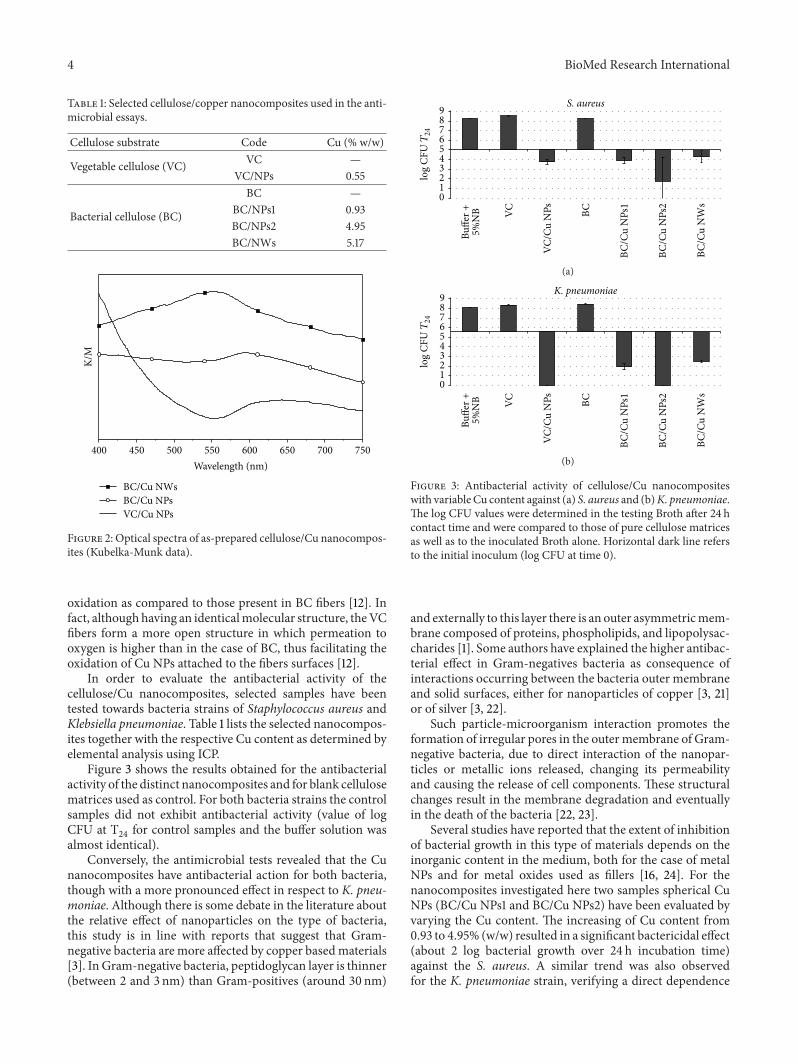
Figure 2 shows the optical spectra of the cellulose/coppernanocomposites. Figure 2 shows that the nanocompositeprepared with BC fibersmaintained the characteristic surfaceplasmon resonance (SPR) band in the visible region (568 nm)assigned to Cu nanoparticles [20], though a slight red-shift was observed as compared to the Cu aqueous colloid.As discussed in our previous work [12], this shift can beexplained by changes in the dielectric function of the sur-rounding medium (from water to cellulose). The CV/Cunanocomposite does not exhibit the SPRbandwhich has beenexplained on the basis of the Cu nanostructures oxidationwhen exposed to air, thus confirming the results observedin Figure 1(b). These differences are in line with the colorobserved for all cellulose/copper nanocomposites; thus theBC nanocomposites maintained the characteristic reddishhue of metallic copper while the VC composites acquired agreenish color shortly after their synthesis, due to oxidizedcopper species [12].
The results above have been explained as consequence ofa less chemical stability of CuNPs grown onVC fibers against
4 BioMed Research International
Table 1: Selected cellulose/copper nanocomposites used in the anti-microbial essays.
Cellulose substrate Code Cu (% w/w)
Vegetable cellulose (VC) VC —VC/NPs 0.55
Bacterial cellulose (BC)
BC —BC/NPs1 0.93BC/NPs2 4.95BC/NWs 5.17
K/M
400 450 500 550 600 650 700 750Wavelength (nm)
BC/Cu NWsBC/Cu NPsVC/Cu NPs
Figure 2: Optical spectra of as-prepared cellulose/Cu nanocompos-ites (Kubelka-Munk data).
oxidation as compared to those present in BC fibers [12]. Infact, althoughhaving an identicalmolecular structure, theVCfibers form a more open structure in which permeation tooxygen is higher than in the case of BC, thus facilitating theoxidation of Cu NPs attached to the fibers surfaces [12].
In order to evaluate the antibacterial activity of thecellulose/Cu nanocomposites, selected samples have beentested towards bacteria strains of Staphylococcus aureus andKlebsiella pneumoniae. Table 1 lists the selected nanocompos-ites together with the respective Cu content as determined byelemental analysis using ICP.
Figure 3 shows the results obtained for the antibacterialactivity of the distinct nanocomposites and for blank cellulosematrices used as control. For both bacteria strains the controlsamples did not exhibit antibacterial activity (value of logCFU at T
24for control samples and the buffer solution was
almost identical).Conversely, the antimicrobial tests revealed that the Cu
nanocomposites have antibacterial action for both bacteria,though with a more pronounced effect in respect to K. pneu-moniae. Although there is some debate in the literature aboutthe relative effect of nanoparticles on the type of bacteria,this study is in line with reports that suggest that Gram-negative bacteria are more affected by copper basedmaterials[3]. In Gram-negative bacteria, peptidoglycan layer is thinner(between 2 and 3 nm) than Gram-positives (around 30 nm)
0123456789 S. aureus
Buffe
r +5%
NB VC
VC/C
u N
Ps BC
BC/C
u N
Ps1
BC/C
u N
Ps2
BC/C
u N
Ws
log
CFUT24
(a)
0123456789
Buffe
r +5%
NB VC
VC/C
u N
Ps BC
BC/C
u N
Ps1
BC/C
u N
Ps2
BC/C
u N
Ws
K. pneumoniae
log
CFUT24
(b)
Figure 3: Antibacterial activity of cellulose/Cu nanocompositeswith variable Cu content against (a) S. aureus and (b)K. pneumoniae.The log CFU values were determined in the testing Broth after 24 hcontact time and were compared to those of pure cellulose matricesas well as to the inoculated Broth alone. Horizontal dark line refersto the initial inoculum (log CFU at time 0).
and externally to this layer there is an outer asymmetricmem-brane composed of proteins, phospholipids, and lipopolysac-charides [1]. Some authors have explained the higher antibac-terial effect in Gram-negatives bacteria as consequence ofinteractions occurring between the bacteria outer membraneand solid surfaces, either for nanoparticles of copper [3, 21]or of silver [3, 22].
Such particle-microorganism interaction promotes theformation of irregular pores in the outermembrane of Gram-negative bacteria, due to direct interaction of the nanopar-ticles or metallic ions released, changing its permeabilityand causing the release of cell components. These structuralchanges result in the membrane degradation and eventuallyin the death of the bacteria [22, 23].
Several studies have reported that the extent of inhibitionof bacterial growth in this type of materials depends on theinorganic content in the medium, both for the case of metalNPs and for metal oxides used as fillers [16, 24]. For thenanocomposites investigated here two samples spherical CuNPs (BC/Cu NPs1 and BC/Cu NPs2) have been evaluated byvarying the Cu content. The increasing of Cu content from0.93 to 4.95% (w/w) resulted in a significant bactericidal effect(about 2 log bacterial growth over 24 h incubation time)against the S. aureus. A similar trend was also observedfor the K. pneumoniae strain, verifying a direct dependence

BioMed Research International 5
of the antibacterial action with the of Cu content in thecomposite. Note that complete killing effect was observed forthe BC/CuNPs2 nanocomposite. Similarly to silvermaterials,the antibacterial activity of Cu nanostructures has beenassociated with the release of ionic species and the formationof reactive oxygen species [1, 5]. The increase of the copperamount in the nanocomposites results in a higher release ofcations, increasing in this way the antibacterial activity of thecorresponding cellulose based nanocomposites.
It is interesting to note that despite the influence ofcopper content, the sample with the higher copper content(BC/CuNWs sample) did not present the higher antibacterialeffect. For both bacteria studied, BC/CuNWsnanocompositepresent an antibacterial activity significantly lower (morethan 2 log bacterial growth) in relation to the nanocompositewith copper NPs with similar copper amount (BC/Cu NPs2).Even BC/Cu NPs1 nanocomposite presents slightly higherantibacterial efficiency, this composite presenting 5 timesless copper amount. Assuming that the antibacterial effect ismainly due to cationic release, this lower efficiency for the CuNWs containing composites, as compared to those incorpo-rating Cu NPs, is probably due to the less surface reactivityof the nanowires, thus leading to lower amounts of solubleand oxidized copper species. In fact, this explanation is inagreement with the observations presented above for theeasier oxidation of the Cu NPs as compared to that of theCu NWs. In fact, the former materials have already oxidizedcopper phases and the surface chemistry is thus markedlydistinct from the Cu NWs.
There is lack of knowledge about the effect of the mor-phology of copper nanostructures on their antibacterial prop-erties, both in the case of individualized nanoparticles andwhen used in composite materials. However, studies per-formed on silver nanostructures with distinct morphologieshave demonstrated that Ag NPs undergo a shape-dependentinteraction with the bacteria [25]. Through the study ofbacteria surface by transmission electron microscopy, theauthors found that spherical NPs exhibit enhanced antibac-terial activity than, for example, Ag nanorods.This effect wasexplained by the higher reactivity of these nanostructuresbecause of higher atomic density. In this case, to obtain sim-ilar antibacterial activity of 12.5mg of spherical NPs werenecessary 50–100mg of nanorods.The higher reactivity of theNPs probably leads to a faster release of metallic ions leadingto an enhancement of the antibacterial activity for this typeof nanostructure.
Finally the effect of the cellulose matrix should also benoted. For this analysis 100mg of the VC/Cu NPs nanocom-posite is used and 50mg of BC/Cu NPs1 so the total amountof inorganic filler is almost the same. For S. aureus the VC/CuNPs nanocomposite presents a similar activity to the BC/CuNPs1 composite. However, comparing the same nanocom-posites for the antibacterial action against K. pneumoniae,the nanocomposite prepared with VC fibers shows a superioreffect. This is an unexpected observation because the CuNPs on the VC fibres are not individualized but forming afilm, as already reported [12], due to the fast oxidation undernormal ambient conditions. Probably this is explained by thepreferential deposition of copper on the VC fibers’ surfaces
which contrasts to the BC matrix in which the NPs also relyon the BC structure due to its three-dimensional internalorganization, thus acting as a protective cage for the Cu NPs.In this case, the release of Cu ions is limited as compared tothe more open structure of the CV nanocomposite. Similarobservations have been reported for materials based oncellulose and Ag NPs, in which for VC/Ag nanocompositesthe release ofAg+was superior compared to the BC analogues[16].
4. Conclusions
A series of cellulose/copper nanocomposites have been pre-pared by varying the type of cellulose used as the matrix(vegetable or bacterial) and also the morphology of coppernanostructures (nanoparticles or nanowires) used as fillers.These composites were investigated for the first time for theirantibacterial activity. Antibacterial activity has been observedfor the nanocomposite samples against both Gram-positive(S. aureus) and Gram-negative (K. pneumoniae) bacteria.Enhancement of the antibacterial activity with increasingcopper content was observed. Among themorphological dis-tinct copper nanostructures used, the nanowires have shownless antibacterial effect that was ascribed to the less reactivesurface towards oxidation. Another parameter that influencesthe antibacterial efficiency of the nanocomposite was thestructure of the cellulose fibers. The results suggest that theuse of copper together with vegetable cellulose fibers resultsin better antibacterial materials against both species of testedbacteria. These results confirm the potential of bionanocom-posites containing copper nanostructures as new antimicro-bial materials. Furthermore this study has shown that formu-lating composites in which both the matrix and the morpho-logical characteristics of the Cu fillers are varied can improvethe antibacterial action of such materials. These parametersseem to influence the antimicrobial mechanism present thatalthough consistent with a cationic release process still needsfurther evidence.
Acknowledgments
Ricardo Pinto thanks the Portuguese Foundation for Scienceand Technology (FCT) for Grant no. SFRH/BPD/89982/2012. The authors also acknowledge FCT (PEst-C/CTM/LA0011/2011), Fundo Social Europeu (FSE), and ProgramaOperacional Potencial Humano (POPH) for funding.Microscopy analysis was supported by Rede Nacional deMicroscopia Eletronica (RNME-Pole UA FCT) ProjectREDE/1509/RME/2005.
References
[1] J. R. Morones, J. L. Elechiguerra, A. Camacho et al., “The bac-tericidal effect of silver nanoparticles,” Nanotechnology, vol. 16,no. 10, pp. 2346–2353, 2005.
[2] R. J. B. Pinto, M. C. Neves, C. Pascoal Neto, and T. Trindade,“Composites of cellulose andmetal nanoparticles,” inNanocom-posites-New Trends and Developments, F. Ebrahimi, Ed., pp. 73–96, InTech, Rijeka, Croatia, 2012.

6 BioMed Research International
[3] J. P. Ruparelia, A. K. Chatterjee, S. P. Duttagupta, and S.Mukherji, “Strain specificity in antimicrobial activity of silverand copper nanoparticles,” Acta Biomaterialia, vol. 4, no. 3, pp.707–716, 2008.
[4] A. Llorens, E. Lloret, P. A. Picouet, R. Trbojevich, and A. Fer-nandez, “Metallic-based micro and nanocomposites in foodcontact materials and active food packaging,” Trends in FoodScience and Technology, vol. 24, no. 1, pp. 19–29, 2012.
[5] M. J. Hajipour, K. M. Fromm, A. A. Ashkarran et al., “Antibac-terial properties of nanoparticles,” Trends in Biotechnology, vol.30, no. 10, pp. 499–511, 2012.
[6] P. V. AshaRani, G. L. K. Mun, M. P. Hande, and S. Valiyaveettil,“Cytotoxicity and genotoxicity of silver nanoparticles in humancells,” ACS Nano, vol. 3, no. 2, pp. 279–290, 2009.
[7] M.V.D. Z. Park,A.M.Neigh, J. P.Vermeulen et al., “The effect ofparticle size on the cytotoxicity, inflammation, developmentaltoxicity and genotoxicity of silver nanoparticles,” Biomaterials,vol. 32, no. 36, pp. 9810–9817, 2011.
[8] N. C. Cady, J. L. Behnke, and A. D. Strickland, “Copper-basednanostructured coatings on natural cellulose: nanocompos-ites exhibiting rapid and efficient inhibition of a multi-drugresistant wound pathogen, A. baumannii, and mammalian cellbiocompatibility in vitro,” Advanced Functional Materials, vol.21, no. 13, pp. 2506–2514, 2011.
[9] J. R. Turnlund, “Human whole-body copper metabolism,”American Journal of Clinical Nutrition, vol. 67, supplement 5, pp.960S–964S, 1998.
[10] K.-Y. Yoon, J. H. Byeon, J.-H. Park, and J. Hwang, “Susceptibilityconstants of Escherichia coli and Bacillus subtilis to silver andcopper nanoparticles,” Science of the Total Environment, vol. 373,no. 2-3, pp. 572–575, 2007.
[11] K. C. Anyaogu, A. V. Fedorov, and D. C. Neckers, “Synthesis,characterization, and antifouling potential of functionalizedcopper nanoparticles,” Langmuir, vol. 24, no. 8, pp. 4340–4346,2008.
[12] R. J. B. Pinto, M. C. Neves, C. Pascoal Neto, and T. Trindade,“Growth and chemical stability of copper nanostructures oncellulosic fibers,” European Journal of Inorganic Chemistry, vol.2012, no. 31, pp. 5043–5049, 2012.
[13] M. Valodkar, P. S. Rathore, R. N. Jadeja, M. Thounaojam, R. V.Devkar, and S. Thakore, “Cytotoxicity evaluation and antimi-crobial studies of starch cappedwater soluble copper nanoparti-cles,” Journal of Hazardous Materials, vol. 201-202, pp. 244–249,2012.
[14] L. F. Qi, Z. R. Xu, X. Jiang, Y. Li, and M. Wang, “Cytotoxicactivities of chitosan nanoparticles and copper-loaded nanopar-ticles,” Bioorganic andMedicinal Chemistry Letters, vol. 15, no. 5,pp. 1397–1399, 2005.
[15] S. V. Manorama, P. Basak, and S. Singh, “Anti-microbial poly-mer nanocomposites,” inNanocomposite Particles for Bio-Appli-cations, T. Trindade and A. L. Daniel-da-Silva, Eds., pp. 249–264, Pan Stanford, Singapora, 2011.
[16] R. J. B. Pinto, P. A. A. P. Marques, C. P. Neto, T. Trindade, S.Daina, and P. Sadocco, “Antibacterial activity of nanocompos-ites of silver and bacterial or vegetable cellulosic fibers,” ActaBiomaterialia, vol. 5, no. 6, pp. 2279–2289, 2009.
[17] R. J. B. Pinto, S. C. M. Fernandes, C. S. R. Freire et al., “Antibac-terial activity of optically transparent nanocomposite filmsbased on chitosan or its derivatives and silver nanoparticles,”Carbohydrate Research, vol. 348, pp. 77–83, 2012.
[18] B. H. Loo, Y. G. Lee, E. J. Liang, and W. Kiefer, “Surface-enhanced Raman scattering from ferrocyanide and ferricyanide
ions adsorbed on silver and copper colloids,” Chemical PhysicsLetters, vol. 297, no. 1-2, pp. 83–89, 1998.
[19] Y. Chang, M. L. Lye, and H. C. Zeng, “Large-scale synthesis ofhigh-quality ultralong copper nanowires,” Langmuir, vol. 21, no.9, pp. 3746–3748, 2005.
[20] D.Mott, J. Galkowski, L.Wang, J. Luo, and C. Zhong, “Synthesisof size-controlled and shaped copper nanoparticles,” Langmuir,vol. 23, no. 10, pp. 5740–5745, 2007.
[21] S. Jadhav, S. Gaikwad,M.Nimse, andA. Rajbhoj, “Copper oxidenanoparticles: synthesis, characterization and their antibacte-rial activity,” Journal of Cluster Science, vol. 22, no. 2, pp. 121–129,2011.
[22] I. Sondi and B. Salopek-Sondi, “Silver nanoparticles as antimi-crobial agent: a case study on E. coli as a model for Gram-negative bacteria,” Journal of Colloid and Interface Science, vol.275, no. 1, pp. 177–182, 2004.
[23] N. A. Amro, L. P. Kotra, K. Wadu-Mesthrige, A. Bulychev, S.Mobashery, and G. Liu, “High-resolution atomic force micros-copy studies of the Escherichia coli outer membrane: structuralbasis for permeability,” Langmuir, vol. 16, no. 6, pp. 2789–2796,2000.
[24] M. A. Vargas-Reus, K. Memarzadeh, J. Huang, G. G. Ren, andR. P. Allaker, “Antimicrobial activity of nanoparticulate metaloxides against peri-implantitis pathogens,” International Jour-nal of Antimicrobial Agents, vol. 40, no. 2, pp. 135–139, 2012.
[25] S. Pal, Y. K. Tak, and J. M. Song, “Does the antibacterial activityof silver nanoparticles depend on the shape of the nanoparticle?A study of the gram-negative bacterium Escherichia coli,”Applied and EnvironmentalMicrobiology, vol. 73, no. 6, pp. 1712–1720, 2007.

Submit your manuscripts athttp://www.hindawi.com
Hindawi Publishing Corporationhttp://www.hindawi.com Volume 2014
Anatomy Research International
PeptidesInternational Journal of
Hindawi Publishing Corporationhttp://www.hindawi.com Volume 2014
Hindawi Publishing Corporation http://www.hindawi.com
International Journal of
Volume 2014
Zoology
Hindawi Publishing Corporationhttp://www.hindawi.com Volume 2014
Molecular Biology International
GenomicsInternational Journal of
Hindawi Publishing Corporationhttp://www.hindawi.com Volume 2014
The Scientific World JournalHindawi Publishing Corporation http://www.hindawi.com Volume 2014
Hindawi Publishing Corporationhttp://www.hindawi.com Volume 2014
BioinformaticsAdvances in
Marine BiologyJournal of
Hindawi Publishing Corporationhttp://www.hindawi.com Volume 2014
Hindawi Publishing Corporationhttp://www.hindawi.com Volume 2014
Signal TransductionJournal of
Hindawi Publishing Corporationhttp://www.hindawi.com Volume 2014
BioMed Research International
Evolutionary BiologyInternational Journal of
Hindawi Publishing Corporationhttp://www.hindawi.com Volume 2014
Hindawi Publishing Corporationhttp://www.hindawi.com Volume 2014
Biochemistry Research International
ArchaeaHindawi Publishing Corporationhttp://www.hindawi.com Volume 2014
Hindawi Publishing Corporationhttp://www.hindawi.com Volume 2014
Genetics Research International
Hindawi Publishing Corporationhttp://www.hindawi.com Volume 2014
Advances in
Virolog y
Hindawi Publishing Corporationhttp://www.hindawi.com
Nucleic AcidsJournal of
Volume 2014
Stem CellsInternational
Hindawi Publishing Corporationhttp://www.hindawi.com Volume 2014
Hindawi Publishing Corporationhttp://www.hindawi.com Volume 2014
Enzyme Research
Hindawi Publishing Corporationhttp://www.hindawi.com Volume 2014
International Journal of
Microbiology