antibacterial activity of mango kernel …antibacterial activity of mango kernel ... be substituted...
TRANSCRIPT

1
ANTIBACTERIAL ACTIVITY OF MANGO KERNEL EXTRACTS
Mohamed E.S. Mirghani1,2*
, Faridah Yosuf1,2
, Nasereldin A. kabbashi1, Jaya
Vejayan3 and Zaimah Bt. Mohamad Yosuf
1
1Department of Biotechnology Engineering, Kulliyyah of Engineering, International Islamic University Malaysia
(IIUM), Gombak, P.O. Box 10, 50728 Kuala Lumpur, Malaysia 2Biomolecular Engineering Research Unit (BioMERU), Kulliyyah of Engineering, IIUM.
3Department of Biochemistry, Faculty of Medicine and Health Sciences, MonashUniversity, Sunway Campus,
Malaysia.
*Corresponding author: [email protected]
ABSTRACT
This study is emphasized specifically on the potential of the mango “Mangifera Indica” seed kernel by discovering
the prospective usage of mango seed kernels as a source of antibacterial compounds against Gram-positive
(Staphylococcus aureus and Bacillus subtilis) and Gram-negative (Escherichia coli and Pseudomonas aeruginosa)
bacterial strains. For this study, three types of mango were used; Blackgold, Lemak, and Waterlily from Malaysia
and Thiland. The approach adopted for this purpose was by implementing agar well diffusion method. The results
were expressed as the average diameter of bacterial inhibition zones surrounding the wells. The required solvents for
the extraction were ethanol, methanol, acetone and phosphate buffer solutions were prepared in a different
concentration; 0.10M, 0.05M, and 0.01M. The results showed that Lemak gave relatively high antibacterial activity
among other types of mango ranging between 1.40 and 2.23 cm. For the known antibiotic, which was Tetracycline,
under the same conditions the diameter of inhibition zones were between 2.30 to 3.30. Then, the minimum inhibition
concentration tests were conducted for Lemak for two solvents extract that gave the highest inhibition zones which
were methanol and acetone. The results showed that the minimum inhibition concentration of extracts that inhibited
the growth of Escherichia coli and Bacillus subtilis was after 10X dilution (1 mM). The results obtained confirmed
the antibacterial potential of mango kernels extracts and this would probably become an alternative source of new
and natural antibacterial agents.
Keywards: Anti-bacterial activity, Bacillus subtilis, Escherichia coli, Mango “Mangifera indica”, Pseudomonas
aeruginosa, Staphylococcus aureus,
Introduction
Mango, Mangifera indica L., is a member of the family Anacardiaceae. Mango has become naturalized and adapted
throughout the tropics and subtropics. There are over 500 classified of mango varieties; some of them have evolved
and have been described throughout the world. The genus of Mangifera consists of 69 species and mostly restricted
to tropical Asia (Ian, 2006).
The highest variety of mango occurs in Malaysia, particularly in peninsular area and about 28 species are found in
this region (Ian, 2006). Malaysia lies wholly within the tropics, which encompasses heavy precipitation, high
temperatures, and high humidity, which are the favoring factors for mango vegetation. There are several varieties of
mango grown in Malaysia; the better known cultivars are Golek (MA 162), Masmuda (MA 204), Maha 65 (MA
165) and Chok Anan (MA 224). Generally, Chok Anan is very suitable for the export market as it has desirable
color and sweetness and good eating quality. The domestic consumption increased from 42,634 MT (2002) to
55,901 MT (2005) (http://pico.neofission.com/). The significant increases of mango consumption in domestic
activity lead to the accumulation of waste. Mango seed kernel usually wasted when processing. However, with
appropriate treatment and study, the kernel might be possibly used as a food ingredient and even in other purposes.
After consumption or industrial processing of mangoes, considerable amounts of mango kernels (seeds) are
discarded as waste. In Kaur, Singh, Sandhu and Guraya study in 2004 (as cited in Sandhu and Lim, 2007),
approximately 40% to 60% waste is generated during processing of mangoes; 12% to 15% consists of peels and
15% to 20% of kernels. According to mango varieties, the seed represents from 10% to 25% of the whole fruit

2
weight. The kernel inside the seed represents from 45% to 75% of the seed and about 20% of the whole fruit.
However, more than one million tons of mango seeds are being treated as wastes.
Compositional quality of mango seed kernel
In a study for Egyptian mango by-product to proximate its compositional quality of mango seed kernel (Abdalla, et
al., 2006), mango seed kernels contained a considerable amount of total phenolic compounds, total lipid,
unsaponifiable matter, and a low amount of crude protein, but the quality of protein was good because it was rich in
all essential amino acids with highest values of leucine, valine and lysine. Eight phenolic compounds were identified
which tannin and vanillin were in highest amounts. Unsaponifiable matter showed the occurrence of high amounts
of squaline followed by sterols and tocopherols. Stearic acid was the main saturated fatty acid, while oleic acid was
the major unsaturated fatty acid in all lipid classes. The fatty acid composition of total lipid and neutral lipid was
similar, while phospholipid had a high amount of palmitic, linoleic and linolenic acids.
Depending on their variety, mango seed kernels contain on a dry weight average 6.0% protein, 11% fat, 77%
carbohydrate, 2.0% crude fiber and 2.0% ash. Mango seed kernels were shown to be a good source of polyphenols,
phytosterols as campesterol, β-sitosterol and tocopherols. Hemavathy, Prabhakar, and Sen (1987) extracted and
fractionated total lipids from Alphonso mango kernel into three lipid classes. Total lipid (11.6% of dry kernel)
consisted of 96.1% neutral and 3.9% polar lipids, which comprised 2.9% glycolipids and 1.0% phospholipids.
Rashwan (1990) extracted and fractionated total lipids from three different mango seed kernel varieties namely
golek, pairi, and hindi. The neutral lipids varied from 95.2% to 96.2%, phospholipids from 2.7% to 3.3% and
glycolipids from 1.1% to 1.4%. Triglycerides constituted the major fraction of the neutral lipids, for all varieties and
accounted from 93.7% to 96.4%. On the nutritional and toxicological studies of the mango seed kernel, Rukmini and
Vijayaraghan (1984) indicated that mango seed kernel fat is promising and a safe source of edible oil and was found
to be nutritious and non-toxic so that it could be substituted for any solid fat without adverse effects. Arogba (1997)
concluded that the Nigerian mango kernel has a good source of high quality fat and protein as well as tannin. When
extracted, these components could be valuable commercially in the vegetable oil industry, in confectionary and in
tanning. Similarly, the processed flour could be principal ingredient in the diets of babies and adults in Nigeria.
Soong et al. (2004) suggested that mango seed kernel could be used as a potential source for functional food
ingredients due to its high quality of fat and protein as well as high levels of natural antioxidants. Furthermore, Zein
et al. (2005) published that soaking and boiling treatments had a great impact in reducing the anti-nutritional factors.
Depending on their variety, mango seed kernels contain on a dry weight average 6.0% protein, 11% fat, 77%
carbohydrate, 2.0% crude fiber and 2.0% ash (Sandhu & Lim, 2007). Since the mango seed kernel contains valuable
elements, it is considerable to utilize it in some way so that hazards could be eliminated and probably valuable
products could be produced.
This research focuses exclusively on antibacterial activity of mango kernel extracts against several of Gram-positive
and Gram-negative bacterial strains.
Approach and methods
Figure 1: Mango seed kernel
Agar well diffusion method
The antibacterial properties of mango seed kernel extracts can be investigated by using the agar well diffusion
method. Through this method, the minimum inhibitory concentration (MIC) of the mango seed kernels against food-
borne pathogenic bacteria can be determined. The MIC is the lowest concentration of a drug or antibiotic that
prevents growth of a particular pathogen.
3
The principle between this assay technique is fairly simple. When an antibiotic is placed inside the well of suitable
gar medium previously inoculated with the test bacterium, the antibiotic diffuses radially outward through the agar,
producing an antibiotic concentration gradient. The antibiotic is present at high concentrations near the well and
affects even minimally susceptible microorganisms. As the distance from the well increases, the antibiotic
concentration drops and only more susceptible pathogens are harmed. A clear zone or ring is present around an
antibiotic well after incubation if the agent inhibits bacterial growth. The wider the zone surrounding a well, the
more susceptible the pathogen is.
The extracts of mango seed kernels display a broad antibacterial spectrum, and are more effective against gram-
positive than the gram-negative bacteria. The antibacterial activity of the mango seed kernels is stable against heat
(121°C, 15 minutes), freezing (-20°C, 16 hours) and pH treatment (pH 3 to 9) normally used in food processing
(Kabuki & Nakajima, et al., 2000).
Methodology
Experimental Materials
Raw material
Three types of mango fruits were purchased from Selayang wet market. The seed kernels were taken out from each
fruits manually and grinded. They were dried before being used.
Bacterial strains
The bacterial strains were obtained from Molecular Biology Laboratory, IIUM. The used strains were:
Gram-positive:- Staphylococcus aureus and Bacillus subtilis
Gram-negative:- Escherichia coli and Pseudomonas aeruginosa
Preparation of mango seed kernels
The seeds were washed and air-dried and the kernels were removed manually from the seeds. The kernels were
chopped or cut into small pieces by using grinder. After that, they were dried in a freezer at -80°C and transferred to
freeze dryer to remove the excessive moisture for several days. Finally, the samples were stored at 4°C until
utilization for the next procedures.
Preparation of extraction reagents and extracts
The required solvents for the extraction (85% ethanol, 85% methanol, 85% acetone, and 0.1 M phosphate buffer of
pH 7) were prepared prior to the extraction process. (Sambrook, & Russell, 2001). The pH of the buffer was
measured again by pH meter.
0.2 g of each sample was weighted and put into a falcon tube. Each solvent was measured according to Table 1
following to the concentration required. The solvent was pipetted into the tubes containing the sample. The tubes
were mixed by using vortex mixer for 30 seconds and centrifuged at 13,000 rpm for 15 minutes. After
centrifugation, the supernatants were transferred into another centrifuge tube to be tested against the selected
bacteria. The extracts were stored at -20˚C if not used.
Preparation of the media
LB Media
37 g of LB agar powder were dissolved in 1 liter of distilled water inside 1 liter of Schott bottle. The pH was
adjusted to 7.0 before being sterilize by autoclaving it for 20 minutes at 15 psi (1.05 kg/cm²) on liquid cycle. Plates
were poured directly from the Schott bottle under laminar flow for about 20-25ml per plate. After the medium
hardened completely, the plates were inverted and stored at 4ºC. When needed, they were removed from storage 1 to
2 hours before use.
To prepare 1 liter of LB liquid broth, 25 g of LB broth powder were dissolved in 1 liter of distilled water inside 1
liter of Schott bottle. The pH was then adjusted to 7.0 before being sterilized by autoclaving it for 20 minutes at 15
psi (1.05 kg/cm²) on liquid cycle. Then, it was stored at 4ºC (Sambrook, & Russell, 2001).
Top agar
For 1 liter of top agar, LB agar and LB broth were mixed together in the ratio of 3:1. They were dissolved in 1 liter
of distilled water. The pH was then adjusted to 7.0 before being sterilize by autoclaving it for 20 minutes at 15 psi
4
(1.05 kg/cm²) on liquid cycle. After autoclaving, the medium was allowed in 40-45ºC dryer prior to use or storage
(Sambrook, & Russell, 2001).
Antibacterial assays
Preparation of cell culture
The bacterial strains were kept in the eppendorf tubes that contain glycerol in -80°C freezer. Prior to use, the
eppendorf tubes were thawed in room temperature for several minutes. Then, a dip of each bacterial strain was
transferred into different bijou bottles that contain 10 ml of LB liquid broth by using a wire loop. The bijou bottles
were put into incubator shaker at 200 rpm and 37°C for overnight. In the next day, a dip of the content of each bijou
bottles were transferred and distributed evenly onto the LB agar plates and put inside incubator at 37°C for
overnight. After there were cell growth on the agar and there were no contaminations, the plates were stored in the
chiller at 4°C until next utilization (Denyer, et al., 2004).
The subcultures must be freshly prepared. A single colony was taken from each plate by using a wire loop. Then, it
was transferred into bijou bottle that contains 10 ml LB liquid broth and was put into incubator shaker at 200rpm
and 37°C, for overnight. In the next day, the optical density (OD) of the bacterial culture was taken. 3 ml of bacterial
culture was loaded into the cuvette. The OD was measured at wavelength 600 nm with LB liquid broth as the blank.
If the OD value was greater than 0.5, the dilution was performed by adding LB liquid broth into the bacterial culture
in the bijou bottle.
Antibacterial screening
The agar well diffusion method was used to analyze and evaluate the antibacterial activity for each extract. About 50
µl of bacteria subcultures were put into each shake flask. Then, 4 ml of top agar (35 to 40°C) were added into the
same flasks and shake gently. The top agar containing the bacterial cultures were poured and distributed evenly onto
the LB agar plates. The plates were transferred into incubator at 37°C. After 15 minutes, 5 wells (5 mm diameter)
were made in each plate using sterile pipette tips. Then, about 50 µl of each solvent extracts were loaded into each
of the well. As a negative control, 50 µl of each solvent (acetone, methanol, ethanol and phosphate buffers) alone
were used. For the positive control, 50 µl of tetracycline was used. All the plates are then were covered with lids and
incubated overnight at 37˚C. The presences of inhibition zones on LB plates containing bacterial culture around the
well were observed. The diameters of each inhibition zone were measured. The type of sample and concentration of
solvent that gave the optimum result were identified (Denyer, et al., 2004).
Minimum Inhibition Concentration (MIC)
The dilutions for the solvents extract were prepared for 10X, 100X, and 1000X. 500 µl of 0.01M sample was
pipetted into a Falcon tube. 5ml of distilled water was added into the tube. The tube was mixed by using vortex
mixture before the previous steps were repeated in order to obtain another dilute sample. Then, the antibacterial
screening was performed towards all the different dilute samples to obtain the minimum inhibition concentration.
The presences of inhibition zones on LB plates containing bacterial culture around the well were observed. The
diameters of each inhibition zone were measured and recorded.
Results and Discussion
The antibacterial activities of Mangifera indica kernel extracts were investigated by using agar well diffusion
method. This study involved different solvents and concentrations in order to evaluate and compare the antibacterial
activity for various circumstances. Antibacterial activities of each solvents extract were expressed in terms of the
average diameter of the presence inhibition zone. The results were shown as in the following tables.
Table1: Antibacterial activity of Mangifera indica kernel extracts against Bacillus subtilis.
Solvent
Diameter of Inhibition Zone (cm)
Blackgold Waterlily Lemak Positive
Control
Negative
Control
Ethanol 1.933 1.567 1.633 2.700 0
Methanol 1.677 1.500 1.967 2.867 0
Acetone 1.600 1.633 2.233 2.700 0
PBS 1.400 1.000 1.600 2.600 0
Table 2: Antibacterial activity of Mangifera indica kernel extracts against Staphylococcus aureus.

5
Solvent
Diameter of Inhibition Zone (cm)
Blackgold Waterlily Lemak Positive
Control
Negative
Control
Ethanol 1.700 1.533 2.000 3.133 0
Methanol 1.700 1.333 1.933 2.567 0
Acetone 1.833 1.500 1.200 2.700 0
PBS 1.567 0.900 1.933 2.600 0
Table 3:Antibacterial activity of Mangifera indica kernel extracts against Pseudomonas aeruginosa
Solvent
Diameter of Inhibition Zone (cm)
Blackgold Waterlily Lemak Positive
Control
Negative
Control
Ethanol 1.300 1.267 1.400 3.167 0
Methanol 1.500 1.800 2.000 3.200 0
Acetone 1.500 1.400 1.333 3.300 0
PBS 1.600 1.500 1.700 3.167 0
Table 4:Antibacterial activity of Mangifera indica kernel extracts against Escherichia coli
Solvent
Diameter of Inhibition Zone (cm)
Blackgold Waterlily Lemak Positive
Control
Negative
Control
Ethanol 1.400 1.500 1.567 2.600 0
Methanol 1.500 1.333 1.867 2.500 0
Acetone 1.700 1.400 1.533 2.600 0
PBS 1.400 1.300 1.600 2.300 0
From the observation and measurement of diameter of inhibition zones formed, it has been demonstrated that the
extracts from the Mangifera indica kernel had significant potential as an antibacterial compounds. The results shown
above were for the extracts concentration of 0.10 M against four types of bacterial strains that signify Gram-positive
and Gram-negative bacteria. The results clearly justified that all of the extracts have shown antibacterial activity. For
the known antibiotic, which was the Tetracycline, the diameter of the inhibition zones were between 2.300 to 3.300.
These measurements were relatively high compared to diameters of inhibition zones for the sample extracts. This is
because Tetracycline is already recognized as the wide-spectrum antibiotic that inhibits the growth of both Gram-
positive and Gram-negative bacteria. The extracts from Lemak gave great results as larger inhibition zones were
formed compared to other types of mango kernel extracts. Thus, further analysis of antibacterial activity was
performed for this specific type of kernel extracts at different concentrations. Later on, the minimum inhibition
concentration (MIC) of the extracts was determined. The results were shown in the following tables.
Table 5: Inhibition zone diameter of the antibacterial activity of Lemak extracts
at different concentrations against Staphylococcus aureus.
Solvent
Diameter of Inhibition Zone (cm)
0.10M 0.05M 0.01M Positive
Control
Negative
Control
Ethanol 1.500 1.300 1.233 2.600 0
Methanol 1.800 1.500 1.400 2.700 0
Acetone 1.500 1.400 1.300 2.700 0
PBS 1.800 1.400 1.400 2.700 0
Table 6: Inhibition zone diameter of the antibacterial activity of Lemak extracts
at different concentrations against Escherichia coli.
Solvent
Diameter of Inhibition Zone (cm)
0.10M 0.05M 0.01M Positive
Control
Negative
Control
6
Ethanol 1.500 1.400 1.300 2.533 0
Methanol 1.500 1.333 1.200 2.300 0
Acetone 1.433 1.367 1.233 2.433 0
PBS 1.467 1.333 1.300 2.200 0
Table 7: Minimum Inhibition Concentration (MIC) of antibacterial activity
of Lemak extracts against Bacillus subtilis.
0.01M Dilution
10x 100x 1000x
Methanol 1.500 1.200 0 0
Acetone 2.550 1.067 0 0
Table 8:Minimum Inhibition Concentration (MIC) of antibacterial
activity of Lemak extracts against Escherichia coli.
0.01M Dilution
10x 100x 1000x
Methanol 1.500 1.067 0 0
Acetone 1.550 1.000 0 0
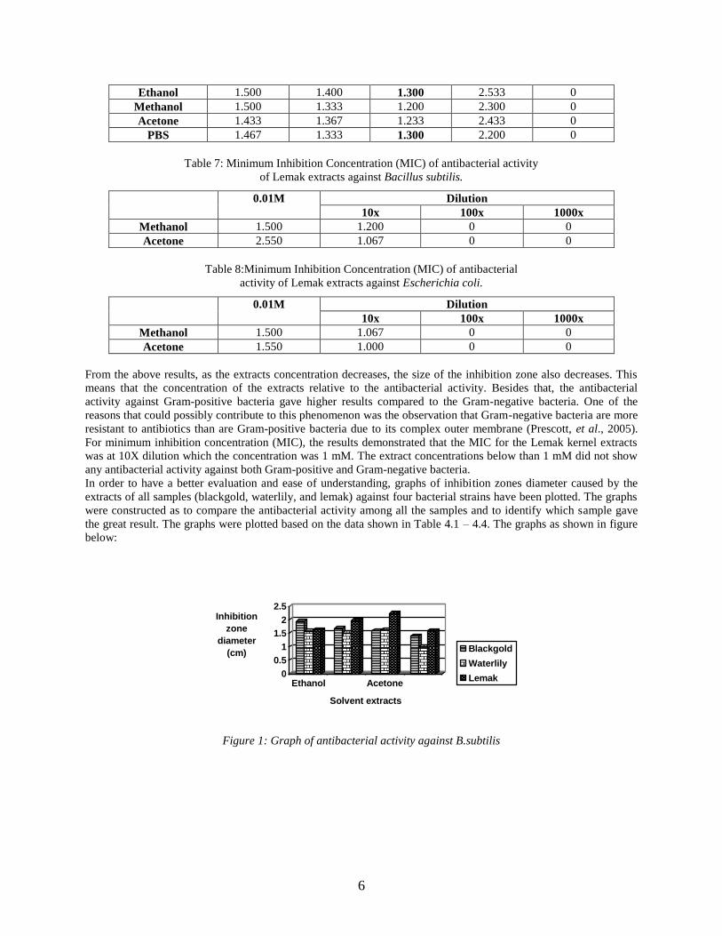
From the above results, as the extracts concentration decreases, the size of the inhibition zone also decreases. This
means that the concentration of the extracts relative to the antibacterial activity. Besides that, the antibacterial
activity against Gram-positive bacteria gave higher results compared to the Gram-negative bacteria. One of the
reasons that could possibly contribute to this phenomenon was the observation that Gram-negative bacteria are more
resistant to antibiotics than are Gram-positive bacteria due to its complex outer membrane (Prescott, et al., 2005).
For minimum inhibition concentration (MIC), the results demonstrated that the MIC for the Lemak kernel extracts
was at 10X dilution which the concentration was 1 mM. The extract concentrations below than 1 mM did not show
any antibacterial activity against both Gram-positive and Gram-negative bacteria.
In order to have a better evaluation and ease of understanding, graphs of inhibition zones diameter caused by the
extracts of all samples (blackgold, waterlily, and lemak) against four bacterial strains have been plotted. The graphs
were constructed as to compare the antibacterial activity among all the samples and to identify which sample gave
the great result. The graphs were plotted based on the data shown in Table 4.1 – 4.4. The graphs as shown in figure
below:
0
0.5
1
1.5
2
2.5Inhibition
zone
diameter
(cm)
Ethanol Acetone
Solvent extracts
Blackgold
Waterlily
Lemak
Figure 1: Graph of antibacterial activity against B.subtilis

7
0
0.5
1
1.5
2
2.5Inhibition
zone
diameter
(cm)
Ethanol Acetone
Solvent extracts
Blackgold
Waterlily
Lemak
Figure 2: Graph of antibacterial activity against S.aureus
0
0.5
1
1.5
2
2.5Inhibition
zone
diameter
(cm)
Ethanol Acetone
Solvent extracts
Blackgold
Waterlily
Lemak
Figure 3: Graph of antibacterial activity against P.aeruginosa
0
0.5
1
1.5
2
2.5Inhibition
zone
diameter
(cm)
Ethanol Acetone
Solvent extracts
Blackgold
Waterlily
Lemak
Figure 4:Graph of antibacterial activity against E.coli
From the graph plotted, it has been identified that Lemak extracts gave the highest antibacterial activity compared to
other sample extracts. Also, it was found that methanol extract has given the optimum activity compared among the
solvents.
The antibacterial activity towards protein extracts that were obtained from other student was also been analyzed.
From the result, only Blackgold showed antibacterial activity against E.coli and B.subtilis whereas the other type of
sample did not show any antibacterial activity. Thus, it can be said that protein did not contribute to the antibacterial
activity for Lemak and Waterlily extracts. The antibacterial activity might come from other components in the
extracts.
Table 4.9:Antibacterial activity of protein extracts
Escherichia coli Bacillus subtilis
Blackgold 1.200 1.467
Waterlily 0 0
Lemak 0 0

8
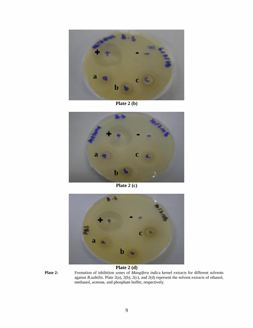
The inhibition zones formed for all samples extracts surrounding the wells on LB plates containing bacterial culture
were shown in the figure as below. For the below figures, (+) sign indicates the positive control (tetracycline), (-) as
negative control (the solvent alone), (a) as blackgold extract, (b) as waterlily extract, and (c) as lemak extract.
Plate 1 (a) Plate 1 (b) Plate 1 (c)
Plate 1 (d)
Plate1: Formation of inhibition zones of Mangifera indica kernel extracts for different solvents
against S.aureus. Plate 1(a), 1(b), 1(c), and 1(d) represent the solvent extracts of ethanol,
methanol, acetone, and phosphate buffer, respectively.
Plate 2 (a)
+ -
a c
b
+ -
a c
b
+ -
a c
b
+ -
a c
b
+ -
a c
b
9
Plate 2 (b)
Plate 2 (c)
Plate 2 (d)
Plate 2: Formation of inhibition zones of Mangifera indica kernel extracts for different solvents
against B.subtilis. Plate 2(a), 2(b), 2(c), and 2(d) represent the solvent extracts of ethanol,
methanol, acetone, and phosphate buffer, respectively.
+ -
a c
b
+ -
a c
b
+ -
a c
b

10
Plate 3 (a)
Plate 3 (b)
Plate 3 (c)
+ -
a c
b
+ -
a c
b
+ -
a c
b

11
Plate 3 (d)
Plate 3: Formation of inhibition zones of Mangifera indica kernel extracts for different solvents
against P.aeruginosa. Plate 3(a), 3(b), 3(c), and 3(d) represent the solvent extracts of
ethanol, methanol, acetone, and phosphate buffer, respectively.
Plate 4 (a)
Plate 4 (b)
+ -
a c
b
+ -
a c
b
+ -
a c
b
12
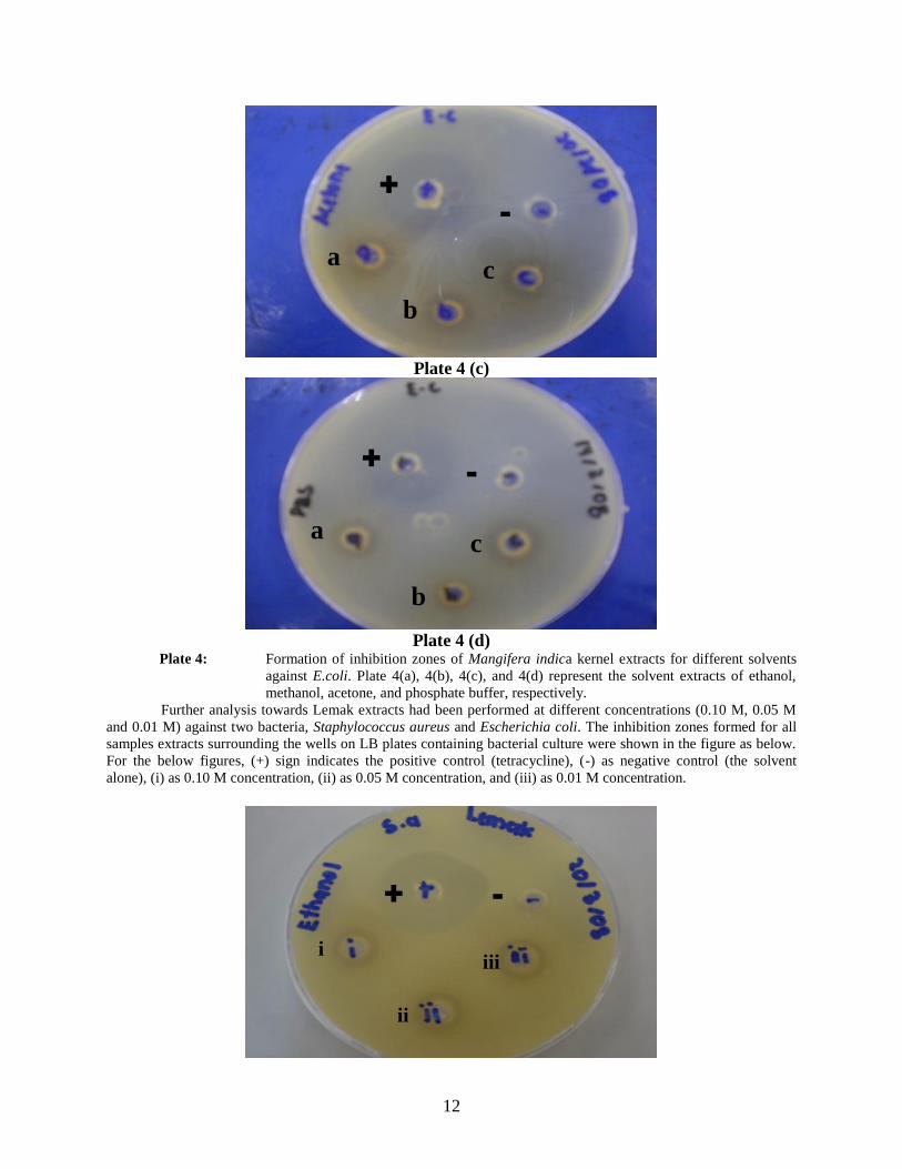
Plate 4 (c)
Plate 4 (d)
Plate 4: Formation of inhibition zones of Mangifera indica kernel extracts for different solvents
against E.coli. Plate 4(a), 4(b), 4(c), and 4(d) represent the solvent extracts of ethanol,
methanol, acetone, and phosphate buffer, respectively.
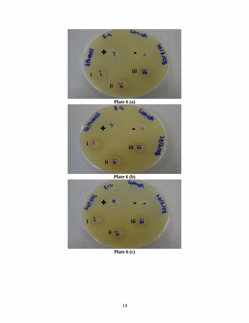
Further analysis towards Lemak extracts had been performed at different concentrations (0.10 M, 0.05 M
and 0.01 M) against two bacteria, Staphylococcus aureus and Escherichia coli. The inhibition zones formed for all
samples extracts surrounding the wells on LB plates containing bacterial culture were shown in the figure as below.
For the below figures, (+) sign indicates the positive control (tetracycline), (-) as negative control (the solvent
alone), (i) as 0.10 M concentration, (ii) as 0.05 M concentration, and (iii) as 0.01 M concentration.
+
-
a c
b
+ -
a c
b
+ -
i iii
ii

13
Plate 5 (a)
Plate 5 (b)
Plate 5 (c)
Plate 5 (d)
Plate 5: Formation of inhibition zones of Lemak kernel extracts for different concentration
against S.aureus. Plate 5(a), 5(b), 5(c), and 5(d) represent the solvent extracts of ethanol,
methanol, acetone, and phosphate buffer, respectively.
+ -
i iii
ii
+ -
i iii
ii
+ -
i iii
ii
14
Plate 6 (a)
Plate 6 (b)
Plate 6 (c)
+ -
i iii
ii
+ -
i iii
ii
+ -
i iii
ii

15
Plate 6 (d)
Plate 6: Formation of inhibition zones of Lemak kernel extracts for different concentration
against E.coli. Plate 6(a), 6(b), 6(c), and 6(d) represent the solvent extracts of ethanol,
methanol, acetone, and phosphate buffer, respectively.
The minimum inhibition concentration (MIC) had been tested for Lemak extracts against Bacillus subtilis
and Escherichia coli for two solvents (methanol and acetone) that gave higher antibacterial activity. The inhibition
zones formed the extracts surrounding the wells on LB plates containing bacterial culture were shown in the figure
as below. For the below figures, (i) as 0.10 M concentration, (ii) as 10X dilution, (iii) as 100X dilution, and (iv) as
1000X dilution.
Plate 7 (a)
+ -
i iii
ii
i
iii
ii
iv

16
Plate 7 (b)
Plate 7: Formation of inhibition zones of Lemak kernel extracts for MIC against B.subtilis. Plate
7(a) and 7(b) represent the solvent extracts of methanol and acetone, respectively.
Plate 8 (a)
Plate 8 (b)
Plate 8: Formation of inhibition zones of Lemak kernel extracts for MIC against E.coli. Plate 8(a)
and 8(b) represent the solvent extracts of methanol and acetone, respectively.
The antibacterial activity towards protein extracts that were obtained from other student was also been
analyzed. The inhibition zones formed for all protein extracts of the samples surrounding the wells on LB plates
i
iii
ii
iv
i
iii
ii
iv
i
iii
ii
iv

17
containing bacterial culture were shown in the figure as below. For the below figures, (a) sign indicates as blackgold
extract, (b) as waterlily extract, and (c) as lemak extract.
Plate 9 (a)
Plate 9 (b)
Plate 9: Formation of inhibition zones of protein extracts for all samples. Plate 9(a) and 9(b)
represent the antibacterial activity against B.subtilis and E.coli.
Conclusion
This research is mainly used to optimize the usage of mango seed kernel by implementing the design of its
extraction for prospective applications that will be beneficial to people. Also, it will help to play a role in
minimizing waste generation worldwide.
In the beginning of this project, several objectives had been set; to perform solvents extraction from the Mangifera
indica seed kernel and to find out any antibacterial activity of the extracts. This project essentially investigated the
antibacterial properties of the Mangifera indica kernel extracts. From the results obtained, the aims of this research
have been successfully achieved. The extracts from Mangifera indica kernel have been proven to contain
antibacterial properties that capable to inhibit the growth of pathogenic bacteria. Through this project, three types of
mango have been used; blackgold, waterlily, and lemak. The antibacterial activities of four solvents extract (ethanol,
methanol, acetone, and phosphate buffers) have been tested against four pathogenic bacteria which signify Gram-
positive and Gram-negative bacteria.
As the results showed constructive values, there is intense possibility for the compound in Mangifera indica kernel
extracts can be utilized as an alternative antibacterial agent in the treatment of infectious disease caused by
pathogenic bacteria. For recommendation in order to improve the results in the future, numerous studies must be
made in order to identify the active compounds in the sample extracts which contribute to the antibacterial activity.
It is crucial to discover the effects of the compounds towards animals and human cells, including the toxicity, the
positive or negative reaction, and the action mechanisms, to make sure that the compounds are safe and has no bad
18
effects on the health. Besides that, the study must involve various extraction reagents at different concentration so
that better comparisons can be achieved.
In conclusion, this project has been successfully finished within the time given. All the objectives have been
accomplished as satisfactory results are obtained. Many knowledge and precious experiences obtained throughout
this project especially regarding to the laboratory works. As the mango seed kernel can be utilized as antibacterial
compounds, thus its usage can be optimized and indirectly can reduce the waste produced by the mango seed
kernels.
References
Abdalla, A.E.M., Darwish, S.M., Ayad, E.H.E., El-Hamahmy, R.M. 2006. Egyptian mango by-product 1.
Compositional quality of mango seed kernel. Journal of Food Chemistry, 103, 1134-1140.
Agro-food business development centre. Mango. Retrieved on August 30, 2007 from:
http://pico.neofission.com/websites/agribdccom/index
Antimicrobial. 2005. Retrieved on March 31, 2008 from: http://www.biocrawler.com/encyclopedia/Antimicrobial
Asia pacific medicinal plant database: Mangifera indica. Retrieved on September 1, 2007 from:
http://wapi/mctweb.dll/
Burman, L.G., Liljequist, B.O. 2001. A global perspective on bacterial infections, antibiotic usage, and the antibiotic
resistance problem. Ins. Hughes, D., Anderson, D.I. (edit). Antibiotic Development and Resistance. New
York: Taylor and Francis, Inc., 1-17.
Denyer, S.P., Hodges, N.A., Gorman, S.P. 2004. Hugo’s and Russell’s pharmaceutical microbiology (edit). 7th
Ed.
United Kingdom: Blackwell Publishing.
Ian, S.E. 2006. Species profiles for pacific island agroforestry. Mangifera indica (mango). (ver. 3.1). Retrieved on
August 30, 2007 from: www.traditionaltree.org
Kabuki, T., Nakajima, H., et al. 2000. Characterization of novel antimicrobial compounds from mango (Mangifera
indica L.) kernel seeds. Journal of Food Chemistry, 71, 61-66.
Karp, G. 2005. Cell and molecular biology.4th
Ed. New York:John Wiley & Sons, Inc.
Mangga. Retrieved on September 1, 2007 from: http://pico.neofission.com/
Mango, from Wikipedia, the free encyclopedia. Retrieved on August 30, 2007 from:
http://en.wikipedia.org/wiki/
Post harvest of mango. 2005. Retrieved on September 1, 2007 from: http://www.fao.org/inpho/
Prescott, L.M., Harley, J.P., Klein, D.A. 2005. Microbiology. 6th
Ed. New York: McGraw-Hill.
Sahu, S., Das, B.K., Pradhan, J., et al. 2006. Effect of Mangifera indica kernel as a feed additive to immunity and
resistance to Aeromonas hydrophila in Labeo rohita fingerlings. Journal of Fish & Shellfish Immunology,
23, 109-118.
Sambrook, J., Russell, D.W. 2001. Molecular cloning: A laboratory manual. (3rd
Ed.). New York: Cold Spring
Harbor Laboratory Press.
Sandhu, K.S., Lim, S.T. 2007. Structural characteristics and in vitro digestibility of Mango kernel starches
(Mangifera indica L). Journal of Food Chemistry.
Singleton, P. 2004. Bacteria in biology, biotechnology and medicine. 6th
Ed. New York: John Wiley & Sons, Inc.
Voet, D., Voet, J.G., Pratt, C.W. 2006. Fundamentals of biochemistry. 2nd
Ed. New York: John Wiley & Sons, Inc.