research areas of interest - usp areas of interest ... (frequency domain intravascular optical...
TRANSCRIPT

Research areas of interest
v MedicalImageProcessingØ UltrasoundQuantitativeTomographicreconstructionØ InverseproblemsingeneralØ UltrasoundØ Segmentationandclassification
v BiomedicalSignalProcessingv Biomedicalsystemsmodeling
Nextpagesshowsummariesofsomeprojects:
On going projects in Medical Image Processing
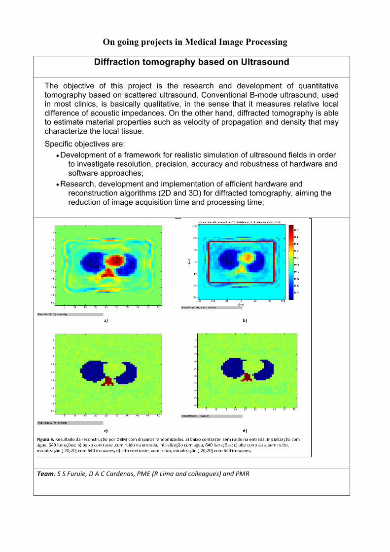
Diffraction tomography based on Ultrasound
The objective of this project is the research and development of quantitative tomography based on scattered ultrasound. Conventional B-mode ultrasound, used in most clinics, is basically qualitative, in the sense that it measures relative local difference of acoustic impedances. On the other hand, diffracted tomography is able to estimate material properties such as velocity of propagation and density that may characterize the local tissue. Specific objectives are:
• Development of a framework for realistic simulation of ultrasound fields in order to investigate resolution, precision, accuracy and robustness of hardware and software approaches;
• Research, development and implementation of efficient hardware and reconstruction algorithms (2D and 3D) for diffracted tomography, aiming the reduction of image acquisition time and processing time;
Team:SSFuruie,DACCardenas,PME(RLimaandcolleagues)andPMR

Quantitative elastography based on Ultrasound
Objective: investigation of quantitative elastography in deep tissues based on ARFI generated by 2D array transducers with large surface. Motivation: Ultrasound elastography is an emerging imaging modality used to provide information about tissue elasticity. It is important because tissue mechanical changes often correlate with tissue pathological changes. Generation of shear waves requires some kind of tissue estimulation in order to induce local displacements. One of these estimulations is via acoustic radiation force (ARFI). There are several studies involving acoustic radiation force and its ability to image the mechanical properties of tissue. However, the application of this technique has been limited to depths smaller than 10 cm. This issue is caused by the inability of the transducer to focus the necessary amount of energy far from the transducer surface.
Figure 1: a, c and e illustrate the accumulated energy distribution in the lateral-axial plane of the simulation of , and , respectively, focused at 120mm depth. b, d and f depict the accumulated energy distribution in the elevational-axial plane of the simulation of , and , respectively, focused at 120mm depth.
F.M.Cardoso,D.SimõesandS.S.Furuie.ACOUSTICRADIATIONFORCEIMPULSEINDEEPTISSUESUSINGMATRICIALARRAYTRANSDUCERS.XXVBrazilianCongressonBiomedicalEngineering–CBEB2016,pg.1075-1078
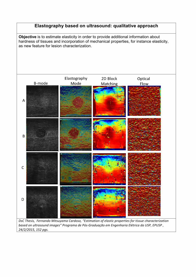
Elastography based on ultrasound: qualitative approach
Objective is to estimate elasticity in order to provide additional information about hardness of tissues and incorporation of mechanical properties, for instance elasticity, as new feature for lesion characterization.
DsCThesis,FernandoMitsuyamaCardoso,“Estimationofelasticpropertiesfortissuecharacterizationbasedonultrasoundimages”ProgramadePós-GraduaçãoemEngenhariaElétricadaUSP,EPUSP,24/2/2015,152pgs.
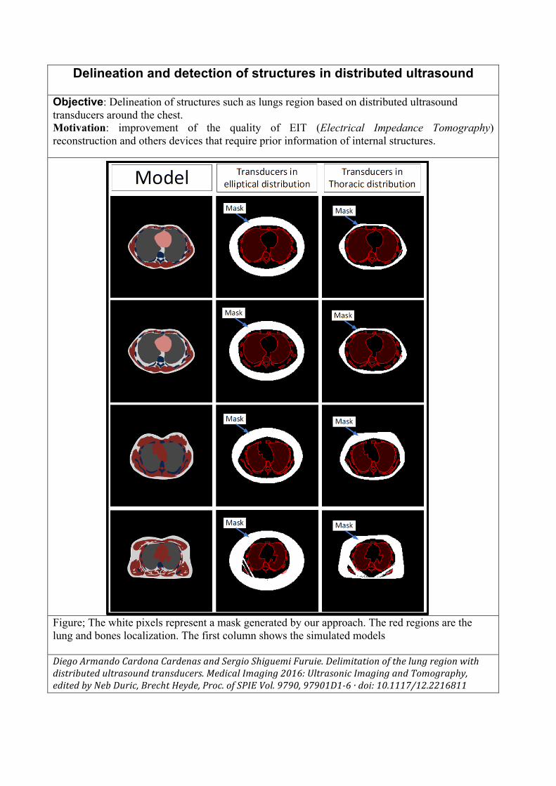
Delineation and detection of structures in distributed ultrasound
Objective: Delineation of structures such as lungs region based on distributed ultrasound transducers around the chest. Motivation: improvement of the quality of EIT (Electrical Impedance Tomography) reconstruction and others devices that require prior information of internal structures.
Figure; The white pixels represent a mask generated by our approach. The red regions are the lung and bones localization. The first column shows the simulated models
DiegoArmandoCardonaCardenasandSergioShiguemiFuruie.Delimitationofthelungregionwithdistributedultrasoundtransducers.MedicalImaging2016:UltrasonicImagingandTomography,editedbyNebDuric,BrechtHeyde,Proc.ofSPIEVol.9790,97901D1-6·doi:10.1117/12.2216811

Framework for Intravascular Ultrasound (IVUS) Simulation
Objective: development of a framework for creating 3D ultrasound numerical deformable phantoms based on Finite Element Method for intravascular applications. Motivation: Intravascular Ultrasound (IVUS) phantoms are important to calibrate and evaluate many IVUS imaging processing tasks. Therefore, we present a framework for creating representative IVUS phantoms, for different intraluminal pressures, based on the Finite Element method and Field II. First, a coronary cross model is selected. Second, the coronary regions are identified to apply the properties. Third, the corresponding mesh is generated. Fourth, the intraluminal force is applied and the deformation computed. Finally, the speckle noise is incorporated. Moreover, the framework toolbox is freely accessible (http://www.leb.usp.br/IVUSSim/), and fully implemented in a single platform.
FernandoMitsuyamaCardoso,MatheusCardosoMoraes,SérgioShiguemiFuruie.RealisticIVUSimagegenerationinDifferentIntraluminalPressures,UltrasoundinMedicineandBiology,Vol.38,No.12,pp.2104–2119,2012pii:S0301-5629(12)00469-3.doi:10.1016/j.ultrasmedbio.2012.08.005.

Analysis and quantification of Intravascular Ultrasound images (IVUS)
The objective of this project is to characterize coronary lesions based on analysis of intravascular ultrasound (IVUS) images. It involves 3D segmentation of lesions, texture analysis and classification.
Intravascular structure characterization is the process of plaque composition inference. An approach using invariant features has been investigated. As the atherosclerotic plaque is a three-dimensional object, consecutive frame information are used in the plaque composition identification process. Current techniques are based only in-plane frame attributes.
FernandoJ.R.Sales,BrenoA.Falcão,JoãoL.A.Falcão,ExpeditoE.Ribeiro,MarcoA.PerinMD,PedroE.Horta,AndréG.Spadaro,JohnA.Ambrose,EulógioE.Martinez,SergioS.Furuie,PedroA.Lemos.EvaluationofPlaqueCompositionbyIntravascularUltrasound“VirtualHistology”:ImpactofDenseCalciumontheEstimationofNecroticTissue.EuroInterventionJournal,ISSN1969-6213–Print,v6(3):394-399

Coronary contraction analysis based on 4D-IVUS
The aim of this project is the analysis of coronary contraction. Conventional IVUS (intravascular ultrasound ) with continuous pullback provide 4D data, but without correct spatial localization. We propose to reconstruct tomographic IVUS image without heart movement artifact for different phases. Moreover, the time synchronization will depend only on the images, and will preserve 4D information. Thus it involves: a) determining images (3D structures) from IVUS sequence that belong to the same phase within the cardiac cycle; b) spatial registration of 3D coronaries; c) 3D interpolation; d) contraction analysis.
MMSMatsumoto,FernandoMitsuyamaCardoso,PLemos,SSFuruie"Coronary3DreconstructionusingIVUSimagesonly:anumericphantomframework."MedicalImaging2010:UltrasonicImaging,Tomography,andTherapy,editedbyJanD'hooge,StephenA.McAleavey,Proc.ofSPIEVol.7629,762915

Visualization and segmentation of coronary arteries in microbubble
contrasted 3D Echocardiographic images The improvement in micro bubble contrasted images in three-dimensional Echocardiography has created new opportunities for non-invasive and relatively inexpensive exams. This objective of this project is the visualization of the 3D coronary tree in a simpler and non-invasive form. There are several challenges such as: a) high noise and natural degradation of the images; b) dynamic nature of the structures; c) poor contrast. The main approach is based on modified version of fuzzy connectedness algorithm.
Team:SSFuruie,JMTsutsui(InCor)

Algorithms for tomographic reconstruction: optimization
The main objective of this project is to sum up efforts and knowledge of several groups that have been working with different aspects of tomography. The investigations basically include: a) practical and new optimized reconstruction algorithms; b) alternative image restoration algorithms; c) robust volume quantification and segmentation processes; and d) clinical applications. Specific objectives are:
• Research, development and implementation of efficient reconstruction algorithms (2D and 3D) for Nuclear Medicine, aiming the reduction of absorbed doses as well as the image acquisition and processing time;
• Research of methods for quantitative tomographic reconstructions in Nuclear Medicine dealing with correction/compensation and restoration of images;
• Research on truly 4D reconstruction algorithms for dynamic structures; • Research, development and evaluation of the whole reconstruction process,
including restoration and quantification, through physical and numerical simulation in 2D, 3D and 4D;
SergioSFuruie.SIMULTANEOUSACTIVITYANDATTENUATIONRECONSTRUCTIONINPET:APPLYINGPARTIALKNOWNATTENUATION.MedicalImaging2009:PhysicsofMedicalImaging,editedbyEhsanSamei,JiangHsieh.Proc.ofSPIEvol.7258,72582F-1a72582F-6.DOI:10.1117/12.811041.

Model identification and analysis in dynamic PET
Analysis in dynamic PET (Positron Emission Tomography) deals with estimation and quantification of parameters for compartmental and non compartmental models. The main topics are related to: input curves, image derived input functions, myocardial metabolic rates of Glucosis, image processing and regions of interest. In a specific approach, an alternative way to assay a noninvasive input function using image derived input functions will be considered.
EdwardFlorezPacheco,HenriquedaFonseca,VaniVijyakumar,SergioShiguemiFuruie.FrameworktoquantifythemetabolicrateintheheartusingMonteCarlosimulationandCompartmentModeling,ComputinginCardiology2015;42:349-352,ISSN2325-8861,6-9set2015,Nice,France.DOI:10.1109/CIC.2015.7408658

IVOCT: neointima estimation
Objective: robust approaches to estimate neointima growth in coronaries, based on IOCT images (Frequency Domain Intravascular Optical Coherence Tomography). Motivation: quantitative follow up of accumulated tissue over the stent (neointimal tissue). It involves segmentation of the lumen and stented area, which is usually done manually by a specialist. We propose two optimal strategies (ellipse fitting algorithm and cylinder fitting algorithm) that are robust even with false positives and false negatives detection of stent struts.
Figure: (a) Lumen contour in green and stent line estimation in blue. (b) Plot of neointima area percentage considering all slices of a set of 25 images.
Veronica,S.S.Furuie.APPROACHESTOSEGMENTSTENTSTRUTS'AREAFROMINTRAVASCULAROPTICALCOHERENCETOMOGRAPHY.XXVBrazilianCongressonBiomedicalEngineering–CBEB2016,pg.971-974

Evaluation of colour deconvolution in histological images with H&E Implementar e comparar quantitativamente resultados de diferentes métodos de separação de corantes
JulianaCesaro,SergioShiguemiFuruie,FilippoMolinari.“Stainseparationofhistologicalslides“.AnaisdoVIISIIM/VISPS(SimpósiodeInstrumentaçãoeImagensMédicas/SimpósiodeProcessamentodeSinais),Campinas,21a23deoutubrode2015

Restoration and calibration of images in teledermatology
Objective: image quality restoration for teledermatology. Motivation: Due to the major expansion of information systems and the utilization of photographic cameras, teledermatology has become a growing option, especially in places of difficult access. However, the increase in its range is hampered by the low fidelity of the acquired image in relation to an in-person consultation. In this manner, a system comprised of a physical device and a software was developed in order to calibrate perspective and colors, in addition to correct possible resolution problems in skin images. An improvement in 85% in colors was obtained, with average values under the limit of visual perception, besides approximately 12% of improvement in resolution using the 1-SSIM metric.
Figure. Left, acquired image with calibration device; right, restored image.
Y.V.Tessaro,S.S.Furuie.METODOLOGIAPARACALIBRARERESTAURARIMAGENSEMTELEDERMATOLOGIA.XXVBrazilianCongressonBiomedicalEngineering–CBEB2016,pg.983-986

Medical image categorization based on wavelet transform and self-
organizing map Images are fundamental data source in modern medicine. These can support doctors and students in diagnostic decisions and provide research and didactic material. The images stored in a database according with categories are an important step for data mining and content-based image retrieval (CBIR). This work addresses a CBIR methodology involving: a) discrete wavelet transform to characterize images; b) Self-Organizing Map (SOM) neural networks to cluster the classes; c) and SOM classification of medical images. This datamining methodology can be used in categorization and diagnostic decision aid.
Team:SSFuruie,RAMoreno(InCor),EDel-Moral-Hernandez(EPUSP).