renal sympathetic nerves in lambs transition fetal to …....
TRANSCRIPT

Role of Renal Sympathetic Nerves in Lambsduring the Transition from Fetal to Newborn LifeFrancine G. Smith, Bruce A. Smith, Edward N. Guillery, and Jean E. RobillardWith the technical assistance of Sherry Flansburg and Oliva J. McWeenyDepartment of Pediatrics and Cardiovascular Center, University of Iowa, Iowa City, Iowa 52242
Abstract
To determine the role of renal sympathetic nerves in influenc-ing renal function during the transition from fetal to newbornlife, studies were carried out in conscious, chronically instru-mented fetal sheep with either bilateral renal denervation (n= 11) or intact renal nerves (n = 12), 3-6 d after surgery. Endo-crine, renal, and cardiovascular parameters were measured be-fore and after delivery of lambs by cesarean section. Bloodpressure and heart rate were similar in intact and denervatedfetuses, and increased after delivery in both groups. There was
also a transient diuresis and natriuresis, in the immediate post-natal period, the response being significantly greater in dener-vated than intact lambs (P < 0.05). By 24 h postnatally, fluidand electrolyte excretions were similar in both groups, and sig-nificantly less than fetal levels. In the absence of renal nerves,
the normal rise in plasma renin activity at birth was attenuated.These data provide evidence that renal sympathetic nerves playan important role during the transition from fetal to newbornlife, and support the premise that birth is associated with sym-
pathetic activation. (J. Clin. Invest. 1991. 88:1988-1994.) Keywords: neonate * renal function * atrial natriuretic factorplasma renin activity birth
Introduction
In the adult animal, renal sympathetic nerves influence reninsecretion, renal hemodynamics, and renal function (1),through activation of adrenergic receptors located on renal ves-
sels and tubules. In the immature animal, stimulation of renalnerves (2) or of renal a,-adrenergic receptors (3), results in a
vasoconstriction, though to a lesser extent than later in life.Similarly, in fetal and newborn sheep, neuronal release of nor-
epinephrine causes renin release in vitro, from renal corticalslices (4). Recent evidence indicates that stimulation of renalnerves of fetal and newborn sheep increases fractional Na+reabsorption (5). These data suggest that renal sympatheticnerves in the immature animal, as in the adult, may regulaterenin release and influence renal hemodynamics and function.
A preliminary report of this manuscript was presented to the Societyfor Pediatric Research, 1991.
Address correspondence Francine G. Smith, Ph.D., Department ofObstetrics/Gynaecology and Medical Physiology, Reproductive Medi-cine Research Group, Heritage Medical Research Building, 3330 Hos-pital Drive NW, Calgary, Alberta T2N 4N1 Canada.
Receivedfor publication 22 April 1991 and in revisedform 22 July1991.
It is well recognized that the transition from fetal to new-born life is associated with an increase in the activity of therenin-angiotensin system and a surge in plasma catechol-amines, these hormonal changes being slightly attentuated incesarean compared with vaginal delivery (6-12). In newbornlambs delivered by cesarean section, there is a marked increasein fractional Na' reabsorption by 24 h postnatally, from fetallevels of 93%of the filtered Na' load to 99%(13-15), similar tothat seen later in life. This rapid rise in fractional Na' reabsorp-tion soon after birth, along with elevated plasma norepineph-rine (NE)' levels and plasma renin activity (PRA) suggests thatthe transition from fetal to newborn life is associated with in-creased sympathetic activity.
To determine whether renal sympathetic innervation influ-ences renal and endocrine function at birth, studies werecarried out in conscious, chronically instrumented fetal sheepwith either bilateral renal denervation or intact renal nerves.Endocrine, renal, and cardiovascular parameters were mea-sured before birth and after delivery of the lamb by cesareansection. Our results show that renal nerves play a major role inregulating renin release and renal function during the transi-tion from fetal to newborn life.
Methods
Pregnant ewes of mixed breeding were obtained from a local sourceand housed in individual pens with free access to alfalfa pellets andwater. Gestational ages were based on the induced ovulation tech-nique (16).
Studies were carried out 3-6 d after surgery in 23 conscious, chroni-cally instrumented fetal sheep aged 140 d, before and after delivery bycesarean section. At surgery, described in detail in the following sec-tion, either bilateral renal denervation (n = I 1) or sham denervation (n= 12) was performed.
Surgical procedures. Surgery was carried out with the ewe and fetusunder general anesthesia (1% halothane, 33% oxygen, 66% nitrous ox-ide), after induction with pentothal (500 mgsodium thiopentone, Ab-bott Laboratories, Irving, TX), as previously described (17). Briefly, auterine incision was made near the fetal hindlimbs; catheters were in-serted into right and left femoral arteries and veins, and into the fetalbladder. Skin incisions were sutured and an additional catheter was
sutured to the fetal skin for later measurement of intraamniotic pres-sure.
In the 11 fetuses submitted to renal denervation, bilateral flankincisions were made and renal nerves were severed and stripped fromalong the aorta, renal arteries, veins, and ureters. This was followed bycareful application of 10% phenol in absolute alcohol to the renalplexus and surrounding area, as previously described (18, 19). Sham-operated fetuses were submitted to the same surgical procedure exceptthat the renal nerves were left intact, and no phenol was applied (18,
1. Abbreviations used in this paper: ANF, atrial natriuretic factor; EPI,epinephrine, GFR, glomerular filtration rate; NE, norepinephrine;PRA, plasma renin activity; RBFV, renal blood flow velocity.
1988 F. G. Smith, B. A. Smith, E. N. Guillery, and J. E. Robillard
J. Clin. Invest.© The American Society for Clinical Investigation, Inc.0021-9738/91/12/1988/07 $2.00Volume 88, December 1991, 1988-1994

19). In both groups, a pulsed Doppler flow probe was placed around theleft renal artery (2).
Fetal skin incisions were then closed and the fetus was returned tothe uterus. Uterine, maternal muscle, and maternal skin were suturedin separate layers. All catheters were exteriorized subcutaneously andplaced in a cloth pouch on the ewe's flank. Ampicillin sodium wasinfused directly into the amniotic cavity (I g) and administered intra-muscularly (1 g) to the ewe at surgery, and at 48-h intervals thereafter.All animals were standing and eating within 1 h of completion of sur-gery.
Dexamethasone (5 mg) was administered intramuscularly to theewe 18 h before experiments to ensure prevention of respiratory dis-tress after delivery of the lamb by cesarean section.
Physiological studies. Before the start of experiments, the ewe wastransferred to the laboratory in a small cart that permitted it to stand inan upright position. The fetal bladder was then drained and a primingdose of [14CJ-inulin (2 MCi) in 0.5 g/liter of dextrose in water was in-fused intravenously, followed by a constant infusion at 0.063 MCi/min,at a rate of 0. 11 ml/min, for later determination of glomerular filtrationrate (GFR). After this 60-min equilibration period, fetal urine was con-tinuously collected for 30 min.
Ewes were then returned to the surgical suite, and a 20-gauge spinalneedle was inserted between the lumbar/sacral intervertebral spaceafter infiltration with local anesthetic (1% lidocaine hydrochloride).This was followed by infusion of 1O ml of 0.5% bupivicaine hydrochlo-ride until analgesia of the lower body was achieved. The lamb wasdelivered by cesarean section within the ensuing 15 min, as previouslydescribed (14, 15, 20). Experiments were resumed 1 h after delivery ofall lambs. During this 1-h recovery period, infusion of ['4C]-inulin wasresumed. An additional solution containing 0.5 g/liter of dextrose, 34mEq/liter of sodium chloride and 30 mEq/liter of potassium chloride,was infused intravenously at 0.07 ml/kg per min, during the recoveryperiod and for the duration of the study. Urinary flow rate was continu-ously collected over 30-min intervals at 1, 4, 8, and 24 h after delivery.
Urinary volumes were recorded and samples stored at -70'C forlater determination of electrolytes (Na+,K+), osmolality, and ['4C]-inulin concentration. At the midpoint of each urinary collection pe-riod, 2.5 ml of arterial blood were removed for immediate determina-tion of pH, Po2 and PCo2, and later determination of hematocrit,plasma electrolytes (Na+,K+), plasma osmolality, and ['4C]-inulin con-centration. Additional arterial blood (9 ml) was removed at the end ofeach urine collection for later determination of plasma atrial natri-uretic factor (ANF), aldosterone, catecholamines (NE and epinephrine[EPI]), and PRA. In lambs, blood samples were replaced with equiva-lent volumes of cord blood obtained at delivery, to avoid any hemody-namic effects of sampling.
During each experiment, mean arterial blood pressure (BP,corrected for intraamniotic pressure before delivery), heart rate (HR),and renal blood flow velocity (RBFV) were monitored continuouslyusing a model P23Db pressure transducer (Statham Instruments,Schiller Park, IL), cardiotachometer, and a Doppler flowmeter. Thevalidity of the pulsed Doppler flowmeter for measurement of RBFVinfetal and newborn sheep has previously been described (2). BP, HR,and RBFVwere continuously recorded on-line to an IBM-XT com-puter using the software package Labtech Notebook (version 2.8; Labo-ratory Technology Corp., Wilmington, MA).
In 10 denervated and eight intact animals, in addition to measure-ments at 1, 4, 8, and 24 h after delivery, urinary flow rate, and Na+excretion were measured continuously for 24 h and sampled at 30-minintervals.
After completion of the study, lambs were killed with a lethal doseof sodium pentobarbitone, and upon postmortem, placement of allcatheters was verified. Right and left kidneys were removed onto ice forlater determination of renal tissue NEcontent to confirm the adequacyof renal denervation.
Analyticalprocedures. Arterial blood for measurement of pH, Pco2and Po2 was collected anaerobically and measured immediately at39.50C for fetuses, or at body temperature for lambs measured before
sampling (37.5-39.00C), using an IL- 1303 pH/blood gas analyzer (Lab-oratory Instruments). Hematocrit was determined in duplicate using amicrometer caliper. Plasma and urinary Na' and K+ concentrationswere determined by flame photometry (No. 430; Corning GlassWorks,Corning, NY). Plasma and urinary osmolalities were measured using amicro-osmometer (3MO; Advanced Instruments, Inc., NeedhamHeights, MA). Concentrations of ['4C]-inulin in plasma and urine weremeasured by liquid scintillation (LS-330; Beckman Instruments, Inc.,Fullerton, CA).
Radioimmunoassays, previously established in our laboratorieswere used to measure plasma aldosterone (21), ANF(22), plasma andtissue catecholamines (23), and PRA(24).
Computations and data analyses. Changes in RBFV(%A) were cal-culated as previously described (2), as percent of fetal levels. GFRwascalculated as the clearance of [14C]-inulin. Fractional excretion of elec-trolytes (FE.) was determined as the ratio of electrolyte clearance (C%)to the clearance of [14C]-inulin (Cl,): FE.(%) = (C1/Cj,) X 100. Freewater clearance (CH2) was calculated as the difference between urinaryflow rate (V) and osmolar clearance (Co.). CHZO= V- Co-..
Data are expressed as mean±SEM. Changes within denervated orintact animals were determined using two-way ANOVAfor repeatedmeasures (25). Where the F value was found to be significant, newborndata were compared with fetal data using Dunnett's multiple compari-son tests (25). Denervated and intact animals were compared usingStudent's nonpaired t tests. For all statistical tests, significance wasaccepted at the 95% confidence interval.
Results
Blood gas status and plasma electrolytes measured in intactand denervated lambs before and after delivery by cesareansection are shown in Table I. Arterial Po2 increased in intact (F= 95.9; P < 0.0001) and denervated lambs (F = 28.7; P< 0.0001) after birth, the levels being higher in intact lambs at 4and 8 h. There was a transient acidosis in the first hour ofpostnatal life in both groups (Table I). Plasma Na+ and K+concentrations, and plasma osmolality remained constantafter cesarean delivery in intact and denervated lambs. Hemato-crit was greater in denervated than intact lambs (P < 0.05)before and after delivery. Hematocrit decreased after cesareandelivery in intact lambs (F = 3.88; P< 0.001) returning to fetallevels by 8 h. In denervated lambs, hematocrit showed no de-crease after delivery (F = 0.61; P > 0.05).
Fig. 1 illustrates the effects of cesarean delivery on BP, HR,and RBFV(%A) measured in intact and denervated lambs. BPand HR were similar in intact and denervated fetuses (P> 0.05). BP increased after cesarean delivery in both groups (P< 0.0001). There was a transient increase in HR1 h after deliv-ery; by 24 h, HRwas less than fetal values (Fig. 1). In lambswith intact renal nerves, RBFVremained constant after deliv-ery (F = 2.22; P > 0.05); in denervated lambs, there was atransient decrease in RBFV at 1 h (F = 4.71; P < 0.001).Changes in RBFVat 1 h were significantly different when in-tact and denervated lambs were compared (t = 2.79; P< 0.005).
Renal function. GFR, urinary flow rate, electrolyte excre-tion rates (UN, V, UKV) and fractional excretion of electrolytes(FENa, FEK) were similar in intact and denervated fetuses (Ta-ble II, Fig. 2). Birth had no effect on UKVor FEK in intact ordenervated lambs (Table II). There was, however, a transientdiuresis and natriuresis 1 h after delivery (Table II, Fig. 2), theresponses being significantly greater in denervated than in in-tact animals (P < 0.05). By 24 h postnatally, FEN. was signifi-cantly less than fetal levels in intact and in denervated lambs.
Role of Renal Sympathetic Nerves at Birth 1989

Table I. Effects of Cesarean Delivery on Blood and Plasma Measurements
Newborn
Time Fetus I h 4 h 8 h 24 h
P02 I 17.4±1.0 40.0±3.0* 71.8±3.4*$ 87.9±6.4** 87.4±4.9*(mmHg) D 17.0±0.8 40.0±6.4* 54.9±5.5* 65.9±6.4* 74.0±6.9*Pco2 I 47.9±0.9 51.9±2.3 44.9±1.1 40.2±1.5 39.6±1.2(mmHg) D 48.6± 1.2 52.8±2.7 46.3±2.3 45.0±2.2 44.4±2.0pH I 7.33±0.01 7.23±0.02* 7.32±0.01 7.36±0.01 7.39±0.01
D 7.35±0.01 7.23±0.03* 7.33±0.01 7.36±0.01 7.37±0.01PNa I 145±1 146±2 148±2 149±1 148±2(mEqlliter) D 146±2 146±1 147±1 149±1 148±2PK I 4.4±0.2 4.2±0.1 4.1±0.1 4.1±0.1 4.31±0.1(mEq/liter) D 4.3±0.1 4.2±0.1 4.3±0.1 4.3±0.1 4.56±0.1Posm I 296±2 295±3 297±2 301±3 300±3(mOsm/kg) D 295±6 301±2 295±3 300±2 295±4Hct I 34.1±1.2* 32.7±1.0** 32.1±1.2*$ 33.6±1.3* 35.0±1.1$(%) D 37.8±1.9 37.3±1.7 36.6±1.6 38.1±1.7 38.0±1.5
Values are mean±SEMmeasured before (fetus) and 1, 4, 8, and 24 h after delivery by cesarean section (newborn). P, plasma concentration; Hct,hematocrit. *P < 0.05 compared with fetus. *P < 0.05 intact (I) compared with denervated (D) animals.
Fig. 3 illustrates urinary flow rates and Na' excretions mea-sured continuously during the 24 h after delivery. Urinary flowrates and Na' excretions were greater in denervated than intactlambs in the 4 h after cesarean delivery.
Urinary osmolality was higher and free water clearancelower in intact than in denervated lambs before and after deliv-ery as shown in Table II. Urinary osmolality increased (F= 4.46; P < 0.001) and free water clearance decreased (F= 3.22; P = 0.02) in intact lambs 1 h postnatally; similarchanges, though not significant, were also seen in denervatedlambs.
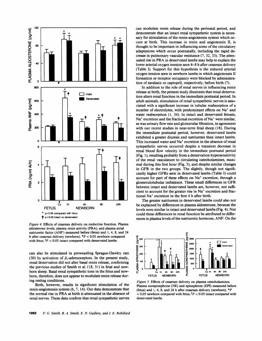
Endocrine function. Effects of delivery on plasma aldoste-rone levels, ANF, and PRA are illustrated in Fig. 4. Plasmaaldosterone levels, PRA, and ANFwere similar in intact anddenervated fetuses (P > 0.05). Plasma aldosterone increased 1h after delivery to similar levels in both groups (Fig. 4), and
remained elevated 24 h after delivery. ANF levels increasedafter delivery in intact (F = 12.39; P< 0.0001) and denervatedlambs (F = 9.94; P < 0.0001), the levels being significantlyhigher in intact than in denervated newborn lambs (P < 0.05;Fig. 4) for up to 24 h after delivery. PRAwas higher in intactthan in denervated animals at 4 and 24 h after delivery (Fig. 4).
Fig. 5 shows plasma levels of NEand EPI measured beforeand after cesarean delivery. Plasma NEand EPI levels tendedto be higher in intact than in denervated lambs before and aftercesarean delivery. There was a transient increase in plasmacatecholamine levels 1 h after birth in both groups, the increasebeing similar in intact and denervated lambs.
Renal tissue NEcontent was 36,044±6,134 pg/g of cortexin intact kidneys (n = 9) and 950±381 pg/g of cortex in dener-vated kidneys (n = 9). This represented a reduction in NEcon-tent of 97.4% in denervated compared with intact kidneys.
Cx lh 4h 8hFETUS NEWBORN
20 -
10 _
0 _RBFV
-10 _
-20
-30
24h
225
200
HR 175(bpm) 150
125
100
-+- Denervated
Intact
Cx lh 4h 8hFETUS NEWBORN
Figure 1. Effects of cesarean delivery on
systemic and renal hemodynamics inintact and denervated lambs. BP, bloodpressure; HR, heart rate; RBFV, renalblood flow velocity (%A), before (fetus)and 1, 4, 8, and 24 h after cesarean de-livery (newborn). *P < 0.05 newborncompared with fetus; *P < 0.05 intactcompared with denervated lambs.
1990 F. G. Smith, B. A. Smith, E. N. Guillery, and J. E. Robillard
65
60
BP 55(mmHg) 50
45
40 I I I I I
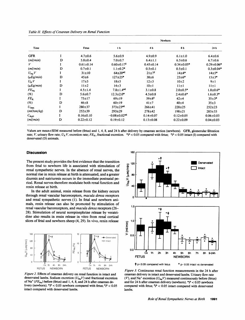
Table IL Effects of Cesarean Delivery on Renal Function
Newbor
Time Fetus h 4 h 8 h 24 h
GFR I 4.7±0.6 5.6±0.9 4.9±0.9 6.1 ± 1.0 6.4±0.6(ml/min) D 5.8±0.4 7.0±0.7 6.4±1.1 6.5±0.6 6.7±0.6V I 0.61±0.14 0.60±0.17* 0.45±0.14 0.34±0.05* 0.29±0.06*(ml/min) D 0.7±0.1 1. 1±0.2* 0.5±0.1 0.5±0.1 0.3±0.04*UNaV I 31±10 64±20** 21±7* 14+4* 14+5*(gEqlmin) D 45±6 127±25* 38±6 23±6* 15±3*UKV I 17±5 18±5 12±3 10±2 9±1(gEq/min) D 11±2 14±3 10±1 11±1 11±1FENa I 4.5±1.4 7.8±1.4*t 3.1±0.8 2.0±0.5* 1.8±0.6*(%) D 5.6±0.7 12.3±2.0* 4.5±0.8 2.4±0.6* 1.6±0.3*FEK I 75±17 69±19 59±8* 42±4 35±3*(%) D 46±8 60±19 41±7 40±4 35±3UOS I 280±37 373±234 266±41 228±25 252±23(mOsm/kg) D 252±39 293±29 278±42 198±21 283±33CH20 I 0.16±0.10 -0.08±0.02*t 0.14±0.07 0.12±0.05 0.06±0.03(ml/min) D 0.22±0.12 0.19±0.12 0.13±0.08 0.22±0.09 0.04±0.03
Values are mean±SEMmeasured before (fetus) and 1, 4, 8, and 24 h after delivery by cesarean section (newborn). GFR, glomerular filtrationrate; V, urinary flow rate; Ux V, excretion rate; FEx, fractional excretion. *P < 0.05 compared with fetus; *P < 0.05 intact (I) compared withdenervated (D) animals.
Discussion
The present study provides the first evidence that the transitionfrom fetal to newborn life is associated with stimulation ofrenal sympathetic nerves. In the absence of renal nerves, thenormal rise in renin release at birth is attenuated, and a greaterdiuresis and natriuresis occurs in the immediate postnatal pe-riod. Renal nerves therefore modulate both renal function andrenin release at birth.
In the adult animal, renin release from the kidney occursthrough renal vascular baroreceptors, macula densa receptorsand renal sympathetic nerves (1). In fetal and newborn ani-mals, renin release can also be promoted by stimulation ofrenal vascular baroreceptors, and maculda densa receptors (26-28). Stimulation of neural norepinephrine release by veratri-dine also results in renin release in vitro from renal corticalslices of fetal and newborn sheep (4, 29). In vivo, renin release
C)E ~~~ ~ t -4-Denervatea
>'SSL~~~~~~~gzX~~LCx lh 4h 8h 24h Cx Ih 4h 8h 24h
FETUS NEWBORN FETUS NEWBORN
Figure 2. Effects of cesarean delivery on renal function in intact anddenervated lambs. Sodium excretion (UN.V) and fractional excretionof Na+ (FEN.) before (fetus) and 1, 4, 8, and 24 h after cesarean de-livery (newborn). *P < 0.05 newborn compared with fetus; *P < 0.05intact compared with denervated lambs.
12 <00 5 copae wtfeu *p.5iacvsdenervated
Figure3.Cotinuos renl funtion easurmentsin the24hafe
±80
coz
~40
0
1.5
1.0
E
>0.5
0.0cx1h 2h 3h 4h 5h 6h 7h 8h 9-24h
FETUS NEWBORN
tf p<0O.05 compared with fetus * p< 0.05 intact vs denervated
Figure 3. Continuous renal function measurements in the 24 h aftercesarean delivery in intact and denervated lambs. Urinary flow rate(V), and Na+ excretion (UN. V) measured continuously before (fetus)and for 24 h after cesarean delivery (newborn). *P < 0.05 newborncompared with fetus; *P < 0.05 intact compared with denervatedlambs.
Role of Renal Sympathetic Nerves at Birth 1991
120
80
40
0
800
't E Intactg 600 _ X - Denervated
1600
~400 -*
E2 200
a.t
6
4
0
Cx lh 4h
FETUS NEWBORN* p<0.05 compared with fetus
t P<0.05 Intact vs denervated
can modulate renin release during the perinatal period, anddemonstrate that an intact renal sympathetic system is neces-
sary for stimulation of the renin-angiotensin system which oc-
curs at birth. This increase in renin and angiotensin II, isthought to be important in influencing some of the circulatoryadaptations which occur postnatally, including the rapid de-crease in pulmonary vascular resistance (7, 32, 33). The atten-uated rise in PRAin denervated lambs may help to explain thelower arterial oxygen tension seen 4-8 h after cesarean delivery(Table I). Support for this hypothesis is the reduced arterialoxygen tension seen in newborn lambs in which angiotensin IIformation or receptor occupancy were blocked by administra-tion of saralasin or captopril, respectively, before birth (7).
In addition to the role of renal nerves in influencing reninrelease at birth, the present study illustrates that renal denerva-tion alters renal function in the immediate postnatal period. Inadult animals, stimulation of renal sympathetic nerves is asso-
ciated with a significant increase in tubular reabsorption of a
number of electrolytes, with predominant effects on Na' andwater reabsorption (1, 34). In intact and denervated fetuses,Na' excretion and the fractional excretion of Na' were similar,as was urinary flow rate and glomerular filtration, in agreementwith our recent studies in near-term fetal sheep (18). Duringthe immediate postnatal period, however, denervated lambsexhibited a greater diuresis and natriuresis than intact lambs.This increased water and Na+ excretion in the absence of renalsympathetic nerves occurred despite a transient decrease inrenal blood flow velocity in the immediate postnatal period(Fig. 1), resulting probably from a denervation hypersensitivityof the renal vasculature to circulating catecholamines, maxi-mal during this first hour (Fig. 5), and despite similar changesin GFR in the two groups. The slightly, though not signifi-cantly higher GFRs seen in denervated lambs (Table I) couldaccount for part of these effects on Na+ excretion, through a
glomerulotubular imbalance. These small differences in GFRbetween intact and denervated lambs are, however, not suffi-cient to account for the greater rise in Na+ excretion and frac-tional Na+ excretion in the first 4 h after birth.
The greater natriuresis in denervated lambs could also notbe explained by differences in plasma aldosterone, because thelevels were similar in intact and denervated lambs (Fig. 4). Norcould these differences in renal function be attributed to differ-ences in plasma levels of the natriuretic hormone, ANF. On the
Figure 4. Effects of cesarean delivery on endocrine function. Plasmaaldosterone levels, plasma renin activity (PRA), and plasma atrialnatriuretic factor (ANF) measured before (fetus) and 1, 4, 8, and 24h after cesarean delivery (newborn). *P < 0.05 newborn comparedwith fetus; tP < 0.05 intact compared with denervated lambs.
can also be stimulated in preweanling Sprague-Dawley rats(30) by activation of j31-adrenoceptors. In the present study,renal denervation did not alter basal renin release, confirmingthe previous studies of Smith et al. ( 18, 31) in fetal and new-
born sheep. Basal renal sympathetic tone in the fetus and new-
born, therefore, does not appear to modulate renin release dur-ing resting conditions.
Birth, however, results in significant stimulation of therenin-angiotensin system (6, 7, 14). Our data demonstrate thatthe normal rise in PRAat birth is attenuated in the absence ofrenal nerves. These data confirm that renal sympathetic nerves
C-
3002
200w
z
W 100
C.
wz
*~~~~~~~i
Cx 1h 4h 8h 24h
FETUS NEWBORN
L3000
. 2500
2000
1500
1000
500
0
0.
* Intact
t Denervated
Cx lh 4h 8h 24h
FETUS NEWBORN
Figure 5. Effects of cesarean delivery on plasma catecholamines.Plasma norepinephrine (NE) and epinephrine (EPI) measured before(fetus) and 1, 4, 8, and 24 h after cesarean delivery (newborn). *P< 0.05 newborn compared with fetus; tP < 0.05 intact compared withdenervated lambs.
1992 F. G. Smith, B. A. Smith, E. N. Guillery, and J. E. Robillard
w
z0
w
(I)
U,
ll
I-.:E.
E
a-
"I
contrary, plasma levels of ANFwere much lower in denervatedthan in intact lambs (Fig. 4).
The reason for the greater ANFlevels in intact lambs afterbirth is not clear. In recent studies by our group (18, 31), ANFwas elevated after volume expansion in fetal sheep and new-born lambs, the levels again being lower in denervated animals.It is plausible to suggest from these observations that there isnormally an interaction between the renal sympathetic systemand ANFrelease. Recent studies in adult WKYrats (35) and inhumans (36) have purported that ANF activates vagal affer-ents, thereby inhibiting renal sympathetic activity (35). It isalso possible that elevated ANF in intact lambs represents acontrol factor for fluid and electrolyte homeostasis at birth,which is disturbed in denervated animals in which a greateramount of Na' is lost at birth. Evidence for this is the decreasein hematocrit seen at birth in intact but not in denervatedlambs. Further studies are needed to determine the interactionbetween ANFand the renal sympathetic system in the perina-tal period.
The greater natriuresis and diuresis seen in the immediatepostnatal period in denervated lambs therefore appears to beattributed to lack of renal sympathetic innervation, and indi-cates that renal sympathetic nerves play an important role inthe adaptation of the kidney to postnatal life. By 24 h afterdelivery, however, the effect of renal denervation on water andNa' excretion at birth was absent. Hence, renal sympatheticnerves do not appear to influence basal renal function in new-born lambs beyond the initial transition phase, as previouslysuggested (18, 31). The normal increase in fractional Na' ex-cretion which occurs by 24 h after cesarean delivery (13-15),was seen in both intact and denervated lambs. Such an effect isnot surprising because, in the adult, it is now generally acceptedthat renal sympathetic nerves are important in influencing Na'homeostasis during stressful conditions, with a minimal role ininfluencing basal renal hemodynamics and function (1).
The increased urinary osmolality and decreased free waterclearance seen in intact lambs in the immediate postnatal pe-riod is in agreement with previous observations (13, 15), andprobably reflects the increased levels of arginine vasopressinduring this time (37). The lack of urinary concentrating capac-ity after birth in denervated lambs is consistent with the hy-pothesis of Kurkus et al. (38) who found that denervated kid-neys had an elevated tissue water content due probably todisruptions in the medullary gradient. Such a disruption inmedullary gradient could prevent the antidiuretic effects of va-sopressin. An alternative explanation is that central vasopres-sin release may be inhibited in renally denervated lambsthrough loss of renal afferent input, because, at least in theadult, neurosecretion of vasopressin is increased by afferentrenal nerve stimulation (39, 40). Although there is presently noevidence that afferent renal nerve activity is increased at birth,circulating bradykinin, a substance known to stimulate affer-ent renal nerves (41), is elevated in the immediate postnatalperiod (32, 33, 42). In the present study, however, neitherplasma vasopressin nor bradykinin levels were measured.
In conclusion, the present study shows that renal sympa-thetic nerves play an important physiological role during thetransition from fetal to newborn life. In the absence of renalnerves, the normal increase in PRAat birth is attenuated, andthere is a greater diuresis and natriuresis in the immediate post-natal period. These data indicate that renal sympathetic nerves
regulate fluid and electrolyte homeostasis during the adapta-tion of the kidney to postnatal demand, and provide the firstevidence that birth is associated with stimulation of the sympa-thetic system.
Acknowledaments
The authors gratefully acknowledge the assistance of Ruth Hurlburt inthe preparation ofthis manuscript, and Donna Farley and the ImmunoCore Laboratory of the University of Iowa in performing the hormonalassays.
This work was supported in part by grants from the National Insti-tute of Health HD-20576, HL-14388, DK-38302, and HL-35600. Ed-ward N. Guillery is supported by National Research Service Award,HL-08366-01.
References
1. DiBona, G. F. 1982. The functions of the renal nerves. Rev. Physiol. Bio-chem. Pharmacol. 94:76-18 1.
2. Robillard, J. E., K. T. Nakamura, M. K. Wilkin, 0. J. McWeeny, and G. F.DiBona. 1987. Ontogeny of renal hemodynamic response to renal nerve stimula-tion in sheep. Am. J. Physiol. 252:F605-F6 12.
3. Robillard, J. E., F. G. Smith, B. Smith, 0. J. McWeeny, and P. A. Jose.1989. Ontogeny of the renal response to alphal-adrenoceptor stimulation insheep. Pediatr. Res. 25:347A. (Abstr.)
4. Nakamura, K. T., J. M. Klinkefus, F. G. Smith, T. Sato, and J. E. Robillard.1989. Ontogeny of neuronally released norepinephrine on renin secretion insheep. Am. J. Physiol. 257:R765-R770.
5. Robillard, J. E., 0. J. McWeeny, B. Smith, and G. F. DiBona. 1988. Ontog-eny of neurogenic regulation of renal tubular sodium reabsorption in sheep.Pediatr. Res. 23:545A. (Abstr.)
6. Lumbers, E. R., and G. C. Reid. 1977. Effects of vaginal delivery andcaesarean section on plasma renin activity and angiotensin II levels in humanumbilical cord blood. Biol. Neonate. 31:127-134.
7. Davidson, D. 1987. Circulating vasoactive substances and hemodynamicadjustments at birth in lambs. J. Appl. Physiol. 63:676-684.
8. Padbury, J. F., D. H. Polk, J. P. Newnham, and R. W. Lam. 1985. Neonataladaptation: greater sympathoadrenal response in preterm than full-term fetalsheep at birth. Am. J. Physiol. 248:E443-E449.
9. Eliot, R. J., A. H. Klein, T. H. Glatz, P. W. Nathanielsz, and D. A. Fisher.1981. Plasma norepinephrine, epinephrine, and dopamine concentrations in ma-ternal and fetal sheep during spontaneous parturition and in premature sheepduring cortisol-induced parturition. Endocrinology. 108:1678-1682.
10. Lagercrantz, H., and P. Bistoletti. 1973. Catecholamine release in thenewborn infant at birth. Pediatr. Res. 1 1:889-893.
11. Eliot, R. J., R. Lam, R. D. Leake, C. J. Hobel, and D. A. Fisher. 1980.Plasma catecholamines concentrations in infants at birth and during the first 48hours of life. J. Pediatr. 96:311-315.
12. Jones, C. M., and F. C. Greiss, Jr. 1982. The effect of labor on maternaland fetal circulating catecholamines. Am. J. Obstet. Gynecol. 144:149-153.
13. Smith, F. G., and E. R. Lumbers. 1989. Comparison of renal function interm fetal sheep and newborn lambs. Biol. Neonate. 55:309-316.
14. Nakamura, K. T., G. P. Matherne, 0. J. McWeeny, B. A. Smith, and J. E.Robillard. 1987. Renal hemodynamics and functional changes during the transi-tion from fetal to newborn life in sheep. Pediatr. Res. 21:229-234.
15. Smith, F. G. 1987. Factors Influencing Fetal and Neonatal Fluid Balance.University of NewSouth Wales, Sydney, Australia. 1 11-132.
16. Jennings, J. J., and J. P. Crowley. 1972. The influence of mating manage-ment on fertility in ewes following progesterone-PMS treatment. Vet. Rec.90:495-498.
17. Robillard, J. E., and R. E. Weitzman. 1980. Developmental aspects of thefetal renal response to exogenous arginine vasopressin. Am. J. Physiol. 238:F407-F414.
18. Smith, F. G., T. Sato, 0. J. McWeeny, J. M. Klinkefus, and J. E. Robillard.1990. Role of renal sympathetic nerves in the response of the ovine fetus tovolume expansion. Am. J. Physiol. 259:R1050-R1055.
19. Robillard, J. E., K. T. Nakamura, and G. F. DiBona. 1986. Effects of renaldenervation on renal responses to hypoxemia in fetal lambs. Am. J. Physiol.250:F294-F30 1.
20. Smith, F. G., and E. R. Lumbers. 1988. Changes in renal function follow-ing delivery of the lamb by caesarian section. J. Dev. Physiol. 10:145-148.
21. Ito, T., J. Woo, R. Haning, and R. Horton. 1972. A radioimmunoassay for
Role of Renal Sympathetic Nerves at Birth 1993
aldosterone in human peripheral plasma including a comparison of alternatetechniques. J. Clin. EndocrinoL. & Metab. 34:106-112.
22. Robillard, J. E., K. T. Nakamura, V. A. Varille, A. A. Andresen, G. P.Matherne, and D. E. Van Orden. 1988. Ontogeny of the renal response to natri-uretic peptide in sheep. Am. J. Physio. 254:F634-F641.
23. Moyer, T. P., and N. S. Jiang. 1978. Optimized isocratic conditions foranalysis of catecholamines by high-performance reversed-phase paired-on chro-matography with amperometric detection. J. Chromatogr. 153:365-372.
24. Haber, E., T. Koerner, L. B. Page, B. Kliman, and A. Purnobe. 1969.Application of a radioimmunoassay for angiotensin I to the physiologic measure-ments of plasma renin activity in normal human subjects. J. Clin. Endocrinol. &Metab. 29:1349-1355.
25. Zar, J. H. 1984. Biostatistical Analysis. Prentice-Hall, Inc., EnglewoodCliffs NJ. 122-235.
26. Smith, F. G., Jr., A. N. Lupu, L. Barajas, R. Bauer, and R. A. Bashore.1974. The renin angiotensin system in the fetal lamb. Pediatr. Res. 8:611-620.
27. Siegel, S. R., and D. A. Fisher. 1980. Ontogeny of the renin-angiotensin-aldosterone system in the fetal and newborn lamb. Pediatr. Res. 14:99-102.
28. Lumbers, E. R., and A. D. Stevens. 1987. The effects of frusemide, sarala-sin and hypotension on fetal plasma renin activity and on fetal renal function. J.Physic!. (Lond.). 393:479-490.
29. Nakamura, K. T., W. V. Page, J. M. Klinkefus, T. Sato, and J. E. Robil-lard. 1989. Ontogeny of isoproterenol-stimulated renin secretion from sheeprenal cortical slices. Am. J. Physiol. 256:R1258-R1263.
30. Kirby, R. F., and A. K. Johnson. 1990. Effects of sympathetic activationon plasma renin activity in the developing rat. J. PharmacoL. Exp. Ther. 253:152-157.
31. Smith, F. G., T. Sato, 0. J. McWeeny, L. Torres, andJ. E. Robillard. 1989.Role of renal nerves in response to volume expansion in conscious, newbornlambs. Am. J. Physio. 257:R 1519-R 1525.
32. Heymann, M. A., A. M. Rudolph, A. S. Nies, and K. L. Melmon. 1969.Bradykinin production associated with oxygenation of fetal lamb. Circ. Res.25:521-534.
33. Mott, J. C. 1975. The place of the renin-angiotensin system before andafter birth. Br. Med. Bull. 31:44-50.
34. Gottschalk, C. W., N. G. Moss, and R. E. Colindres. 1985. Neural controlof renal function in health and disease. In The Kidney: Physiology and Pathophys-iology. D. W. Seldin and G. Giebisch, editors. Raven Press, NewYork. 581-61 1.
35. Imaizumi, T., A. Takeshita, H. Higashi, and M. Nakamura. 1987. Alpha-ANPalters reflex control of lumbar and renal sympathetic nerve activity andheart rate. Am. J. Physiol. 253:H1 136-H1 140.
36. Ebert, T. J., and A. W. Cowley, Jr. 1988. Atrial natriuretic factor attenu-ates carotid baroreflex-mediated cardioacceleration in humans. Am. J. Physiol.254:R590-R594.
37. Leffler, C. W., J. Crofton, D. P. Brooks, L. Share, J. R. Hessler, and R. S.Green. 1985. Changes in plasma arginine vasopressin during transition from fetusto newborn following minimal trauma delivery of lambs and goats. Biol. Neonate.48:43-48.
38. Kurkus, J., J. Sadowski, R. Gellert, and S. Krus. 1980. Influence of renaldenervation on urine concentration in awake and anesthetized dogs. Eur. J. Clin.Invest. 10:463-467.
39. Day, T. A., and J. Ciriello. 1987. Effects of renal receptor activation onneurosecretory vasopressin cells. Am. J. Physiol. 253:R234-R241.
40. Caverson, M. M., and J. Ciriello. 1987. Effect of stimulation of afferentrenal nerves on plasma levels of vasopressin. Am. J. Physiol. 252:R801-R807.
41. Smits, J. F. M., and M. J. Brody. 1984. Activation of afferent renal nervesby intrarenal bradykinin in conscious rats. Am. J. Physiol. 247:R1003-R1008.
42. Melmon, K. L., M. J. Cline, T. Hughes, and A. S. Nies. 1968. Kinins:possible mediators of neonatal circulatory changes in man. J. Clin. Invest.57:1295-1302.
1994 F. G. Smith, B. A. Smith, E. N. Guillery, and J. E. Robillard