renal review for rnc 2018-final - bchsfoutreach.ucsf.edu. renal... · angiotensin pathway diabetes...
TRANSCRIPT

5/31/2018
1
RNC Renal Review
Elizabeth Rex, MS, NNP-BC with thanks to Carlos Botas, MD
Abbreviations
• RBF = Renal Blood Flow
• UOP = Urine Output
• BUN = Blood Urea Nitrogen
• MAP = Mean arterial blood pressure
• GFR = Glomerular Filtration Rate
15 Things To Know About Baby Kidneys
1. Most babies have two kidneys
2. The kidneys filter the blood and produce urine.
About 20% of all cardiac output goes to the kidneys.
3. Urine output is closely related to renal blood flow:
Good renal blood flow = good UOP
Poor renal blood flow = poor UO

5/31/2018
2
4. Urine Output is an excellent method for monitoring overall circulation / organ perfusion
5. Premature infants have immature ‘dumb’ kidneys that are not good at reabsorbing electrolytes (esp. HCO3ˉ)
=
6. Blockage of the drainage of urine results in Hydronephrosis (dilated, swollen kidneys)
7. Certain medications can decrease RBF, resulting in decreased urine output (example, Indocin)
8. Certain medications increase urine output and are very useful in patient with too much fluid (diuretics)
9. Serum creatinine and BUN are measurements of renal function. An elevated creatinine suggest renal disease or poor renal blood flow.
10. The kidneys are very important during fetal development and produce most of the amniotic fluid. A fetus without functional kidneys has minimal amniotic fluid.

5/31/2018
3
11. Embryology of the kidney is basically useless (or at least very confusing)
12. An infant only needs ONE good kidney to be OK.
13. Hypertension can be a sign of renal problems.
14. And most importantly, the great majority of infants have happy and healthy kidneys Even in sick babies, the kidney is usually not the problem. The kidneys are usually markers of other problems and not the primary problem.
15. Isolated renal disease in infants is fairly rare.
Kidney Function
Non excretory• Produces Renin
• Produces Erythropoietin
• Metabolizes Vitamin D
• Degrades insulin
• Produces Prostaglandins
Excretory• Maintains plasma osmolality
• Maintains electrolyte imbalance
• Maintains water balance
• Excretes nitrogenous end products
The Nephron
Basic functional unit of the kidney: 10,000 nephrons / kidney
Glomerulus–filters the blood, produces filtrate
PCT–reabsorption of Na, Cl, water, glucose, amino acids
Loop of Henle–concentration of Na,Cl in interstitium
DCT–active reabsorption of Na, Cl, HCO3. Controlled by aldosterone, parathyroid hormone and atrial naturietic factor

5/31/2018
4
What is the Glomerular Filtration Rate (GFR)?
The volume of fluid filtered from the renal (kidney) glomerular capillaries into the into Bowman's capsule per unit time
GFR = Renal Plasma Flow x Filtration Fraction
Normal Urine Output after Birth
• 97% of healthy infants urinate in the first 24 hours (100% by 48 hours)
• First 24 hours: UOP is often < 1 ml/kg/hr
• After 24 hours: Normal UOP 1-5 ml/kg/hr
Oliguria: UOP less than 1 ml/kg/hr
Anuria: No UOP

5/31/2018
5
What is creatinine ?
• Product of creatine which comes from muscle and brain
• Excreted through the kidneys related to serum concentration
It is a marker of Glomerular Filtration Rate (GFR) Renal Plasma Flow x Filtration Fraction
• Inversely related to GFR
• High GFR = Low creatinine
• Low GFR = High creatinine Every time creatinine doubles there is a 50% renal loss
0.5 100% function
1.0 50% function
2.0 25% function
4.0 12.5% function
GFR rises over first 4 weeks
Creatinine drops over first 4 weeks
What is a normal creatinine in a baby ?
1-2
days
8-9
days
15-16
days
22-23
days
1000 -2000 gms 1.1 0.7 0.6 0.4
>2000 gms 0.9 0.5 0.4 0.3
What is a normal BUN (Blood urea Nitrogen in a baby?
Preterm Term
BUN 3 - 25 4 - 12

5/31/2018
6
AKI Acute Kidney Injury / ARF Acute Renal Failure3 types
1. Prerenal (80%) volume-responsive or functional AKI is caused by reduced renal perfusion and is the most common form of neonatal AKI. It is typically due to hypovolemia or reduction of effective circulation.
Hypovolemia - Neonates are at increased risk for hypovolemia due to their increased insensible water loss, limited urinary concentration ability, and concomitant conditions that result in excessive fluid loss:
- Bleeding
- Diarrhea
- Increased evaporative fluid loss due to use of radiant warmers, phototherapy, compromise of skin integrity (abdominal wall defect), or thinness of the skin seen in extremely low birth weight infants (BW <1000 g)
AKI/ARF – 3 types
1. Prerenal 80%Reduction of effective circulation – neonatal conditions associated with reduced effective circulation include:
- Impaired cardiac output - myocardial injury, perinatal asphyxia, critical congenital heart defects, complete heart block
- Sepsis
- Third spacing (movement of fluid from the vascular space into the interstitium) from acute intestinal injury (necrotizing enterocolitis)
- Hypoalbuminemia due to nephrotic syndrome or hepatic failure
- Capillary leak due to perinatal asphyxia or hydrops fetalis
AKI/ARF
2.Intrinsic or intrarenal (11%)• Tubular and/or interstitial injury –
• Although hypoperfusion usually results in prerenal AKI, prolonged and/or severe renal hypoperfusion causes direct tubular endothelial and epithelial cell injury from ischemia and inflammation
• Prenatal and postnatal nephrotoxic exposures
• Renal vasculature disease
• Bilateral renal vascular thrombosis can result in intrinsic renal failure.
• A more common manifestation of less severe renal vascular thrombosis is hypertension.
• Glomerular disease AKI due to glomerular disease in the newborn is rare. More commonly, neonatal renal dysfunction due to glomerular disorders is due to chronic kidney disease (CKD) caused by congenital anomalies of the kidney and urinary tract, genetic disorders (polycystic kidney), and congenital nephrotic syndrome, which can present in a similar manner as AKI.

5/31/2018
7
ATN - Acute Tubular Necrosis
• Death of tubular cells (in the PCT / DCT)
• Why tubular cells?
• Tubular cells are very metabolically active and depend on oxygen
• Tubular cells transport 99% of the filtrate back into the bloodstream.
• Inadequate oxygen delivery causes death of tubular cells
ATN - Acute Tubular Necrosis
Damage due to toxins, ischemia, lack of oxygen
Toxins include:• Gentamicin
• Amphotericin B
• Vancomycin
• Prostaglandin synthesis inhibitors (indomethacin)
• Radiopaque dye
• Cisplatin, etc.
ATN - Acute Tubular Necrosis
• Acute Tubular Necrosis (ATN)
• Results in decreased urine output and elevated creatinine
• Usually reversible in neonates
• Signs and symptoms
- Decreased urine output
- Increasing BUN and creatinine
- Blood and protein may be present in urine after initial injury

5/31/2018
8
Management of ATN
• Careful Fluid Management
• Monitor closely and treat:
- Hyperkalemia
- Metabolic acidosis
- Hypocalcemia
- Hyperphosphatemia
- Hyponatremia
- Hypertension
• Consider Dialysis
AKI / ARF
3. Post Renal (3%)
- Due to obstruction of urinary flow from both kidneys or obstruction of the urinary tract of a solitary kidney.
Therefore, postrenal neonatal AKI is usually due to - anatomic bladder obstruction (e.g. posterior urethral valves) or - bilateral anomalies of the renal pelvis or ureter.
Hydronephrosis
• Dilatation of the upper urinary tract
• Commonly seen on prenatal US
• Mild dilatation (pelviectasis) is common in late gestation and not the same as hydronephrosis
• Most common causes• Physiological hydronephrosis (spontaneously resolves)
• Ureteropelvic junction obstruction (ureter – kidney junction)
• Ureterovesical junction obstruction (ureter – bladder junction)
• Posterior urethral valves (in boys)
• Vesicoureteral Reflux (reflux from bladder to ureter)
• Duplication anomalies

5/31/2018
9

5/31/2018
10
PKD Polycystic Kidney Disease2 types
1. Autosomal Recessive (ARPKD)- Occurs 1/10,000-40,000 births
- Initial prenatal sonogram may be normal, with progressively enlarged kidneys
- Oligohydramnios develops during late 2nd trimester
- Respiratory failure due to pulmonary hypoplasia is common
- Mutations of PKHD1 gene – chromosome 6p21 – codes for fibrocystin protein
- Usually have hepatic fibrosis as well
2 types of Polycystic Kidney Disease
2. Autosomal Dominant (ADPKD)
- Much more common than ADPKD, but rarely presents in the neonatal period
- Progressive bilateral renal cyst formation variable in size
- Clinical spectrum ranges from severe neonatal problems to asymptomatic during lifetime
- Long term survival is usually excellent

5/31/2018
11
Management
• AKI/ARF • Prerenal - volume resuscitation and treat specific causes • Intrinsic - supportive care, fluid restriction, electrolyte balance• Postrenal - drain bladder with indwelling catheter and consult urology
• Fluid Challenge• 10-20 ml NS IV over 1 – 2 hours• Lasix 1-2 mg/kg IV should oliguria persist• Response is defined as UOP > 2ml/hr
• Maintaining Euvolemia• IWL (insensible water loss) + urinary losses• Preterm – up to 70 ml/kg + ml/ml UOP• Full term – 30 ml/kg + ml/ml UOP
• Formula – low phosphorus PM 60/40
Angiotensin Pathway
Diabetes Insipidus
5/31/2018
12
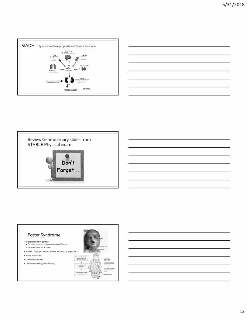
SIADH – Syndrome of inappropriate antidiuretic hormone
Review Genitourinary slides from STABLE Physical exam
Potter Syndrome
• Bilateral Renal Agenesis• Occurs 1: 4,000 to 1: 8,000 infants and fetuses
• 2 x more common in males
• Severe Oligohydramnios Severe Pulmonary Hypoplasia
• Facial anomalies
• Limb contractures
• Lethal anomaly (40% stillborn)

5/31/2018
13
Prune Belly syndromeAKA Eagle-Barrett syndrome
1/30,000 births
Features:• Lack of development of abdominal muscles,
causing the skin to wrinkle like a prune
• Renal and urinary tract anomalies
• Dilated bladder/ureters, often tortuous, reflux common
• Infection common due to urinary stasis
• Intestinal malrotation common
• 95% are male infants with bilateral undescended testicles
cryptorchidism
• Less common limb and cardiac anomalies
• Most infants have 2 umbilical arteries and 1 umbilical vein
• Approx. 0.3% of infant have only 1 umbilical artery
• Approximately 5 – 16% of infants with single umbilical artery have renal abnormalities
• Most common is vesicouretric reflux (reflux from bladder to ureter)
• IUGR is as high as 28% of cases of single umbilical artery
• Placental anomalies found in 16 – 78% of cases (most common small placenta and abnormal cord insertion)
Single Umbilical Artery
Patent uracus
• Presents with urine leaking from the umbilicus.
• Communication between the bladder and umbilicus when the urachal tube fails to close
• More common in males than females
• Complications include UTIs, excoriation, and infection
• May close spontaneously or require surgical closure

5/31/2018
14
Bladder extrophy
• Inner posterior wall of bladder protruding through a defect in the anterior pelvic wall
• Repair before 48 hours may be more successful due to maternal hormone relaxin
Hypospadias / Epispadias
• Other anomalies undescended testes and inguinal hernias
• Constriction of orifice, urinary tract obstruction
• UTIs, impaired reproductive function
dorsal
ventral
Common diureticsFurosemide (Lasix®) - loop diuretic
- blocks reabsorption of Cl-
- impairs Ca++ and Mg++ reabsorption• 0.5 –1 mg/kg/dose varies IV
• 1 – 2 mg/kg/dose varies PO
• Management of edema associated with heart failure and hepatic or renal disease; acute pulmonary edema.
• Hypertension.

5/31/2018
15
Common diureticsChlorothiazide (Diuril) - thiazide diuretic
- acts on distal tubule - augments potassium wasting - stimulates Ca++ reabsorption
• 10 to 40 mg/kg/day in 2 divided doses PO
• 5 to 10 mg/kg/day in 2 divided doses IV
Congenital hyperinsulinism (adjunct therapy in combination with diazoxide
Diabetes insipidus (central), need to titrate dose to target urine osmolality
Common diuretics
Spironolactone (Aldactone) - potassium sparing diuretic (potassium retained)
- competitive inhibition of aldosterone• Bronchopulmonary dysplasia
• 1.5 mg/kg/dose every 12 hours PO; used in combination with a thiazide diuretic
• Edema• 1 to 3 mg/kg/day PO in divided doses every 12 to 24 hours; used in combination with a thiazide
diuretic
• Dosing: • Dosing adjustment in renal impairment: Spironolactone is substantially excreted by the
kidney; use with caution; monitor serum potassium closely; consider extended dosing intervals for moderate renal impairment. Avoid use in severe renal impairment
• Not stable at room temperature, must be refrigerated

5/31/2018
16
Electrolytes
Na+- sodium is the major extracellular cation.
- Hyponatremia causes intravascular hypotonicity and fluid shifts into the cells causing cellular edema and intravascular dehydration.
- Hypernatremia results intravascular hypertonicity and a fluid shift into the extracellular space causing cellular dehydration and intravascular overload.
Hypernatremia signs: nausea and vomiting, lethargy, weakness, restlessness, hyperreflexia, spasticity, hyperthermia, seizures, and coma. Intracranial hemorrhage, venous sinus thrombosis, and acute renal tubular necrosis may occur.
Hyponatremia signs: nausea and vomiting, apathy, headache, seizures, hypothermia, weakness. Infants with hyponatremic dehydration may appear quite ill, because hyponatremia causes disproportionate reductions in ECF volume
If the sodium concentration drops quickly to critical levels, seizures, coma, and death may occur
To differentiate between hemodilution and excessive Na loss a urine osmolality and serum Na should be done
Electrolytes
K+- potassium is the major cation in the cells, 90% of intracellular fluid.
- If the pH is high, K will be driven into the cells, causing serum K level to drop - Insulin will also force K back into the cells by stimulating the Na/K pump causing a decreased
serum K - Tissue damage, bruising, or cephalohematoma may increase serum K level - K may be falsely elevated with hemolysis - K excretion is via the kidneys based on GFR, UO, and aldosterone sensitivity
Hypokalemia signs: abdominal distension, ileus, hypotonia, flattened T waves, and ST depression
Hyperkalemia signs: cardiac irritability, peaked T waves, and widened QRS

5/31/2018
17
Electrolytes
Mg ++– magnesium is the second-most abundant intracellular cation and
overall, the fourth-most abundant cation.
It plays a fundamental role in many functions of the cell, including energy transfer, storage, and use; protein, carbohydrate, and fat metabolism; maintenance of normal cell membrane function; and the regulation of parathyroid hormone (PTH) secretion.
• Hypomagnesemia signs: hyperreflexia and irritability, abnormal eye movements (nystagmus), and convulsion
• Hypermagnesemia signs: smooth muscle relaxation causing respiratory, neuromuscular, and gastrointestinal depression
ElectrolytesCl – Chloride is the major anion seen in both, the blood and the extracellular fluid
(2/3 of all serum anions)
- Chloride is secreted by the stomach's mucosa as hydrochloric acid - It provides an acid medium that aids digestion and activation of enzymes.- It participates in maintaining acid-base and body water balances - Influences the osmolality or tonicity of extracellular fluid - Plays a role in the exchange of oxygen and carbon dioxide in red blood cells- Helps activate salivary amylase (which, in turn, activates the digestive process)
Hypochloremia causes: Adrenal insufficiency, renal failure, administration of dextrose I.V. without electrolytes. Loss of hydrochloric acid in gastric secretions due to vomiting, gastric suctioning, or gastric surgery. Pyloric obstruction, draining fistula, ileostomy, prolonged diarrhea or diaphoresis resulting in dehydration. Edematous states (CHF). Loop and thiazide diuretics. Corticosteroids.
Due to volume depletions, the chloride level decreases. Hence, the kidneys retain the bi-carbonate and sodium ions for balancing the incurred loss. As a result, bicarbonate accumulates in the ECF, thereby raising the pH level leading to hypochloremic metabolic alkalosis.
Hypochloremia signs: Muscle weakness, dry skin, twitching, tetany, shallow, depressed breathing, respiratory arrest, hyperactive deep tendon reflexes, cardiac arrhythmias, seizures, and coma
Hyperchloremia causes: – usually occurs as a result of dehydration or excess administration of sodium or other chloride. Hypernatremia, diabetes insipidus, hyperparathyroidism, metabolic acidosis, ATN. Dehydration from vomiting, prolonged diarrhea, kidney diseases, loss of pancreatic secretion.
Hyperchloremia signs: The clinical presentation is not specific for hyperchloremia; it depends on the underlying disease and hydration
Electrolytes
Ca++- Acidosis results in an increase of free ionized Ca, inhibits the Ca binding
ability with protein, thereby increases serum Ca levels.
Hypocalcemia signs: may see stridor, high-pitched cry, twitching, jitteriness not related to low glucose, seizure activity, poor cardiac output.
IDM have delayed neonatal parathyroid production. Asphyxia results in calcitonin release, which prohibits parathyroid action. Tissue damage releases phosphorus into the blood which further inhibits Ca uptake.
Hypercalcemia:
Untreated maternal hypoparathyroidism = overproduction of parathyroid hormone in infant
PTH - Parathyroid hormone increases serum Ca by increasing the breakdown of bone, increasing intestinal absorption, and inhibiting renal excretion.
Calcitonin is secreted by the thyroid C cells to lower serum Ca by counteracting PTH.