regulation of c-fos translation 1 signaling pathways leading to
TRANSCRIPT
Regulation of c-fos translation 1�
Signaling pathways leading to transcription and translation cooperatively regulate the
transient increase in expression of c-Fos protein1�
Kenji Takeuchi‡, Sayumi Shibamoto‡†2, Kentaro Nagamine‡, Ichiro Shigemori‡, Satoshi
Omura*, Naomi Kitamura¶, and Fumiaki Ito‡
‡Department of Biochemistry, Faculty of Pharmaceutical Sciences, Setsunan University,
Hirakata, Osaka 573-0101, Japan
*Research Center for Biological Function, The Kitasato Institute, Shirokane, Minato-ku,
Tokyo 108-8642, Japan.
¶Department of Life Science, Faculty of Bioscience and Biotechnology, Tokyo Institute of
Technology, Nagatsuta, Midori-ku, Yokohama 226-8501, Japan.
† Present address: Genentech Inc., Department of Molecular Oncology, 1 DNA Way, South
San Francisco, CA 94080
Running title: Regulation of c-fos translation
Corresponding author: Sayumi Shibamoto
Genentech Inc., 1 DNA Way, MS#40, South San Francisco, CA 94080
Fax: 650-225-5716, Tel: 650-225-8494
E-mail: [email protected]
Copyright 2001 by The American Society for Biochemistry and Molecular Biology, Inc.
JBC Papers in Press. Published on May 14, 2001 as Manuscript M102704200 by guest on February 18, 2018
http://ww
w.jbc.org/
Dow
nloaded from

Regulation of c-fos translation 2�
Summary
�
� The mechanisms by which growth factors trigger signal transduction pathways
leading to the regulation of c-Fos expression are of great interest. In this study we
investigated the effect of hepatocyte growth factor (HGF/SF) and epidermal growth factor
(EGF) on the expression of c-fos and its product, c-Fos, in human epithelial cell line
MKN74. The expression level of c-Fos protein in HGF/SF-stimulated cells was 5 to 10-
fold higher than that in EGF-stimulated cells, whereas the level of c-fos mRNA induced by
HGF/SF was similar to that by EGF.
� The hyperphosphorylation of eukaryotic initiation factor 4E-binding protein 1 (4E-
BP1), indicative of an increased number of translation initiation complexes, was detected
only in HGF/SF-induced MKN74 cells. Activation of phosphatidylinositol-3'-OH kinase
(PI3-K) and FKBP-rapamycin-associated protein/ mammalian target of rapamycin
(FRAP/mTOR) was observed after the treatment with HGF/SF. Pretreatment with an
inhibitor of either one, i.e., LY294002 for PI3-K, or rapamycin for FRAP/mTOR,
completely inhibited 4E-BP1 phosphorylation, and decreased the c-Fos synthesis induced
by HGF/SF down to the level found in EGF-induced cells.
These results suggest that the phosphorylation of 4E-BP1 is stimulated by HGF/SF
in a manner requiring both PI3-K-dependent and FRAP/mTOR-dependent pathways,
thereby stimulating c-fos mRNA translation. Regulation of the translation process of c-fos
mRNA, in addition to the immediate activation of c-fos transcription, is necessary for the
transient increase in the level of c-Fos protein to stimulate cell proliferation.
by guest on February 18, 2018http://w
ww
.jbc.org/D
ownloaded from
Regulation of c-fos translation 3�
Introduction�
�
� c-Fos, a transcription factor, is a product of a member of the immediate-early gene
family and heterodimerizes with a transcription factor of the Jun family. The heterodimeric
complex, termed activator protein-1 (AP-1), binds to the 12-O-tetradecanoylphorbol 13-
acetate (TPA) response element (TRE) of certain genes crucial for cellular proliferation and
differentiation and thereby activates transcription of such genes (1,2).
� The regulatory mechanism of c-fos expression by extracellular signaling molecules
has been studied in great detail at the step of transcription activation. Ligands such as
growth factors bind to their specific receptors and activate the Ras-mitogen-activated
protein (MAP)3 kinase cascade. MAP kinase phosphorylates ternary complex factors such
as p62TCF or Elk-1 (3), which binds together with serum response factor (SRF) to the cis-
acting regulatory element of the c-fos gene, termed SRE (serum response element),
resulting in the induction of c-fos transcription.
The expression level of c-Fos is also regulated at post-translationally. c-Fos is an
intrinsically unstable protein, and its degradation appears to occur via the ubiquitin-
mediated proteolysis system (4,5). The phosphorylation state of c-Fos at serine residues,
which are modified via the MAP-kinase pathway, is suggested to be involved in the
stability of the c-Fos protein (6).
Recently, signaling pathways involved in translation control have become better-
understood (7-9). Growth factor-regulated signaling pathways downstream of
phosphatidylinositol-3'-OH kinase (PI3-K) and FKBP-rapamycin-associated protein/
mammalian target of rapamycin (FRAP/mTOR) are suggested to be involved in the
by guest on February 18, 2018http://w
ww
.jbc.org/D
ownloaded from

Regulation of c-fos translation 4�
regulation of translation initiation. A signaling pathway that regulates the transcription of c-
fos, the Ras-MAP kinase pathway, has been well characterized (10-12). In contrast, it has
not been determined whether c-Fos expression is regulated during the translation of c-fos
mRNA.
Hepatocyte growth factor (HGF/SF) and epidermal growth factor (EGF) have been shown
to stimulate the proliferation of epithelial cells. These factors bind to their intrinsic,
membrane-spanning receptors, which are tyrosine kinases; and this binding is followed by
the activation of the kinase via autophosphorylation of the receptor for individual factors.
The receptor capable of transducing the signal of HGF/SF is the c-met proto-oncogene
product (13,14). Two tyrosine residues of its β-chain are phosphorylated upon HGF/SF
binding, which allows the receptor to transmit signals via association with p60src, PI3-K,
Grb2/SOS complex, and Gab1 (15-20). On the other hand, phosphotyrosine residues of the
EGF receptor are associated with signaling molecules such as phospholipase Cγ, Shc, and
Gab1 (21-25). Although both receptors for HGF/SF and EGF appear to be able to activate
common signaling pathways, such as PI3-K and Ras-MAP kinase pathways (26-31), the set
of pathways activated in response to HGF/SF or EGF depends on the individual systems
used for experiments.
In our ongoing studies on the effects of HGF/SF and EGF on the growth of several
gastric carcinoma cell lines, we have found that the growth of MKN74 cells was
significantly stimulated by HGF/SF, but was not changed by EGF. Moreover, the
expression level of c-Fos in EGF-induced cells was far less than that in HGF/SF-induced
cells, whereas the c-fos mRNA level was similar in cells treated with either factor. In the
present study, we analyzed this differential expression of c-Fos in response to HGF/SF and
by guest on February 18, 2018http://w
ww
.jbc.org/D
ownloaded from
Regulation of c-fos translation 5�
EGF, and found that the expression level of c-Fos was regulated not only at the
transcription step but also at the translation step. Our results strongly suggest that the c-fos
translation is regulated signaling pathways leading to the phosphorylation of 4E-BP1, the
eukaryotic initiation factor 4E-binding protein 1, which phosphorylation leads to the
increase in translation necessary for the transient increase in c-Fos protein expression in
response to growth factors.
by guest on February 18, 2018http://w
ww
.jbc.org/D
ownloaded from

Regulation of c-fos translation 6�
Experimental procedures
�
Materials�
Recombinant human HGF/SF was provided by the Research Center of Mitsubishi-Tokyo
Pharmaceuticals, Inc. (32,33). Lactacystin was prepared as described previously (34,35).
EGF (ultrapure) from mouse submaxillary glands was purchased from Toyobo Co., Ltd.
(Osaka, Japan); and fetal calf serum (FCS), from JRH Biosciences (Lenexa, KS).
Phenylmethylsulfonyl fluoride (PMSF), pepstatin A, p-toluenesulfonyl-L-arginine methyl
ester (TAME), and leupeptin came from Sigma (St. Louis). RPMI1640 was from Nissui
Pharmaceutical Co., Ltd. (Tokyo, Japan). Anti-c-Fos antiserum (Ab-2) was purchased from
Oncogene Science, Inc. (Cambridge, MA), anti-phosphotyrosine antibody (PY20) from
Transduction Laboratories (Lexington, KY), anti-4E-BP1 antibody (R-113) from Santa
Cruz Biotechnology, Inc. (Santa Cruz, CA), anti-mTOR antibody from Oncogene research
products (Cambridge, MA), and anti-c-Myc antibody from PharMingen (San Diego, CA).
Swine horseradish peroxidase (HRP)-linked anti-rabbit Ig antibody was obtained from
DAKO (Denmark). Recombinant tumor necrosis factor was purchased from R&D
(Minneapolis, MN).
Cell cultures�
Human gastric adenocarcinoma cell line MKN74 was kindly provided by Dr. E. Tahara
(Hiroshima University School of Medicine). MKN74 cells were cultured to subconfluence
in RPMI1640 supplemented with 10 % FCS and used for all of the experiments. FS-4 cells
derived from human foreskin fibroblasts were cultured in DMEM supplemented with 10 %
by guest on February 18, 2018http://w
ww
.jbc.org/D
ownloaded from
Regulation of c-fos translation 7�
FCS and treated with recombinant tumor necrosis factor as described previously (36,37).
Cell proliferation assay
MKN74 cells were seeded at a density of 1 x10⁴ cells/well in 96-well plates and cultured
for 2 days in RPMI1640 supplemented with 5 % FCS. The cells were subsequently treated
with HGF/SF or EGF for 2 days, and cell proliferation was quantified by a colorimetric
assay using the WST-1 reagent according to the manufacturer's instruction (Dojindo
Laboratories).
Preparation of cellular lysates and immunoblotting�
Cells were seeded at a density of 1.6 x 10⁵ cell / 35-mm-diameter dish and cultured for 2
days. The cells were washed with buffer A (20 mM Hepes / NaOH, pH 7.4, 150 mM NaCl)
containing a cocktail of protease inhibitors (0.1 mg/ml PMSF, 2 µg/ml leupeptin, 1 µg/ml
pepstatin A, 1 µg/ml TAME). Subsequently, the cells were lysed with Laemmli SDS
sample buffer (38). Total cellular lysates were resolved by SDS-PAGE and transferred to
an Immobilon-P membrane (Millipore). The membranes were sequentially incubated first
with primary antibody for 2 h and then with HRP-conjugated species-specific Ig for 1 h;
samples were subsequently developed with Western Blot Chemiluminescence Reagent
(Renaissance; NEN Life Science Products, Inc., Boston, MA) and exposed to
autoradiography film (REFLECTION; NEN Life Science Products, Inc.). Tyrosine
phosphorylation of HGF/SF receptor and EGF receptor was examined as described
previously (29).
�
by guest on February 18, 2018http://w
ww
.jbc.org/D
ownloaded from
Regulation of c-fos translation 8�
Northern blot analysis
Cells were treated with 10 ng/ml HGF/SF or EGF, and total RNA was obtained by
use of Isogen (Nippon Gene). Total RNA was separated electrophoretically and transferred
to a Hybond-N+ membrane (Amersham Pharmacia Biotech Inc.). The blots were
hybridized with the full-length human c-fos cDNA supplied by the Japan Cancer Research
Resources Bank, which had been labeled with [32P]dCTP by use of a multiprime labeling
system (Amersham Pharmacia Biotech Inc.). The intensity of each band on the
autoradiogram was determined by densitrometry (AE-9600; Atto Corp.).
�
Sequencing of c-fos mRNA in MKN74 cells
Polyadenylated (poly (A)+) RNA was isolated with QuickPrep Micro mRNA
Purification Kit (Amersham Pharmacia Biotech Inc.) according to the manufacture’s
instruction. Amplification of c-fos cDNA was performed from first-strand cDNA with
primers, 5'-CAGCGAGCAACTGAGA-3' (position 12-27) and 5'-
TTGACAATGTCTTGGAA-3' (position 2065-2080). The PCR products were directly
sequenced with Dye Terminator Cycle Sequencing Ready Reaction kit and ABI PRISM
377 DNA Sequencer (Perkin Elmer, Foster City, CA).
To sequence 5'-region of c-fos mRNA, we used a 5'RACE System (Gibco Life
Technologies, Inc.) according to the manufacturer's instruction. Briefly, first-strand cDNA
was reverse-transcribed with a specific primer for human c-fos cDNA (lower primer, 5'-
AATGAACCCAATAGAT-3'; position 1617-1634). A homopolymeric tail was then added
to the 3'-end of the cDNA by using terminal deoxynucleotidyl transferase (TdT) and dCTP,
and amplified with a deoxyinosine-containing Abridged Anchor Primer as upper primer
by guest on February 18, 2018http://w
ww
.jbc.org/D
ownloaded from

Regulation of c-fos translation 9�
and a human c-fos cDNA specific primer (lower primer, 5'-GGTAGGTGAAGACGAA-3';
position 1161-1176). The primary PCR product was reamplified with an Abridged
Universal Amplification Primer and a human c-fos cDNA specific primer (lower primer, 5'-
GGAAGCCCAGGTCATC-3'; position 762-777), and sequenced as described above.
For 3'RACE, first-strand cDNA was synthesized with an oligo (dT)-containing
adapter primer (5'-GGCCACGCGTCGACTAGTAC TTTTTTTTTTTTTTTTT-3'), and
amplified with a primer for human c-fos cDNA (upper primer, 5'-
GGTCACTGCCATCTCG-3'; position 344-359) and with the adapter (5'-
GGCCACGCGTCGACTAGTAC-3') as lower primer. The primary PCR products were
reamplified with a primer for human c-fos cDNA (upper primer, 5'-
GCTGGAGCCCCTGTGC-3'; position 1112-1127) and with the adapter as lower primer,
and sequenced as described above.
Assay for PI3-kinase activity
Activity of PI3-K was examined according to the method of Tanimura et al.(28). Briefly,
MKN74 cells were stimulated with either HGF/SF or EGF for 3 min and 5 min.
Immunoprecipitates prepared from cell lysates with anti-phosphotyrosine antibody (PY20)
were incubated in an assay mixture containing 40 mM Tris/HCl, pH 7.4, 0.5 mM EGTA, 5
mM MgCl2, 0.2 mM phosphatidylinositol 4, 5-diphosphate (PI4,5P2, Sigma), 0.2 mM
phosphatidylserine (Avanti Polar-Lipids, Inc.), 50 µM ATP, 25 µCi of [γ-32P] ATP (3000
Ci/mmol; NEN Life Science Products, Inc.), and resolved by thin layer chromatography
(TLC) in CHCl3/methanol/acetone/acetic acid/H2O (7:5:2:2:2). Authentic
phosphatidylinositol 3, 4, 5-triphosphate (PI3,4,5P3) was purchased from Matreya Inc.,
by guest on February 18, 2018http://w
ww
.jbc.org/D
ownloaded from
Regulation of c-fos translation 10�
(Pleasant Gap, PA). Aliquots of each extract were analyzed by immunoblotting probed
with mouse monoclonal anti- PI3-Kinase antibody (clone AB6; MBL, Nagoya, Japan)
which recognizes p85 subunit of PI3-Kinase to determine the amount of the subunit.
FRAP/mTOR kinase activity
MKN74 cells were pretreated with or without rapamycin for 90 min, stimulated with either
HGF/SF or EGF for 3 min, and washed with buffer A containing a cocktail of protease
inhibitors. Subsequently, the cells were homogenized in lysis buffer B (20 mM Tris/HCl,
pH7.4, 100 mM KCl, 20 mM γ -glycerophosphate, 1 mM dithiothreitol (DTT), 0.25 mM
sodium orthovanadate, 10 mM sodium fluoride, 1 mM EDTA, 1 mM EGTA, 10 nM
okadaic acid, 1 mM PMSF, and 1 % Nonidet P-40). For immunoprecipitation of mTOR,
the cell extracts were incubated with anti-mTOR antibody and protein G-sepharose
(Amersham Pharmacia Biotech Inc,) for 2 h at 4 �C. The immune complexes were washed
three times with lysis buffer B containing 0.4 M KCl and equilibrated with kinase buffer
(40 mM Tris/ HCl, pH7.4, 50 mM NaCl, 20 % glycerol, 0.1 % Triton X-100, 10 mM
MgSO4, and 1 mM DTT); and then a kinase reaction mixture containing 100 µM ATP, 20
µCi of [γ -32P] ATP (3000 Ci/ mmol; NEN Life Science Products, Inc.), and 2 µg of
recombinant glutathione S-transferase (GST)-fused-HA-4E-BP was added to the
equilibrated immune complexes. The reaction was carried out at 30 �C with rocking for 15
min and stopped by addition of Laemmli SDS sample buffer (38). Samples were reduced
and resolved by SDS-PAGE. The radioactive bands were visualized with a Fujix
Bioimaging analyzer BAS 1500.
by guest on February 18, 2018http://w
ww
.jbc.org/D
ownloaded from
Regulation of c-fos translation 11�
Results and Discussion�
�
� MKN74 cells were treated with HGF/SF or EGF for 2 days, and the mitogenic
response was then determined (Fig. 1A). HGF/SF significantly stimulated the cell growth,
as we had previously reported (39). In contrast, EGF did not affect the growth of MKN74
cells, even at the concentration of 100 ng/ml. Both growth factors elicit their biological
activities through the activation of their intrinsic receptors induced by autophosphorylation
in response to these factors. Thus, it is likely that EGF did not induce autophosphorylation
of its specific receptor in MKN74 cells. To explore this possibility, we treated cells with
EGF or HGF/SF for 5 min, and then immunoprecipitated the receptors in the cell lysates
with antibody against EGF- or HGF/SF-receptor. Immunoblot analysis for
phosphotyrosine of their receptors showed, in fact, that both receptors were tyrosine
phosphorylated in response to their specific ligands (Fig. 1B). This result suggests that the
EGF receptor can transmit a signal to intracellular signaling molecules in MKN74 cells.
� To address the mechanism by which HGF/SF, but not EGF, increased cell
proliferation of MKN74 cells, signaling pathways downstream of the receptors were
analyzed in HGF/SF- and EGF-treated cells. We first examined the induction of c-Fos
protein by these growth factors. Cells were incubated in the presence of HGF/SF or EGF
for 1, 2, or 3 h, and cell lysates were prepared from these cells to determine the c-Fos
expression by immunoblotting (Fig. 1C). At the concentration of 10 ng/ml both growth
factors induced c-Fos expression, and its level reached maximum 1 h after the start of
treatment with either factor and subsequently decreased. However, EGF induced a
significantly smaller amount of c-Fos than HGF/SF; i.e., densitometric analysis revealed
by guest on February 18, 2018http://w
ww
.jbc.org/D
ownloaded from

Regulation of c-fos translation 12�
that the amount of c-Fos in the HGF/SF-treated cells was approximately 5 to 10-fold higher
than that in the EGF-treated ones 1 h after the start of treatment. A higher concentration
(100 ng/ml) of EGF did not increase the expression level of c-Fos protein above that
obtained with 10 ng/ml of EGF (data not shown).
� Next, we compared HGF/SF- and EGF-induced c-fos mRNA levels to examine the
upstream response of MKN74 cells. Cells were incubated with 1, 10, or 100 ng/ml of
HGF/SF or EGF for 40 min, and total RNA was then analyzed by Northern hybridization.
The expression level of c-fos mRNA reached its maximum at 10 ng/ml of HGF/SF or EGF
(data not shown). To examine the time course of c-fos mRNA expression, we treated
MKN74 cells with 10 ng/ml HGF/SF or EGF for different time periods, and then examined
their total RNA (Fig. 2, upper panel). The expression of c-fos mRNA increased in response
to not only HGF/SF but also EGF: the mRNA level reached its maximum 40 min after the
start of treatment with either growth factor and thereafter rapidly decreased. Densitometric
determination showed that the level of c-fos mRNA induced by either factor was similar at
all time points (Fig.2, lower panel). Next, we compared the nucleotide sequences of c-fos
mRNA induced by HGF/SF and EGF. c-fos mRNA was isolated from HGF/SF- and EGF-
induced cells, and the sequence of c-fos cDNA from the 5' cap to the poly A signal was
determined and found to be identical (data not shown). Therefore, a difference in the
sequence of c-fos mRNA can not account for the difference in c-Fos expression level
between HGF/SF- and EGF-stimulated MKN74 cells. Taken together, the data suggest the
c-Fos expression level to be regulated by extracellular stimuli at the post-transcriptional
level as well as at that of transcription activation.
The expression level of c-Fos protein in HGF/SF-stimulated cells was higher than
by guest on February 18, 2018http://w
ww
.jbc.org/D
ownloaded from

Regulation of c-fos translation 13�
that in EGF-stimulated cells, whereas the level of c-fos mRNA induced by HGF/SF was
similar to that by EGF. These results allowed us to postulate two possibilities: HGF/SF,
but not EGF, may activate signals leading to an increase in the c-Fos protein level.
Alternatively, EGF may activate signals to decrease the expression level of c-Fos. In the
latter case, the c-Fos level in HGF/SF-treated cells should be decreased in the presence of
EGF. To know if this is the case, we treated MKN74 cells with both HGF/SF and EGF for
1 h, and then prepared total cell lysates to determine the c-Fos expression. As shown in
Fig. 3A, there was no difference in c-Fos expression between cells treated with HGF/SF
alone or together with EGF. Also, Northern hybridization showed that c-fos mRNA level
in the cells treated with a combination of HGF/SF and EGF was equal to that in cells
treated with HGF/SF alone (data not shown). Therefore, HGF/SF seems to activate the
signaling pathway leading to the regulation of the c-Fos expression level.
c-Fos protein is unstable and subjected to degradation via the ubiquitin-proteasome
pathway (4,5). This observation prompted us to investigate whether the degradation
process is inhibited by HGF/SF, as this might be the reason that c-Fos expression level in
HGF/SF-stimulated cells was higher than that in EGF-stimulated cells. To test this
possibility, we determined the effect of a proteasome inhibitor, lactacystin (40), on c-Fos
expression in HGF/SF- and EGF-treated cells (Fig. 3B). As is shown in Fig. 1C, the c-Fos
level in these cells gradually decreased with time in the absence of lactacystin. On the
other hand, this decrease in c-Fos level was not observed in lactacystin-treated cells. Even
when the proteasome degradation system was inhibited by lactacystin, the c-Fos level
induced by EGF was still low compared with that by HGF/SF. The fact that a proteasome
inhibitor did not increase the expression level of c-Fos in EGF-induced cells to the level in
by guest on February 18, 2018http://w
ww
.jbc.org/D
ownloaded from
Regulation of c-fos translation 14�
HGF/SF-treated cells strongly argues that the higher-level expression of c-Fos protein in
HGF/SF-induced cells is not due to the prevention of c-Fos degradation via proteasome
action, suggesting that HGF/SF may regulate the c-Fos expression at the level of translation
of c-fos mRNA rather than at the post-translation step.
Next, we examined whether signal transduction pathways activated by HGF/SF
might control the process of c-fos translation. Recently, pathways that transmit signals
from extracellular stimuli to translation initiation machinery have been identified (7-9),
such as phosphatidylinositol-3'-OH kinase (PI3-K) -dependent and FKBP-rapamycin-
associated protein/ mammalian target of rapamycin (FRAP/mTOR) -dependent pathways
(41-44). We tested whether PI3-K and FRAP/mTOR were activated in HGF/SF-treated
cells. First, we performed an in vitro kinase assay of FRAP/mTOR isolated from cell
lysates of HGF/SF- or EGF-treated cells. Since eIF4E-binding protein 1 (4E-BP1) is
known to be phosphorylated by FRAP/mTOR in vitro, recombinant 4E-BP1 was used as a
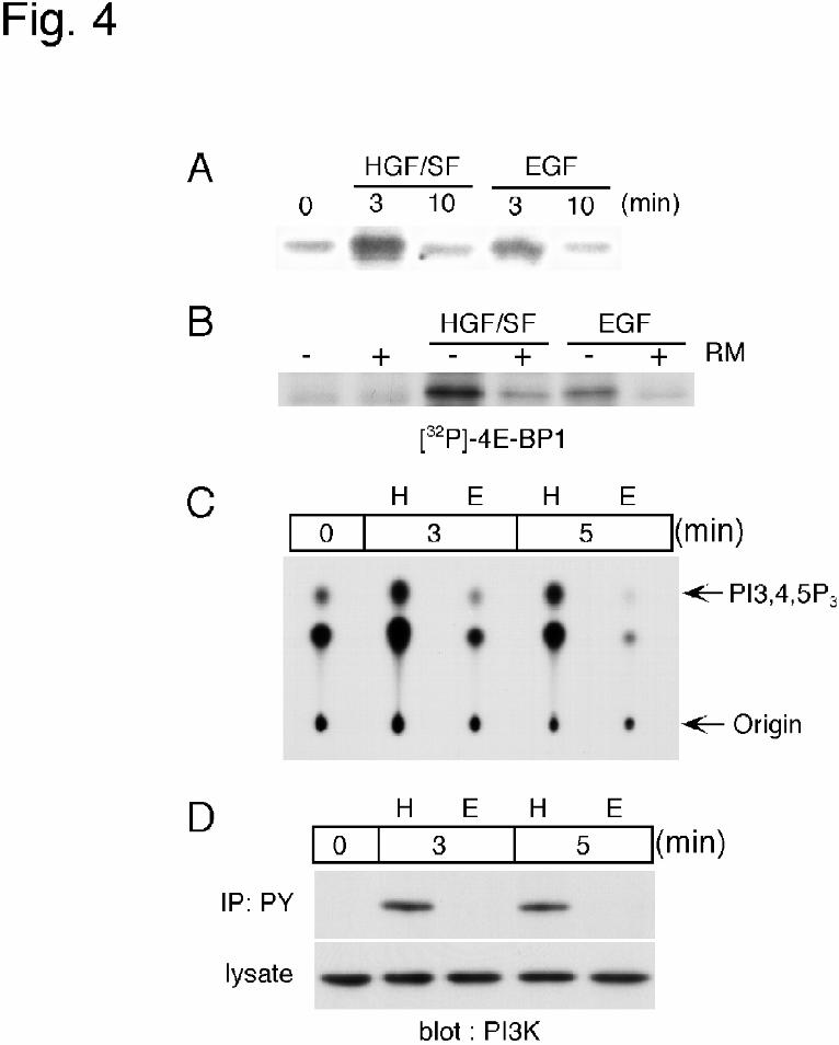
substrate for FRAP/mTOR. Incorporation of 32P was increased after 3 min of the
stimulation, and the band after the stimulation with HGF/SF was stronger than that obtained
with EGF (Fig. 4A). Pretreatment with rapamycin significantly decreased the incorporation
of 32P into 4E-BP1(Fig. 4B), suggesting that FRAP/mTOR was activated and could
phosphorylate 4E-BP1 in HGF/SF-treated MKN-74 cells.
Next, we determined the PI3-K activity in MKN74 cells. Cell lysates were
immunoprecipitated with anti-phosphotyrosine antibody and then used for an in vitro
kinase assay of PI3-K, since it is known that the kinase is activated via phosphorylation of
tyrosine residues in its regulatory subunit. As shown in Fig. 4C, when the cells were
stimulated with HGF/SF for 3 or 5 min, increased incorporation of 32P into the PI3,4,5P3
by guest on February 18, 2018http://w
ww
.jbc.org/D
ownloaded from

Regulation of c-fos translation 15�
was observed. In contrast, no PI3-K activity was detectable in EGF-stimulated cells.
Immunoblot analysis revealed that the regulatory subunit of PI3-K was not detected in
phosphotyrosine immunoprecipitates of EGF-treated cells (Fig. 4D), also suggesting that
PI3-K was not phosphorylated by EGF.
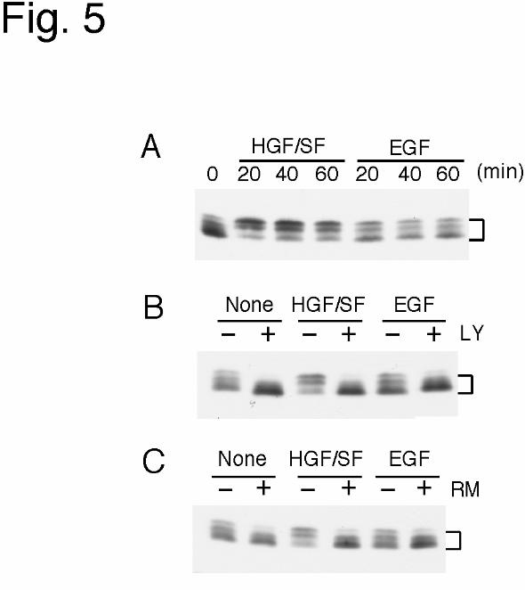
Translation initiation, which is regulated by a set of eukaryotic initiation factors
(eIF’s), is the rate-limiting step and a major target of translational control. Among
initiation factors, eIF4E directly binds to the 5’ cap structure of mRNA and is obligatory for
the start of cap-dependent translation initiation (45,46). 4E-BP1 is able to prevent binding
of eIF4E to the 5' cap structure by binding with eIF4E (44,47). Binding of 4E-BP1 to eIF4E
is reversible and dependent on the phosphorylation status of 4E-BP1; i.e.,
hypophosphorylated 4E-BP1 strongly interacts with eIF4E, whereas hyperphosphorylated
4E-BP1 does not. The phosphorylation of 4E-BP1 induced by extracellular stimuli is thus
suggested to trigger the release of 4E-BP1 from eIF4E, resulting in the stimulation of
translation initiation (48,49). Therefore, we examined the effect of HGF/SF and EGF on
the phosphorylation of 4E-BP1. Cells were treated with HGF/SF or EGF for 20, 40, or 60
min, and thereafter 4E-BP1 in the cell lysates was detected by immunoblotting analysis
(Fig. 5A). Among multiple bands of 4E-BP1 detected in each lane, the density of slower
migrating bands, which represent hyperphosphorylated forms of 4E-BP1, were increased by
HGF/SF, but not by EGF.
� Recently, the involvement of PI3-K- and FRAP/mTOR-dependent pathways in 4E-
BP1 phosphorylation has been established (41,50-52). FRAP/mTOR can directly
phosphorylate 4E-BP1; however, a direct link between the PI3-K pathway and
FRAP/mTOR remains to be proven. To examine the involvement of PI3-K and/or
by guest on February 18, 2018http://w
ww
.jbc.org/D
ownloaded from

Regulation of c-fos translation 16�
FRAP/mTOR in the phosphorylation of 4E-BP1 induced by HGF/SF, we treated cells with
a PI3-K inhibitor, LY294002 or with a FRAP/mTOR inhibitor, rapamycin, prior to the
incubation with HGF/SF or EGF, and assayed the phosphorylation of 4E-BP1 (Fig. 5B,C).
The HGF/SF-induced phosphorylation of 4E-BP1 was completely inhibited by LY294002
or rapamycin, indicating that this phosphorylation was dependent on the activities of PI3-K
and FRAP/mTOR.
We next examined the effect of these two inhibitors on c-Fos expression induced by
HGF/SF and EGF. HGF/SF-induced expression of c-Fos was inhibited by either
LY294002 or rapamycin, and its level was decreased to that induced by EGF (Fig. 6A,B
upper panel). We confirmed that neither inhibitor changed the level of c-fos mRNA
induced by HGF/SF or EGF by Northern blotting (Fig. 6A,B lower panel). These results
suggest that both PI3-K-dependent and FRAP/mTOR-dependent pathways are involved in
the translation of c-fos mRNA by HGF/SF in MKN74 cells.
� In the present study, 4E-BP1 was significantly phosphorylated after HGF/SF
treatment of MKN74 cells, and pretreatment with a PI3-K inhibitor, LY294002, or a
FRAP/mTOR inhibitor, rapamycin, inhibited 4E-BP1 phosphorylation (Fig.5). Moreover,
HGF/SF-induced c-Fos expression was decreased by LY294002 or rapamycin down to the
level found in EGF-stimulated cells (Fig.6). These results suggest that the HGF/SF signal
can induce phosphorylation of 4E-BP1 via the PI3-K- and FRAP/mTOR-dependent
pathways and thereby stimulate the translation initiation rate of c-fos mRNA.
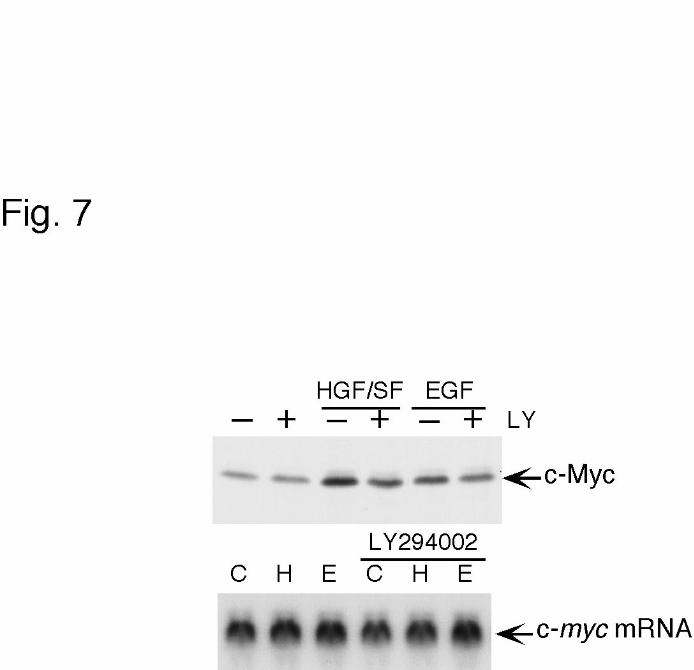
We further investigated whether c-fos mRNA was the only one molecule
specifically regulated in the translation process by PI3-K- and FRAP/mTOR-dependent
pathways in MKN74 cells. Recently, West et al.(53) reported that translation induction of
by guest on February 18, 2018http://w
ww
.jbc.org/D
ownloaded from
Regulation of c-fos translation 17�
c-myc was activated by 4E-BP1 phosphorylation. Therefore, we examined the change in
expression levels of c-myc mRNA and c-Myc protein after the treatment of MKN74 cells
with HGF/SF or EGF (Fig 7). The mRNA level of c-myc was not changed irrespective of
the stimulus used. The expression of c-Myc protein, however, was increased after the
treatment with HGF/SF and EGF, although the expression level in HGF/SF-stimulated cells
was twofold of that in EGF-stimulated cells. In addition, the inhibitor of PI3-K decreased
the c-Myc level in HGF/SF-stimulated cells down to the level in EGF-stimulated cells.
These results are consistent with the observation we showed in this report as to the
regulation of c-Fos induction by HGF/SF.
In the case that the PI3-K- and FRAP/mTOR-dependent pathways are not activated,
as in EGF-stimulated MKN74 cells, c-fos mRNA would not be translated effectively,
resulting in less accumulation of c-Fos protein, no matter how great an amount of mRNA is
transcribed. The present results suggest the notion that the activation of signaling pathways
affecting the translation initiation is essential, as well as the activation of transcription, for
the induction of c-Fos to an expression level sufficient for stimulating cell growth.
To test this notion, we examined the expression levels of c-fos mRNA and c-Fos
protein in another type of human cell line, foreskin fibroblast FS-4 cells. It is known that
proliferation of FS-4 cells is stimulated when the quiescent cells cultured under serum-
starved condition are treated with a cytokine, e.g., tumor necrosis factor (TNF) (54). TNF
increased the level of c-fos mRNA in quiescent FS-4 cells (Fig.8B), as Lin and Vilcek
reported (36). In these cells, the expression level of c-Fos was also increased by TNF, and
this increase was inhibited by LY294002 and rapamycin (Fig.8A). In contrast, the c-fos
mRNA level was not changed by the inhibitors as in the case of HGF/SF-stimulated
by guest on February 18, 2018http://w
ww
.jbc.org/D
ownloaded from

Regulation of c-fos translation 18�
MKN74 cells (Fig. 6). 4E-BP1 phosphorylation was significantly increased in TNF-
stimulated FS-4 cells, and was inhibited by LY294002 or rapamycin (Fig.8C). These results
indicate that our observation regarding regulation of c-Fos translation in MKN74 cells was
particular to neither the cell line or HGF/SF, and could support the notion that the
translation initiation of c-Fos as well as the activation of the gene transcription is regulated
when cell proliferation is stimulated. From all of the data taken together, we propose the
concept that the signaling pathway leading to 4E-BP1 phosphorylation in concert with the
Ras-MAP kinase pathway, which activates the transcription of c-fos, is necessary for the
functional activation for c-Fos-mediated transcription of target genes.
The effect of growth factors on cell proliferation generally depends on individual
cell lines and factors. In the case of MKN74 cells, EGF was not able to activate PI3-K,
although many reports have shown that the EGF receptor can transmit signals to activate
PI3-K(55,56). Adapter proteins such as Gab1 (25), c-Cbl (55), and a member of the EGF
receptor family, ErbB3 (56), are necessary for association between EGF receptor and PI3-
K, suggesting a possibility that the activated EGF-receptor may not be able to recruit an
adapter protein due to lack of the adapter or some mutation in the EGF receptor in these
cells. As is the case for the EGF effect on MKN74 cells, the differential actions of a given
factor on the proliferation of cells might reflect the susceptibility of a particular signaling
molecule involved in translation stimulation in individual cell lines.
by guest on February 18, 2018http://w
ww
.jbc.org/D
ownloaded from
Regulation of c-fos translation 19�
References
�
1. Sassone-Corsi, P., and Verma, I. M. (1987) Nature 326, 507-510
2. Greenberg, M. E., Ziff, E. B., and Greene, L. A. (1986) Science 234, 80-83
3. Shaw, P. E., Schroter, H., and Nordheim, A. (1989) Cell 56, 563-572
4. Tsurumi, C., Ishida, N., Tamura, T., Kakizuka, A., Nishida, E., Okumura, E., Kishimoto,
T., Inagaki, M., Okazaki, K., Sagata, N., Ichikawa, A., and Tanaka, K. (1995) Mol. Cell.
Biol. 15, 5682-5687
5. Stancovski, I., Gonen, H., Orian, A., Schwartz, A. L., and Ciechanover, A. (1995) Mol.
Cell. Biol. 15, 7106-7116
6. Okazaki, K., and Sagata, N. (1995) EMBO J. 14, 5048-5059
7. Brown, E. J., and Schreiber, S. L. (1996) Cell 86, 517-520
8. Pain, V. M. (1996) Eur. J. Biochem. 236, 747-771
9. Kleijn, M., Scheper, G. C., Voorma, H. O., and Thomas, A. A. (1998) Eur. J. Biochem.
253, 531-544
10. Gille, H., Sharrocks, A. D., and Shaw, P. E. (1992) Nature 358, 414-417
11. Marais, R., Wynne, J., and Treisman, R. (1993) Cell 73, 381-393
12. Gille, H., Kortenjann, M., Thomae, O., Moomaw, C., Slaughter, C., Cobb, M. H., and
Shaw, P. E. (1995) EMBO J. 14, 951-962
13. Naldini, L., Vigna, E., Narsimhan, R. P., Gaudino, G., Zarnegar, R., Michalopoulos, G.
K., and Comoglio, P. M. (1991) Oncogene 6, 501-504
14. Bottaro, D. P., Rubin, J. S., Faletto, D. L., Chan, A. M., Kmiecik, T. E., Vande Woude,
G. F., and Aaronson, S. A. (1991) Science 251, 802-804
by guest on February 18, 2018http://w
ww
.jbc.org/D
ownloaded from
Regulation of c-fos translation 20�
15. Ponzetto, C., Bardelli, A., Maina, F., Longati, P., Panayotou, G., Dhand, R., Waterfield,
M. D., and Comoglio, P. M. (1993) Mol. Cell. Biol. 13, 4600-4608
16. Graziani, A., Gramaglia, D., dalla Zonca, P., and Comoglio, P. M. (1993) J. Biol.
Chem. 268, 9165-9168
17. Weidner, K. M., Sachs, M., and Birchmeier, W. (1993) J. Cell. Biol. 121, 145-154
18. Ponzetto, C., Bardelli, A., Zhen, Z., Maina, F., dalla Zonca, P., Giordano, S., Graziani,
A., Panayotou, G., and Comoglio, P. M. (1994) Cell 77, 261-271
19. Weidner, K. M., Di Cesare, S., Sachs, M., Brinkmann, V., Behrens, J., and Birchmeier,
W. (1996) Nature 384, 173-176
20. Bardelli, A., Longati, P., Gramaglia, D., Stella, M. C., and Comoglio, P. M. (1997)
Oncogene 15, 3103-3111
21. Nishibe, S., Wahl, M. I., Rhee, S. G., and Carpenter, G. (1989) J. Biol. Chem. 264,
10335-10338
22. Margolis, B., Rhee, S. G., Felder, S., Mervic, M., Lyall, R., Levitzki, A., Ullrich, A.,
Zilberstein, A., and Schlessinger, J. (1989) Cell 57, 1101-1107
23. Meisenhelder, J., Suh, P. G., Rhee, S. G., and Hunter, T. (1989) Cell 57, 1109-1122
24. Ruff-Jamison, S., McGlade, J., Pawson, T., Chen, K., and Cohen, S. (1993) J. Biol.
Chem. 268, 7610-7612
25. Holgado-Madruga, M., Emlet, D. R., Moscatello, D. K., Godwin, A. K., and Wong, A.
J. (1996) Nature 379, 560-564
26. Khwaja, A., Lehmann, K., Marte, B. M., and Downward, J. (1998) J. Biol. Chem. 273,
18793-18801
27. Chatani, Y., Itoh, A., Tanaka, E., Hattori, A., Nakamura, T., and Kohno, M. (1992)
by guest on February 18, 2018http://w
ww
.jbc.org/D
ownloaded from

Regulation of c-fos translation 21�
Biochem. Biophys. Res. Commun. 185, 860-866
28. Tanimura, S., Chatani, Y., Hoshino, R., Sato, M., Watanabe, S., Kataoka, T.,
Nakamura, T., and Kohno, M. (1998) Oncogene 17, 57-65
29. Nagamine, K., Shibamoto, S., Takeuchi, K., Miyazawa, K., Kitamura, N., Chatani, Y.,
Kohno, M., and Ito, F. (1996) Eur. J. Biochem. 236, 476-481
30. Bjorge, J. D., Chan, T. O., Antczak, M., Kung, H. J., and Fujita, D. J. (1990) Proc. Natl.
Acad. Sci. U. S. A. 87, 3816-3820
31. Rossomando, A. J., Payne, D. M., Weber, M. J., and Sturgill, T. W. (1989) Proc. Natl.
Acad. Sci. U. S. A. 86, 6940-6943
32. Miyazawa, K., Tsubouchi, H., Naka, D., Takahashi, K., Okigaki, M., Arakaki, N.,
Nakayama, H., Hirono, S., Sakiyama, O., Takahashi, K., Gohda, E., Daikuhara, Y., and
Kitamura, N. (1989) Biochem. Biophys. Res. Commun. 163, 967-973
33. Strain, A. J., Ismail, T., Tsubouchi, H., Arakaki, N., Hishida, T., Kitamura, N.,
Daikuhara, Y., and McMaster, P. (1991) J. Clin. Invest. 87, 1853-1857
34. Omura, S., Fujimoto, T., Otoguro, K., Matsuzaki, K., Moriguchi, R., Tanaka, H., and
Sasaki, Y. (1991) J. Antibiot. 44, 113-116
35. Omura, S., Matsuzaki, K., Fujimoto, T., Kosuge, K., Furuya, T., Fujita, S., and
Nakagawa, A. (1991) J. Antibiot. 44, 117-118
36. Lin, J.-X., and Vilcek, J. (1987) J. Biol. Chem. 262, 11908-11911
37. Tsujimoto, M., Yip, Y. K., and Vilcek, J. (1986) J. Immunol. 136, 2441-2444
38. Laemmli, U. K. (1970) Nature 227, 680-685
39. Shibamoto, S., Hayakawa, M., Hori, T., Oku, N., Miyazawa, K., Kitamura, N., and Ito,
F. (1992) Cell Struct. Funct. 17, 185-190
by guest on February 18, 2018http://w
ww
.jbc.org/D
ownloaded from
Regulation of c-fos translation 22�
40. Fenteany, G., Standaert, R. F., Lane, W. S., Choi, S., Corey, E. J., and Schreiber, S. L.
(1995) Science 268, 726-731
41. Mendez, R., Myers, M. G. Jr., White, M. F., and Rhoads, R. E. (1996) Mol. Cell. Biol.
16, 2857-2864
42. Gingras, A.-C., Kennedy, S. G., O'Leary, M. A., Sonenberg, N., and Hay, N. (1998)
Genes Dev. 12, 502-513
43. Brunn, G. J., Hudson, C. C., Sekulic, A., Williams, J. M., Hosoi, H., Houghton, P. J.,
Lawrence, J. C. Jr., and Abraham, R. T. (1997) Science 277, 99-101
44. Haghighat, A., Mader, S., Pause, A., and Sonenberg, N. (1995) EMBO J. 14, 5701-5709
45. Sonenberg, N., Morgan, M. A., Merrick, W. C., and Shatkin, A. J. (1978) Proc. Natl.
Acad. Sci. U. S. A. 75, 4843-4847
46. Rhoads, R. E. (1988) Trends Biochem. Sci. 13, 52-56
47. Mader, S., Lee, H., Pause, A., and Sonenberg, N. (1995) Mol. Cell. Biol. 15, 4990-4997
48. Pause, A., Belsham, G. J., Gingras, A.-C., Donze, O., Lin, T.-A., Lawrence, J. C. Jr.,
and Sonenberg, N. (1994) Nature 371, 762-767
49. Lin, T.-A., Kong, X., Haystead, T. A., Pause, A., Belsham, G., Sonenberg, N., and
Lawrence, J. C. Jr. (1994) Science 266, 653-656
50. von Manteuffel, S. R., Gingras, A.-C., Ming, X.-F., Sonenberg, N., and Thomas, G.
(1996) Proc. Natl. Acad. Sci. U. S. A. 93, 4076-4780
51. Lin, T.-A., Kong, X., Saltiel, A. R., Blackshear, P. J., and Lawrence, J. C. Jr. (1995) J.
Biol. Chem. 270, 18531-18538
52. Kleijn, M., Korthout, M. M., Voorma, H. O., and Thomas, A. A. (1996) FEBS Lett.
396, 165-171
by guest on February 18, 2018http://w
ww
.jbc.org/D
ownloaded from

Regulation of c-fos translation 23�
53. West, M. J., Stoneley, M., and Willis, A. E. (1998) Oncogene 17, 769-780
54. Vilcek, J., Palombella, V.J., Henriksen-DeStefano, D., Swenson, C., Feinman, R., Hirai,
M., and Tsujimoto, M.(1986) J. Exp. Med. 163, 632-643
55. Soltoff, S.P. and Cantley, L.C. (1996) J. Biol. Chem. 271, 563-567
56. Kim, H.-H., Sierke, S.L., and Koland, J.G. (1994) J. Biol. Chem. 269, 24747-24755
by guest on February 18, 2018http://w
ww
.jbc.org/D
ownloaded from

Regulation of c-fos translation 24�
�
Footnotes
�
1This work was supported in part by a Grant-in-Aid for scientific research from the
Ministry of Education, Science, and Culture of Japan.
2To whom correspondence should be addressed: Genentech Inc.,1 DNA Way, MS#40,
South San Francisco, CA 94080, E-mail: [email protected]
3Abbreviations used are the following: HGF/SF, hepatocyte growth factor/ scatter factor;
EGF, epidermal growth factor; eIF, eukaryotic initiation factor; 4E-BP1, eIF4E-binding
protein 1; PI3-K, phosphatidylinositol-3'-OH kinase; FRAP/mTOR, FKBP-rapamycin-
associated protein/ mammalian target of rapamycin; MAP-K, mitogen-activated protein
kinase.
Acknowledgments
We thank Mitsubishi-Tokyo Pharmaceuticals, Inc. for providing human HGF/SF, Dr. E.
Tahara for supplying MKN74 cells, Drs. K. Nishikawa and S. Tanimura for technical
advice, Drs. K. Miyazawa and M. Hayakawa for critical comments, and M. Urushihara and
S. Yasuda for technical assistance.�
by guest on February 18, 2018http://w
ww
.jbc.org/D
ownloaded from

Regulation of c-fos translation 25�
Figure legends
�
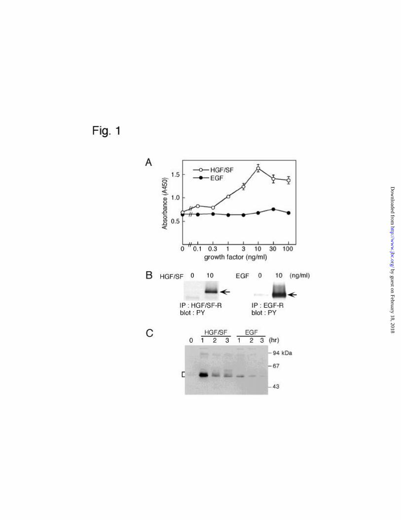
Figure 1
(A) Proliferation of MKN 74 cells in the presence of HGF/SF or EGF.
MKN74 cells were treated with each concentration of HGF/SF or EGF, as indicated, for 2
days. Cell proliferation of MKN74 treated with HGF/SF (open circle) and EGF (closed
circle) was determined by a colorimetric assay. Each point represents the mean of six
different samples. (B) Tyrosine phosphorylation of HGF/SF receptor and EGF receptor.
MKN74 cells were treated with 10 ng/ml HGF/SF (left panel) or 10 ng/ml EGF (right
panel). Immunoprecipitates of HGF/SF receptor and EGF receptor were probed with anti-
phosphotyrosine antibody. Similar results were obtained from three separate experiments.
Arrows indicate tyrosine-phosphorylated receptors. (C) Time course of c-Fos expression in
HGF/SF- and EGF-treated cells. MKN74 cells were treated with 10 ng/ml HGF/SF or 10
ng/ml EGF for the indicated times. Total cellular lysates were resolved by SDS-PAGE, and
the immunoblot was probed with anti-c-Fos antibody. Molecular size markers are indicated
at the right. Bracket indicates c-Fos protein.
�
Figure 2�
Effect of HGF/SF or EGF on the expression of c-fos.
Upper panel shows results of Northern blot analysis of c-fos mRNA in MKN74 cells. Total
RNA was isolated at the indicated times after the addition of 10 ng/ml HGF/SF or 10 ng/ml
EGF. Lower panel shows results of densitometric analysis of the bands in the upper panel.
�
by guest on February 18, 2018http://w
ww
.jbc.org/D
ownloaded from
Regulation of c-fos translation 26�
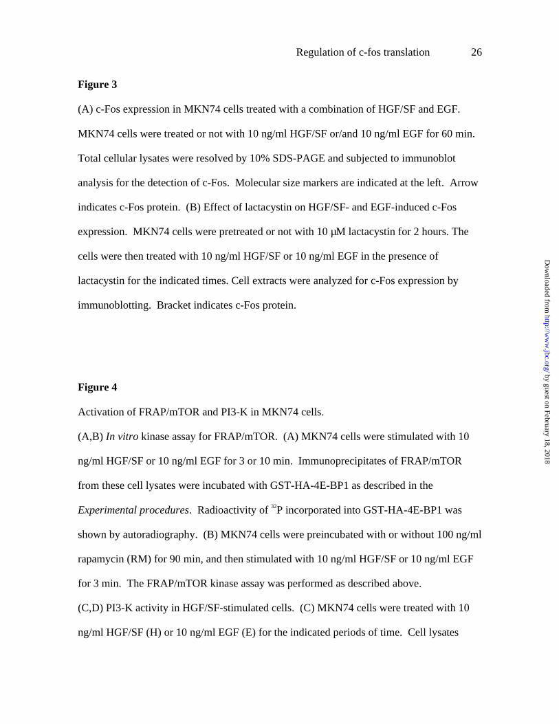
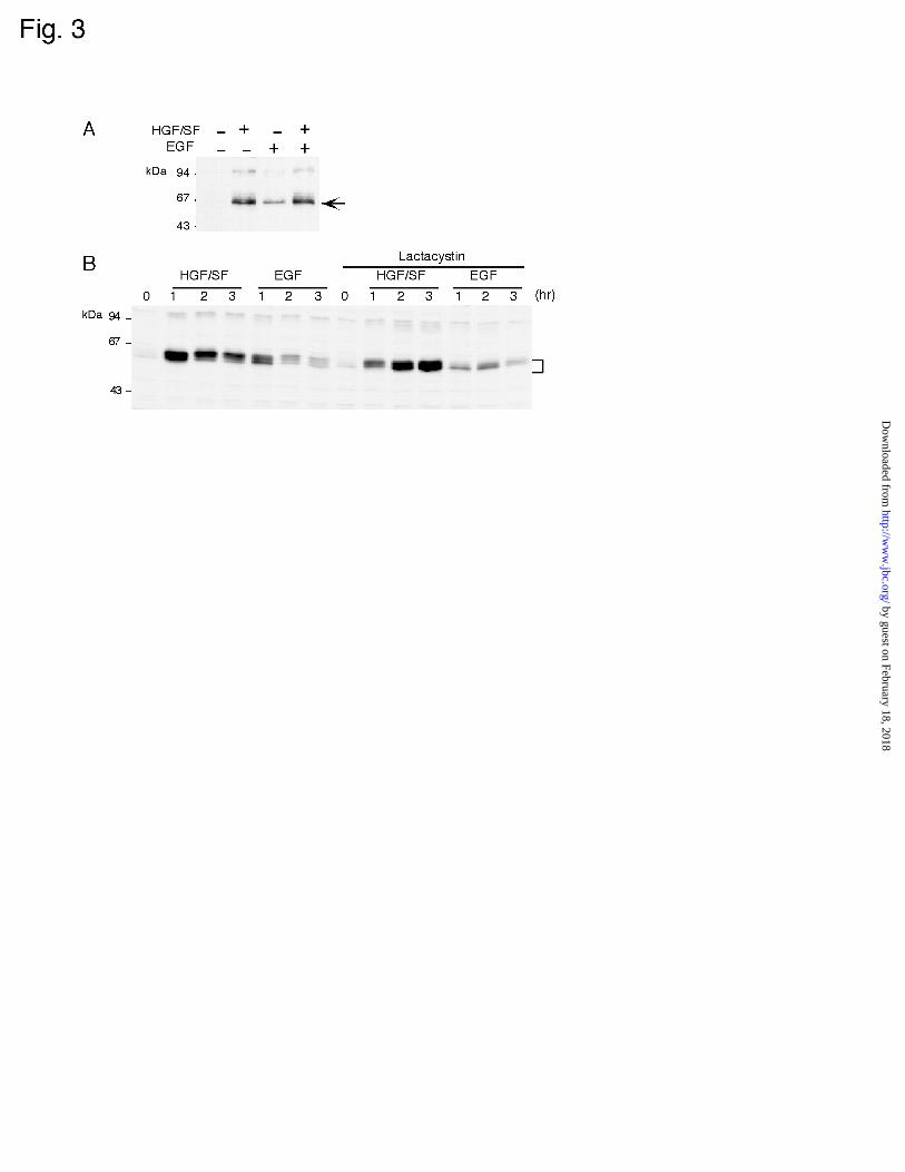
Figure 3
(A) c-Fos expression in MKN74 cells treated with a combination of HGF/SF and EGF.
MKN74 cells were treated or not with 10 ng/ml HGF/SF or/and 10 ng/ml EGF for 60 min.
Total cellular lysates were resolved by 10% SDS-PAGE and subjected to immunoblot
analysis for the detection of c-Fos. Molecular size markers are indicated at the left. Arrow
indicates c-Fos protein. (B) Effect of lactacystin on HGF/SF- and EGF-induced c-Fos
expression. MKN74 cells were pretreated or not with 10 µM lactacystin for 2 hours. The
cells were then treated with 10 ng/ml HGF/SF or 10 ng/ml EGF in the presence of
lactacystin for the indicated times. Cell extracts were analyzed for c-Fos expression by
immunoblotting. Bracket indicates c-Fos protein.
�
�
Figure 4
Activation of FRAP/mTOR and PI3-K in MKN74 cells.
(A,B) In vitro kinase assay for FRAP/mTOR. (A) MKN74 cells were stimulated with 10
ng/ml HGF/SF or 10 ng/ml EGF for 3 or 10 min. Immunoprecipitates of FRAP/mTOR
from these cell lysates were incubated with GST-HA-4E-BP1 as described in the
Experimental procedures. Radioactivity of 32P incorporated into GST-HA-4E-BP1 was
shown by autoradiography. (B) MKN74 cells were preincubated with or without 100 ng/ml
rapamycin (RM) for 90 min, and then stimulated with 10 ng/ml HGF/SF or 10 ng/ml EGF
for 3 min. The FRAP/mTOR kinase assay was performed as described above.
(C,D) PI3-K activity in HGF/SF-stimulated cells. (C) MKN74 cells were treated with 10
ng/ml HGF/SF (H) or 10 ng/ml EGF (E) for the indicated periods of time. Cell lysates
by guest on February 18, 2018http://w
ww
.jbc.org/D
ownloaded from

Regulation of c-fos translation 27�
were incubated with anti-phosphotyrosine (PY) antibody and used for the kinase reaction as
described in the Experimental procedures. The position of authentic phosphatidylinositol
3, 4, 5-triphosphate (PI3,4,5P3) is indicated. (D) PI3-K in PY immunoprecipitates and total
cell lysate was analyzed by immunoblotting.
�
�
Figure 5
(A) 4E-BP1 phosphorylation in response to growth factor treatment.
MKN74 cells were treated with 10 ng/ml HGF/SF or EGF for the indicated time periods,
and the resulting cell extracts were resolved by 13.5 % SDS-PAGE and subjected to
immunoblot analysis for the detection of 4E-BP1. Bracket indicates multiple bands of 4E-
BP1 representing differently phosphorylated forms of the molecule. (B,C) Inhibition of 4E-
BP1 phosphorylation by LY294002 (LY) and rapamycin (RM). MKN74 cells were
pretreated for 90 min with 10 µM LY294002 (B) or 100 ng/ml rapamycin (C), and then
incubated with 10 ng/ml HGF/SF or 10 ng/ml EGF in the presence of the inhibitor for 60
min. Bracket indicates multiple forms of 4E-BP1. – indicates incubation without inhibitor.�
�
�
Figure 6
Inhibition of c-Fos expression by LY294002 and rapamycin.
MKN74 cells were pretreated with 10 µM LY294002 (A) or 100 ng/ml rapamycin (B) for
90 min, and then incubated with 10 ng/ml HGF/SF or EGF for 60 min for immunoblotting
by guest on February 18, 2018http://w
ww
.jbc.org/D
ownloaded from
Regulation of c-fos translation 28�
analysis of c-Fos (upper panel). Total RNA was isolated after a 40-min stimulation with
either of the factors for Northern blotting analysis of c-fos (lower panel). C indicates no
stimulation with the factors.
�
�
Figure 7�
Effect of HGF/SF and EGF on the expression of c-Myc in MKN74 cells.
MKN74 cells were pretreated or not with 10 µM LY294002 (LY) for 90 min prior to the
stimulation with 10 ng/ml HGF/SF or EGF, and analyzed for c-Myc protein and c-myc
mRNA expression by immunoblotting (upper panel) and Northern blotting (lower panel),
respectively. C indicates no stimulation with the factors. The results shown are
representative of three independent experiments.
�
�
Figure 8
Inhibition of TNF-induced c-Fos expression by LY294002 and rapamycin in FS-4 cells.
FS-4 cells were pretreated with 10 µM LY294002 (LY) or 100 ng/ml rapamycin (RM) for
90 min and then incubated with 30 ng/ml TNF in the presence of the inhibitor for 60 min.
Cell extracts were prepared for immunoblotting analysis of c-Fos (A) and 4E-BP1 (C) and
Northern blotting analysis of c-fos (B). The cells were treated with TNF under the same
condition as described above except that the cells were incubated with TNF for 40 min.
by guest on February 18, 2018http://w
ww
.jbc.org/D
ownloaded from


Naomi Kitamura and Fumiaki ItoKenji Takeuchi, Sayumi Shibamoto, Kentaro Nagamine, Ichiro Shigemori, Satoshi Omura,
transient increase in expression of c-Fos proteinSignaling pathways leading to transcription and translation cooperatively regulate the
published online May 14, 2001J. Biol. Chem.
10.1074/jbc.M102704200Access the most updated version of this article at doi:
Alerts:
When a correction for this article is posted•
When this article is cited•
to choose from all of JBC's e-mail alertsClick here
by guest on February 18, 2018http://w
ww
.jbc.org/D
ownloaded from