regional 12 lead ecg and stemi triage short … · 12 lead ecg and stemi triage protocols 5.1 &...
TRANSCRIPT
All levels / MedicsAll levels / Medics
12 Lead ECG and STEMI Triage12 Lead ECG and STEMI TriageProtocols 5.1 & 4.27Protocols 5.1 & 4.27
Revised 3/3/2008
References provided on last slide
Revised 3/3/2008
References provided on last slide
All levelsAll levels
ObjectivesObjectives
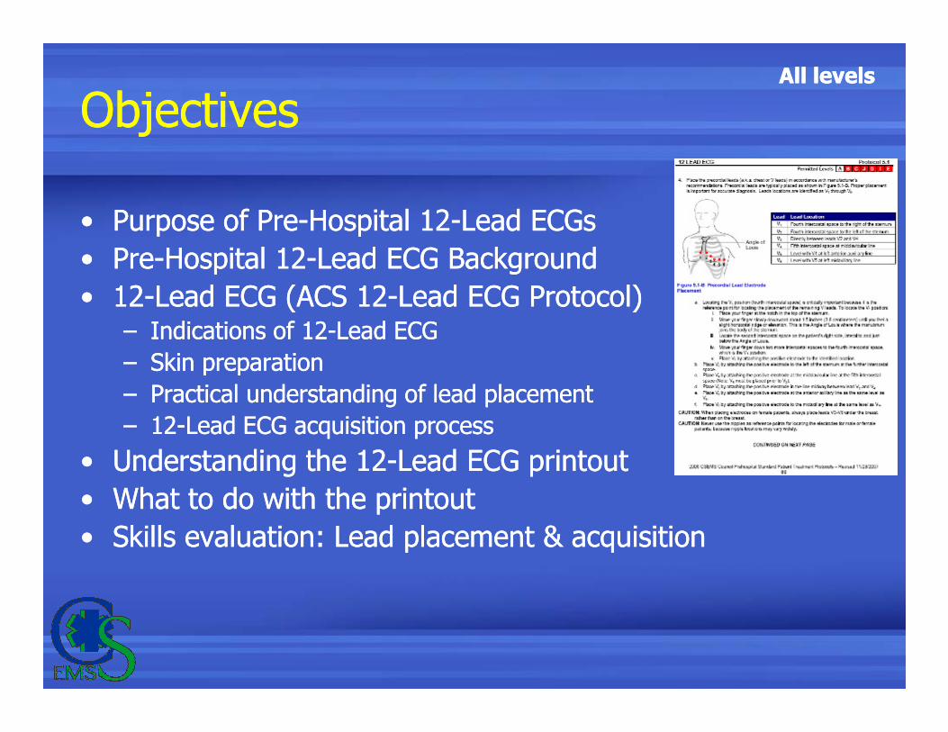
• Purpose of Pre-Hospital 12-Lead ECGs
• Pre-Hospital 12-Lead ECG Background
• 12-Lead ECG (ACS 12-Lead ECG Protocol)– Indications of 12-Lead ECG
– Skin preparation
• Purpose of Pre-Hospital 12-Lead ECGs
• Pre-Hospital 12-Lead ECG Background
• 12-Lead ECG (ACS 12-Lead ECG Protocol)– Indications of 12-Lead ECG
– Skin preparation– Skin preparation
– Practical understanding of lead placement
– 12-Lead ECG acquisition process
• Understanding the 12-Lead ECG printout
• What to do with the printout
• Skills evaluation: Lead placement & acquisition
– Skin preparation
– Practical understanding of lead placement
– 12-Lead ECG acquisition process
• Understanding the 12-Lead ECG printout
• What to do with the printout
• Skills evaluation: Lead placement & acquisition
All levelsAll levels
Pre-Hospital 12-Lead ECG PurposePre-Hospital 12-Lead ECG Purpose
• What is this all for?– Earlier selection and transport to definitive care facility(5)
• STEMI Triage Protocol will determine the receiving facility
– Speed diagnosis & treatment to reduce the amount of tissue death by reperfusing the heart sooner(5)
• Fibrinolysis (meds) or Percutaneous Coronary Intervention (PCI)
• What is this all for?– Earlier selection and transport to definitive care facility(5)
• STEMI Triage Protocol will determine the receiving facility
– Speed diagnosis & treatment to reduce the amount of tissue death by reperfusing the heart sooner(5)
• Fibrinolysis (meds) or Percutaneous Coronary Intervention (PCI)• Fibrinolysis (meds) or Percutaneous Coronary Intervention (PCI)
– Overall decrease in mortality(5)
• Less deaths from AMI overall
• VF & VT most likely to occur in first 4 hours after onset of s/s
– Preserve Left Ventricular function(5)
• Thus preserving lifestyle for patient (with less CHF)
– Decrease long term overall incidence of CHF and its financial impact on society
• Fibrinolysis (meds) or Percutaneous Coronary Intervention (PCI)
– Overall decrease in mortality(5)
• Less deaths from AMI overall
• VF & VT most likely to occur in first 4 hours after onset of s/s
– Preserve Left Ventricular function(5)
• Thus preserving lifestyle for patient (with less CHF)
– Decrease long term overall incidence of CHF and its financial impact on society
All levelsAll levels
Pre-Hospital 12-Lead ECG PurposePre-Hospital 12-Lead ECG Purpose
• What is this not for?
– Ruling out an AMI (heart attack)
• Only about 50% of AMIs have ST Segment Elevation(1)
– Deciding how to treat your patient
• Do not let the 12 lead decide how you are going to proceed with
• What is this not for?
– Ruling out an AMI (heart attack)
• Only about 50% of AMIs have ST Segment Elevation(1)
– Deciding how to treat your patient
• Do not let the 12 lead decide how you are going to proceed with • Do not let the 12 lead decide how you are going to proceed with treatment
• Follow the Adult –Cardiac: Chest Pain (Suspected Myocardial Event) Protocol
– Providing Oxygen, Aspirin, assisting with prescribed Nitroglycerin and calling for ALS are all required (within indications and contraindications) by the Chest Pain (Non-Traumatic) protocol regardless of 12 lead ECG interpretation
– ALS: INT or IV, Nitroglycerin, Morphine and Metoprolol as necessary
• Do not let the 12 lead decide how you are going to proceed with treatment
• Follow the Adult –Cardiac: Chest Pain (Suspected Myocardial Event) Protocol
– Providing Oxygen, Aspirin, assisting with prescribed Nitroglycerin and calling for ALS are all required (within indications and contraindications) by the Chest Pain (Non-Traumatic) protocol regardless of 12 lead ECG interpretation
– ALS: INT or IV, Nitroglycerin, Morphine and Metoprolol as necessary
All levelsAll levels
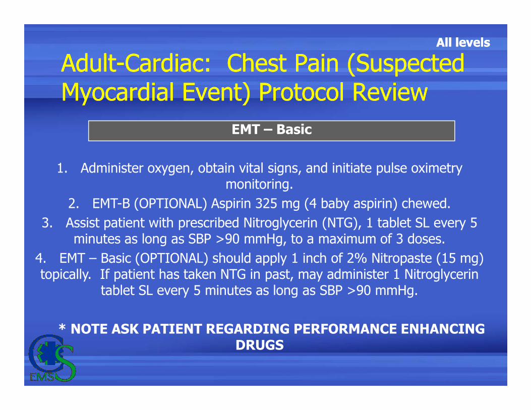
Adult-Cardiac: Chest Pain (Suspected Myocardial Event) Protocol ReviewAdult-Cardiac: Chest Pain (Suspected Myocardial Event) Protocol Review
EMT – Basic
1. Administer oxygen, obtain vital signs, and initiate pulse oximetry monitoring.
2. EMT-B (OPTIONAL) Aspirin 325 mg (4 baby aspirin) chewed.2. EMT-B (OPTIONAL) Aspirin 325 mg (4 baby aspirin) chewed.
3. Assist patient with prescribed Nitroglycerin (NTG), 1 tablet SL every 5 minutes as long as SBP >90 mmHg, to a maximum of 3 doses.
4. EMT – Basic (OPTIONAL) should apply 1 inch of 2% Nitropaste (15 mg) topically. If patient has taken NTG in past, may administer 1 Nitroglycerin
tablet SL every 5 minutes as long as SBP >90 mmHg.
* NOTE ASK PATIENT REGARDING PERFORMANCE ENHANCING DRUGS
All levelsAll levels
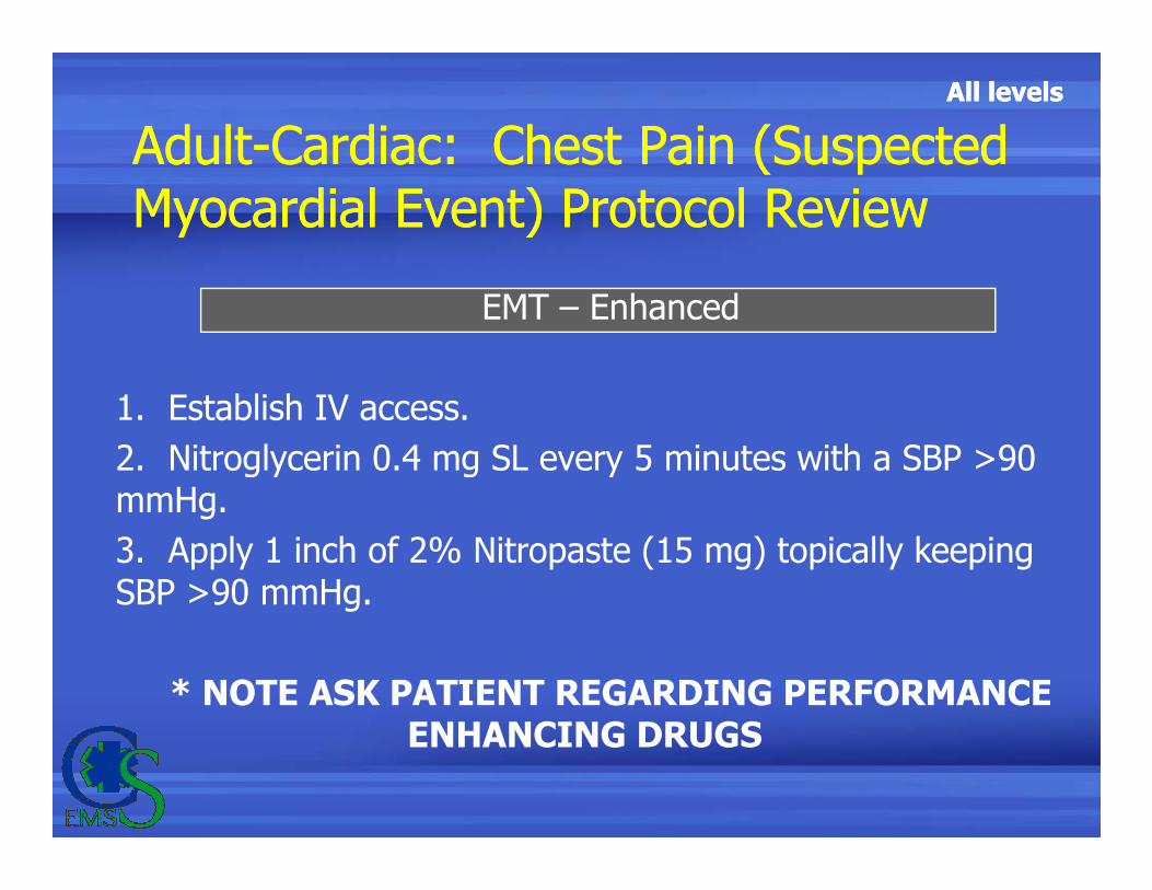
Adult-Cardiac: Chest Pain (Suspected Myocardial Event) Protocol ReviewAdult-Cardiac: Chest Pain (Suspected Myocardial Event) Protocol Review
EMT – Enhanced
1. Establish IV access.
2. Nitroglycerin 0.4 mg SL every 5 minutes with a SBP >90 2. Nitroglycerin 0.4 mg SL every 5 minutes with a SBP >90 mmHg.
3. Apply 1 inch of 2% Nitropaste (15 mg) topically keeping SBP >90 mmHg.
* NOTE ASK PATIENT REGARDING PERFORMANCE ENHANCING DRUGS
All levelsAll levels
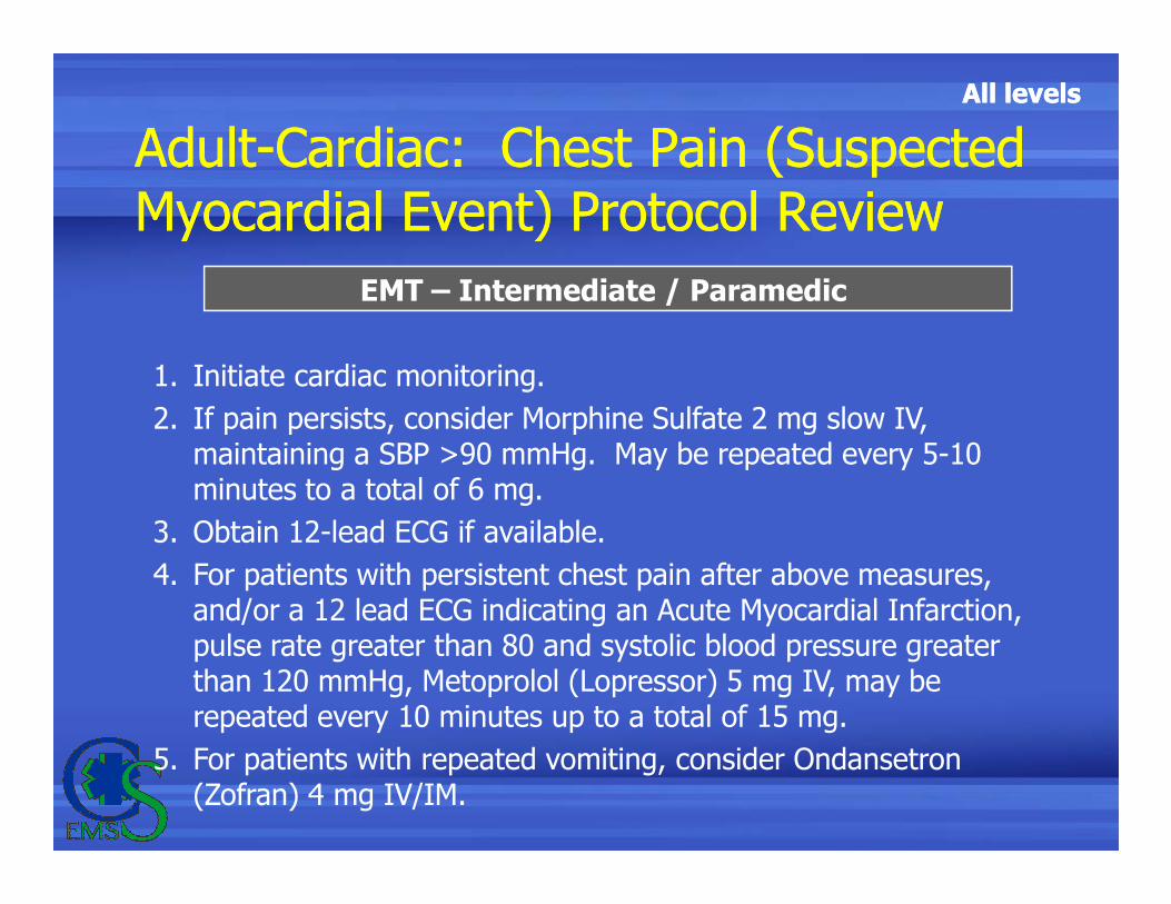
Adult-Cardiac: Chest Pain (Suspected Myocardial Event) Protocol ReviewAdult-Cardiac: Chest Pain (Suspected Myocardial Event) Protocol Review
EMT – Intermediate / Paramedic
1. Initiate cardiac monitoring.
2. If pain persists, consider Morphine Sulfate 2 mg slow IV, maintaining a SBP >90 mmHg. May be repeated every 5-10 maintaining a SBP >90 mmHg. May be repeated every 5-10 minutes to a total of 6 mg.
3. Obtain 12-lead ECG if available.
4. For patients with persistent chest pain after above measures, and/or a 12 lead ECG indicating an Acute Myocardial Infarction, pulse rate greater than 80 and systolic blood pressure greater than 120 mmHg, Metoprolol (Lopressor) 5 mg IV, may be repeated every 10 minutes up to a total of 15 mg.
5. For patients with repeated vomiting, consider Ondansetron (Zofran) 4 mg IV/IM.
All levelsAll levels
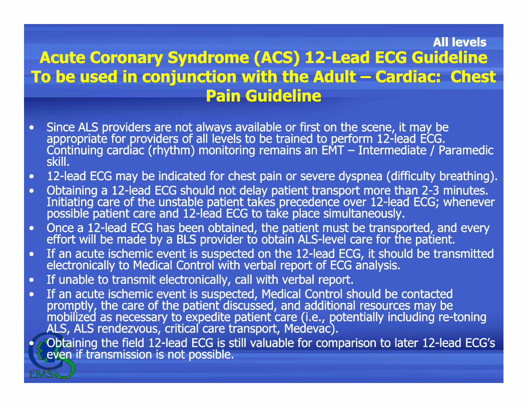
Acute Coronary Syndrome (ACS) 12-Lead ECG GuidelineTo be used in conjunction with the Adult – Cardiac: Chest
Pain Guideline
Acute Coronary Syndrome (ACS) 12-Lead ECG GuidelineTo be used in conjunction with the Adult – Cardiac: Chest
Pain Guideline
• Since ALS providers are not always available or first on the scene, it may be appropriate for providers of all levels to be trained to perform 12-lead ECG. Continuing cardiac (rhythm) monitoring remains an EMT – Intermediate / Paramedic skill.
• 12-lead ECG may be indicated for chest pain or severe dyspnea (difficulty breathing).• Obtaining a 12-lead ECG should not delay patient transport more than 2-3 minutes.
Initiating care of the unstable patient takes precedence over 12-lead ECG; whenever
• Since ALS providers are not always available or first on the scene, it may be appropriate for providers of all levels to be trained to perform 12-lead ECG. Continuing cardiac (rhythm) monitoring remains an EMT – Intermediate / Paramedic skill.
• 12-lead ECG may be indicated for chest pain or severe dyspnea (difficulty breathing).• Obtaining a 12-lead ECG should not delay patient transport more than 2-3 minutes.
Initiating care of the unstable patient takes precedence over 12-lead ECG; whenever Initiating care of the unstable patient takes precedence over 12-lead ECG; whenever possible patient care and 12-lead ECG to take place simultaneously.
• Once a 12-lead ECG has been obtained, the patient must be transported, and every effort will be made by a BLS provider to obtain ALS-level care for the patient.
• If an acute ischemic event is suspected on the 12-lead ECG, it should be transmitted electronically to Medical Control with verbal report of ECG analysis.
• If unable to transmit electronically, call with verbal report.• If an acute ischemic event is suspected, Medical Control should be contacted
promptly, the care of the patient discussed, and additional resources may be mobilized as necessary to expedite patient care (i.e., potentially including re-toning ALS, ALS rendezvous, critical care transport, Medevac).
• Obtaining the field 12-lead ECG is still valuable for comparison to later 12-lead ECG’s even if transmission is not possible.
Initiating care of the unstable patient takes precedence over 12-lead ECG; whenever possible patient care and 12-lead ECG to take place simultaneously.
• Once a 12-lead ECG has been obtained, the patient must be transported, and every effort will be made by a BLS provider to obtain ALS-level care for the patient.
• If an acute ischemic event is suspected on the 12-lead ECG, it should be transmitted electronically to Medical Control with verbal report of ECG analysis.
• If unable to transmit electronically, call with verbal report.• If an acute ischemic event is suspected, Medical Control should be contacted
promptly, the care of the patient discussed, and additional resources may be mobilized as necessary to expedite patient care (i.e., potentially including re-toning ALS, ALS rendezvous, critical care transport, Medevac).
• Obtaining the field 12-lead ECG is still valuable for comparison to later 12-lead ECG’s even if transmission is not possible.
All levelsAll levels
Acute Coronary Syndrome (ACS) 12-Lead ECG GuidelineTo be used in conjunction with the Adult – Cardiac: Chest
Pain Guideline
Acute Coronary Syndrome (ACS) 12-Lead ECG GuidelineTo be used in conjunction with the Adult – Cardiac: Chest
Pain Guideline
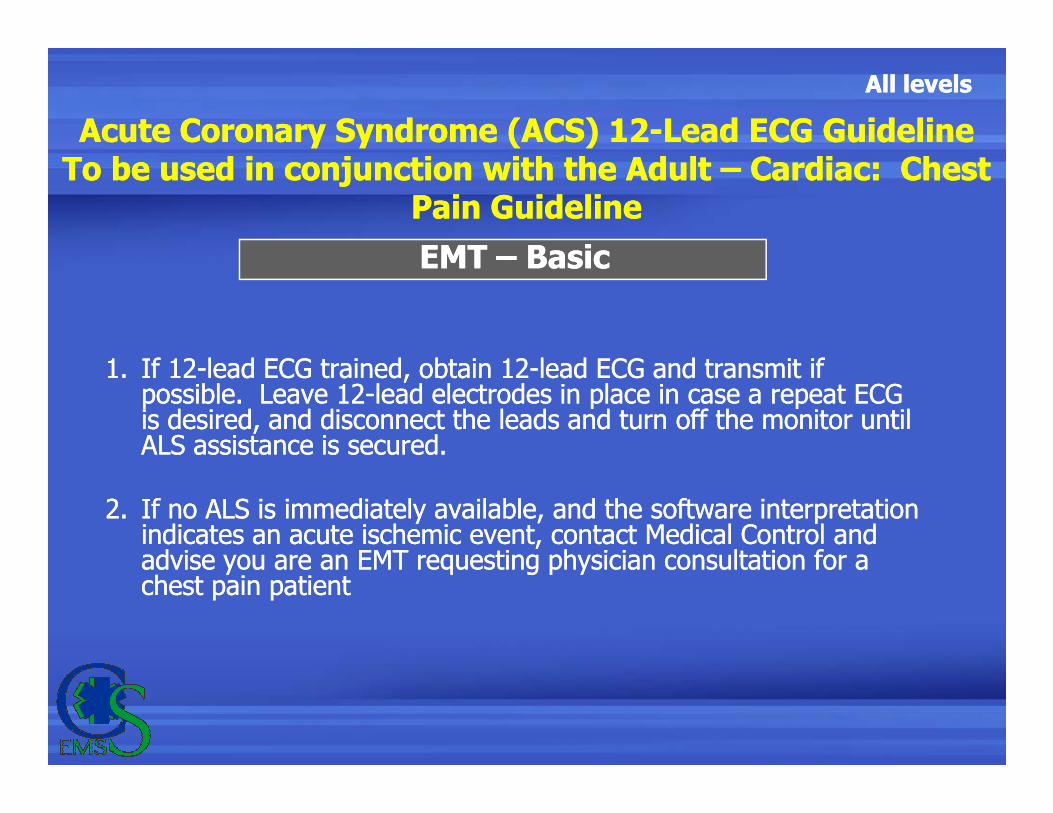
EMT – Basic
1. If 12-lead ECG trained, obtain 12-lead ECG and transmit if possible. Leave 12-lead electrodes in place in case a repeat ECG is desired, and disconnect the leads and turn off the monitor until
EMT – Basic
1. If 12-lead ECG trained, obtain 12-lead ECG and transmit if possible. Leave 12-lead electrodes in place in case a repeat ECG is desired, and disconnect the leads and turn off the monitor until possible. Leave 12-lead electrodes in place in case a repeat ECG is desired, and disconnect the leads and turn off the monitor until ALS assistance is secured.
2. If no ALS is immediately available, and the software interpretation indicates an acute ischemic event, contact Medical Control and advise you are an EMT requesting physician consultation for a chest pain patient
possible. Leave 12-lead electrodes in place in case a repeat ECG is desired, and disconnect the leads and turn off the monitor until ALS assistance is secured.
2. If no ALS is immediately available, and the software interpretation indicates an acute ischemic event, contact Medical Control and advise you are an EMT requesting physician consultation for a chest pain patient
All levelsAll levels
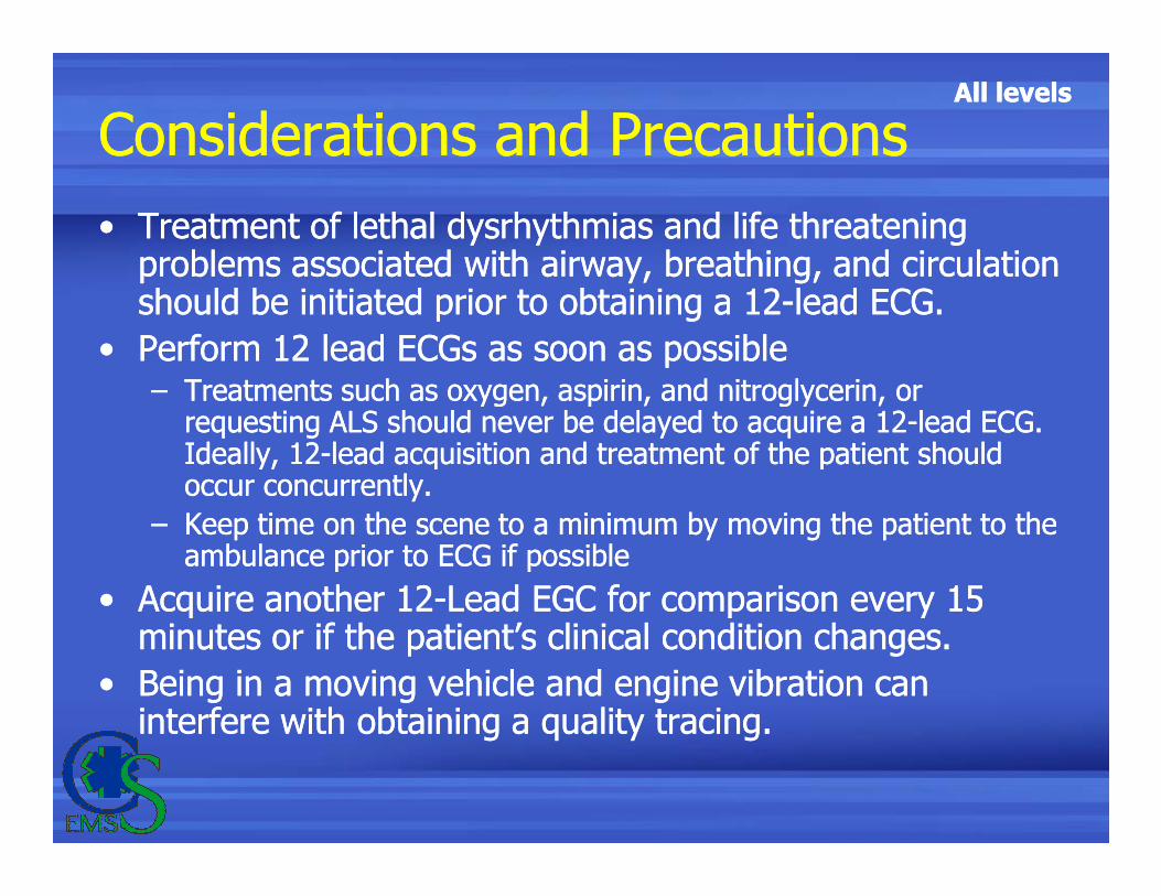
Considerations and PrecautionsConsiderations and Precautions
• Treatment of lethal dysrhythmias and life threatening problems associated with airway, breathing, and circulation should be initiated prior to obtaining a 12-lead ECG.
• Perform 12 lead ECGs as soon as possible– Treatments such as oxygen, aspirin, and nitroglycerin, or
requesting ALS should never be delayed to acquire a 12-lead ECG. Ideally, 12-lead acquisition and treatment of the patient should
• Treatment of lethal dysrhythmias and life threatening problems associated with airway, breathing, and circulation should be initiated prior to obtaining a 12-lead ECG.
• Perform 12 lead ECGs as soon as possible– Treatments such as oxygen, aspirin, and nitroglycerin, or
requesting ALS should never be delayed to acquire a 12-lead ECG. Ideally, 12-lead acquisition and treatment of the patient should Ideally, 12-lead acquisition and treatment of the patient should occur concurrently.
– Keep time on the scene to a minimum by moving the patient to the ambulance prior to ECG if possible
• Acquire another 12-Lead EGC for comparison every 15 minutes or if the patient’s clinical condition changes.
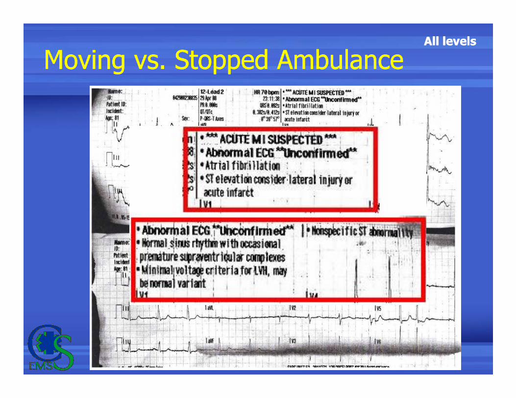
• Being in a moving vehicle and engine vibration can interfere with obtaining a quality tracing.
Ideally, 12-lead acquisition and treatment of the patient should occur concurrently.
– Keep time on the scene to a minimum by moving the patient to the ambulance prior to ECG if possible
• Acquire another 12-Lead EGC for comparison every 15 minutes or if the patient’s clinical condition changes.
• Being in a moving vehicle and engine vibration can interfere with obtaining a quality tracing.

All levelsAll levels
Lead Placement GeneralitiesLead Placement Generalities
• Prepare all equipment in advance (i.e. check your equipment routinely)
• Skin preparation issues– Dirt, dead skin, oil, sweat, hair on the skin can interfere with
obtaining a quality tracing
• Prep the skin
• Prepare all equipment in advance (i.e. check your equipment routinely)
• Skin preparation issues– Dirt, dead skin, oil, sweat, hair on the skin can interfere with
obtaining a quality tracing
• Prep the skin– Dry sweat and water
– Buff the placement area• Foam dots… abrasive pad on the removable cover of some electrodes
• Alcohol preps
– Buff or rub briskly… removes dirt and oil, stimulates capillary blood to decrease diaphoresis. A clean 4x4 gauze works great!
– Diaphoresis… use “all hands” if necessary
– Shave… battery clippers do work best• Disposable razors clog up with one stroke
– Dry sweat and water
– Buff the placement area• Foam dots… abrasive pad on the removable cover of some electrodes
• Alcohol preps
– Buff or rub briskly… removes dirt and oil, stimulates capillary blood to decrease diaphoresis. A clean 4x4 gauze works great!
– Diaphoresis… use “all hands” if necessary
– Shave… battery clippers do work best• Disposable razors clog up with one stroke
All levelsAll levels
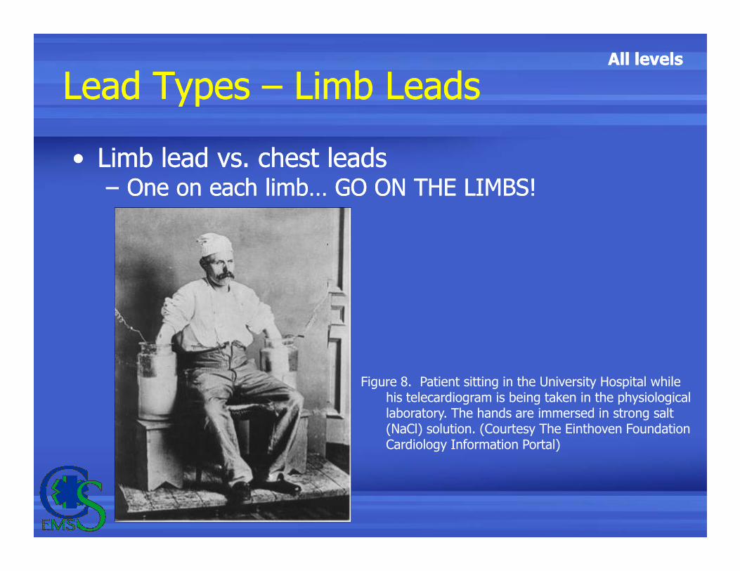
Lead Types – Limb LeadsLead Types – Limb Leads
• Limb lead vs. chest leads– One on each limb… GO ON THE LIMBS!
• Limb lead vs. chest leads– One on each limb… GO ON THE LIMBS!
Figure 8. Patient sitting in the University Hospital while his telecardiogram is being taken in the physiological laboratory. The hands are immersed in strong salt (NaCl) solution. (Courtesy The Einthoven Foundation Cardiology Information Portal)
All levelsAll levels
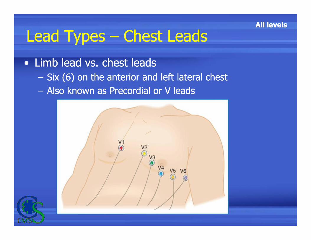
Lead Types – Chest LeadsLead Types – Chest Leads
• Limb lead vs. chest leads
– Six (6) on the anterior and left lateral chest
– Also known as Precordial or V leads
• Limb lead vs. chest leads
– Six (6) on the anterior and left lateral chest
– Also known as Precordial or V leads
All levelsAll levels
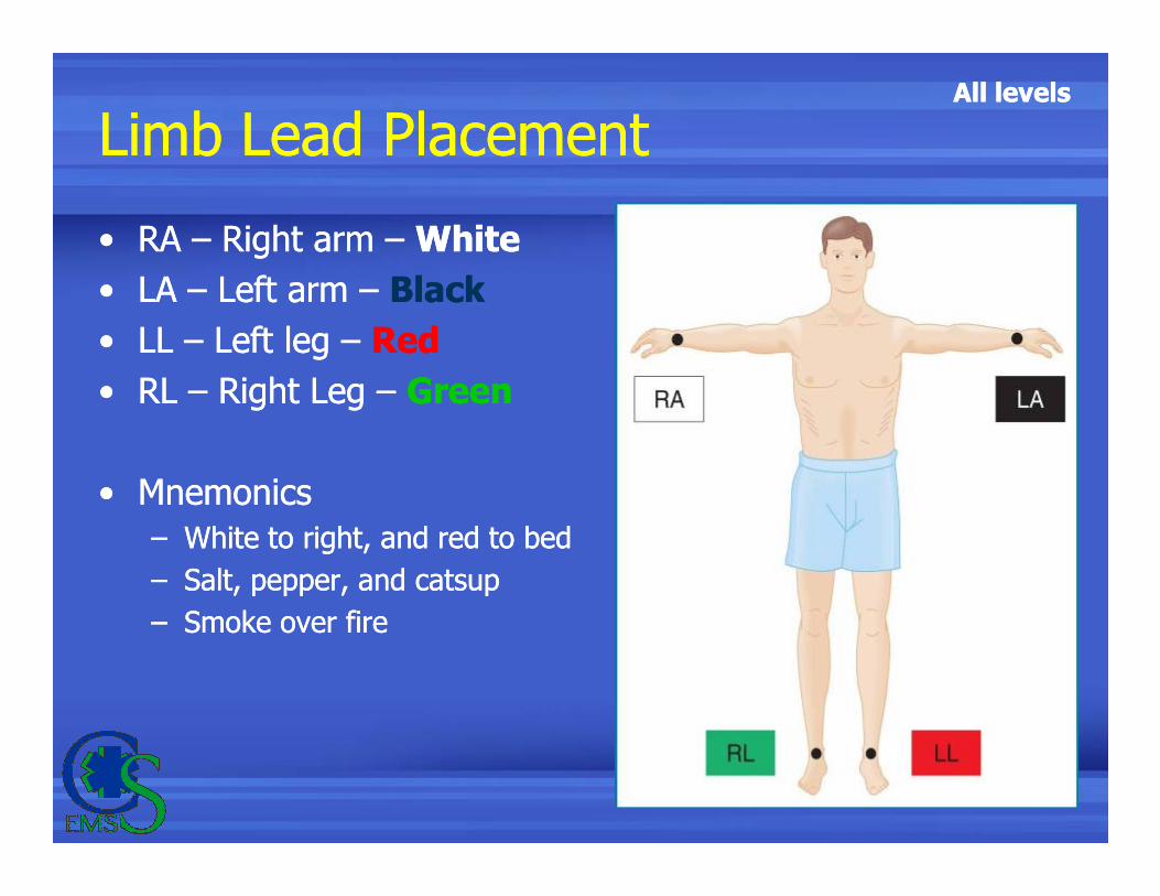
Limb Lead PlacementLimb Lead Placement
• RA – Right arm – White
• LA – Left arm – Black
• LL – Left leg – Red
• RL – Right Leg – Green
• RA – Right arm – White
• LA – Left arm – Black
• LL – Left leg – Red
• RL – Right Leg – Green
• Mnemonics
– White to right, and red to bed
– Salt, pepper, and catsup
– Smoke over fire
• Mnemonics
– White to right, and red to bed
– Salt, pepper, and catsup
– Smoke over fire
All levelsAll levels
Limb Lead PlacementLimb Lead Placement
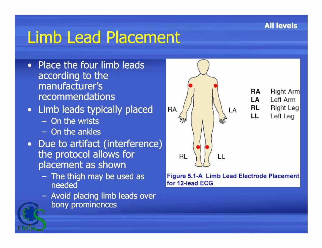
• Place the four limb leads according to the manufacturer’s recommendations
• Limb leads typically placed – On the wrists
• Place the four limb leads according to the manufacturer’s recommendations
• Limb leads typically placed – On the wrists– On the wrists
– On the ankles
• Due to artifact (interference) the protocol allows for placement as shown– The thigh may be used as
needed
– Avoid placing limb leads over bony prominences
– On the wrists
– On the ankles
• Due to artifact (interference) the protocol allows for placement as shown– The thigh may be used as
needed
– Avoid placing limb leads over bony prominences
All levelsAll levels
Chest Lead PlacementChest Lead Placement
All levelsAll levels
Chest Lead PlacementChest Lead Placement
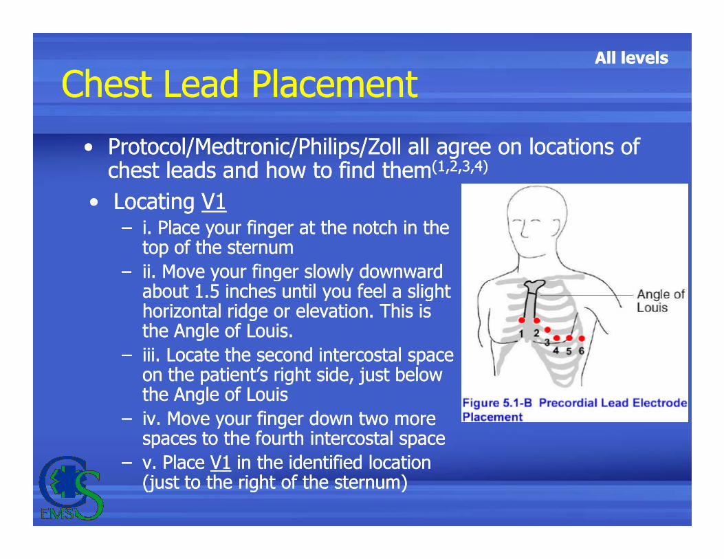
• Locating V1– i. Place your finger at the notch in the
top of the sternum
– ii. Move your finger slowly downward about 1.5 inches until you feel a slight
• Locating V1– i. Place your finger at the notch in the
top of the sternum
– ii. Move your finger slowly downward about 1.5 inches until you feel a slight
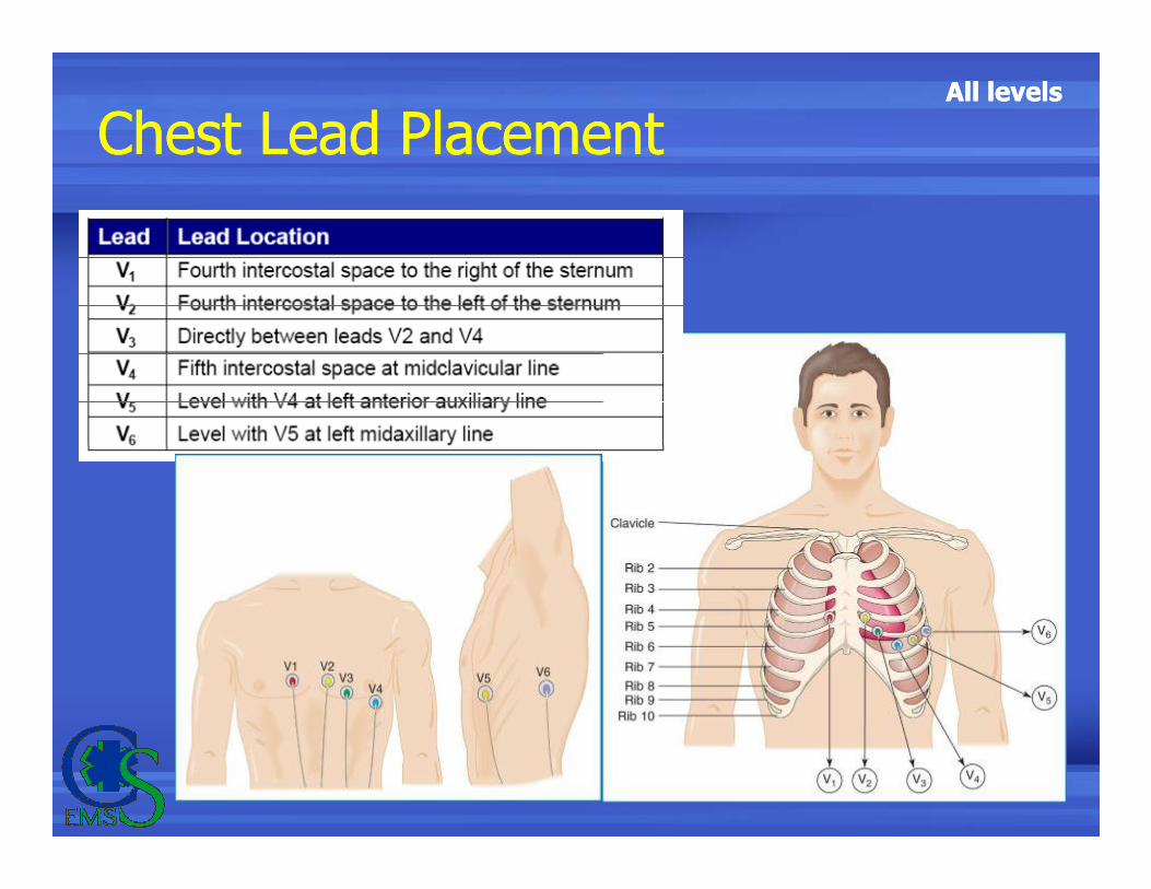
• Protocol/Medtronic/Philips/Zoll all agree on locations of chest leads and how to find them(1,2,3,4)
• Protocol/Medtronic/Philips/Zoll all agree on locations of chest leads and how to find them(1,2,3,4)
– ii. Move your finger slowly downward about 1.5 inches until you feel a slight horizontal ridge or elevation. This is the Angle of Louis.
– iii. Locate the second intercostal space on the patient’s right side, just below the Angle of Louis
– iv. Move your finger down two more spaces to the fourth intercostal space
– v. Place V1 in the identified location (just to the right of the sternum)
All levelsAll levels
Chest Lead PlacementChest Lead Placement
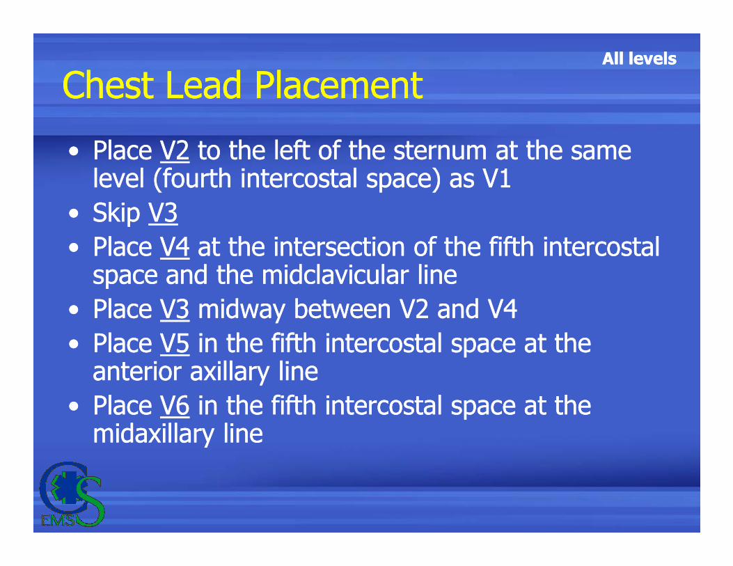
• Place V2 to the left of the sternum at the same level (fourth intercostal space) as V1
• Skip V3
• Place V4 at the intersection of the fifth intercostal space and the midclavicular line
• Place V2 to the left of the sternum at the same level (fourth intercostal space) as V1
• Skip V3
• Place V4 at the intersection of the fifth intercostal space and the midclavicular linespace and the midclavicular line
• Place V3 midway between V2 and V4
• Place V5 in the fifth intercostal space at the anterior axillary line
• Place V6 in the fifth intercostal space at the midaxillary line
space and the midclavicular line
• Place V3 midway between V2 and V4
• Place V5 in the fifth intercostal space at the anterior axillary line
• Place V6 in the fifth intercostal space at the midaxillary line
All levelsAll levels
Notes on Chest Lead PlacementNotes on Chest Lead Placement
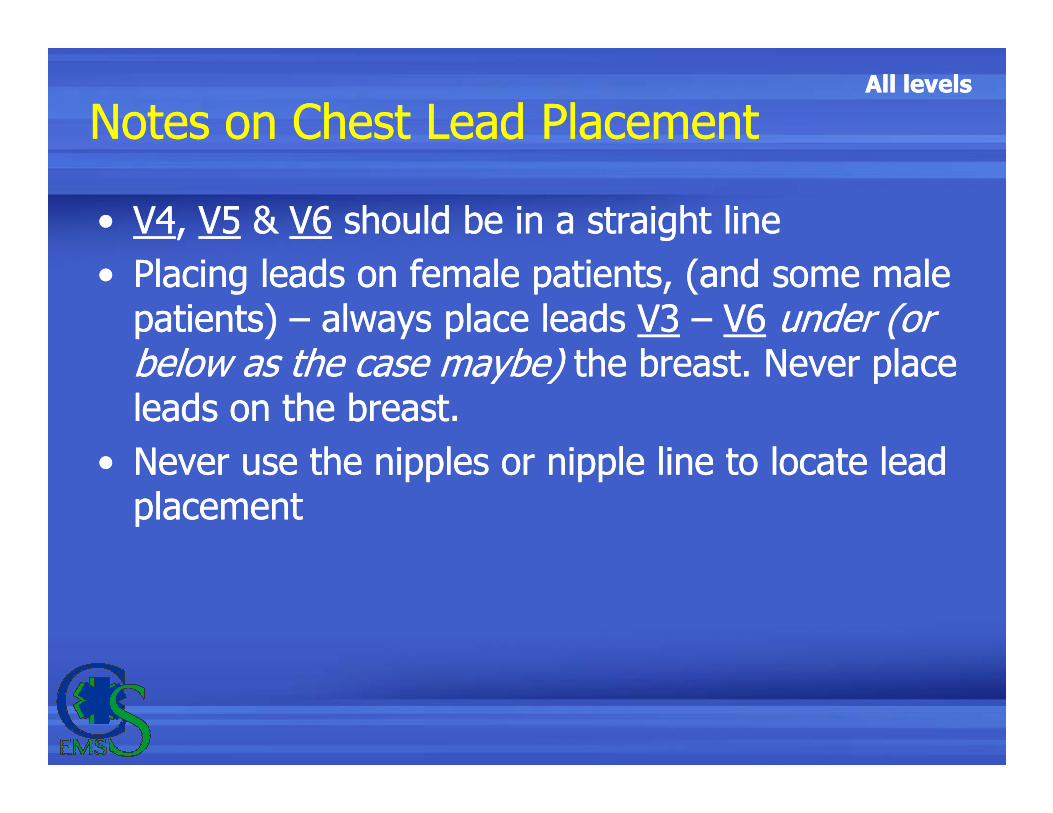
• V4, V5 & V6 should be in a straight line
• Placing leads on female patients, (and some male patients) – always place leads V3 – V6 under (or below as the case maybe) the breast. Never place leads on the breast.
• V4, V5 & V6 should be in a straight line
• Placing leads on female patients, (and some male patients) – always place leads V3 – V6 under (or below as the case maybe) the breast. Never place leads on the breast.leads on the breast.
• Never use the nipples or nipple line to locate lead placement
leads on the breast.
• Never use the nipples or nipple line to locate lead placement
All levelsAll levels
Acquisition of 12 LeadsAcquisition of 12 Leads
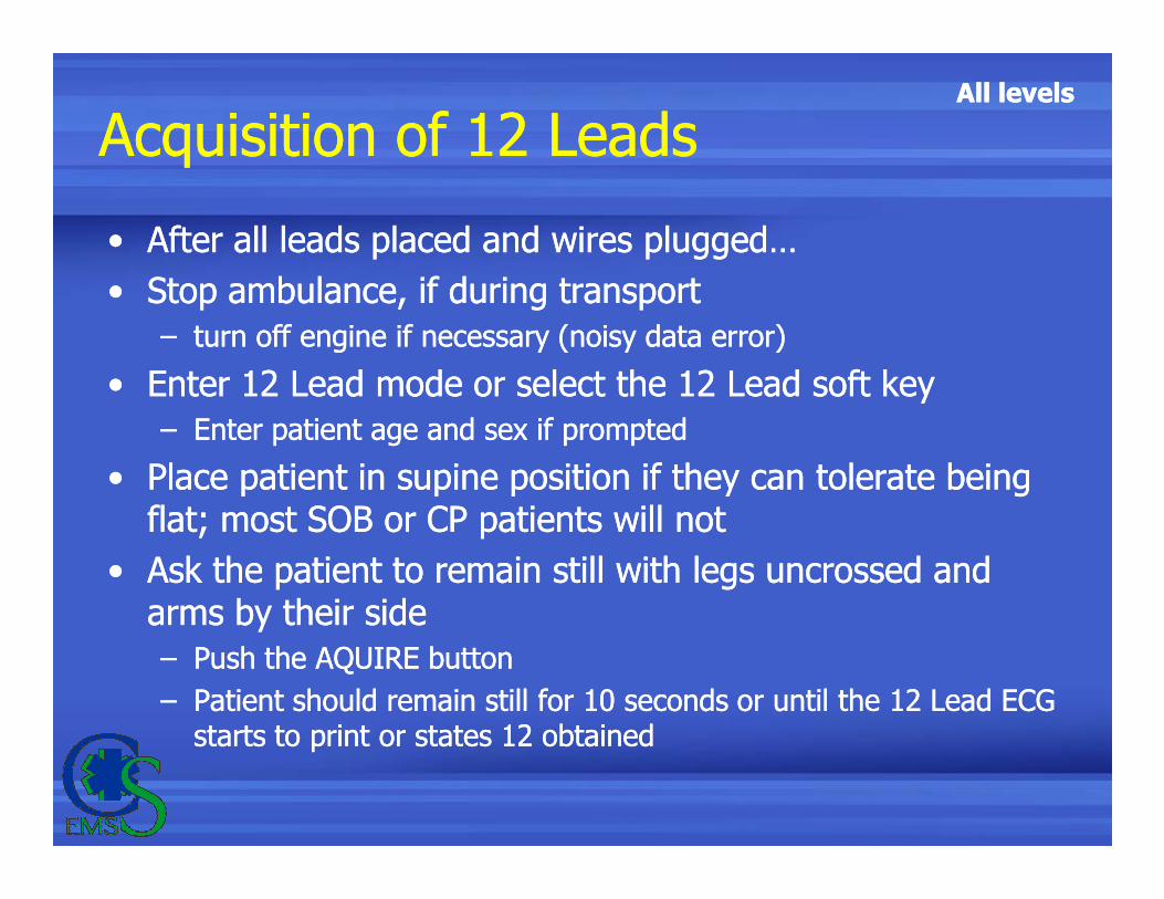
• After all leads placed and wires plugged…
• Stop ambulance, if during transport
– turn off engine if necessary (noisy data error)
• Enter 12 Lead mode or select the 12 Lead soft key
– Enter patient age and sex if prompted
• After all leads placed and wires plugged…
• Stop ambulance, if during transport
– turn off engine if necessary (noisy data error)
• Enter 12 Lead mode or select the 12 Lead soft key
– Enter patient age and sex if prompted
• Place patient in supine position if they can tolerate being flat; most SOB or CP patients will not
• Ask the patient to remain still with legs uncrossed and arms by their side
– Push the AQUIRE button
– Patient should remain still for 10 seconds or until the 12 Lead ECG starts to print or states 12 obtained
All levelsAll levels
Moving vs. Stopped AmbulanceMoving vs. Stopped Ambulance
All levelsAll levels
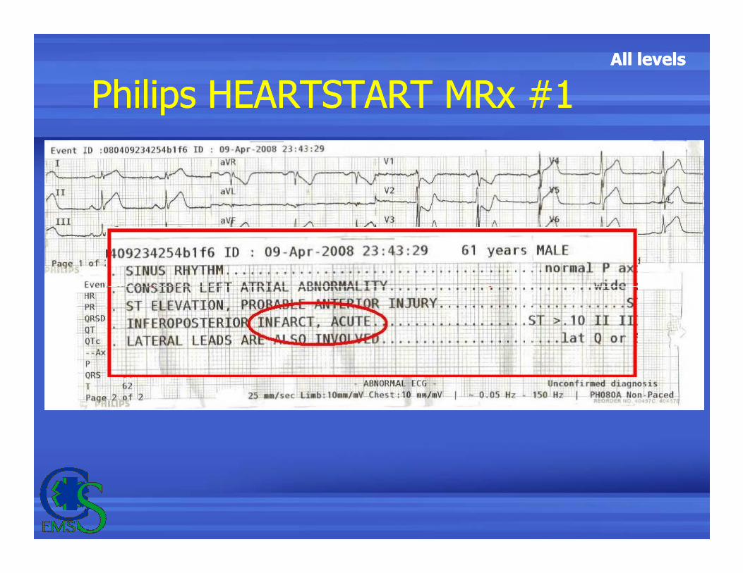
Philips HEARTSTART MRx #1Philips HEARTSTART MRx #1
All levelsAll levels
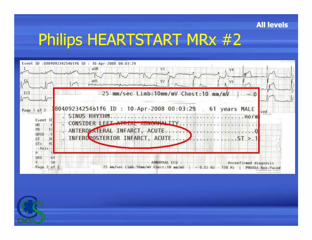
Philips HEARTSTART MRx #2Philips HEARTSTART MRx #2
All levelsAll levels
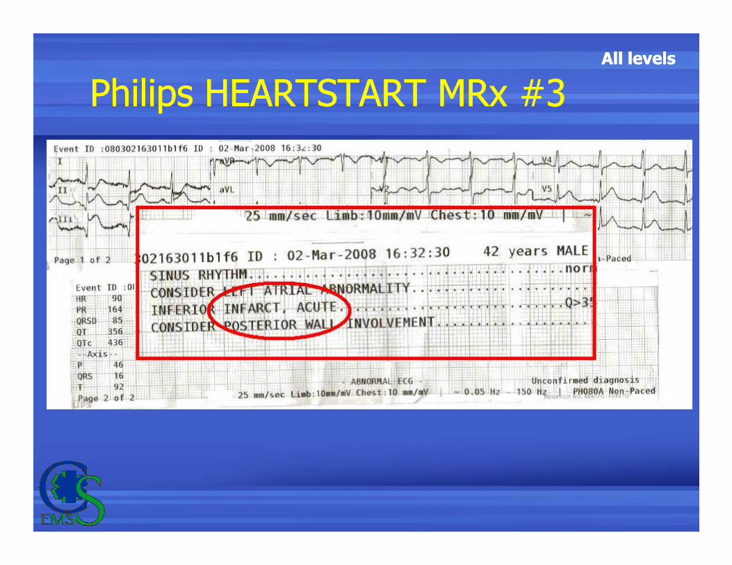
Philips HEARTSTART MRx #3Philips HEARTSTART MRx #3
All levelsAll levels
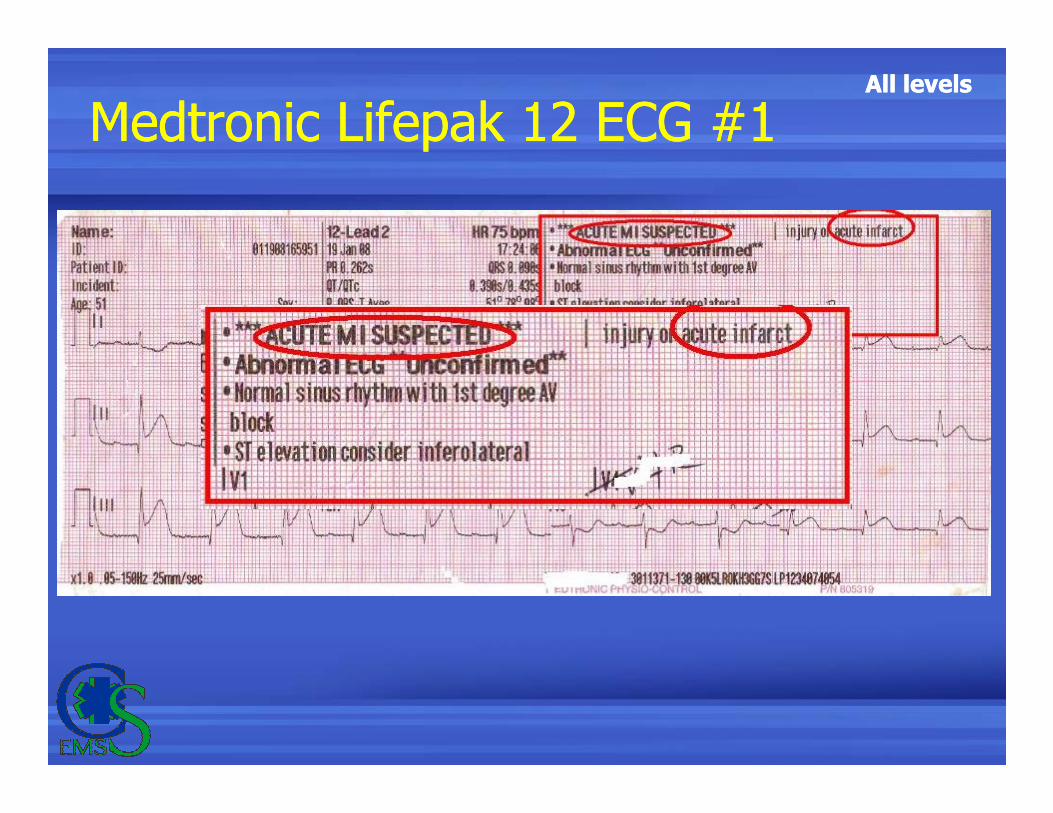
Medtronic Lifepak 12 ECG #1Medtronic Lifepak 12 ECG #1
All levelsAll levels
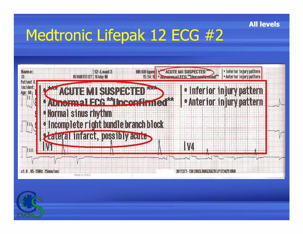
Medtronic Lifepak 12 ECG #2Medtronic Lifepak 12 ECG #2
All levelsAll levels
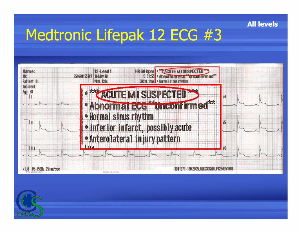
Medtronic Lifepak 12 ECG #3Medtronic Lifepak 12 ECG #3
All levelsAll levels
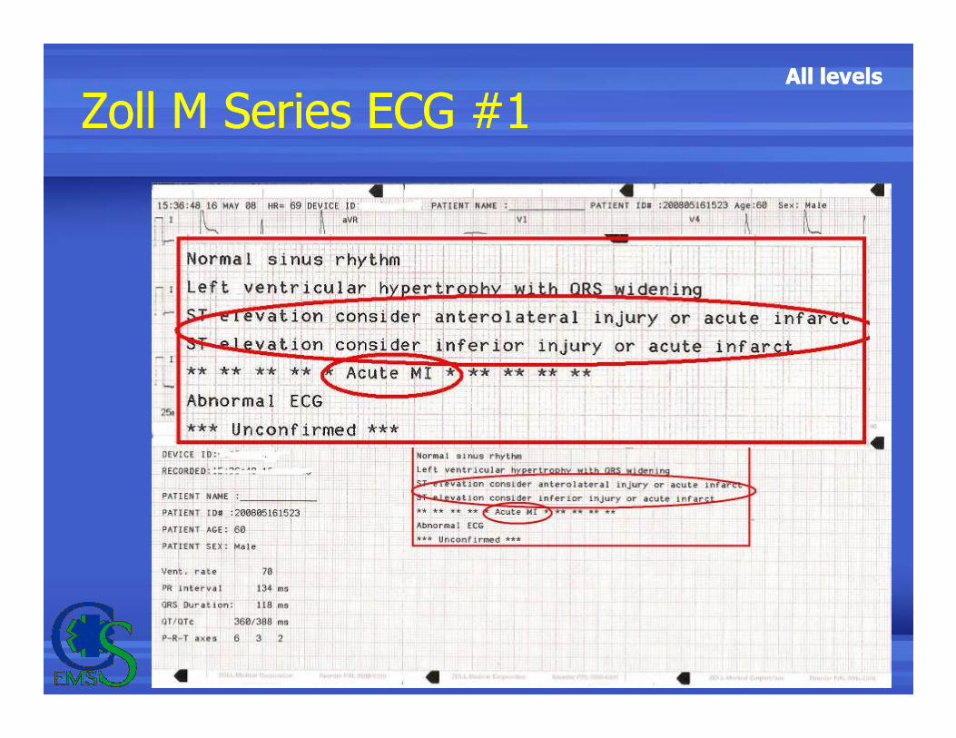
Zoll M Series ECG #1Zoll M Series ECG #1
All levelsAll levels
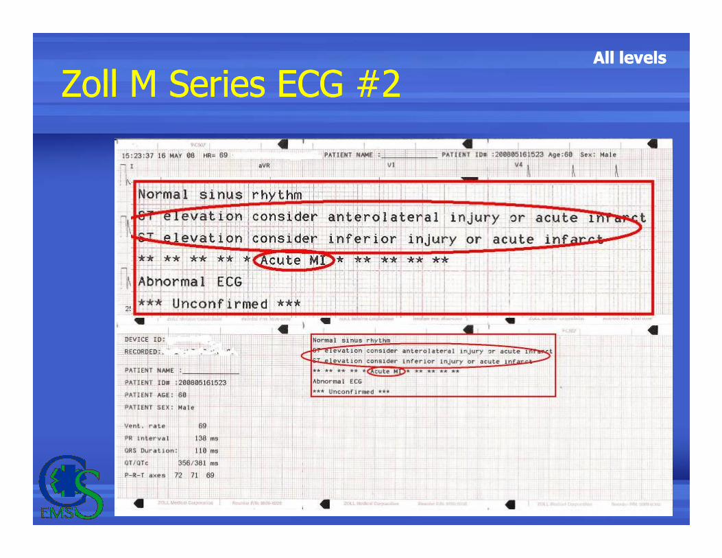
Zoll M Series ECG #2Zoll M Series ECG #2
All levelsAll levels
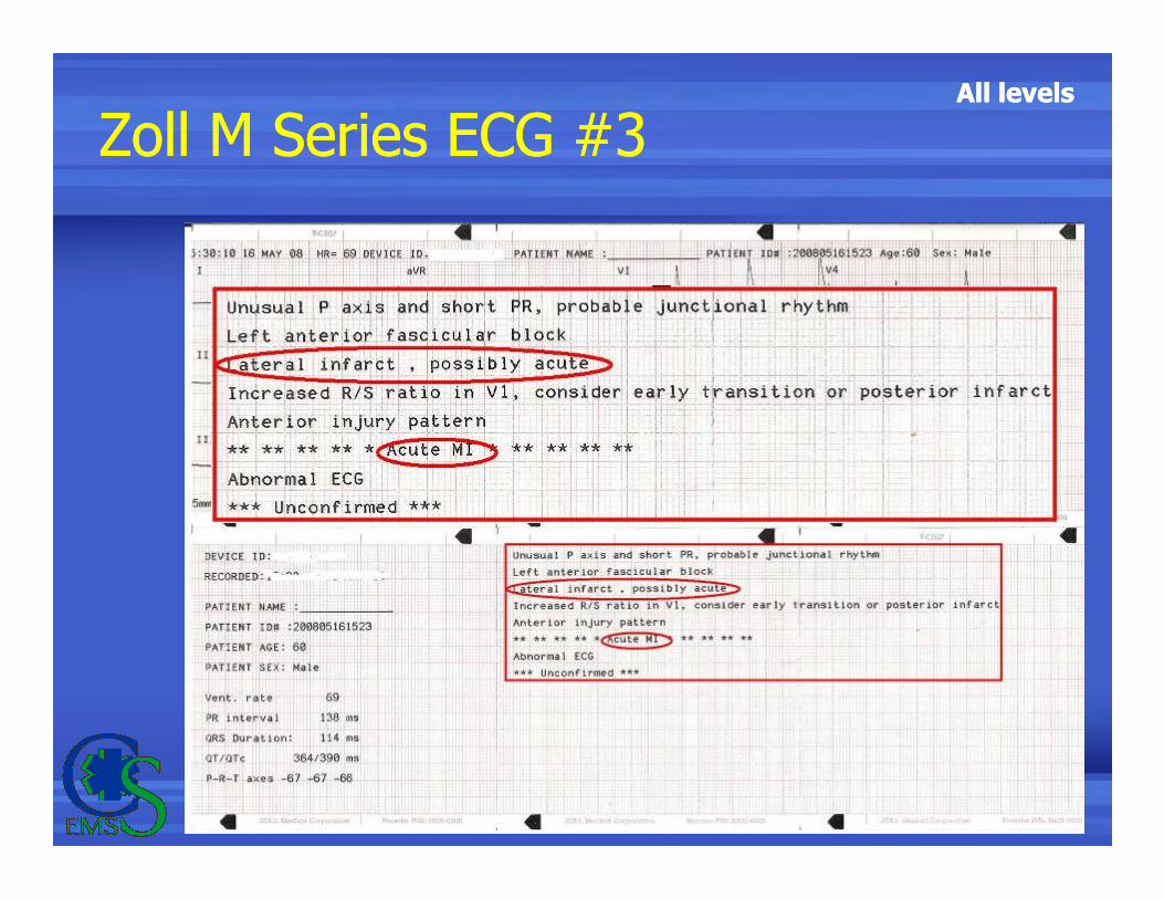
Zoll M Series ECG #3Zoll M Series ECG #3
All levelsAll levels
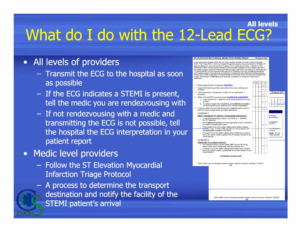
What do I do with the 12-Lead ECG?What do I do with the 12-Lead ECG?
• All levels of providers
– Transmit the ECG to the hospital as soon as possible
– If the ECG indicates a STEMI is present, tell the medic you are rendezvousing with
– If not rendezvousing with a medic and
• All levels of providers
– Transmit the ECG to the hospital as soon as possible
– If the ECG indicates a STEMI is present, tell the medic you are rendezvousing with
– If not rendezvousing with a medic and – If not rendezvousing with a medic and transmitting the ECG is not possible, tell the hospital the ECG interpretation in your patient report
• Medic level providers
– Follow the ST Elevation Myocardial Infarction Triage Protocol
– A process to determine the transport destination and notify the facility of the STEMI patient’s arrival
– If not rendezvousing with a medic and transmitting the ECG is not possible, tell the hospital the ECG interpretation in your patient report
• Medic level providers
– Follow the ST Elevation Myocardial Infarction Triage Protocol
– A process to determine the transport destination and notify the facility of the STEMI patient’s arrival
All levelsAll levels
Transmitting the 12-Lead ECGTransmitting the 12-Lead ECG
• The process for each type of monitor is a little different
• Some monitors can be programmed to transmit automatically (sometimes to more than one location)
• The process for each type of monitor is a little different
• Some monitors can be programmed to transmit automatically (sometimes to more than one location)location)
• Therefore transmitting is device specific
• You must practice with and be familiar with your agencies configuration
location)
• Therefore transmitting is device specific
• You must practice with and be familiar with your agencies configuration
All levelsAll levels
Questions?Questions?
All levelsAll levelsReferences:
1. Page, 12-Lead ECG for Acute and Critical Care Providers © 2006 by Pearson Education, Inc. Upper Saddle River, NJ
2. Zoll M Series 12-Lead ECG Monitoring © 2002 by ZOLL Medical Corporation. (9650-0215-01 Rev. H)
3. LIFEPAK 12 Defibrillator/Monitor Series Operating Instructions © 1998 –2004 by Medtronic Emergency Response Systems, Inc.
4. HEARTSTART MRx Instructions for Use, Edition 2 © 2004 by Koninklijke Philips Electronics N.V. (M3535-91900)
5. Part 8: Stabilization of the Patient With Acute Coronary Syndromes Circulation 2005 © 2005 by American Heart Association
6. 2006 Prehospital Standard Patient Treatment Protocols by Central
References:
1. Page, 12-Lead ECG for Acute and Critical Care Providers © 2006 by Pearson Education, Inc. Upper Saddle River, NJ
2. Zoll M Series 12-Lead ECG Monitoring © 2002 by ZOLL Medical Corporation. (9650-0215-01 Rev. H)
3. LIFEPAK 12 Defibrillator/Monitor Series Operating Instructions © 1998 –2004 by Medtronic Emergency Response Systems, Inc.
4. HEARTSTART MRx Instructions for Use, Edition 2 © 2004 by Koninklijke Philips Electronics N.V. (M3535-91900)
5. Part 8: Stabilization of the Patient With Acute Coronary Syndromes Circulation 2005 © 2005 by American Heart Association
6. 2006 Prehospital Standard Patient Treatment Protocols by Central 6. 2006 Prehospital Standard Patient Treatment Protocols by Central Shenandoah EMS Council; November 27, 2007 revision and January 1, 2008 Addendum
Questions?
6. 2006 Prehospital Standard Patient Treatment Protocols by Central Shenandoah EMS Council; November 27, 2007 revision and January 1, 2008 Addendum
Questions?