12 lead ecg workbook
TRANSCRIPT

STAT 12 Lead ECG in ACS Workshop 5/13/2019
Download Today’s Presentation To go: www.ECGtraining.org
from the MENU BAR at left side of screen, select “Downloads PDF,”
and then select:
12 Lead ECG Workbook: •Part 1: Basic Fundamentals •Part 2: ACS

STAT 12 Lead ECG in ACS Workshop 5/13/2019
STAT 12 Lead ECG Workshop
The T Wave SHOULD NOT be:
• Inverted in ___________CONTIGUOUS LEADS
• __________ (“Pointy” tipped)
• ________ (half above and half below isoelectric line)
STAT 12 Lead ECG in ACS Workshop 5/13/2019
ECG Indicators of NORMAL myocardial perfusion include:
• J Point isoelectric, or within_____of the ISOELECTRIC LINE
• ST Segment has a slight_______ inclination where ST Segment and T Wave merge, the shape is ________ (bowed downward).
• The T Wave is ________ (in all leads except for AVR), is not taller than ______, and is _____________(NOT “pointy”).
P-R Interval
• The P-R Interval should be between ___-___ms, (which is 3 – 5 little squares).

STAT 12 Lead ECG in ACS Workshop 5/13/2019
QRS Duration (width):
• The Normal QRS should be NO WIDER than ___ms (3 little squares).
QRS Duration (width):
• If the QRS is WIDER than 120ms, it indicates the VENTRICLES are ____________ __________.
• If the Ventricles are DEPOLARIZING ABNORMALLY, it causes them to _____________________.
STAT 12 Lead ECG in ACS Workshop 5/13/2019
QRS Duration (width):
• When the VENTRICLES REPOLARIZE ABNORMALLY due to the QRS being TOO WIDE, it often causes ______________:
– ______
– _________
– ______
• These changes are known as Secondary Repolarization Abnormalities.
Leads V1 & V2 on 12 Lead ECG:
• Proper lead placement of precordial Leads V1 and V2 are __________________on opposite sides of the sternum.
• Incorrect placement of Leads V1 and V2 will result in the presence of Q Waves (indicator of necrosis) leading to misdiagnosis of _____________________________.
STAT 12 Lead ECG in ACS Workshop 5/13/2019
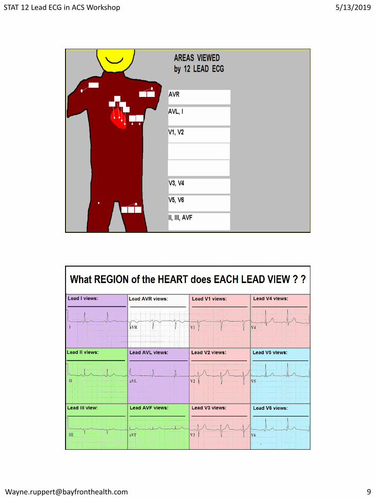
Leads V1-V4:
• V1 – V4 view the ______________of the Left Ventricle.
• V1 and V2 also view the ___________
• V1 – V3 view the ______________via Reciprocal Changes.
Leads V5 & V6:
• V5 & V6 view the _____________of the Left Ventricle.
STAT 12 Lead ECG in ACS Workshop 5/13/2019
Leads I and AVL:
• Leads I and AVL view the PROXIMAL aspect of the _______ and ________ WALLS
• I and AVL can be associated with EITHER the ____________, the _____________, or BOTH the _______ and _____________.
Leads II, III, and AVF:
• Leads, II, III, and AVF view the _____________ of the Left Ventricle.
STAT 12 Lead ECG in ACS Workshop 5/13/2019
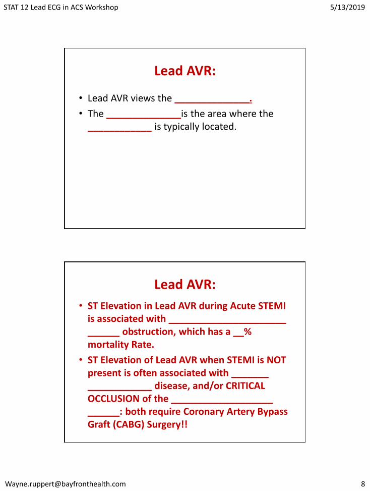
Lead AVR:
• Lead AVR views the ______________.
• The ______________is the area where the ____________ is typically located.
Lead AVR:
• ST Elevation in Lead AVR during Acute STEMI is associated with ______________________ ______ obstruction, which has a __% mortality Rate.
• ST Elevation of Lead AVR when STEMI is NOT present is often associated with _______ ____________ disease, and/or CRITICAL OCCLUSION of the ___________________ ______: both require Coronary Artery Bypass Graft (CABG) Surgery!!
STAT 12 Lead ECG in ACS Workshop 5/13/2019
The INDICATION for obtaining a RIGHT VENTRICULAR ECG is
___________________.
The INDICATION for obtaining a
POSTERIOR LEAD ECG is: __________________________.
Left Anterior Descending Artery
The LAD supplies blood to the ANTERIOR and SEPTAL walls, and includes the following CRITICAL STRUCTURES:
• Approximately ____of the Left Ventricle
• ___________
• _____________

STAT 12 Lead ECG in ACS Workshop 5/13/2019
Circumflex (Cx) Artery
In patients with a Right Dominant coronary artery system, the Circumflex supplies blood to:
• Approximately 20-30% of the Left Ventricle, which includes:
–__________of Left Ventricle
–______________________
• On rare occasion, the __________
Right Coronary Artery (RCA)
In patients with a RIGHT DOMINANT system, the RCA supplies blood to the following cardiac structures:
• __________
• _____________
• _______
• Approximately _____%of the Left Ventricle
– INFERIOR Wall
– ½ POSTERIOR WALL

STAT 12 Lead ECG in ACS Workshop 5/13/2019
If the patient has TWO or more of the following, ACS should be RULED OUT:
• ____________
• __________ for Heart Disease (3 or more, or KNOWN history of heart disease)
• _______________ (ST- T wave changes)
• _____________ (Troponin) elevated.
“Classic” cardiac chest pain:
• Location: _________
• ___ or __________ in nature
• Does not change with ______________
STAT 12 Lead ECG in ACS Workshop 5/13/2019
All patients with ACS symptoms . . .
STAT 12 Lead ECG; obtain and have read within _________ !!!
ACC/AHA Guideline!
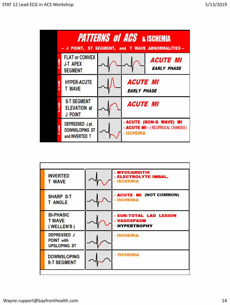
ECG Indicators of ABNORMAL PERFUSION
(possible ischemia / infarction) in Patients with
Normal Width QRS Complexes (QRS duration < 120 ms)
Normal Width
STAT 12 Lead ECG in ACS Workshop 5/13/2019
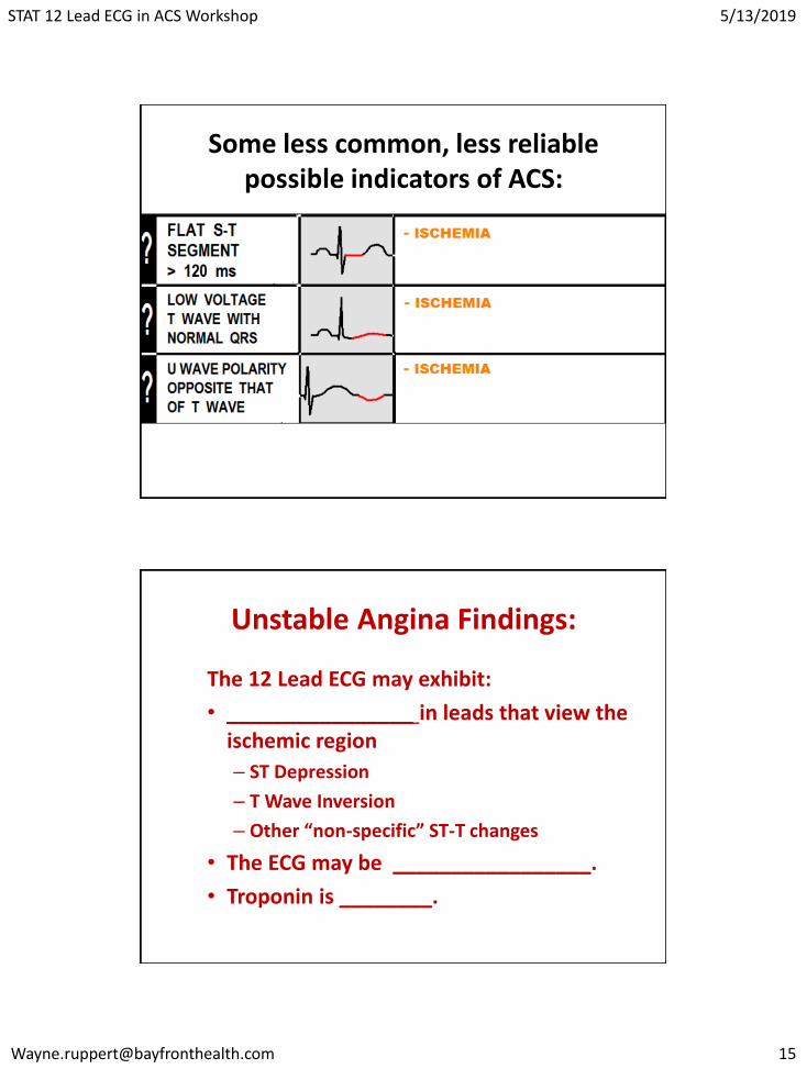
Some less common, less reliable possible indicators of ACS:
Unstable Angina Findings:
The 12 Lead ECG may exhibit:
• ________________ in leads that view the ischemic region
– ST Depression
– T Wave Inversion
– Other “non-specific” ST-T changes
• The ECG may be _________________.
• Troponin is ________.
STAT 12 Lead ECG in ACS Workshop 5/13/2019
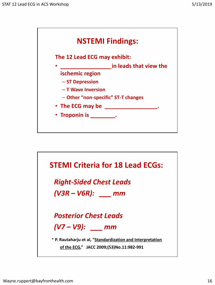
NSTEMI Findings:
The 12 Lead ECG may exhibit:
• ________________ in leads that view the ischemic region
– ST Depression
– T Wave Inversion
– Other “non-specific” ST-T changes
• The ECG may be _________________.
• Troponin is ________.
STEMI Criteria for 18 Lead ECGs:
Right-Sided Chest Leads
(V3R – V6R): ___ mm
Posterior Chest Leads
(V7 – V9): ___ mm
* P. Rautaharju et al, “Standardization and Interpretation
of the ECG,” JACC 2009;(53)No.11:982-991