reeve-irvine research center - spinal cord injury research ...brain computer interfaces (bcis) or...
TRANSCRIPT

On February 6, 2010, Anthony Purcell
dove into Miami Beach waters and didn’t
come back up. Having grown up in
Florida, he knew the waters very well and
thought the water was deeper than it
actually was. His head hit the sand
breaking his neck and bruising his cervical
spinal cord. Thanks to his cousin Bernie
who pulled him ashore, and the medical
personnel who acted swiftly, Anthony
survived this life threatening accident, but
was tragically paralyzed. Walking With
Anthony was founded in 2010 by
Anthony’s mother, Micki Purcell. This
charitable organization is devoted to
helping people believe in the hope to someday cast aside their wheelchairs.
Through Micki’s experiences with Anthony’s injury, she learned that many spinal
cord injury victims and their families have no idea where to turn for help. Sadly,
the majority of SCI injured individuals have limited resources once their health
benefits expire. Walking With Anthony’s mission is to forever change the
recovery outcome of spinal cord injury, currently perceived as unchangeable.
The charity is committed to educate/raise awareness, support SCI research,
expand rehabilitation centers and provide assistance to individuals with SCI.
Walking with Anthony honored Os Steward for his lifetime achievements in spinal
cord injury research at the “Movement for Change” Charity Event held at Siren
Studios in Hollywood California. The Reeve-Irvine Research Center would like
to congratulate Os for this award in the recognition of his dedication to
researching potential treatments that can improve the lives of those who suffer
from the repercussions of spinal cord injury.
In this issue...From the Director Pg 2
Late Breaking News Pg 3
Anatomy 101 Pgs 4-5, 8-9
PTEN Suppression Pgs 10-11
Roman Reed Awards Pg 14
Meet the Scientists Pg 15
Unsung Heroes Pgs 18-19
CA Legislative Update Pg 22
Number 21, Summer 2012
REEVE–IRVINE RESEARCH CENTER
Reeve-Irvine Research Center Publication
Continued on page 2...
Dr. Steward Receives Lifetime Achievement Award
On February 6, 2010, Anthony Purcell
dove into Miami Beach waters and didn’t
come back up. Having grown up in
Florida, he knew the waters very well and
thought the water was deeper than it
actually was. His head hit the sand
breaking his neck and bruising his cervical
spinal cord. Thanks to his cousin Bernie
who pulled him ashore, and the medical
personnel who acted swiftly, Anthony
survived this life threatening accident, but
was tragically paralyzed. Walking With
Anthony was founded in 2010 by
Anthony’s mother, Micki Purcell. This
charitable organization is devoted to
helping people believe in the hope to someday cast aside their wheelchairs.
Through Micki’s experiences with Anthony’s injury, she learned that many spinal
cord injury victims and their families have no idea where to turn for help. Sadly,
the majority of SCI injured individuals have limited resources once their health
benefits expire. Walking With Anthony’s mission is to forever change the
recovery outcome of spinal cord injury, currently perceived as unchangeable.
The charity is committed to educate/raise awareness, support SCI research,
expand rehabilitation centers and provide assistance to individuals with SCI.
Walking with Anthony honored Os Steward for his lifetime achievements in spinal
cord injury research at the “Movement for Change” Charity Event held at Siren
Studios in Hollywood California. The Reeve-Irvine Research Center would like
to congratulate Os for this award in the recognition of his dedication to
researching potential treatments that can improve the lives of those who suffer
from the repercussions of spinal cord injury.
Micki Purcell Honors
Dr. Oswald Steward Please consider including
Reeve-Irvinein your estate plans.
Your planned gift
can help create
tomorrow’s cures.
For information please contact:
Tania Cusack,Director of Community Development
(949) 824-5925 or email [email protected]
D GE IN VIN NA GLP
Are you considering including Reeve-Irvine in your estate plans?
Your planned gift can help create tomorrow’s cures.
For information please contact:
Tania Jope, Director of Community Development(949) 824-5925 or email [email protected]
John Salley, event MC
MBA Basketball Star & Talkshow HostAnthony Purcell
Continued from cover...
“Movement for Change” Charity Event
FROMTHE Director...
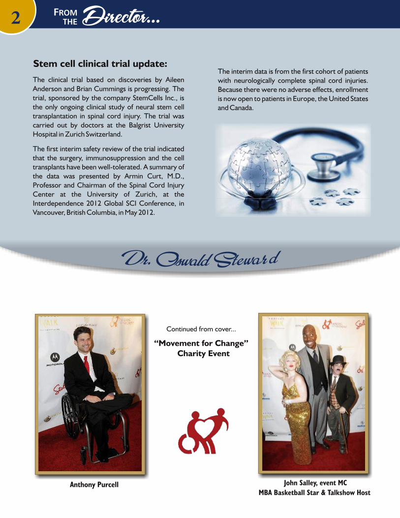
The clinical trial based on discoveries by Aileen Anderson and Brian Cummings is progressing. The trial, sponsored by the company StemCells Inc., is the only ongoing clinical study of neural stem cell transplantation in spinal cord injury. The trial was carried out by doctors at the Balgrist University Hospital in Zurich Switzerland.
The first interim safety review of the trial indicated that the surgery, immunosuppression and the cell transplants have been well-tolerated. A summary of the data was presented by Armin Curt, M.D., Professor and Chairman of the Spinal Cord Injury Center at the University of Zurich, at the Interdependence 2012 Global SCI Conference, in Vancouver, British Columbia, in May 2012.
The interim data is from the first cohort of patients with neurologically complete spinal cord injuries. Because there were no adverse effects, enrollment is now open to patients in Europe, the United States and Canada.
Stem cell clinical trial update:
2
Please consider including
Reeve-Irvinein your estate plans.
Your planned gift
can help create
tomorrow’s cures.
For information please contact:
Tania Cusack,Director of Community Development
(949) 824-5925 or email [email protected]
D GE IN VIN NA GLP
Are you considering including Reeve-Irvine in your estate plans?
Your planned gift can help create tomorrow’s cures.
For information please contact:
Tania Jope, Director of Community Development(949) 824-5925 or email [email protected]
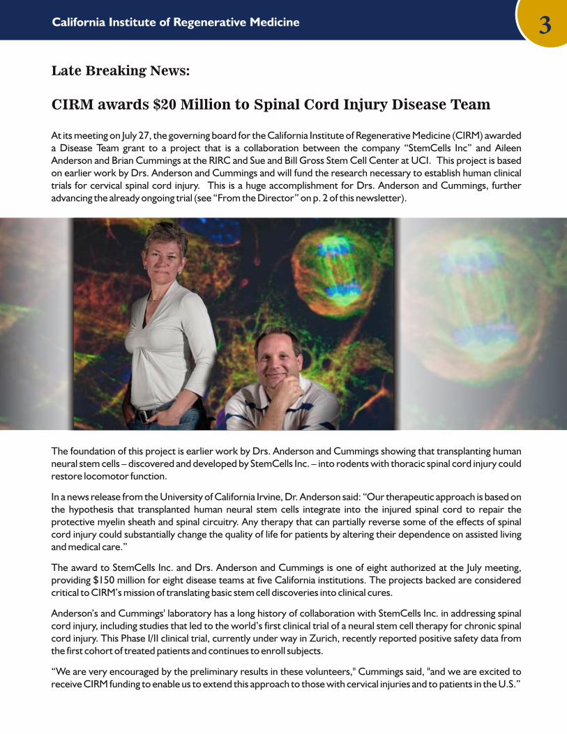
At its meeting on July 27, the governing board for the California Institute of Regenerative Medicine (CIRM) awarded a Disease Team grant to a project that is a collaboration between the company “StemCells Inc” and Aileen Anderson and Brian Cummings at the RIRC and Sue and Bill Gross Stem Cell Center at UCI. This project is based on earlier work by Drs. Anderson and Cummings and will fund the research necessary to establish human clinical trials for cervical spinal cord injury. This is a huge accomplishment for Drs. Anderson and Cummings, further advancing the already ongoing trial (see “From the Director” on p. 2 of this newsletter).
The foundation of this project is earlier work by Drs. Anderson and Cummings showing that transplanting human neural stem cells – discovered and developed by StemCells Inc. – into rodents with thoracic spinal cord injury could restore locomotor function.
In a news release from the University of California Irvine, Dr. Anderson said: “Our therapeutic approach is based on the hypothesis that transplanted human neural stem cells integrate into the injured spinal cord to repair the protective myelin sheath and spinal circuitry. Any therapy that can partially reverse some of the effects of spinal cord injury could substantially change the quality of life for patients by altering their dependence on assisted living and medical care.”
The award to StemCells Inc. and Drs. Anderson and Cummings is one of eight authorized at the July meeting, providing $150 million for eight disease teams at five California institutions. The projects backed are considered critical to CIRM’s mission of translating basic stem cell discoveries into clinical cures.
Anderson’s and Cummings' laboratory has a long history of collaboration with StemCells Inc. in addressing spinal cord injury, including studies that led to the world’s first clinical trial of a neural stem cell therapy for chronic spinal cord injury. This Phase I/II clinical trial, currently under way in Zurich, recently reported positive safety data from the first cohort of treated patients and continues to enroll subjects.
“We are very encouraged by the preliminary results in these volunteers," Cummings said, "and we are excited to receive CIRM funding to enable us to extend this approach to those with cervical injuries and to patients in the U.S.”
Late Breaking News from the CIRM
CIRM awards $20 Million to Spinal Cord Injury Disease Team
Late Breaking News:
3California Institute of Regenerative Medicine
Please consider including
Reeve-Irvinein your estate plans.
Your planned gift
can help create
tomorrow’s cures.
For information please contact:
Tania Cusack,Director of Community Development
(949) 824-5925 or email [email protected]
D GE IN VIN NA GLP
Are you considering including Reeve-Irvine in your estate plans?
Your planned gift can help create tomorrow’s cures.
For information please contact:
Tania Jope, Director of Community Development(949) 824-5925 or email [email protected]

44 Anatomy 101
Ibrain computer interfaces (BCIs) or regeneration. Since functional recovery, or the ability to regain partial or complete control of a limb or organ, after spinal cord injury is a priority of patients and the ultimate goal of SCI research we thought it would be good to define what these terms mean.
In the past decade the idea that machines or robots can be controlled by our thoughts, much like Alex Murphy in robocop, have emerged from the realm of science fiction to reality and serious scientific thought. These types of devices are generally called BCIs or brain-computer interfaces and nowadays they are much different than the Robocop example. BCIs are computers that are able to recognize some form of electrical signal in the brain and then process and use the signals to communicate with or control some other element of the outside world that enacts the intentions of the original signal from the brain. In 2000 the first International Meeting on Brain Computer Interface Technology defined a BCI as "A communication system that does not depend on the brain’s normal output pathways of peripheral nerves and muscles." Simply put BCIs are a computer based system that can acquire signals from the brain, analyze them, and then translate them into commands or actions that can make something move. This means that things like cochlear implants that transfer external signals like sound into an auditory perception are not BCIs because they only take information into the brain or auditory system. A BCI has to be able to both acquire information as well as provide an output or command. Indeed, BCIs are highly specialized and specifically designed to distinguish specific signals that are different from normal brain signals. It is also important to point out that a BCI cannot read a persons' mind and extract unwilling or unsuspecting information. Instead the user and the BCI work together, providing feedback that enables fine tuning and accuracy, which requires a lot of training for proper use.
BCIs have the potential to drastically improve the quality of life for disabled individuals including those with spinal cord injury since they can bypass areas of dysfunction or impairment. For example, in an uninjured individual the thought about grabbing an object creates a specific electrical signal in the brain that is transmitted to the spinal cord. The spinal cord then enables the proper nerves and muscles in the arm and fingers to grab. For spinal cord injured individuals the signal is unable to get past the injury in the spinal cord and the arm therefore does not receive the signal to grasp despite the initiation of the thought in the brain. BCI technology can allow a signal from the brain to bypass the injured spinal cord and enable the limb to move or grab. Signals from the BCI can also be transferred to an external monitor, prosthetic limb or wheelchair. Keep in mind that although BCI technology may help improve a persons' independence, it does not actually alter or improve the damaged spinal cord itself. It only provides a work around.
There are many different types of BCIs but all BCI systems have 3 major components: (1) Signal acquisition, (2) signal extraction and translation and (3) device output or command.
Signal acquisition: A sensor (which can be on the scalp, beneath the skull or within the brain) measures and detects specific brain signals or electrical activity. These signals are then amplified and transmitted to a computer or device.
n the Asilomar article (see page18) we mentioned the debate about whether the future of functional recovery was in
What is a BCI anyway?
What can a BCI do for me?
How do they work?
Anatomy 101: Are You Ready to be a Cyborg? By Maya Hatch, PhD
The subject is wearing a cap with electrodesthat pick up brain waves
and activate the mechanical glove.
Please consider including
Reeve-Irvinein your estate plans.
Your planned gift
can help create
tomorrow’s cures.
For information please contact:
Tania Cusack,Director of Community Development
(949) 824-5925 or email [email protected]
D GE IN VIN NA GLP
Are you considering including Reeve-Irvine in your estate plans?
Your planned gift can help create tomorrow’s cures.
For information please contact:
Tania Jope, Director of Community Development(949) 824-5925 or email [email protected]

5
Signal extraction and translation: This is the process of analyzing and converting a distinguished signal or intent to the appropriate command for the output device. To do this extraneous content or information must be filtered and then compacted and translated into an actual output command.
Device output: This is generally an external device that can receive the commands or algorithm from the signal translation that code for a function like letter selection on a screen, cursor control on a screen/wheelchair or robotic/prosthetic arm operation. The device operation then provides feedback to the user to close the circuit.
Although there are many different external output devices (computers, prosthetic arms, wheelchairs, etc) that a BCI can communicate with there are currently only 3 clinically applicable ways to extract signals from the brain.
EEG-based or scalp recording systems EEG (electroencephalography) BCIs take recordings noninvasively from the scalp, which is separated from the brain tissue where the signal is initiated by the skull and subcutaneous tissue. This distance degrades the original signal significantly and limits its resolution. Yet, these BCIs are widely used and the most popular to date due to their minimal risk, ease of use and convenience for conducting studies. Since these BCIs take recordings from the scalp they are generally limited to basic communication like using a computer for typing or browsing the internet. In some cases it can also allow basic operations of a prosthetic limb or help guide a wheelchair in a predetermined path. The use of these BCIs is restrictive since they require the user to be stationary.
ECoG-based recordings ECoG (electrocorticogram) activity is recorded from the cortical surface of the brain and is therefore implanted underneath the skull but not in the brain tissue itself, generally in the form of a small electrode array. These have better resolution than the EEG systems and their signal is much more robust. These BCIs allow slightly higher capabilities such as more defined finger, hand and arm movements compared to the EEG systems. They can also provide better mobility since they are implanted inside the skull. However, they are considered to be invasive and somewhat unstable making them riskier than the EEG systems.
BCIs within the brain These are electrode arrays or chips that are implanted into the actual brain tissue. They can take recordings from single neurons or ensembles of neurons. The signal quality and accuracy of these invasive BCIs is far superior to the others and they can provide signals for complex movements that will mimic normal movement. These BCIs also allow user mobility however, their downfall is that they are not very stable and they may cause some tissue damage in the implanted area.
Although BCI technology is relatively new, it has moved at an extraordinary pace in the past decade and has the potential to improve the quality of life for people with disabilities. All of the BCI systems mentioned still have room for improvement. However, given the rapid progression of these types of technologies over the past decade coupled with the rapid ascent of computer technologies and biomedical engineering, these obstacles will likely be easily overcome. Ultimately it will be up the disabled individual to weigh out the benefits and associated risks of using one of these BCIs.
Current BCI systems
Are you considering including Reeve-Irvine in your estate plans?
Your planned gift can help create tomorrow’s cures.
For information please contact:
Tania Jope, Director of Community Development(949) 824-5925 or email [email protected]
Please consider including
Reeve-Irvinein your estate plans.
Your planned gift
can help create
tomorrow’s cures.
For information please contact:
Tania Cusack,Director of Community Development
(949) 824-5925 or email [email protected]
D GE IN VIN NA GLP
Are you considering including Reeve-Irvine in your estate plans?
Your planned gift can help create tomorrow’s cures.
For information please contact:
Tania Jope, Director of Community Development(949) 824-5925 or email [email protected]

6
Spinal cord injuries commonly result in bladder dysfunction in addition to paralysis, loss of sensation, and pain. In recent years, researchers have become increasingly aware of the importance of bladder function for people who have suffered a spinal cord injury. In many experimental studies, researchers have been working on ways to re-connect injured nerve fibers. At the Reeve-Irvine Research Center (RIRC), another strategy to improve bladder function is currently also being evaluated. In the Havton Laboratory at RIRC,Dr. Harriet Chang and Dr. Leif Havton have developed a model to evaluate novel drug treatments that may enhance the function of spared nerve connections after incomplete injuries to the lowermost portion of the spinal cord and associated nerve roots. These injuries are known as conus medullaris and cauda equina injuries. Drs. Chang and Havton have demonstrated that pharmacologic activation of spinal cord connections can improve bladder function. These translational research studies use physiologic tests that are similar to those used by urologists when examining patients with bladder problems like a “cystometrogram” and electromyography (EMG) recordings of bladder muscles. A cystometrogram shows the bladder pressure during rest and during voiding. It is used to evaluate how well the bladder can contract its wall muscles and empty itself.
Drs. Chang and Havton showed that an experimental drug known as 8-OH-DPAT can improve the function of the spared nerve fibers and restore much of voiding efficiency after incomplete, sp ina l cord in jur ies . This pharmacological treatment stimulates a group of receptors in the brain and spinal cord that are activated by a normally occurring "brain chemical" called serotonin. "This new pharmacological approach and strategy represents a new opportunity to develop novel treatments for people with incomplete injuries affecting the sacral portion of the spinal cord and the cauda equina nerve roots", according to the research team. Preliminary results of these studies have already been presented at the Annual Meeting for the Society of Neuroscience, and a comprehensive manuscript was recently submitted by Drs. Chang and Havton to a research journal for peer review and publication.
A novel approach to improve bladder functionin laboratory studies
Cystometrogram recordings of bladder pressureand electromyogram (EMG) recordings of the
external urethral muscle showing that the drug,8-OH-DPAT, has an effect on bladder function.
The voiding efficiency improved and EMG activityincreased after drug administration in a rat
with a cauda equina injury.
Please consider including
Reeve-Irvinein your estate plans.
Your planned gift
can help create
tomorrow’s cures.
For information please contact:
Tania Cusack,Director of Community Development
(949) 824-5925 or email [email protected]
D GE IN VIN NA GLP
Are you considering including Reeve-Irvine in your estate plans?
Your planned gift can help create tomorrow’s cures.
For information please contact:
Tania Jope, Director of Community Development(949) 824-5925 or email [email protected]

7
Research in the laboratory of RIRC member Dr. Brian Cummings focuses on understanding interactions between transplanted human neural stem cell populations and the damaged microenvironment that results from traumatic central nervous system injury - spinal cord injury (SCI) and traumatic brain injury (TBI). Integral to this research is a focus on how neural stem cells interpret the “niche” as well as how transplanted stem cells integrate with the host. By learning more about these interactions, we can better understand how best to use neural stem cells to treat CNS injuries.
We are developing new human neural stem cell lines with the potential to treat neurotraumatic injuries. Daniel Haus, a graduate student in the Cummings Lab, has worked out the cell culture methods needed to grow human embryonic stem cells (hESCs) in media that contains no contaminants from animal sources, meaning that they would be suitable for use in humans. These hESCs are then transitioned to neural stem cells (hNSCs) and sorted via positive and negative cell surface markers in collaboration with Dr. Hal Nguyen in the Anderson Lab. In the last year, we have succeeded in making four distinct populations of human ESC derived neural stem cells and confirmed that these cell populations are genetically normal. Over next year, Mr. Haus will transplant these hNSCs into rats with traumatic brain injuries to determine if they are capable of repairing damaged tissue and improving function on a battery of behavioral tasks.
Using hNSCs in an animal model requires an understanding of how the cells interact with the host. One potential way to analyze interactions between hNSCs and the injured microenvironment or “niche” is to use live cell, time-lapse imaging in an incubator. Such imaging can track cell lineage and fate over days or weeks without the need to use animals, and can reveal individual cell responses to differentiation factors, inflammatory cues, and/or drug treatments.
In parallel with live cell imaging studies, Post-doctoral fellow Luci Velazquez and graduate student Eric Gold are refining a model of traumatic brain injury using Athymic nude rats. These rats lack an immune system; if human cells are transplanted into their brains, the foreign cells have a better chance of surviving and not being rejected by the host’s immune system. Before a stem cell therapy can be tested in these rats, Dr. Velazquez and Mr. Gold need to determine how ATN rats respond to TBI and obtain baseline data on their normal motor and cognitive recovery to a standardized injury. Transplants of hNSCs is scheduled to begin in the fall of 2012.
Live cell image analysis using Imaris computer software to trackcell migration and division.
(image by Daniel Haus, RIRC graduate student)
Brian Cummings Lab Update
Please consider including
Reeve-Irvinein your estate plans.
Your planned gift
can help create
tomorrow’s cures.
For information please contact:
Tania Cusack,Director of Community Development
(949) 824-5925 or email [email protected]
D GE IN VIN NA GLP
Are you considering including Reeve-Irvine in your estate plans?
Your planned gift can help create tomorrow’s cures.
For information please contact:
Tania Jope, Director of Community Development(949) 824-5925 or email [email protected]

Anatomy 101
Anatomy 101: Regeneration
In this article we will be discussing the second part of the Asilomar debate; regeneration. Although there are many types of regeneration we will only be addressing axonal regeneration within the central nervous system (CNS) in the context of spinal cord injury.
Medical and scientific terms were first developed during the renaissance era when new medical and scientific advances were being made. Medical scientists at the time turned to the vocabulary of early Greek and Roman physicians like Hippocrates, Galen and Celsus to coin words for their new diseases and discoveries. Therefore a system of Latin and Greek prefixes, combining forms and suffixes were used to create new scientific and medical terms, creating an easy to use, universally accepted language. If we break down the term regeneration into its Latin and Greek root words we can easily understand what it means. Regeneration can be broken down into re-gener-a-tion where re- is the prefix meaning back or again; gener- means to produce or come into being; and the suffix tion- means the act of. Putting that together regeneration is the act of producing something again. In the context of spinal cord injury, axonal regeneration means the act of producing axons again, or reproducing axons after injury.
Axons are long fibers extending from neurons that transmit information from one place to another, much like electrical wires. Within the CNS there are descending axons that transfer messages from the brain to the spinal cord and ascending axons that transmit information from the spinal cord back to the brain. When these axons are damaged or cut, as is the case in spinal cord injury (SCI), the information that they are transferring slows or stops altogether. This axonal damage is the main cause of many of the impairments associated with SCI and it was thought these damaged or cut CNS axons were incapable of regeneration. Pioneering work by Aguayo and colleagues in 1980 showed for the first time that injured CNS axons were able to regenerate when they were provided the permissive environment of peripheral nerve grafts. This work suggested that injured axons could in fact regenerate when given the right environment or signals. Follow-up experiments confirmed that there were growth promoting factors within the grafts and growth inhibitory factors in the injured spinal cord. These discoveries changed the idea that a damaged CNS could not regenerate and it changed the course of SCI research and our understanding of CNS axons.
In the past 30 years there has been significant progress in identifying a number of the factors found to inhibit or limit axonal regeneration in the cord. The main culprits proven to limit or stop axonal regeneration in the CNS after SCI are listed on the facing page.
What's in the name?
Axonal regeneration and the central nervous system
The culprits
By Maya Hatch, PhD
Astroglial Scar
Nucleus
BRAIN
Soma
Myelin
DyingNeuron
Axon
8
Please consider including
Reeve-Irvinein your estate plans.
Your planned gift
can help create
tomorrow’s cures.
For information please contact:
Tania Cusack,Director of Community Development
(949) 824-5925 or email [email protected]
D GE IN VIN NA GLP
Are you considering including Reeve-Irvine in your estate plans?
Your planned gift can help create tomorrow’s cures.
For information please contact:
Tania Jope, Director of Community Development(949) 824-5925 or email [email protected]
9
Myelin-associated inhibitors: Myelin-associated inhibitors (MAIs) are proteins expressed by oligodendrocytes and components of myelin. The main MAIs are Nogo-A, myelin-associated glycoprotein (MAG), and oligodendrocyte myelin glycoprotein (OMgp). All of these interact with receptors found on neurons that activate intracellular mechanisms that inhibit or limit axon growth.
Chondroitin sulfate proteoglycans: After a spinal cord injury, cells called astrocytes form a physical barrier around the injury site called an astroglial scar. It is thought that this forms around the injury to wall off or protect the rest of the spinal cord. Chondroitin sulfate proteoglycans (CSPGs) are one of the main molecules found within the astroglial scar both on the membranes of astrocytes and in the extracellular space. Members from this class of inhibitors are neurocan, versican, brevican, phosphacan, aggrecan and NG2. All of these proteoglycans have relatively large sugar side chains and it is thought that these side chains are the culprits in axonal regeneration inhibition.
Other extracellular matrix components: There are many other components within the injury site that may have inhibitory effects on axonal regeneration and they are generally noted as inhibitory extracellular matrix (ECM) components. Included in this list are semaphorins, ephrins, slit proteins and tenascin. All of these ECM components inhibit the growth, guidance or function of axons and some, like the semaphorins, may also bind to chains on CSPGs adding to the scar around the injury site. The exact role of these factors is currently being studied and not well known yet.
Limited growth state of neurons: After a spinal cord injury, many different inhibitory molecules are present that inhibit the ability of axons to regenerate but the presence of these external molecules are not the only factors involved in regeneration failure. It is also believed that the intrinsic growth state of the neuron itself is altered after axonal injury. Once an axon is cut or damaged, signals are transferred from the axon back up to the soma or nucleus of the neuron that may alter the expression or functioning of various proteins. The alteration of these proteins has been suggested to limit the intrinsic growth state of the neuron.
Now that some of the factors for axonal regeneration failure have been identified, a variety of new therapies targeted at removing these inhibitory factors are being investigated. Last year at our "Meet the Scientist" event Dr. Jerry Silver explained that the spinal cord is like a freeway that cars (or axons) can travel on to get from one place to the next. When an accident occurs (a spinal cord injury) there are car parts and debris all over the road inhibiting cars from passing through. Once the debris and vehicles are removed the cars would be able to get through to the other side. Additionally, it might also be required to not only clear the debris out of the way but also bring in road guides or officers that could help guide the cars through a safe and correct path while the vehicles and debris are being cleared. Many regenerative therapies for SCI are now being carried out in a very similar way. Many researchers now believe that a combinatorial approach involving the removal of inhibitory factors and the enhancement of growth promoting factors, will ultimately be what is needed to reverse the impairments seen in SCI and lead to marked functional improvement. It does not seem to be enough to only remove the inhibitory factors or signals from the injury site.
Unlike the BCI technology on page 4-5, regenerative therapies change or alter the injured environment within the spinal cord. This is not a simple task and it will take time to get these therapies exactly right. However, we believe that many of the current regenerative therapies being explored have great potential to significantly improve a persons' life in a major way. These therapies may not be available for use right now, but their future potential should prove to be worth the wait.
Richardson PM, McGuinness UM, Aguayo AJ, 1980, Axons from CNS neurons regenerate into PNS grafts, Nature 284(5753):264-265.
Enhancing regeneration
Please consider including
Reeve-Irvinein your estate plans.
Your planned gift
can help create
tomorrow’s cures.
For information please contact:
Tania Cusack,Director of Community Development
(949) 824-5925 or email [email protected]
D GE IN VIN NA GLP
Are you considering including Reeve-Irvine in your estate plans?
Your planned gift can help create tomorrow’s cures.
For information please contact:
Tania Jope, Director of Community Development(949) 824-5925 or email [email protected]

Number 21, Summer 201210
Potential therapies for spinal cord injury take into account the changes that occur in the spinal cord following injury, including axon degeneration (Figure 1A) and cavity (or cyst) formation (Figure 1B). Not surprisingly, axon regeneration is an intense research area. As many of you know, a collaborative study between Os Steward’s group and the group of Zhigang He from Harvard showed that genetic deletion of the PTEN gene in neurons of the motor cortex (Figure 1C) promotes corticospinal tract axon regeneration after spinal cord injury (Liu et al., 2010).
A second important research area is repair of the cavity that forms in the spinal cord after injury. Some researchers are concerned that regenerating axons will be unable to grow through the cavity. Thus, scientists are seeking ways to repair the cavity. At UCI, Drs. Lisa Flanagan (Sue and Bill Gross Stem Cell Research Center) and Kelli Sharp (RIRC)recently reported that salmon fibrin treatment in the spinal cord injury site promotes functional recovery (Sharp et al., 2012). In this article, I will discuss a potential therapy that combines PTEN suppression in the motor cortex and salmon fibrin treatment in the spinal cord.
AAV-shPTEN suppresses PTEN expression in the motor cortex of adult rats.
So, how do we begin to translate PTEN gene deletion in genetically altered mice to PTEN gene suppression in patients? One way is use a viral vector expressing a small hairpin RNA molecule (shRNA) to selectively suppress PTEN gene expression in rats (See Anatomy 101 in Spinal Connections Number 20, page 4 for a review of the basic principles of gene therapy using viral vectors). In fact, Drs. Gail Lewandowski and Os Steward (RIRC) worked with the University of Pennsylvania Vector Core to develop an adeno-associated virus (AAV) vector expressing an shRNA for the PTEN gene (AAV-shPTEN). In a recent proof-of-concept experiment we injected AAV-shPTEN into the left hemisphere of the cortex of adult rats. Three weeks later we determined whether the injected AAV-shPTEN suppressed PTEN protein expression. To do this we took brain sections from the rats and detected PTEN protein using a process called immunohistochemistry. Additionally, AAV-shPTEN also expresses a bright green reporter gene called zsGreen. Visualization of zsGreen indicates the exact location of our AAV-shPTEN injection (Figure 2B). As our exciting results in Figure 2 show, AAV-shPTEN injections suppress PTEN protein expression by nearly 100% in the region of the injection (Figure 2). This significant result means that we now have a way to specifically suppress PTEN expression. Importantly, we have cleared the first translational hurdle.
Salmon Fibrin treatment promotes repair of the cavity after spinal injury in rats.
After spinal cord injury in humans and rats, a cavity forms in the area of the injury (Figure 3B). The cavity presents a real obstruction to extending regenerating axons from above the injury to the segment below the injury. Based on the work of Drs. Kelli Sharp and Lisa Flanagan,Dr. Lewandowski, in conjunction with Dr. Flanagan,
Axon regeneration via PTEN
suppression in the motor cortex
and fish fibrin treatment to
repair the spinal cord cavity
A winning combination therapy for spinal cord injury?Gail Lewandowski, Ph.D
Overview of the system of spinal cord injury. After spinal cord injury there is degeneration of axons (A) that originate from neurons in the cortex (C). Additionally, a cavity (cyst) forms in the area of injury (B).The cavity area can expand over time.
Intra-cortical injections ofAAV-shPTEN suppress PTENgene expression adult rats.
This figure shows the PTEN expression in the brain of a rat 3 weeks after AAV-shPTEN injection. Panel A shows a brain section stained for a general stain called DAPI; note that both the AAV-shPTEN side and uninjected sides are similar in appearance. The area of AAV-shPTEN injection is apparent from the expression of the zsGreen reporter gene (B). Importantly, PTEN protein expression is suppressed nearly 100% in the region corresponding to AAV-shPTEN injection (C and D).
Continued on the next page...

11did an experiment to determine whether salmon fibrin treatment at the time of spinal cord injury would help repair the cavity. We injected salmon fibrin into the spinal cord injury site of adult rats and then three weeks later examined the spinal cords for cavity formation. In Figure 3A we show an example of a section through a spinal cord injury site from an animal treated with salmon fibrin. Notice that the injury site, while still apparent, is “filled-in”. In contrast, a large cavity or hole is obvious in the section taken from the injury site of an untreated animal (Figure 3B). These results are very encouraging and indicated that salmon fibrin promotes the repair of the cavity formed after spinal cord injury.
Putting it all together for a combination therapy after spinal cord injury.
The next step is an experiment to determine if AAV-shPTEN injection into the cortex in combination with salmon fibrin treatment into the spinal cord is a synergistic therapy that will promote axon regeneration through a repaired injury site. Recently, Dr. Lewandowski with help from members of the Steward lab and Dr. Flanagan injected AAV-shPTEN into the cortex of adult rats that also received a cervical spinal cord injury. Half of the rats were also treated with salmon fibrin at the time of injury. For ten weeks after treatment the rats were assessed for forepaw motor function in a skilled reaching task. Next, we take a close look at the spinal cords and determine whether there is axon regeneration and cavity repair. Since we are a few weeks away from finishing the experiment, we do not yet have the results. It will take us a few months to put it all together, so stay tuned for the next installment!
Salmon fibrin promotes the repair of the lesion cavity after spinal
cord injury in rats. After spinal cord injury in humans and rats a cavity is formed in the area of the original lesion. Panel B shows the cavity area in a section taken through the injury site in the spinal cord of an untreated rat. Note the large and obvious cavity. Panel A is a section taken through the injury site in the spinal cord of a rat treated with salmon fibrin at the time of injury. Notice that the injury site is still apparent, but there is not a cavity.
Nearly 6 million people in the United States live with some form of paralysis. About 1.3 million are paralyzed as a result of spinal cord injury (SCI), with approximately 11,000 new injuries occurring each year. These numbers are staggering because SCI's are life altering, affecting not only one's ability to walk or use their hands, but in most cases, also impairing bladder, bowel, sexual function, ability to control body temperature, and sometimes even the ability to breathe independently.
Until recently, SCI was considered untreatable. Today, however, our researchers are on the cusp of some promising and exciting discoveries focusing on these deficiencies. Research findings could also be instrumental in treating a myriad of other neurological diseases such as Parkinson's, Huntington's, Alzheimer's, Multiple Sclerosis and stroke. We urgently need funding to help our researchers launch new studies and propel existing ones forward.
Seed Money - Out-of-the-box research ideas ineligible for Federal funding without proven scientific data.
Complementary Projects - Relating to an existing project improving the quality of the potential treatment.
Underfunded Projects - Advance projects that are underfunded
Accelerate Progress - Exciting new studies may have funding limitations or distributions over several years. Our researchers are anxious to move research forward without delay so they are able to test potential therapies.
Support Services - Research projects must order lab items, track finances, prepare animal research protocols, and write progress reports. Restricted research funds cannot be used for these critical support services.
Equipment Acquisition - State-of-the-art equipment may be restricted to a single research project or costs may exceed budget allowances. Private gifts help procure equipment bringing research to an entirely different level.
Education - Events such as our "Meet the Scientists" provide opportunities for the SCI community to speak directly with our scientists asking specific questions relating to their injury and observe the latest research through on-site tours.
Unrestricted gifts allow RIRC maximum flexibility to rapidly address new opportunities, bolster ongoing programs, and meet unexpected financial needs. Help us achieve breakthroughs and improve the lives of those suffering these devastating injuries and neurological conditions!
Why is Unrestricted Giving Important?

find treatments. In order to create a more accurate cellular model for research, human blastocysts carrying the mHtt gene were acquired and a HD stem cell line was derived. Stem cells from mHtt diseased and normal lines were then cultured in the lab and differentiated into MSN. Developing these genetically diseased (or mHtt) MSNs is a new and important tool for HD research. For example these mHtt MSNs will enable experiments investigating how the mHtt gene affects the natural growth, death and behavior of these neurons. Additionally, cell-based assays and transplantation studies to discover new drugs to treat MSN death and/or prevent HD will be possible. While the paper coming out will only focus on the differentiation procedure of these MSNs, further experiments investigating the behavior of these neurons will be performed and hopefully open the door to new discoveries for HD research.
“DARPP-32 positive medium spiny neuronsof the striatum differentiated in vitro from
human embryonic stem cells"
Keirstead Lab Works toUnlock the Complexitiesof Huntington’s Disease
The Keirstead lab primarily works on therapies or discoveries related to spinal cord injury but there's more to the Keirstead lab than spinal cord injury. Recently Julie Harness has been working on cellular therapies for Huntington's disease (HD) with a collaborator at UCI, Dr. Leslie Thompson, and they will soon publish a paper describing how to make medium spiny neurons of the striatum from human embryonic stem cells.
HD is a deadly, progressive neurodegenerative disorder that is caused by a genetic abnormality in the Huntington gene (called huntingtin or Htt). The mutant form of the gene (mHtt) contains an expanded series of CAG repeats, which codes for the amino acid glutamine. This expansion of glutamines makes the protein fold over in an unnatural way, affecting how it interacts with other elements within the cell.
The most pronounced effect of the mHtt gene is death of a particular type of neuron in the striatum called medium spiny projection neurons (MSN). The death of these neurons is important because they help modulate motor commands. Once there is a significant loss of MSN, which occurs in HD, the striatum is no longer able to properly regulate movement and afflicted individuals will often display "chorea" or constant dancing as a result. As the disease progresses widespread cell death in other areas of the brain like the cerebral cortex, substantia nigra, hypothalamus, thalamus and cerebellum occurs as well.
Currently there are no treatments that address the death of the MSN in HD. Indeed, we still do not understand how the mutant mHtt gene leads to MSN death or how to prevent it. Additionally, although cellular models of the disease currently exist, none of them accurately recapitulate the human disease, which further limits our ability to
12

13Reeve-Irvine Research Center
For questions regarding oureducational and scientific programs
or general information on theReeve-Irvine Research Center,
please contact:
Please contact:
Kelli Sharp, DPT
Tania Jope
Interested in fundraisingor making a donation?
Spinal Connectionsis a publication of the
Reeve-Irvine Research CenterUniversity of California, Irvine
2107 Gillespie Neuroscience Research FacilityIrvine, CA 92697-4292
945-824-5739www.reeve.uci.edu
May 5, 2012 -- Oswald Steward, PhD, director of the Reeve-Irvine Research Center, has been appointed Senior Associate Dean of Research for UC Irvine School of Medicine. The appointment took effect June 1st. In making the announcement, Dr. Ralph V. Clayman, dean of the School of Medicine, said Steward's "leadership will be tremendously beneficial....I truly look forward to working with him on many upcoming scientific issues and research endeavors."
Steward, a professor in the Departments of Anatomy and Neurobiology, Neurobiology and Behavior and Neurological Surgery, was recruited to UC Irvine in 1999 to be the founding director of the Reeve-Irvine Research Center. With Steward at the helm, the center "has excelled on both a national and international level," said
Clayman. In addition to its research activities, the center promotes the coordination and cooperation of scientists from around the world who are seeking innovative treatments for diseases and injuries affecting the spinal cord.
The overall responsibility of the Senior Associate Dean for Research is to promote research activities in the School of Medicine and work to improve the infrastructure for research support. Among the projects Steward will be handling in his new role as Senior Associate Dean of Research will be issues of space utilization, instrumentation inventory and availability, and development of a searchable research database to easily identify faculty by the area of interest and grant funding.
The position of Senior Associate Dean for Research is half time. By adjusting some of his other academic commitments, Dr. Steward will still be able to focus on his research projects on axon regeneration after spinal cord injury. In fact, his projects in collaboration with Dr. Zhigang He that are testing ways to manipulate PTEN/mTOR are gaining momentum, with two new postdoctoral fellows joining the group in the fall (to be introduced in the Fall Newsletter).
Steward Named Senior AssociateDean of Research
Dr. Oswald Steward

2012 Roman Reed Research Awards
This year is the 11th year of the Roman Reed Research Program. In a Spinal Connections article (2010, volume 17) we mentioned that this state funded program was not renewed by the state legislature due to continuing economic turmoil in California. Previously, this program was funded by the Roman Reed Spinal Cord Injury Research Act (AB1794). In this bill the State of California provided funds to the University of California (UC), which then through the UC office of the President, allowed the Reeve Irvine Research Center to administer the funds. The program funded Roman Reed Research Grants and the Roman Reed Core Laboratory at RIRC.
Although the state funding for this bill was not renewed, we were fortunate to receive funding from the University of California to continue the program for one more year. We are extremely grateful for the recognition by the University of the value of this program.
In December a panel of experts from the spinal cord injury field was assembled to provide outside peer review of Roman Reed Grant applications. All grants were evaluated for scientific merit and the top 10 were funded. Congratulations recipients!!
One More Year for Roman Reed Research Funding
Reggie Edgerton, PhD Identifying Interneurons in the Locomotor and $ 50,000UC Los Angeles Postural Circuitries of Adult Spinal Rats
Zhigang He, PhD Assessing the Effects of Combined PTEN and $ 100,000UC Berkeley SOCS3 Deletion on Promoting CST Regeneration
Yuh-Nung Jan, PhD Identification and Characterization of a Novel $ 99,500UC San Francisco Inhibitory Factor for CNS Axon Regeneration
in Drosophila and Mice
Leif Havton, MD, PhD Modulation of Voiding Function by Epidural $ 50,000UC Irvine Stimulation After Acute and Chronic
Spinal Cord Injury in Rats
Hans Keirstead, PhD PTEN Inhibition within Human $ 50,000UC Irvine Embryonic Stem Cell Derived Neurons
David Luo, MD, PhD Thrombospondin-4 and Calcium Channel a2g1 $ 99,811UC Irvine Protein Interactions in Mediating Spinal Cord Injury Pain
David Reinkensmeyer, PhD Enhancing Use-Dependent Hand Motor Recovery $ 100,000UC Irvine During Spinal Cord Injury Regeneration with a Grasp
Training Robot and Implanted Hand Grasp Sensor
Michael Sofroniew, MD, PhD Injectable Biomaterials to Manipulate and Bridge $ 50,000UC Los Angeles Glial Scars After Spinal Cord Injury
Yimin Zou, PhD Combinatorial Approach for $ 50,000UC San Diego Corticospinal Tract Regeneration
Binhai Zheng, PhD Enhancing Axon Sprouting and Functional Recovery $ 100,000UC San Diego After Cervical Spinal Cord Injury in Mice
Total $ 749,311
14

15Meet the Scientists Forum IncludesSpecial Guest Dr. John A. Kessler
Reeve-Irvine prides itself on its relationship with the spinal cord injured community. Our belief is that our continued friendships with the community and their families/caregivers will enhance our research to provide more targeted research approaches. Additionally we hope to provide a place where researchers, clinicians and the public can come together to collaborate.
Each Year the RIRC holds its annual Meet the Scientists Forum. This forum typically brings in approximately 250-300 attendees who have an open forum to ask their burning questions of our world renowned researchers. At the conclusion of the forum the RIRC opens its laboratories to the public to provide education on the latest research advances at our Center.
This year the Reeve-Irvine Research Center was honored to have special guest speaker Dr. John A. Kessler from Northwestern University’s Feinberg School of Medicine in Chicago IL. Dr. Kessler is known for his recent work on the glial scar and has recently used nanofibers for promoting axonal regeneration after spinal cord injury. He also has extensive knowledge in neuronal development and neurogenesis. Also present to speak with the public but not on the forum panel was Dr. Minoru Fujiki from Japan who’s work specializes in magnetic stimulation therapies and Dr. Adam Ferguson from University of San Francisco Department of Neurosurgery who’s work specializes in sophisticated statistical analysis of data in spinal cord injury. We would like to extend a special thanks to Drs. Kessler, Ferguson, and Fujiki for their contributions to this year’s forum.
For those of you who were unable to attend the Meet the Scientists event, the talks and some information regarding the tours are available on video on our website www.reeve.uci.edu We hope that you will visit the site to learn more about the event!

16
We’re pleased to announce recipients of the Lloyd Guth Award for excellence in graduate research in spinal cord injury. The Lloyd Guth Award recognizes outstanding research conducted and published by graduate students. Assessment is based on a paper, published or accepted for publication, on which the student is the first author. The award provides $1000, which can be used to support travel expenses to a scientific meeting.
The RIRC established the Lloyd Guth award in 2003 to honor Dr. Guth’s many contributions and leadership in spinal cord injury research. The RIRC can trace its lineage through Dr. Guth to the very origins of modern spinal cord injury research just after WWII. Dr. Guth began research at the National Institutes of Health in the early 1950’s soon after he received his
M.D. He was one of the first scientists to join the newly formed Section on Development and Regeneration at the NIH headed by Dr. William Windle, formerly of the University of Pennsylvania. Dr. Windle himself had carried out seminal experiments during the 1940’s on axon regeneration after spinal cord injury. His research group at the University of Pennsylvania discovered that treatment with piromen, an agent that induces inflammation, enhances axon growth after injury. This work presaged later studies showing that under some conditions, inflammatory cells can release growth factors that stimulate axon growth.
Dr. Guth made many fundamental discoveries during his time at NIH. In 1970, he co-authored a report with Dr. Windle entitled “The Enigma of Central Nervous System Regeneration”, which summarized the findings reported at a meeting in Palm Beach Florida. This report, published in the journal “Experimental Neurology” marked a turning point in SCI research because for the first time, scientists in attendance were optimistic that regeneration could eventually be achieved. This report was an important part of the reason that NIH increased the level of funding for regeneration research, which propelled the huge surge in discovery in the next decades. Dr. Guth became Chair of the Department of Anatomy at the University of Maryland in 1975. While at Maryland, Dr. Guth fostered the careers of several young scientists including Dr. Paul Reier and Dr. Barb Bregman, who are now leaders in SCI research.
Dr. Steward and Dr. Guth first became acquainted in the late 1970’s when Dr. Steward served his first stint
on an NIH review panel. Dr. Guth was a senior member of that panel, and helped Dr. Steward learn the ropes. Their friendship grew when Drs. Steward and Guth and their wives Kathy and Jo toured Israel and Egypt in 1983 after a scientific meeting on nerve regeneration.
When Dr. Steward decided to devote a major portion of his effort to spinal cord injury in the early 1990’s, he sought out Dr. Guth for help. Dr. Guth trained Dr. Steward in many of the techniques required for SCI research, and the two co-authored several papers together. When the RIRC was established, Dr. Guth was one of the core instructors of our summer course in Spinal Cord Injury Research Techniques, which trained another generation of SCI researchers, many of whom are now making major contributions.
Lloyd Guth Award Winners
Mitra Hooshmand
Mitra Hooshmand won the award for her paper entitled “Analysis of Host-Mediated Repair Mechanisms After Human CNS-Stem Cell Transplantation for Spinal Cord Injury: Correlation of Engraftment with Recovery”. This paper was published in PLoS One. Aileen Anderson was Dr. Hooshmand’s dissertation advisor.
Jessica Nielson won the award for her paper entitled “Unexpected Survival of Neurons of Origin of the Pyramidal Tract After Spinal Cord Injury”. This paper was published in the Journal of Neuroscience. Os Steward was Dr. Nielson’s dissertation advisor.
Sharyn Rossi won the award for her paper entitled “Histological and Func t iona l Bene f i t Fo l l ow ing Transplantation of Motor Neuron Progenitors to the Injured Rat Spinal Cord”. This paper was published in PLoS One. Hans Keirstead was Dr. Rossi’s dissertation advisor.
Jessica Nielson
Sharyn Rossi

The Reeve-Irvine Research Center was proud to host the Reeve-Irvine Research Medal Symposium, which is held annually to honor the recipients of the Reeve-Irvine Research Medal. This year’s Reeve-Irvine Research Medal was awarded to Drs. Michael Fehlings and Charles Tator for their groundbreaking research on mechanisms of spinal cord injury and repair, as well as their many contributions to the treatment of people who have suffered spinal cord injury. Dr. Fehlings and Tator are both neurosurgeons who actually treat people with acute spinal cord injuries and carry out basic research on mechanisms of repair. Today, it is extremely rare for people to be accomplished in both neurosurgery and research, and Drs. Fehlings and Tator are role models in this regard.
The Reeve-Irvine Medal Symposium was a day long event in which the award recipients delivered plenary lectures and other leading scientists from throughout the country presented their own, related work. The symposium was attended by both scientists and clinicians seeking to update their knowledge on the latest approaches to SCI care. The Reeve-Irvine Research Medal award is now recognized as one of the most prestigious awards given specifically to researchers who specialize in spinal cord injury research. The Reeve-Irvine Research Medal recognizes an individual, or individuals, who have made highly meritorious scientific contributions in the area of spinal cord repair, and whose research has stood the test of time and scrutiny. The medal includes a $50,000 cash award generously provided by Joan Irvine Smith and the Athalie R. Clarke Foundation. Their kindness has made it possible to continue to recognize the work of pioneering investigators whose work has brought us closer to cures for afflictions affecting the spinal cord. Since 1996, twenty-one exceptional researchers have received this esteemed award.
The medal and cash prize were presented at a celebratory dinner after the symposium by Morton and Marianne Smith, who represented the Joan Irvine Smith and Athalie Clarke Foundation that sponsors the award.
Marianne Smith, Dr. Charles Tator, Dr. Michael Fehlings,Morton Smith, Dr. Oswald Steward
Brownstone Photography
“Emerging Therapies for the Treatment of Spinal Cord Injury”
Reeve-Irvine Research Medal Symposium and Awards Ceremony 17

Number 21, Summer 201218
The Reeve-Irvine Research Center was originally established by Christopher Reeve and Joan Irvine Smith to study injuries and diseases of the spinal cord that result in paralysis or other neurological dysfunction, with the goal of funding a cure for spinal cord injury (SCI). To accomplish this the RIRC was set up with a fairly small group of principle investigators (PIs) that work within the center (Drs. Aileen Anderson, Brian Cummings, Leif Havton, Hans Keirstead and Oswald Steward) and a larger group of RIRC Associates.
In each of our newsletters we highlight ongoing work by our center PIs and associated members. All of these members work on SCI research itself or discoveries that are related to spinal cord injury repair. But the RIRC is not just composed of these top researchers. There are many other individuals in the center you may not be aware of and rarely see in the newsletters. Who are the other members of the center you might ask? Well, we have administrative staff members, technicians and post-doctorates that actually do the work that leads to discovery. We also have graduate or PhD students. The PI’s are the leaders, or maestros of their laboratory if you will, instructing everyone in the lab and making sure that each person or part of the lab is operating in the correct tone or manner so that the entire laboratory works together harmoniously. They guide everyone. The graduate students however, are often the ones performing the experiments making sure they are done correctly from start to finish. Sure, they are assisted by technicians and undergraduate students but the majority of work performed in a lab is by graduate students themselves. This is why we call them the unsung heroes of the lab. Graduate students work long hours to get everything done - weekends, nights and holiday's and they do it all basically for free. Yes, they are getting a degree for their work but they work really hard for that degree and are often not mentioned or seen much at all. You may be able to catch a glimpse of a few of them in various background pictures for the newsletter or if they win an award or perhaps when they graduate. However, a majority of the time they are not seen or heard much at all. Until now.
We appreciate all our graduate students and their contributions. We want to say Thank You, and introduce you to them and their research projects.
Kristen FousekI am a second year graduate student in the Master's of Biotechnology program, and I am completing my master's research project in the lab of Dr. Aileen Anderson. In my research I am using histological and stereological techniques to investigate the fate and migration of human central nervous system stem cells transplanted into a clinically relevant cervical spinal cord injury model.
Sheri PetersonI'm a 5th year graduate student in Dr. Aileen Anderson's lab. My projects aim to investigate the role of the inflammatory response in the pathology and motor deficit following spinal cord injury. Specifically, I am testing the effects of complement proteins on: 1) neurite outgrowth of primary neurons in culture, and 2) axon regeneration and locomotor recovery in rodent models of spinal cord injury.
Daniel HausI am a third year graduate student in the laboratory of Dr. Brian Cummings and my current projects involve generating neural stem cell populations from human embryonic stem cells and testing their efficacy in models of CNS injury (spinal cord injury and traumatic brain injury). I am also developing methods, such as live cell imaging, to assess the impact that excitotoxic molecules have on human neural stem cell fate and migration.
The Unsung Heroesof the
Reeve-IrvineResearch Center

19
Chris SontagI am a 6th year graduate student and in the process of writing my dissertation. The focus of my dissertation has been on the effect of the injured spinal cord microenvironment and pharmacological immunosuppression on human neural stem cell engraftment, survival, migration, and differentiation.
Melda BuyukozturkI am a second year graduate student in the PhD program in Dr. Steward's lab. The PTEN tumor suppressor is known to play an important role in cell survival and regeneration capacity in the nervous system, but its specific effects over the long term are still unclear. My research involves investigating these effects in the context of the motor cortex and its role in spinal cord injury.
Patty SalgadoI'm a third year student in Dr. Oswald Steward's lab. My research project involves looking at the phosphorylation and localization of ribosomal protein S6 ( r p S 6 ) . S t u d i e s s u g g e s t s t h a t phosphorylation of rpS6 may serve as an initiation signal to synaptic activity. Understanding how this phosphorylation works can highlight what other roles rpS6 plays in other processes like memory and axon regeneration after spinal cord injury.
Julie HarnessI am a 5th year graduate student in Dr. Hans Keirstead's lab and I characterize different types of pluripotent cells including Huntington's disease lines. I am also deriving specialized cells called medium spiny neurons for Huntington's disease research (see article on page 12).
Eric GoldI am a rotating, first-year graduate student in the Interdepartmental Neuroscience Program. Concluding the program, I will be joining Dr. Brian Cummings laboratory studying human neural stem cell transplantation as a therapeutic for CNS injury. My research is focused on understanding how different injury niches effect neural stem cell differentiation post-transplantation in the brain.
Rafer WillenbergI am a third year graduate student in Dr. Oswald Steward's lab and I am using 3D imaging to examine the differences between uninjured and regenerated axons. I am also assessing the validity of genetically labeled mice for studies in spinal cord injury.
Diane SuI'm a 2nd year graduate student in the Anatomy and Neurobiology department studying in Dr. Aileen Anderson's lab. My research is focused on studying the efficacy of Recombinant Human C1 inhibitor as a therapeutic treatment for traumatic brain injury.
TheUnsungHeroes

This past December, 2011 RIRC researchers and their students headed out to the Asilomar Conference Center in Pacific Grove, California to attend the 14th International Symposium on Neural Regeneration (ISNR). ISNR began in 1985 and has been held on a biennial basis since that time. The primary sponsor has been the Department of Veterans Affairs with co-sponsors being the NIH, the Paralyzed Veterans of America, and recently the California Institute for Regenerative Medicine (CIRM) and the International Society for Neurochemistry.
This specific conference is revered by many in the field of neural regeneration not only because of the beautiful scenery of rustic lodges and cozy cottages among forested hills, or because of its beachfront location where you can take a short walk or run on the wooded path in between talks, but mostly because of the high caliber and breath of current research in neural regeneration. This is accomplished over 3 ½ days of presentations spread out from 8 am to 8 pm with lunch and dinner breaks. The primary purpose of ISNR has always been "to present current work in neural regeneration, especially in areas where there has been some notable recent progress.”
To this end, many of the talks this year were directed towards spinal cord injury research. The keynote presentation this year was by Dr. Mark Tuszynski from the University of California, San Diego on "Combinatorial Strategies for Regeneration after Spinal Cord Injury". Dr. Tuszynski has an MD and PhD in Neuroscience and he is well known in the scientific and medical community. In his keynote address, Dr. Tuszynski showed that successful regeneration of axons (for more details on regeneration see article on page 8) into the injury site and beyond required multiple strategies and no single strategy is sufficient. Previous work by Dr. Tuszynski has shown that growth factor secreting cells grafted into the injury site were only able to attract axons from above the injury into the injury site (and graft) but not further. Although getting axons to extend into the injury site was exciting news, axon extension past the injury site is essential if we are to reconnect the injured spinal cord and improve functional recovery. Dr. Tuszynski's current work showed that successful growth of axons through the injury site and beyond required the combination of grafted cells (supported by an agarose scaffold), growth factor administration in and around the grafts, and a priming or "conditioning" lesion performed outside of the spinal cord that is able to activate or enhance the growth of axonslocated within the spinal cord. The work that he showed
RIRC researchers head to the central coast to attend the14th International Symposium on Neural Regeneration
Asilomar Conference Center
exemplified the progress that the field has made in regards to SCI regeneration.
After Dr. Tuszynski's keynote kickoff, the program was then divided into four major sessions: 1) Brain Machine Interface (BMI), 2) Stem Cells - The promises and challenges, 3) Translational Approaches and 4) ISN symposium on Neurochemistry and Neurobiology of Repair. Other sessions included two poster sessions where students presented their recent work, a new session called "Route 28 summit" where students got into groups and presented their plans on novel ways to exploit stem cells for recovery of CNS function, and a debate. The debate is one of the most interesting sessions at this symposium and it is focused on ongoing or new points of controversy in the field. This year the debate was entitled "the future of functional recovery is robotics and not regeneration". The pro or "robotics team", and the con or "regeneration team" went back and forth presenting data on the benefits of either robots, also known as BMIs, or regeneration for functional recovery. The main point by the "robotics team" was that there are BMIs that currently exist that can help SCI patients' quality of life now.
The main point put forth by the "regeneration team" was that these BMI robots will not actually restore the damage or injury and they are concerned that the machines are unreliable and cumbersome for patients. In the end both teams put out solid and justified concerns, as well as benefits, of BMIs and regenerative therapies. Neither team was declared a winner in the debate as both sides had ”valid points".
20
continued on the page 23
Maya Hatch, PhD

From Mice to Men: Lessons from the Lab to Cure Paralysis
Thursday, September 27, 2012
5:00 p.m. - 7:00 p.m.
Sue & Bill Gross Hall: A CIRM Institute, UC Irvine Campus845 Health Sciences Road, 4th Floor, Irvine, CA 92697
Oswald Steward, Ph.D.Director, Reeve-Irvine Research CenterProfessor, Department of Anatomy &
Neurobiology
Ranjan Gupta, M.D.Professor and Chair,Orthopaedic Surgery
invites you to join him for
RSVP to Karen Kirkbride: 949.824.1677 / [email protected]
2012 DEAN ’S QUARTERLY LECTURE SERIES
Ralph V. Clayman, M.D.Dean, School of Medicine
Shaping the Future of Your Health

As this newsletter goes to press, the bill to renew funding of the Roman Reed Spinal Cord Injury Research Program of the State of California is making its way through the legislature. AB 1657 provides for a $1 traffic ticket add-on (moving violations only). At $1 per ticket, judging by DMV count of last year's traffic tickets, the program could result in approximately $3.4 million a year. The bill has passed all relevant committees in the Assembly (Judicial, Health, Appropriations) and the Senate Transportation Committee. On August 6, the bill was passed by the Senate Appropriations Committee with a 4 - 2 vote. A full Senate vote is the next step. If it passes the Senate, it goes to concurrence (for the Assembly to vote on the minor changes made since they saw it)-- and then to the Governor.
Renewed funding of the Roman Reed Program would have an enormous positive impact on the future of spinal cord injury research in California. As most of you know, the Roman Reed Spinal Cord Injury Research Program was funded by the California legislature for 10 years (2000-2010). The funding (approximately $1.5 million per year) supported a grants program administered by the RIRC that supported a host of novel research projects that have been reported in previous newsletters. This seed funding enabled scientists anywhere in the state to gather preliminary data to support grant proposals to NIH and other funding agencies. These seed grants have so far produced approximately $89 million in additional funding—new money for California that not only drives further research but also provides jobs. By any measure, the Roman Reed Spinal Cord Injury Research Program has been a huge success!
We thank Don and Roman Reed for all their hard work in Sacramento in moving the bill forward, and Assembly member Bob Wieckowski (20th District) for his leadership in introducing the new bill. We are all hopeful that the California legislature and the governor will restore funding to this critical program.
California Legislative Update: 22
Stay Connected to the Reeve-Irvine Research Group...
Exclusive Updates on Spinal Cord Injury Advances
Get upcoming Events on your calendar
Network with the SCI community
facebook.com/ReeveIrvineResearchCenter
Join Us On
Senate Committee Approves Funding for Roman Reed Program
Assembly member Bob Wieckowski at the podium.
From left to right: Don Reed, Roman Reed, Sara AndersonDr. Grahan Creasey, Professor of Neurosurgery, Stanford University
Dr. Benton Giap, Chair of Physical Medicine and Rehabilitation,Santa Clara Valley Medical Center

23
6th Annual Tara Llanes Classic
September 29-30th
Northstar Resort Lake Tahoe
Novice to professional
downhill mountain bike race
and kids race!
For more information www.tarallanesclassic.org
Congratulationsto the
Recent RIRC Grads!
Shannon Farris fromOs Steward’s Lab
Michelle Wedemeyer fromHans Keirstead’s Lab
Tanya Wyatt from Hans Keirstead’s Lab
Melissa Strong fromOs Steward’s Lab
Overall there was much to be learned and discussed at the conference and it looked as though attendees enjoyed themselves. This symposium is truly unique. The way that the conference is set up fosters an atmosphere that encourages the exchange of ideas and interactions between seasoned investigators and students within the field, which helps move our field and research forward. The information gathered at this conference undoubtedly helped guide and improve future experiments for all who attended as well as fostering new collaborations.
See International Symposium on Neural Regeneration newsletter by Roger Madison,
http://www.rehab.research.va.gov/ISNR/
continued from page 20

We would like to say a special thanks to
Shad Davis a personal friend of Roman Reed
for donating his time to update our website!
Thank you Shad!
12 Reeve-Irvine Research Center - University of California, Irvine
Reeve-Irvine Research CenterUniversity of California, Irvine
HOME ABOUT RIRC RESEARCH EDUCATION NEWS GIVING
Reeve-Irvine Research Center - University of California, Irvine
www.reeve.uci.edu
Since there are a variety of ways one can support the Reeve-Irvine Research Center at the University of California, Irvine, it’s important you choose the options that are most appropriate for you. Planned giving enables a donor to arrange charitable contributions in ways that maximize his or her personal objectives while minimizing the after-tax cost. Listed below are just a few ways to send your gift to support the critical spinal cord injury research happening today and in years to come.
Should you have questions or if you would like to receive more information on giving, please contact
Those wishing to make a donation directly may send checks payable to the UCI Foundation/Reeve-Irvine to the address below:
Or donate on line by visiting our website at
Tania Jope
(949) 824-5925 or [email protected].
Tania Jope,Director of Community DevelopmentReeve-Irvine Research CenterUniversity of California, Irvine2107 GNRFIrvine, CA 92620-4292
www.reeve.uci.edu
Ways to Give....
Check out our website!
REEVE–IRVINE RESEARCH CENTER
Monthly Lab Tours
For more informationon touring the laboratories
and hearing more about our research programsplease contact
Kelli Sharp, [email protected] or call (949) 824-5145
New at the RIRC!
Study to understandtrunk stability and control.
• Subjects with spinal cord injury
• The session will be held in Irvine on the UC Irvine campus.
• Participants will receive $10.00 for completing each session.
• If your SCI occurred at least 1 year ago you may be eligible.
will receive truck stability testing.
If interested, contact Kelli Sharp, [email protected] or call (949) 824-5145
All personal information will be kept confidential.
3 session minimum. 10 session maximum.