rapid triacylglycerol turnover in chlamydomonas ... · rapid triacylglycerol turnover in...
TRANSCRIPT
Rapid Triacylglycerol Turnover in Chlamydomonas reinhardtiiRequires a Lipase with Broad Substrate Specificity
Xiaobo Li,a,b Christoph Benning,c and Min-Hao Kuoc
MSU-DOE Plant Research Laboratory, Michigan State University, Lansing, Michigan, USAa; Department of Plant Biology, Michigan State University, Lansing, Michigan,USAb; and Department of Biochemistry and Molecular Biology, Michigan State University, Lansing, Michigan, USAc
When deprived of nitrogen (N), the photosynthetic microalga Chlamydomonas reinhardtii accumulates large quantities of tri-acylglycerols (TAGs), making it a promising source of biofuel. Prominent transcriptional changes associated with the conditionsleading to TAG accumulation have been found, suggesting that the key enzymes for TAG metabolism might be among those thatfluctuate in their expression during TAG synthesis and breakdown. Using a Saccharomyces cerevisiae lipase null mutant strainfor functional complementation, we identified the CrLIP1 gene from Chlamydomonas based on its ability to suppress the lipasedeficiency-related phenotypes of the yeast mutant. In Chlamydomonas, an inverse correlation was found between the CrLIP1transcript level and TAG abundance when Chlamydomonas cultures were reversibly deprived of N. The CrLIP1 protein ex-pressed and purified from Escherichia coli exhibited lipolytic activity against diacylglycerol (DAG) and polar lipids. The lipasedomain of CrLIP1 is most similar to two human DAG lipases, DAGL� and DAGL�. The involvement of CrLIP1 in Chlamydomo-nas TAG hydrolysis was corroborated by reducing the abundance of the CrLIP1 transcript with an artificial micro-RNA, whichresulted in an apparent delay in TAG lipolysis when N was resupplied. Together, these data suggest that CrLIP1 facilitates TAGturnover in Chlamydomonas primarily by degrading the DAG presumably generated from TAG hydrolysis.
Plants, animals, and fungi accumulate triacylglycerols (TAGs)to store excess carbon and energy. For industrial biofuel pro-
duction, triacylglycerol can be converted into fatty acid methylesters (FAMEs), the major components of biodiesel, throughtransesterification (12). TAGs harvested from cells can thus be asource of biofuel. Thanks to their higher growth rate and higheroil content, microalgae can, in theory, produce TAG at yields sur-passing those of plants (29). However, the details of lipid metab-olism in algae are poorly understood.
Chlamydomonas reinhardtii has a long history of serving as amodel system for studying cell motility and photosynthesis (18,31, 41). The recent development of genetic and molecular biolog-ical tools, such as nuclear genome transformation (21) and genesilencing with artificial micro-RNA (35), has also made Chlamy-domonas an excellent model alga for a wider spectrum of researchtopics, including lipid metabolism and biofuel production. InChlamydomonas, the glycerolipid composition was explored (16),and the lipid metabolism pathways have been annotated in silico(38). Certain enzymes for TAG metabolism have also been char-acterized experimentally (7). Like many other algae, Chlamy-domonas accumulates TAG under nitrogen deprivation condi-tions (34, 52).
In plants, TAG biosynthesis starts with the plastid productionof fatty acids, which are then esterified to glycerol-3-phosphate,forming phosphatidic acid (PtdOH). The endoplasmic reticulum(ER) is another site for lipid assembly and fatty acid modification(44). Diacylglycerol is produced after the removal of the phos-phate group by a PtdOH phosphatase. Diacylglycerol acyltrans-ferases (DGATs) and phospholipid:diacylglycerol acyltransferases(PDATs) then transfer an acyl chain from either an acyl coenzymeA or a phospholipid to diacylglycerol (DAG) to synthesize TAG(53), which can be considered to be a vehicle for carbon storage.TAG turnover (lipolysis) is initiated by the action of TAG lipases,which generate DAG and free fatty acids (13). DAG can be hydro-lyzed further into monoacylglycerol and glycerol. Certain lipases
hydrolyze all acyl chains from the glycerol backbone, while othersmay act specifically on one kind of glycerolipid (50). To engineerTAG content, both TAG biosynthetic and hydrolytic enzymeshave to be considered. For example, overexpressing an ArabidopsisDGAT increases seed oil content (19), whereas deleting the majorTAG lipases in the budding yeast Saccharomyces cerevisiae resultsin enhanced accumulation of TAG in cells (4, 5, 24). In Chlamy-domonas, the functions of two DGATs and a PDAT have beendescribed previously (7, 11, 26). The lipases for the TAG turnoverchain reactions have yet to be identified.
As with many other microalgae, Chlamydomonas reinhardtiicells do not maintain a large TAG pool during vegetative growthbut do so following nutrient deprivation, most notably depriva-tion of nitrogen (N). Seventy-two hours after N removal, TAGaccounts for 50% of all cellular fatty acids, which results from bothincreased fatty acid biosynthesis and membrane remodeling (33).These metabolic flux changes are accompanied by genome-widetranscription adaptation, including a large number of putativelipase-encoding genes that are up- or downregulated (33). Wereasoned that the genes repressed during TAG accumulation mayencode lipases that degrade TAG or its initial breakdown products(i.e., diacylglycerol and monoacylglycerol). The N deprivation-induced lipase genes, on the other hand, might be involved inshuffling fatty acids from the membrane lipids to TAG. Here, wedescribe the identification of a lipase-encoding gene required forrapid TAG turnover in Chlamydomonas.
Received 27 September 2012 Accepted 27 September 2012
Published ahead of print 5 October 2012
Address correspondence to Min-Hao Kuo, [email protected].
Supplemental material for this article may be found at http://ec.asm.org/.
Copyright © 2012, American Society for Microbiology. All Rights Reserved.
doi:10.1128/EC.00268-12
December 2012 Volume 11 Number 12 Eukaryotic Cell p. 1451–1462 ec.asm.org 1451
on June 20, 2019 by guesthttp://ec.asm
.org/D
ownloaded from
MATERIALS AND METHODSYeast methods. Yeast cells were grown according to standard procedures(48). Yeast transformation was performed using the lithium acetatemethod as described previously (15). The diameters of the yeast cells weredetermined using the MicroSuite Basic software (Olympus) after micros-copy. A uniform lower limit of 4.1 �m for the long-axis cell length wasarbitrarily set to exclude young daughter cells. All yeast strains (Table 1)were derived from the yMK839 (23) background, a derivative of theS288C strain. To delete TGL4, PCR was done with the primers 4DF and4DR (all primer sequences are shown in Table S1 in the supplementalmaterial) to amplify Kluyveromyces lactis TRP1 from pBS1479 (40). ThePCR product was then transformed into strain yMK839 for tryptophanprototroph selection to obtain strain yXL001. The deletion was verified bygenomic PCR. To delete TGL3 from yXL001, PCR was performed to am-plify the KanMX6 sequence from pFA6a-KanMX6 (51). The PCR productwas then gel purified and used as the template for another PCR with theprimers 3DF and 3DR. The new PCR product was transformed to yXL001,and the cells were plated onto yeast extract-peptone-dextrose (YPD) with200 �g/ml G418 for selection, creating yXL005 after genomic PCR verifi-cation of the tgl3�::KanMX allele. In some cases, yeast cells were grown inCasamino Acids (CAA) medium minus uracil (CAA � Ura), which wasessentially identical to the synthetic complete medium (48) except for thesupplement of 0.5% Casamino Acids and uracil dropout.
To create a cDNA clone for CrLIP1 expression in yeast, Chlamydomo-nas RNA was prepared with the RNeasy plant minikit (Qiagen). Reversetranscription was conducted with Superscript II reverse transcriptase (In-vitrogen) to obtain cDNA. The CrLIP1 coding sequence was amplifiedusing Phusion polymerase (New England BioLabs) and the primers 595Fand 595R. The PCR product was cotransformed into yeast cells togetherwith a NotI-linearized yeast expression vector pMK595 (29) by homolo-gous recombination (30). A single clone of pMK595 with CrLIP1 inte-grated (plasmid pMK595CrLIP1) was obtained through transformationof the yeast crude DNA into Escherichia coli and sequenced to confirmthat it was correct (Table 2). Vector pMK595 and plasmidpMK595CrLIP1 were transformed into strain yMK839 to obtain yXL023(referred to as WT-vect in Fig. 1 and 2) and yXL026 (WT-CrLIP1), andinto yXL005 to obtain yXL077 (tgl3�tgl4�-vect) and yXL080 (tgl3�tgl4�-CrLIP1), respectively.
Chlamydomonas strains and growth conditions. The cell wall-lessstrain dw15.1 (cw15, nit1, and mt�), obtained from Arthur Grossman
(Carnegie Institution, Stanford, CA), was used for all experiments per-formed on Chlamydomonas. The liquid and solid cultures were grown inTris-acetate-phosphate (TAP) medium as described previously (34). ForN deprivation or resupply, the cells were collected by centrifugation at3,000 � g (4°C, 3 min), washed twice, and resuspended in TAP-N (TAPmedium with NH4Cl omitted) or TAP medium, respectively.
PCR. Quantitative real-time PCR (qRT-PCR) on CrLIP1 was per-formed using the primers qRT-F and qRT-R on the Applied Biosystems7500 Fast real-time PCR (RT-PCR) system. The data were normalized tothe commonly used RACK1 gene using the primers RACK1-F (receptorfor activated C kinase 1 forward) and RACK1 reverse (RACK1-R) (9). The2���CT method (28) was employed for data analysis. RT-PCR for theagarose gel electrophoresis was performed using the same primers withthe real-time PCR for the RACK1 and CrLIP1 genes. For the DGTT1 gene,the primers DGTT1-F and DGTT1-R were used.
Western blotting. To examine the expression of the CrLIP1 cDNA inyeast, log-phase yeast cells were collected by centrifugation (3,000 � g, 5min) and then resuspended in 2� SDS-PAGE loading dye (0.12 M Tris-HCl [pH 6.8], 0.04% bromophenol blue, 4% SDS, 20% glycerol, 5.6%�-mercaptoethanol) with the same volume of acid-washed glass beads(0.45 mm; Sigma-Aldrich). The mixture was boiled in a water bath for 5min followed by 5 min of vortexing. After an additional cycle of boilingand vortexing, the lysates were centrifuged at 21,000 � g for 1 min, and thesupernatant was analyzed by SDS-PAGE. To examine the subcellular lo-calization of the recombinant CrLIP1 protein, yeast cell pellets were sus-pended in 0.1 M phosphate-buffered saline (PBS) (pH 7.4) containing theComplete protease inhibitor cocktail (Roche) (one tablet for every 10 mlof buffer). Glass beads (0.45 mm) were added, followed by vigorous agi-tation in a Mini-BeadBeater (BioSpec) for 45 s and a 1-min incubation inice. The beating was repeated 3 times. After the last bead beating, thelysates were collected by spinning them through a pinhole punched at thebottom of the tube (1,000 � g, 5 min, 4°C). The supernatant of the low-speed spinning was fractionated by 100,000 � g centrifugation (90 min,4°C). The secondary supernatant was defined as the soluble fraction in Fig.1. The membrane-enriched pellet was suspended in PBS containing theprotease inhibitor tablet. For immunoblotting, the 12CA5 monoclonalantibody (Roche) was used to probe for proteins with a hemagglutinin(HA) epitope tag, and the histidine (His) tag monoclonal antibody (cat-alog no. H1029; Sigma-Aldrich) was used for the hexahistidine-taggedprotein in E. coli.
Lipid analysis. To extract lipids, Chlamydomonas and E. coli cells wereharvested and resuspended in the lipid extraction solvent containingmethanol, chloroform, and formic acid (88%) (2:1:0.1 [vol/vol/vol]). Inaddition, the bacterial cells expressing CrLIP1 were shaken at 26°C for 6 hafter overnight IPTG (isopropyl-�-D-thiogalactopyranoside) inductionbefore the cells were collected (described below). We found that this extrastep allowed for better lipase action in E. coli. For lipid extraction, Chla-mydomonas and E. coli cells were vortexed for 30 s in the extraction solventfor cell lysis. For yeast, the cells were resuspended in the solvent, and glassbeads (0.45 mm) were added. Yeast lipids were extracted by using a Mini-BeadBeater (BioSpec) as described above for protein preparation, exceptthat 0.5 volume of lipid extraction buffer (1 M KCl, 0.2 M H3PO4) wasadded to the organic phase after its collection by spinning it through a
TABLE 1 Yeast strains used in this study
Strain Relevant genotype Source or reference
yMK839 MATa leu2-3 trp1 ura3-52 23yXL001 MATa leu2-3 trp1 ura3-52tgl4�::K.l. TRP1 This studyyXL005 MATa leu2-3 trp1 ura3-52tgl4�::K.l. TRP1tgl3�::KanMX This studyyXL023 MATa leu2-3 trp1 ura3-52/pMK595 [2� URA3] This studyyXL026 MATa leu2-3 trp1 ura3-52/pMK595CrLIP1 [2� URA3] This studyyXL077 MATa leu2-3 trp1 ura3-52tgl4�::K.l. TRP1tgl3�::KanMX/pMK595 [2� URA3] This studyyXL080 MATa leu2-3 trp1 ura3-52tgl4�::K.l. TRP1tgl3�::KanMX/pMK595CrLIP1 [2� URA3] This study
TABLE 2 Plasmid constructs used in this study
Plasmid Main featureSource orreference
pMK595 2�URA3/PADH1-3� HA 29pMK595CrLIP1 2�URA3/PADH1-3� HA-CrLIP1 This studypMK1006 ori Ampr pT7-His6 UnpublishedpMK1006CrLIP1 ori Ampr pT7-His6-CrLIP1 This studypChlamiRNA3int ori Ampr aphVIII pPSAD 35pChlamiRNA3intCrLIP1 ori Ampr aphVIII pPSAD-CrLIP1
targeting oligonucleotideThis study
Li et al.
1452 ec.asm.org Eukaryotic Cell
on June 20, 2019 by guesthttp://ec.asm
.org/D
ownloaded from
pinhole at the bottom of the tube. For thin-layer chromatography (TLC),lipids were loaded onto silica gel 60 plates (catalog no. 5721-7; EMDChemicals) and developed in petroleum ether-diethyl ether-acetic acid(80:20:1 [vol/vol/vol], to separate neutral lipids) or chloroform-metha-nol-acetic acid-H2O (75:13:9:3 [vol/vol/vol/vol], for the separation of po-lar lipids). Lipids were visualized by exposure to iodine vapor. For thequantification of fatty acid methyl esters derived from a certain lipid spe-cies on the TLC plate, the corresponding spots were scraped and subjected
to transesterification and gas-liquid chromatography (GLC) as describedpreviously (43).
Yeast metabolic labeling. To measure the rate of lipid synthesis, yeastcells were grown in CAA � Ura medium for 48 h at 26°C to early station-ary phase. To each 5-ml culture, 20 �Ci [14C]acetate (specific activity, 45to 60 mCi/mmol; Perkin Elmer) was added. The cultures were then con-tinually shaken (approximately 150 rpm in a Lab-Line floor incubator) at26°C and were harvested 25 min, 40 min, 65 min, 100 min, and 140 min
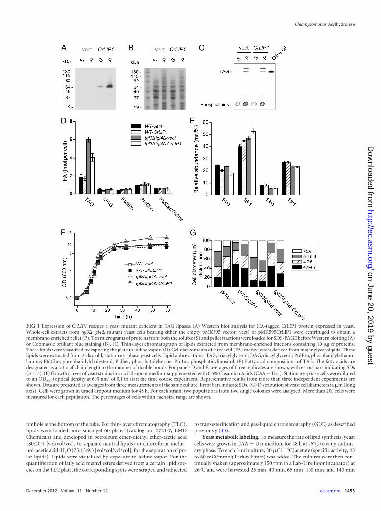
FIG 1 Expression of CrLIP1 rescues a yeast mutant deficient in TAG lipases. (A) Western blot analysis for HA-tagged CrLIP1 protein expressed in yeast.Whole-cell extracts from tgl3� tgl4� mutant yeast cells bearing either the empty pMK595 vector (vect) or pMK595CrLIP1 were centrifuged to obtain amembrane-enriched pellet (P). Ten micrograms of proteins from both the soluble (S) and pellet fractions were loaded for SDS-PAGE before Western blotting (A)or Coomassie brilliant blue staining (B). (C) Thin-layer chromatograph of lipids extracted from membrane-enriched fractions containing 10 �g of proteins.These lipids were visualized by exposing the plate to iodine vapor. (D) Cellular contents of fatty acid (FA) methyl esters derived from major glycerolipids. Theselipids were extracted from 2-day-old, stationary-phase yeast cells. Lipid abbreviations: TAG, triacylglycerol; DAG, diacylglycerol; PtdEtn, phosphatidylethano-lamine; PtdCho, phosphatidylcholesterol; PtdSer, phosphatidylserine; PtdIns, phosphatidylinositol. (E) Fatty acid compositions of TAG. The fatty acids aredesignated as a ratio of chain length to the number of double bonds. For panels D and E, averages of three replicates are shown, with errors bars indicating SDs(n � 3). (F) Growth curves of yeast strains in uracil dropout medium supplemented with 0.5% Casamino Acids (CAA � Ura). Stationary-phase cells were dilutedto an OD600 (optical density at 600 nm) of 0.1 to start the time course experiment. Representative results from more than three independent experiments areshown. Data are presented as averages from three measurements of the same culture. Error bars indicate SDs. (G) Distribution of yeast cell diameters in �m (longaxis). Cells were grown in uracil dropout medium for 48 h. For each strain, two populations from two single colonies were analyzed. More than 200 cells weremeasured for each population. The percentages of cells within each size range are shown.
Chlamydomonas Acylhydrolase
December 2012 Volume 11 Number 12 ec.asm.org 1453
on June 20, 2019 by guesthttp://ec.asm
.org/D
ownloaded from
after acetate supplementation. Total lipids were extracted (see above) andresolved by TLC resolution (petroleum ether-diethyl ether-acetic acid,80:20:1 [vol/vol/vol]) that separated neutral lipids. TAG, DAG, and TLCorigins (containing a mixture of polar lipids) were isolated for radioactiv-ity measurement by liquid scintillation counting. For yeast cellular TAGhydrolysis analyses, 50 �Ci (0.83 to 1.11 �mol) [14C]acetate was added to5-ml cultures grown for 48 h at 26°C. After 4 h of labeling, the cells werecollected by centrifugation, washed with fresh medium, and diluted 28-fold with fresh CAA � Ura medium to an optical density at 600 nm(OD600) of 0. 4 to 0.6. The cells were shaken for another 5 h with aliquotsfrozen at each time point. The growth curves were determined with cellsgrown under exactly the same conditions except that 1 �mol nonlabeledacetate was used. Lipid analysis was performed as described above for lipidsynthesis rate analysis.
Recombinant protein production. The CrLIP1 coding sequence wasamplified using the primers ligation-independent cloning forward (LICF)and LIC reverse (LICR) and the Phusion polymerase. The PCR productwas then integrated into expression vector pMK1006 through ligation-independent cloning (3). E. coli strain BL21-CodonPlus (Stratagene)transformed with empty vector pMK1006 or pMK1006CrLIP1 was grownto the log phase at 37°C, and protein production was induced by theaddition of IPTG to a final concentration of 0.5 mM. After 16 h of shakingat 220 rpm at 16°C, the cells were harvested by centrifugation (6,000 � g,5 min, 4°C). For protein preparation, the cells were collected and sus-pended in the lysis buffer (20 mM Tris-HCl [pH 7.9], 10% glycerol, 150mM NaCl, 1 mM dithiothreitol) and subjected to three freeze-thaw cycles,followed by sonication on ice (90 times of alternating 1-s pulses and 0.3-srests, with 1 min of ice chilling after every 15 cycles; a Branson digitalsonicator 250 with 25% energy output was used). The cell extracts wereobtained through centrifugation at 21,000 � g for 15 min and applied to anickel-nitrilotriacetate (Ni-NTA) affinity purification column (Qiagen).The CrLIP1 protein was eluted with the lysis buffer supplemented with200 mM imidazole. A spectrophotometric assay with the Bio-Rad proteinassay dye reagent concentrate (catalog no. 500-006 EDU; Bio-Rad) (8)was used to determine protein concentrations.
Lipase assay. For the lipase assay with various substrates, lipids dis-solved in organic solvents were dried under a stream of nitrogen gas. Thedried lipid was then dissolved in 350 �l of 0.1 M phosphate-bufferedsaline (PBS) (pH 7.4) containing various concentrations of Triton X-100(see below) by sonication (Sonicator 3000 with a Misonix microprobe) 6times for 10 s each (power setting, 1.5). Purified CrLIP1 protein or thesame volume of protein storage buffer was added. Fresh dithiothreitol(DTT) was added to the final concentration of 2 mM. The mixture wasthen vortexed and incubated at room temperature.
For the TAG lipase assay, 10 �Ci triolein (specific activity, 30 to 120Ci/mmol) (PerkinElmer) and 5.6 nmol olive oil were dried and resus-pended in 350 �l PBS containing 0.11 mM Triton X-100. The mixture wassplit into halves, and 18 �g of purified CrLIP1 in 20 �l protein storagebuffer was added to one of the aliquots. The mixture was then vortexedvigorously for 5 s and incubated at room temperature for 12 h. We triedmultiple other conditions for the assay, with varied amounts of olive oil (0to 250 nmol), DTT, or supplementation of CaCl2. Emulsifiers, including aphosphatidylcholine-phosphatidylinositol mix and bovine serum albu-min (BSA) were also used to replace Triton X-100 without any observableeffects on the results. For the DAG lipase assay, 150 nmol diolein (AvantiPolar Lipids) was dried and sonicated as described for TAG, except that0.53 mM, instead of 0.11 mM, Triton X-100 was used. Eighteen micro-grams of purified CrLIP1 was added in 50 �l protein storage buffer.The mixture was then vortexed briefly and incubated at room temper-ature for 16 h.
For assays containing radioactive polar lipids, Chlamydomonas cellswere grown to log phase in TAP medium, harvested, and suspended inmedium with 30 �Ci of [14C]acetate, as mentioned above (final concen-tration, 20 to 23.3 �M). The cells were harvested after another 4 h ofstandard growth (see above). Total lipids were extracted and resolved by
TLC developed in chloroform-methanol-acetic acid-H2O (75:13:9:3 [vol/vol/vol/vol]). Individual lipids were visualized by radiography and thenscraped off the plate before extraction with chloroform-methanol (1:1[vol/vol]) from the silica gel. Lipids (35 nmol with total radioactivities of10,000 to 40,000 dpm) were dried and dissolved as described for TAG. Forthe lipase assays, the emulsified lipid substrates were divided into halves,with 18 �g purified CrLIP1 added to one set and the same volume ofprotein storage buffer added to the other set. The mixture was incubatedfor 12 h at room temperature. To prepare radioactive steryl esters for invitro lipolysis reactions, 1 ml of early stationary-phase tgl3�tgl4�-vectyeast cells were diluted to 25 ml fresh CAA � Ura medium with an addi-tional 30 �Ci of [14C]acetate and grown for 48 h at 26°C. Total lipids wereextracted and resolved by TLC as described above. The steryl esters werethen isolated from the TLC plate. The assay conditions were the same asthose for the lipids derived from Chlamydomonas cells, with 35 nmollabeled steryl esters (10,000 dpm) as the substrates. For the assays withphosphatidylcholine (PtdCho), 60 nmol 18:1�9/16:0 PtdCho (Sigma-Al-drich) or 18:1�9/16:0 PtdCho (Avanti Polar Lipids) was dried and dis-solved as described for TAG. CrLIP1 (36 �g) was added, and aliquots wereflash frozen at 0, 6, and 16 h. The 0-h control was used to verify theintactness of PtdCho. The mixture was incubated for 12 h at room tem-perature. The background levels of fatty acids carried over with purifiedCrLIP1 protein were estimated in a control reaction without a substratelipid supplied and subtracted from the free fatty acids data obtained witha substrate.
All the lipase reactions were quenched by adding 2 volumes of lipidextraction solvent. The lipid products were then analyzed by TLC as de-scribed above. For Rhizopus arrhizus lipase (Sigma-Aldrich) digestion,200 �g of the enzyme preparation was used, and the incubation time wasshortened to 20 min.
amiRNA construct. The artificial micro-RNA (amiRNA) constructneeded to silence CrLIP1 expression was generated according to refer-ence 35. Briefly, four primers were designed using the MicroRNA De-signer protocols (http://wmd3.weigelworld.org/cgi-bin/webapp.cgi?page�Help). The primers were annealed by 5 min of boiling andovernight cooling to form a double-stranded fragment with CrLIP1-tar-geting sequences and overhangs compatible with the SpeI digestion site.This fragment was then integrated into the SpeI-linearizedpChlamiRNA3int vector (35) through ligation and transformation. Togenerate CrLIP1 knockdown lines, the artificial micro-RNA construct(pChlamiRNA3intCrLIP1) or the empty vector was digested with KpnI(New England BioLabs) and transformed into the dw15.1 Chlamydomo-nas strain using the glass bead transformation method (21). TAP agarcontaining 10 �g/ml paromomycin was used for selection. The real-timePCR described above was used to screen for lines with reduced mRNAabundance of CrLIP1.
Bioinformatics. A BLAST (1) search of the CrLIP1 protein sequencein the human genome was performed on the National Center for Biotech-nology Information website (http://blast.ncbi.nlm.nih.gov/). Protein se-quences were aligned with the ClustalW2 program (25).
RESULTSHeterologous expression of CrLIP1 complemented a yeast mu-tant deficient in TAG lipase activity. To identify the Chlamy-domonas lipases involved in TAG turnover, we used a yeast mutantdeficient in TAG lipase for functional complementation. In yeast,the major TAG lipases are encoded by TGL3 and TGL4 (4, 5, 24).Their deletion results in a hyperaccumulation of TAG in station-ary-phase cells. This phenotype can be rescued by an ectopic ex-pression of a mouse adipose triglyceride lipase (24). To chooseChlamydomonas candidate genes for a lipase that suppress theyeast lipid overstocking phenotype, putative lipases based onGene Ontology analyses of the Joint Genome Institute (JGI) Chla-mydomonas 3.0 database were cross-referenced to our transcrip-
Li et al.
1454 ec.asm.org Eukaryotic Cell
on June 20, 2019 by guesthttp://ec.asm
.org/D
ownloaded from
tomic studies of N-replete and -depleted cells (33) and to the datafrom a lipid droplet proteomics study (34). Eight genes were se-lected for the initial studies. The respective cDNAs were placedunder the control of the constitutive ADH1 promoter from a yeasthigh-copy-number plasmid. An N-terminal triple-HA tag wasadded to the coding sequence. These genes were tentatively namedCrLIP1 through LIP8 (summarized in Table S2 in the supplemen-tal material). The candidate genes CrLIP1, LIP3, LIP5, LIP6, andLIP8 were successfully cloned and expressed in the tgl3� tgl4�double-knockout mutant. Of the five genes tested, CrLIP1 (pro-tein identification no. 184308 and 519543; JGI Chlamydomonas4.0; also known as FAP12 [36]) (see Discussion) consistentlyshowed reduction of TAG and changes in cell growth (see below).For the remainder of this work, we focused on the CrLIP1 gene forits role in lipolysis.
We first examined the expression of CrLIP1 in yeast. As ex-pected for a lipase, the majority of recombinant CrLIP1 was asso-ciated with the membrane-enriched fraction that also includedlipid droplets and TAG (Fig. 1A to C). Importantly, the TAG over-accumulation phenotype of the tgl3� tgl4� mutant (5) was par-tially suppressed by CrLIP1 overexpression (Fig. 1D). There wasno discernible change in the steady-state levels of DAG or majorphospholipids, initially suggesting that CrLIP1 primarily affectedthe TAG accumulation or hydrolysis found in yeast. AlthoughCrLIP1 did not decrease the TAG content in a wild-type back-ground to a statistically significant level (Fig. 1D), it caused con-sistent differences in the fatty acid compositions of TAG, in boththe wild type and the tgl3� tgl4� mutant, with an increase in therelative amount of 16:1 and a decrease in 16:0 (Fig. 1E). This mightbe due to substrate specificity of CrLIP1 if it is a bona fide lipase.Besides these lipid phenotypes, we also noticed that the tgl3� tgl4�mutant grew to a higher optical density (OD) in the stationaryphase (Fig. 1F). Overexpressing CrLIP1 prevented this OD in-crease. One of the underlying reasons for this phenomenon couldbe the changes in cell size, as revealed by the measurement ofcellular diameter under a light microscope (Fig. 1G). Overall, thedouble lipase knockout strain had a higher number of large cellsthan did the wild-type strain. Larger particles should absorb morelight, resulting in higher ODs. Overexpressing CrLIP1 in the tgl3�tgl4� background caused the overall cell size distribution to ap-proach that of the wild-type cells. Together, these results demon-strate that the Chlamydomonas CrLIP1 protein was able to func-tionally complement the yeast TAG lipases Tgl3 and Tgl4.
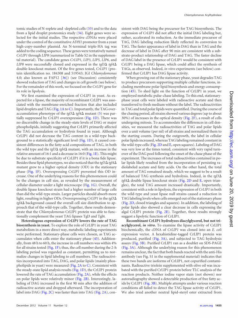
Heterologous expression of CrLIP1 lowers the rate of TAGbiosynthesis in yeast. To explore the role of CrLIP1 in yeast TAGmetabolism in a more direct way, metabolic labeling experimentswere performed. Stationary-phase cells were chosen, as TAG ac-cumulates when cells enter the stationary phase (45). Addition-ally, from 48 h to 60 h, the increase in cell numbers was within 4%for all strains tested (Fig. 1F); thus, the cell number during the 2-hlabeling period was regarded as constant, permitting us to nor-malize changes in lipid labeling to cell numbers. The radioactivi-ties incorporated into TAG, DAG, and polar lipids (mainly phos-pholipids in yeast) were measured (Fig. 2A to C). Consistent withthe steady-state lipid analysis results (Fig. 1D), the CrLIP1 proteinlowered the rate of TAG accumulation (Fig. 2A), while the effectson polar lipids were relatively minor (Fig. 2B). Interestingly, la-beling of DAG increased in the first 90 min after the addition ofradioactive acetate and dropped afterward. The incorporation oflabel into DAG (Fig. 2C) was faster than into TAG (Fig. 2A), con-
sistent with DAG being the precursor for TAG biosynthesis. Theexpression of CrLIP1 did not affect the initial DAG labeling but,rather, accelerated its reduction. As the immediate precursor ofTAG, DAG labeling reduction likely reflected its conversion toTAG. The faster appearance of label in DAG than in TAG and thedecrease of label in DAG after 90 min are consistent with a sub-strate-product relationship of DAG and TAG. The faster declineof DAG label in the presence of CrLIP1 would be consistent withCrLIP1 being a DAG lipase, which could affect the synthesis ofTAGs, as observed. Indeed, in vitro experiments (see below) con-firmed that CrLIP1 has DAG lipase activity.
When growing out of the stationary phase, yeast degrades TAGto produce precursors supporting multiple cellular functions, in-cluding membrane polar lipid biosynthesis and energy consump-tion (45). To shed light on the function of CrLIP1 in yeast, weemployed metabolic pulse-chase labeling. To this end, stationary-phase yeast cells were labeled with radioactive acetate and thentransferred to fresh medium without the label. The radioactivitiesin TAG and total polar lipids were quantified. During the course ofthe chasing period, all strains showed various degrees (up to about30%) of increases in the optical density (Fig. 2F), a result of cellsundergoing mitosis. To accommodate the differences in cell den-sities, we measured the radioactivities of TAG and polar lipidsover a unit volume (per ml) of all strains and normalized them tothe starting counts. During the outgrowth, the label in cellularpolar lipids increased, whereas TAG labeling remained constant inthe wild-type cells (Fig. 2D and E, open squares). Labeling of DAGwas very low at the times tested, consistent with very rapid turn-over of the DAG pool following the onset of the chase phase of theexperiment. The increases of total radioactivities contained in po-lar lipids likely resulted from the incorporation of persisting ra-diolabeled acetate and fatty acids. On the other hand, the totalamount of TAG remained steady, which we suggest to be a resultof balanced TAG synthesis and hydrolysis. Indeed, in the tgl3�tgl4� double lipase knockout strain (Fig. 2D and E, open trian-gles), the total TAG amount increased drastically. Importantly,consistent with a role in lipolysis, the expression of CrLIP1 in boththe wild-type and yeast lipase null strains caused a reduction inTAG labeling levels when cells emerged out of the stationary phase(Fig. 2D, closed triangles and squares). In addition, the labeling ofpolar lipids also showed a clear decrease in cells expressing thealgal CrLIP1 protein (Fig. 2E). Together, these results stronglysuggest a lipolytic function of CrLIP1.
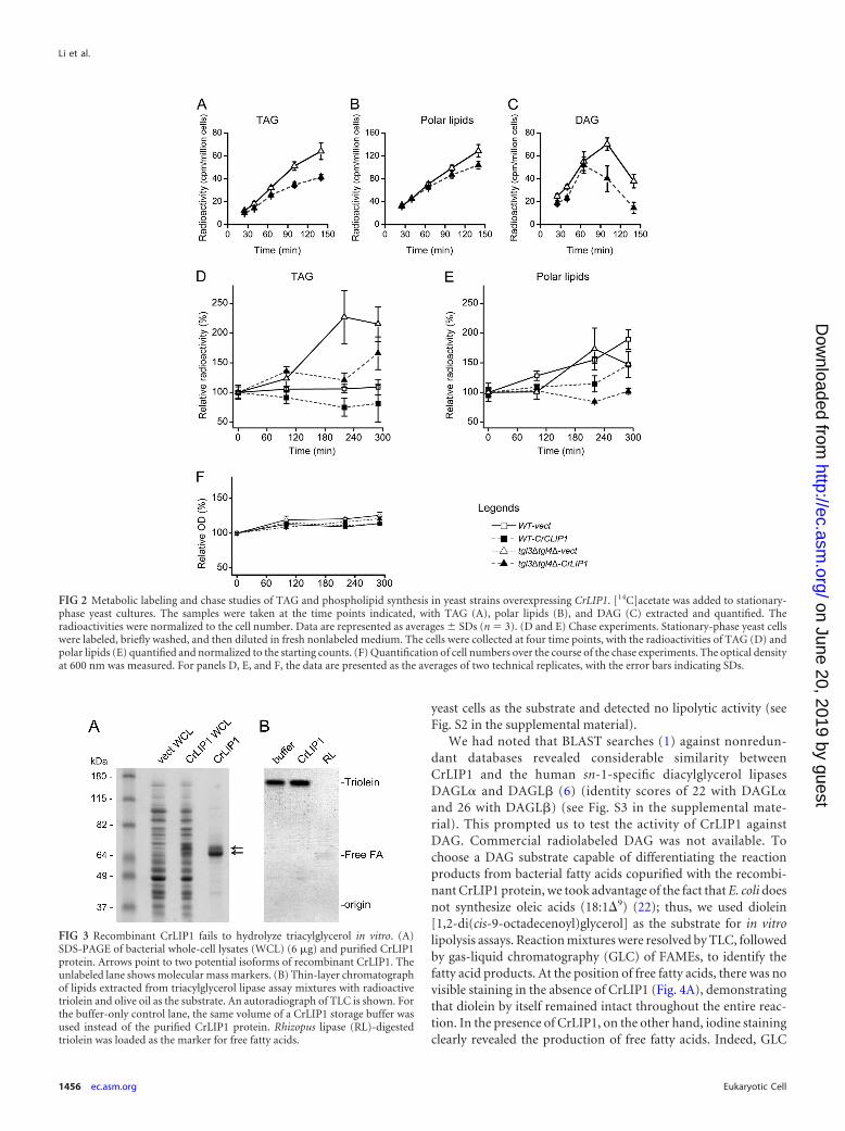
Recombinant CrLIP1 hydrolyzes diacylglycerol, but not tri-acylglycerol, in vitro. To examine the lipase activity of CrLIP1biochemically, the cDNA of CrLIP1 was cloned into an E. coliexpression vector. A hexahistidine-tagged CrLIP1 protein wasproduced, purified (Fig. 3A), and subjected to TAG hydrolysisassays (Fig. 3B). Purified CrLIP1 ran as a doublet on SDS-PAGE(Fig. 3A). Although the underlying reason for this phenomenonremains unclear, the fact that both bands reacted with the anti-Hisantibody (see Fig. S1 in the supplemental material) indicates thatthese two bands are isoforms of CrLIP1, not copurified contami-nants. Radioactive triolein supplemented with olive oil was incu-bated with the purified CrLIP1 protein before TLC analysis of thereaction products. Neither iodine vapor stain (not shown) norautoradiography showed a detectable production of free fatty ac-ids by CrLIP1 (Fig. 3B). Multiple attempts under various reactionconditions all failed to detect the TAG lipase activity of CrLIP1.We also tried another neutral lipid-steryl ester extracted from
Chlamydomonas Acylhydrolase
December 2012 Volume 11 Number 12 ec.asm.org 1455
on June 20, 2019 by guesthttp://ec.asm
.org/D
ownloaded from
yeast cells as the substrate and detected no lipolytic activity (seeFig. S2 in the supplemental material).
We had noted that BLAST searches (1) against nonredun-dant databases revealed considerable similarity betweenCrLIP1 and the human sn-1-specific diacylglycerol lipasesDAGL� and DAGL� (6) (identity scores of 22 with DAGL�and 26 with DAGL�) (see Fig. S3 in the supplemental mate-rial). This prompted us to test the activity of CrLIP1 againstDAG. Commercial radiolabeled DAG was not available. Tochoose a DAG substrate capable of differentiating the reactionproducts from bacterial fatty acids copurified with the recombi-nant CrLIP1 protein, we took advantage of the fact that E. coli doesnot synthesize oleic acids (18:1�9) (22); thus, we used diolein[1,2-di(cis-9-octadecenoyl)glycerol] as the substrate for in vitrolipolysis assays. Reaction mixtures were resolved by TLC, followedby gas-liquid chromatography (GLC) of FAMEs, to identify thefatty acid products. At the position of free fatty acids, there was novisible staining in the absence of CrLIP1 (Fig. 4A), demonstratingthat diolein by itself remained intact throughout the entire reac-tion. In the presence of CrLIP1, on the other hand, iodine stainingclearly revealed the production of free fatty acids. Indeed, GLC
FIG 3 Recombinant CrLIP1 fails to hydrolyze triacylglycerol in vitro. (A)SDS-PAGE of bacterial whole-cell lysates (WCL) (6 �g) and purified CrLIP1protein. Arrows point to two potential isoforms of recombinant CrLIP1. Theunlabeled lane shows molecular mass markers. (B) Thin-layer chromatographof lipids extracted from triacylglycerol lipase assay mixtures with radioactivetriolein and olive oil as the substrate. An autoradiograph of TLC is shown. Forthe buffer-only control lane, the same volume of a CrLIP1 storage buffer wasused instead of the purified CrLIP1 protein. Rhizopus lipase (RL)-digestedtriolein was loaded as the marker for free fatty acids.
FIG 2 Metabolic labeling and chase studies of TAG and phospholipid synthesis in yeast strains overexpressing CrLIP1. [14C]acetate was added to stationary-phase yeast cultures. The samples were taken at the time points indicated, with TAG (A), polar lipids (B), and DAG (C) extracted and quantified. Theradioactivities were normalized to the cell number. Data are represented as averages SDs (n � 3). (D and E) Chase experiments. Stationary-phase yeast cellswere labeled, briefly washed, and then diluted in fresh nonlabeled medium. The cells were collected at four time points, with the radioactivities of TAG (D) andpolar lipids (E) quantified and normalized to the starting counts. (F) Quantification of cell numbers over the course of the chase experiments. The optical densityat 600 nm was measured. For panels D, E, and F, the data are presented as the averages of two technical replicates, with the error bars indicating SDs.
Li et al.
1456 ec.asm.org Eukaryotic Cell
on June 20, 2019 by guesthttp://ec.asm
.org/D
ownloaded from
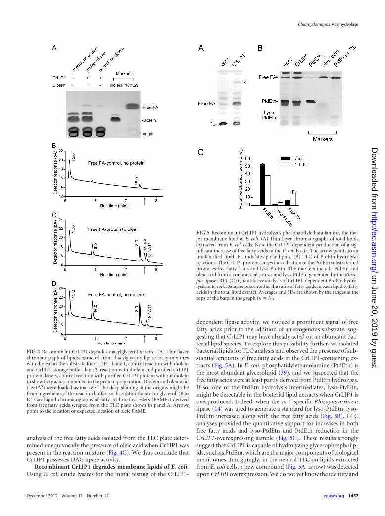
analysis of the free fatty acids isolated from the TLC plate deter-mined unequivocally the presence of oleic acid when CrLIP1 waspresent in the reaction mixture (Fig. 4C). We thus conclude thatCrLIP1 possesses DAG lipase activity.
Recombinant CrLIP1 degrades membrane lipids of E. coli.Using E. coli crude lysates for the initial testing of the CrLIP1-
dependent lipase activity, we noticed a prominent signal of freefatty acids prior to the addition of an exogenous substrate, sug-gesting that CrLIP1 may have already acted on an abundant bac-terial lipid species. To explore this possibility further, we isolatedbacterial lipids for TLC analysis and observed the presence of sub-stantial amounts of free fatty acids in the CrLIP1-containing ex-tracts (Fig. 5A). In E. coli, phosphatidylethanolamine (PtdEtn) isthe most abundant glycerolipid (39), and we suspected that thefree fatty acids were at least partly derived from PtdEtn hydrolysis.If so, one of the PtdEtn hydrolysis intermediates, lyso-PtdEtn,might be detectable in the bacterial lipid extracts when CrLIP1 isoverproduced. Indeed, when the sn-1-specific Rhizopus arrhizuslipase (14) was used to generate a standard for lyso-PtdEtn, lyso-PtdEtn increased along with the free fatty acids (Fig. 5B). GLCanalyses provided the quantitative support for increases in bothfree fatty acids and lyso-PtdEtn and PtdEtn reduction in theCrLIP1-overexpressing sample (Fig. 5C). These results stronglysuggest that CrLIP1 is capable of hydrolyzing glycerophospholip-ids, such as PtdEtn, which are the major components of biologicalmembranes. Intriguingly, in the neutral TLC on lipids extractedfrom E. coli cells, a new compound (Fig. 5A, arrow) was detectedupon CrLIP1 overexpression. We do not yet know the identity and
FIG 4 Recombinant CrLIP1 degrades diacylglycerol in vitro. (A) Thin-layerchromatograph of lipids extracted from diacylglycerol lipase assay mixtureswith diolein as the substrate for CrLIP1. Lane 1, control reaction with dioleinand CrLIP1 storage buffer; lane 2, reaction with diolein and purified CrLIP1protein; lane 3, control reaction with purified CrLIP1 protein without dioleinto show fatty acids contained in the protein preparation. Diolein and oleic acid(18:1�9) were loaded as markers. The deep staining at the origins might befrom ingredients of the reaction buffer, such as dithiothreitol or glycerol. (B toD) Gas-liquid chromatographs of fatty acid methyl esters (FAMEs) derivedfrom free fatty acids scraped from the TLC plate shown in panel A. Arrowspoint to the location or expected location of oleic FAME.
FIG 5 Recombinant CrLIP1 hydrolyzes phosphatidylethanolamine, the ma-jor membrane lipid of E. coli. (A) Thin-layer chromatographs of total lipidsextracted from E. coli cells. Note the CrLIP1-dependent production of a sig-nificant increase of free fatty acids in the E. coli lysate. The arrow points to anunidentified lipid. PL indicates polar lipids. (B) TLC of PtdEtn hydrolysisreactions. The CrLIP1 protein causes the reduction of the PtdEtn substrate andproduces free fatty acids and lyso-PtdEtn. The markers include PtdEtn andoleic acid from a commercial source and lyso-PtdEtn generated by the Rhizo-pus lipase (RL). (C) Quantitative analysis of CrLIP1-dependent PtdEtn hydro-lysis in E. coli. Data are presented as the ratio of fatty acids in each lipid to fattyacids in the total lipid extract. Averages and SDs are shown by the ranges at thetops of the bars in the graph (n � 3).
Chlamydomonas Acylhydrolase
December 2012 Volume 11 Number 12 ec.asm.org 1457
on June 20, 2019 by guesthttp://ec.asm
.org/D
ownloaded from
origin of this species. While this phenomenon is likely related tothe biochemical activity of CrLIP1, a definitive identification ofthis bacterial lipid species will require additional efforts that arebeyond the scope of the current work.
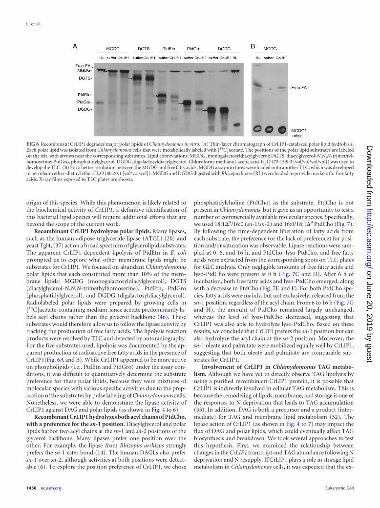
Recombinant CrLIP1 hydrolyzes polar lipids. Many lipases,such as the human adipose triglyceride lipase (ATGL) (20) andyeast Tgl4, (37) act on a broad spectrum of glycerolipid substrates.The apparent CrLIP1-dependent lipolysis of PtdEtn in E. coliprompted us to explore what other membrane lipids might besubstrates for CrLIP1. We focused on abundant Chlamydomonaspolar lipids that each constituted more than 10% of the mem-brane lipids: MGDG (monogalactosyldiacylglycerol), DGTS(diacylglycerol-N,N,N-trimethylhomoserine), PtdEtn, PtdGro(phosphatidylglycerol), and DGDG (digalactosyldiacylglycerol).Radiolabeled polar lipids were prepared by growing cells in[14C]acetate-containing medium, since acetate predominantly la-bels acyl chains rather than the glycerol backbone (46). Thesesubstrates would therefore allow us to follow the lipase activity bytracking the production of free fatty acids. The lipolysis reactionproducts were resolved by TLC and detected by autoradiography.For the five substrates used, lipolysis was documented by the ap-parent production of radioactive free fatty acids in the presence ofCrLIP1(Fig. 6A and B). While CrLIP1 appeared to be more activeon phospholipids (i.e., PtdEtn and PtdGro) under the assay con-ditions, it was difficult to quantitatively determine the substratepreference for these polar lipids, because they were mixtures ofmolecular species with various specific activities due to the prep-aration of the substrates by pulse labeling of Chlamydomonas cells.Nonetheless, we were able to demonstrate the lipase activity ofCrLIP1 against DAG and polar lipids (as shown in Fig. 4 to 6).
Recombinant CrLIP1 hydrolyzes both acyl chains of PtdCho,with a preference for the sn-1 position. Diacylglycerol and polarlipids harbor two acyl chains at the sn-1 and sn-2 positions of theglycerol backbone. Many lipases prefer one position over theother. For example, the lipase from Rhizopus arrhizus stronglyprefers the sn-1 ester bond (14). The human DAGLs also prefersn-1 over sn-2, although activities at both positions were detect-able (6). To explore the position preference of CrLIP1, we chose
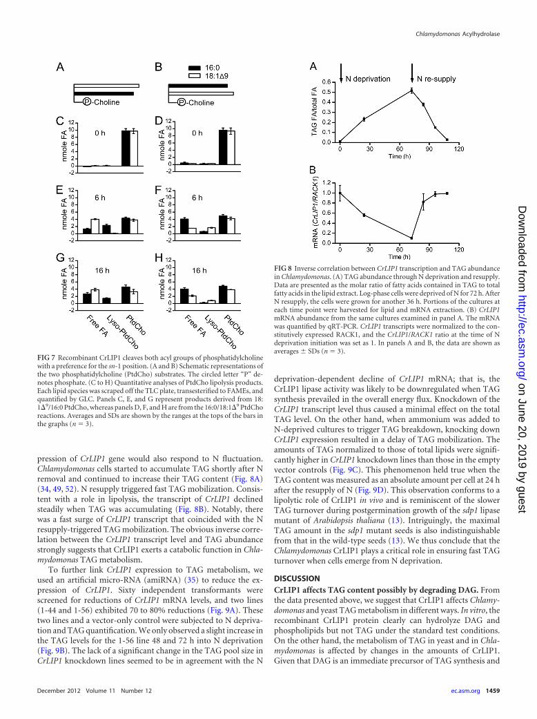
phosphatidylcholine (PtdCho) as the substrate. PtdCho is notpresent in Chlamydomonas, but it gave us an opportunity to test anumber of commercially available molecular species. Specifically,we used 18:1�9/16:0 (sn-1/sn-2) and 16:0/18:1�9 PtdCho (Fig. 7).By following the time-dependent liberation of fatty acids fromeach substrate, the preference (or the lack of preference) for posi-tion and/or saturation was observable. Lipase reactions were sam-pled at 0, 6, and 16 h, and PtdCho, lyso-PtdCho, and free fattyacids were extracted from the corresponding spots on TLC platesfor GLC analysis. Only negligible amounts of free fatty acids andlyso-PtdCho were present at 0 h (Fig. 7C and D). After 6 h ofincubation, both free fatty acids and lyso-PtdCho emerged, alongwith a decrease in PtdCho (Fig. 7E and F). For both PtdCho spe-cies, fatty acids were mainly, but not exclusively, released from thesn-1 position, regardless of the acyl chain. From 6 to 16 h (Fig. 7Gand H), the amount of PtdCho remained largely unchanged,whereas the level of lyso-PtdCho decreased, suggesting thatCrLIP1 was also able to hydrolyze lyso-PtdCho. Based on theseresults, we conclude that CrLIP1 prefers the sn-1 position but canalso hydrolyze the acyl chain at the sn-2 position. Moreover, thesn-1 oleate and palmitate were mobilized equally well by CrLIP1,suggesting that both oleate and palmitate are comparable sub-strates for CrLIP1.
Involvement of CrLIP1 in Chlamydomonas TAG metabo-lism. Although we have yet to directly observe TAG lipolysis byusing a purified recombinant CrLIP1 protein, it is possible thatCrLIP1 is indirectly involved in cellular TAG metabolism. This isbecause the remodeling of lipids, membrane, and storage is one ofthe responses to N deprivation that leads to TAG accumulation(33). In addition, DAG is both a precursor and a product (inter-mediate) for TAG and membrane lipid metabolism (32). Thelipase action of CrLIP1 (as shown in Fig. 4 to 7) may impact theflux of DAG and polar lipids, which could eventually affect TAGbiosynthesis and breakdown. We took several approaches to testthis hypothesis. First, we examined the relationship betweenchanges in the CrLIP1 transcript and TAG abundance following Ndeprivation and N resupply. If CrLIP1 plays a role in storage lipidmetabolism in Chlamydomonas cells, it was expected that the ex-
FIG 6 Recombinant CrLIP1 degrades major polar lipids of Chlamydomonas in vitro. (A) Thin-layer chromatograph of CrLIP1-catalyzed polar lipid hydrolysis.Each polar lipid was isolated from Chlamydomonas cells that were metabolically labeled with [14C]acetate. The positions of the polar lipid substrates are labeledon the left, with arrows near the corresponding substrates. Lipid abbreviations: MGDG, monogalactosyldiacylglycerol; DGTS, diacylglycerol-N,N,N-trimethyl-homoserine; PtdGro, phosphatidylglycerol; DGDG, digalactosyldiacylglycerol. Chloroform-methanol-acetic acid-H2O (75:13:9:3 [vol/vol/vol/vol]) was used todevelop the TLC. (B) For a better resolution between the MGDG and free fatty acids, MGDG assay mixtures were loaded onto another TLC, which was developedin petroleum ether-diethyl ether-H2O (80:20:1 [vol/vol/vol]). MGDG and DGDG digested with Rhizopus lipase (RL) were loaded to provide markers for free fattyacids. X-ray films exposed to TLC plates are shown.
Li et al.
1458 ec.asm.org Eukaryotic Cell
on June 20, 2019 by guesthttp://ec.asm
.org/D
ownloaded from
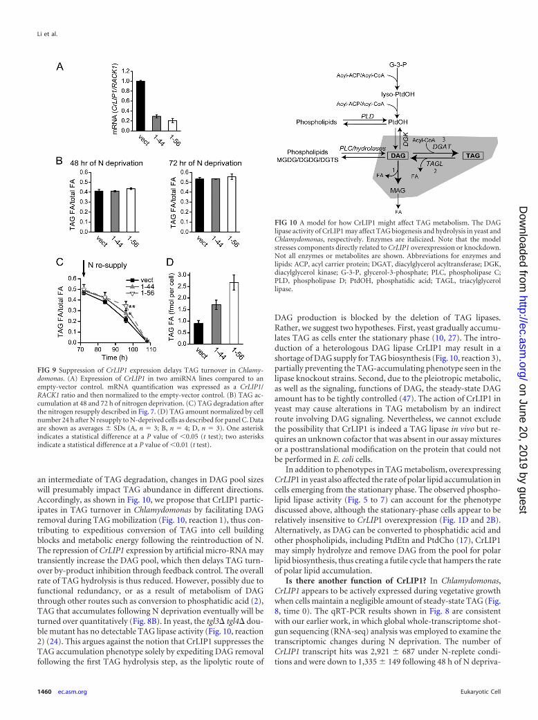
pression of CrLIP1 gene would also respond to N fluctuation.Chlamydomonas cells started to accumulate TAG shortly after Nremoval and continued to increase their TAG content (Fig. 8A)(34, 49, 52). N resupply triggered fast TAG mobilization. Consis-tent with a role in lipolysis, the transcript of CrLIP1 declinedsteadily when TAG was accumulating (Fig. 8B). Notably, therewas a fast surge of CrLIP1 transcript that coincided with the Nresupply-triggered TAG mobilization. The obvious inverse corre-lation between the CrLIP1 transcript level and TAG abundancestrongly suggests that CrLIP1 exerts a catabolic function in Chla-mydomonas TAG metabolism.
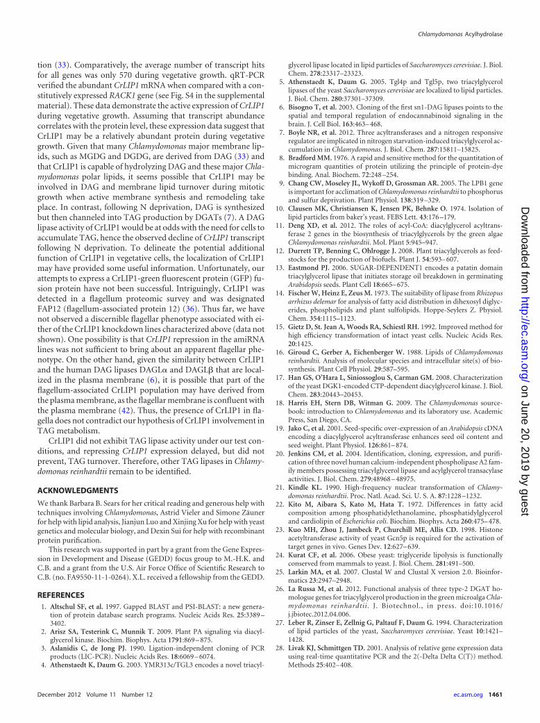
To further link CrLIP1 expression to TAG metabolism, weused an artificial micro-RNA (amiRNA) (35) to reduce the ex-pression of CrLIP1. Sixty independent transformants werescreened for reductions of CrLIP1 mRNA levels, and two lines(1-44 and 1-56) exhibited 70 to 80% reductions (Fig. 9A). Thesetwo lines and a vector-only control were subjected to N depriva-tion and TAG quantification. We only observed a slight increase inthe TAG levels for the 1-56 line 48 and 72 h into N deprivation(Fig. 9B). The lack of a significant change in the TAG pool size inCrLIP1 knockdown lines seemed to be in agreement with the N
deprivation-dependent decline of CrLIP1 mRNA; that is, theCrLIP1 lipase activity was likely to be downregulated when TAGsynthesis prevailed in the overall energy flux. Knockdown of theCrLIP1 transcript level thus caused a minimal effect on the totalTAG level. On the other hand, when ammonium was added toN-deprived cultures to trigger TAG breakdown, knocking downCrLIP1 expression resulted in a delay of TAG mobilization. Theamounts of TAG normalized to those of total lipids were signifi-cantly higher in CrLIP1 knockdown lines than those in the emptyvector controls (Fig. 9C). This phenomenon held true when theTAG content was measured as an absolute amount per cell at 24 hafter the resupply of N (Fig. 9D). This observation conforms to alipolytic role of CrLIP1 in vivo and is reminiscent of the slowerTAG turnover during postgermination growth of the sdp1 lipasemutant of Arabidopsis thaliana (13). Intriguingly, the maximalTAG amount in the sdp1 mutant seeds is also indistinguishablefrom that in the wild-type seeds (13). We thus conclude that theChlamydomonas CrLIP1 plays a critical role in ensuring fast TAGturnover when cells emerge from N deprivation.
DISCUSSIONCrLIP1 affects TAG content possibly by degrading DAG. Fromthe data presented above, we suggest that CrLIP1 affects Chlamy-domonas and yeast TAG metabolism in different ways. In vitro, therecombinant CrLIP1 protein clearly can hydrolyze DAG andphospholipids but not TAG under the standard test conditions.On the other hand, the metabolism of TAG in yeast and in Chla-mydomonas is affected by changes in the amounts of CrLIP1.Given that DAG is an immediate precursor of TAG synthesis and
FIG 7 Recombinant CrLIP1 cleaves both acyl groups of phosphatidylcholinewith a preference for the sn-1 position. (A and B) Schematic representations ofthe two phosphatidylcholine (PtdCho) substrates. The circled letter “P” de-notes phosphate. (C to H) Quantitative analyses of PtdCho lipolysis products.Each lipid species was scraped off the TLC plate, transesterified to FAMEs, andquantified by GLC. Panels C, E, and G represent products derived from 18:1�9/16:0 PtdCho, whereas panels D, F, and H are from the 16:0/18:1�9 PtdChoreactions. Averages and SDs are shown by the ranges at the tops of the bars inthe graphs (n � 3).
FIG 8 Inverse correlation between CrLIP1 transcription and TAG abundancein Chlamydomonas. (A) TAG abundance through N deprivation and resupply.Data are presented as the molar ratio of fatty acids contained in TAG to totalfatty acids in the lipid extract. Log-phase cells were deprived of N for 72 h. AfterN resupply, the cells were grown for another 36 h. Portions of the cultures ateach time point were harvested for lipid and mRNA extraction. (B) CrLIP1mRNA abundance from the same cultures examined in panel A. The mRNAwas quantified by qRT-PCR. CrLIP1 transcripts were normalized to the con-stitutively expressed RACK1, and the CrLIP1/RACK1 ratio at the time of Ndeprivation initiation was set as 1. In panels A and B, the data are shown asaverages SDs (n � 3).
Chlamydomonas Acylhydrolase
December 2012 Volume 11 Number 12 ec.asm.org 1459
on June 20, 2019 by guesthttp://ec.asm
.org/D
ownloaded from
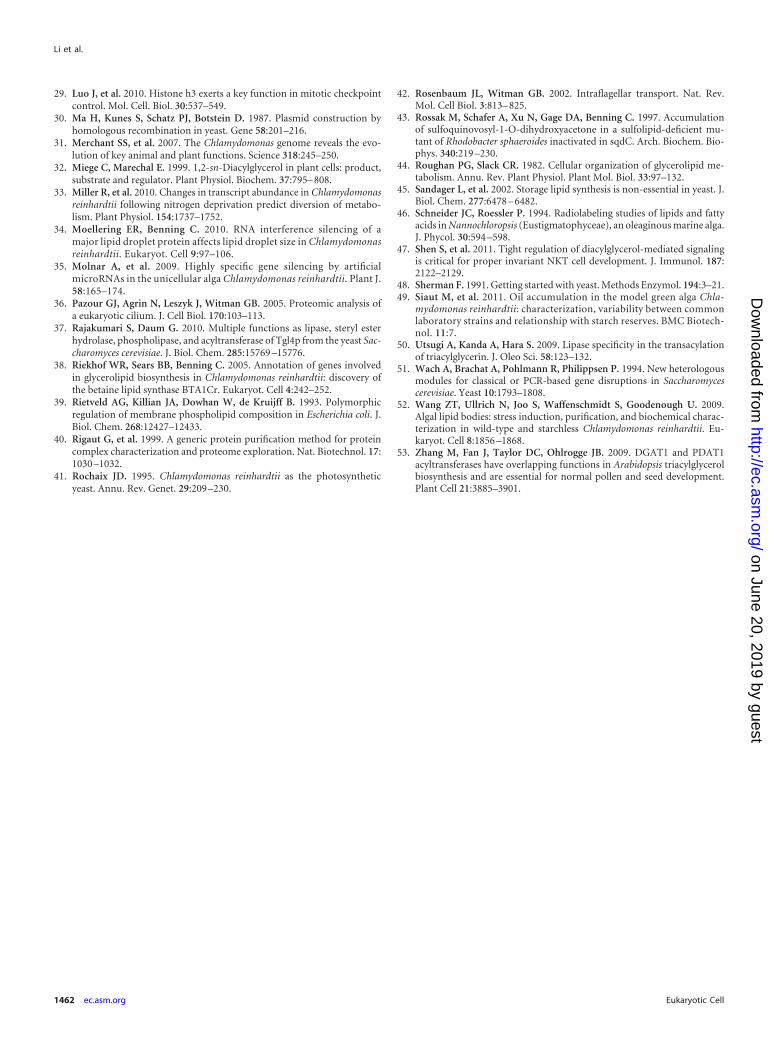
an intermediate of TAG degradation, changes in DAG pool sizeswill presumably impact TAG abundance in different directions.Accordingly, as shown in Fig. 10, we propose that CrLIP1 partic-ipates in TAG turnover in Chlamydomonas by facilitating DAGremoval during TAG mobilization (Fig. 10, reaction 1), thus con-tributing to expeditious conversion of TAG into cell buildingblocks and metabolic energy following the reintroduction of N.The repression of CrLIP1 expression by artificial micro-RNA maytransiently increase the DAG pool, which then delays TAG turn-over by-product inhibition through feedback control. The overallrate of TAG hydrolysis is thus reduced. However, possibly due tofunctional redundancy, or as a result of metabolism of DAGthrough other routes such as conversion to phosphatidic acid (2),TAG that accumulates following N deprivation eventually will beturned over quantitatively (Fig. 8B). In yeast, the tgl3� tgl4� dou-ble mutant has no detectable TAG lipase activity (Fig. 10, reaction2) (24). This argues against the notion that CrLIP1 suppresses theTAG accumulation phenotype solely by expediting DAG removalfollowing the first TAG hydrolysis step, as the lipolytic route of
DAG production is blocked by the deletion of TAG lipases.Rather, we suggest two hypotheses. First, yeast gradually accumu-lates TAG as cells enter the stationary phase (10, 27). The intro-duction of a heterologous DAG lipase CrLIP1 may result in ashortage of DAG supply for TAG biosynthesis (Fig. 10, reaction 3),partially preventing the TAG-accumulating phenotype seen in thelipase knockout strains. Second, due to the pleiotropic metabolic,as well as the signaling, functions of DAG, the steady-state DAGamount has to be tightly controlled (47). The action of CrLIP1 inyeast may cause alterations in TAG metabolism by an indirectroute involving DAG signaling. Nevertheless, we cannot excludethe possibility that CrLIP1 is indeed a TAG lipase in vivo but re-quires an unknown cofactor that was absent in our assay mixturesor a posttranslational modification on the protein that could notbe performed in E. coli cells.
In addition to phenotypes in TAG metabolism, overexpressingCrLIP1 in yeast also affected the rate of polar lipid accumulation incells emerging from the stationary phase. The observed phospho-lipid lipase activity (Fig. 5 to 7) can account for the phenotypediscussed above, although the stationary-phase cells appear to berelatively insensitive to CrLIP1 overexpression (Fig. 1D and 2B).Alternatively, as DAG can be converted to phosphatidic acid andother phospholipids, including PtdEtn and PtdCho (17), CrLIP1may simply hydrolyze and remove DAG from the pool for polarlipid biosynthesis, thus creating a futile cycle that hampers the rateof polar lipid accumulation.
Is there another function of CrLIP1? In Chlamydomonas,CrLIP1 appears to be actively expressed during vegetative growthwhen cells maintain a negligible amount of steady-state TAG (Fig.8, time 0). The qRT-PCR results shown in Fig. 8 are consistentwith our earlier work, in which global whole-transcriptome shot-gun sequencing (RNA-seq) analysis was employed to examine thetranscriptomic changes during N deprivation. The number ofCrLIP1 transcript hits was 2,921 687 under N-replete condi-tions and were down to 1,335 149 following 48 h of N depriva-
FIG 9 Suppression of CrLIP1 expression delays TAG turnover in Chlamy-domonas. (A) Expression of CrLIP1 in two amiRNA lines compared to anempty-vector control. mRNA quantification was expressed as a CrLIP1/RACK1 ratio and then normalized to the empty-vector control. (B) TAG ac-cumulation at 48 and 72 h of nitrogen deprivation. (C) TAG degradation afterthe nitrogen resupply described in Fig. 7. (D) TAG amount normalized by cellnumber 24 h after N resupply to N-deprived cells as described for panel C. Dataare shown as averages SDs (A, n � 3; B, n � 4; D, n � 3). One asteriskindicates a statistical difference at a P value of 0.05 (t test); two asterisksindicate a statistical difference at a P value of 0.01 (t test).
FIG 10 A model for how CrLIP1 might affect TAG metabolism. The DAGlipase activity of CrLIP1 may affect TAG biogenesis and hydrolysis in yeast andChlamydomonas, respectively. Enzymes are italicized. Note that the modelstresses components directly related to CrLIP1 overexpression or knockdown.Not all enzymes or metabolites are shown. Abbreviations for enzymes andlipids: ACP, acyl carrier protein; DGAT, diacylglycerol acyltransferase; DGK,diacylglycerol kinase; G-3-P, glycerol-3-phosphate; PLC, phospholipase C;PLD, phospholipase D; PtdOH, phosphatidic acid; TAGL, triacylglycerollipase.
Li et al.
1460 ec.asm.org Eukaryotic Cell
on June 20, 2019 by guesthttp://ec.asm
.org/D
ownloaded from
tion (33). Comparatively, the average number of transcript hitsfor all genes was only 570 during vegetative growth. qRT-PCRverified the abundant CrLIP1 mRNA when compared with a con-stitutively expressed RACK1 gene (see Fig. S4 in the supplementalmaterial). These data demonstrate the active expression of CrLIP1during vegetative growth. Assuming that transcript abundancecorrelates with the protein level, these expression data suggest thatCrLIP1 may be a relatively abundant protein during vegetativegrowth. Given that many Chlamydomonas major membrane lip-ids, such as MGDG and DGDG, are derived from DAG (33) andthat CrLIP1 is capable of hydrolyzing DAG and these major Chla-mydomonas polar lipids, it seems possible that CrLIP1 may beinvolved in DAG and membrane lipid turnover during mitoticgrowth when active membrane synthesis and remodeling takeplace. In contrast, following N deprivation, DAG is synthesizedbut then channeled into TAG production by DGATs (7). A DAGlipase activity of CrLIP1 would be at odds with the need for cells toaccumulate TAG, hence the observed decline of CrLIP1 transcriptfollowing N deprivation. To delineate the potential additionalfunction of CrLIP1 in vegetative cells, the localization of CrLIP1may have provided some useful information. Unfortunately, ourattempts to express a CrLIP1-green fluorescent protein (GFP) fu-sion protein have not been successful. Intriguingly, CrLIP1 wasdetected in a flagellum proteomic survey and was designatedFAP12 (flagellum-associated protein 12) (36). Thus far, we havenot observed a discernible flagellar phenotype associated with ei-ther of the CrLIP1 knockdown lines characterized above (data notshown). One possibility is that CrLIP1 repression in the amiRNAlines was not sufficient to bring about an apparent flagellar phe-notype. On the other hand, given the similarity between CrLIP1and the human DAG lipases DAGL� and DAGL� that are local-ized in the plasma membrane (6), it is possible that part of theflagellum-associated CrLIP1 population may have derived fromthe plasma membrane, as the flagellar membrane is confluent withthe plasma membrane (42). Thus, the presence of CrLIP1 in fla-gella does not contradict our hypothesis of CrLIP1 involvement inTAG metabolism.
CrLIP1 did not exhibit TAG lipase activity under our test con-ditions, and repressing CrLIP1 expression delayed, but did notprevent, TAG turnover. Therefore, other TAG lipases in Chlamy-domonas reinhardtii remain to be identified.
ACKNOWLEDGMENTS
We thank Barbara B. Sears for her critical reading and generous help withtechniques involving Chlamydomonas, Astrid Vieler and Simone Zäunerfor help with lipid analysis, Jianjun Luo and Xinjing Xu for help with yeastgenetics and molecular biology, and Dexin Sui for help with recombinantprotein purification.
This research was supported in part by a grant from the Gene Expres-sion in Development and Disease (GEDD) focus group to M.-H.K. andC.B. and a grant from the U.S. Air Force Office of Scientific Research toC.B. (no. FA9550-11-1-0264). X.L. received a fellowship from the GEDD.
REFERENCES1. Altschul SF, et al. 1997. Gapped BLAST and PSI-BLAST: a new genera-
tion of protein database search programs. Nucleic Acids Res. 25:3389 –3402.
2. Arisz SA, Testerink C, Munnik T. 2009. Plant PA signaling via diacyl-glycerol kinase. Biochim. Biophys. Acta 1791:869 – 875.
3. Aslanidis C, de Jong PJ. 1990. Ligation-independent cloning of PCRproducts (LIC-PCR). Nucleic Acids Res. 18:6069 – 6074.
4. Athenstaedt K, Daum G. 2003. YMR313c/TGL3 encodes a novel triacyl-
glycerol lipase located in lipid particles of Saccharomyces cerevisiae. J. Biol.Chem. 278:23317–23323.
5. Athenstaedt K, Daum G. 2005. Tgl4p and Tgl5p, two triacylglycerollipases of the yeast Saccharomyces cerevisiae are localized to lipid particles.J. Biol. Chem. 280:37301–37309.
6. Bisogno T, et al. 2003. Cloning of the first sn1-DAG lipases points to thespatial and temporal regulation of endocannabinoid signaling in thebrain. J. Cell Biol. 163:463– 468.
7. Boyle NR, et al. 2012. Three acyltransferases and a nitrogen responsiveregulator are implicated in nitrogen starvation-induced triacylglycerol ac-cumulation in Chlamydomonas. J. Biol. Chem. 287:15811–15825.
8. Bradford MM. 1976. A rapid and sensitive method for the quantitation ofmicrogram quantities of protein utilizing the principle of protein-dyebinding. Anal. Biochem. 72:248 –254.
9. Chang CW, Moseley JL, Wykoff D, Grossman AR. 2005. The LPB1 geneis important for acclimation of Chlamydomonas reinhardtii to phosphorusand sulfur deprivation. Plant Physiol. 138:319 –329.
10. Clausen MK, Christiansen K, Jensen PK, Behnke O. 1974. Isolation oflipid particles from baker’s yeast. FEBS Lett. 43:176 –179.
11. Deng XD, et al. 2012. The roles of acyl-CoA: diacylglycerol acyltrans-ferase 2 genes in the biosynthesis of triacylglycerols by the green algaeChlamydomonas reinhardtii. Mol. Plant 5:945–947.
12. Durrett TP, Benning C, Ohlrogge J. 2008. Plant triacylglycerols as feed-stocks for the production of biofuels. Plant J. 54:593– 607.
13. Eastmond PJ. 2006. SUGAR-DEPENDENT1 encodes a patatin domaintriacylglycerol lipase that initiates storage oil breakdown in germinatingArabidopsis seeds. Plant Cell 18:665– 675.
14. Fischer W, Heinz E, Zeus M. 1973. The suitability of lipase from Rhizopusarrhizus delemar for analysis of fatty acid distribution in dihexosyl diglyc-erides, phospholipids and plant sulfolipids. Hoppe-Seylers Z. Physiol.Chem. 354:1115–1123.
15. Gietz D, St. Jean A, Woods RA, Schiestl RH. 1992. Improved method forhigh efficiency transformation of intact yeast cells. Nucleic Acids Res.20:1425.
16. Giroud C, Gerber A, Eichenberger W. 1988. Lipids of Chlamydomonasreinhardtii. Analysis of molecular species and intracellular site(s) of bio-synthesis. Plant Cell Physiol. 29:587–595.
17. Han GS, O’Hara L, Siniossoglou S, Carman GM. 2008. Characterizationof the yeast DGK1-encoded CTP-dependent diacylglycerol kinase. J. Biol.Chem. 283:20443–20453.
18. Harris EH, Stern DB, Witman G. 2009. The Chlamydomonas source-book: introduction to Chlamydomonas and its laboratory use. AcademicPress, San Diego, CA.
19. Jako C, et al. 2001. Seed-specific over-expression of an Arabidopsis cDNAencoding a diacylglycerol acyltransferase enhances seed oil content andseed weight. Plant Physiol. 126:861– 874.
20. Jenkins CM, et al. 2004. Identification, cloning, expression, and purifi-cation of three novel human calcium-independent phospholipase A2 fam-ily members possessing triacylglycerol lipase and acylglycerol transacylaseactivities. J. Biol. Chem. 279:48968 – 48975.
21. Kindle KL. 1990. High-frequency nuclear transformation of Chlamy-domonas reinhardtii. Proc. Natl. Acad. Sci. U. S. A. 87:1228 –1232.
22. Kito M, Aibara S, Kato M, Hata T. 1972. Differences in fatty acidcomposition among phosphatidylethanolamine, phosphatidylglyceroland cardiolipin of Escherichia coli. Biochim. Biophys. Acta 260:475– 478.
23. Kuo MH, Zhou J, Jambeck P, Churchill ME, Allis CD. 1998. Histoneacetyltransferase activity of yeast Gcn5p is required for the activation oftarget genes in vivo. Genes Dev. 12:627– 639.
24. Kurat CF, et al. 2006. Obese yeast: triglyceride lipolysis is functionallyconserved from mammals to yeast. J. Biol. Chem. 281:491–500.
25. Larkin MA, et al. 2007. Clustal W and Clustal X version 2.0. Bioinfor-matics 23:2947–2948.
26. La Russa M, et al. 2012. Functional analysis of three type-2 DGAT ho-mologue genes for triacylglycerol production in the green microalga Chla-mydomonas reinhardtii. J. Biotechnol., in press. doi:10.1016/j.jbiotec.2012.04.006.
27. Leber R, Zinser E, Zellnig G, Paltauf F, Daum G. 1994. Characterizationof lipid particles of the yeast, Saccharomyces cerevisiae. Yeast 10:1421–1428.
28. Livak KJ, Schmittgen TD. 2001. Analysis of relative gene expression datausing real-time quantitative PCR and the 2(-Delta Delta C(T)) method.Methods 25:402– 408.
Chlamydomonas Acylhydrolase
December 2012 Volume 11 Number 12 ec.asm.org 1461
on June 20, 2019 by guesthttp://ec.asm
.org/D
ownloaded from
29. Luo J, et al. 2010. Histone h3 exerts a key function in mitotic checkpointcontrol. Mol. Cell. Biol. 30:537–549.
30. Ma H, Kunes S, Schatz PJ, Botstein D. 1987. Plasmid construction byhomologous recombination in yeast. Gene 58:201–216.
31. Merchant SS, et al. 2007. The Chlamydomonas genome reveals the evo-lution of key animal and plant functions. Science 318:245–250.
32. Miege C, Marechal E. 1999. 1,2-sn-Diacylglycerol in plant cells: product,substrate and regulator. Plant Physiol. Biochem. 37:795– 808.
33. Miller R, et al. 2010. Changes in transcript abundance in Chlamydomonasreinhardtii following nitrogen deprivation predict diversion of metabo-lism. Plant Physiol. 154:1737–1752.
34. Moellering ER, Benning C. 2010. RNA interference silencing of amajor lipid droplet protein affects lipid droplet size in Chlamydomonasreinhardtii. Eukaryot. Cell 9:97–106.
35. Molnar A, et al. 2009. Highly specific gene silencing by artificialmicroRNAs in the unicellular alga Chlamydomonas reinhardtii. Plant J.58:165–174.
36. Pazour GJ, Agrin N, Leszyk J, Witman GB. 2005. Proteomic analysis ofa eukaryotic cilium. J. Cell Biol. 170:103–113.
37. Rajakumari S, Daum G. 2010. Multiple functions as lipase, steryl esterhydrolase, phospholipase, and acyltransferase of Tgl4p from the yeast Sac-charomyces cerevisiae. J. Biol. Chem. 285:15769 –15776.
38. Riekhof WR, Sears BB, Benning C. 2005. Annotation of genes involvedin glycerolipid biosynthesis in Chlamydomonas reinhardtii: discovery ofthe betaine lipid synthase BTA1Cr. Eukaryot. Cell 4:242–252.
39. Rietveld AG, Killian JA, Dowhan W, de Kruijff B. 1993. Polymorphicregulation of membrane phospholipid composition in Escherichia coli. J.Biol. Chem. 268:12427–12433.
40. Rigaut G, et al. 1999. A generic protein purification method for proteincomplex characterization and proteome exploration. Nat. Biotechnol. 17:1030 –1032.
41. Rochaix JD. 1995. Chlamydomonas reinhardtii as the photosyntheticyeast. Annu. Rev. Genet. 29:209 –230.
42. Rosenbaum JL, Witman GB. 2002. Intraflagellar transport. Nat. Rev.Mol. Cell Biol. 3:813– 825.
43. Rossak M, Schafer A, Xu N, Gage DA, Benning C. 1997. Accumulationof sulfoquinovosyl-1-O-dihydroxyacetone in a sulfolipid-deficient mu-tant of Rhodobacter sphaeroides inactivated in sqdC. Arch. Biochem. Bio-phys. 340:219 –230.
44. Roughan PG, Slack CR. 1982. Cellular organization of glycerolipid me-tabolism. Annu. Rev. Plant Physiol. Plant Mol. Biol. 33:97–132.
45. Sandager L, et al. 2002. Storage lipid synthesis is non-essential in yeast. J.Biol. Chem. 277:6478 – 6482.
46. Schneider JC, Roessler P. 1994. Radiolabeling studies of lipids and fattyacids in Nannochloropsis (Eustigmatophyceae), an oleaginous marine alga.J. Phycol. 30:594 –598.
47. Shen S, et al. 2011. Tight regulation of diacylglycerol-mediated signalingis critical for proper invariant NKT cell development. J. Immunol. 187:2122–2129.
48. Sherman F. 1991. Getting started with yeast. Methods Enzymol. 194:3–21.49. Siaut M, et al. 2011. Oil accumulation in the model green alga Chla-
mydomonas reinhardtii: characterization, variability between commonlaboratory strains and relationship with starch reserves. BMC Biotech-nol. 11:7.
50. Utsugi A, Kanda A, Hara S. 2009. Lipase specificity in the transacylationof triacylglycerin. J. Oleo Sci. 58:123–132.
51. Wach A, Brachat A, Pohlmann R, Philippsen P. 1994. New heterologousmodules for classical or PCR-based gene disruptions in Saccharomycescerevisiae. Yeast 10:1793–1808.
52. Wang ZT, Ullrich N, Joo S, Waffenschmidt S, Goodenough U. 2009.Algal lipid bodies: stress induction, purification, and biochemical charac-terization in wild-type and starchless Chlamydomonas reinhardtii. Eu-karyot. Cell 8:1856 –1868.
53. Zhang M, Fan J, Taylor DC, Ohlrogge JB. 2009. DGAT1 and PDAT1acyltransferases have overlapping functions in Arabidopsis triacylglycerolbiosynthesis and are essential for normal pollen and seed development.Plant Cell 21:3885–3901.
Li et al.
1462 ec.asm.org Eukaryotic Cell
on June 20, 2019 by guesthttp://ec.asm
.org/D
ownloaded from