rapid healing of a patient with dramatic subacute combined...
TRANSCRIPT
Roessler and Wolff BMC Res Notes (2017) 10:18 DOI 10.1186/s13104-016-2344-4
CASE REPORT
Rapid healing of a patient with dramatic subacute combined degeneration of spinal cord: a case reportFlorian C. Roessler1,2* and Stephanie Wolff1
Abstract
Background: Prevalence of cobalamin deficiency is high especially in older patients and an immediate therapy start is necessary to prevent irreversible neurological damages. Unfortunately, the diagnosis of cobalamin deficiency is dif-ficult and at present, there is no consensus for diagnosis of this deficiency. Therefore, we aim to elucidate a meaning-ful diagnostic pathway by a case report with an initially misleading medical history.
Case presentation: A 57 year-old Caucasian man suffering from dramatic myelosis of the cervical posterior columns. Apart from associated neurological symptoms (tactile hypaesthesia, reduced vibration sensation, loss of stereognosis and of two-point-discrimination) there were no further complaints; especially no gastrointestinal, haematological or psychiatric disorders were provable. Cobalamin (vitamin B12) serum level was normal. The diagnosis of subacute combined degeneration of spinal cord was confirmed by an elevated methylmalonic acid, and hyperhomocysteine-mia. Cobalamin deficiency was caused by asymptomatic chronic atrophic inflammation of the stomach with a lack of intrinsic factor producing gland cells. This was revealed by increased gastrin and parietal cell antibodies and finally confirmed by gastroscopy. Parenteral substitution of cobalamin rapidly initiated regeneration.
Conclusions: This case demonstrates that normal cobalamin serum levels do not rule out a cobalamin deficiency. In contrast, path-breaking results can be achieved by determining homocysteine, holotranscobalamin, and methyl-malonic acid.
Keywords: Subacute combined degeneration of spinal cord, Cobalamin, Methylmalonic acid, Holotranscobalamin, Homocysteine, Autoimmune gastritis
© The Author(s) 2017. This article is distributed under the terms of the Creative Commons Attribution 4.0 International License (http://creativecommons.org/licenses/by/4.0/), which permits unrestricted use, distribution, and reproduction in any medium, provided you give appropriate credit to the original author(s) and the source, provide a link to the Creative Commons license, and indicate if changes were made. The Creative Commons Public Domain Dedication waiver (http://creativecommons.org/publicdomain/zero/1.0/) applies to the data made available in this article, unless otherwise stated.
BackgroundPrevalence of cobalamin deficiency in general population is about 4% [1]. In older patients (>65 years) functional cobalamin deficiency was found in 10–30% of all cases [2, 3]. Frequently, the diagnosis of cobalamin deficiency is difficult, because anaemia or macrocytosis are frequently absent, cobalamin concentrations are mostly borderline [4], and solely psychiatric syndromes are present which are sometimes variable, unspecific, subtle, and uneven in rate [5, 6]. Therefore, the exact prevalence of clinically significant cobalamin deficiency is not known [7]. Early
diagnostic and an immediate therapy start are necessary to prevent irreversible neurological damages [8]. How-ever, at present, there is no consensus or guideline for the diagnosis of this deficiency.
Humans are not able to synthesize cobalamin. Food of animal source is the only natural source of cobala-min in human diet. In the stomach, ingested cobala-min is detached from its protein-binding by pepsin and hydrochloric acid. Then, it is bound to the glycopro-teins haptocorrin and intrinsic factor (IF) secreted by gastric mucosa. The majority of the required cobalamin uptake takes place in the terminal ileum by binding of the cobalamin-IF-complex to receptors of the mucosa cells [9]. Inside the enterocytes cobalamin is released and bound to its carrier protein transcobalamin II. Thereby,
Open Access
BMC Research Notes
*Correspondence: [email protected] 2 Klinik und Poliklinik für Neurologie, Universitätsklinikum Standort Gießen, Klinikstraße 33, 35385 Gießen, GermanyFull list of author information is available at the end of the article

Page 2 of 7Roessler and Wolff BMC Res Notes (2017) 10:18
holotranscobalamin (holoTC) originates. In this way cobalamin circulates in the blood and gets absorbed by body cells. In the cytoplasm, the released cobalamin is converted into methylcobalamin. In the mitochondria, cobalamin is converted into adenosylcobalamin.
Methylcobalamin and folates are co-factors in the methionine-synthase mediated conversion of homo-cysteine to methionine, which is essential for nucleotide synthesis and genomic and non-genomic methylation [5]. Therefore, a lack of methylcobalamin leads to a dis-turbed cell multiplication. Homocysteine accumulates at the same time. High concentrations of homocysteine are associated with an increased cardiovascular risk [10–13]. Furthermore, homocysteine seems to have neurotoxic properties causing vascular dementia and Alzheimer’s disease [14, 15]. Adenosylcobalamin is a co-factor for methylmalonyl-CoA mutase converting methylmalonyl-CoA to succinyl-CoA. Succinyl-CoA plays a decisive role in the citric acid cycle. Therefore, a lack of adeno-sylcobalamin disrupts the proliferation, maturation, and regeneration of neurons and leads to an accumulation of methylmalonic acid (MMA).
Clinically, cobalamin deficiency is manifested mainly by haematological and neuropsychiatric symptoms. Fre-quently, these symptoms arise before the lower limit value of cobalamin is reached [16]. In contrast, mac-rocytosis evolves later. Subacute combined degenera-tion of spinal cord (SACD) is a frequent consequence of cobalamin deficiency. In most cases this disease is restricted to the posterior columns of the upper cervical and thoracic segments associated with tactile sensibility loss and proprioceptive problems [4, 17, 18]. White mat-ter damages comply with an abnormal myelination [19] probably caused by (1) reduced methyl group availabil-ity resulting from a lack of methylcobalamin [17, 20] and (2) nonphysiological fatty acids toxicity resulting from a decreased activity of the adenosylcobalamin dependent methylmalonyl-CoA mutase [20]. Focal gliosis results from homocysteine-induced toxicity to the endothe-lium [20]. Subsequently, a less extended demyelination of the spinocerebellar tracts and also an involvement of the lateral columns and pyramidal tracts can be seen, which typically gets started in the thoracic cord, but can extend to involve other levels [17]. This might lead to ataxia, paresis, hyperreflexia, and bladder dysfunction. Later, peripheral nerves, cerebrum, and in rare cases also optic nerves are damaged [17]. Additionally, patients may become depressed or suffer from psychosis [4, 18]. Many patients have a macrocytosis [21], glossitis and cutane-ous manifestations like hyperpigmentation, hair and nail changes [22].
Common reasons for cobalamin deficiencies can be divided into four groups (Table 1): diminished supply
of cobalamin, disruption of cobalamin processing in the stomach (caused for instance by an autoimmune gastri-tis resulting from antibodies reacting with parietal cells leading to a decreased emission of intrinsic factor), intes-tinal resorption problems, and defective transport and intracellular metabolism [21, 23, 24].
Finally, cobalamin deficiency with inconspicuous blood values for cobalamin and holoTC is common in patients suffering from kidney diseases [25]. Probably, this is caused by a disrupted cellular absorption of holoTC and by secondary accumulation of holoTC resulting from a disturbed filtration of transcobalamin in the kidneys [26]. This leads to an intracellular lack of cobalamin and to increased values of cobalamin dependent metabolites (MMA and homocysteine). On the other hand, patients suffering from kidney diseases might also have increased values of MMA although no cobalamin deficiency is pre-sent [2, 25]. Therefore, Herrmann et al. recommend in cases of renal dysfunction to verify a real cobalamin defi-ciency by detection of a significant reduction of MMA (ΔMMA > 200 nmol/l) after probatory substitution with cobalamin [7] (Fig. 1).
Clinical improvement and full recovery from myelopa-thy can occur when substitution of cobalamin and folic acid is started in the early stages of the disease [27].
We would like to present a case, in which medical history and imaging initially pointed to a traumatic or malignant cause of solely neurological complaints. This
Table 1 Common reasons for cobalamin deficiencies
Diminished supply
Vegan nutrition
Alcohol abuse
Parasitic infections (e.g. fish tapeworm)
Reduced food intake (older people)
Pregnancy (relative deficit)
Disruption of cobalamin processing in the stomach
Gastric bypass/post-gastrectomy
Chronic gastritis (e.g. induced by alcohol abuse, helicobacter pylori infection)
Autoimmune gastritis resulting from antibodies reacting with parietal cells (pernicious anaemia)
Proton pump inhibitors and H2-receptor antagonists (sustained release of cobalamin)
Metformin, cytostatics, methyldopa, aminoglycosides e.g. (medicinal side effect)
Intestinal resorption problems
Intestinal bypass/ileal resection
Pathogenic intestinal flora
Ulcerating colitis
Crohn’s disease
Zollinger-Ellison syndrome
Imerslund-Gräsbeck syndrome
Defective transport and intracellular metabolism
Congenital deficiency in transcobalamin II
Congenital deficiency in various intracellular enzymes

Page 3 of 7Roessler and Wolff BMC Res Notes (2017) 10:18
case illustrates the need of a targeted laboratory diagnos-tic when clinical examination raises reasonable suspicion of SACD.
Case presentationMedical historySeven months before presentation, a 57 year-old Cauca-sian man fell off a two meter high roof, suffering from a left-hand serial rip fracture and a fracture of the proces-sus transversi of the thoracic vertebral bodies 6 and 7. Three months later, he recognized for the first time a sus-tainable tactile hypaesthesia and paraesthesia beginning in both hands and extending to both shoulders and to the thorax double-sided within the following months. He also described a narrowed sensation within the thorax.
Three and a half months after the start of sensibility loss a magnetic resonance tomography of the cervical spine was performed outward. There were no other diseases or allergies and no sustained medication intake. Nutritional status was normal with no restrictive dietary habits.
Physical examinationThe patient suffered from a symmetric hypaesthesia of both arms reaching from the fingers up to the mid-dle of the upper arms and double-sided at the thorax from Th2 to Th10. He had a pathological two-point-discrimination at both arms and at the thorax (he only recognized distances >7 cm) and a disturbed stereogno-sis: the patient was not able to distinguish a pen from a rolled-up bandage. Additionally, he offered a reduced
Fig. 1 Diagnostic pathway to prove cobalamin deficiency. Up to now, no consensus exists about the best diagnostic pathway to prove cobalamin deficiency [7]. This pathway is a modification of recommendations made by Herrmann et al. [7]. MMA methylmalonic acid, holoTC holotranscobala-min. Limit values for MMA and holoTC specified here are in accordance with those of other authors [7, 24, 29, 32]. For follow-up we recommend the measurement of homocysteine (normal: 5.0–15.0 µmol/l; pathological threshold: >25 µmol/l) [28]
Page 4 of 7Roessler and Wolff BMC Res Notes (2017) 10:18
pallaesthesia: vibration sensibility was reduced to 3–4/8 on both sides of the distal radius, and to 0/8 on both malleoli mediales and to 6/8 on both tibiae. Perception of temperature and pain and sense of position were not affected. Further examination did not reveal any abnor-malities. Gait was unremarkable, reflexes were normal: There were no pyramidal tract symptoms, no pareses and no mental abnormalities. The patient was of good general condition with a normal weight. He had no glossitis.
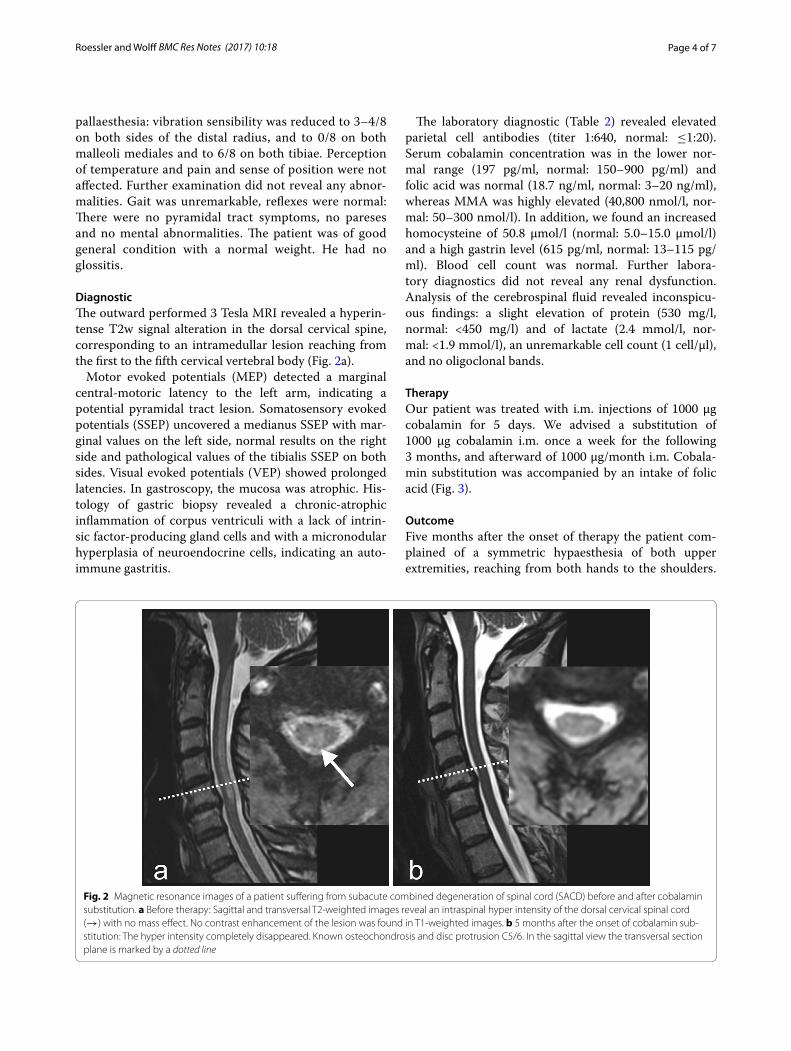
DiagnosticThe outward performed 3 Tesla MRI revealed a hyperin-tense T2w signal alteration in the dorsal cervical spine, corresponding to an intramedullar lesion reaching from the first to the fifth cervical vertebral body (Fig. 2a).
Motor evoked potentials (MEP) detected a marginal central-motoric latency to the left arm, indicating a potential pyramidal tract lesion. Somatosensory evoked potentials (SSEP) uncovered a medianus SSEP with mar-ginal values on the left side, normal results on the right side and pathological values of the tibialis SSEP on both sides. Visual evoked potentials (VEP) showed prolonged latencies. In gastroscopy, the mucosa was atrophic. His-tology of gastric biopsy revealed a chronic-atrophic inflammation of corpus ventriculi with a lack of intrin-sic factor-producing gland cells and with a micronodular hyperplasia of neuroendocrine cells, indicating an auto-immune gastritis.
The laboratory diagnostic (Table 2) revealed elevated parietal cell antibodies (titer 1:640, normal: ≤1:20). Serum cobalamin concentration was in the lower nor-mal range (197 pg/ml, normal: 150–900 pg/ml) and folic acid was normal (18.7 ng/ml, normal: 3–20 ng/ml), whereas MMA was highly elevated (40,800 nmol/l, nor-mal: 50–300 nmol/l). In addition, we found an increased homocysteine of 50.8 µmol/l (normal: 5.0–15.0 µmol/l) and a high gastrin level (615 pg/ml, normal: 13–115 pg/ml). Blood cell count was normal. Further labora-tory diagnostics did not reveal any renal dysfunction. Analysis of the cerebrospinal fluid revealed inconspicu-ous findings: a slight elevation of protein (530 mg/l, normal: <450 mg/l) and of lactate (2.4 mmol/l, nor-mal: <1.9 mmol/l), an unremarkable cell count (1 cell/µl), and no oligoclonal bands.
TherapyOur patient was treated with i.m. injections of 1000 µg cobalamin for 5 days. We advised a substitution of 1000 µg cobalamin i.m. once a week for the following 3 months, and afterward of 1000 µg/month i.m. Cobala-min substitution was accompanied by an intake of folic acid (Fig. 3).
OutcomeFive months after the onset of therapy the patient com-plained of a symmetric hypaesthesia of both upper extremities, reaching from both hands to the shoulders.
Fig. 2 Magnetic resonance images of a patient suffering from subacute combined degeneration of spinal cord (SACD) before and after cobalamin substitution. a Before therapy: Sagittal and transversal T2-weighted images reveal an intraspinal hyper intensity of the dorsal cervical spinal cord (→) with no mass effect. No contrast enhancement of the lesion was found in T1-weighted images. b 5 months after the onset of cobalamin sub-stitution: The hyper intensity completely disappeared. Known osteochondrosis and disc protrusion C5/6. In the sagittal view the transversal section plane is marked by a dotted line

Page 5 of 7Roessler and Wolff BMC Res Notes (2017) 10:18
The hypaesthesia of the thorax had regressed. There were no other neurological problems and no behavioural abnormalities. At that time, serum level of cobalamin was 710 pg/ml (Table 2).
MRI of the cervical spine revealed a complete regres-sion of the intraspinal myelon lesion (Fig. 2b). Elec-troneurography showed an axonal sensorimotor polyneuropathy of the upper and lower extremities.
After 11 months the patient reported paresthesia of both hands, the hypaesthesia of both arms had disap-peared. Stereognosis of hands and feet was normal and two-point-discrimination was better (he recognized a distance of 4–5 cm at both arms and at the thorax). Bimalleolar vibration sense had improved from 0/8 to 4–5/8 as assessed by the scale of the vibration tune (0 no sense, 8 full vibration sense).
Gastrin level was still elevated (730 pg/ml, normal: 13–115 pg/ml), folic acid (10 ng/ml, normal: 3–20 ng/ml), cobalamin (410 pg/ml, normal: 150–900 pg/ml), homocysteine (6.9 µmol/l, normal: 5.0–15.0 µmol/l) and MMA (178 nmol/l, normal: 50–300 nmol/l) were of normal range (Table 2). Blood picture was unremark-able. MEP, SSEP and VEP revealed no abnormalities.
ConclusionsInitially, the report of an accident three months before the onset of symptoms combined with a cervical myelon lesion suggested a traumatic injury. A neoplastic cause was considered for differential diagnosis. However, the lesion of the cervical spine cord only affected the dorsal part of the myelon without any mass effect and did not show any uptake of contrast agent. Furthermore, a sub-acute progress of disease was reported by the patient. Clinical examination revealed only sensory qualities conveyed by the posterior columns: hypaesthesia, reduc-tion of two-point-discrimination, disturbed stereogno-sis, and reduced vibration sensation. Therefore, SACD became probable. Now, diagnosis had to be confirmed by
laboratory tests. The patient had a cobalamin level in the lower normal range. Several publications described the determination of serum cobalamin to be unreliable [2, 3, 5, 18, 23, 24, 28, 29]. Therefore, other diagnostic markers are needed:
Adenosylcobalamin converts MMA to succinyl coen-zyme A. Hence, cobalamin deficiency causes an excess of MMA [23]. Increased MMA values are highly sensitive and highly specific for cobalamin deficiency [28, 30].
For the degradation of homocysteine methylcobalamin, pyridoxine, and folic acid are needed. Therefore, hyper-homocysteinemia gives a hint of a deficiency of all these vitamins, and has a high sensitivity but low specificity for cobalamin deficiency [28, 30]. The homocysteine level is suitable for follow-up and therapy monitoring [31]. It needs to be considered that blood has to be cooled for determining homocysteine levels.
Finally, a deficit of cobalamin causes a reduction of holoTC [2, 23, 32]. Lowered serum holoTC concentra-tion is the earliest marker of cobalamin deficiency and is reduced even before any clinical symptoms are apparent [2, 32].
In our case increased values for MMA and homocyst-eine were determined. After cobalamin substitution both values returned to normal. HoloTC was not measured, although we recommend its determination (Fig. 1).
The subsequent diagnostic provided prolonged laten-cies for the left-hand medianus and double-sided tibialis SSEP. Read in conjunction with the diagnostic imaging this can be explained by lesions in the fasciculus gra-cilis and cuneatus. MEP detected a central-motoric latency to the left arm pointing to an additional damage of the pyramidal tract. The elongated VEP give a hint for an undergoing demyelination of both optic nerves, which is sporadically associated with SACD. Neverthe-less, our patient did not notice any visual limitations. In addition to the lesion in the cervical myelon, elec-troneurography revealed the existence of an axonal
Table 2 Laboratory values before and 11 month after cobalamin substitution
Despite distinct clinical findings of a subacute combined degeneration of spinal cord (SACD) and a profound intramedullar lesion shown by MRI, cobalamin was still in the normal range before therapy started. In contrast, homocysteine and methylmalonic acid are suitable parameters for SACD diagnostic. Methylmalonic acid is the most specific marker of a cobalamin deficiency. Homocysteine is suitable for follow-up and therapy monitoring. Just 5 month after therapy start clinical symptoms were declining and all pathological changes found by MRI in the spinal cord disappeared
Before cobalamin substitution 11 month after cobalamin substitution Normal range
Cobalamin (pg/ml) 197 410 150–900
Homocysteine (µmol/l) 50.8 6.9 5.0–15.0
Methylmalonic acid (nmol/l) 40,800 178 50–300
Folic acid (ng/ml) 18.7 10 3–20
Gastrin (pg/ml) 615 730 13–115
Parietal cell antibodies 1:640 – ≤1:20

Page 6 of 7Roessler and Wolff BMC Res Notes (2017) 10:18
sensorimotor polyneuropathy, which is also common for a lack of cobalamin [33]. As expected in this context, liquor analysis did not reveal an infectious or chronic inflammatory process.
Gastroscopy and histology proved a type A gastritis with an increased value of gastrin. Serum gastrin is usu-ally markedly increased as a result of gastric atrophy and the increase of pH value. Appropriately, parietal cell antibodies were found. Therefore, in this patient SACD was caused by a disruption of cobalamin processing in the stomach due to parietal cell antibodies inducing an increased pH-value and a decreased production of intrin-sic-factor. Consequently, cobalamin could not dissolve out of the protein-bindings of the ingested food and was not bound to intrinsic factor.
Our patient presented solely sensory disturbances. No psychological disorders, no rhagades or gastrointestinal symptoms like Hunter glossitis, jaundice, diarrhea, dys-pepsia or increased values of bilirubin were found. Moreo-ver, no haematological alterations like macrocytosis were determined. A significant inverse correlation between the degree of anaemia and the severity of neurological involve-ment that was independent of the duration of symptoms is known [5, 27]. The reasons for this finding are unclear.
Therefore, it is important to think of SACD when only sensory disturbances can be found, even if the value of cobalamin is normal.
Vitamin substitution removed the lesion and nearly all clinical symptoms of cervical spine cord within 5 months and provoked a restitution of hypaesthesia of the thorax within eleven months. Therapy recommendations con-cerning dosage and mode of administration of cobalamin are inconsistent and depend on the underlying reason of vitamin deficiency [7, 24]. Figure 3 condenses our pre-ferred treatment regime.
We reported on a patient with a large lesion in cer-vical spine cord that matched with a subacute com-bined degeneration of spinal cord (SACD). SACD
was diagnosed by clinical signs and laboratory tests. When clinical signs suggest a lack of cobalamin, val-ues of cobalamin might still be in the normal range. Therefore, it is important to determine more sensi-tive parameters: HoloTC is the earliest and MMA the most specific marker of a cobalamin deficiency. Meas-urement of homocysteine is inexpensive and there-fore suitable for follow-up and therapy monitoring. Early and appropriate treatment reversed pathologi-cal changes in the spinal cord and dissolved associated clinical symptoms.
AbbreviationsholoTC: holotranscobalamin; IF: intrinsic factor; MEP: motor evoked potentials; MMA: methylmalonic acid; SACD: subacute combined degeneration of spinal cord; SSEP: somatosensory evoked potentials; VEP: visual evoked potentials.
Authors’ contributionsSW and FR cared for the patient and conceived all diagnostic investigations. Together they performed the therapy of the patient and wrote the article. Both authors read and approved the final manuscript.
Author details1 Department of Neurology, Justus-Liebig-University Giessen, Klinikstraße 33, 35385 Gießen, Germany. 2 Klinik und Poliklinik für Neurologie, Universitätsklini-kum Standort Gießen, Klinikstraße 33, 35385 Gießen, Germany.
AcknowledgementsWe are grateful to Ana Ivasioc and Petra Dietz-Ruckstuhl for critical reading of the manuscript and for language-editing.
Competing interestsThe authors declare that they have no competing interests.
Availability of data and materialsAll data supporting the findings of this work can be found in this article.
DeclarationsAll authors have read and agreed to the manuscript as written. There are no conflicts of interest. There was no source of funding.
Ethics approval and consent to participate and to publishWritten informed consent was obtained from the patient for publication of this Case Report and any accompanying images. This study was notified to the ethics committee of the Justus-Liebig-University. The ethics committee stated that an ethics approval is not necessary.
Fig. 3 Treatment concept of subacute combined degeneration of spinal cord (SACD) as it is practiced in our clinic. The concept follows recommen-dations made by Herrmann et al. [7]. Early start of therapy is decisive for better treatment outcomes [5]

Page 7 of 7Roessler and Wolff BMC Res Notes (2017) 10:18
• We accept pre-submission inquiries
• Our selector tool helps you to find the most relevant journal
• We provide round the clock customer support
• Convenient online submission
• Thorough peer review
• Inclusion in PubMed and all major indexing services
• Maximum visibility for your research
Submit your manuscript atwww.biomedcentral.com/submit
Submit your next manuscript to BioMed Central and we will help you at every step:
Ethics approvalNot applicable.
FundingNot applicable.
Received: 16 August 2016 Accepted: 14 December 2016
References 1. Qi YP, Do AN, Hamner HC, Pfeiffer CM, Berry RJ. The prevalence of low
serum vitamin B-12 status in the absence of anemia or macrocytosis did not increase among older U.S. adults after mandatory folic acid fortifica-tion. J Nutr. 2014;144:170–6.
2. Herrmann W, Obeid R, Schorr H, Geisel J. The usefulness of holotransco-balamin in predicting vitamin B12 status in different clinical settings. Curr Drug Metab. 2005;6:47–53.
3. Obeid R, Schorr H, Eckert R, Herrmann W. Vitamin B12 status in the elderly as judged by available biochemical markers. Clin Chem. 2004;50:238–41.
4. Lindenbaum J, Healton EB, Savage DG, Brust JC, Garrett TJ, Podell ER, Marcell PD, Stabler SP, Allen RH. Neuropsychiatric disorders caused by cobalamin deficiency in the absence of anemia or macrocytosis. N Engl J Med. 1988;318:1720–8.
5. Reynolds E. Vitamin B12, folic acid, and the nervous system. Lancet Neu-rol. 2006;5:949–60.
6. Wong CW. Vitamin B12 deficiency in the elderly: is it worth screening? Hong Kong Med J. 2015;21:155–64.
7. Herrmann W, Obeid R. Causes and early diagnosis of vitamin B12 defi-ciency. Dtsch Arztebl. 2008;105:680–5.
8. Graham SM, Arvela OM, Wise GA. Long-term neurologic consequences of nutritional vitamin B12 deficiency in infants. J Pediatr. 1992;121:710–4.
9. Andrès E, Loukili NH, Noel E, Kaltenbach G, Abdelgheni MB, Perrin AE, Noblet-Dick M, Maloisel F, Schlienger JL, Blicklé JF. Vitamin B12 (cobala-min) deficiency in elderly patients. Can Med Assoc J. 2004;171:251–9.
10. Homocysteine Studies Collaboration. Homocysteine and risk of ischemic heart disease and stroke: a meta-analysis. JAMA. 2002;288:2015–22.
11. Fowler B. Homocysteine—an independent risk factor for cardiovascular and thrombotic diseases. Ther Umsch. 2005;62:641–6.
12. Pang H, Han B, Fu Q, Zong Z. Association of high homocysteine levels with the risk stratification in hypertensive patients at risk of stroke. Clin Ther. 2016;38:1184–92.
13. Stanger O, Herrmann W, Pietrzik K, Fowler B, Geisel J, Dierkes J, Weger M. Clinical use and rational management of homocysteine, folic acid, and B vitamins in cardiovascular and thrombotic diseases. Z Kardiol. 2004;93:439–53.
14. Irizarry MC, Gurol ME, Raju S, Diaz-Arrastia R, Locascio JJ, Tennis M, Hyman BT, Growdon JH, Greenberg SM, Bottiglieri T. Association of homocyst-eine with plasma amyloid beta protein in aging and neurodegenerative disease. Neurology. 2005;65:1402–8.
15. Nagy ZS, Smith MZ, Esiri MM, Barnetson L, Smith AD. Hyperhomocyst-einaemia in Alzheimer’s disease and expression of cell cycle markers in the brain. J Neurol Neurosurg Psychiatr. 2000;69:565–6.
16. Kuzminski AM, Del Giacco EJ, Allen RH, Stabler SP, Lindenbaum J. Effec-tive treatment of cobalamin deficiency with oral cobalamin. Blood. 1998;92:1191–8.
17. Katsaros VK, Glocker FX, Hemmer B, Schumacher M. MRI of spinal cord and brain lesions in subacute combined degeneration. Neuroradiology. 1998;40:716–9.
18. Stabler SP, Allen RH, Savage DG, Lindenbaum J. Clinical spectrum and diagnosis of cobalamin deficiency. Blood. 1990;76:871–81.
19. van der Knaap MS, Valk J. Magnetic resonance oy myelin, myelination, and myelin disorders. 2nd ed. Berlin: Springer; 1995. p. 223–30.
20. Rossi A, Cerone R, Biancheri R, Gatti R, Schiaffino MC, Fonda C, Zam-marchi E, Tortori-Donati P. Early-onset combined methylmalonic aciduria and homocystinuria: neuroradiologic findings. Am J Neuroradiol. 2001;22:554–63.
21. Belghith A, Mahjoub S, Ben Romdhane N. Causes of vitamin B12 defi-ciency. Tunis Med. 2015;93(11):678–82.
22. Brescoll J, Daveluy S. A review of vitamin B12 in dermatology. Am J Clin Dermatol. 2015;16:27–33.
23. Herrmann W, Obeid R. Cobalamin deficiency. Subcell Biochem. 2012;56:301–22.
24. Hvas AM, Nexo E. Diagnosis and treatment of vitamin B12 deficiency—an update. Haematologica. 2006;91:1506–12.
25. Obeid R, Kuhlmann MK, Köhler H, Herrmann W. Response of homocyst-eine, cystathionine, and methylmalonic acid to vitamin treatment in dialysis patients. Clin Chem. 2005;51:196–201.
26. Obeid R, Kuhlmann M, Kirsch CM, Herrmann W. Cellular uptake of vitamin B12 in patients with chronic renal failure. Nephron Clin Pract. 2005;99:c42–8.
27. Healton EB, Savage DG, Brust JC, Garrett TJ, Lindenbaum J. Neurologic aspects of cobalamin deficiency. Medicine. 1991;70:229–45.
28. Savage DG, Lindenbaum J, Stabler SP, Allen RH. Sensitivity of serum meth-ylmalonic acid and total homocysteine determinations for diagnosing cobalamin and folate deficiencies. Am J Med. 1994;96:239–46.
29. Vashi P, Edwin P, Popiel B, Lammersfeld C, Gupta D. Methylmalonic acid and homocysteine as indicators of vitamin B-12 deficiency in cancer. PLoS ONE. 2016. doi:10.1371/journal.pone.0147843.
30. Moelby L, Rasmussen K, Jensen MK, Pedersen KO. The relationship between clinically confirmed cobalamin deficiency and serum methyl-malonic acid. J Intern Med. 1990;228:373–8.
31. Rajan S, Wallace JI, Brodkin KI, Beresford SA, Allen RH, Stabler SP. Response of elevated methylmalonic acid to three dose levels of oral cobalamin in older adults. J Am Geriatr Soc. 2002;50:1789–95.
32. Herrmann W, Schorr H, Obeid R, Geisel J. Vitamin B12 status, particularly holotranscobalamin II and methylmalonic acid concentrations, and hyperhomocysteinemia in vegetarians. Am J Clin Nutr. 2003;78:131–6.
33. Dalla Torre C, Lucchetta M, Cacciavillani M, Campagnolo M, Manara R, Briani C. Reversible isolated sensory axonal neuropathy due to cobalamin deficiency. Muscle Nerve. 2012;45:428–30.