rapid biological synthesis of silver nanoparticles...
TRANSCRIPT
Research ArticleRapid Biological Synthesis of Silver Nanoparticles fromOcimum sanctum and Their Characterization
M Z H Khan1 F K Tarek1 M Nuzat1 M A Momin2 andM R Hasan1
1Department of Chemical Engineering Jessore University of Science and Technology Jessore 7408 Bangladesh2Department of Applied Physics and Electronics Islamic University Kushtia Bangladesh
Correspondence should be addressed to M Z H Khan zavedkhanyahoocom
Received 17 April 2017 Revised 23 June 2017 Accepted 12 July 2017 Published 20 August 2017
Academic Editor Tian Xia
Copyright copy 2017 M Z H Khan et al This is an open access article distributed under the Creative Commons Attribution Licensewhich permits unrestricted use distribution and reproduction in any medium provided the original work is properly cited
With development of nanotechnology the biological synthesis process deals with the synthesis characterization andmanipulationof materials and further development at nanoscale which is the most cost-effective and eco-friendly and rapid synthesis processas compared to physical and chemical process In this research silver nanoparticles (AgNPs) were synthesized from silver nitrate(AgNO
3) aqueous solution through eco-friendly plant leaf broth of Ocimum sanctum as reactant as well as capping agent and
stabilizer The formation of AgNPs was monitored by ultraviolet-visible spectrometer (UV-vis) and Fourier transform infrared(FTIR) spectroscopy X-ray diffraction (XRD) and scanning electronic microscopy (SEM) have been used to characterize themorphology of prepared AgNPs The peaks in XRD pattern are in good agreement with that of face-centered-cubic (FCC) formof metallic silver Thermal gravimetric analysisdifferential thermal analysis (TGADTA) results confirmed the weight loss and theexothermic reaction due to desorption of chemisorbedwaterThe average grain size of silver nanoparticles is found to be 29 nmTheFTIR results indicated that the leaf broths containing the carboxyl hydroxyl and amine groups are mainly involved in fabricationof silver AgNPs and proteins which have amine groups responsible for stabilizing AgNPs in the solution
1 Introduction
Nanotechnology is a branch of science and technology whichconcerns with the development of process for the design syn-thesis andmanipulation of particle structure different shapesize and controlled disparity Due to their unique physico-chemical properties metal nanoparticles with a dimension ofapproximately 1ndash100 nm have received considerable attentionin last few decades [1] Silver nanoparticles (AgNPs) haveattracted significant interest due to their wide variety ofapplications and their unique optical electrical and thermalproperties [2ndash4] In the synthesis of AgNPs numerous chem-ical biological and physical methods have been developedHowever conventional physical and chemical methods areexpensive as well as resulting in low yield and poor sizedistribution [1]
Over the recent years biosynthesis method has beenwidely studied for stable metal nanoparticles synthesis withcontrolled size and shape and considered as a ldquogreenrdquo
approach [5ndash8] Among them plant leaf extract mediatedbiological process has attracted much attention due to thesimple and inexpensive protocol [9ndash12] In addition theproteins or polysaccharides or secondary metabolites foundin leaf extracts can reduce the Ag+ ions to Ag0 state and formsilver nanoparticles [1]
Traditional chemical methods of synthesizing silvernanoparticles include the use of ethylene glycol pyridine andsodium borohydride The chemicals used in these method-ologies can be toxic and highly reactive posing a risk tothe environment and humans or the procedures are tooexpensive to be feasible at an industrial scale Therefore therehas been a search for inexpensive reliable safe and ldquogreenrdquoapproach to the synthesis of stable metal nanoparticles withcontrolled size and shape As a result some novel methodshave recently developed using (i) biologically derived reduc-ing agents such as chitosan glucose and polysaccharides (ii)microbes such as bacteria and fungus and (iii) a variety of
HindawiJournal of NanoscienceVolume 2017 Article ID 1693416 6 pageshttpsdoiorg10115520171693416
2 Journal of Nanoscience
(a) (b)
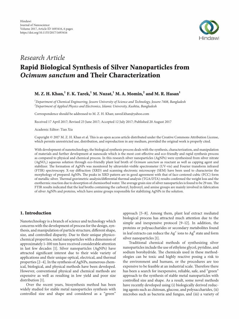
Figure 1 (a) Ocimum sanctum leaves and (b) leaf broth
plant (seed and leaf as well as tuber) extracts for the synthesisof metal nanoparticles Among them plant leaf extractmediated biological process has been widely investigated dueto the inexpensive and simple protocol
We have used Ocimum sanctum leaf broth to synthesizeAgNPs The used plant is wild herbaceous with medicinalvalues and available in all tropical countries [13 14] and tradi-tionally thought to have strong antimicrobial and antioxidantactivity and be widely used to stimulate the appetite and easestomach upset Recently Singhal et al [15] and others [16ndash18]synthesized silver nanoparticles using Ocimum sanctum leafextract which showed significant antibacterial activity againstE coli and Staphylococcus aureus
The objective of this study was to find out a cost-effectiveand eco-friendly technique for biological synthesis of AgNPsusing Ocimum sanctum leaf broth It was also aimed attackling the optical structural and thermal characteristics ofsynthesized nanoparticles
2 Materials and Methods
21 Preparation of Leaf Broth The Ocimum sanctum plantleaves were collected from Jessore district in BangladeshAbout 20 gm of fresh leaves was thoroughly washed threetimes with deionized water and chopped into small piecesThen 100ml of deionized water was added in 250ml conicalflask stirred and boiled for 20min at 60∘C During boiling acondenser was used as a vapor recovery device After boilingthe leaf broth was cooled and filtered yielding transparentyellow color leaf broth and these were stored at 4∘CThe plantmaterial and leaf broth are showed in Figure 1
22 Synthesis of Silver Nanoparticles In a typical synthesisof AgNPs the leaf extract (10ml) of Ocimum sanctumwas added to 90ml of 0001M AgNO
3and 001M AgNO
3
(9999) aqueous solution respectively and kept at 33∘CThe experiment was done in triplicate for reproducibilityAfter 10 minutes the color of the solution changed fromcolorless to yellow indicating the formation of AgNPs Thebioreduced AgNPs solution was collected and monitored byperiodic sampling of aliquots (5ml) of aqueous component
and measuring UV-visible spectra of the solution Due tohigh optical density of the nanoparticles solution it wasdiluted to 10 times with DI water to avoid errors
23 Characterization The bioreduction of AgNPs fromAgNO
3ions in solution was carried out using UV-visible
(UV-vis) spectrophotometer (Shimadzu UV-1800 Japan) byrecording absorption spectra of the samples in the wave-length range 190ndash800 nm To identify the presence of poten-tial biomolecule and functional groups Fourier transforminfrared spectra (FTIR) study was used FTIR spectra weremeasured by Perkin Elmer spectrometer having a resolution4 cmminus1 in the wavenumber range 500ndash4000 cmminus1 The for-mation crystalline behavior and quality of synthesized silvernanoparticles powder were investigated by X-ray diffraction(XRD) spectrum with Cu-K120572 radiation of 0154187 nm wave-lengthThe scanning was performed in the region of 2120579 from30∘ to 80∘ at 002∘min and the time constant was 2 s TheDebye-Scherer equation was used to calculate the size ofsilver nanoparticles Scanning electronic microscope (SEM)was used to investigate the surface morphology and particlesize of synthesized AgNPs powder Thermal gravimetricanalysisdifferential thermal analysis (TGADTA) thermalsystem was used to examine the reaction type and weightloss of synthesized silver nanoparticles powderThe spectrumof TGADTA has been recorded in temperature range fromroom temperature to 1000∘C where Al
2O3was used for
heating andmeasurement carried out in air atmosphere at theheating rate 200∘Cmin
3 Results and Discussion
31 UV-Vis Study The formation of the AgNPs was con-firmed by change in the color of the solution mixture bythe bioreduction of Ag+ to Ag0 which is presented inFigure 2 A color change occurred from transparent yellowto reddish brown which indicates the formation of AgNPsduring stirring UV-visible spectra of synthesized AgNPsshow the characteristic band at 440ndash455 nm which indicatescrystalline spherical nature of AgNPs Here we investigate theeffect of reaction time on the synthesis of AgNPs
Journal of Nanoscience 3
(a) (b) (c) (d)
Figure 2 Image of synthesized AgNPs solution over reaction times (a) 15min (b) 30min (c) 60min and (d) 75min usingOcimum sanctumleaf broth
400 450 500 550 600350Wavelength (nm)
1
15
2
25
Abso
rban
ce
15 min30 min
60 min75 min
Figure 3 Absorption peak of synthesized AgNPs solution usingOcimum sanctum leaf broth
During stirring ofmixer ofAgNO3solution and leaf broth
the possible chemical reactions for synthesizing the AgNPsare as follows
Ag+ (aq) + 119874119888119894119898119906119898 119904119886119899119888119905119906119898 997888rarr
[Ag (119874119888119894119898119906119898 119904119886119899119888119905119906119898)]+(1)
[Ag (119874119888119894119898119906119898 119904119886119899119888119905119906119898)]+ + R-CHO 997888rarr
[Ag (119874119888119894119898119906119898 119904119886119899119888119905119906119898)] + R-COOH(2)
Aftermixing ofAgNO3solution and leaf broth the dispersion
of silver ion in the Ocimum sanctum aqueous solution mixer(1) reacted with the Ag to form [Ag(Ocimum sanctum)]+complex which reacted with aldehyde in the molecularstructure to form [Ag(Ocimum sanctum)] due to reductionof silver ions (2)
Figure 3 shows the UV-visible spectra that were recordedat different time intervals for monitoring the reaction of
silver nitrate reduction by the leaf extract The appearanceof the surface plasmon resonance (SPR) band increased inintensity with time Due to the unique optical properties ofsilver nanoparticles a great deal of information about thephysical state can be obtained by analyzing the spectra Thespectra clearly show the increase in intensity of silver solutionwith time indicating the formation of increased number ofAgNPs in the solution The sharp bands of silver colloidswere observed at sim450 nm The intensity of absorption bandincreases with increasing time period of aqueous componentand consequent color changes were observed from yellow toreddish brown shown in Figure 2 This characteristic colorvariation is due to the excitation of the SPR in the metalnanoparticles The broadening of peak indicated that theparticles are spherical and polydispersed The SPR band inthe AgNPs solution remains close to 450 nm throughout thereaction period indicating that the particles are dispersed inthe aqueous solution with no evidence for aggregation
The optimum temperature required for the completionof reaction was investigated to be 60∘C Upon a furtherincrease in temperature (up to 75∘C) no further absorbanceincrease was observed and shows an increase in AgNPs sizeFurther increase in temperature caused the broadening ofthe peak revealing the increased size of nanoparticles Thistemperature dependent increase in the peak intensity showedthe dependence of the silver ion reduction on the reactiontemperature It was observed that reduction rate of silver ionsincreased by increasing temperature
32 FTIR Study FTIR measurements were responsible foridentifying the functional group which were responsiblefor the capping and efficient stabilization of synthesizedsilver nanoparticles by the leaf broth Absorption peaks ofsynthesized AgNPs using Ocimum sanctum were observed at67027 cmminus1 assigned to C-H stretch in alkenes 95092 cmminus1assigned to C-H bending in alkenes 106208 cmminus1 assigned to
4 Journal of Nanoscience
Table 1 Distribution of particle size of nanopowder using Ocimum sanctum leaf broth
2120579 values (degree) hkl FWHM (120573) in radians Particle size (nm)322 (110) 0005765 253812 (111) 000433 344434 (200) 000433 354624 (200) 0005765 265482 (211) 000742 215752 (211) 0007343 226456 (220) 000433 386734 (221) 0005765 297444 (310) 000742 23768 (311) 000433 41
750100012501500175020002500300035004000(1cm)
45
60
75
90
105
120
T 39
425
738
548
037
477
5
3339
80
3237
57
2788
15
2343
55
2070
62
1704
14
1272
08
1183
35
1062
80
950
92
670
27
Figure 4 FTIR spectrum of synthesized AgNPs using Ocimumsanctum leaf broth
C-O stretch in ester 118335 and 127208 cmminus1 assigned to C-N stretch in amines 170414 cmminus1 assigned to C=O stretch incarbonyl 278818 cmminus1 assigned to O-H stretch in carboxylicacid and 323757 and 33938 cmminus1 assigned to O-H stretch inalcohol (Figure 4) This analysis strongly supported the cap-ping behavior of synthesized silver nanoparticles byOcimumsanctum leaf broth to stabilize the silver nanoparticles [19]
Still up to date there is no proper mechanism for thesynthesis of silver nanoparticles The proposed hypotheticalmechanism behind the synthesis of nanoparticles is anenzymatic reaction in which the plant extract contains thecomplex of reducing enzymes which reduce the chemicalssuch as silver nitrate into silver ions and nitrate ions Plantscontain a complex network of antioxidant metabolites andenzymes that work together to prevent oxidative damageto cellular components It was reported that plants extractscontain biomolecules including polyphenols ascorbic acidflavonoids sterols triterpenes alkaloids alcoholic com-pounds polysaccharides saponins 120573-phenylethylamines
600
2
710
8
631
8
505
447
38
679
2
568
6
647
6
442
2
726
6
394
8
489
6
742
4
521
2
363
233
16
315
8
426
4
663
4
552
8
410
6
347
4
584
4
758
278
98
695
537
458
616
379
77430
2 (degree)
0
300
600
900
1200
1500
1800
Inte
nsity
Figure 5 XRD pattern of synthesized silver nanoparticles usingOcimum sanctum leaf broth
glucose and fructose and proteinsenzymes which could beused as reductant to react with silver ions and therefore usedas scaffolds to direct the formation of AgNPs in the solutionHypothetically biosynthetic products or reduced cofactorsplay an important role in the reduction of respective salts tonanoparticles
33 XRD Study Figure 5 shows the XRD pattern of synthe-sized silver nanoparticles using Ocimum sanctum leaf brothThe 119889-spacing is calculated by Braggrsquos law that is 119889 =1205822 sin 120579 where 120582 is wavelength in nm The intense peaksface-centered-cubic (FCC) plans full width at half maximum(FWHM) and particle size are calculated mathematicallyand shown in Table 1 The intense peaks at 2120579 degree values3812∘C 4434∘C 6456∘C and 768∘C correspond to (111)(200) (220) and (311) for FCC plans of silver nanoparticlesusing Ocimum sanctum [20] The particle size of the nano-powder has been calculated by Debye Scherrer formula 119863 =119896120582120573 cos 120579 where 119863 is particle diameter size 119896 is Scherrerconstant (09) 120582 is wavelength of X-rays (01541 nm) 120573 iswidth at half of reflection at Braggrsquos angle 2120579 and 120579 is Braggangle The average particle size is 29 nm for using of Ocimumsanctum leaf broth
34 SEM Study The surface morphological and nanos-tructural studies of synthesized silver nanoparticles using
Journal of Nanoscience 5
Figure 6 SEM image of synthesized silver nanoparticles usingOcimum sanctum leaf broth
2459636239223208402
35730763742945225261326743421321943207
1265471073373141359
44039715193177708867
632384397418746280351549881261
20879222721300150382
1193097571282927912138304031
19330389949212774695
0102030405060708090
100
Freq
uenc
y
2 3 4 5 6 7 8 9 10 11 12 13 14 15 16 17 18 19 201Size (nm)
Figure 7 Particle size distribution of nanopowder using Ocimumsanctum
Ocimum sanctum leaf broth were investigated by SEM InSEM image of synthesized silver nanoparticles using Oci-mum sanctum is shown in Figure 6 This indicates thatthe monodispersive and crystalline silver nanoparticles areobtained The spherical size of synthesized silver nanoparti-cles using Ocimum sanctum leaf broth is 29 nm as estimatedfrom the SEM picture similar to the particle size calculatedfrom Debye Scherrer formula (Figure 7) Some nanoparticlessize is larger because silver nanoparticles have the tendency toagglomerate due to their high surface energy and high surfacetension of the ultrafine nanoparticles
35 DTTGA Study A ceramic crucible (Al2O3) was used for
heating andmeasurementswere carried out in air atmosphereat the heating rate of 20∘Cmin Curves of TGA and DTA ofsynthesized silver nanoparticles using Ocimum sanctum leafbroth are shown in Figure 8Theweight loss of silver nanopar-ticles using Ocimum sanctum is observed in TGA curvewhich occurs from 200∘C to 400∘C and again a loss occursfrom 700∘C to 1000∘C which is shown in figure DTA curveof synthesized silver nanoparticles using Ocimum sanctumleaf broth shows intense endothermic peakwhen temperatureincreases DTA curve shows the thermal decomposition andcrystallization of synthesized silver nanoparticles
TG
DTA
+00
261 2000 4000 6000 8000 9999
+00
minus6666
minus4000
minus2000
000
2000
4000
6000
8000
10000
13333+00
minus1250
minus1000
minus800
minus600
minus400
minus200
00
200
400
600
800
1000
Wei
ght (
)
Temperature (∘C)
Hea
t flow
(V
)
Figure 8 TGA and DTA curve of synthesized silver nanoparticlesusing Ocimum sanctum leaf broth
4 Conclusion
Thebiological synthesis process is a reliable eco-friendly andcost-effective process for the synthesis of nanoparticles InUV-vis spectrometer detected peaks at 450 nm confirm theformation of AgNPs FTIR spectrum confirms the functionalgroup of organic compoundwhich is responsible for cappingformation and stabilization of AgNPs The XRD analysisconfirms the FCC plans of the synthesized nanoparticles andthe particles size of 29 nm usingOcimum sanctum leaf brothSEM images confirm the morphological and nanostructuralcharacteristics of nanoparticles with average size distributioncalculated by Debye Scherrer formula DTATGA curveshows the weight loss of AgNPs with temperature whichalso confirms the thermal decomposition and crystallizationof nanoparticles The resulting synthesized AgNPs werecapped by a thin layer of proteins and metabolites havingfunctional groups of amines alcohol ketones aldehydes andcarboxylic acids which are responsible for formation andstabilization of nanoparticles Prepared nanoparticles can beused as bactericidal wound healing and water purificationagents and in medicine field Due to these applications thismethod is potentially exciting for the large-scale synthesis ofnanoparticles
Conflicts of Interest
The authors declare that there are no conflicts of interest
Acknowledgments
The financial support from Jessore University of Scienceand Technology (Research Budget Code 4829) is greatlyacknowledged This work was done under the banner of theLaboratory of Nano-Bio and Advanced Materials Engineer-ing (NAME) SEM report from the Department of Glass
6 Journal of Nanoscience
and Ceramic Engineering BUET Bangladesh is greatlyappreciated
References
[1] S Gurunathan J W Han D-N Kwon and J-H KimldquoEnhanced antibacterial and anti-biofilm activities of silvernanoparticles against Gram-negative and Gram-positive bacte-riardquo Nanoscale Research Letters vol 9 no 1 pp 1ndash17 2014
[2] A Saxena R M Tripathi F Zafar and P Singh ldquoGreensynthesis of silver nanoparticles using aqueous solution of Ficusbenghalensis leaf extract and characterization of their antibac-terial activityrdquoMaterials Letters vol 67 no 1 pp 91ndash94 2012
[3] A K Mittal D Tripathy A Choudhary et al ldquoBio-synthesisof silver nanoparticles using Potentilla fulgens Wall ex Hookand its therapeutic evaluation as anticancer and antimicrobialagentrdquoMaterials Science and Engineering C vol 53 pp 120ndash1272015
[4] J L Lopez-Miranda M Vazquez N Fletes R Esparza andG Rosas ldquoBiosynthesis of silver nanoparticles using a Tamarixgallica leaf extract and their antibacterial activityrdquo MaterialsLetters vol 176 pp 285ndash289 2016
[5] A Saravanakumar M Ganesh J Jayaprakash and H T JangldquoBiosynthesis of silver nanoparticles using Cassia tora leafextract and its antioxidant and antibacterial activitiesrdquo Journalof Industrial and Engineering Chemistry vol 28 pp 277ndash2812015
[6] T Y Suman S R Radhika Rajasree A Kanchana and S BElizabeth ldquoBiosynthesis characterization and cytotoxic effectof plant mediated silver nanoparticles using Morinda citrifoliaroot extractrdquo Colloids and Surfaces B Biointerfaces vol 106 pp74ndash78 2013
[7] P P N V Kumar S V N Pammi P Kollu K V V Satya-narayana and U Shameem ldquoGreen synthesis and character-ization of silver nanoparticles using Boerhaavia diffusa plantextract and their anti bacterial activityrdquo Industrial Crops andProducts vol 52 pp 562ndash566 2014
[8] L Wang C-C Liu Y-Y Wang H Xu H Su and X ChengldquoAntibacterial activities of the novel silver nanoparticles biosyn-thesized using Cordyceps militaris extractrdquo Current AppliedPhysics vol 16 no 9 pp 969ndash973 2016
[9] A D Dwivedi and K Gopal ldquoBiosynthesis of silver and goldnanoparticles using Chenopodium album leaf extractrdquo Colloidsand Surfaces A Physicochemical and Engineering Aspects vol369 no 1ndash3 pp 27ndash33 2010
[10] C S Espenti K S V K Rao and K M Rao ldquoBio-synthesisand characterization of silver nanoparticles using Terminaliachebula leaf extract and evaluation of its antimicrobial poten-tialrdquoMaterials Letters vol 174 pp 129ndash133 2016
[11] D Inbakandan C Kumar M Bavanilatha D N RavindraR Kirubagaran and S A Khan ldquoUltrasonic-assisted greensynthesis of flower like silver nanocolloids usingmarine spongeextract and its effect on oral biofilm bacteria and oral cancer celllinesrdquoMicrobial Pathogenesis vol 99 pp 135ndash141 2016
[12] N Basavegowda A Idhayadhulla and Y R Lee ldquoPhyto-synthesis of gold nanoparticles using fruit extract of Hoveniadulcis and their biological activitiesrdquo Industrial Crops andProducts vol 52 pp 745ndash751 2014
[13] P Prakash andN Gupta ldquoTherapeutic uses ofOcimum sanctumLinn (tulsi) with a note on eugenol and its pharmacologicalactions a short reviewrdquo Indian Journal of Physiology andPharmacology vol 49 pp 125ndash131 2005
[14] S Mahata S Maru S Shukla et al ldquoAnticancer property ofBryophyllum pinnata (Lam) Oken leaf on human cervicalcancer cellsrdquo BMC Complementary and Alternative Medicinevol 12 article 15 2012
[15] G Singhal R Bhavesh K Kasariya A R Sharma and RP Singh ldquoBiosynthesis of silver nanoparticles using Ocimumsanctum (Tulsi) leaf extract and screening its antimicrobialactivityrdquo Journal of Nanoparticle Research vol 13 no 7 pp 2981ndash2988 2011
[16] KMallikarjun G Narasimha G R Dillip et al ldquoGreen synthe-sis of silver nanoparticles using ocimum leaf extract and theircharacterizationrdquoDigest Journal of Nanomaterials and Biostruc-tures vol 6 no 1 pp 181ndash186 2011
[17] C Ramteke T Chakrabarti B K Sarangi and R-A PandeyldquoSynthesis of silver nanoparticles from the aqueous extract ofleaves of ocimum sanctum for enhanced antibacterial activityrdquoJournal of Chemistry Article ID 278925 7 pages 2013
[18] Y Rout ldquoGreen synthesis of silver nanoparticles using Ocimumsanctum (Tulashi) and study of their antibacterial and antifun-gal activitiesrdquo Journal ofMicrobiology and Antimicrobials vol 4no 6 pp 103ndash109 2012
[19] L Christensen S Vivekanandhan M Misra and A KMohanty ldquoBiosynthesis of silver nanoparticles using Murrayakoenigii (curry leaf) an investigation on the effect of broth con-centration in reductionmechanism and particle sizerdquoAdvancedMaterials Letters vol 2 no 6 pp 429ndash434 2011
[20] P Phanjom andG Ahmed ldquoBiosynthesis of silver nanoparticlesby Aspergillus oryzae (MTCC No 1846 ) and its characteriza-tionsrdquo Nanoscience and Nanotechnology vol 5 no 1 pp 14ndash202015
Submit your manuscripts athttpswwwhindawicom
ScientificaHindawi Publishing Corporationhttpwwwhindawicom Volume 2014
CorrosionInternational Journal of
Hindawi Publishing Corporationhttpwwwhindawicom Volume 2014
Polymer ScienceInternational Journal of
Hindawi Publishing Corporationhttpwwwhindawicom Volume 2014
Hindawi Publishing Corporationhttpwwwhindawicom Volume 2014
CeramicsJournal of
Hindawi Publishing Corporationhttpwwwhindawicom Volume 2014
CompositesJournal of
NanoparticlesJournal of
Hindawi Publishing Corporationhttpwwwhindawicom Volume 2014
Hindawi Publishing Corporationhttpwwwhindawicom Volume 2014
International Journal of
Biomaterials
Hindawi Publishing Corporationhttpwwwhindawicom Volume 2014
NanoscienceJournal of
TextilesHindawi Publishing Corporation httpwwwhindawicom Volume 2014
Journal of
NanotechnologyHindawi Publishing Corporationhttpwwwhindawicom Volume 2014
Journal of
CrystallographyJournal of
Hindawi Publishing Corporationhttpwwwhindawicom Volume 2014
The Scientific World JournalHindawi Publishing Corporation httpwwwhindawicom Volume 2014
Hindawi Publishing Corporationhttpwwwhindawicom Volume 2014
CoatingsJournal of
Advances in
Materials Science and EngineeringHindawi Publishing Corporationhttpwwwhindawicom Volume 2014
Smart Materials Research
Hindawi Publishing Corporationhttpwwwhindawicom Volume 2014
Hindawi Publishing Corporationhttpwwwhindawicom Volume 2014
MetallurgyJournal of
Hindawi Publishing Corporationhttpwwwhindawicom Volume 2014
BioMed Research International
MaterialsJournal of
Hindawi Publishing Corporationhttpwwwhindawicom Volume 2014

2 Journal of Nanoscience
(a) (b)
Figure 1 (a) Ocimum sanctum leaves and (b) leaf broth
plant (seed and leaf as well as tuber) extracts for the synthesisof metal nanoparticles Among them plant leaf extractmediated biological process has been widely investigated dueto the inexpensive and simple protocol
We have used Ocimum sanctum leaf broth to synthesizeAgNPs The used plant is wild herbaceous with medicinalvalues and available in all tropical countries [13 14] and tradi-tionally thought to have strong antimicrobial and antioxidantactivity and be widely used to stimulate the appetite and easestomach upset Recently Singhal et al [15] and others [16ndash18]synthesized silver nanoparticles using Ocimum sanctum leafextract which showed significant antibacterial activity againstE coli and Staphylococcus aureus
The objective of this study was to find out a cost-effectiveand eco-friendly technique for biological synthesis of AgNPsusing Ocimum sanctum leaf broth It was also aimed attackling the optical structural and thermal characteristics ofsynthesized nanoparticles
2 Materials and Methods
21 Preparation of Leaf Broth The Ocimum sanctum plantleaves were collected from Jessore district in BangladeshAbout 20 gm of fresh leaves was thoroughly washed threetimes with deionized water and chopped into small piecesThen 100ml of deionized water was added in 250ml conicalflask stirred and boiled for 20min at 60∘C During boiling acondenser was used as a vapor recovery device After boilingthe leaf broth was cooled and filtered yielding transparentyellow color leaf broth and these were stored at 4∘CThe plantmaterial and leaf broth are showed in Figure 1
22 Synthesis of Silver Nanoparticles In a typical synthesisof AgNPs the leaf extract (10ml) of Ocimum sanctumwas added to 90ml of 0001M AgNO
3and 001M AgNO
3
(9999) aqueous solution respectively and kept at 33∘CThe experiment was done in triplicate for reproducibilityAfter 10 minutes the color of the solution changed fromcolorless to yellow indicating the formation of AgNPs Thebioreduced AgNPs solution was collected and monitored byperiodic sampling of aliquots (5ml) of aqueous component
and measuring UV-visible spectra of the solution Due tohigh optical density of the nanoparticles solution it wasdiluted to 10 times with DI water to avoid errors
23 Characterization The bioreduction of AgNPs fromAgNO
3ions in solution was carried out using UV-visible
(UV-vis) spectrophotometer (Shimadzu UV-1800 Japan) byrecording absorption spectra of the samples in the wave-length range 190ndash800 nm To identify the presence of poten-tial biomolecule and functional groups Fourier transforminfrared spectra (FTIR) study was used FTIR spectra weremeasured by Perkin Elmer spectrometer having a resolution4 cmminus1 in the wavenumber range 500ndash4000 cmminus1 The for-mation crystalline behavior and quality of synthesized silvernanoparticles powder were investigated by X-ray diffraction(XRD) spectrum with Cu-K120572 radiation of 0154187 nm wave-lengthThe scanning was performed in the region of 2120579 from30∘ to 80∘ at 002∘min and the time constant was 2 s TheDebye-Scherer equation was used to calculate the size ofsilver nanoparticles Scanning electronic microscope (SEM)was used to investigate the surface morphology and particlesize of synthesized AgNPs powder Thermal gravimetricanalysisdifferential thermal analysis (TGADTA) thermalsystem was used to examine the reaction type and weightloss of synthesized silver nanoparticles powderThe spectrumof TGADTA has been recorded in temperature range fromroom temperature to 1000∘C where Al
2O3was used for
heating andmeasurement carried out in air atmosphere at theheating rate 200∘Cmin
3 Results and Discussion
31 UV-Vis Study The formation of the AgNPs was con-firmed by change in the color of the solution mixture bythe bioreduction of Ag+ to Ag0 which is presented inFigure 2 A color change occurred from transparent yellowto reddish brown which indicates the formation of AgNPsduring stirring UV-visible spectra of synthesized AgNPsshow the characteristic band at 440ndash455 nm which indicatescrystalline spherical nature of AgNPs Here we investigate theeffect of reaction time on the synthesis of AgNPs
Journal of Nanoscience 3
(a) (b) (c) (d)
Figure 2 Image of synthesized AgNPs solution over reaction times (a) 15min (b) 30min (c) 60min and (d) 75min usingOcimum sanctumleaf broth
400 450 500 550 600350Wavelength (nm)
1
15
2
25
Abso
rban
ce
15 min30 min
60 min75 min
Figure 3 Absorption peak of synthesized AgNPs solution usingOcimum sanctum leaf broth
During stirring ofmixer ofAgNO3solution and leaf broth
the possible chemical reactions for synthesizing the AgNPsare as follows
Ag+ (aq) + 119874119888119894119898119906119898 119904119886119899119888119905119906119898 997888rarr
[Ag (119874119888119894119898119906119898 119904119886119899119888119905119906119898)]+(1)
[Ag (119874119888119894119898119906119898 119904119886119899119888119905119906119898)]+ + R-CHO 997888rarr
[Ag (119874119888119894119898119906119898 119904119886119899119888119905119906119898)] + R-COOH(2)
Aftermixing ofAgNO3solution and leaf broth the dispersion
of silver ion in the Ocimum sanctum aqueous solution mixer(1) reacted with the Ag to form [Ag(Ocimum sanctum)]+complex which reacted with aldehyde in the molecularstructure to form [Ag(Ocimum sanctum)] due to reductionof silver ions (2)
Figure 3 shows the UV-visible spectra that were recordedat different time intervals for monitoring the reaction of
silver nitrate reduction by the leaf extract The appearanceof the surface plasmon resonance (SPR) band increased inintensity with time Due to the unique optical properties ofsilver nanoparticles a great deal of information about thephysical state can be obtained by analyzing the spectra Thespectra clearly show the increase in intensity of silver solutionwith time indicating the formation of increased number ofAgNPs in the solution The sharp bands of silver colloidswere observed at sim450 nm The intensity of absorption bandincreases with increasing time period of aqueous componentand consequent color changes were observed from yellow toreddish brown shown in Figure 2 This characteristic colorvariation is due to the excitation of the SPR in the metalnanoparticles The broadening of peak indicated that theparticles are spherical and polydispersed The SPR band inthe AgNPs solution remains close to 450 nm throughout thereaction period indicating that the particles are dispersed inthe aqueous solution with no evidence for aggregation
The optimum temperature required for the completionof reaction was investigated to be 60∘C Upon a furtherincrease in temperature (up to 75∘C) no further absorbanceincrease was observed and shows an increase in AgNPs sizeFurther increase in temperature caused the broadening ofthe peak revealing the increased size of nanoparticles Thistemperature dependent increase in the peak intensity showedthe dependence of the silver ion reduction on the reactiontemperature It was observed that reduction rate of silver ionsincreased by increasing temperature
32 FTIR Study FTIR measurements were responsible foridentifying the functional group which were responsiblefor the capping and efficient stabilization of synthesizedsilver nanoparticles by the leaf broth Absorption peaks ofsynthesized AgNPs using Ocimum sanctum were observed at67027 cmminus1 assigned to C-H stretch in alkenes 95092 cmminus1assigned to C-H bending in alkenes 106208 cmminus1 assigned to
4 Journal of Nanoscience
Table 1 Distribution of particle size of nanopowder using Ocimum sanctum leaf broth
2120579 values (degree) hkl FWHM (120573) in radians Particle size (nm)322 (110) 0005765 253812 (111) 000433 344434 (200) 000433 354624 (200) 0005765 265482 (211) 000742 215752 (211) 0007343 226456 (220) 000433 386734 (221) 0005765 297444 (310) 000742 23768 (311) 000433 41
750100012501500175020002500300035004000(1cm)
45
60
75
90
105
120
T 39
425
738
548
037
477
5
3339
80
3237
57
2788
15
2343
55
2070
62
1704
14
1272
08
1183
35
1062
80
950
92
670
27
Figure 4 FTIR spectrum of synthesized AgNPs using Ocimumsanctum leaf broth
C-O stretch in ester 118335 and 127208 cmminus1 assigned to C-N stretch in amines 170414 cmminus1 assigned to C=O stretch incarbonyl 278818 cmminus1 assigned to O-H stretch in carboxylicacid and 323757 and 33938 cmminus1 assigned to O-H stretch inalcohol (Figure 4) This analysis strongly supported the cap-ping behavior of synthesized silver nanoparticles byOcimumsanctum leaf broth to stabilize the silver nanoparticles [19]
Still up to date there is no proper mechanism for thesynthesis of silver nanoparticles The proposed hypotheticalmechanism behind the synthesis of nanoparticles is anenzymatic reaction in which the plant extract contains thecomplex of reducing enzymes which reduce the chemicalssuch as silver nitrate into silver ions and nitrate ions Plantscontain a complex network of antioxidant metabolites andenzymes that work together to prevent oxidative damageto cellular components It was reported that plants extractscontain biomolecules including polyphenols ascorbic acidflavonoids sterols triterpenes alkaloids alcoholic com-pounds polysaccharides saponins 120573-phenylethylamines
600
2
710
8
631
8
505
447
38
679
2
568
6
647
6
442
2
726
6
394
8
489
6
742
4
521
2
363
233
16
315
8
426
4
663
4
552
8
410
6
347
4
584
4
758
278
98
695
537
458
616
379
77430
2 (degree)
0
300
600
900
1200
1500
1800
Inte
nsity
Figure 5 XRD pattern of synthesized silver nanoparticles usingOcimum sanctum leaf broth
glucose and fructose and proteinsenzymes which could beused as reductant to react with silver ions and therefore usedas scaffolds to direct the formation of AgNPs in the solutionHypothetically biosynthetic products or reduced cofactorsplay an important role in the reduction of respective salts tonanoparticles
33 XRD Study Figure 5 shows the XRD pattern of synthe-sized silver nanoparticles using Ocimum sanctum leaf brothThe 119889-spacing is calculated by Braggrsquos law that is 119889 =1205822 sin 120579 where 120582 is wavelength in nm The intense peaksface-centered-cubic (FCC) plans full width at half maximum(FWHM) and particle size are calculated mathematicallyand shown in Table 1 The intense peaks at 2120579 degree values3812∘C 4434∘C 6456∘C and 768∘C correspond to (111)(200) (220) and (311) for FCC plans of silver nanoparticlesusing Ocimum sanctum [20] The particle size of the nano-powder has been calculated by Debye Scherrer formula 119863 =119896120582120573 cos 120579 where 119863 is particle diameter size 119896 is Scherrerconstant (09) 120582 is wavelength of X-rays (01541 nm) 120573 iswidth at half of reflection at Braggrsquos angle 2120579 and 120579 is Braggangle The average particle size is 29 nm for using of Ocimumsanctum leaf broth
34 SEM Study The surface morphological and nanos-tructural studies of synthesized silver nanoparticles using
Journal of Nanoscience 5
Figure 6 SEM image of synthesized silver nanoparticles usingOcimum sanctum leaf broth
2459636239223208402
35730763742945225261326743421321943207
1265471073373141359
44039715193177708867
632384397418746280351549881261
20879222721300150382
1193097571282927912138304031
19330389949212774695
0102030405060708090
100
Freq
uenc
y
2 3 4 5 6 7 8 9 10 11 12 13 14 15 16 17 18 19 201Size (nm)
Figure 7 Particle size distribution of nanopowder using Ocimumsanctum
Ocimum sanctum leaf broth were investigated by SEM InSEM image of synthesized silver nanoparticles using Oci-mum sanctum is shown in Figure 6 This indicates thatthe monodispersive and crystalline silver nanoparticles areobtained The spherical size of synthesized silver nanoparti-cles using Ocimum sanctum leaf broth is 29 nm as estimatedfrom the SEM picture similar to the particle size calculatedfrom Debye Scherrer formula (Figure 7) Some nanoparticlessize is larger because silver nanoparticles have the tendency toagglomerate due to their high surface energy and high surfacetension of the ultrafine nanoparticles
35 DTTGA Study A ceramic crucible (Al2O3) was used for
heating andmeasurementswere carried out in air atmosphereat the heating rate of 20∘Cmin Curves of TGA and DTA ofsynthesized silver nanoparticles using Ocimum sanctum leafbroth are shown in Figure 8Theweight loss of silver nanopar-ticles using Ocimum sanctum is observed in TGA curvewhich occurs from 200∘C to 400∘C and again a loss occursfrom 700∘C to 1000∘C which is shown in figure DTA curveof synthesized silver nanoparticles using Ocimum sanctumleaf broth shows intense endothermic peakwhen temperatureincreases DTA curve shows the thermal decomposition andcrystallization of synthesized silver nanoparticles
TG
DTA
+00
261 2000 4000 6000 8000 9999
+00
minus6666
minus4000
minus2000
000
2000
4000
6000
8000
10000
13333+00
minus1250
minus1000
minus800
minus600
minus400
minus200
00
200
400
600
800
1000
Wei
ght (
)
Temperature (∘C)
Hea
t flow
(V
)
Figure 8 TGA and DTA curve of synthesized silver nanoparticlesusing Ocimum sanctum leaf broth
4 Conclusion
Thebiological synthesis process is a reliable eco-friendly andcost-effective process for the synthesis of nanoparticles InUV-vis spectrometer detected peaks at 450 nm confirm theformation of AgNPs FTIR spectrum confirms the functionalgroup of organic compoundwhich is responsible for cappingformation and stabilization of AgNPs The XRD analysisconfirms the FCC plans of the synthesized nanoparticles andthe particles size of 29 nm usingOcimum sanctum leaf brothSEM images confirm the morphological and nanostructuralcharacteristics of nanoparticles with average size distributioncalculated by Debye Scherrer formula DTATGA curveshows the weight loss of AgNPs with temperature whichalso confirms the thermal decomposition and crystallizationof nanoparticles The resulting synthesized AgNPs werecapped by a thin layer of proteins and metabolites havingfunctional groups of amines alcohol ketones aldehydes andcarboxylic acids which are responsible for formation andstabilization of nanoparticles Prepared nanoparticles can beused as bactericidal wound healing and water purificationagents and in medicine field Due to these applications thismethod is potentially exciting for the large-scale synthesis ofnanoparticles
Conflicts of Interest
The authors declare that there are no conflicts of interest
Acknowledgments
The financial support from Jessore University of Scienceand Technology (Research Budget Code 4829) is greatlyacknowledged This work was done under the banner of theLaboratory of Nano-Bio and Advanced Materials Engineer-ing (NAME) SEM report from the Department of Glass
6 Journal of Nanoscience
and Ceramic Engineering BUET Bangladesh is greatlyappreciated
References
[1] S Gurunathan J W Han D-N Kwon and J-H KimldquoEnhanced antibacterial and anti-biofilm activities of silvernanoparticles against Gram-negative and Gram-positive bacte-riardquo Nanoscale Research Letters vol 9 no 1 pp 1ndash17 2014
[2] A Saxena R M Tripathi F Zafar and P Singh ldquoGreensynthesis of silver nanoparticles using aqueous solution of Ficusbenghalensis leaf extract and characterization of their antibac-terial activityrdquoMaterials Letters vol 67 no 1 pp 91ndash94 2012
[3] A K Mittal D Tripathy A Choudhary et al ldquoBio-synthesisof silver nanoparticles using Potentilla fulgens Wall ex Hookand its therapeutic evaluation as anticancer and antimicrobialagentrdquoMaterials Science and Engineering C vol 53 pp 120ndash1272015
[4] J L Lopez-Miranda M Vazquez N Fletes R Esparza andG Rosas ldquoBiosynthesis of silver nanoparticles using a Tamarixgallica leaf extract and their antibacterial activityrdquo MaterialsLetters vol 176 pp 285ndash289 2016
[5] A Saravanakumar M Ganesh J Jayaprakash and H T JangldquoBiosynthesis of silver nanoparticles using Cassia tora leafextract and its antioxidant and antibacterial activitiesrdquo Journalof Industrial and Engineering Chemistry vol 28 pp 277ndash2812015
[6] T Y Suman S R Radhika Rajasree A Kanchana and S BElizabeth ldquoBiosynthesis characterization and cytotoxic effectof plant mediated silver nanoparticles using Morinda citrifoliaroot extractrdquo Colloids and Surfaces B Biointerfaces vol 106 pp74ndash78 2013
[7] P P N V Kumar S V N Pammi P Kollu K V V Satya-narayana and U Shameem ldquoGreen synthesis and character-ization of silver nanoparticles using Boerhaavia diffusa plantextract and their anti bacterial activityrdquo Industrial Crops andProducts vol 52 pp 562ndash566 2014
[8] L Wang C-C Liu Y-Y Wang H Xu H Su and X ChengldquoAntibacterial activities of the novel silver nanoparticles biosyn-thesized using Cordyceps militaris extractrdquo Current AppliedPhysics vol 16 no 9 pp 969ndash973 2016
[9] A D Dwivedi and K Gopal ldquoBiosynthesis of silver and goldnanoparticles using Chenopodium album leaf extractrdquo Colloidsand Surfaces A Physicochemical and Engineering Aspects vol369 no 1ndash3 pp 27ndash33 2010
[10] C S Espenti K S V K Rao and K M Rao ldquoBio-synthesisand characterization of silver nanoparticles using Terminaliachebula leaf extract and evaluation of its antimicrobial poten-tialrdquoMaterials Letters vol 174 pp 129ndash133 2016
[11] D Inbakandan C Kumar M Bavanilatha D N RavindraR Kirubagaran and S A Khan ldquoUltrasonic-assisted greensynthesis of flower like silver nanocolloids usingmarine spongeextract and its effect on oral biofilm bacteria and oral cancer celllinesrdquoMicrobial Pathogenesis vol 99 pp 135ndash141 2016
[12] N Basavegowda A Idhayadhulla and Y R Lee ldquoPhyto-synthesis of gold nanoparticles using fruit extract of Hoveniadulcis and their biological activitiesrdquo Industrial Crops andProducts vol 52 pp 745ndash751 2014
[13] P Prakash andN Gupta ldquoTherapeutic uses ofOcimum sanctumLinn (tulsi) with a note on eugenol and its pharmacologicalactions a short reviewrdquo Indian Journal of Physiology andPharmacology vol 49 pp 125ndash131 2005
[14] S Mahata S Maru S Shukla et al ldquoAnticancer property ofBryophyllum pinnata (Lam) Oken leaf on human cervicalcancer cellsrdquo BMC Complementary and Alternative Medicinevol 12 article 15 2012
[15] G Singhal R Bhavesh K Kasariya A R Sharma and RP Singh ldquoBiosynthesis of silver nanoparticles using Ocimumsanctum (Tulsi) leaf extract and screening its antimicrobialactivityrdquo Journal of Nanoparticle Research vol 13 no 7 pp 2981ndash2988 2011
[16] KMallikarjun G Narasimha G R Dillip et al ldquoGreen synthe-sis of silver nanoparticles using ocimum leaf extract and theircharacterizationrdquoDigest Journal of Nanomaterials and Biostruc-tures vol 6 no 1 pp 181ndash186 2011
[17] C Ramteke T Chakrabarti B K Sarangi and R-A PandeyldquoSynthesis of silver nanoparticles from the aqueous extract ofleaves of ocimum sanctum for enhanced antibacterial activityrdquoJournal of Chemistry Article ID 278925 7 pages 2013
[18] Y Rout ldquoGreen synthesis of silver nanoparticles using Ocimumsanctum (Tulashi) and study of their antibacterial and antifun-gal activitiesrdquo Journal ofMicrobiology and Antimicrobials vol 4no 6 pp 103ndash109 2012
[19] L Christensen S Vivekanandhan M Misra and A KMohanty ldquoBiosynthesis of silver nanoparticles using Murrayakoenigii (curry leaf) an investigation on the effect of broth con-centration in reductionmechanism and particle sizerdquoAdvancedMaterials Letters vol 2 no 6 pp 429ndash434 2011
[20] P Phanjom andG Ahmed ldquoBiosynthesis of silver nanoparticlesby Aspergillus oryzae (MTCC No 1846 ) and its characteriza-tionsrdquo Nanoscience and Nanotechnology vol 5 no 1 pp 14ndash202015
Submit your manuscripts athttpswwwhindawicom
ScientificaHindawi Publishing Corporationhttpwwwhindawicom Volume 2014
CorrosionInternational Journal of
Hindawi Publishing Corporationhttpwwwhindawicom Volume 2014
Polymer ScienceInternational Journal of
Hindawi Publishing Corporationhttpwwwhindawicom Volume 2014
Hindawi Publishing Corporationhttpwwwhindawicom Volume 2014
CeramicsJournal of
Hindawi Publishing Corporationhttpwwwhindawicom Volume 2014
CompositesJournal of
NanoparticlesJournal of
Hindawi Publishing Corporationhttpwwwhindawicom Volume 2014
Hindawi Publishing Corporationhttpwwwhindawicom Volume 2014
International Journal of
Biomaterials
Hindawi Publishing Corporationhttpwwwhindawicom Volume 2014
NanoscienceJournal of
TextilesHindawi Publishing Corporation httpwwwhindawicom Volume 2014
Journal of
NanotechnologyHindawi Publishing Corporationhttpwwwhindawicom Volume 2014
Journal of
CrystallographyJournal of
Hindawi Publishing Corporationhttpwwwhindawicom Volume 2014
The Scientific World JournalHindawi Publishing Corporation httpwwwhindawicom Volume 2014
Hindawi Publishing Corporationhttpwwwhindawicom Volume 2014
CoatingsJournal of
Advances in
Materials Science and EngineeringHindawi Publishing Corporationhttpwwwhindawicom Volume 2014
Smart Materials Research
Hindawi Publishing Corporationhttpwwwhindawicom Volume 2014
Hindawi Publishing Corporationhttpwwwhindawicom Volume 2014
MetallurgyJournal of
Hindawi Publishing Corporationhttpwwwhindawicom Volume 2014
BioMed Research International
MaterialsJournal of
Hindawi Publishing Corporationhttpwwwhindawicom Volume 2014