rajesh˜k. sani r.˜navanietha˜krishnaraj editors
TRANSCRIPT

Rajesh K. SaniR. Navanietha Krishnaraj Editors
Extremophilic Enzymatic Processing of Lignocellulosic Feedstocks to Bioenergy

Extremophilic Enzymatic Processingof Lignocellulosic Feedstocks to Bioenergy

Rajesh K. Sani • R. Navanietha Krishnaraj
Editors
Extremophilic EnzymaticProcessing of LignocellulosicFeedstocks to Bioenergy

EditorsRajesh K. SaniDepartment of Chemical and Biological
EngineeringSouth Dakota School of Mines andTechnology
Rapid City, South DakotaUSA
R. Navanietha KrishnarajDepartment of Chemical and Biological
EngineeringSouth Dakota School of Mines andTechnology
Rapid City, South DakotaUSA
ISBN 978-3-319-54683-4 ISBN 978-3-319-54684-1 (eBook)DOI 10.1007/978-3-319-54684-1
Library of Congress Control Number: 2017942740
© Springer International Publishing AG 2017This work is subject to copyright. All rights are reserved by the Publisher, whether the whole or part ofthe material is concerned, specifically the rights of translation, reprinting, reuse of illustrations,recitation, broadcasting, reproduction on microfilms or in any other physical way, and transmissionor information storage and retrieval, electronic adaptation, computer software, or by similar ordissimilar methodology now known or hereafter developed.The use of general descriptive names, registered names, trademarks, service marks, etc. in thispublication does not imply, even in the absence of a specific statement, that such names are exemptfrom the relevant protective laws and regulations and therefore free for general use.The publisher, the authors and the editors are safe to assume that the advice and information in thisbook are believed to be true and accurate at the date of publication. Neither the publisher nor theauthors or the editors give a warranty, express or implied, with respect to the material containedherein or for any errors or omissions that may have been made. The publisher remains neutral withregard to jurisdictional claims in published maps and institutional affiliations.
Printed on acid-free paper
This Springer imprint is published by Springer NatureThe registered company is Springer International Publishing AGThe registered company address is: Gewerbestrasse 11, 6330 Cham, Switzerland

Preface
Biochemical processes have been realized as the ideal option for replacing physi-
cochemical processes in an efficient, eco-friendly, and economical manner. The
understanding of the enzymes, their catalysis, and their applications are mandate
for the engineers working in the industry. Today, most industries which were
making use of chemical processes have replaced several of their processes with
bioprocesses because of the several advantages. Hence, it becomes equally impor-
tant for the engineers to understand the bioprocess on a par with chemical pro-
cesses. Enzymes are widely used for several industrial processes these days.
Different enzymes have been explored for real-time applications in various indus-
tries such as biofuel, detergent, brewing, culinary, dairy, paper industry, food
processing, starch, molecular biology research, as well as biosensor development.
Enzymes have been thought to be less advantageous than microbial whole cell
catalysts as they are fragile and get denatured easily. However, with the findings of
a new way for exploiting enzymes that can operate in severe operating conditions
from extremophilic organisms, the scope of using enzymes for industrial applica-
tions has improved tremendously.
The extremophilic enzymes can operate at lower or higher pH conditions,
different range of temperatures, and different pressures, etc. The idea of exploring
the enzymes from extremophiles is not new. For example, Taq polymerase, a
thermostable enzyme with a half-life of greater than 2 h at 92.5 �C and which can
function at around 70–80 �C, was isolated from a thermophilic bacteria Thermusaquaticus in 1976. The Taq polymerase is being used for amplification of DNA in
polymerase chain reaction for over four decades. Thermophilic enzymes will likely
have major applications in the selective synthesis of economically valuable com-
pounds in a large-scale setup. These enzymes are promising candidates for the
development of amperometric biosensors for the detection of analyte for diagnosis,
biomedical, food industry, etc. Extremophilic enzymes are promising candidates
for carrying out operations in adverse conditions such as space, mining (biomining/
bioleaching), deep sea, etc. All these motivated us to write the current text focused
on industrially important extremophilic enzymes. The knowledge of extremophilic
v

enzymes is essential for chemists, biochemists, chemical/biochemical/bioprocess
engineers, biotechnologists, molecular biologists, genetic engineers, as well as
computational biologists. The research activities are going on at rapid speed in
identifying the sources and applications of extremophilic enzymes; however, we
are still in infancy in terms of taking extremophilic enzyme technologies for real-
time application. There are a few extremophilic enzymes which have been taken up
for real-time applications, while hundreds of extremophilic enzymes will be used
for industrial applications in the near future.
It has been established that extremophilic enzymes play important roles in many
kinds of bioprocessing, e.g., in conversion of biomass into biofuels. Existing enzy-
matic technologies (e.g., hydrolysis of lignocellulose into sugars) have several
limitations including very slow enzymatic hydrolysis rates, low yields of products
(often incomplete hydrolysis), high dosages of enzymes, and sensitive to microbial
contamination problems. These limitations can be overcome using extremophilic
enzymes. This book introduces the fundamentals of enzymatic processes, various
renewable energy resources, and their pretreatment processes. This book presents
in-depth review of extremophilic enzymes which can be used in several biotechno-
logical processes. In addition, the book provides the knowledge on how to engineer
enzymes for enhanced conversion of lignocellulosic feedstocks to biofuels. This book
will support the readers to get a clear understanding on this upcoming field of science
and engineering of extremophilic enzymes in such a way besides understanding the
concept that they will be in position to design the bioprocesses for production of the
suitable/desired enzyme from the ideal source for their desired application. This book
can be used for academia, research, and industry. Utmost care has been taken to
address the basic concepts in extremophilic enzymatic processing so that it would be
useful for the beginners. The activities and key questions are also included at the end
of every chapter to improve the reasoning ability of the reader in a specific topic.
Chapter 1 is the introduction to the book. It begins with the growing demand for
the enzymatic processes and the advantages of the extremophilic enzymes over others.
It covers the various sources of extremozymes such as thermophiles, psychrophiles,
barophiles, acidophiles, alkaliphiles, desiccation-resistant microorganisms, etc. It
emphasizes the need for knowledge, understanding, and skill on working with
extremophilic enzymes. By the end of the chapter, the beginnerwill get a clear essence
of identifying the ideal source for the extremophilic enzyme, identifying the suitable
enzyme for the desired bioprocess operation, and application of the extremozymes.
Chapter 2 deals with the basic concepts in enzymatic bioprocesses. It is impor-
tant for the readers to first understand the microbial catalysis and only then they will
be able to recognize the advantages of the enzymatic processes or extremophilic
enzymatic systems. Hence, Chap. 2 is planned to cover the basic concepts of
enzymes are introduced and finally the concepts about extremophiles,
extremophilic enzymes, and their advantages over others are described.
Chapter 3 deals with the different approaches for the pretreatment of lignocellulosic
feedstocks. Lignocellulosic biomass is the most abundantly available feedstock that
comes from agricultural, forestry, municipal, and domestic sources. The use of ligno-
cellulosic biomass for microbial/enzymatic process can greatly help in lowering down
vi Preface
the costs of operation, but its recalcitrant nature is its major limitation. The chapter
beginswith the purpose of pretreatment and gives a detailed description and comparison
of various pretreatment methods of lignocellulosic biomass including physical, chem-
ical, physiochemical, and biological. Physical processes such as mechanical comminu-
tion and extrusion, chemical pretreatment processes such as acid-based pretreatments,
alkali-based pretreatment, and organosolv are elaborately described. Physicochemical
processes such as steam explosion pretreatment, hydrothermal pretreatment, and
ammonia fiber explosion (AFEX) are also addressed. The chapter provides information
about the effect of pretreatment of lignocellulosic biomass on the process as well as
product yield. The chapter also gives a clear idea about the economic and environmental
evaluation of pretreatment processes for treating lignocellulosic biomass.
Chapters 4–13 describe various extremophilic enzymes that are used for differ-
ent applications including lignocellulosics hydrolysis and saccharification. Each
chapter discusses about an extremophilic enzyme, its source and molecular struc-
ture, catalysis, and its applications. These chapters also provide relevant literature
on those selected extremophilic enzymes. Chapter 4 deals with extremophilic
cellulases which have an elevated industrial demand especially from paper industry
and biofuel sector. The chapter describes glycosyl hydrolases, which are involved
in the hydrolysis of lignocellulosics and their classification and structural features.
The chapter discusses the metagenomic approaches for isolating novel cellulases
genes. The chapter also covers important aspects such as methods for isolation of
cellulase producers and cellulase activity assays as well as approaches for strain
improvement which are very important for the industry personnel or researchers.
Chapter 5 covers extremophilic xylanases, their applications, their production,
and properties. The chapter discusses about the structure and occurrence of xylan, a
substrate for xylanase. The chapter also addresses the approaches for improving
microbial xylanases. Chapter 6 describes in detail about the lytic polysaccharide
monooxygenases, their occurrence, classification, structure, types, mechanism of
reaction catalysis, as well as their applications.
Chapter 7 covers the various concepts about extremophilic amylases and their
occurrences in detail from various sources such as Thermophiles/Thermoacidophiles,
Psychrophiles/Psychrohalophiles, Alkaliphiles, Halophiles, and Archaea. The chapter
also discusses about the genetic engineering approaches that are used for enhancing
the amylase activity. Finally, this chapter covers the various applications of amylases
in food industry, detergents, fermentation industry, etc.
Chapter 8 deals with another demanding extremophilic lignolytic enzymes.
Lignin acts as a cement and hinders the hydrolysis of cellulose present in plant
biomass. This chapter covers the major lignolytic enzymes, namely, manganese
peroxidase, lignin peroxidase, and laccase. The molecular structure, catalytic cycle,
mode of action, common substrate, microbial source, and effect of various operat-
ing conditions for these enzymes are discussed in detail.
Chapter 9 covers with the extremophilic pectinases. The chapter covers two
aspects: pectin and pectin-degrading enzymes. The first section covers the occur-
rence and distribution of pectic substances, structure, and their classification.
The second part of the chapter covers the nomenclature of the pectinase enzymes,
Preface vii

their classification, microbial source, in vitro assays, as well as their applications.
The chapter also covers different extremophilic pectinases such as acidic, alkaline,
thermostable, and cold-active pectinases. The chapter ends with the state of the art
in an industrial/commercial perspective and the future prospects of extremophilic
pectinases.
Chapter 10 deals with extremophilic esterases and discusses about its
major types such as thermophilic, psychrophilic, halophilic, piezophilic, and
polyextremophilic esterases in detail. The chapter discusses about the stability of
the esterases against temperatures, chemicals, their characteristics, and immobili-
zation strategies. Chapter 11 makes a special discussion about the relevance of
esterases to “Lignocellulosic Feedstocks.” It deals with extremophilic esterases for
bioprocessing of lignocellulosic feedstocks. It discusses about the structure and
mode of action of the esterases. It covers different types of esterases, namely, acetyl
xylan esterases, acetyl mannan esterases, feruloyl esterases, glucuronoyl esterases,
and complexed hemicellulases.
Chapter 12 discusses about the chitinases from different sources such as bacteria,
fungi, plants, and insects. The chapter also describes about chitin and its deriva-
tives. The chapter provides clear insights about catalysis mechanism of chitinases,
chitinase production, applications of chitinases, and molecular biology/genetic
engineering approaches for improving the extremophilic chitinases. Chapter 13
deals with extremophilic lipases, their structures, and catalytic mechanisms. It
covers the different types of extremophilic lipases, namely, thermophilic, psychro-
philic, alkaliphilic, acidophilic, and halophilic lipases. It also discusses the struc-
tural characteristics of extremophilic lipases with a special emphasis on structural
features that contribute to stability. Two major sources of extremophilic lipases,
namely, lipase P1 from Bacillus stearothermophilus and lipase from Archeoglobusfulgidus, are discussed.
The final Chap. 14 deals with bioprospection of extremozymes for conversion of
lignocellulosic feedstocks to bioethanol and other biochemicals. It covers the
various interesting topics such as different approaches, e.g., microbial, enzymatic,
and metagenomic, in search of extremozymes. It also discusses in detail about the
protein engineering strategies such as rational design and directed evolution of
extremophilic glycoside hydrolases and semi-rational protein engineering and
design for improving the catalytic rates of the enzymes.
viii Preface
Contents
1 Introduction to Extremozymes . . . . . . . . . . . . . . . . . . . . . . . . . . . . 1
R. Navanietha Krishnaraj and Rajesh K. Sani
2 Fundamentals of Enzymatic Processes . . . . . . . . . . . . . . . . . . . . . . 5
R. Navanietha Krishnaraj, Aditi David, and Rajesh K. Sani
3 Pretreatment of Lignocellulosic Feedstocks . . . . . . . . . . . . . . . . . . 31
Antonio D. Moreno and Lisbeth Olsson
4 Approaches for Bioprospecting Cellulases . . . . . . . . . . . . . . . . . . . 53
Baljit Kaur and Bhupinder Singh Chadha
5 Extremophilic Xylanases . . . . . . . . . . . . . . . . . . . . . . . . . . . . . . . . . 73
Hemant Soni, Hemant Kumar Rawat, and Naveen Kango
6 Lytic Polysaccharide Monooxygensases . . . . . . . . . . . . . . . . . . . . . 89
Madhu Nair Muraleedharan, Ulrika Rova, and Paul Christakopoulos
7 Recent Advances in Extremophilic α-Amylases . . . . . . . . . . . . . . . 99
Margarita Kambourova
8 Extremophilic Ligninolytic Enzymes . . . . . . . . . . . . . . . . . . . . . . . 115
Ram Chandra, Vineet Kumar, and Sheelu Yadav
9 Extremophilic Pectinases . . . . . . . . . . . . . . . . . . . . . . . . . . . . . . . . 155
Prasada Babu Gundala and Paramageetham Chinthala
10 An Overview on Extremophilic Esterases . . . . . . . . . . . . . . . . . . . . 181
Roberto Gonzalez-Gonzalez, Pablo Fuci~nos, and Marıa Luisa Rua
11 Extremophilic Esterases for Bioprocessing of LignocellulosicFeedstocks . . . . . . . . . . . . . . . . . . . . . . . . . . . . . . . . . . . . . . . . . . . . 205
Juan-Jose Escuder-Rodrıguez, Olalla Lopez-Lopez, Manuel Becerra,
Marıa-Esperanza Cerdan, and Marıa-Isabel Gonzalez-Siso
ix
12 An Overview on Extremophilic Chitinases . . . . . . . . . . . . . . . . . . . 225
Mohit Bibra, R. Navanietha Krishnaraj, and Rajesh K. Sani
13 Extremophilic Lipases . . . . . . . . . . . . . . . . . . . . . . . . . . . . . . . . . . . 249
Marcelo Victor Holanda Moura, Rafael Alves de Andrade,
Leticia Dobler, Karina de Godoy Daiha, Gabriela Coelho Breda,
Cristiane Dinis AnoBom, and Rodrigo Volcan Almeida
14 Bioprospection of Extremozymes for Conversion of LignocellulosicFeedstocks to Bioethanol and Other Biochemicals . . . . . . . . . . . . . 271
Felipe Sarmiento, Giannina Espina, Freddy Boehmwald,
Rocıo Peralta, and Jenny M. Blamey
Questions . . . . . . . . . . . . . . . . . . . . . . . . . . . . . . . . . . . . . . . . . . . . . . . . 299
Index . . . . . . . . . . . . . . . . . . . . . . . . . . . . . . . . . . . . . . . . . . . . . . . . . . . 301
x Contents

List of Contributors
Rodrigo Volcan Almeida Departamento de Bioquımica, Instituto de Quımica,
Laboratorio de Microbiologia Molecular e Proteınas, Programa de Pos-graduac~aoem Bioquımica, Universidade Federal do Rio de Janeiro, Rio de Janeiro, RJ, Brazil
Cristiane Dinis AnoBom Departamento de Bioquımica, Instituto de Quımica,
Laboratorio de Biologia Estrutural de Proteınas, Programa de Pos-graduac~ao em
Bioquımica, Universidade Federal do Rio de Janeiro, Rio de Janeiro, RJ, Brazil
Manuel Becerra Facultade de Ciencias, Departamento de Bioloxıa Celular e
Molecular, Grupo EXPRELA, Centro de Investigacions Cientıficas Avanzadas
(CICA), Universidade da Coru~na, A Coru~na, Spain
Mohit Bibra Department of Chemical and Biological Engineering, South Dakota
School of Mines and Technology, Rapid City, SD, USA
Jenny M. Blamey Swissaustral USA, Athens, GA, USA
Fundacion Cientıfica y Cultural Biociencia, Nu~noa, Santiago, Chile
Faculty of Chemistry and Biology, University of Santiago, Santiago, Chile
Freddy Boehmwald Fundacion Cientıfica y Cultural Biociencia, Nu~noa,Santiago, Chile
Gabriela Coelho Breda Departamento de Bioquımica, Instituto de Quımica,
Laboratorio de Microbiologia Molecular e Proteınas, Programa de Pos-graduac~aoem Bioquımica, Universidade Federal do Rio de Janeiro, Rio de Janeiro, RJ, Brazil
Marıa Esperanza Cerdan Facultade de Ciencias, Departamento de Bioloxıa
Celular e Molecular, Grupo EXPRELA, Centro de Investigacions Cientıficas
Avanzadas (CICA), Universidade da Coru~na, A Coru~na, Spain
Bhupinder Singh Chadha Department of Microbiology, Guru Nanak Dev Uni-
versity, Amritsar, India
xi

Ram Chandra Environmental Microbiology Division, Indian Institute of Toxi-
cology Research, Lucknow, UP, India
Paramageetham Chinthala Department of Microbiology, Sri Venkateswara
University, Tirupati, India
Paul Christakopoulos Biochemical and Chemical Process Engineering, Division
of Chemical Engineering, Department of Civil, Environmental and Natural
Resources Engineering, Lulea University of Technology, Lulea, Sweden
Aditi David Department of Chemical and Biological Engineering, South Dakota
School of Mines and Technology, Rapid City, SD, USA
Rafael Alves de Andrade Departamento de Bioquımica, Instituto de Quımica,
Laboratorio de Microbiologia Molecular e Proteınas, Programa de Pos-graduac~aoem Bioquımica, Universidade Federal do Rio de Janeiro, Rio de Janeiro, RJ, Brazil
Departamento de Bioquımica, Instituto de Quımica, Laboratorio de Biologia
Estrutural de Proteınas, Programa de Pos-graduac~ao em Bioquımica, Universidade
Federal do Rio de Janeiro, Rio de Janeiro, RJ, Brazil
Karina de Godoy Daiha Departamento de Bioquımica, Instituto de Quımica,
Laboratorio de Microbiologia Molecular e Proteınas, Programa de Pos-graduac~aoem Bioquımica, Universidade Federal do Rio de Janeiro, Rio de Janeiro, RJ, Brazil
Leticia Dobler Departamento de Bioquımica, Instituto de Quımica, Laboratorio
de Microbiologia Molecular e Proteınas, Programa de Pos-graduac~ao em
Bioquımica, Universidade Federal do Rio de Janeiro, Rio de Janeiro, RJ, Brazil
Juan Jose Escuder Facultade de Ciencias, Departamento de Bioloxıa Celular e
Molecular, Grupo EXPRELA, Centro de Investigacions Cientıficas Avanzadas
(CICA), Universidade da Coru~na, A Coru~na, Spain
Giannina Espina Fundacion Cientıfica y Cultural Biociencia, Nu~noa, Santiago,Chile
Pablo Fuci~nos International Iberian Nanotechnology Laboratory (INL), Braga,
Portugal
Roberto Gonzalez-Gonzalez Department of Food and Analytical Chemistry,
University of Vigo, Ourense, Spain
Marıa-Isabel Gonzalez-Siso Facultade de Ciencias, Departamento de Bioloxıa
Celular e Molecular, Grupo EXPRELA, Centro de Investigacions Cientıficas
Avanzadas (CICA), Universidade da Coru~na, A Coru~na, Spain
Prasada Babu Gundala Department of Botany, Sri Venkateswara University,
Tirupati, India
Margarita Kambourova Institute of Microbiology, Bulgarian Academy of
Sciences, Sofia, Bulgaria
xii List of Contributors

Naveen Kango Department of Applied Microbiology, Dr. HariSingh Gour
Vishwavidyalaya (A Central University), Sagar, MP, India
Baljit Kaur Department of Microbiology, Guru Nanak Dev University, Amritsar,
India
Vineet Kumar Department of Environmental Microbiology, Babasaheb Bhima
Rao Ambedkar Central University, Lucknow, UP, India
Olalla Lopez-Lopez Facultade de Ciencias, Departamento de Bioloxıa Celular e
Molecular, Grupo EXPRELA, Centro de Investigacions Cientıficas Avanzadas
(CICA), Universidade da Coru~na, A Coru~na, Spain
Antonio D. Moreno Department of Biology and Biological Engineering, Indus-
trial Biotechnology, Chalmers University of Technology, Gothenburg, Sweden
Department of Energy, Biofuels Unit, Ciemat, Madrid, Spain
Marcelo Victor Holanda Moura Departamento de Bioquımica, Instituto de
Quımica, Laboratorio de Microbiologia Molecular e Proteınas, Programa de Pos-
graduac~ao em Bioquımica, Universidade Federal do Rio de Janeiro, Rio de Janeiro,
RJ, Brazil
Madhu Nair Muraleedharan Biochemical and Chemical Process Engineering,
Division of Chemical Engineering, Department of Civil, Environmental and Nat-
ural Resources Engineering, Lulea University of Technology, Lulea, Sweden
R. Navanietha Krishnaraj Department of Chemical and Biological Engineering,
South Dakota School of Mines and Technology, Rapid City, SD, USA
Lisbeth Olsson Department of Biology and Biological Engineering, Industrial
Biotechnology, Chalmers University of Technology, Gothenburg, Sweden
Rocıo Peralta Fundacion Cientıfica y Cultural Biociencia, Nu~noa, Santiago, Chile
Marıa Luisa Rua Department of Food and Analytical Chemistry, University of
Vigo, Ourense, Spain
Ulrika Rova Biochemical and Chemical Process Engineering, Division of Chem-
ical Engineering, Department of Civil, Environmental and Natural Resources
Engineering, Lulea University of Technology, Lulea, Sweden
Rajesh K. Sani Department of Chemical and Biological Engineering, South
Dakota School of Mines and Technology, Rapid City, SD, USA
Felipe Sarmiento Swissaustral USA, Athens, GA, USA
Hemant Soni Department of Applied Microbiology, Dr. HariSingh Gour
Vishwavidyalaya (A Central University), Sagar, MP, India
Sheelu Yadav Department of Environmental Microbiology, Babasaheb Bhima
Rao Ambedkar Central University, Lucknow, UP, India
List of Contributors xiii

About the Editors
Rajesh K. Sani is an Associate Professor in the Department of Chemical and
Biological Engineering and Chemistry and Applied Biological Sciences at the
South Dakota School of Mines and Technology, South Dakota. He joined the
South Dakota School of Mines and Technology as an Assistant Professor in 2006.
Prior to this, he worked as a Postdoctoral Researcher and Research Assistant
Professor at the Washington State University, Pullman, WA, and focused his
research on Waste Bioprocessing. He also served as an Associate Director of
NSF Center for Multiphase Environmental Research at the Washington State
University. He received his BS in Mathematics from the Meerut University in
India, his MS in Enzyme Biotechnology from Devi Ahilya University in India,
and his PhD in Environmental Biotechnology from the Institute of Microbial
Technology in India.
Due to his interdisciplinary background, Sani has been integrating engineering
with biological sciences in his teaching as well as research endeavors. For over
12 years, Sani has engaged in a constant endeavor to improve his teaching skills
to become an effective instructor and communicator. In Washington State
University’s School of Chemical and Bioengineering and Center for Multiphase
Environmental Research, he taught a variety of engineering courses including
Integrated Environmental Engineering for Chemical Engineers, Bioprocess Engi-
neering, and Current Topics in Multiphase Environmental Research—a team taught
interdisciplinary course to undergraduate and graduate students. Over the last
9 years at the South Dakota School of Mines and Technology, he has been teaching
various science and engineering courses including Microbiology for Engineers,
Biochemistry Laboratories, Bioinformatics, Molecular Biology for Engineers,
Microbial Genetics, and Microbial and Enzymatic Processing to students of various
disciplines of Chemical Engineering, Environmental Engineering, Applied Biolog-
ical Sciences, Chemistry, Interdisciplinary Studies, Biology, Medical, and Paleon-
tology. Sani has received several awards including the outstanding student research
(India), Department of Biotechnology Scholarship (India), the Council of Scientific
and Industrial Research (India), and Science and Technology Agency (Japan).
xv

Sani group’s research includes extremophilic bioprocessing of lignocellulose-
based renewables for biofuels and bioproducts and bioprospecting of extremophilic
microorganisms for developing more efficient and cost-effective biofuel
(bioenergy) production technologies. Over the past 11 years, he has been the PI
or co-PI on over $12 million in funded research. Several of his accomplishments in
research and advising include (i) postdocs supervised (7); (ii) graduate students
supervised (MS students, 10; and PhD, 6), and (iii) undergraduate students and K12
teachers supervised (over 35). He has one patent and five invention disclosures, and
he has published over 55 peer-reviewed articles in high impact factor journals and
contributed in several book chapters. He is currently acting as editor and coeditor
for three textbooks which will be published by Springer International Publishing
AG. In addition, he has been a proposal reviewer and panelist for the Federal
Agencies: (i) National Science Foundation, (ii) U.S. Army Research Office, (iii)
Department of Energy, (iv) U.S. Geological Survey, and (v) User Facility—Envi-
ronmental Molecular Sciences Laboratory. He also serves the Industrial Microbi-
ology profession as “Biocatalysis Program Committee Member” of the Society for
Industrial Microbiology and Biotechnology (SIMB) and technical session chair at
the Annual American Institute of Chemical Engineers (AIChE) and SIMB and is an
associate editor.
R. Navanietha Krishnaraj is currently a B-ACER fellow and Research Professor
at the Department of Chemical and Biological Engineering, South Dakota School of
Mines and Technology, USA. Prior to this, he worked at the Department of
Biotechnology, National Institute of Technology Durgapur, India. He received his
B.Tech in Biotechnology and PhD in Chemical Engineering in the field of micro-
bial fuel cells from the CSIR—Central Electrochemical Research Institute,
Karaikudi, India. He recently received the prestigious Bioenergy Award for Cutting
Edge Research (B-ACER) from the Department of Biotechnology, Government of
India, and the Indo-U.S. Science and Technology Forum. His areas of research
interest include bioelectrocatalysis and bioenergy. He has taught bioinformatics and
computational biology courses to undergraduate students. He is a life member of
several renowned professional societies. He is the faculty advisor for the Indian
Society for Technical Education, Durgapur Chapter.
xvi About the Editors
Chapter 1
Introduction to Extremozymes
R. Navanietha Krishnaraj and Rajesh K. Sani
What Will You Learn from This Chapter?
This chapter introduces the basic concepts of enzymes, applications of enzymes and
advantages of microbial bioprocesses over the enzymatic bioprocesses. The chapter
gives an introduction about the extremozymes, its sources, and advantages of
extremozymes over other enzymatic processes. This chapter also explains the
different types of extremozymes from thermophilic, hyperthermophilic, psychro-
philic, barophilic, acidophilic, alkaliphilic, xenophilic, halophilic as well as metal-
resistant microorganisms. This chapter gives a broad outline about extremophilic
enzymatic processes which is a prerequisite for the readers to understand the
following chapters.
Biotechnology and bioprocess engineering are a boon to mankind. Biochemical
engineers make use of the microorganisms as catalysts for the wide range of
applications including food processing, water treatment, solid waste disposal, and
production of organic acids, vitamins, antibiotics, and therapeutic molecules.
Microorganisms utilizes the substrates as the source of energy and produce primary
and secondary metabolites. They convert the substrate to product either in a single
reaction or a linear/complex series of reactions. Each of these reactions are carried
out by a single or a set of enzymes.
Biotechnology research and bioprocess industry have grown at rapid pace to
incredible heights that they have developed bioprocesses for almost all traditional
chemical processes. The bioprocesses, which are ecofriendly and economical, are
green alternatives to the chemical processes. They can operate at ambient physical
conditions such as temperature, pH and pressure unlike chemical processes which
demands very high temperature, pressure, or a specific pH. The biological processes
R. Navanietha Krishnaraj • R.K. Sani (*)
Department of Chemical and Biological Engineering, South Dakota School of Mines and
Technology, 501 East St. Joseph Street, Rapid City, SD 57701-3995, USA
e-mail: [email protected]
© Springer International Publishing AG 2017
R.K. Sani, R.N. Krishnaraj (eds.), Extremophilic Enzymatic Processing ofLignocellulosic Feedstocks to Bioenergy, DOI 10.1007/978-3-319-54684-1_1
1

also do not require any special apparatus or sophisticated processes for the produc-
tion of desired product as in the case of chemical process. Besides these, the
microbial processes can make use of the waste organic materials such as effluent
or solid waste from agri-food or any other industry as the substrate. This helps
greatly in reducing the costs of operation besides bioremediation/disposal of wastes
from the environment. The major issue with the microbial processes is that their
metabolic pathways are very complex leading to several undesired products.
Therefore purification of the products especially in the case of therapeutic mole-
cules/foods/single cell proteins is very difficult. In some cases, microorganisms can
also release some toxins into the reaction system. These major limitations can be
circumvent by the use of specific enzymes which can confer sensitivity and
selectivity to the reaction.
Enzymes, also known as biocatalysts, produced by the microorganisms that can
catalyze a particular reaction or a set of reactions. Enzymes are generally proteins,
however, ribozyme is an exception. Some enzymes need cofactors or coenzymes for
their catalytic activity. The use of enzymes for different applications have been
explored well. Technologies have improved in such a way that there is innumerable
number of products based on enzymes. Enzymes are indispensable to research as well
as modern life. However, these enzymes have also certain limitations. They are so
fragile they get denatured easily because they are mainly composed of proteins. Few
enzymes can only be operated under very narrow operating conditions. In addition,
purification of enzymes is a tedious job. The use of extremophilic enzymes can help to
overcome some of the limitations (Rothschild and Mancinelli 2001).
Extremophilic enzymes can be operated under adverse conditions such as a high
or low temperature, pressure, extreme radiations and pH conditions. The operating
conditions of most enzymes depend on the microorganisms from which they are
isolated. These enzymes can be isolated from thermophiles, hyperthermophiles,
psychrophiles, barophiles, acidophiles, alkaliphiles, xenophiles, halophiles as well
as metal-resistant microorganisms. Figure 1.1 shows the various sites in USA (South
Dakota, Wyoming, and Washington) and India (Himachal Pradesh and Haryana)
where extremophiles are present. The extremophilic enzymes have several advan-
tages over the mesophilic enzymes. These extremophilic enzymes can operate a
much broader range of conditions besides being stable, and have much longer shelf
life. These enzymes also possess higher activity and high rate the catalysis when
compared with the normal enzymes. They are more resistant to proteolysis and are
more robust to organic solvents. They can be overexpressed to very high levels using
heterologous host-vector systems. The high structural stability of the extremozyme
also helps in engineering the enzymes by genetic engineering or site directed
mutagenesis/protein engineering approaches. It also helps in improving the immo-
bilization processes onto the wide range of carriers either by surface immobilization
by functionalization/covalent bonding/adsorption by weak Vander walls forces or
entrapment/encapsulation avoiding themass transfer limitations leading to improve-
ment in effectiveness. The extremozymes can be produced from extremophilic
microorganisms including bacteria, algae, fungi or even from plants growing in
adverse conditions (Anitori 2012; Atomi 2005).
2 R. Navanietha Krishnaraj and R.K. Sani

The extremophilic enzymes have wide range of applications when compared
with the normal enzymes. For example, thermostable enzymes have several poten-
tial advantages e.g., higher specific activity and greater half-lives. Carrying out
hydrolysis at higher temperature can ultimately lead to improved performance
through decreased enzyme dosage and reduced hydrolysis time, thus, resulting in
decreased hydrolysis costs. Besides these, high temperature can also help in
avoiding the mesophilic contamination and thus prevent from undesired
reactions. Thermophilic proteases find its applications in hydrolysis in food, feed,
brewing, and baking. Thermophilic glycosyl hydrolases namely amylases,
pullulanase, glucoamylases, glucosidases, cellulases and xylanases are shown to
have applications in processing the polysaccharides such as starch, cellulose, chitin,
pectin, and textiles. Thermophilic lipases, proteases and esterases have been widely
used in detergent industry. Thermophilic xylanases are used for bleaching paper.
Thermophilic DNA polymerases has been used in molecular biology for PCR. Like
thermophilic enzymes, the psychrophilic proteases, amylases and cellulases have
also been used in detergent industry. Extremophilic oxidoreductases are widely
used for the real time development of electrochemical biosensors. Halophilic
peptidases have been used for peptide synthesis. Acidophilic proteases have been
used for detergent, food, and feed industry.
Several investigations have been carried out to understand the structural features
that confer better stability to thermophilic enzymes when compared with
mesophilic enzymes. literature suggest that the increased surface charge, increased
protein core hydrophobicity, and replacement of exposed ‘thermolabile’ amino
acids together can lead to the increase in the stability of the thermophilic enzymes.
Halophilic enzymes can exhibit catalysis at a very high salt concentration (e.g. KCl
concentrations of 4 M and NaCl concentrations of >5 M). The halophilic enzymes
Fig. 1.1 Presence of extremophiles at various sites in USA (South Dakota, Wyoming, and
Washington) and India (Himachal Pradesh and Haryana)
1 Introduction to Extremozymes 3

have a relatively large number of negatively charged amino acid residues on their
surfaces which helps them to adapt to this environmental pressure without getting
precipitated. However, halophilic enzymes have several limitations that they are
not soluble in surroundings with lower salt concentrations which hinders the use of
halophiles in these environment (Egorova and Antranikian 2005; Demirjian et al.
2001).
Extremozymes have immense potential for applications in industries including
agricultural, chemical, and pharmaceutical. So far, a very small percentage of the
extremozymes have been explored for industrial applications. With research
advancements in highly stable extremozymes from different organisms, the number
of biotechnology products may also increase. In summary, there is no doubt that
extremozymes will significantly improve the scope of biotechnology towards real
time applications.
Take Home Message
• The bioprocesses have several advantages over chemical processes. The
bioprocess operations can operate at ambient physical conditions such as tem-
perature, pH and pressure whereas chemical processes require very high tem-
perature, pressure, or a specific pH. The bioprocesses are also ecofriendly and
economical.
• The bioprocess operations can be mediated by enzymes or microorganisms.
Enzymatic processes have advantages such as high rate of catalysis, specificity
and selectivity; however, suffers from limitations such as high cost and narrow
range of operating conditions. Enzymatic processes are prone to denature at high
temperature. The use of extremozymes will help to circumvent these
shortcomings.
• Extremozymes are those enzymes which can be operated under adverse condi-
tions such as a high or low temperature and pressure and extreme radiations and
pH conditions. These enzymes can be isolated from thermophiles,
hyperthermophiles, psychrophiles, barophiles, acidophiles, alkaliphiles, xeno-
philes, halophiles as well as metal-resistant microorganisms. The extremozymes
can be used for wide range of applications including agricultural, chemical, and
pharmaceutical sectors.
References
Anitori RP (ed) (2012) Extremophiles: microbiology and biotechnology. Caister Academic Press,
Norfolk. isbn:978-1-904455-98-1
Atomi H (2005) Recent progress towards the application of hyperthermophiles and their enzymes.
Curr Opi Chem Biol 9(2):166–173
Demirjian DC, Morıs-Varas F, Cassidy CS (2001) Enzymes from extremophiles. Curr Opin Chem
Biol 5(2):144–151
Egorova K, Antranikian G (2005) Industrial relevance of thermophilic Archaea. Curr Opin
Microbiol 8(6):649–655
Rothschild LJ, Mancinelli RL (2001) Life in extreme environments. Nature 409(6823):1092–1101
4 R. Navanietha Krishnaraj and R.K. Sani

Chapter 2
Fundamentals of Enzymatic Processes
R. Navanietha Krishnaraj, Aditi David, and Rajesh K. Sani
What Will You Learn from This Chapter?
A basic and clear understanding about enzymes is essential for the readers before
they begin to learn about the extremozymes (enzymes in extreme conditions). The
chapter begins with the basic concepts of enzymes, roles of enzymes in biological
systems, components of enzymes, detailed list of applications of the enzymes and
the history of enzymology. Specificity is the key characteristic of the enzyme and
has crucial role in terms of selectivity and catalytic activity of the enzyme. The
section on specificity of enzymes explains five different types of specificity namely
Absolute Substrate specificity, Broad specificity (Group specificity), Bond speci-
ficity (Relative specificity), Stereochemical specificity and Reaction specificity.
The chapter covers the different methods of classification of enzymes and enzyme
nomenclature. The chapter gives a clear explanation about the mechanisms of
enzyme- substrate interactions with special emphasis on Lock and Key Theory
and Induced Fit Hypothesis. Different units of enzyme activity (Katal, IU, Turnover
number), different models of enzyme kinetics, types of enzyme inhibition and
different strategies for immobilization of are also addressed in this chapter. Finally,
the chapter describes the various applications of extremozymes.
R. Navanietha Krishnaraj • A. David • R.K. Sani (*)
Department of Chemical and Biological Engineering, South Dakota School of Mines
and Technology, 501 East St. Joseph Street, Rapid City, SD 57701-3995, USA
e-mail: [email protected]
© Springer International Publishing AG 2017
R.K. Sani, R.N. Krishnaraj (eds.), Extremophilic Enzymatic Processing ofLignocellulosic Feedstocks to Bioenergy, DOI 10.1007/978-3-319-54684-1_2
5

2.1 Introduction
Enzymes are biocatalysts produced by all living organisms such as plants, animals,
human beings including microorganisms. Enzymes help them to carry out their
metabolic reactions. They are generally proteins in nature. All enzymes are not
proteins. Ribozyme is an enzyme that is made up of nucleic acids. Ribozymes are
involved in the cleavage of phosphodiester bond in the hydrolysis of hnRNA to
mRNA. There are over 20,000 genes that are coding for the proteins in the human
genomes. Most of these genes code for enzymes which are involved in various
metabolic reactions. For instance, the saliva contains several enzymes such as
amylase, protease, lipase, DNase, and RNase which are helpful in the digestion
process. Enzyme like lysozyme confers natural immunity to our body.
The enzymes help in accelerating the biochemical reaction which converts the
substrate into product. The substance on which the enzyme acts is called a substrate.
The region of the enzyme on which the substrate binds is called as the active site of
the enzyme. The enzyme has two components. Prosthetic group is the non-protein
component of the enzyme and the apoenzyme is the protein part of the enzyme. The
apoenzyme and the prosthetic group are together known as the holoenzyme. If
prosthetic group is inorganic, then it is called cofactor and if the prosthetic group is
organic, it is called coenzyme. The enzymes help in decreasing the activation
energy required for the catalytic reaction. Lower the activation energy, higher is
the reaction catalysis. The enzyme–substrate interaction causes the redistribution of
electrons in the chemical bonds of the substrate. Generally, the enzymes are larger
than their substrate. However, there are some exceptions like DNA polymerase.
The enzymes have several advantages over the chemical catalysts. They have
very high catalytic rates and high specificity when compared with the chemical
catalysts.
Carbonic anhydrase hydrolyses carbon dioxide to carbonic acid and it can
catalyze the hydration of 105 carbon dioxide molecules per second. Most of the
chemical catalysts are not very specific and they catalyze related compounds as
well and end up in producing undesired products. Both enzymes and chemical
catalysts help in lowering the activation energy but there are some differences. The
chemical catalysts may be simple organic or inorganic molecules/compounds/
materials and have low molecular weights. Majority of enzymes are high molecular
weight globular proteins with a few exceptions. The chemical catalysts require a
very high operating conditions such as high temperature, high pressure etc. How-
ever, enzymatic processes can occur in normal mild operating conditions of tem-
perature, pressure, pH etc. Certain enzymes are produced in inactive forms such as
trypsinogen, chymotrypsinogen, pepsinogen etc. and they are called zymogens.
These enzymes after getting activated, are termed as trypsin, chymotrypsin, pepsin,
respectively. If these enzymes are not produced in inactive forms, then these
enzymes may damage the proteins of the host cells/tissues.
Enzymes are indispensable to all biological systems. They play a key role in all
anabolic and catabolic reactions in the biological systems. Enzymes operate the
6 R. Navanietha Krishnaraj et al.

central dogma of life. The upregulation and downregulation of enzymes in the
biological systems lead to genetic diseases. For instance, deficiency of
glucocerebrosidase, an enzyme which breaks down of a glucocerebroside leads to
Gaucher’s disease. This disease can be treated using recombinant imiglucerase
enzyme or glucosylceremide synthetase inhibitor. Similarly, deficiency of the
enzyme alpha-galactosidase A leads to Fabrys disease which leads to progressive
accumulation of lipids in kidney, heart, and other organs. Another inherited disease
state namely type 1 mucopolysaccharidosis is caused by the deficiency of the
enzyme alpha-L-iduronidase.
Enzymes are used for the treatment of wide range of diseases. For example,
asparaginase is used for the treatment of leukemia. Leukemic cells are devoid of the
essential amino acid, asparagine. Generally, the cancer cells receive asparagine from
the normal cells. When the asparaginase is provided to the cancer patient, the enzyme
utilizes asparagine in normal cells and the tumor cells do not receive asparagine.
Similarly, collagenase is used for the treatment of skin ulcers. Rhodonase is used for
the treatment of cyanide, and can be used for cyanide poisoning. Glutaminase is used
for the treatment of leukemia. Urease hydrolyses urea and is used for hyperureamia.
α-amylase can be used to hydrolyze starch and treatment of digestive disorders.
Bilirubin oxidase is used for the treatment of hyoerbilirubinemia. The enzyme uricase
helps in the oxidation of uric acid to 5-hydroxyisourate, and can be used for the
treatment of gout. Streptokinase and urokinase comes under the group of fibrinolytics
and can be used for thrombolysis and treatment of myocardial infarction. Enzymes are
also used for the diagnosis of different diseases. The enzyme levels are used as the
markers for several diseases. Enzymes are also used for the development of
biosensors. For instance, liver cirrhosis can be diagnosed with the levels of liver
enzymes such as alanine aminotransferase (ALT), alkanine protease (ALP) and
bilirubin oxidase.
Enzymes are also used for the development of electrochemical sensors such as
amperometric sensor, impedance sensor etc. In amperometry sensor, the enzymes
are used for the oxidation or reduction of the substrates. The analyte concentration
is estimated from the correlation between the current and the concentration of the
analyte at the specific oxidation/reduction potential. Reports are also available for
the detection and destruction of explosives (2,4-Dinitroanisole) using immobilized
enzymes (e.g., DNAN demethylase encapsulated in biogenic silica). Enzymes are
playing an important role in the research areas. Without enzymes it would be
impossible to understand and elucidate the molecular mechanisms in biological
systems. A thermostable enzyme, Taq polymerase plays a key role in the polymer-
ase chain reactions. Molecular scissors are yet another wonder molecules which are
restriction enzymes that can help in excising the DNA in the specific position of the
nucleotides. Scientists from NASA have developed a thermostable cellulolytic
enzyme and a synthetic cellulosome (rosettazyme) by genetic engineering
approaches for the hydrolysis of cellulose in space.
2 Fundamentals of Enzymatic Processes 7
2.2 History of Enzymology
Enzymes have been using in biological processes since 400 BC. Yeast is well
known for the fermentation for the production of alcoholic beverages. Payen and
Persoz, for the first time in 1883, showed that the extracts of germinating barley
hydrolyzed starch to dextrin and sugar which provided the clues about the enzy-
matic catalysis. They showed that very small amount of the extracts of germinating
barley was able to liquefy large amount of starch indicating the very high rates of
catalytic activity of the extract (Payen and Persoz 1833). Further, the extract was
shown to be thermos-labile and the active catalytic substance could be precipitated
out from aqueous solution using alcohol. This active catalytic agent obtained from
the extract of germinating barley was called as diastase. Later, it was realized that
the extract dextrin was composed of a mixture of amylases. The diastase was then
used to produce dextrin which is used for the production of alcoholic beverages
from fruits and bread. Similarly, malt extract (amylases, amyloglucosidases) was
used for the hydrolytic processes. Until, Berzilius made his hypothesis, the catalytic
agents were termed as ferments. Berzelius was the first to hypothesize that ferments
were catalysts. Further, Wagner classified ferments as organized and unorganized
ferments in 1857. Finally, in 1878, Kuhne coined the term enzymes for the catalytic
agent which were previously called as ferments. The term “Enzyme” was derived
from “In-Zymase”. Zymase was the enzyme produced by yeast which converts
sugar to alcohol and carbon dioxide. However, the scientific community was not
convinced about the concept of enzymatic catalysis for long time. The enigma in
the theory of fermentation existed for a long time as some vital factor which is
different from the chemical forces is present in the extracts of the living organisms
and this mediates the reactions. In fact, Liebig and group believed fermentation is
simply a decay process.
The first company for the production of enzyme for cheese making was started
by Christian Hansen’s Laboratory in 1874 (Buchholz and Poulson 2000). Two
decades later, Emil Fischer started his investigations on the specificity of the
enzymes in 1894. He carried out a series of experiments to assess the specificity
of different enzymes using several glycosides and oligosaccharides. He showed that
invertin extracted from yeast can hydrolyze the α-methyl-D-glucoside but not
β-methyl-D-glucoside. Whereas, emulsion (a commercial product of Merck) was
able to hydrolyze the β-methyl-D-glucoside and not α-methyl-D-glucoside. With
these results, Fischer proposed the “lock and key mechanism” on the interactions of
the enzyme on the substrate. Further, in 1894 it was Fisher who first proposed that
the enzymes were proteins in nature. It took more than 20 years for the scientific
community to get convinced with this concept. Like Fisher, Buchner also made
significant contributions in the field of fermentation and enzymology. He used the
cell free extracts of yeast cells for the fermentation of sugar into alcohol and carbon
dioxide. The concept of Buchner was contradictory to the theory of Louis Pasteur
which states that the alcoholic fermentation was mediated by the presence and
action of living cells and the required vital force which they termed as “avis vitalis”.
8 R. Navanietha Krishnaraj et al.

However, Buchner showed that the fermentation can be carried out using enzymatic
catalysis without the use of live cells or other vital forces.
The first biochemical process for the production of isomaltose from yeast extract
(α-glycosidase) was demonstrated by Croft-Hill in 1898 (Sumner and Somers
1953). The work of Sumner laid the foundation for the use of enzymes for
biochemical processes. Sumner has made significant contribution for enzymology
and he is called as the father of enzymology. Sumner confirmed that the enzymes
are proteins in nature. He isolated urease from jack beans and crystallized urease in
1926. He also developed a general crystallization method for the enzymes. With the
crystallization method developed by Sumner, Northrop from Rockfeller institute
crystallized pepsin. In 1946 Sumner and Northrop got Nobel Prize for their discov-
ery that “enzymes can be crystallized”. In 1948, Sumner’s contributions were
recognized and he was elected to the National Academy of Sciences, USA.
In summary, enzymes are key players in biological systems. Nobel prizes have
been awarded to several scientists working in different areas directly or indirectly
related to enzymes. Some of their contributions are briefly discussed here. In 1965,
Francois Jacob, Andre Lwoff, and Jacques Monod jointly received the Nobel Prize
in Physiology or Medicine for their discoveries concerning genetic control of
enzyme and virus synthesis. Restriction enzymes have key roles in molecular
genetics, and for this discovery Werner Arber, Daniel Nathans and Hamilton
O. Smith jointly received the Nobel Prize in Physiology or Medicine in 1978.
Elizabeth H. Blackburn, Carol W. Greider and Jack W. Szostak jointly received
the Nobel Prize in Physiology or Medicine in 2009 “for the discovery of how
chromosomes are protected by telomeres and the enzyme telomerase”.
2.3 Specificity of Enzymes
Specificity is the inherent property of enzymes, and is one of the crucial factors that
make enzymes advantageous over the chemical catalysis and microbial processes.
Specificity of the enzyme to the substrate is a very interesting molecular recognition
mechanism. It is based on the structural and configurational complementarity
between the enzyme and the substrate. The ratio of kcat/Km provides the information
about enzyme specificity where kcat refers to the turnover number and Km refers to
the Michaelis Menten constant. Km is the substrate concentration required by the
enzyme to operate at half its maximum velocity. The details about turnover number
and Michaelis Menten constant are discussed in detail in the later part of the chapter
under sections “Enzyme Activity and Enzyme Kinetics”. These properties make the
enzymes useful for diagnostic and research applications. Enzymes are highly
specific for the substrate (also called reactant) and the reaction they catalyze.
Different enzymes exhibit different degrees of specificity to the substrate. Enzyme
specificity is due to the way an enzyme interacts with the substrate molecule to form
an enzyme–substrate complex (also called transition-state complex). In the
enzyme–substrate complex, the substrate binds to a specific site on the enzyme
2 Fundamentals of Enzymatic Processes 9

called the active site through weak, non-covalent interactions (hydrogen bonding
and Van der Waals interactions). The difference in the energy of the free substrates
and the enzyme–substrate complex sites of enzyme are very selective for a partic-
ular substrate molecule. This is the reason for the high specificity of enzymes. After
the formation of the enzyme substrate complex, the reaction proceeds and the
products are formed.
Enzymes have been widely used for the development of biosensors for the
selective detection of different analytes at very low concentration. Specificity of
the enzyme also helps in preventing the unwanted reactions in the bioprocess
operations/fermentations. The microorganisms mediate a wide range of reactions
in the system and produce undesired products and even toxins which necessitates
the need for a challenging downstream process. Enzymes that are used for thera-
peutic applications should be highly specific to the desired molecule. However, it is
not always that highly specific enzymes have greater industrial importance. In some
cases, like microbial fuel cell or treatment of wastewater, an enzyme which has
a broad range of specificity is preferred. Specificity of the enzymes can be
classified into five different types as described in the Manual of Clinical Enzyme
Measurements published in 1972. The specificities of enzymes can be categorized
as follows:
Absolute Substrate Specificity The enzyme with absolute substrate specificity
can act on only a specific substrate and will mediate only a specific reaction. For
example, the enzyme urease can mediate hydrolysis of urea, but it cannot act on
thiourea. The enzyme urease is very specific for the substrate and fails to hydrolyze
if the methyl and alkyl groups are replaced by NH2 groups or oxygen is replaced by
sulfur molecules. For example, lactase can act on lactose, maltase can act on
maltase, and sucrase can act on sucrose. Carbonic anhydrase can act only on
carbonic acid. Uricase can act only on uric acid. Arginase can act only on arginine.
Broad Specificity or Group Specificity Group specific enzyme can act on a group
of substrates that have specific functional groups, such as amino, phosphate, and
methyl groups. Certain enzymes can act not only on the specific bond, but on the
structure surrounding it. For example, hexokinase will not only act on glucose but
also on other hexoses. Peptidases act on peptide bonds, but differ in their specificity
depending on the amino acids making these bonds. Pepsin is an endopeptidase that
acts on the peptide bonds formed by the aromatic amino acids such as phenylala-
nine, tyrosine and tryptophan. Trypsin is another endopeptidase that acts on the
peptide bonds in which amino groups contributed by basic amino acids such as
histidine, arginine and lysine. In the same way, chymotrypsin acts on the peptide
bonds having carboxyl group of the aromatic amino acids. Aminopeptidase is an
exopeptidase that specifically hydrolyses the peripheral bond on the amino terminal
of the polypeptide chain. Carboxypeptidase specifically hydrolyses the peripheral
bond on the carboxyl terminal of the polypeptide chain.
Relative Specificity or Bond Specificity Bond specificity of the enzyme refers to
the activity of the enzyme on specific bonds. For example, proteases act on peptide
10 R. Navanietha Krishnaraj et al.

bonds formed by any amino acid in the protein. Amylase can act on
α-1,4-glycosidic bonds in dextrin, starch and glycogen. Lipase can mediate the
hydrolysis of different ester bonds in triglycerides.
Stereochemical Specificity The stereochemical specificity is the most interesting
and important characteristic of enzymes. An enzyme can act not only on a particular
substrate but also on a specific optical configuration. For example, the enzyme α-glycosidase can act only on α-glycosidic bonds of glycogen, starch and dextrin. Theenzyme, β-glycosidase can act only on β-glycosidic bonds of the cellulose. Simi-
larly, L-amino acid oxidase can act on L-amino acids but not on D-amino acids.
D-amino acid oxidase can act on D-amino acids but not on L-amino acids.
Reaction Specificity A substrate is acted upon by different enzymes and each one
gives rise to different products. Different enzymes act on a single substrate and
gives rise to different products. This kind of specificity is called reaction specificity.
There are few other categories of enzyme specificity namely geometric speci-
ficity and cofactor specificity which are covered in the Manual of Clinical Enzyme
Measurements. In geometrical specificity, different substrates having similar
molecular geometry can be acted upon by a single enzyme. For example, alcohol
dehydrogenase can oxidize ethanol and methanol as ethanol and methanol have
similar molecular geometry.
Co-factors specificity is not between the enzyme and the substrate and it is
between the enzymes and the cofactors. Certain enzymes require cofactors for their
activity. However, each enzyme requires the specific cofactor for their activity. The
appropriate combination of enzyme and co-factor is required to mediate the catal-
ysis of substrate in the enzymatic reaction.
2.4 Classification of Enzymes
The terms classification and nomenclature are often used synonymously in enzy-
mology. The classification refers to the grouping of enzymes based on certain
common properties in relatively lesser number of groups. The term Nomenclature
refers to systematic and scientific method of classification to identify the specific
enzymes based on its detailed biocatalytic reactions.
Enzymes can be classified by different ways such as depending on the constit-
uents of enzyme, role of metal ions, substrate, reaction, reaction and substrate and
so on. One simple way of classifying enzymes is as simple enzymes and conjugated
enzymes. Simple enzymes are composed of only proteins. The hydrolysis of simple
enzymes gives amino acids. The conjugated enzymes are made up of protein part
called apoenzyme and the non-protein part called prosthetic group. The cofactor
may be organic or inorganic. If the cofactor is organic, then it is called as the
coenzyme and if the cofactor of the conjugated enzyme is inorganic, it is termed as
cofactor. Coenzymes are mainly composed of vitamins or vitamin derivatives.
2 Fundamentals of Enzymatic Processes 11

Some of the examples of coenzymes are NAD+, NADP+ and FAD+. Inorganic metal
ions such as iron, magnesium or zinc act as cofactors for the enzymes.
Many enzymes have metal ions as the cofactor and it is required for the
activity of the enzyme. The enzymes having metal ion as cofactor may be
classified as metalloenzymes and metal activated enzymes. Metals helps in medi-
ating the biocatalytic activity of the enzymes by different ways. Actually the
metals are not involved in the catalytic activity of the enzyme directly but they
activate the enzyme by changing its shape. Metal activated enzymes have metals
ions that are loosely bound onto the enzyme and this enzyme are more prone to
lose the metal ion during purification and in turn may lose enzymatic activity.
These enzymes always require the higher concentration of metal ions than the
concentration of the enzyme. Loss of metal ion will lead to decrease in the
enzymatic activity but not the loss of activity. In contrast, the metalloenzymes
loses the activity when the cofactor metal ion is lost. The metal ion is tightly
bound to the apoenzyme in the metalloenzymes. Some metalloenzymes, require
one or more metal ion for its activity. For example, the enzyme superoxide
dismutase require Cu2+ and Zn2+ for its activity. Fe, Zn, Cu and Mn are some
of the cofactors of metalloenzymes.
Certain enzymes are produced by the cells of the organisms at all times and are
called as normal enzymes. These enzymes help in the normal metabolic processes.
For example, amylase in the saliva helps in the digestion of starch and lysozyme
helps in the innate immune response of our body. However, certain enzymes are
produced only when they are exposed to some drugs and are called drug metabo-
lizing enzyme. Cytochrome P450, cytochrome b5, and NADPH-cytochrome P450
reductase are the examples of drug metabolizing enzymes.
The other simple way of classifying the enzyme is based on the site of release of
the enzyme. The enzymes that are produced extracellularly are called
exozymes. Certain enzymes are produced within the cells and are called
endozymes. These terms should not be confused with isozymes. If a specific
reaction can be catalyzed by two different enzymes, then these enzymes are called
isozymes or isoenzymes. The isozymes are homologous enzymes and they have
different amino acid sequences. They have different kinetic and regulatory features.
They also differ in their KM and Vmax values. The isoenzymes of Lactate dehydro-
genase are typical examples. The Lactate dehydrogenase produced by different
organs differ in their amino acid sequence and their levels of expression.
As in the case of classification of enzymes, there are different ways for naming
the enzymes. Generally the names of the enzymes ends with the suffix “ase”. But, it
is not always the case. The name of the enzymes namely pepsin, trypsin, rennin,
papain etc. do not end with the suffix “ase”. Depending on the substrate on which
the enzyme acts, the enzyme can be classified as amylase (if it acts of starch),
cellulase (if enzyme acts on cellulose), protease (if it acts on protein), lipase
(if enzyme acts on lipid) and so on.
The enzymes can also be named depending on the type of reaction that it
mediates. For example, the enzymes mediating oxidase reaction are termed oxi-
dases, the enzymes mediating reduction reactions are reductases, the enzymes
12 R. Navanietha Krishnaraj et al.
mediating dehydrogenation are dehydrogenases, the enzymes mediating transami-
nation reaction are transaminases etc. Sometimes, the enzymes are named based on
the substrate and the reaction it catalyzes. Pyruvate dehydrogenase is an example
for this type.
The different types of conventions used for naming enzymes created lot of
confusions among the researchers. Sometime, a single enzyme is named by two
different names by two different conventions. Hence there was a need for a rational
classification system for naming the enzymes. This was initiated by Enzyme
Commission. Since many enzymes have more than one common name (based on
different conventions), EC numbers were introduced and each enzyme was given
with the specific EC number. Every enzyme code consists of the letters “EC”
followed by four numbers separated by periods. The EC nomenclature classified
enzymes based on the type of reaction it mediates. Each category covers the group
of enzymes that catalyses a similar group of reactions. In an EC number code, the
first digit indicates the general type of reaction mediated by the enzyme. The first
digit ranges from 1 to 6 indicating the six different types of reactions.
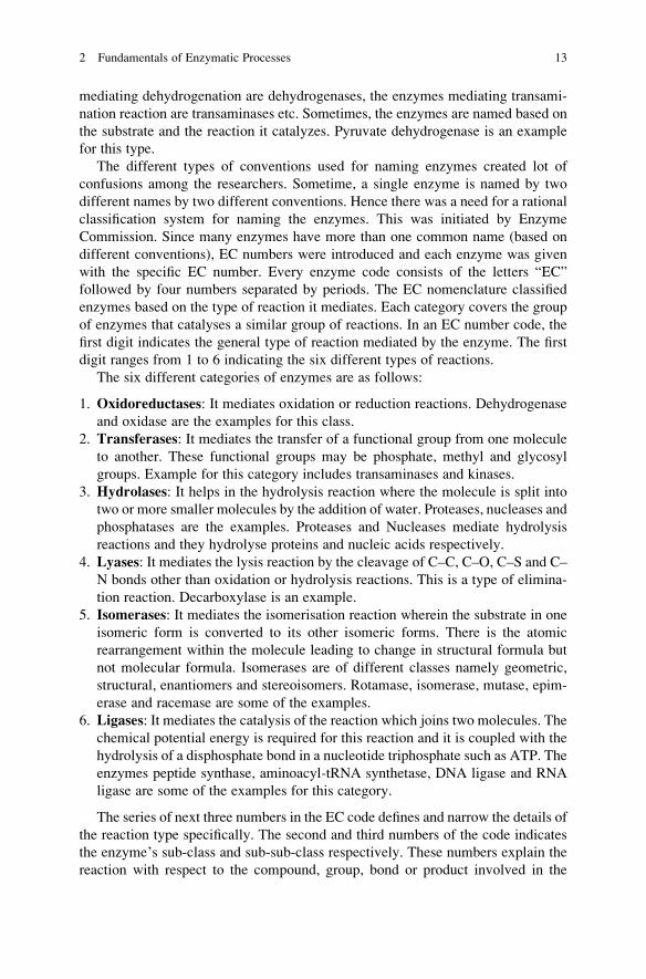
The six different categories of enzymes are as follows:
1. Oxidoreductases: It mediates oxidation or reduction reactions. Dehydrogenase
and oxidase are the examples for this class.
2. Transferases: It mediates the transfer of a functional group from one molecule
to another. These functional groups may be phosphate, methyl and glycosyl
groups. Example for this category includes transaminases and kinases.
3. Hydrolases: It helps in the hydrolysis reaction where the molecule is split into
two or more smaller molecules by the addition of water. Proteases, nucleases and
phosphatases are the examples. Proteases and Nucleases mediate hydrolysis
reactions and they hydrolyse proteins and nucleic acids respectively.
4. Lyases: It mediates the lysis reaction by the cleavage of C–C, C–O, C–S and C–
N bonds other than oxidation or hydrolysis reactions. This is a type of elimina-
tion reaction. Decarboxylase is an example.
5. Isomerases: It mediates the isomerisation reaction wherein the substrate in one
isomeric form is converted to its other isomeric forms. There is the atomic
rearrangement within the molecule leading to change in structural formula but
not molecular formula. Isomerases are of different classes namely geometric,
structural, enantiomers and stereoisomers. Rotamase, isomerase, mutase, epim-
erase and racemase are some of the examples.
6. Ligases: It mediates the catalysis of the reaction which joins two molecules. The
chemical potential energy is required for this reaction and it is coupled with the
hydrolysis of a disphosphate bond in a nucleotide triphosphate such as ATP. The
enzymes peptide synthase, aminoacyl-tRNA synthetase, DNA ligase and RNA
ligase are some of the examples for this category.
The series of next three numbers in the EC code defines and narrow the details of
the reaction type specifically. The second and third numbers of the code indicates
the enzyme’s sub-class and sub-sub-class respectively. These numbers explain the
reaction with respect to the compound, group, bond or product involved in the
2 Fundamentals of Enzymatic Processes 13

reaction. The final digit of the EC code is called as the serial identifier and it
provides insights about specific metabolites and cofactors involved.
2.5 Mechanism of Enzymes
The binding of the substrate to the active site of the enzyme and its interaction is the
basic mechanism of enzymatic catalysis. The active site is the catalytic region of the
enzyme on which the substrate binds. Once the substrate binds to the active site of
the enzyme, it causes the redistribution of electrons in the chemical bonds of the
substrate. This redistribution of electrons in the substrate causes the biochemical
transformation of the substrate to form products. Once the substrate is converted
into product in the active site of the enzyme, the products are released from the
enzyme and the next substrate molecule binds on the active site of the enzyme again
and this cycle continues. The substrate can interact with the active site of the
enzyme by different ways based on opposite charges, hydrogen bonding, hydro-
phobic non-polar interaction, and coordinate covalent bonding.
The unique geometric shape of the active site provides clue for the specificity of
the enzyme to the specific substrate. There is a correlation between the geometric
shapes of substrate to the geometric shape of active site of the enzyme. Two major
hypothesis namely lock and key theory and Induced fit hypothesis have been
proposed to explain the mechanism of interaction of the enzyme and substrate
depending on the geometric shape.
Lock and Key Theory The specific interaction between the enzyme and its
substrate was first postulated by Emil Fisher in 1894 using Lock and Key analogy.
In this hypothesis, Email Fisher postulated that the enzyme has a rigid structure and
its active site has a defined geometric shape. Only the substrate whose geometric
shape is complementary to the active site will be able to bind to the active site of the
enzyme and gets catalysed. It is similar mechanism of the lock and the key. The
enzyme acts as the lock and the substrate acts as the key. If the key exactly suits the
lock, only then the key can used to open the lock. Even a very small changes in the
shape/size of the key or if the key is not positioned properly, then the lock cannot be
opened.
Induced Fit Theory However, the Fisher’s lock and key theory which explained
enzyme as the rigid structure had limitations and failed to support all the experi-
mental evidences on enzyme–substrate interactions. To circumvent this issue, a
new theory called the induced-fit hypothesis has been developed. In contrast the
lock and key mechanism, the induced fit hypothesis proposed that the enzyme
structure is flexible and not rigid.
It is proposed that the substrate has a crucial role in determining the structure of
the enzyme and the shape of the active site. The structure of the enzyme is partially
flexible. On binding of the substrate to the enzyme, the enzyme structure changes
accordingly and mediates the catalysis of the substrate. This theory also describes
14 R. Navanietha Krishnaraj et al.
the reason behind the irreversible inhibition of enzyme wherein inhibitors can bind
to the enzyme and causes the distortion of the enzyme. The substrate molecules
which has a smaller geometric size when compared with the geometric size of the
active site of the enzyme, cannot induce the structural change of the enzyme and
they could not react/get catalysed. The specific substrate can only induce the change
in the structure of the specific enzyme and get catalysed.
The enzyme activities are regulated in a highly systematic manner in the living
systems. Certain enzymes are produced in inactive forms and they get activated
whenever their catalytic activity is required. Enzymes such as pepsin, trypsin and
chymotrypsin are produced in in active forms namely pepsinogen, trypsinogen and
chymotrypsinogen respectively. These are proenzymes and it is inappropriate to
term them as inactive enzymes. The term “inactive enzymes” refers to those
enzymes which have lost their activity due to physical/chemical/metabolic factors
or any other reasons. But zymogens are molecules that needs to be activated to
make it an active enzyme. It is apt to term zymogens as the inactive precursor of
enzymes. Some of the digestive enzymes and coagulation factors are synthesized as
zymogens. The synthesis of digestive enzymes as zymogens is a safe mechanism as
most of these digestive enzymes are proteolytic and if the enzymes are synthesized
in active form, they have greater chance of hydrolyzing the proteins in the cells
synthesizing them. If the zymogens are synthesized in actively forms, it leads to
certain diseases in biological systems. Acute pancreatitis is one such example
wherein the pancreatic enzymes e.g. trypsin, phospholipase A2, and elastase are
activated in premature state.
Allosteric enzymes have a different interesting regulatory mechanism when
compared with the normal enzymes. Allosteric enzymes have two different sites
namely catalytic site and the regulatory site. The molecules which binds to these
regulatory sites are called modulators or effectors. The positive modulators will
mediate catalysis and negative modulators will inhibit the catalysis. Unlike the
normal enzymes, the allosteric enzymes do not follow the Michaelis–Menten
Kinetics as they have multiple active sites. Binding of the substrate to one active
site of allosteric enzyme will influence the binding of the substrate to its next active
site and this phenomenon is called cooperativity. Cooperativity is an interesting
feature of allosteric enzyme. If the binding of first substrate onto the one active site
facilitates binding of subsequent substrate molecule onto the next active site of the
substrate molecule, then it is called “positive cooperativity”. On the contrary, if the
binding of the first substrate to its active site decelerates the binding of the next
substrate to other active site, it is termed as “negative cooperativity”. In some cases,
various enzymes combine to form supramolecular complexes that allows the direct
transfer of metabolites from one enzyme to the other without entering the bulk
solution and this process is termed as metabolic channelling. The multi-enzyme
complex systems may be static or dynamic.
2 Fundamentals of Enzymatic Processes 15

2.6 Enzyme Activity
To assess the activity of the enzyme, it is more important to understand the units of
the enzyme. Generally, the enzymes are quantified based on the unit of activity
rather than in terms of the amount that is physically present (weight). This is
mainly because of two reasons. It is difficult to purify the enzyme and the enzyme
is easily prone to denaturation or loss of activity. The activity of the enzyme is
generally represented as International Unit (IU) which is widely used. The unit is
defined as the amount of enzyme which catalyzes the transformation of 1 micromole
of the substrate per minute under standard conditions. The other unit of enzyme
activity is Katal (referred as to kat). It is the amount which catalyzes the transfor-
mation of one mole of substance per second (1 kat ¼ 60,000,000 U). There are few
non-standard units for enzyme activity namely Soxhlet, Anson, and Kilo Novo
units. However, these units are not generally used. The activity of the enzyme is a
tool to assess the quality of the enzyme in terms of catalytic activity and it is of use
in industrial applications. The specific activity is another important parameter for
enzyme activity. Specific activity is the number of enzyme units per mg of enzyme
protein. It can be denoted as units/mg (U mg�1). Generally the definitions of
activity are described with reference to the term “standard conditions”. The term
“standard conditions” refer to optimal conditions of properties such as pH, ionic
strength, temperature, substrate concentration, and the concentration of cofactors/
coenzymes. However, these conditions depends on the type of application and
experimental conditions.
The enzyme activity depends on several factors such as temperature, pH,
pressure, cofactor like metal ions etc.
Temperature The enzymatic reactions can operate at a narrow range of temper-
ature. With increase in temperature up to the optimum temperature, the enzymatic
reaction rate increases but after optimal temperature is reached, the reaction rate
decreases with increasing temperature. The increase in temperature causes the
increase in collision between the molecules with higher energy (increase in kinetic
energy) leading to enhanced enzymatic catalytic rates. Like other catalytic reac-
tions, enzymatic reactions also obey Arrhenius equation which states that rate of
reaction will exponentially increase with increase in temperature. The Arrhenius
equation is as follows:
k ¼ Ae�ΔG∗=RT
where k is the kinetic rate constant for the reaction, A is the Arrhenius constant or
frequency factor, G* is the standard free energy of activation (kJ M�1) which
depends on entropic and enthalpic factors, R is the gas law constant and T is the
absolute temperature. Different enzymes have different optimal temperatures and
even a single enzyme from different organisms might differ in their optimal
temperature. Actually the enzymes cannot decrease the activation energy of the
16 R. Navanietha Krishnaraj et al.

same barrier. The enzyme directs the reaction in an alternate chemical pathway
which has a lower activation energy. Most enzymes are stable for months if they are
refrigerated at 0–4 �C. However, storing the enzymes below 0 �C is not advisable.
Storing the enzymes below 0 �C requires the additives such as glycerol and such
very low temperature often causes denaturation due to the stress and pH variation
caused by ice-crystal formation.
Effect of pH on Enzyme Activity The enzymes can remain active and accelerate
catalytic reactions only in a narrow range of pH. The drastic changes in pH from its
optimal levels will change the stability of the enzymes. With increase or decrease in
the pH, the various acid and amine groups on side chains of the protein is disturbed
which in return affects the structure of the enzyme and its catalysis. The pH of the
solution affects the state of ionization of acidic or basic amino acids. The change in
the state of ionization of amino acids in a protein leads to changes in the ionic bonds
which in turn affects the structure of the enzyme. Different enzymes differ in their
optimal pH values. For example, lipase in pancreas has the optimal pH of 8.0 and
lipase from stomach has the optimal pH of 4.0 to 5.0. Pepsin has an optimal pH
value of 1.5–1.6 and trypsin has an optimal pH value of 7.8–8.7. The changes in
ionic strength in the enzymatic system also affect the activity of the enzyme and its
catalytic rates.
The changes in pressure also affects the enzymes in two different ways. The high
pressure applied to the system will affect the conformation of the enzyme leading to
loss of catalytic activity. On the other hand, if the enzymatic reaction systems (such
as in oxygenases and decarboxylases enzyme systems) involve dissolved gases, the
increase in pressure will lead to increase in gas solubility. This causes the distur-
bance in the reaction system leading to shift in the equilibrium position of the
reaction due to any difference in molar volumes between the reactants and products.
2.7 Enzyme Kinetics
The enzyme catalyzed reaction can be of zero, first or second order. For an
elementary reaction occurring in one direction, the order of reaction is equal to
the molecularity. For non-elementary reaction, the rate order is experimentally
determined to analyze what happens to the rate of reaction as the concentration of
one of the reactants change. For first order reaction, if you double the concentration
of the reactant A, the rate also doubles (Rate α [A]). For second order reaction, if
you double the concentration of A the reaction rate will increase by 4 times (Rate αk[A]2). For zero order reaction, the rate is apparently independent of the reactant
concentration.
Enzyme kinetics involves the study of mechanisms and rates of enzyme cata-
lyzed reactions at different reactions conditions such as varying the substrate and
enzyme concentrations. Various environmental factors influence the Enzyme activ-
ity, including substrate concentration, pH, temperature and presence of inhibitors
2 Fundamentals of Enzymatic Processes 17

and activators. Substrate concentration is one of the most important influencing
factor. The rate of enzyme catalyzed reaction increases with increase in substrate
concentration up to the optimal substrate concentration. This is because more the
amount of substrate molecules present, an enzyme binds substrate more often, and
the rate of product formation (reaction velocity) is greater than at lower substrate
concentration. After reaching an optimal substrate concentration, the further
increase in substrate concentration causes no further increase in reaction velocity
because no more active sites are available on the enzyme molecules, that is, the
enzyme is saturated with the substrate and is converting substrate to product as
rapidly as possible. This means enzyme is working at maximum velocity (Vmax).
Relationship between reaction velocity and substrate concentration is shown in
Fig. 2.1.
Michaelis–Menten Kinetics: The relation between the substrate concentration
and reaction rate/velocity is described by the following Michaelis–Menten equation
v ¼ Vmax � S½ �= Kmþ S½ �ð Þ
The Michaelis constant (Km) is that substrate concentration at which the reac-
tion rate (velocity) is half its maximum. Its significance is that it acts as indicator for
the affinity of the enzyme for its substrate. The lower the Km value, more is the
affinity of the enzyme to that substrate. In other words, a low Km means that the
enzyme achieves its maximum catalytic efficiency at low substrate concentrations
and therefore is more efficient as a catalyst or has more affinity for that particular
substrate.
Lineweaver–Burk plot is obtained by taking the reciprocal of the Michaelis–
Menton equation. It was widely used to calculate the Km and Vmax before
non-linear regression software were developed. Figure 2.2 depicts the Lineweaver–
Burk plot.
Fig. 2.1 Relationship
between reaction velocity
and substrate concentration
18 R. Navanietha Krishnaraj et al.

Although it is still used for representation of kinetic data, non-linear regression
or alternative linear forms of the Michaelis–Menten equation such as the Hanes–
Woolf plot or Eadie–Hofstee plot are generally used for the calculation of param-
eters. The drawback for Lineweaver–Burk plot is that it is error prone as small
errors in measurement gets magnified as y-axis takes the reciprocal of the rate of
reaction
Hanes–Woolf plot (shown in Fig. 2.3) is also obtained on rearranging the
Michaelis–Menton equation to get the ratio of the initial substrate concentration
[S] to the reaction velocity v. It linearizes the Michaelis–Menten equation but has a
drawback that neither ordinate nor abscissa represent independent variables: both
are dependent on substrate concentration.
Enzymes can have more than one substrate binding site. When such enzymes
catalyze reactions, the rate increases in a sigmoidal manner as the substrate
concentration increases instead of the hyperbola curve in case of enzymes with
just one binding site. The Hill equation is commonly used to study the kinetics of
reactions that exhibit a sigmoidal behavior. The rate of many transporter-mediated
processes can be analyzed by the Hill equation. Hyperbola curve demonstrates
saturation of the enzyme or transporter at high substrate concentrations. Saturation
is caused by the fact that there is a fixed number of enzyme or transporter
molecules, each with a fixed number of substrate binding sites. At high substrate
Fig. 2.2 Lineweaver–Burk
plot
Fig. 2.3 Hanes–Woolf plot
2 Fundamentals of Enzymatic Processes 19

concentrations, all of the binding sites have substrate bound and each enzyme or
transporter molecule is working as fast as its intrinsic rate to catalyze the reaction.
Reactions that exhibit a sigmoidal curve also exhibit saturation at high substrate
concentration. However, at low substrate concentrations, a very different behavior
is observed (compared to a hyperbolic relationship). At low substrate concentra-
tions, the rate increases only incrementally with increases in the substrate concen-
tration. As the substrate concentration increases further, small increases in the
substrate concentration lead to large increases in the reaction rate. At very high
substrate concentrations, the rate exhibits saturation, where additional increases in
the substrate concentration no longer increase the reaction velocity. This type of
saturation kinetics is adequately described by the Hill equation.
2.8 Enzyme Inhibitors and Types
Enzyme inhibitors are molecules that prevents the enzyme catalysis by interacting
with the enzyme molecule. Enzyme inhibitors have several applications in the
development of drugs, pesticides etc. However, some of the inhibitors inhibit the
key enzymes of our body and causes diseases and even death.
The enzyme’s catalytic activity can be altered by binding various small mole-
cules to it, sometimes at its active site, and sometimes at a site distant from the
active site. Usually these alterations involve a reduction in the enzyme’s ability to
accelerate the reaction; less commonly, they give rise to an increase in the enzyme’sability to accelerate a reaction. Enzyme inhibitors are molecules which interact in
some way with the enzyme and consequently slow down, or in some cases stop
catalysis. Enzyme inhibitors can be specific or non-specific. Non-specific methods
of inhibition include any physical or chemical changes which ultimately denatures
the enzyme. The specific inhibitors act on a single enzyme or closely related
enzymes and are further characterized into three main types: competitive, uncom-
petitive and non-competitive. These types of enzyme inhibition can be explained on
the basis of their effect on the formation of enzyme–substrate complex and is
explained in the following paragraphs.
Competitive Inhibitors These inhibitor molecules are similar, in chemical struc-
ture and molecular geometry, to the substrate of the enzyme. Therefore, as the name
suggests, they compete with the substrate for binding to the active side of the
enzyme. The inhibitor binds to the active site of the enzyme and thus prevent the
formation of the Enzyme–Substrate complex. In other words, enzyme–inhibitor
(E-I) complex is formed in instead of enzyme–substrate complex. Therefore,
binding of substrate to enzyme in inhibited and the product formation is decreased.
However, Competitive inhibition is reversible and levels of inhibition depends upon
the relative concentrations of the substrate and the inhibitor molecules as the
inhibitors compete with the substrate to bind the active site of the enzyme.
20 R. Navanietha Krishnaraj et al.

Drug disulfuran (Antabuse) which is used to help people to get rid of alcoholism,
acts as a competitive inhibitor for the aldehyde oxidase enzyme which causes the
accumulation of acetaldehyde resulting in unpleasant side effects such as nausea
and vomiting. Another example of a useful application of competitive inhibition is
in the case of methanol poisoning. Ethanol is given as an antidote in these cases as it
acts as a competitive inhibitor to methanol, thus preventing its oxidation to form-
aldehyde and formic acid. Formic acid is the actual toxic compound that effects the
optic nerve causing blindness and its production is stopped by administration of
ethanol.
It is important to note that the competitive inhibitor affects the binding of the
substrate but not the reaction velocity i.e. it affects the Km of the enzyme but not the
Vmax. As discussed earlier, Km value is a measure of the amount of the substrate
required to reach half the maximum reaction velocity (Vmax). It is also known
that the presence of the competitive inhibitor, forces to increase the substrate
concentration in order to achieve that half maximal velocity. It therefore, increases
the Km.
Non-competitive Inhibitors A non-competitive inhibitor interacts with the
enzyme at a site other than the active site called the allosteric site. This site could
be very close to the active site or far from it. Non-competitive inhibitors are usually
reversible, but are not influenced by concentrations of the substrate since they do
not compete with substrate molecules as in the case for a reversible competitive
inhibitor. Therefore, Km is unchanged, but Vmax is reduced. A non-competitive
inhibitor reacts with the enzyme–substrate complex, and slows the rate of reaction
to form the enzyme–product complex. This means that increasing the concentration
of substrate will not relieve the inhibition, since the inhibitor reacts with the
enzyme–substrate complex. An example of non-competitive inhibition is the inhi-
bition of cytochrome enzymes by cyanide.
Uncompetitive Inhibitors Uncompetitive inhibition is a very rare class of inhibi-
tion. An uncompetitive inhibitor binds to the enzyme and enhances the binding of
substrate (so reducing Km), but the resultant enzyme–inhibitor-substrate complex
only undergoes reaction to form the product slowly, so that Vmax is also reduced.
While uncompetitive inhibition requires that an enzyme–substrate complex must be
formed, non-competitive inhibition can occur with or without the substrate present.
In addition, uncompetitive inhibition works best when substrate concentration is
high. Lithium in the phosphoinositide cycle is an example of uncompetitive inhi-
bition which ameliorates manic-depressive psychosis.
2.9 Immobilization of Enzymes
Enzyme immobilisation is a technique for entrapment of the enzyme onto a distinct
support or matrix. The support or matrix on which the enzymes are immobilized is
called a carrier and it allows the substrate or effector or inhibitor molecules to
2 Fundamentals of Enzymatic Processes 21

interact with the immobilised enzyme. The enzyme amino acylase of Aspergillusoryzae was the first enzyme to be immobilized for the production of L-amino acids.
The enzyme can be immobilised onto the carrier either permanently or temporarily
for a certain period of time. Three different polymers such as natural polymers,
synthetic polymers and inorganic materials can be used as carriers for the
immobilisation of enzymes. Alginate, chitosan, collagen, carrageenan, gelatin,
cellulose, starch, and pectin are some of the natural polymers that are used for
immobilisation. Alginate is an inert natural polymer and has good water retaining
capacity. Calcium or magnesium alginate are the commonly used polymers for the
preparation of natural matrices. Chitosan and chitin are the structural polysaccha-
rides occurring naturally in the cell wall of fungi that are widely used polymers for
immobilisation. Collagen is the proteinaceous support with good porosity and water
retaining capacity. The side chains of the amino acids in the collagen and that of
enzyme can form covalent bonds to permanently hold the enzyme to the support.
Gelatin is the partially hydrolyzed collagen with good water holding capacity and is
used for immobilisation of enzymes. Carrageenan, is a sulphated polysaccharide
extracted from red algae, is a gelling agent that can hold proteins.
Polyvinyl chloride (PVC), Diethylaminoethyl cellulose (DEAE cellulose), and
UV activated Polyethylene glycol (PEG) are some of the synthetic polymers that
are used for the enzyme immobilisation. Zeolites, ceramics, diatomaceous earth,
glass, silica, activated carbon, and charcoal are some of the examples for the
inorganic materials.
There are a different types of carriers or supports that are used for immobiliza-
tion of enzymes. The carrier used should be cheap and easily available. The ideal
carrier should also have ease of functionalisation and should not inhibit the catalytic
activity of the enzyme. The good carrier should offer good mass transfer charac-
teristics and better diffusion rates leading to enhanced effectiveness. The effective-
ness can be calculated by the ratio of rate of the reaction to the rate of diffusion. The
immobilization of enzymes onto the carrier has several advantages such as
enhanced functional efficiency of enzyme, reuse of enzyme, decreasing the reaction
time, decreasing the cost of operation, high enzyme substrate ratio, and enhanced
reproducibility of the process.
The immobilisation of enzymes has certain disadvantages. In some cases, the
immobilisation of enzymes causes the loss of catalytic activity. The immobilisation
of enzyme involves high cost and technical difficulties in the recovery of active
enzyme and purification. There is a high chance that enzyme becoming unstable
and losing its catalytic activity after it is recovered from the carrier. The
immobilisation strategy does not suit for most industrial operations.
The immobilisation of enzyme can be carried out by five different methods
namely adsorption, covalent bonding, entrapment, copolymerization and encapsu-
lation. Adsorption is the simplest technique for the immobilisation of enzyme onto
carrier. It is based on formation of weak (low energy) bonds to stabilise the
enzymes onto the external surface of the carrier. Ionic interaction, hydrogen
bonds and Van der Waal forces are the weak bonds involved in the adsorption
process. Adsorption is the most traditional method for enzyme immobilisation.
22 R. Navanietha Krishnaraj et al.

Charcoal was used as the carrier for immobilisation of invertase in 1916. Materials
such as aluminium oxide, clay, starch, and ion exchange resins are used as
adsorbants/carriers. The smaller the size of the carrier, the larger is the surface
area and higher is the rate of immobilisation and catalysis. The adsorption process
also have the advantage that it will not suffer from pore diffusion limitations since
the enzymes are immobilised on the external surface of the carrier. The adsorption
of enzyme onto the carrier has several advantages such as simplicity, low cost for
immobilisation, no need for reagents, minimum activation steps and no loss of
enzyme structure/catalytic activity than other immobilisation methods.
The adsorption of enzyme onto the carrier can be carried out by four different
methods namely static loading process, dynamic process, reactor loading process
and electrode position process. Adsorption of enzyme onto the carrier by static
process involves the carrier to be placed in the solution containing the enzyme
without stirring. In dynamic process, the carrier is placed in the solution and mixed
by agitation/stirring. Reactor loading process involves the transfer of enzyme
solution from the carrier to the reactor by continuous agitation process. Electrode
position process makes use of the electric field for the migration of the enzyme to
the surface of the carrier. The enzyme immobilisation by adsorption technique has
certain disadvantages such as the enzyme is prone to desorb from the carrier and the
efficiency of this immobilization technique is poor.
The second important technique for immobilisation of the enzyme on the matrix
by forming covalent bond between the chemical groups in enzyme and to the
chemical groups on the support. Chemical bonding is an important and stable
method for enzyme immobilization. The choice of the carrier is very important
for immobilisation using this covalent bonding technique. The carrier should have
chemical groups such as amino groups, imino groups, hydroxyl groups, carboxyl
groups, thiol groups, methylthiol groups, guanidyl groups, imidazole groups or
phenol ring which can help in forming the covalent group with the enzyme.
Similarly the enzyme should have functional groups such as alpha carboxyl group
at ‘C’ terminal of enzyme, alpha amino group at ‘N’ terminal of enzyme, phenol
ring of Tyrosine, thiol group of Cysteine, hydroxyl groups of serine and threonine,
imidazole group of histidine and indole ring of tryptophan. Different matrices such
as cellulose, agarose, DEAE cellulose, polyacrylamide, collagen, gelatin, amino
benzyl cellulose, porous glass, cyanogen bromide etc can be used as support for
enzyme immobilization. The covalent bonding can be mediated by diazotion,
peptide bond formation and using poly functional reagents. The covalent bonding
technique is a simple method that provides a strong linkage of enzyme to the
carrier/matrix and avoids leakage/desorption of enzymes. The covalent bonding
technique for enzyme immobilisation is widely used for several industrial applica-
tions. But, in some cases, covalent bonding leads to enzyme inactivation/chemical
modification of the enzyme which in turn affects the conformation of the enzyme
and its catalytic activity.
Entrapment is another method for enzyme immobilisation wherein the enzymes
are physically entrapped within a porous carrier. Generally, water soluble polymer
is used for the preparation of carriers for entrapment of enzymes. The enzymes are
2 Fundamentals of Enzymatic Processes 23

immobilised onto the matrix either covalently or non-covalently. The nature of the
carrier and pore size are some of the criteria for choice of matrix. Pore size of the
carrier can be modified based on the concentration of the polymer used. Entrapment
technique is not suitable for the low molecular weight enzymes from the matrix as it
causes leakage. Polyacrylamide gels, agar, alginate, gelatin, and carrageenan are
some of the matrices that are used as matrices for entrapment. Entrapment of
enzymes can be made in the gels, fibers or in microcapsules. This is a simple,
quick, and economical method of immobilisation. It requires mild operating con-
ditions for the entrapment and has lesser change of conformational changes in
structure of the enzyme. Entrapment techniques has certain limitations such as
leakage/loss of the enzyme, loss by diffusion, and is easily prone to microbial
contamination.
The next method of immobilisation is cross linking method in which functional
groups between the enzymes are cross linked directly by covalent bonds.
Polyfunctional reagents such as glutaraldehyde and diazonium salt are generally
used as crosslinking agents for the immobilisation by crosslinking or
copolymerisation technique. It is a simple and an economical method for
immobilisation where pure enzymes are not required. It is widely used for several
industrial and commercial applications. This technique has certain limitations—the
crosslinking agents that are used for co-polymerisation of enzymes are prone to
denature the structure or activity of the enzyme.
The fifth method for immobilisation is the encapsulation. It is done by
immobilising the enzymes within membrane capsule. Semi permeable membranes
such as nitro cellulose or nylon are generally used as capsules. The mass transfer
shortcoming is the major shortcoming of this technique. This technique allows
immobilisation of a large quantity of enzyme within the capsule. It is a simple and
inexpensive method. The membrane capsule allows only the substrate of smaller
size to enter the capsule and this technique has pore size limitation.
Applications of Immobilized Enzymes Immobilized enzymes are widely used
for several industrial biotechnology applications such as production of alcohols,
organic acids, amino acids, drugs, antibiotics etc. The use of immobilized enzymes
greatly helps to cut down the costs of bioprocesses. Immobilized enzymes are also
used widely in the food industry for the production of corn syrup, jams, jellies etc.
Immobilised pectinases and cellulases are widely used in food industries. Biomed-
ical sector is one major area where the immobilized enzymes are widely used in the
diagnosis of ailments and therapy. Immobilized enzymes based biosensors are
widely used for detection of different analytes such as glucose, urea, nitric oxide,
dopamine, phenol etc. Enzyme Immobilization strategies are also very useful for
the targeted drug delivery and controlled drug release at the site of infection.
Immobilised enzymes are also widely used in research especially in molecular
biology. The other areas where the immobilised enzyme can be used are for the
production of bio-diesel from vegetable oils, treatment of wastewater, textile
industry and detergent industry.
24 R. Navanietha Krishnaraj et al.

2.10 Application of Extremozymes
Extremophiles are microorganisms that survive under harsh environmental condi-
tions that can include atypical temperature, pH, salinity, pressure, nutrient, oxygen,
water, and radiation levels. Thus, extremophiles are a robust group of organisms
producing stable enzymes, which are often capable of tolerating changes in envi-
ronmental conditions such as high/low pH and temperature. These organisms and
their enzymes have wide range of potential applications in biotechnology. One
application is that extremophilic enzymes, also called extremozymes can be
exploited for conversion of biomass to biofuel. Biomass is biological material
derived from living, or recently living organisms. It most often refers to plants or
plant-based materials which are specifically called lignocellulosic biomass. As an
energy source, biomass can either be used directly via combustion to produce heat,
or indirectly after converting it to various forms of biofuel. Biomass can be
converted to other usable forms of energy like methane gas or transportation fuels
like ethanol and biodiesel.
The five most basic type of extremozymes and their applications are as follows:
Thermophilic Enzymes High temperatures for chemical reaction is favorable due
to the increase in substrate solubility, better mixing, high reaction rates, and low
viscosity. Another important advantage of carrying out reactions at high tempera-
tures is pathogen removal. Thus, thermophilic enzymes which are active at high
temperatures can be used to catalyze such high temperature chemical processes. An
enzyme or protein is considered thermostable when they have a high defined
unfolding (transition) temperature (Tm), or a long half-life at a selected high
temperature. The use of such enzymes in maximizing reactions accomplished in
the food and paper industry, detergents, drugs, toxic wastes removal, and drilling
for oil is being studied extensively. Thermophilic enzymes specifically hemicellu-
lose degrading is employed in treatment of wood to obtain pulp as it is carried out at
high temperatures. Thermophilic xylanases (xylan degrading) also reduce the
consumption of Chlorine in the bleaching process. Thus, decreasing the release of
hazardous organic halogens to the environment. Thermostable enzymes have also
been used in the pharmaceutical industry along with conventional chemical syn-
thesis for production of drugs. L-aminoacylase from Thermococcus litoralis devel-oped at Exeter is used for the resolution of amino acids and analogues (Toogood
et al. 2002). The gamma lactamase from Sulfolobus solfataricus is used for the
production of optically pure gamma lactam—the building block for anti-viral
carbocyclic nucleotides (Toogood et al. 2004). The alcohol dehydrogenase from
Aeropyrum pernix for the production of optically pure alcohols (Guy et al. 2003).
Another major applications for thermostable enzymes are starch liquefaction using
amylases from thermophilic Bacillus sp. and proteases for food processing and
detergents. Thermostable DNA polymerases are greatly useful in the PCR process.
Psychrophilic Enzymes These enzymes are produced by psychrophilic (organ-
isms that thrive at very low temperatures, usually below 5 �C). Psychrophilic
2 Fundamentals of Enzymatic Processes 25

enzymes are therefore cold active and heat labile. They have a number of advan-
tages in the field of biotechnology, majorly because of their high kcat at low to
moderate temperatures. Also, they can be used at lower concentration as they have
high activities, and thus reduce the cost of producing large amount of the enzyme.
So additional heating costs for their optimum activity is not required. Due to this,
these enzymes, typically belonging to the lipase and protease class have been used
as additives to detergents for washing at room temperature. Further, due to their
heat lability, they can be selectively deactivated easily by slight increase in tem-
peratures. Their industrial applications have been widely explored.
Psychrophilic enzymes finds immense applications in food industry for improv-
ing the digestibility and removing hemicellulose from feed, meat tenderizing,
ripening of cheese, dough fermenting, stabilizing wine and beverages (Bialkowska
et al. 2009; Collins et al. 2006; Tutino et al. 2009; Wang et al. 2010). They also find
applications in detergent industries, (Tutino et al. 2009; Wang et al. 2010); Biofuels
and energy production (Dahiya et al. 2006; Hildebrandt et al. 2009; Ueda et al.
2010); pharmaceutical, (Dahiya et al. 2006; Joseph et al. 2008); Textile industries
(Collins et al. 2006; Ueda et al. 2010); environmental biotechnology for bioreme-
diation; biobleaching of pulp and paper and tanning (Joseph et al. 2008; Wang et al.
2010) and chemical synthesis (of peptides, epoxides, oligosaccharides and other
organic compounds) by reverse hydrolysis in organic solvents (Aurilia et al. 2008;
Joseph et al. 2008).
Acidophilic Enzymes Acidophilic enzymes derived from acidophiles are adapted
to work under low pH. In other words the optimum pH for their activity lies is the
acidic range. An acidophilic β-galactosidase enzyme purified from TeratosphaeriaacidothermaAIU BGA-1 has been found to show high activity at extremely low pH
of 1 (Chiba et al. 2015). Another acidophilic enzyme, β-mannanase from
Gloeophyllum trabeum CBS900.73 with significant transglycosylation activity
have been found recently and posseses feed digesting ability (Wang et al. 2016).
Xylanase produced by an acidophile Penicillium oxalicum GZ-2 has great potential
to be used in biofuels, animal feed, and food industry applications (Liao et al. 2012).
Alkaline Enzymes Alkaliphiles are microorganisms that can grow in alkaline
environments, i.e. pH > 9.0 Alkaline enzymes obtained from organisms living in
these environments are able to function under high alkaline pH values because of
their stability/activity under these conditions. Alkaline enzymes often show activ-
ities in a broad pH range, thermostability, and tolerance to oxidants compared to
neutral enzymes (Fujinami and Fujisawa 2010).
From many decades, many alkaline protease enzymes have been tested for their
compatibility with commercial detergents (Devi et al. 2016; Gupta et al. 2002;
Haddar et al. 2009; Phadatare et al. 1993). They also have wide applications in
tannery and food industries, medicinal formulations, and processes like waste
treatment, silver recovery and resolution of amino acid mixtures (Agrawal et al.
2004; Horikoshi 1999; Sinha et al. 2014). Alkaline proteases have been used in the
preparation of protein hydrolysates of high nutritional value. The protein hydroly-
sates play an important role in blood pressure regulation and are used in infant food
26 R. Navanietha Krishnaraj et al.

formulations. The possible utilization of alkaline protease secreted by Penicilliumsp. for hydrolysis of soy protein, a byproduct of soybean industries has also been
done (Agrawal et al. 2004; Fujinami and Fujisawa 2010). Other alkaline enzymes,
e.g. alkaline cellulases, alkaline amylases, and alkaline lipases, are also adjuncts to
detergents for improving cleaning efficiency.
Barophilic Enzymes These enzymes are produced by deep sea Piezophilic/
barophilic microorganisms and are stable at high pressures. They can be used for
enzymatic processes at high pressure such as in food processing industry for
sterilization of food, deep sea waste disposal, production of novel natural products
and catabolic activities. These enzymes can also be used for other high pressure
bioreactors. The application of these enzymes in various industrial processes is still
in its budding stage.
Take Home Message
• Enzymes are biological catalysts that can mediate the biological reactions. They
are produced by all living organisms. Most enzymes are proteins except
ribozymes. All enzymes are not proteins and all proteins are not enzymes.
• Enzyme Commission has classified enzymes into six classes: Oxidoreductases,
Transferase, Hydrolase, Lyase, Isomerase and Ligase.
• Specificity is the inherent charecetristic of the enzyme. The enzyme specificity
can be categorized into five groups: Absolute Substrate specificity, Broad
specificity (Group specificity), Bond specificity (Relative specificity),
Sterochemical specificity, and Reaction specificity.
• Active site is the region of the enzyme where the substrate binds. Lock and Key
Theory states both the structure of enzyme and the substrate are rigid whereas
Induced Fit Theory describes that the structure of enzyme is partially flexible.
The binding of the substrate onto enzyme causes structural changes in the
enzyme and mediates the catalysis of the substrate.
• The activity of the enzyme can be represented as International Unit (IU), Katal,
and Turnover number.
• The relation between the substrate concentration and reaction rate/velocity is
described by Michaelis–Menten equation.
• Enzyme inhibition can be reversible or irreversible. The inhibitors of enzymes
can be classified as competitive, non- competitive, and un-competitive.
• The enzymes can be immobilized onto carriers by different methods such as
adsorption, covalent bonding, cross linking, entrapment, and encapsulation.
References
Agrawal D, Patidar P, Banerjee T, Patil S (2004) Production of alkaline protease by Penicillium
sp. under SSF conditions and its application to soy protein hydrolysis. Process Biochem
39:977–981
2 Fundamentals of Enzymatic Processes 27

Aurilia V, Parracino A, D’Auria S (2008) Microbial carbohydrate esterases in cold adapted
environments. Gene 410:234–240
Buchholz K, Poulson PB (2000) Overview of history of applied biocatalysis. In: Straathof AJJ,
Adlercreutz P (eds) Applied biocatalysis. Harwood Academic Publishers, Amsterdam
Bialkowska AM, Cieslinski H, Nowakowska KM, Kur J, Turkiewicz M (2009) A new beta-
galactosidase with a low temperature optimum isolated from the Antarctic arthrobacter
sp. 20B: gene cloning, purification and characterization. Arch Microbiol 191:825–835
Chiba S, Yamada M, Isobe K (2015) Novel acidophilic beta-galactosidase with high activity at
extremely acidic pH region from Teratosphaeria acidotherma AIU BGA-1. J Biosci Bioeng
120:263–267
Collins T, Hoyoux A, Dutron A, Georis J, Genot B, Dauvrin T, Arnaut F, Gerday C, Feller G
(2006) Use of glycoside hydrolase family 8 xylanases in baking. J Cereal Sci 43:79–84
Dahiya N, Tewari R, Hoondal GS (2006) Biotechnological aspects of chitinolytic enzymes: a
review. Appl Microbiol Biotechnol 71:773–782
Devi SG, Fathima AA, Sanitha M, Iyappan S, Curtis WR, Ramya M (2016) Expression and
characterization of alkaline protease from the metagenomic library of tannery activated sludge.
J Biosci Bioeng 122:694–700
Fujinami S, Fujisawa M (2010) Industrial applications of alkaliphiles and their enzymes – past,
present and future. Environ Technol 31:845–856
Gupta R, Beg QK, Lorenz P (2002) Bacterial alkaline proteases: molecular approaches and
industrial applications. Appl Microbiol Biotechnol 59:15–32
Guy JE, Isupov MN, Littlechild JA (2003) The structure of an alcohol dehydrogenase from the
hyperthermophilic Archaeon aeropyrum pernix. J Mol Biol 331:1041–1051
Haddar A, Agrebi R, Bougatef A, Hmidet N, Sellami-Kamoun A, Nasri M (2009) Two detergent
stable alkaline serine-proteases from Bacillus mojavensis A21: purification, characterization
and potential application as a laundry detergent additive. Bioresour Technol 100:3366–3373
Hildebrandt P, Wanarska M, Kur J (2009) A new cold-adapted beta-D-galactosidase from the
Antarctic arthrobacter sp. 32c – gene cloning, overexpression, purification and properties.
BMC Microbiol 9:151
Horikoshi K (1999) Alkaliphiles: some applications of their products for biotechnology. Microbiol
Mol Biol Rev 63:735–750
Joseph B, Ramteke PW, Thomas G (2008) Cold active microbial lipases: some hot issues and
recent developments. Biotechnol Adv 26:457–470
Liao H, Xu C, Tan S, Wei Z, Ling N, Yu G, Raza W, Zhang R, Shen Q, Xu Y (2012) Production
and characterization of acidophilic xylanolytic enzymes from Penicillium oxalicum GZ-2.
Bioresour Technol 123:117–124
Payen A, Persoz JF (1833) Memoire su la diastase, les principaux produits de ses reactions, et leurs
applications aux arts industriels. Ann Chim Phys 53:73–92
Phadatare SU, Deshpande VV, Srinivasan MC (1993) High activity alkaline protease from
Conidiobolus coronatus (NCL 86.8.20): enzyme production and compatibility with commer-
cial detergents. Enzyme Microb Technol 15:72–76
Sinha R, Srivastava AK, Khare SK (2014) Efficient proteolysis and application of an alkaline
protease from halophilic Bacillus sp. EMB9. Prep Biochem Biotechnol 44:680–696
Sumner JB, Somers GF (1953) Chemistry and methods of enzymes, third edition. Soil Sci 76
(2):166
Toogood HS, Hollingsworth EJ, Brown RC, Taylor IN, Taylor SJ, McCague R, Littlechild JA
(2002) A thermostable L-aminoacylase from Thermococcus litoralis: cloning, overexpression,
characterization, and applications in biotransformations. Extremophiles 6:111–122
Toogood HS, Brown RC, Line K, Keene PA, Taylor SJC, McCague R, Littlechild JA (2004) The
use of a thermostable signature amidase in the resolution of the bicyclic synthon (rac)–γlactam.
Tetrahedron 60:711–716
Tutino ML, di Prisco G, Marino G, de Pascale D (2009) Cold-adapted esterases and lipases: from
fundamentals to application. Protein Pept Lett 16:1172–1180
28 R. Navanietha Krishnaraj et al.

Ueda M, Goto T, Nakazawa M, Miyatake K, Sakaguchi M, Inouye K (2010) A novel cold-adapted
cellulase complex from Eisenia foetida: characterization of a multienzyme complex with
carboxymethylcellulase, beta-glucosidase, beta-1,3 glucanase, and beta-xylosidase. Comp
Biochem Physiol B Biochem Mol Biol 157:26–32
Wang F, Hao J, Yang C, Sun M (2010) Cloning, expression, and identification of a novel
extracellular cold-adapted alkaline protease gene of the marine bacterium strain YS-80-122.
Appl Biochem Biotechnol 162:1497–1505
Wang C, Zhang J, Wang Y, Niu C, Ma R, Wang Y, Bai Y, Luo H, Yao B (2016) Biochemical
characterization of an acidophilic beta-mannanase from Gloeophyllum trabeum CBS900.73
with significant transglycosylation activity and feed digesting ability. Food Chem 197:474–481
2 Fundamentals of Enzymatic Processes 29
Chapter 3
Pretreatment of Lignocellulosic Feedstocks
Antonio D. Moreno and Lisbeth Olsson
Abbreviations
AFEX ammonia fiber explosion
ARP ammonia recycled percolation
CELF co-solvent enhanced lignocellulosic fractionation
COSLIF cellulose and organic solvent-based lignocellulosic fractionation
CrI cellulose crystallinity index
CSF combined severity factor
Cu-AHP copper-catalyzed alkaline hydrogen peroxide
DP degree of polymerization of cellulose
EA extractive ammonia
GHG greenhouse gas
ILs ionic liquids
LCA life-cycle assessment
LCCs lignin-carbohydrate complexes
NMMO N-methylmorpholine-N-oxide
SAA soaking in aqueous ammonia
SF severity factor
SPORL sulfite pretreatment to overcome recalcitrance of lignocellulose
WRV water retention value
A.D. Moreno (*)
Department of Biology and Biological Engineering, Industrial Biotechnology,
Chalmers University of Technology, 412 96 Gothenburg, Sweden
Department of Energy, Biofuels Unit, Ciemat, Avda. Complutense 40, 28040 Madrid, Spain
e-mail: [email protected]; [email protected]
L. Olsson
Department of Biology and Biological Engineering, Industrial Biotechnology,
Chalmers University of Technology, 412 96 Gothenburg, Sweden
© Springer International Publishing AG 2017
R.K. Sani, R.N. Krishnaraj (eds.), Extremophilic Enzymatic Processing ofLignocellulosic Feedstocks to Bioenergy, DOI 10.1007/978-3-319-54684-1_3
31

3.1 Introduction
After the first oil crisis in the mid-late twentieth century, the need for a more
sustainable and renewable energy system became clear. Today, 40 years later, a
continuous increase in the energy demand and the urgency in reducing carbon
emissions are still good reasons for us to give up our dependence on non-renewable
fossil resources. Consequently, the scientific community is in the process of helping
to develop and implement novel technologies and lay the basis of a new bio-based
economy with relevant products for the energy, chemical, material, and food
sectors. In this context, lignocellulosic biomass represents a great source of renew-
able raw material for the implementation of this bio-based economy since it is
widely available, relatively inexpensive and do not compete with food production.
Lignocellulose is the most abundant renewable organic matter in nature, with an
estimated annual production of more than 1010 MT worldwide (Sanchez and
Cardona 2008). It includes agricultural wastes, forest products, and energy crops,
and its chemical composition varies depending on the raw material (Sun and Cheng
2002). The main components of lignocellulosic biomass are cellulose, hemicellu-
lose, and lignin. From the biochemical point of view, cellulose and hemicellulose
incorporate sugars as monomers (glucose, xylose, mannose, arabinose, etc.), while
lignin is built up of three primary phenylpropane units ( p-coumaryl, coniferyl, and
sinapyl alcohol). Due to their different properties, each polymer has a specific role
in lignocellulosic materials. Cellulose fibers interact to each other by hydrogen
bonds, forming a compact, crystal structure that confers rigidity and stability.
Hemicellulose, on the other hand, acts as a link between cellulose and lignin,
while lignin provides a recalcitrant matrix together with cellulose and hemicellu-
lose and protects these two components from chemical and biological degradation.
Lignin is also able to form ether- and ester-type covalent bonds with hemicellulose,
forming lignin-carbohydrate complexes (LCCs) (Jørgensen et al. 2007). These
intrinsic properties of lignocellulose render a three-dimensional structure that is
difficult to disrupt. For an optimal use of lignocellulosic feedstocks as an energy
source, a pretreatment step to improve its digestibility is therefore essential.
In a sugar biorefinery platform, lignocellulose is converted to several value-
added compounds such as ethanol, organic acids, or lipids, by enzymatic hydrolysis
and microbial/chemical catalysis processes (Fig. 3.1). Enzymatic hydrolysis of
cellulose is carried out by cellulases, which major components are endo-1,4-β-D-glucanases (EC 3.2.1.4., attack cellulose regions with low crystallinity, creating
new free chain ends), exo-1,4-β-D-glucanases (EC 3.2.1.91., degrade the cellulose
molecule by releasing cellobiose units from the free chain ends), and 1,4-β-D-glucosidases (EC 3.2.1.21., hydrolyze cellobiose to produce glucose) (Jørgensen
et al. 2007). Similarly, hemicellulases [endo-1,4-β-D-xylanases (EC 3.2.1.8), endo-
1,4-β-D-mannanases (EC 3.2.1.78), α-L-arabinofuranosidases (EC 3.2.1.55), etc.]
are responsible for breaking down hemicellulose polymer (Jørgensen et al. 2007).
However, the recalcitrant structure of lignocellulosic materials limits the accessi-
bility of hydrolytic enzymes to cellulose and hemicellulose and leaves a demand of
32 A.D. Moreno and L. Olsson
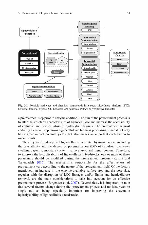
a pretreatment step prior to enzyme addition. The aim of the pretreatment process is
to alter the structural characteristics of lignocellulose and increase the accessibility
of cellulose and hemicellulose to hydrolytic enzymes. The pretreatment is most
certainly a crucial step during lignocellulosic biomass processing, since it not only
has a great impact on final yields, but also makes an important contribution to
overall costs.
The enzymatic hydrolysis of lignocellulose is limited by many factors, including
the crystallinity and the degree of polymerization (DP) of cellulose, the water
swelling capacity, moisture content, surface area, and lignin content. Therefore,
to improve the hydrolisability of lignocellulosic feedstocks, one or more of these
parameters should be modified during the pretreatment process (Karimi and
Taherzadeh 2016). The mechanisms responsible for the effectiveness of
pretreatment vary according to the nature of the pretreatment itself. Of the factors
mentioned, an increase in the enzyme-available surface area and the pore size,
together with the disruption of LCC linkages and/or lignin and hemicellulose
removal, are the main considerations to take into account for an effective
pretreatment process (Jørgensen et al. 2007). Nevertheless, it is important to note
that several factors change during the pretreatment process and no factor can be
single out as being especially important for improving the enzymatic
hydrolysability of lignocellulosic feedstocks.
Fig. 3.1 Possible pathways and chemical compounds in a sugar biorefinery platform. BTX:
benzene, toluene, xylene; C6: hexoses; C5: pentoses; PHAs: polyhydroxyalkanoates
3 Pretreatment of Lignocellulosic Feedstocks 33
3.2 Pretreatment Technologies
A large number of diverse pretreatment technologies have been suggested to
overcome the complex physicochemical, structural, and compositional barriers
that hinder the digestibility of lignocellulosic biomass (Hendriks and Zeeman
2009; Alvira et al. 2010; Tomas-Pejo et al. 2011; Bensah and Mensah 2013;
Moreno et al. 2015; Akhtar et al. 2016). Still, it is not possible to identify the best
pretreatment method, and the choice of an adequate pretreatment technology will
depend on factors such as type of lignocellulosic biomass, the final targeted product
(s), economics, and environmental impact. For instance, in the case of bioethanol
production, pretreatment methods offering lignin recovery at the end of the process
are preferred since it can potentially offer an added value (Fig. 3.1).
Pretreatment technologies can be classified into physical, chemical, and biolog-
ical methods. The combination of different pretreatment technologies has been also
proposed in several cases, due to the added benefits of the synergistic effects and the
potential cost reductions (Moreno et al. 2015; Bensah and Mensah 2013). Thus,
combined physicochemical pretreatments reduce the amounts of chemicals or
solvents required during the process, use milder process conditions, and decrease
enzyme loadings in the subsequent saccharification process. Biological pretreat-
ments can be also combined with chemical/physicochemical pretreatments in order
to increase delignification yields. Table 3.1 summarizes all pretreatment technolo-
gies developed up to date, listing the advantages and disadvantages of each one.
3.2.1 Physical Pretreatments
3.2.1.1 Mechanical Comminution and Refining
During mechanical pretreatment, the structure of the cell wall is disrupted by three
main stress factors: cutting, shearing and compression. Basically, there is a reduc-
tion in particle size and the outer walls of fibers are pulled out and primary walls are
removed. These effects increase the enzyme-available surface area and reduce the
cellulose crystallinity index (CrI) and DP, improving the accessibility of carbohy-
drates to hydrolytic enzymes (Gharehkhani et al. 2015). The final particle size of the
material will depend on the technique used. For instance, 10- to 30-mm particles are
obtained after chipping processes while a particle size of 0.2–2 mm is usually
observed after milling or grinding (Sun and Cheng 2002). Methods such as stirred
ball milling, hammer milling, disk refining, roller (Szego) milling, high-pressure
homogenization, and mechanical jet smash are the main techniques used for these
pretreatment processes (Alvira et al. 2010; Kim et al. 2016). Among them, ball
mills, disk mills and roller (Zsego) mills are the major scalable methods and can be
adapted for dry and wet samples. The industrial use of mechanical pretreatments is
however limited due to the long milling times and the high energy demands, which
34 A.D. Moreno and L. Olsson

Table
3.1
Comparisonofdifferenttechnologiesdeveloped
forpretreatm
entoflignocellulosicfeedstocks
Pretreatm
ent
Modeofaction
Saccharification
yieldsa
Biomass
degradationb
Biomass
spectra/
effectivenessc
Additional
observations
Reference(s)
Physicalpretreatm
ents
Mechanical
Reduce
CrI,DP,andparti-
clesize
Low
�++
Highoperational
costs.
Effectivewhen
combined
withhydrothermal/chem
i-
caltechnologies
Gharehkhani
etal.(2015),
Kim
etal.(2016)
Extrusion
Increase
enzyme-available
surfacearea
andreduce
CrI
andDP
Medium/very
high
�++;HB
Higher
saccharification
yieldswhen
combined
withchem
ical
catalysts
Zhengand
Rhem
ann
(2014),Duque
etal.(2014)
Chem
ical
pretreatm
ents
Acid-based
pretreatm
ent
Hem
icellulose
solubiliza-
tion;lignin
modification
andcellulose
hydrolysis
(dependentonacid
concentration)
Medium/very
high
+++
+++;
HB/HWB
Dilute-acidpretreatm
ents
aremore
advantageous
than
strongacid
pretreatm
ents
Kootstraet
al.
(2009),Tomas-
Pejoetal.(2011)
Alkali-based
pretreatm
ent
Delignification;cellulose
swellingandpartial
hem
i-
cellulose
modification
High
�/+
+;HB
Higher
saccharification
yieldswhen
combined
withother
pretreatm
ent
technologies
Zhao
etal.
(2008),Karim
i
andTaherzadeh
(2016)
Organosolv
Lignin
removal
andpartial
hydrolysisofhem
icellulose
Medium
+/++
+++;
HB/SWB/
HWB
High-qualitylignin
Pan
etal.(2005),
Mesaet
al.
(2011),
Wildschutet
al.
(2013)
Ionic
liquids(ILs)
Biomassfractionation/lig-
nin
removal
andhydrolysis
ofhem
icellulose
High/veryhigh
+++;
HB/SWB/
HWB
Possibilityoflignin
revalorization.Anefficient
recoverysystem
isneeded
Brandtet
al.
(2013) (continued)
3 Pretreatment of Lignocellulosic Feedstocks 35

Table
3.1
(continued)
Pretreatm
ent
Modeofaction
Saccharification
yieldsa
Biomass
degradationb
Biomass
spectra/
effectivenessc
Additional
observations
Reference(s)
Soakingin
aqueous
ammonia
(SAA)
Lignin
removal
High
�+;HB
Verylongpretreatm
ent
times
Kim
andLee
(2005)
Cellulose
andorganic
solvents-based
ligno-
cellulosicfractionation
(COSLIF)
Biomassfractionation
Veryhigh
�+++;
HB/HWB
Mildreactionconditions
Zhanget
al.
(2007)
Co-solventenhanced
lignocellulosicfrac-
tionation(CELF)
Lignin
removal
andhydro-
lysisofhem
icellulose
High
�++;HB
Higher
yieldsobtained
in
simultaneoussaccharifica-
tionandferm
entation
processes
Nguyen
etal.
(2016)
Ozonolysis
Lignin
removal
Medium/very
high
�/+
+/++;
HB/HWB
Highcostoflargeam
ount
ofozoneneeded
Travainiet
al.
(2015)
Aqueous
N-m
ethylm
orpholine-
N-oxide
Biomassfractionation;cel-
lulose
swellingand
increasedporosity
High/veryhigh
�/+
++;
HB/HWB
Possibilityofsimultaneous
pretreatm
entand
saccharification
Bensahand
Mensah(2013)
Combined
physicochem
ical
pretreatm
ents
Steam
explosion
Hem
icellulose
hydrolysis
andlignin
redistribution
Medium/very
high
+/+++
+++;
HB/HWB
Lowenvironmentalim
pact
Wanget
al.
(2015)
Hydrothermal
pretreatm
ent
Hem
icellulose
hydrolysis
andlignin
redistribution
Medium/very
high
++/+++
++;HB
Lower
biomassdegrada-
tionat
pH4-7
Alviraet
al.
(2010)
Ammonia
fiber
explo-
sion(A
FEX)
Cellulose
swellingand
deacetylationof
hem
icellulose
Veryhigh
�++;HB
Smallparticlesize
needed
Akhtaret
al.
(2016)
36 A.D. Moreno and L. Olsson

Extrativeam
monia
(EA)
Lignin
solubilizationand
cellulose
decrystallization
Veryhigh
�++;HB
Lignin
withhighpurity
recovered
andintact,
nativelignin
functionality
daCostaSousa
etal.(2016)
Wet
oxidation
Lignin
oxidationandhem
i-
cellulose
hydrolysis
Medium
�/+
++;HB
Lowenergydem
anddueto
anexothermic
reaction
Klinkeet
al.
(2002),Olsson
etal.(2005)
Microwave
pretreatm
ent
Delignificationand/or
hem
icellulose
hydrolysis
Medium/high
+/++
+++
Needforadditionof
chem
icals
Xu(2015)
Ultrasound
pretreatm
ent
Lignin
removal
Medium
+++
Possibilityofintegrating
ultrasonicationandenzy-
matic
hydrolysis
Bussem
aker
and
Zhang(2013)
Ammonia
recycleper-
colation(A
RP)
Lignin
removal
andpartial
hydrolysisofhem
icellulose
Veryhigh
�+;HB/HWB
Bussem
aker
and
Zhang(2013)
Supercritical
fluids
Delignificationandhydro-
lysisofhem
icellulose
Medium
+++
Higher
delignification
when
combined
with
organic
solvents
Schachtet
al.
(2008)
Sulfite
pretreatm
entto
overcomerecalci-
trance
oflignocellu-
lose
(SPORL)
Lignin
sulfonationand
hydrolysisofhem
icellulose
Medium/high
+++
Bensahand
Mensah(2013)
Biological
pretreatm
ents
Microbial
pretreatm
ent
Delignification
Low/high
�+
Reducedcellulose
content
Morenoet
al.
(2015)
Enzymatic
pretreatm
ent
Delignification
Low/high
�+/++;
HB/HWB
Shortreactiontimes
Morenoet
al.
(2015)
aLow:yieldslower
than
30%;Medium:yieldsof30–60%;High:yieldsof60–80%;Veryhigh:yieldshigher
than
80%.
b�:
nobiomassdegradation;+:lowbiomassdegradation;++:medium
biomassdegradation;+++:highbiomassdegradation.
c+:lowapplicabilityto
differenttypes
ofbiomass;++:itcanbeapplied
tosomeextentto
differenttypes
ofbiomass;+++:highapplicabilityto
differenttypes
ofbiomass;HB:effectiveonherbaceousbiomass;SWB:effectiveonsoftwoodbiomass;HWB:effectiveonhardwoodbiomass
3 Pretreatment of Lignocellulosic Feedstocks 37

implies in general an increase in the overall costs (Hendriks and Zeeman 2009; Kim
et al. 2016). Nevertheless, mechanical pretreatments are commonly used to reduce
particle size in all type of lignocellulosic feedstocks before subjecting raw materials
to other pretreatment technologies.
3.2.1.2 Extrusion
Extrusion is a thermo-mechanical pretreatment process. It is based on the effect
exerted by the tight rotation of a single or a twin screw at a certain temperature. The
equipment configuration can vary by using different types of screw elements. Also,
it can be coupled to the addition of chemical and biological catalysts (Zheng and
Rhemann 2014; Duque et al. 2014). Depending on the screw speed and barrel
temperature, the structure of lignocellulose is modified by promoting defibrillation
and shortening of fibers, which—in a similar way to milling processes—increases
the surface area available to hydrolytic enzymes and reduces CrI and DP. The
integration of chemical catalysts (acid or alkali) and/or biological catalysts (hydro-
lytic enzymes) results in a water-swollen effect—that increases water retention
values (WRV) of fibers—and a more efficient biomass fractionation, leading to
higher sugar yields in the subsequent saccharification step (Zheng and Rhemann
2014; Duque et al. 2014). Extrusion process has been applied to a wide range of
lignocellulosic feedstocks (eucalyptus, pine sawdust, switchgrass, wheat straw,
municipal waste, sugarcane bagasse, etc.) with and without acid or alkali as
catalysts, increasing sugar yields during the saccharification steps by 40–95%
(Zheng and Rehmann 2014). Also, when combining alkali-extrusion and
bioextrusion, 73% sugar yields where obtained from barley straw (Duque et al.
2014).
In contrast to the high energy demanding mechanical methods, extrusion is
considered a promising pretreatment technology for biomass use because of its
adaptability to process modifications (alkali or enzyme addition) and its versatility
regarding the use of different raw materials.
3.2.2 Chemical Pretreatments
3.2.2.1 Acid-Based Pretreatments
Acid-based pretreatments can use either concentrated or dilute acids as catalysts.
The main objective of both acid pretreatment methods is to solubilize hemicellu-
lose, increasing the porosity and making cellulose more accessible to enzymes. In
general, concentrated-acid pretreatments (acid concentration higher than 30%) are
less popular for industrial applications than dilute-acid pretreatments (acid concen-
tration between 0.5% and 5%), due to equipment corrosion problems, difficulties in
acid recovery, and considerable degradation products formation (Tomas-Pejo et al.
38 A.D. Moreno and L. Olsson

2011). Dilute-acid pretreatment is usually performed at temperatures above 100 �C,and it also promotes biomass degradation.
The most common acid catalyst is H2SO4. Still, HCl, H3PO4, HNO3 and organic
acids such as acetic acid, maleic acid, and acetic acid have been tested (Kootstra
et al. 2009). The use of organic acids has shown advantages for reducing the
formation of degradation products. Nevertheless, these pretreated materials usually
show lower hydrolysability, and high temperatures (>170 �C) are required to reachglucose yields comparable to those obtained with strong acids (Kootstra et al.
2009). Although dilute-acid pretreatment is especially suitable for biomass with
low lignin content, this technology has been assessed with a wide range of ligno-
cellulosic feedstocks (wheat straw, olive tree, pine, municipal solid waste, switch-
grass, aspen, corn cob), showing about 70–90% solubilization of hemicellulose
(in both xylans and mannans), and about 60–80% of glucose yield from the
enzymatic hydrolysis of pretreated materials (Cara et al. 2008; Satimanont et al.
2012; Arora and Carrier 2015).
3.2.2.2 Alkali-Based Pretreatments
Alkaline pretreatments improve the accessibility of lignocellulosic biomass by
mainly removing lignin polymer. Also, partial hemicellulose modification, higher
WRV and lower CrI values are usually observed (Karimi and Taherzadeh 2016).
The effectiveness of these pretreatment processes depends on the lignin content,
and they are more effective on agricultural residues (such as wheat straw, corn
stover, or sugarcane bagasse) than on woody materials due to the lower lignin
content. Although some inhibitory compounds are generated during the process,
alkaline pretreatments cause less sugar degradation than acid pretreatments.
The most common alkali compounds used for biomass pretreatment are NaOH,
KOH, lime [Ca(OH)2], NH3 and NH4OH. These processes are preferably performed
at room temperature or temperatures below 60 �C, with residence times varying
from seconds to days. Due to the lower costs and less stringent safety requirements,
lime—which can be easily recovered at the end of the process with CO2—and
ammonia are the preferred alkali compounds (Zhao et al. 2008; da Costa Sousa
et al. 2016).
Alkali-based pretreatments have shown to remove 20–60% lignin content (Zhao
et al. 2008; Akhtar et al. 2016). Alkali-pretreated feedstocks usually show sacchar-
ification yields of about 50–70%. However, higher saccharification yields (up to
95%) can be obtained by combining alkali-based processes with other pretreatment
methods (e.g. mechanical or biological pretreatments) or with oxidant agents such
as H2O2 or copper-catalyzed alkaline hydrogen peroxide (Cu-AHP) (Moreno et al.
2015; Akhtar et al. 2016; Bhalla et al. 2016). Due to the lower lignin content, the
non-specific binding of cellulases is reduced and lower enzyme loadings can be
used in comparison to dilute-acid pretreatments.
3 Pretreatment of Lignocellulosic Feedstocks 39

Several ammonia-based pretreatments, including soaking in aqueous ammonia
(SAA), ammonia fiber explosion (AFEX; described in Sect. 3.2.3.3), extractive
ammonia (EA, described in Sect. 3.2.3.4), or ammonia-recycle percolation (ARP;
described in Sect. 3.2.3.5), have been developed during last years, showing very
promising results (Akhtar et al. 2015; da Costa Sousa 2016). SAA is an interesting
chemical-based technology with mild reaction conditions. During SAA
pretreatment, biomass is submerged in aqueous ammonia solution at temperatures
below 75 �C for an extended period of time (up to several weeks). Optimal
ammonia concentrations and solid:liquid ratios will depend on the feedstock
used, ranging from 15% to 30% and from 1:2 to 1:15, respectively (Kim and Lee
2005). This pretreatment can efficiently remove lignin polymer (up to 75%) with
low formation of degradation products, and keeping both cellulose (100%) and
hemicellulose (85%) fractions in the solid residue. The presence of hemicellulose in
the pretreated material makes the addition of hemicellulases imperative for improv-
ing saccharification yields.
3.2.2.3 Organosolv
Organosolv pretreatment uses organic or aqueous solvents to remove lignin poly-
mer and hydrolyze hemicellulosic sugars. This method is performed at tempera-
tures of about 100–250 �C. Acids (HCl, H2SO4, oxalic acid, or salicylic acid) bases
(mainly NaOH) or salts [MgCl2, Fe2(SO4)3] can also be added as catalysts (Bensah
and Mensah 2013). Organosolv-pretreated materials can be split into three different
fractions: (1) cellulose fibers, (2) solid lignin (which is obtained after removing the
solvent from the liquid phase), and (3) a liquid phase containing hemicellulosic
sugars (Tomas-Pejo et al. 2011). Ethanol, methanol, acetone, ethylene glycol, and
other low-molecular-weight alcohols are the main solvents used in organosolv
pretreatment. Solvents must be recovered after each pretreatment process for its
reutilization and to avoid the inhibitory effect that these compounds exert on
hydrolytic enzymes and fermentative microorganisms. Although organosolv
pretreatment can be potentially used for any feedstock (e.g. softwoods, sugarcane
bagasse, or wheat straw) with high lignin removal (60–90%, with high purity) and
saccharification yields (up to 90%), it implies high capital costs and promotes the
formation of degradation compounds (Pan et al. 2005; Mesa et al. 2011; Wildschut
et al. 2013).
3.2.2.4 Other Chemical Pretreatments
In addition to acid, alkali, and organosolv pretreatment methods, ionic liquids (ILs),
cellulose and organic solvents-based lignocellulosic fractionation (COSLIF),
co-solvent enhanced lignocellulosic fractionation (CELF), ozonolysis, or aqueous
N-methylmorpholine-N-oxide (NMMO) are also considered as chemical
pretreatment technologies:
40 A.D. Moreno and L. Olsson

ILs ILs are organic or inorganic cations and anions that remain in liquid form
below 100 �C. They are considered green solvents because they are non-flammable,
non-volatile, and recyclable, and can be used for either partial or complete biomass
deconstruction (Brandt et al. 2013). When complete biomass dissolution is desired,
acidic or acidified ILs can be used for the hydrolysis of carbohydrates. On the other
hand, ILs promote lignin removal (up to 70%) and partial hemicellulose hydrolysis
(up to 85%) during partial deconstruction of biomass. Similar to organosolv
pretreatment, ILs offer the possibility of lignin revalorization, with the advantage
of handling non-volatile, non-odorous, and relatively safe liquors. In spite of
producing low amounts of degradation products, ILs can act as inhibitory com-
pounds themselves. Certain ILs have shown to inhibit cellulase activity by 70–85%
with a concentration of 10% (v/v) (Brandt et al. 2013). Also, the economic
feasibility of these pretreatment technologies is another drawback, and an energy-
efficient recycling method is therefore required to compensate the high costs of ILs.
Imidazolium salts are very commom ILs (Brandt et al. 2013). Recently, tertiary
amines such as [FurEt2NH][H2PO4] and [p-AnisEt2NH][H2PO4] (also called
bioionic liquids), have been synthesized by reductive amination of aldehydes
derived from lignin and hemicellulose (Socha et al. 2014). In presence of these
lignin-derived ILs, enzymatic hydrolysis of switchgrass released 90–96% of poten-
tial glucose and 70–76% of potential xylose. However, they showed to be less
effective than [C2mim][OAc] toward lignin removal.
COSLIF The COSLIF technology is a pretreatment method that can effectively
fractionate lignocellulosic biomass into amorphous cellulose, lignin, hemicellulose,
and acetic acid (Zhang et al. 2007). It is based on the sequential use of a non-volatile
cellulose solvent [concentrated (80–90%) phosphoric acid], a highly volatile
organic solvent (acetone or ethanol), and water. In a similar way than SAA,
COSLIF pretreatment requires mild reaction conditions (50 �C and atmospheric
pressure), but with the great advantage that the overall process takes place in few
hours. COSLIF processes also give other benefits, including less degradation
products formation, the possibility of obtaining high sugar yields with fast hydro-
lysis rates (up to 97% yield within 24 h of saccharification at low enzyme loadings),
and the possibility of isolating high-value lignin, acetic acid, and hemicellulose.
Moreover, it can be applied to a wide range of lignocellulosic feedstocks with only
minor modifications of the operation conditions. All these favorable consider-
ations—quite apart from the need of an efficient solvent recycling process—make
COSLIF technology an attractive pretreatment process for the biorefinery concept.
CELF Similar to COSLIF technology, CELF pretreatment combines an aqueous
solution of a biomass-sourced green solvent, THF (compound regenerated catalyt-
ically from the biomass degradation product furfural), with dilute-acid
pretreatment, promoting delignification and solubilization of biomass with minimal
sugar degradation (Nguyen et al. 2016). In comparison to dilute acid pretreatment
alone [0.5% (w/w) H2SO4], the present of THF [1:1 (v:v) THF:water ratio] in CELF
pretreatment enhanced final ethanol titers and yields by 25%, during a simultaneous
saccharification and fermentation process of pretreated corn stover (Nguyen et al.
3 Pretreatment of Lignocellulosic Feedstocks 41

2016). Due to extensive biomass deconstruction, low enzyme loadings are required
in the subsequent saccharification process. Still, further research is needed to fully
understand the beneficial mechanisms promoted by CELF in altering the physico-
chemical properties of lignocellulose.
Ozonolysis Ozone can be used as oxidizing agent to hydrolyze and remove lignin
polymer from lignocellulosic biomass with slight modifications of cellulose and
hemicellulose. Among other feedstocks, ozonolysis has been successfully applied
to wheat straw, sugarcane bagasse and poplar, reducing the lignin content by up to
60% (Travaini et al. 2015). The process is in general performed at room tempera-
ture and low amounts of degradation products are generated. The main disadvan-
tage of ozonolysis is the high amount of ozone required for the process, which
limits its industrial application.
Aqueous NMMO NMMO is an environmentally friendly solvent that in a similar
way to ILs can fractionate biomass at moderate temperatures (80–130 �C). Thissolvent disrupts hydrogen bonds in cellulose fibers, increasing porosity and WRV,
and reduces CrI and DP by partial cellulose solubilization. In addition, NMMO is
easy to recover and generates low amount of degradation products. During the
enzymatic hydrolysis of NMMO-pretreated biomass, >90% sugar yields have been
observed (Bensah and Mensah 2013). At concentrations of about 15–20% (w/w),
NMMO does not affect cellulase activity, offering the possibility of a simultaneous
pretreatment and saccharification process.
3.2.3 Combined Physicochemical Pretreatments
Physicochemical pretreatments are technologies that combines both physical and
chemical effects to improve the hydrolysability of raw materials. These technolo-
gies include, among others, steam explosion, hydrothermal methods, AFEX, EA,
microwave, ultrasound, or wet oxidation.
3.2.3.1 Steam Explosion Pretreatment
Steam explosion pretreatment combines the physical effect of an explosive decom-
pression with a chemical autohydrolysis promoted by the solubilization of acetyl
groups. The process is performed at high temperatures and pressures (160–250 �C,6–50 bar), reached by injecting saturated steam into the reactor. Steam explosion
promotes hydrolysis of hemicellulosic sugars and redistribution and partial solubi-
lization of lignin polymer. Residence time (t) and temperature (T ) are the main
factors influencing the effectiveness of this pretreatment method. The reaction
ordinate (Ro) correlates these two parameters in a single Eqn. ((3.1)) and it is
used to estimate the severity factor (SF) ((3.2)) for the evaluation and comparison
between pretreatment processes.
42 A.D. Moreno and L. Olsson

Ro ¼ t� e T�100=14:75ð Þ ð3:1ÞSF ¼ Log Roð Þ ð3:2Þ
Relatively low capital investment, low environmental impact, and complete
sugar recovery have positioned steam explosion technology as one of the most
widely used for lignocellulose pretreatment. As major drawback, the harsh condi-
tions applied during the process cause severe biomass degradation and generate
abundant degradation products (Wang et al. 2015). In order to compromise the
better biomass accessibility and the lower biomass degradation, SF values between
3 and 4.5 have been considered to be optimal. Steam explosion has been used to
pretreat almost the full range of lignocellulosic feedstocks, showing sugar yields of
about 90% in a subsequent enzymatic hydrolysis (Tomas-Pejo et al. 2011; Wang
et al. 2015). It is remarkable that the process can be performed without a previous
reduction in chip size and without the addition of chemical catalysts, offering a
substantial benefit for cost savings. Notwithstanding, an acid catalyst is often used,
especially in softwoods, to reduce pretreatment temperatures and residence times,
and decrease the amount of degradation products formed during the process. When
a catalyst is used, a combined severity factor (CSF) ((3.3)), which includes the pH
parameter in the previous Eqn. ((3.2)), is used to compare acid-catalyzed steam
explosion pretreatments.
CSF ¼ Log R0ð Þ � pH ð3:3Þ
3.2.3.2 Hydrothermal Pretreatment
Hydrothermal pretreatment (also known as liquid hot water) uses high pressures to
keep water in the liquid phase at temperatures ranging from 160 �C to 240 �C.Similar to steam explosion, these conditions favors hemicellulose autohydrolysis
and redistribution and partial solubilization of lignin, enhancing the accessibility of
the cellulose polymer. Hydrothermal pretreatment also promotes the formation of
high amounts of degradation products. As an attempt to control biomass degrada-
tion, the pH can be maintained between 4 and 7 to guarantee a mild autohydrolysis
process.
Using the hydrothermal technology, about 80% of the hemicellulosic fraction
has been removed from materials such as corn stover, sugarcane bagasse, and wheat
straw, improving the digestibility of these lignocellulosic materials (Alvira et al.
2010).
3 Pretreatment of Lignocellulosic Feedstocks 43

3.2.3.3 Ammonia Fiber Explosion (AFEX)
The AFEX pretreatment is another physicochemical process that uses anhydrous
ammonia at high pressures and temperatures between 60 �C and 100 �C (Akhtar
et al. 2016). The temperature range can be further extended to 140 �C without the
need of external heat, due to exothermal reactions between ammonia and water
(da Costa Sousa et al. 2016). Equivalent to steam explosion pretreatment, a fast
depressurization of the reactor leads to a rapid expansion of the ammonia gas,
which causes the physical disruption of lignocellulosic fibers. In consequence,
biomass digestibility is increased due to higher WRVs and lower CrI and
DP. Also, deacetylation of hemicellulose polymer is usually observed. Recovered
pretreated materials consist of a solid fraction (since ammonia is completely
evaporated) with slight modifications in hemicellulose and lignin content.
Hemicellulases are therefore required to increase enzymatic saccharification yields
when using AFEX as pretreatment method.
AFEX technology has a greater effect on agricultural residues, in contrast to
woody biomass, where it has shown to be less effective. One major advantage of
AFEX technology is that no degradation products are generated during the process.
In spite of minor lignin or hemicellulose removal, enzymatic hydrolysis can result
in high sugar yields (above 90% in agricultural residues), even at low enzyme
loadings (da Costa Sousa et al. 2016). This result can be explained by the fact that
ammonia affects lignin in a way that reduces its ability to non-specifically adsorb
hydrolytic enzymes.
3.2.3.4 Extractive Ammonia (EA)
EA represents a step forward in the use of ammonia for lignocellulose pretreatment.
This pretreatment uses liquid ammonia at elevated temperatures to solubilize lignin
polymer and to modify crystalline cellulose, making it more accessible to hydro-
lytic enzymes. During EA pretreatment, biomass with low moisture levels [up to
10% (w/w)] are mixed with ammonia up to 6:1 ammonia:biomass ratio and heated
to about 120 �C for 30 min (da Costa Sousa et al. 2016). A nitrogen overpressure is
used to maintain ammonia in the liquid phase. In contrast to AFEX pretreatment,
EA requires external heat to reach reaction temperatures due to the lower moisture
levels. After pretreatment, ammonia can be recovered and recycle. However,
0.022 g of ammonia per 100 g biomass is lost during each cycle due to ammonia-
biomass interactions. EA pretreatment has shown to solubilize about 44% (w/w)
lignin from corn stover with high purity and high proportion of intact, native lignin
functionality (e.g., β-O-4 linkages) (da Costa Sousa et al. 2016). Although process
conditions have to be optimized, EA pretreatment represents a very promising
technology since it requires about 60% lower enzyme loadings to reach similar
saccharification yields than AFEX, ammonia is an inexpensive commodity
44 A.D. Moreno and L. Olsson

chemical with easy recycling, detoxification of pretreated biomass is not necessary,
and it offers the possibility of lignin revalorization.
3.2.3.5 Other Physicochemical Pretreatments
With the aim of enhancing biomass digestibility, other physicochemical
pretreatment processes, including wet oxidation, the use of microwave energy in
the presence of chemical reagents, ultrasound pretreatment, ARP, supercritical
fluids, and sulfite pretreatment to overcome recalcitrance of lignocellulose
(SPORL) have been also described:
Wet Oxidation Wet oxidation is an oxidative pretreatment method that takes
place at high temperatures (170–200 �C) and pressures (10–15 bar), and uses
oxygen or air as catalyst (Olsson et al. 2005). It promotes lignin oxidation and
disruption of LCCs bonds, leading to 60–70% lignin removal (Olsson et al. 2005).
The presence of oxygen at high temperatures makes the process exothermic, thus
reducing the energy input. This technology has been applied to agricultural and
wood feedstocks and it can be also combined with Na2CO3 for enhancement of
cellulose and hemicellulose recovery yields (up to 96% and 70%, respectively)
(Klinke et al. 2002).
Microwave Pretreatment This method uses electromagnetic waves, with fre-
quencies ranging from 0.3 to 300 GHz, to irradiate lignocellulosic materials. To
avoid interference with telecommunications, 915 MHz (or 896 MHz in the United
Kingdom) and 2450 MHz are the most common microwave frequencies use for
industrial purposes (Xu 2015). By interacting with lignocellulose, microwaves
encourage thermal and also non-thermal effects for enhancing the accessibility of
cellulose to hydrolytic enzymes. Microwave pretreatment has been applied to a
wide range of lignocellulosic biomass feedstocks, including both agricultural and
woody biomass. During microwave pretreatment, biomass is submerged in water or
in solution with alkalis, acids, ILs, or salts, to increase the effectiveness of
pretreatment process. Microwave pretreatement has shown to increase saccharifi-
cation yields from 1.5 to 4 times in comparison with non-pretreated biomass,
obtaining 50–98% of the theoretical glucose (Xu 2015).
Ultrasound Pretreatment Ultrasonication is a pretreatment technology that con-
sists of rapid compression and decompression cycles of sonic waves to generate
cavitation (formation, growth, and subsequent collapse of microbubbles, resulting
in localized temperatures and pressures of about 5000 �C and 1000 bar). Ultrasound
can be applied at low frequencies (<50 kHz) and it affects lignocellulosic biomass
through mechanoacoustic (physical) and sonochemical (chemical) effects, allowing
lignin extraction (Bussemaker and Zhang 2013). Although working at higher
temperatures would improve pretreatment efficiency, the cavitation effect is max-
imized at temperatures between 30 �C and 70 �C. These temperatures allow
integration of ultrasonication and enzymatic hydrolysis, thus decreasing overall
3 Pretreatment of Lignocellulosic Feedstocks 45
process time and increasing saccharification yields. As for microwave pretreatment,
ultrasound pretreatment is usually combined with other pretreatment technologies,
such as hydrogen peroxide or alkali- and acid-based pretreatments, for increasing
delignification efficiency up to 90% (Bussemaker and Zhang 2013).
ARP ARP is based on passing aqueous ammonia at a concentration of 5–15%
through a reactor packed with biomass at 140–210 �C (Kim et al. 2008). This
process mainly promotes lignin removal and hemicellulose solubilization, with
little loss in cellulose content. Depending on the process conditions, about
30–80% of both lignin and hemicellulose can be removed (Kim et al. 2008).
Supercritical Fluids Supercritical fluid pretreatment, also known as CO2 explo-
sion, uses CO2 compressed at temperatures above its critical point to liquid-like
density (Schacht et al. 2008). It is employed as a delignification method for
enhancement of biomass accessibility. In order to increase the delignification
efficiency, the use of organic solvents such as ethanol and methanol has been
combined with this technology. One of the main advantages of this pretreatment
process is the possibility of cost reduction by reutilization of the CO2 produced
during other microbial processes, such as bioethanol production. In addition, CO2 is
non-toxic, non-flammable, and easy to recover.
SPORL This process uses an aqueous sulfite solution at 160–180 �C and low pH
(between 2 and 4) for about 30 min, combined with a mechanical step using a disk
mill (Bensah and Mensah 2013). It mainly causes hemicellulose removal and lignin
sulfonation. Even working at high temperatures, the concentration of degradation
products is lower than other pretreatment technologies such as dilute acid or steam
explosion. Sulfite pretreatment has been successfully tested at pilot scales with
several types of biomass including corn stover, switchgrass, agave stalk or
lodgepole pine. In the particular case of corn stover, SPORL pretreatment has
shown to remove up to 92% lignin, which enables to obtain 78.2% hydrolysis
yield (Bensah and Mensah 2013).
3.2.4 Biological Pretreatments
Several microorganisms, including white rot Basidiomycetes (Ceriporiopsissubvermispora, Trametes versicolor, Pycnoporus cinnabarinus, or Phanerochaetechrysosporium), Ascomycetes (Trichoderma reesei or Aspergillus terreus) and
bacteria (Bacillus macerans, Cellulomonas cartae, or Zymomonas mobilis),and/or their ligninolytic enzyme systems (mainly laccases or laccase-mediator
systems), have been used as single biological pretreatment methods or combined
with other pretreatment technologies to improve biomass hydrolysability (Moreno
et al. 2015). This pretreatment process mainly focusses on delignification of
lignocellulose and requires mild reaction conditions (15–40 �C and pH 4–5),
which promotes few side reactions, lowers the energy demand, and there are no
46 A.D. Moreno and L. Olsson

strict reactor requirements to resist pressure and/or corrosion. In addition to lignin
removal, partial degradation of hemicellulose and cellulose can be observed.
Biological pretreatment does not generate degradation products, but requires long
reaction times in case of using microorganisms (up to several weeks). In contrast,
when using ligninolytic enzymes the overall process time is reduced (4–24 h), but
the addition of external enzymes represents an extra cost. Lignocellulosic feed-
stocks such as eucalyptus, wheat straw, pine, or corn stover have been subjected to
biological pretreatment, showing delignification efficiency up to 97% (Moreno
et al. 2015). Although further research is needed to overcome current bottlenecks
and optimize these processes, biological pretreatments—especially enzymatic
delignification—are a very interesting alternative for future biorefineries.
3.3 Challenges in Converting Pretreated Biomass
Biomass pretreatment usually requires harsh conditions (high temperatures and
pressures, the use of solvents or the addition of chemical catalysts), leading to
biomass degradation and/or leaving residual chemicals that limit the subsequent
saccharification and fermentation steps. Biomass degradation generates several
byproducts during pretreatment processes that are inhibitory for hydrolytic
enzymes and fermenting microorganisms. These inhibitory compounds can be
classified according to their chemical nature into three major groups: (1) weak
organic acids, (2) furan derivatives, and (3) phenolic compounds (Taherzadeh and
Karimi 2011; Moreno et al. 2015). Furan derivatives include 2-furaldehyde (furfu-
ral) and 5-hydroxymethylfurfural (HMF), which come from degradation of pentose
and hexose sugars, respectively. Weak acids (acetic acid, formic acid, and levulinic
acid) are generated from the hydrolysis of acetyl groups and further degradation of
furfural and HMF. Finally, a wide variety of phenolic compounds such as vanillin,
syringaldehyde, and hydroxycinnamic acids (the composition varies depending on
feedstocks) are released from lignin. In the particular case of wet oxidation
pretreatment, phenols are not end-products and are further degraded to carboxylic
acids. In addition to the formation of inhibitory compounds, biomass degradation
also affects biomass recovery yields. This is another important parameter since any
loss in cellulose and/or hemicellulose (the sugar source) has a direct impact in the
concentration of the final desired products, resulting in lower revenues. Also, the
presence of residual solvents and/or chemical catalysts affects both enzymes and
fermenting microorganisms and should be therefore taken into account (Bensah and
Mensah 2013; Brandt et al. 2013). In this context, an optimal pretreatment process
must compromise biomass hydrolysability, minimizing biomass degradation and
maximizing the recovery yields of chemical catalysts. Several physical, chemical,
and biological detoxification methods have been also proposed and studied for
reducing the inhibitory effect of degradation compounds, and improving the con-
version of pretreated biomass (Taherzadeh and Karimi 2011; Moreno et al. 2015).
However, these detoxification methods should be avoided whenever possible since
3 Pretreatment of Lignocellulosic Feedstocks 47

they increase the overall process costs. Another strategy that is being implemented
is the development of genetic and evolutionary engineering strategies to obtain
robust microorganisms with the ability to convert/tolerate higher concentrations of
inhibitory compounds (Koppram et al. 2014). In a similar way, the use of
extremophillic microorganisms and/or enzymes that can perform the conversion
processes on such challenging media (e.g. those microorganisms/enzymes that are
able to catalyze reactions in acid or alkali environments) are also very promising
alternatives to be considered (Miller and Blum 2010).
3.4 Economic and Environmental Evaluation
of Pretreatment Processes
In addition of balancing biomass accessibility with sugar recoveries and biomass
degradation, the pretreatment process must be evaluated from the economic and
environmental point of view to meet sustainability criteria. Economic evaluations
of energy consumption and process costs, and the environmental impact are usually
estimated by techno-economic analyses and life-cycle assessments (LCAs), respec-
tively. Although both parameters are independent of each other, there is a tendency
to couple them and quantify the impact of research progress from an economic and
environmental point of view.
Pretreatment can represent about 30–40% of total costs (Tomas-Pejo et al.
2011). During an economic analysis of pretreatment technologies, the energy
demand and the cost of chemicals (including solvents, acids, alkalis, ligninolytic
enzymes and/or the nutrients required for microorganism growth) are the major
variables to be considered. Thus, pretreatment technologies with low energy and
chemical requirements would represent a better choice. Pretreatment methods such
as mechanical pretreatment and ozonolysis are considered economically unviable
because of the high energy demand and the large amount of ozone required during
the process, respectively (Hendriks and Zeeman 2009; Travaini et al. 2015; Kim
et al. 2016). Similar drawbacks are found in wet oxidation and other chemical
pretreatment technologies such as organosolv, or alkali-based pretreatments. In the
case of AFEX or EA, although ammonia recycling is feasible even despite its high
volatility, these pretreatments can be hindered by ammonia recovery yields and the
environmental concerns derived of the use of this chemical (Wang et al. 2015). A
reduction in ammonia concentration together with a decrease in enzyme loading
would aid in reducing overall costs of AFEX/EA processes, making these technol-
ogies effective alternative for biomass fractionation and revalorization (da Costa
Sousa et al. 2016). ILs, COSLIF and CELF are also quite attractive technologies
from the biorefinery point of view due to the possibility of obtaining certain high
value-added products such as lignin-derived compounds. Nevertheless, these pro-
cesses are limited by solvent prices, biomass loading, recovery yields, especial
48 A.D. Moreno and L. Olsson

reactor requirements to resist corrosion, and the need of neutralizing pretreated
materials.
Among all different processes, dilute-acid and steam-explosion have been
reported to be cost-effective technologies, and have been commercially used to
pretreat several lignocellulosic feedstocks. Steam explosion has been a competitive
pretreatment technology since the 1980s (Wang et al. 2015). The effectiveness of
steam explosion depends directly on the SF values applied to biomass. SF values of
3–4.5 are considered optimal, but lower SF values can be beneficial for reducing
biomass degradation. However, lower SF values results in less hemicellulose
solubilization, and higher enzyme loadings are required for reaching similar sac-
charification yields, which increases the overall costs. Dilute-acid pretreatment is
considered to be a simple, low-cost and effective pretreatment technology. In
contrast, additional steps after pretreatment, such as neutralization, inhibitor
removal, salt disposal, and acid recovery increase final production costs. The use
of extremophiles microorganisms and/or enzymes that can tolerate acid and/or salty
environments might help in the cost-effectiveness of the process (Miller and Blum
2010). Extrusion is another versatile and energy-efficient technology for lignocel-
lulosic pretreatment (Zheng and Rehmann 2014; Duque et al. 2014). This physical
technology produce very low amounts of degradation products, can be adapted to
different process configurations, and allows the possibility to add chemical and/or
biological catalysts to boost biomass accessibility. The combination of different
pretreatment technologies has been also considered to be a suitable choice for
reaching high sugar yields, using milder process conditions, lower concentrations
of costly solvents, and lower enzyme loadings (Bensah and Mensah 2013). In this
context, extrusion has been successfully combined—even at industrial scale—with
a continuous steam explosion process (Fang et al. 2011).
Regarding environmental evaluation, greenhouse gas (GHG) emissions (includ-
ing CO2, NO2, SO2, and CH4), water requirements, wastewater produced, the use of
chemicals, and the energy demand associated with fossil fuels are the main vari-
ables to be considered. So far, chemical pretreatments have a higher impact on the
environment in comparison with other pretreatment technologies, such as steam
explosion and hydrothermal pretreatment. On the contrary, biological
delignification is a promising technology with very low environmental impact,
high product yields, mild reaction conditions, few side reactions, less energy
demand, and reduced reactor requirements (Moreno et al. 2015). However, before
scaling up biological pretreatment technologies, shorter reaction times and/or lower
prices for ligninolytic enzymes are required to meet the economic needs.
3.5 Concluding Remarks
Lignocellulosic biomass is an appropriate feedstock for developing a bio-based
economy, relying on sugar-related products. Different physical, chemical, physi-
cochemical, and biological pretreatments technologies have been developed and
3 Pretreatment of Lignocellulosic Feedstocks 49
evaluated to alter the highly recalcitrant three-dimensional structure of lignocellu-
lose, and increase its digestibility for optimal biomass conversion. After consider
economic and environmental impact, only few pretreatment methods fulfill the
sustainability criteria and are suitable for their use at commercial scale. Focusing on
the needs of local biorefineries, a common pretreatment technology would be of
benefit to make use of all lignocellulosic feedstocks available in the nearby areas,
thus avoiding transportation costs. Although there is no best pretreatment technol-
ogy, dilute-acid pretreatment, steam explosion, extrusion, COSLIF, CELF, ILs,
Cu-AHP and certain ammonia-based technologies such as EA, are considered to be
effective methods that can be applied to a wide range of lignocellulosic feedstocks.
In addition, they also offer the possibility of providing other high value-added
product (such as lignin-derived compounds), contributing to the economy of the
process. In contrast, although some of these technologies are now in commercial
scale (e.g. steam explosion and dilute-acid pretreatment), certain parameters such
as biomass degradation, enzyme loadings required in the subsequent saccharifica-
tion step, or efficient recycling processes must be optimized to make these pre-
treatments viable from the economic and environmental point of view. The
combination of different pretreatment processes such as extrusion and steam
explosion, alkali-based pretreatment and enzymatic delignification, solvents and
acid-based pretreatment (COSLIF, CELF), ILs and microwave, etc., also offers
possibilities for enhancing the effectiveness of pretreatment processes by promot-
ing synergistic effects (e.g. higher lignin and hemicellulose solubilization in com-
parison of using single processes, and/or the need of lower enzyme loadings), and
reducing the environmental impact due to milder process conditions. With this
respect, further research at pilot and demonstration scale should be performed to
evaluate the feasibility and full potential for already established pretreatment
technologies, and at laboratory scale to further develop the non-efficient but
promising technologies in order to meet the economic and environmental require-
ments, giving them the possibility of representing an actual choice.
Acknowledgment Authors are grateful to the Swedish Energy Agency (Energimyndigheten) for
the financial support.
References
Alvira P, Tomas-Pejo E, Ballesteros M, Negro MJ (2010) Pretreatment technologies for an
efficient bioethanol production process based on enzymatic hydrolysis: a review. Bioresour
Technol 101:4851–4861
Akhtar N, Gupta K, Goyal D, Goyal A (2015) Recent advances in pretreatment technologies for
efficient hydrolysis of lignocellulosic biomass. Environ Prog Sustain Energy 35(2):489–511
Akhtar N, Gupta K, Goyal D, Goyal A (2016) Recent advances in pretreatment technologies for
efficient hydrolysis of lignocellulosic biomass. Environ Prog Sustain Energy 35(2):489–511
Arora A, Carrier DJ (2015) Understanding the pine dilute acid pretreatment system for enhanced
enzymatic hydrolysis. ACS Sustain Chem Eng 3(10):2423–2428
50 A.D. Moreno and L. Olsson

Bensah EC, Mensah M (2013) Chemical pretreatment methods for the production of cellulosic
ethanol: technologies and innovations. Int J Chem Eng 2013:719607
Bhalla A, Bansal N, Stoklosa RJ, Fountain M, Ralph J, Hodge DB, Hegg EL (2016) Effective
alkaline metal-catalyzed oxidative delignification of hybrid poplar. Biotechnol Biofuels 9:34
Brandt A, Grasvik J, Halletta JP, Welton T (2013) Deconstruction of lignocellulosic biomass with
ionic liquids. Green Chem 15:550–583
Bussemaker MJ, Zhang D (2013) Effect of ultrasound on lignocellulosic biomass as a pretreatment
for biorefinery and biofuel applications. Ind Eng Chem Res 52(10):3563–3580
Cara C, Ruiz E, Oliva JM, Saez F, Castro E (2008) Conversion of olive tree biomass into
fermentable sugars by dilute acid pretreatment and enzymatic saccharification. Bioresour
Technol 99:1869–1876
da Costa SL, Jin M, Chundawat SPS, Bokade V, Tang X, Azarpira A, Lu F, Avci U, Humpula J,
Uppugundla N, Gunawan C, Pattathil S, Cheh AM, Kothari N, Kumar R, Ralph J, Hahn MG,
Wyman CE, Singh S, Simmons BA, Dale BE, Balan V (2016) Next-generation ammonia
pretreatment enhances cellulosic biofuel production. Energy Environ Sci 9:1215–1223
Duque A, Manzanares P, Ballesteros I, Negro MJ, Oliva JM, Gonzalez A, Ballesteros M (2014)
Sugar production from barley straw biomass pretreated by combined alkali and enzymatic
extrusion. Bioresour Technol 158:262–268
Fang H, Deng J, Zhang X (2011) Continuous steam explosion of wheat straw by high pressure
mechanical refining system to produce sugars for bioconversion. Bioresources 6(4):4468–4480
Gharehkhani S, Sadeghinezhad E, Kazi SN, Yarmand H, Badarudin A, Safaei MR, Zubir MNM
(2015) Basic effects of pulp refining on fiber properties—a review. Carbohydr Polym
115:785–803
Hendriks ATWM, Zeeman G (2009) Pretreatments to enhance the digestibility of lignocellulosic
biomass. Bioresour Technol 100:10–18
Jørgensen H, Kristensen JB, Felby C (2007) Enzymatic conversion of lignocellulose into ferment-
able sugars: challenges and opportunities. Biofuels Bioprod Biorefin 1:119–134
Karimi K, Taherzadeh MJ (2016) A critical review on analysis in pretreatment of lignocelluloses:
degree of polymerization, adsorption/desorption, and accessibility. Bioresour Technol
203:348–356
Kim JS, Kim H, Lee JS, Lee JP, Park SC (2008) Pretreatment characteristics of waste oak wood by
ammonia percolation. Appl Biochem Biotechnol 148:15–22
Kim SM, Dien BS, Singh V (2016) Promise of combined hydrothermal/chemical and mechanical
refining for pretreatment of woody and herbaceous biomass. Biotechnol Biofuels 9:97
Kim TH, Lee YY (2005) Pretreatment of corn stover by soaking in aqueous ammonia. Appl
Biochem Biotechnol 124(1–3):1119–1131
Klinke HB, Ahring BK, Schmidt AS, Thomsen AB (2002) Characterization of degradation
products from alkaline wet oxidation of wheat straw. Bioresour Technol 82:15–26
Kootstra AMJ, Beeftink HH, Scott EL, Sanders JPM (2009) Comparison of dilute mineral and
organic acid pretreatment for enzymatic hydrolysis of wheat straw. Biochem Eng J 46:126–131
Koppram R, Tomas-Pejo E, Xiros C, Olsson L (2014) Lignocellulosic ethanol production at high-
gravity: challenges and perspectives. Trends Biotechnol 32(1):46–53
Mesa L, Gonzalez E, Cara C, Gonzalez M, Castro E, Mussatto SI (2011) The effect of organosolv
pretreatment variables on enzymatic hydrolysis of sugarcane bagasse. Chem Eng J
168:1157–1162
Miller PS, Blum PH (2010) Extremophile-inspired strategies for enzymatic biomass saccharifica-
tion. Environ Technol 31(8-9):1005–1015
Moreno AD, Ibarra D, Alvira P, Tomas-Pejo E, Ballesteros M (2015) A review of biological
delignification and detoxification methods for lignocellulosic bioethanol production. Crit Rev
Biotechnol 35(3):342–354
Nguyen TY, Cai CM, Osman O, Kumar R, Wyman CE (2016) CELF pretreatment of corn stover
boosts ethanol titers and yields from high solids SSF with low enzyme loadings. Green Chem
18:1581–1589
3 Pretreatment of Lignocellulosic Feedstocks 51
Olsson L, Jørgensen H, Krogh KBR, Roca C (2005) Bioethanol production from lignocellulosic
material. In: Dimitriu S (ed) Polysaccharides structural diversity and functional versatility.
Marcel Dekker, New York, pp 957–993
Pan X, Arato C, Gilkes N, Gregg D, Mabee W, Pye K, Xiao Z, Zhang X, Saddler J (2005)
Biorefining of softwoods using ethanol organosolv pulping: preliminary evaluation of process
streams for manufacture of fuel-grade ethanol and co-products. Biotechnol Bioeng 90
(4):473–481
Sanchez OJ, Cardona CA (2008) Trends in biotechnological production of fuel ethanol from
different feedstocks. Bioresour Technol 99:5270–5295
Satimanont S, Luengnaruemitchai A, Wongkasemjit S (2012) Effect of temperature and time on
dilute acid pretreatment of corn cobs. Int J Chem Mol Nucl Mater Metall Eng 6(4):316–320
Schacht C, Zetzl C, Brunner G (2008) From plant materials to ethanol by means of supercritical
fluid technology. J Supercrit Fluids 46:299–321
Socha AM, Parthasarathi R, Shi J, Pattathil S, Whyte D, Bergeron M, George A, Tran K, Stavila V,
Venkatachalam S, Hahn MG, Simmons BA, Singh S (2014) Efficient biomass pretreatment
using ionic liquids derived from lignin and hemicellulose. Proc Natl Acad Sci U S A 111(35):
E3587–E3595
Sun Y, Cheng J (2002) Hydrolysis of lignocellulosic materials for ethanol production: a review.
Bioresour Technol 83(1):1–11
Taherzadeh MJ, Karimi K (2011) Fermentation inhibitors in ethanol processes and different
strategies to reduce their effects. In: Pandey A, Larroche C, Ricke SC et al (eds) Biofuels.
Alternative feedstocks and conversion processes. Elsevier, Amsterdam, pp 287–311
Tomas-Pejo E, Alvira P, Ballesteros M, Negro MJ (2011) Pretreatment technologies for
lignocellulose-to-bioethanol conversion. In: Pandey A, Larroche C, Ricke SC et al (eds)
Biofuels. Alternative feedstocks and conversion processes. Elsevier, Amsterdam, pp 149–176
Travaini R, Marangon-Jardim C, Colodette JL, Morales-Otero M, Bolado-Rodrıguez S (2015)
Ozonolysis. In: Pandey A, Negi S, Binod P, Larroche C (eds) Pretreatment of biomass.
Processes and technologies. Elsevier, Amsterdam, pp 105–135
Wang K, Chen J, Sun SN, Sun RC (2015) Steam Explosion. In: Pandey A, Negi S, Binod P,
Larroche C (eds) Pretreatment of biomass. Processes and technologies. Elsevier, Amsterdam,
pp 75–104
Wildschut J, Smit AT, Reith JH, Huijgen WJ (2013) Ethanol-based organosolv fractionation of
wheat straw for the production of lignin and enzymatically digestible cellulose. Bioresour
Technol 135:58–66
Xu J (2015) Microwave pretreatment. In: Pandey A, Negi S, Binod P, Larroche C (eds)
Pretreatment of biomass. Processes and technologies. Elsevier, Amsterdam, pp 157–172
Zhang YH, Ding SY, Mielenz JR, Cui JB, Elander RT, Laser M, Himmel ME, McMillan JR, Lynd
LR (2007) Fractionating recalcitrant lignocellulose at modest reaction conditions. Biotechnol
Bioeng 97(2):214–223
Zhao Y, Wang Y, Zhu JY, Ragauskas A, Deng Y (2008) Enhanced enzymatic hydrolysis of spruce
by alkaline pretreatment at low temperature. Biotechnol Bioeng 99(6):1320–1328
Zheng J, Rehmann L (2014) Extrusion pretreatment of lignocellulosic biomass: a review. Int J Mol
Sci 15(10):18967–18984
52 A.D. Moreno and L. Olsson
Chapter 4
Approaches for Bioprospecting Cellulases
Baljit Kaur and Bhupinder Singh Chadha
What Will You Learn from This Chapter?
Cellulases are industrially important enzymes with a market share of 500 million
dollars that is expected to rise to 1.5 billion dollars by 2018. Cellulases play a
crucial role in generating sugar feedstock for lignocellulosic based biorefinery
platform. In addition, their demand in textile, paper, feed and food industries is
rising steadily. However, these industrial applications require thermostable, cata-
lytically highly efficient cellulases for making the processes commercially viable.
Therefore, search and discovery for novel sources of cellulases is continued area of
research. This chapter highlights some of the approaches for bioprospecting for
novel cellulases.
4.1 Introduction
Lignocellulosics constitutes key component of plant biomass (forestry and agricul-
tural wastes) and municipal solid wastes (MSW) that primarily comprises of
polymeric cellulose (40–50%), hemicellulose (20–30%), and lignin (10–25%)
that varies according to their sources or genetic makeup (Fig. 4.1). Owing to their
relative abundance and being a rich inherent source of sugar moieties, crop residues
(corn stover, corn cobs, rice straw, wheat straw, barley husks, sugarcane bagasse,
etc.) form an important feedstock for biotechnological intervention to produce
B. Kaur • B.S. Chadha (*)
Department of Microbiology, Guru Nanak Dev University, Amritsar 143005, India
e-mail: [email protected]; [email protected]
© Springer International Publishing AG 2017
R.K. Sani, R.N. Krishnaraj (eds.), Extremophilic Enzymatic Processing ofLignocellulosic Feedstocks to Bioenergy, DOI 10.1007/978-3-319-54684-1_4
53

value added products. Being a rich inherent source of sugar moieties, lignocellu-
losics constitute important feedstock for the production of second generation
cellulosic ethanol. However, the release of sugars from complex lignocellulosic
substrate requires hydrolysis of pretreated substrates. The pretreatment processes
employed bring about structural changes and loosens up the complex have been
reported in literature as well as at commercial scale. This chapter would only
discuss about the cellulases and enzymatic hydrolysis to achieve sugars for subse-
quent fermentation to ethanol (Xiao et al. 2012)
4.2 Glycosyl Hydrolases Involved in Hydrolysis
of Lignocellulosics
The hydrolysis of lignocellulosics into simpler form of sugars (hexoses/and pen-
toses) is accomplished by the wide array of enzymes collectively termed as glycosyl
hydrolases (GH). These enzymes are responsible for synthesis, modification and
degradation of carbohydrates and are clustered as carbohydrate-active enzymes
(CAZymes). CAZymes are classified on the basis their similarities in amino acid
sequences, enzymatic mechanism and protein folding and include Glycosyl hydro-
lases (GH), Polysaccharidelyases (PL), Carbohydrate esterases (CE) and Glycosyl
transferases (GT). In addition, glycosyl hydrolases do include chitinolytic,
pectinolytic and amylolytic enzymes which is not discussed in this chapter. These
enzymes are grouped into 133 GH families in the continually updated CAZymes.
Interestingly, in spite of a vast diversity of GHs their active site topologies essen-
tially remain the same and can be divided into three groups: pocket, cleft and tunnel
(Davies and Henrissat 1995).
Fig. 4.1 Diagrammatic illustration of the framework of lignocelluloses; cellulose, hemicellulose
and lignin (Menon and Rao 2012)
54 B. Kaur and B.S. Chadha

4.3 Cellulose Binding Modules
Most cellulolytic enzymes comprises of a catalytic domain connected to a
cellulose-binding domain (CBD) through a linker segment rich in Pro/Ser/Thr-
amino acids. Bacterial system is rich in proline content whereas, high serine
residues are found in eukaryotic linkers. CBM are amino acid sequences with a
unique folding patterns involved in recognizing and adhering to polysaccharides.
CBMs are appended to CAZymes that degrade insoluble polysaccharides and are
classified into 71 families on the basis of amino acid sequences. CBMs actively
participate in targeting the enzyme towards specific substrates and enhance the
penetration of enzyme to cellulose for efficient hydrolysis. On contrary to this, one
of the recent reports suggest that CBM lacking cellulases are advantageous in
enzyme recycling as higher share of unbound enzyme can be recovered. Kraulis
and co-workers (1989) elucidated the first structure of family 1 CBM from
Trichoderma reesei (TrCel7A) belonging to family GH7 (Fig. 4.2). The structural
analysis of CBM revealed the presence of β-sheet with two disulphide bridges and aflat surface linked with polar and aromatic amino acids.
The catalytic domains can exhibit both N- and O-linked glycans, where the
amino acids in linker contain O-linked glycosylation, which has long been impli-
cated in exhibiting protease protection and more recently implicated in substrate
binding (Payne et al. 2013). Linkers connecting cellulase domains from different
families exhibit different average lengths. It has been reported that linker length has
significant influence on enzymatic activity and binding affinity such as in T. reeseiCel7A where shorter linker length promotes processivity, whereas the longer linker
lengths in Cel6A enable it to search for hydrolytic site.
Fig. 4.2 The NMR structure of the Family 1 CBM and the top layer of cellulose (Kraulis et al.
1989). The tyrosine residues are shown in purple. The O-linked mannoses are shown in cyan and
blue
4 Approaches for Bioprospecting Cellulases 55

4.4 Classification of Cellulases and Their Structural
Features
Cellulose degradation is primarily attributed through multi-component cellulolytic
enzyme system that works in a synergistic manner to degrade the complex poly-
meric structure of cellulose. These enzymes include endoglucanases (EC 3.2.1.4),
exoglucanases (EC 3.2.1.74 and 3.2.1.91), and β-glucosidases (EC 3.2.1.21).
Endoglucanase (EG) mainly attack amorphous cellulose by cleaving the internal
glycosidic bonds in a random fashion and releases oligomers cellobiose, cellotriose,
cellotetraose as the products (Zhang et al. 2006). The active sites of most EGs are
open cleft shaped and may possess carbohydrate binding modules (modulator) or
other domains (Sweeney and Xu 2012). Cellobiohydrolases (CBH) act processively
on the reducing (CBHI) and non-reducing (CBHII) termini of cellulose fibers to
primarily release cellobiose and are abundant in the secretome of cellulolytic fungi.
EGs are widely classified on the basis of their structural and functional character-
ization into glycosyl hydrolases families 5, 6, 7, 8, 9, 12, 45 and 74.
Cellobiohydrolases are represented by families GH 6, 7, 8, 9 and 48.
Crystal structures of the cellulase belonging to GH families 5–9, 12, 45 and
48 have been elucidated and revealed variation in folding topology (Table 4.1).
Enzymes of all GH families except GH 9 and 12 are modular multi-domain pro-
teins. GH family 5 and 7 enzymes catalyze the hydrolysis of glycosidic bonds with
retention mechanism, while GH families 6, 9, and 48 mediated hydrolysis results in
inverting configuration (Dodd and Cann 2009; Miotto et al. 2014). The processivity
of enzyme action is not only limited to CBH but are also found in GH 5 and GH
9. The processive bifunctional EG (GH5), exhibiting both “endo” and “exo” type
activities, have been reported from diverse organisms such as in brown rot basid-
iomycete Gloeophyllum trabeum and the marine bacterium Saccharophagusdegradans 2–40. The structure of GH5 endoglucanase comprises of catalytic
domain solely with β/α topology and seven subsites (�4 to þ3) which are present
at the C-terminal end of the barrel.
The structure of cellulolytic enzyme of family GH6 is composed of a single
domain with a distorted α/β barrel topology; the active site that resides in a long
cleft at the C-terminal end of the parallel β-strands that make up the barrel
(Harhangi et al. 2003). The solved structures of catalytic domains of cellulases
belonging to family GH7, suggested the presence of two distinct groups i.e. EG and
CBHI. The solved structure showed the presence of common β-jelly roll fold with
two face-to-face packed antiparallel β-sheets which form a curved β-sandwich.Nakamura and co-workers (2014) solved the crystal structure of cellulase of
T. reesei (TrCel7A, ascomycete) and Phanerochaete chrysosporium (PcCel7C
and PcCel7D, basidiomycete) belonging to family GH7. The structural and func-
tional relationship between CBH and EG was resolved and found that in ascomy-
cete CBHs possess four loops that cover the active subsite resulting in stronger
binding affinity and increased processivity. These enzymes possess the ability to
degrade crystalline cellulose “processively” as they have tunnel like active sites.
56 B. Kaur and B.S. Chadha
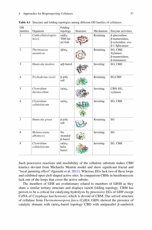
Such processive reactions and insolubility of the cellulose substrate makes CBH
kinetics deviant from Michaelis Menton model and show significant fractal and
“local jamming effect” (Igarashi et al. 2011). Whereas, EGs lack two of these loops
and exhibited open cleft shaped active sites. In comparison CBHs in basidiomycete
lack one of the loops that cover the active subsite.
The members of GH8 are evolutionary related to members of GH48 as they
share a similar tertiary structure and displays (α/α)6 folding topology. CBM has
proven to be a critical for catalyzing hydrolysis by processive EGs of GH9 except
Cel9A of Cytophaga hutchensonii, which is devoid of CBM. The solved structure
of cellulase from Thermomonospora fusca (CelE4, GH9) showed the presence of
catalytic domain with (α/α)6-barrel topology CBD with antiparallel β-sandwich
Table 4.1 Structure and folding topologies among different GH families of cellulases.
GH
families Organism
Folding
topology Structure Mechanism Enzyme activities
1 Caldicellulosiruptorbescii
(α/β)8-TIM bar-
rel fold
Retaining β-glucosidase,β-mannosidase,
β-xylosidase, exoβ-1-4glucanase
2 Thermoascusauranticus
(β/α)8 Retaining EG, CBH,
Xylanase,
β-mannosidase,
β-mannanase
3 Humicola insolens α/β-barrel Inverting EG, CBH
4 Trichoderma reesei β-jellyroll
Retaining EG,CBH
5 Clostridiumthermocellum
(α/α)6 Inverting CBH, EG,
xylanase
6 Clostridiumcellulolyticum
(α/α)6 Inverting EG, CBH
7 Humicola grisea β-jellyroll
Retaining EG
8 Melanocarpusalbomyces
Six
stranded
β-barrel
Inverting EG
9 Clostridiumcellulolyticum
(α/α)6-helix
barrel
Inverting EG, CBH
4 Approaches for Bioprospecting Cellulases 57

fold. It was observed that deletion of CBM3c led to loss in the processivity and
cellulose degradation ability (Irwin et al. 1998). Whereas, processivity of EGs in
S. degradans 2–40 was found to be independent of CBM. Enzymes from family
GH12 hydrolyze β-1-4 and β-1,3-1,4 linkages with retaining mechanism possessing
catalytic domains and lacks CBMs.
β-glucosidases (βG) exhibit an exo-type action that hydrolyze cellobiose to
glucose from the non-reducing ends. βG catalytic core features pocket shaped
active sites, topology that equips βG to act on non-reducing ends. β-glucosidasesbelong to the GH1, 3 and 9 families and play a key role in the efficient hydrolysis as
its action on cellobiose mitigates product inhibition on CBH and EG (Opassiri et al.
2007). The family1 beta-glucosidases are also classified as members of the 4/7
super family with a common (α/β)8 fold barrel motif. Family 3 of glycosyl hydro-
lases consists of nearly 44 beta-glucosidases and hexosaminidases of bacterial,
mold, and yeast origin. Most of the fungal beta-glucosidases studied belong to the
family 3 of glycosyl hydrolases. GH family 3 is one of the most abundant families
of carbohydrate active enzymes and includes members that possess distinct enzy-
matic activities, including β-D-glucosidase, β-D-xylosidase, α-L-arabinofuranosidase, and N-acetyl-β-D-glucosaminidase activities. βGs are well
characterized, biologically important enzymes that catalyze the transfer of glycosyl
group between oxygen nucleophiles either reverse hydrolysis or transglycosylation
(Bhatia et al. 2002).
Recent discovery suggests the role of novel class of oxidative enzymes respon-
sible for enhancing cellulose degradation which are identified and classified as
auxiliary activity (AA) enzymes in CAZy database (Fig. 4.3). These oxidative
enzymes are referred as lytic polysaccharide monooxygenases (LPMOs), a term
coined by Horn and co-workers (2012) and classified into three families in CAZy
database. The first one is AA9, formerly known as GH 61 that belongs to fungal
enzymes specifically. The second family is AA10 which was previously known as
carbohydrate binding module 33 (CBM 33) and it is dominated in bacteria and
viruses, the third family is AA11 which acts on chitin and shares some structural
and spectroscopic characteristics with AA9 and AA10. The fourth family is starch
degrading LPMO referred as AA13.
4.5 Metagenomics for Isolating Novel Cellulases
In addition to culturable microbes there is growing understanding of the fact that the
source of novel cellulases lies with enormous unculturable microbial diversity that
resides within unique ecological niches and, therefore, screening of metagenomic/
environmental DNA libraries that potentially harbour many of the open reading
frames (ORF) including those coding for the desired novel enzymes including
cellulases is being advocated (Voget et al. 2003). In last one-decade commercial-
ization of number of metagenomic technologies by companies such as Diversa,
Cubist, BRAIN, BASF, Genecor, Prokaria for enzyme production has come
58 B. Kaur and B.S. Chadha
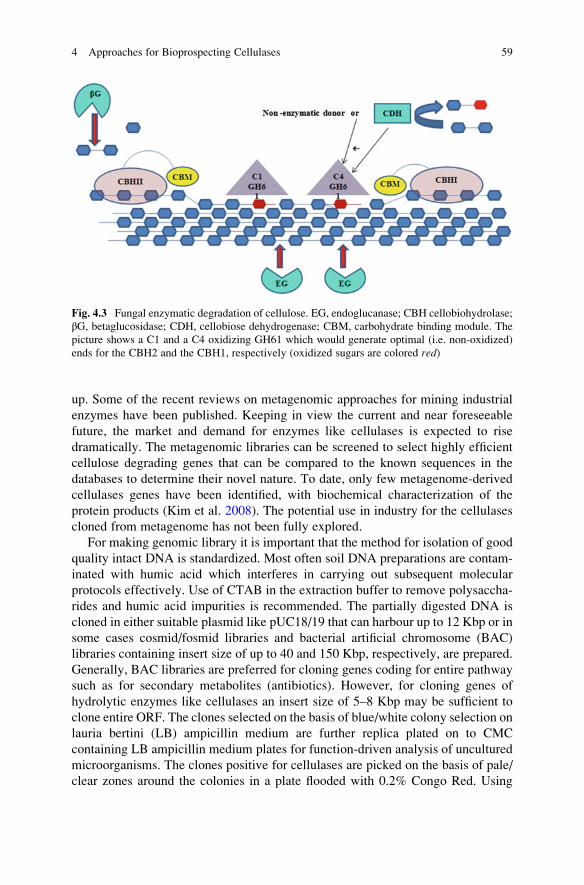
up. Some of the recent reviews on metagenomic approaches for mining industrial
enzymes have been published. Keeping in view the current and near foreseeable
future, the market and demand for enzymes like cellulases is expected to rise
dramatically. The metagenomic libraries can be screened to select highly efficient
cellulose degrading genes that can be compared to the known sequences in the
databases to determine their novel nature. To date, only few metagenome-derived
cellulases genes have been identified, with biochemical characterization of the
protein products (Kim et al. 2008). The potential use in industry for the cellulases
cloned from metagenome has not been fully explored.
For making genomic library it is important that the method for isolation of good
quality intact DNA is standardized. Most often soil DNA preparations are contam-
inated with humic acid which interferes in carrying out subsequent molecular
protocols effectively. Use of CTAB in the extraction buffer to remove polysaccha-
rides and humic acid impurities is recommended. The partially digested DNA is
cloned in either suitable plasmid like pUC18/19 that can harbour up to 12 Kbp or in
some cases cosmid/fosmid libraries and bacterial artificial chromosome (BAC)
libraries containing insert size of up to 40 and 150 Kbp, respectively, are prepared.
Generally, BAC libraries are preferred for cloning genes coding for entire pathway
such as for secondary metabolites (antibiotics). However, for cloning genes of
hydrolytic enzymes like cellulases an insert size of 5–8 Kbp may be sufficient to
clone entire ORF. The clones selected on the basis of blue/white colony selection on
lauria bertini (LB) ampicillin medium are further replica plated on to CMC
containing LB ampicillin medium plates for function-driven analysis of uncultured
microorganisms. The clones positive for cellulases are picked on the basis of pale/
clear zones around the colonies in a plate flooded with 0.2% Congo Red. Using
Fig. 4.3 Fungal enzymatic degradation of cellulose. EG, endoglucanase; CBH cellobiohydrolase;
βG, betaglucosidase; CDH, cellobiose dehydrogenase; CBM, carbohydrate binding module. The
picture shows a C1 and a C4 oxidizing GH61 which would generate optimal (i.e. non-oxidized)
ends for the CBH2 and the CBH1, respectively (oxidized sugars are colored red)
4 Approaches for Bioprospecting Cellulases 59

these approaches halo tolerant cellulases from soil, sediments and surrounding soda
lakes of Wadi el Natrun in Libyan Desert and multifunctional hybrid glycosyl
hydrolases from metagenomic library of the ruminant gut and uncultured microor-
ganisms in rabbit cecum has been discovered (Palackal et al. 2007). Recent report
on the metagenomic and proteogenomic analyses of a compost-derived bacterial
consortium adapted to switchgrass at elevated temperature with high levels of
glycoside hydrolase activities have been isolated. Near-complete genomes were
reconstructed for the most abundant populations, which included composite
genomes for populations closely related to sequenced strains of Thermusthermophilus and Rhodothermus marinus, and for novel populations that are relatedto thermophilic Paenibacilli and an uncultivated subdivision of the little studied
Gemmatimonadetes phylum. Partial genomes were also reconstructed for a number
of lower abundance thermophilic Chloroflexi populations. Identification of genes forlignocellulose processing and metabolic reconstructions suggested Rhodothermus,Paenibacillus and Gemmatimonadetes as key groups for deconstructing biomass.
Mass spectrometry-based proteomic analysis of the consortium was used to identify
3000 proteins in fractionated samples from the cultures, and confirmed the impor-
tance of proteins from Paenibacillus and Gemmatimonadetes in biomass
deconstruction.
4.6 Cellulase Producing Microorganisms
Complete cellulose degradation involves a dense interconnection between different
cellulolytic microbial populations. Broad range of microbial diversity, that includes
fungi, bacteria, and protozoans, is known that secretes these hydrolytic enzymes
either freely or in complexed form that efficiently degrades such complex poly-
saccharides. Bioprospecting of diverse range of ecological niches for isolating
novel cellulolytic microorganisms including the human, herbivore, arthropods
and termite gut, terrestrial and aquatic environments have been explored. Such as
wide array of bacterial strains which include Cellulomonas sp., Bacillus cereus,Bacillus licheniformis, Bacillus pumilus were isolated from mangrove areas of
Philippines. A novel bacterial isolate, identified as Cellulomonas composti sp.,showing high sequence similarity (98.5%) to Cellulomonas uda DSM 20107(T),
was isolated from compost at a cattle farm near Daejeon, Republic of Korea and
possesses endoglucanase andβ-glucosidase activities. Potent novel species were
isolated from forest soil, which were found to be closely related to Betaproteobacteria and Pseudo gulbenkiania on the basis of 16SrRNA sequencing.
Marinobacter sp. (MSI032), isolated from the marine sponge Dendrillanigra, pro-duces an extracellular alkaline cellulase at pH 9.0 at early stage of growth which
facilitates it for industrial process. Another potent cellulase producing strain named
as Clostridium phytofermentans was isolated from forest soil and was found to be a
processive endoglucanase, active on both crystalline cellulose and soluble CMC
(Warnick et al. 2002) that was later developed for consolidated bioprocessing of
60 B. Kaur and B.S. Chadha
cellulose into ethanol and has been demonstrated for commercialization by Qteros
at Masuchesset institute of technology. In order to obtain high-purity cellulase and
facilitate its production, the cel9 gene from C. phytofermentans was expressed in
Escherichia coli, and the recombinant protein was purified and characterized.
Caulobacteria sp. FMC-1, a facultative mesophilic strain, was isolated from shal-
low freshwater and was observed to produce cellulase under aerobic and anaerobic
conditions.
The marine isolate S. degradans is presently being researched for spectrum of
cell wall degrading enzymes. The genome of this bacterium has been sequenced to
completion, and more than 180 open reading frames have been identified that
encode carbohydrases. There is a distinct difference in cellulolytic strategy between
aerobic and anaerobic bacteria. The anaerobic bacteria such as Clostridium,Butyrivibrio fibrisolvens, Acetovibrio cellulolyticus, Bacteroides cellulosolvens,Ruminococcus albus and Ruminococcus flavefaciens utilizes multiprotein com-
plexes for achieving total degradation of cellulose.
Cellulolytic fungi are the most suited option for cellulose degradation as they
have capability to produce copious amounts of extracellular enzymes and this
significant characteristic of cellulolytic fungi attracts researcher’s interest over
bacteria. The best known cellulase producing fungi include Trichoderma sp.,
Aspergillus sp., Fusarium sp., Penicillium sp., Rhizopus sp., and Alternariasp. Most of the commercial cellulases available are produced from T. reesei andAspergillus niger but T. reesei lack sufficient amount of β- glucosidase to perform a
proper and complete hydrolysis. In the search of indigenous cellulose degrading
fungi, Barro and coworkers (2010) revealed the production of cellulases and
xylanases by thermotolerant fungi Acrophialophora nainiana and Ceratocystisparadoxa using shake flask culture. The novel isolate Aspergillus glaucus XC9
produced extracellular cellulase and shares common characteristics with those from
industrial cellulase-producing fungi, such as A. niger and T. reesei suggesting its
possible use in industry. The enzyme was found to be stable over a wide pH range
(3.5–7.5) and at temperatures below 55 �C. Two extracellular endoglucanases,
named RCE1 and RCE2, produced by Rhizopus oryzae FERM BP-6889 isolated
from soil, were identified and purified. The molecular masses of the two enzymes
were 41.0 and 61.0 kDa, respectively. Like bacterial cellulosomes, anaerobic fungi
anaerobic produce large multienzyme complexes which can depolymerise both
amorphous and crystalline cellulose. Cellulosome-type complexes with
endoglucanase, xylanase, mannanase, and β-glucosidase activities containing at
least 10 proteins have been found in Neocallimastix frontalis, Piromyces, andOrpinomyces.
The ecological niches having high temperatures such as hot springs and
composting heaps are also an attractive source for thermophilic cellulase producing
microorganisms. A thermophilic bacterium Aneurinibacillus thermoaerophilusWBS2 which produces extracellular thermophilic cellulases was isolated from hot
spring in India and the strain was subjected to optimization to enhance cellulase
production (Acharya and Chaudhary 2012). Cellulases isolated from various ther-
mophilic fungi include, Chaetomium thermophilum, Humicola insolens, Humicola
4 Approaches for Bioprospecting Cellulases 61

grisea, Myceliopthora thermophila, Talaromyces emersonii, Thermoascusauranticus, Sporotrichum thermophile, Melanocarpus albomyces.
In addition to the this, certain ruminants, termites and herbivorous animals
e.g. cattle, goats, sheep, buffalo, deer and camels etc., contain fungal and bacterial
group of microorganisms including Trichonympha, Clostridium, Actinomycetes,Bacteroides succinogenes, B. fibrisolvens, R. albus, and Methanobrevibacterruminantium have been documented for cellulose degradation (Ni and Tokuda
2013).
4.7 Methods for Isolation of Cellulase Producers
and Cellulase Activity Assays
The physical heterogeneity of the cellulosic substrate and complexity of enzyme
system makes cellulase assay and the interpretation of results a formidable problem
(Wood and Bhat 1988). The measurement of cellulases has been subject of intense
lab experiments. Various workers have published details of standardized methods
(Wood and Bhat 1988) to name a few. The work of Mary Mandel has been revisited
in a recent review republished in Biotechnology for Biofuels issue of September
2009 (Eveleigh et al. 2009). An excellent review on methodology by Zhang and
Lynd (2004) critically examines the methods being employed for assay of enzymes.
For isolating and selecting diverse cellulase producing microorganisms, screen-
ing of metagenomic libraries or clones, the most commonly employed method is
based on plate assay. This method relies on flooding the plates with Congo Red to
detect visible solubilization of substrate particles and the formation of halos on petri
dishes. Clearing zones around ball milled filter paper containing plates has also
been used for isolating cellulolytic microorganisms. A method involving use of
mixture of dyed polysaccharides has been used for simultaneous detection of
carbohydrase activities (Ten et al. 2004). Dyed cellulose is prepared by mixing
cellulose with a variety of dyes, such as Remazol Brilliant Blue, Reactive Orange,
Reactive Blue 19, and fluorescent dye 5-4,6- dichlorotriazinyl) aminoflurescein.
Because of large variations in the surface areas of cellulose and the binding
conditions, the quantitative relationship between released dye and reducing sugars
must be established for each batch of dyed cellulose. Insoluble cellulose deriva-
tives, such as slightly substituted CMC, can be mixed with a variety of dyes,
including Cibacron Blue 3GA and Reactive Orange 14 to produce insoluble
dyed-CMC (Ten et al. 2004). Insoluble cellulose derivatives can also be chemically
substituted with trinitrophenyl groups to produce chromogenic trinitrophenyl-
carboxymethyl cellulose (TNP-CMC) and fluorophoric fluram cellulose. These
methods are semi-quantitative, and are well suited to monitoring large numbers
of samples. The plate assay is convenient and easy to perform, rapid, and more
adaptable for screening of a large number of samples but does not show quantitative
62 B. Kaur and B.S. Chadha

analyses of protein. Whereas, quantitative approaches make it possible to accu-
rately estimate the saccharifying activities of crude cellulase.Quantitative cellulase activity assays can be divided into three types: (1) the
formation of products after hydrolysis, (2) the reduction in substrate quantity, and
(3) the change in the physical properties of substrates. The two basic approaches to
measuring cellulase activity are (1) measuring the individual cellulase
(endoglucanases, exoglucanases, and β-glucosidases) activities, and (2) measuring
the total cellulase activity. In general, hydrolase enzyme activities are expressed in
form of initial hydrolysis rate for individual enzyme component within a short time,
or the end-point hydrolysis for the total enzyme mixture to achieve a fixed hydro-
lysis degree within a given time. For cellulase activity assays, there is always a gap
between initial cellulase activity assays and final hydrolysis measurement.
Endoglucanases cleave intramolecular β-1,4-glucosidic linkages randomly, and
their activities are often measured on a soluble high degree of polymerization
(DP) cellulose derivative, such as CMC with the lowest ratio of FRE/Fa (the
fraction of β-glucosidic bond accessible to cellulase (Fa), the fraction of reducing
ends (FRE), and relative ratio of FRE/Fa). The modes of actions of endoglucanases
and exoglucanases differ in that endoglucanases decreases the specific viscosity of
CMC significantly with little hydrolysis due to intramolecular cleavages, whereas
exoglucanases hydrolyze long chains from the ends in a processive process (Zhang
and Lynd 2004). Endoglucanase activities can be measured based on a reduction in
substrate viscosity and/or an increase in reducing ends determined by a reducing
sugar assay. Because exoglucanases also increase the number of reducing ends, it is
strongly recommended that endoglucanase activities be measured by both methods
(viscosity and reducing ends). Soluble oligosaccharides and their chromophore
substituted substrates, such as p-nitrophenyl glucosides and
methylumbelliferyl-β-D-glucosides, are also used to measure endoglucanase activ-
ities based on the release of chromophores or the formation of shorter oligosaccha-
ride fragments, which are measured by HPLC or TLC. The methodologies that are
based on polarography for detection of cellulase activity by coupling the liberation
of glucose with oxygen consumption have been described by various researchers.
The polarographic assay couples cellulase with an excess mixture of
β-1,4-glucosidase (EC 3.2.1.21), glucose oxidase (EC 1.1.3.4), mutarotase
(EC 5.1.3.3), and catalase (EC 1.11.1.6). Glucose oxidase couples β-D-glucoseformation, the end product of cellulose hydrolysis, with the consumption of oxygen.
Thus, the enzyme-coupled system provides a means of continuously monitoring
cellulase activity, or the susceptibility of cellulosic substrates to enzymatic degra-
dation, by measuring oxygen consumption in the reaction medium.
Exoglucanases cleave the accessible ends of cellulose molecules to liberate
glucose and cellobiose. T. reesei cellobiohydrolase (CBH) I and II act on the
reducing and non-reducing cellulose chain ends, respectively (Zhang and Lynd
2004). Avicel has been used for measuring exoglucanase activity because it has the
highest ratio of FRE/Fa among insoluble cellulosic substrates. During chromato-
graphic fractionation of cellulase mixtures, enzymes with little activity on soluble
CMC, but showing relatively high activity on avicel, are usually identified as
4 Approaches for Bioprospecting Cellulases 63

exoglucanases. Unfortunately, amorphous cellulose and soluble cellodextrins are
substrates for both purified exoglucanases and endoglucanases. Therefore, unlike
endoglucanases and β- glucosidases, there are no substrates specific for
exoglucanases within the cellulase mixtures (Wood and Bhat 1988). In some
reports it has been described that 4-methylumbelliferyl-β-D-lactoside was an effec-tive substrate for T. reesei CBH I, yielding lactose and phenol as reaction products,
but it was not a substrate for T. reesei CBH II. Some endoglucanases of T. reeseiEG I, structurally homologous to CBH I, also cleaves 4-methylumbelliferyl- β-D-lactoside, yet these enzymes can be differentiated by adding cellobiose, an inhibitor
that strongly suppresses cellobiohydrolase activity. T. reesei CBH II does not
hydrolyze 4-methylumbelliferyl-β-D-aglycones of either glucose or cellobiose
units, but does cleave 4-methylumbelliferyl-β-D-glycosides with longer glucose
chains.
β-D-glucosidases predominantly act on cellobiose to release glucose as the
major product which can be measured employing glucose oxidase and peroxidase
(GOD-POD) kit. Whereas other simple sensitive assay methods are based on
colored or fluorescent products released from para nitrophenyl β-D-1,4-glucopyranoside, β-naphthyl-β-D-glucopyranoside, 6-bromo-2-naphthyl-β-D-glucopyranoside, and 4-methylumbelliferyl-β-D-glucopyranoside. Also, β-D-glu-cosidase activities can be measured using cellobiose, which is not hydrolyzed by
endoglucanases and exoglucanases, and using longer cellodextrins, which are
hydrolyzed by endoglucanases and exoglucanases (Zhang and Lynd 2004).
The total cellulase system consists of endoglucanases, exoglucanases, and β-D-glucosidases, all of which hydrolyze crystalline cellulose synergically. Total cellu-
lase activity assays are always measured using insoluble substrates, including pure
cellulosic substrates such as Whatman No. 1 filter paper, cotton linter, microcrys-
talline cellulose, bacterial cellulose, algal cellulose; and cellulose-containing sub-
strates such as dyed cellulose, α-cellulose, and pretreated lignocellulose. The
heterogeneity of insoluble cellulose and the complexity of the cellulase system
cause formidable problems in measuring total cellulase activity. Experimental
results show that the heterogeneous structure of cellulose (filter paper and bacterial
cellulose) gives rise to a rapid decrease in the hydrolysis rate within a short time
(less than an hour), even when the effects of cellulase deactivation and product
inhibition are taken into account. The most common total cellulase activity assay is
the filter paper assay (FPA) using Whatman No. 1 filter paper as the substrate,
which was established and published by the International Union of Pure and
Applied Chemistry (IUPAC). This assay requires a fixed amount (2 mg) of glucose
released from a 50-mg sample of filter paper (i.e., 3.6% hydrolysis of the substrate),
which ensures that both amorphous and crystalline fractions of the substrate are
hydrolyzed. A series of enzyme dilution solutions is required to achieve the fixed
degree of hydrolysis. The strong points of this assay are (1) it is based on a widely
available substrate, (2) it uses a substrate that is moderately susceptible to cellu-
lases, and (3) it is based on a simple procedure (the removal of residual substrate is
not necessary prior to the addition of the DNS reagent).
64 B. Kaur and B.S. Chadha

Recent trends show that automated methods for screening large microbial/
libraries are being employed. Decker et al. (2003) reported automated filter paper
assay method based on a Cyberlabs C400 robotics deck which is equipped with
customized incubation, reagent storage and plate-reading capabilities that allow
rapid evaluation of cellulases acting on cellulose. A functional proteomic assay in a
multiplexed setting on an integrated plasmid-based robotic work cell for high-
throughput screening of clones expressing mutants with improved endoglucanase
F from the anaerobic fungus Orpinomyces PC-2 was reported by Hughes et al.
(2006).The multiplex method using an integrated automated platform for high-
throughput screening in a functional proteomic assay allows rapid identification of
plasmids containing optimized clones ready for use in subsequent applications
including transformations to produce improved strains or cell lines. A high-
throughput assay employing microplate method to study the digestibility of ligno-
cellulosic biomass as a function of biomass composition, pre-treatment severity,
and enzyme composition was optimized for crystalline cellulose (Avicel) by
Chundawat et al. (2008). The method is most suitable for delivering milled biomass
to the microplate through multi-pipetting slurry suspensions. Similarly, miniaturi-
zation of DNS based detection method employing microtitre plate for screening
cellulase producing clones of entire gut of Reticulitermes flavipes have been
employed. Xia et al. (2005) reported microplate-based carboxymethylcellulose
assay for endoglucanase activity. A rapid bio-enzymatic, spectrophotometric
assay can be used to determine fermentable sugars. The entire procedure was
automated using a robotic pipetting workstation. A new method to determine the
activity of cellulase has been developed using a quartz crystal microbalance (QCM)
technique. The QCM technique provides results closer to those obtained by mea-
suring the actual reducing sugars.
4.8 Secretome Analysis of Glycosyl Hydrolases
Recent progress in various -omics tools has recently established a fundament for a
system biological approach towards an understanding of cellulase and hemicellu-
lose production. Proteomics is an excellent tool in profiling, discovering, and
identifying proteins produced in response to a particular cellular environment as
well as to study the distinct glycosyl hydrolases produced by industrially important
cellulolytic strains. Secretome analysis in the past has focused on few of the
representative fungal genera belonging to Ascomycetes (Aspergillus, Penicillum,Trichoderma) and basidiomycetes (P. chrysosporium). The list of basidiomycetes
has expanded with the reports of the secretomes from Pleurotus sapidus, Trametesversicolor and Coprinopsis cinerea and Phanerocheate carnosa. Perhaps most well
studied fungi is Aspergillus where the whole-genome sequencing projects has been
completed for A. aculeatus Aspergillus carbonarius A. clavatus A. fumigatus,A. terreus, A. parasiticus, A. flavus. In one of the initial researches on secretome
73 secreted proteins of A. flavus were identified. Many of these proteins were
4 Approaches for Bioprospecting Cellulases 65

proteases, metabolic proteins or proteins involved in electron/proton transport.
Comparative secretome analyses is usually focused on fungal strains grown under
submerged and solid state fermentation, or grown in the presence of different
carbon sources or for comparisons among wood-rotters or phytopathogens. This
comprehensive approaches been used to reveal the differential protein expression
profiling in the parent and heterokaryons, developed through inter-specific proto-
plast fusion between Aspergillus nidulans and Aspergillus tubingensis (Kaur et al.2013). It has also been reported in one of the study that expression of β-glucosidaseactivity in the 2DE gel can be detected using methylumbelliferyl glucoside. Two
novel β-glucosidases of A. fumigatus were identified by this in situ activity stainingmethod, and the gene coding for a novel β-glucosidase (EAL88289) was cloned andheterologously expressed. The expressed β-glucosidase showed far superior heat
stability to the previously characterized β-glucosidases of A. niger and Aspergillusoryzae. In a recent report Sharma et al. (2010) has reported a methodology for
detection of (CBHI/EGI) in secretome of A. fumigatus using methyl umbelliferyl
β-D lactopyranoside as substrate.
Chundawat et al. (2011) explored the protein composition of several commercial
cellulase and xylanase preparations from T. reesei using a proteomics approach
with high throughput quantification using liquid chromatography–tandem mass
spectrometry (LC–MS/MS). As expected, Cel7A (former CBH1) was the predom-
inant cellulase in all major commercial enzymes, followed by Cel6A (former CBH
2). Interestingly, proteomic approach has been employed to detect several intracel-
lular enzymes in the culture filtrate of T. reesei which indicates that enzyme
secretion is accompanied by considerable autolysis or mycelia fragmentation,
whose role for high enzyme production has not yet been investigated. The
secretome of white rot fungus Bjerkandera adusta produced in the presence of
water-soluble olive mill extractives and the influence of the latter on the oxidore-
ductase expression pattern was investigated. Distinct changes in the protein com-
position of oxidoreductases, namely diverse class-II peroxidases and aryl alcohol
oxidases were observed. The secretome analysis of anaerobic bacteria Clostridiumcellulolyticum and Cellulomonas flavigena has also been reported. In addition to itscapability of complementing transcriptome level changes, proteomics can also
detect translational and post-translational regulations, thereby providing new
insights into complex biological phenomena such as abiotic stress responses in
plant roots. Recently, a novel technique termed as iTRAQ (isobaric tags for relative
and absolute quantitation) has been adapted for proteomic quantitation which
overcomes some of the limitations of 2D gel electrophoresis and also improves
the throughput of proteomic studies (Liu et al. 2014).
The secretome analysis is not limited to fungi; few of bacterial systems have also
been studied. The secretome including that of Bacillus stereothermophilus,C. cellulolyticum, C. flavigena, Thermobifida fusca have also been reported. The
secretome analysis of C. cellulolyticum showed presence ofat least 30 dockerin-
containing proteins (designated cellulosomal proteins) and 30 noncellulosomal
components. Most of the known cellulosomal proteins, including CipC, Cel48F,
Cel8C, Cel9G, Cel9E, Man5K, Cel9M, and Cel5A, were identified by using
66 B. Kaur and B.S. Chadha

two-dimensional Western blot analysis with specific antibodies, whereas Cel5N,
Cel9J, and Cel44O were identified by using N-terminal sequencing. Unknown
enzymes having carboxymethyl cellulase or xylanase activities were detected by
zymogram analysis of two-dimensional gels. Using Trap-Dock PCR and DNA
walking, seven genes encoding new dockerin-containing proteins were cloned
and sequenced. Some of these genes belonging to glycoside hydrolase families
GH2, GH9, GH10, GH26, GH27, and GH59 are clustered.
4.9 Strain Improvement Programme
Some of the wild type fungal strains are known to produce copious amount of
cellulolytic/hemicellulolytic enzymes. However, they are fall of being considered
ideal for commercialization as the enzyme titres are low from industrial point of
view. These cellulolytic strains T. reesei, Penicillium decumbens, Acremoniumcellulolyticus have been subjected to continuous strain improvement programme,
primarily involving repeated mutagenesis, selection and rational screening
approaches (Liu et al. 2013). These procedures involve physical/chemical muta-
gens followed by rational screening strategies for enhancing the levels of cellulase
and other cell wall degrading enzymes. Ethane methane sulphate (EMS) N-methyl-
N0nitro-N-nitrosoguanidine (NTG), acriflavin are widely used chemical mutagens
and UV radiation is primarily employed for physical treatment. The genealogy of
these strain development program shows that either these mutagens have been used
alone or in combination. In some cases, combined mutagenesis involving both
chemical and physical treatments have resulted in the overproduction of cellulolytic
enzymes. The developed mutants are picked randomly or on the basis of phenotypic
traits such as resistance to antimetabolite, morphological differences, developmen-
tal (lacking cell differentiation to spores). The synthesis of cellulases is known to be
primarily regulated through carbon catabolite repression (CCR) where expression
of various transcriptional factors and expression of kinases and phosphatases that
senses different carbon sources have important role to control cellular energy states.
2-Deoxy-D-glucose (2DG), a toxic analogue of glucose is widely used as selection
marker for selecting deregulated mutants. Catabolite derepression is associated
with the mutation in creI gene as described in T. reesei and P. decumbens. Expres-sion of cellulolytic and hemicellulolytic genes in the presence of glucose in the
developed mutants carries truncated creI gene encoded only one zinc finger protein.
Whereas, A. nidulans in addition to CreA also contain CreB, CreC and CreD genes
which actively participate in CCR. It has also been reported that high protein
secretion is generally associated with alterations in certain organelles that are
involved in the secretory pathway such as six to seven fold higher endoplasmic
reticulum (ER) was observed in one the developed mutant (RUT-C30) of T. reesei.Several reports are available on the characterization of the selected mutants at
genomic and proteomic level. To uncover the genetic changes in the mutants
4 Approaches for Bioprospecting Cellulases 67

sequencing approach is widely employed for comparative analysis of parent and
mutant strains to indicate the deletions or insertions in the nucleotide sequence. The
expression profiling through SDS-PAGE of wild and developed strains also indi-
cated the up/downregulated proteins which were identified through peptide mass
fingerprinting (PMF) (Kaur et al. 2014).
Protoplast fusion is a powerful approach through which potential strains with
desirable properties could be obtained with minimal disturbance in their physiology
(Savitha et al. 2010). Most of the laboratories engaging in fungal genetics are using
gene manipulation procedures based on protoplasts. Isolation of protoplasts are
carried out by the digestion of cell wall with the aid of different hydrolytic enzymes,
such as Novozyme234, chitinase, lysozyme etc., in the presence of osmotic stabi-
lizer. Osmotic stabilizers play crucial role in maintaining the integrity of the pro-
toplasts and protect the lysis of protoplasts. Inorganic osmotic stabilizers have been
reported to be effective in Thermomyces lanuginosus, Graphium putredinis,Trichoderma harzianum, Aspergillus sp. Genetic manipulations can successfully
be achieved through fusion of protoplasts in filamentous fungi that lack the capacity
for sexual reproduction. The protoplast fusion is possible at intra-specific, inter-
specific and inter-generic level involving two or more complex parental genomes.
Therefore, a number of desirable genes from divergent strains can potentially be
recombined into a fusant strain by this method. This technique was successfully
employed as useful tool for genetic manipulation of desirable traits, for enhancing
the cellulase production in T. ressei by inter-specific protoplast fusion. It has been
studied that morphological markers (colony morphology and spore size and shape)
and genetical markers like, mycelial protein pattern, restriction digestion pattern
and random amplified polymorphic DNA (RAPD) analysis indicates the genetic
recombination. RAPD method has proven to be efficient in describing DNA
polymorphism in fusants obtained through the inter-specific protoplast fusion
between A. carbonarius and A. niger for overproduction of pectinase. Protein
expression analysis through SDS-PAGE is also effectively used as one of the
markers which indicates the genetic recombination between parents and fusants.
The presence or absence of protein bands between the parents and the hybrids
(fusants) confirm the hybrid formation (Savitha et al. 2010). Similar genomic and
proteomic approaches were carried to study the genetic relatedness in the hetero-
karyons developed through inter-specific protoplast fusion between two cellulolytic
strains of A. nidulans (AN) and A. tubingensis (Dal8) (Kaur et al. 2013). One of theextensions of this breeding approach is genome shuffling that uses alternative
cycles of genome recombination and selection to combine the useful alleles of
many parental strains into single cells showing the desired phenotype. This tech-
nique offers the advantage of simultaneous genetic changes at different positions
throughout the entire genome without the necessity for genome sequence
information.
Cloning of genes by complementation cloning techniques has been used for the
isolation of some enzyme genes in yeast and bacteria. However, complementation
cloning is dependent on host strains having appropriate mutations and is therefore
68 B. Kaur and B.S. Chadha

not of general use in the cloning of non-essential enzyme genes. In contrast
expression cloning results in cloning full length genes using good quality mRNA
and cDNA derived thereof is fast and efficient method of cloning of enzyme genes
from fungi that are known express a variety of glycosyl hydrolases under inducible
conditions. The advancement in genome sequencing has further made this tech-
nique more powerful and relevant where specific primers can amplify the entire
ORF coding for the gene. It is possible to over-express individual enzyme compo-
nents in different host systems using strong promoters such as AOX1 in Pichiapastoris, GLA in A. oryzae, CBH in T. reesei to name a few (Dalboge 1997).
4.10 Future Perspectives
The search for catalytically efficient thermostable cellulases from diverse
extremophilic niches (culturable/unculturable) using wet lab and bioinformatics
tools for mining data for prospecting unique and novel cellulases is foreseen as an
area of future research. The use of system biology approaches for understanding
and discovery of new set of proteins that can usher new concepts as deviation from
the existing paradigm of cellulose degradation is also anticipated. Developing new
and more efficient expression platforms for commercial exploitation of the identi-
fied genes is also an area that would draw the attention of the researchers and
industrial houses alike.
Take Home Message
• Lignocellulosic wastes are primarily composed of polymeric cellulose
(40–50%), hemicellulose (20–30%), and lignin (10–25%) that varies depending
on their sources. Glycosyl hydrolases (GH) are those enzymes that can be used
for the hydrolysis of lignocellulosics into simpler form of sugars (hexoses/and
pentoses).
• These enzymes are responsible for synthesis, modification and degradation of
carbohydrates and are clustered as carbohydrate-active enzymes (CAZymes). It
includes Glycosyl hydrolases (GH), Polysaccahridelyases (PL), Carbohydrate
esterases (CE), Glycosyl transferases (GT). It also includes chitinolytic,
pectinolytic and amylolytic enzymes. The cellulases includes endoglucanases
(EC 3.2.1.4), exoglucanases (EC 3.2.1.74 and 3.2.1.91), and β-glucosidases(EC 3.2.1.21). Endoglucanase acts on amorphous cellulose by cleaving the
internal glycosidic bonds in a random fashion and releases oligomers such as
cellobiose, cellotriose, and cellotetraose. β-glucosidases hydrolyze cellobiose toglucose from the non-reducing ends. Cellobiohydrolase hydrolyze the 1,4-β-D-glycosidic bonds of cellulose. Cellulolytic fungi are the most suited option for
cellulose degradation as they have capability to produce greater amounts of
extracellular enzymes when compared with the other group of microorganisms.
4 Approaches for Bioprospecting Cellulases 69
References
Acharya S, Chaudhary A (2012) Alkaline cellulase produced by a newly isolated thermophilic
Aneurinibacillus thermoaerophilus WBS2 from hot spring, India. Afr J Microbiol Res 6
(26):5453–5458
Barros RR, Oliveira RA, Gottschalk LM, Bon EP (2010) Production of cellulolytic enzymes by
fungi Acrophialophora nainiana and Ceratocystis paradoxa using different carbon sources.
Appl Biochem Biotechnol 161:448–454
Bhatia Y, Mishra S, Bisaria VS (2002) Microbial β-glucosidases: cloning, properties, and appli-
cations. Crit Rev Biotechnol 22(4):375–407
Chundawat SPS, Balan V, Dale BE (2008) High-throughput microplate technique for enzymatic
hydrolysis of lignocellulosic biomass. Biotechnol Bioengineer 99:1281–1294
Chundawat SP, Lipton MS, Purvine SO, Uppugundla N, Gao D, Balan V, Dale BE (2011)
Proteomics-based compositional analysis of complex cellulase/hemicellulase mixture. J Pro-
teome Res 10:4365–4372
Dalboge H (1997) Expression cloning of fungal enzyme genes; a novel approach for efficient
isolation of enzyme genes of industrial relevance. FEMS Micribiol Rev 21:29–42
Davies G, Henrissat B (1995) Structures and mechanisms of glycosyl hydrolases. Structure
3:853–859.
Decker SR, Adney WS, Jennings E, Vinzant TB, Himmel ME (2003) Automated filter paper assay
for determination of cellulase activity. Appl Biochem Biotechnol 105–108:689–703
Dodd D, Cann IO (2009) Enzymatic deconstruction of xylan for biofuel production. GCB
Bioenerg 1:2–17
Eveleigh DE, Mandels M, Andreotti R, Roche C (2009) Measurement of saccharifying cellulase.
Biotechnol Biofuels 2:21. doi:10.1186/1754-6834-2-21
Harhangi HR, Freelove ACJ, Ubhayasekera W, Dinther MV, Steenbakkers PJM, Akhmanova A,
van der Drift C, Jetten MSM, Mowbray SL, Gilbert HJ, Op den Camp HJM (2003) Cel6A, a
major exoglucanase from cellulosome of the anearobic fungi Piromyces sp. E2 and Piromycesequi. Biochem Biophys Acta 1628:30–39
Horn SJ, Vaaje-Kolstad G, Westereng B, Eijsink VGH (2012) Novel enzymes for the degradation
of cellulose. Biotechnol Biofuels 5:45
Hughes SR, Riedmuller SB, Mertens A, Li XL, Bischoff KM, Qureshi N, Cotta MA, Philip J,
Farrelly PJ (2006) High-throughput screening of cellulase F mutants from multiplexed plasmid
sets using an automated plate assay on a functional proteomic robotic work cell. Proteome Sci
4:1–14
Igarashi K, Uchihashi T, Koivula A, Wada M, Kimura S et al (2011) Traffic jams reduce
hydrolytic efficiency of cellulase on cellulose surface. Science 333:1279–1282
Irwin D, Shin DH, Zhang S, Barr BK, Sakon J, Karplus PA, Wilson DB (1998) Roles of catalytic
domain and two cellulose binding domains of Thermomonospora fusca E4 in cellulose
hydrolysis. J Bacterio 180(7):1709–1714
Kaur B, Sharma M, Soni R, Oberoi HS, Chadha BS (2013) Proteome-based profiling of
hypercellulase – producing strains developed through interspecific protoplast fusion between
Aspergillus nidulans and Aspergillus tubingensis. Appl Biochem Biotechnol 169:393–407
Kaur B, Oberoi HS, Chadha BS (2014) Enhanced cellulase producing mutants developed from
heterokaryotic Aspergillus strain. Bioresour Technol 156:100–107Kim Y, Nandakumar MP, Marten MR (2008) The state of the proteome profiling in the fungal
genus Aspergillus. Brief Funct Genomic Proteomic 7:87–94
Kraulis J et al (1989) Determination of the three-dimensional solution structure of the C-terminal
domain of cellobiohydrolase I from Trichoderma reesei. A study using nuclear magnetic
resonance and hybrid distance geometry-dynamical simulated annealing. Biochem 28
(18):7241–7257
70 B. Kaur and B.S. Chadha
Liu GD et al (2013) Long-term strain improvements accumulate mutations in regulatory elements
responsible for hyper-production of cellulolytic enzymes. Sci Rep 3:1569. doi:10.1038/
srep01569
Liu GT, Ma LDW, Wang BC, Li JH, Xu HG, Yan XQ, Yan BF, Li SH, Wang LJ (2014)
Differential proteomic analysis of grapevine leaves by iTRAQ reveals responses to heat stress
and subsequent recovery. BMC Plant Biol 14:110
Menon V, Rao M (2012) Trends in bioconversion of lignocellulose: biofuels, platform chemicals
and biorefinery concept. Prog Energy Combust Sci 38(4):522–550
Miotto LS, de Rezende CA, Bernardes A, Serpa VI, Tsang A et al (2014) The characterization of
the endoglucanase Cel12A from Gloeophyllum trabeum reveals an enzyme highly active on
β-Glucan. PLos One 9(9):e108393Nakamura A, Watanabe H, Ishida T, Uchihashi T, Wada M, Ando T, Igarashi K, Samejima M
(2014) Trade-off between processivity and hydrolytic velocity of cellobiohydrolases at the
surface of crystalline cellulose. J Am Chem Soc 136:4584–5492
Ni J, Tokuda G (2013) Lignocellulose-degrading enzymes from termites and their symbiotic
microbiota. Biotechnol Adv 31:838–850
Opassiri R, Pomthong B, Akiyama T, Nakphaichit M, Onkoksoong T, Ketudat-Cairns M, Ketudat
CJR (2007) A stress-induced rice beta-glucosidase represents a new subfamily of glycosyl
hydrolase family 5 containing a fascin-like domain. Biochem J 408:241–249
Palackal N, Lyon CS, Zaidi S, Luginbuhl P, Dupree P, Goubet F, Macomber JL, Short JM,
Hazlewood GP, Robertson DE, Steer BA (2007) A multifunctional hybrid glycosyl hydrolase
discovered in an uncultured microbial consortium from ruminant gut. App Microbiol
Biotechnol 74:113–124
Payne CM et al (2013) Glycosylated linkers in multimodular lignocellulose-degrading enzymes
dynamically bind to cellulose. Proc Natl Acad Sci USA 110(36):14646–14651
Savitha S, Sadhasivam S, Swaminathan K (2010) Regeneration and molecular characterization of
an intergeneric hybrid between Graphium putredinis and Trichoderma harzianum by proto-
plasmic fusion. Biotehnol Adv 28:285–292
Sharma M, Soni R, Nazir A, Oberai HS, Chadha BS (2010) Evalution of glycosyl hydrolases in
secretome of Aspergillus fumigatus and saccharification of Alkali- Treated Rice Straw. Appl
Biochem Biotechnol 163(5):577–591
Sweeney MD, Xu F (2012) Biomass converting enzymes as industrial biocatalysts for fuels and
chemicals: recent developments. Catalysts 2:244–263
Ten LN, Im WT, Kim MK, Kang MS, Lee ST (2004) Development of a plate technique for
screening of polysaccharide-degrading microorganisms by using a mixture of insoluble chro-
mogenic substrates. J Microbiol Methods 56:375–382
Voget S, Leggewie C, Uesbeck A, Raasch C, Jaeger KE, Streit WR (2003) Prospecting for novel
biocatalysts in a soil metagenome. Appl Environ Microbiol 69:6235–6242
Warnick TA, Methe BA, Leschine SB (2002) Clostridium phytofermentans sp. nov., a cellulolyticmesophile from forest soil. Int J Sys Evol Micr 52:1155–1160
Wood TM, Bhat KM (1988) Methods for measuring cellulase activities. In: Wills AM, Scott TK
(eds) Methods enzymol 160. Academic, San Diego, CA, pp 87–112
Xia Z, Storms R, Tsang A (2005) Microplate-based carboxymethylcellulose assay for
endoglucanase activity. Anal Biochem 342:176–178
Xiao W, Wang Y, Xia S, Ma P (2012) The study of factors affecting the enzymatic hydrolysis of
cellulose after ionic liquid pretreatment. Carbohydr Polym 87:2019–2023
Zhang YHP, Lynd LR (2004) Toward an aggregated understanding of enzymatic hydrolysis of
cellulose: noncomplexed cellulase systems. Biotechnol Bioeng 88:797–824
Zhang YHP, Himmel ME, Mielenz JR (2006) Outlook for cellulase improvement: Screening and
selection strategies. Biotechnol Adv 24:452–481
4 Approaches for Bioprospecting Cellulases 71
Chapter 5
Extremophilic Xylanases
Hemant Soni, Hemant Kumar Rawat, and Naveen Kango
What Will You Learn from This Chapter?
Xylanolytic enzymes find wide range of applications in pulp and paper, food and
feed and textile industries. Xylanases replace chlorine based bleaching in pulp
industry thus avoiding release of harmful organo-chloro chemicals. Realization of
industrial applications necessitates that xylanase should be optimally active under
process conditions. Pertaining to high operational temperature and pH of the
industrial processes, xylanases showing high thermal and alkali stability are desir-
able. A few such xylanases are reported from different groups of microorganisms
including thermophilic fungi, extremophilic bacteria and archaea
(e.g. Thermomyces lanuginosus, Geobacillus thermodenitrificans, Thermotogamaritima etc.). These domains are further being explored for identifying hyperther-
mophilic microorganisms harboring robust xylanases. Culture-independent or
metagenomic approaches have also yielded some thermostable xylanases. Another
approach of obtaining industrially useful extremophilic xylanases involves design-
ing of tailored xylanases using protein engineering and computational tools. This
chapter focuses on sources, properties and development of extremophilic xylanases.
5.1 Introduction
Efficient enzymatic lignocellulose hydrolysis for generation of value-added prod-
ucts such as fermentable sugars and various other industrial objectives has been a
matter of intense research (Subramaniyan and Prema 2002). Lignocellulose is
H. Soni • H.K. Rawat • N. Kango (*)
Department of Microbiology, Dr. Harisingh Gour Vishwavidyalaya (A Central University),
Sagar, MP 470003, India
e-mail: [email protected]
© Springer International Publishing AG 2017
R.K. Sani, R.N. Krishnaraj (eds.), Extremophilic Enzymatic Processing ofLignocellulosic Feedstocks to Bioenergy, DOI 10.1007/978-3-319-54684-1_5
73

composed of three major structural polymers viz. lignin, cellulose and hemicellu-
lose and hence demands action of ligninases, cellulases and hemicellulases (Sluiter
et al. 2010). Among these, hydrolysis of heteropolymeric hemicelluloses such as
xylan and mannan, on account of a variety of potential industrial applications, has
attracted attention of several workers (Kulkarni et al. 1999; Kango and Jain 2005;
Juturu and Wu 2012).
Xylanases have potential applications in a wide range of industrial processes,
covering food industry (fruit and vegetable processing, brewing, wine production,
baking), animal feeds (monogastric-swine and poultry and ruminant feeds), paper
and pulp industry (biobleaching of kraft pulps, bio-mechanical pulping), coffee
extraction, preparation of soluble coffee, detergents, production of pharmacologi-
cally active polysaccharides for use as antimicrobial agents etc. (Collins et al.
2005). Biobleaching applications in pulp and paper industry require cellulase-free
xylanase, while in many other applications combination with other hydrolases such
as proteases, oxidases, isomerases is desirable (Verma and Satyanarayana 2012;
Thomas et al. 2014). Xylanases from extremophilic sources have tremendous utility
in many biotechnological processes. In particular, thermostable and alkalistable
xylanases could be used in industrial applications where high temperature and
alkaline pH are an integral part of process (Viikari et al. 1994). For instance,
pulping in paper industry and pelleting process in animal feed industry, where
high temperature (70–90 �C) is required, necessitates use of thermostable
xylanases. Acidophilic and alkaliphilic enzymes would obviously be beneficial in
processes where extreme pH conditions are required or where adjustment of pH to
neutral conditions is uneconomical. Alkaliphilic xylanases are also required for
detergent applications where high pH is typically used (Viikari et al. 1994; Verma
and Satyanarayana 2012). Archaea, bacteria and fungi isolated from extreme
environments such as Antarctic region, hot water springs and oceans depths are
understood to be a possible source of psychrophilic, thermophilic and halophilic
xylanases, respectively (Collins et al. 2005). Some microbial sources of
extremophilic xylanases and their properties are listed in Table 5.1.
As newer enzymes and their applications are being explored, their scope and
market continues to grow. Hydrolases cover approximately 75% of the market
share of industrial enzymes and among them xylanases make a significant part.
Industrial relevance of xylanases can be appreciated on account of various patents
being filed in the developed and developing countries (Soni and Kango 2013).
Although conventional screening methods have led to the identification of
several potential xylanase producers, newer approaches, such as amino acid mod-
ification, functional PCR screening of environmental DNA libraries, DNA shuf-
fling, error prone PCR and site directed mutagenesis are now being explored
routinely for improvement of xylanases enabling their applicability at very high
temperature and pH (Verma and Satyanarayana 2012).
74 H. Soni et al.

Table 5.1 Some microbial sources of extremophilic xylanases
S.N. Microorganism Xylanase characteristics Reference
Thermophilic
1 Myceliophthora thermophile Thermostable 1 h at 65 �C Maijala et al. (2012)
2 Chaetomium thermophilum Thermostable Hakulinen et al.
(2003)
3 Talaromyces thermophiles Thermostable (4 h at 80 �C, 2 h
at 90 �C, and 1 h at 100 �C)Romdhane et al.
(2010)
4 Talaromyces cellulolyticus Thermostable Inoue et al. (2015)
5 Thermoauscus aurantiacus Stable 6 h at 70 �C Jain et al. (2015)
6 Humicola insolens Stable below 50 �C Du et al. (2013)
7 Thermoanaerobacteriumthermosaccharolyticum
Thermostable for 1 h at 71 �Cwith broad pH range (4–8)
Li et al. (2014)
8 Caldicoprobacter algeriensis Stable at 50, 60, 70, and 80 �Cwith half-life of 10, 9, 8, and 4 h
Bouacem et al.
(2014)
9 Geobacillusthermodenitrificans
Thermostable and alkalistable Verma et al. (2013)
10 Clostridium beijerinckiiG117
Thermostable at 40–50 �C Ng et al. (2015)
11 Streptomyces thermovulgaris Xylanase cultivation at 50 �C Chaiyaso et al.
(2011)
12 Geobacillus thermoleovorans Thermostability at 40–100 �Cand stable at broad pH range
6–12
Verma and
Satyanarayana
(2012)
13 Thermotoga maritima Stable at 95 �C for 22 min and
had a half-life of 57% and stable
at broad pH range 5.4–8.5
Xue and Shao
(2004)
Halophilic
14 Halorhabdus utahensis Highly active xylanase and
β-xylosidase over a broad NaCl
range (0–30% NaCl)
Wainø and
Ingvorsen (2003)
15 Bacillus halodurans S7 Alkalistable (pH 9) and thermo-
stable at 65 �CMamo et al. (2006)
16 Bacillus halodurans TSEV1 Stable at pH 4.0 and 11.0 and
thermostable at 70 and 80 �C had
half-life of 40 and 15 min
respectively
Kumar and
Satyanarayana
(2014)
Psychrophilic
17 Anthrobacter sp. Active at low temperature
0–30 �CZhou et al. (2015)
Alkalophilic
18 Penicillium sp. Xylanase production at alkaline
pH (8–10). Maximum xylanase
activity at 60 �C (65.4%)
Bajaj et al. (2011)
19 Bacillus pumilus Asha Poorna and
Prema (2007)
20 Cellulosimicrobium cellulans Walia et al. (2015)
5 Extremophilic Xylanases 75

5.2 Xylan: Structure and Occurrence
Next to cellulose, hemicellulose is the most abundant structural polysaccharide. In
plant hemicelluloses, xylan content in hardwood is about 15–30% (e.g. Betulaverrucosa—27.5%) with a degree of polymerization of 150–200 while it is upto
7–15% in softwood (e.g. Picea abies 8.6%) with a degree of polymerization of
70–130 (Casey 1980; Kulkarni et al. 1999; Ek et al. 2007). In this polysaccharide,
xylopyranose residues are linked by β-1,4 linkages and also have various other sidechain substituents such as 4-O-methyl glucuronic acid (glucuronoxylan). It is an
important component of dicots (15%–30%) in which 4-O-methyl glucuronic acid is
linked with α-1,2 linkage at C-2 position (Collins et al. 2005). When substituted
with L-arabinofuranose, xylan is called arabinoxylan in which L-arabinofuranose
residues are attached by α 1 ! 2 or α 1 ! 3 linkages to the xylose units of main
chain at C-3 position.
5.3 Xylanases and Their Applications
Xylan is acted upon by endoxylanases, β-xylosidases, arabinofuranosidases and
acetyl xylan esterases (Fig. 5.1). Endoxylanase (1,4-β-D-Xylan xylanohydrolase,
EC 3.2.1.8) is understood to be the chief enzyme hydrolyzing the backbone of
xylan macromolecule while other enzymes act upon side groups of the
heteropolysaccharide (Collins et al. 2005). Endoxylanases belong to family GH
10 or 11 (a few also belong to GH 5, 8, 43) and hydrolyze bonds of xylan backbone
in a random way and yielding xylooligosaccharides. These are the best studied
enzymes among the group of xylanolytic enzymes. Xylosidases (1,4-β-D-Xylan
1αMeGlcA
4Xylb1 4Xylb1
Araf AcEndoxylanase
β–xylosidase
Glucuronidase
AXEArabinosidase
4Xylb1 4Xylb1 4Xylb1 4Xylb1 4Xylb1 4Xylb14Xylb1
Fig. 5.1 Xylan being acted upon by xylanolytic enzymes (Xyl xylose; Araf arabinofuranose; Acacetyl; MeglcA methylglucuronic acid; AXE acetyl xylan esterase)
76 H. Soni et al.

xylohydrolases, EC 3.2.1.37, GH 3, 39, 43, 52 and 54) hydrolyze xylobiose
or xylooligosaccharides and yield D-xylose. Arabinofuranosidases (α-L-arabinofuranosidase, EC 3.2.1.55) hydrolyze α-L-arabinofuranosidic linkages in
arabinoxylan. Glucuronidase (α-4-o-methyl-D-glucuronidase, EC 3.2.1.1) catalyze
cleavage of α-4-o-methyl-D-glucuronic side chains of glucuronoxylan. Another
accessory enzyme, acetyl xylan esterase (EC 3.1.1.6) releases acetic acid from
acetylxylan (Shallon and Shoham 2003).
Several patents disclosing enhancement of bleachability of pulp by microbial
xylanases indicate their potential in biobleaching (Soni and Kango 2013). Several
benefits of xylanase pretreatment of pulp include higher brightness ceilings, signif-
icant reductions in the amounts of chlorine based bleaching thus avoiding conse-
quential release of organo-chlorine compounds in bleach plant effluents (Collins
et al. 2005). Xylanase pretreatment of food and feed with significant xylan content
brings improvement in their nutritional and textural properties. A potential appli-
cation of xylanases in combination with pectinolytic enzymes lies in debarking of
bast fibres such as flax, jute and hemp (Kango et al. 2003).
Xylanases also find use in manufacturing of functional xylo-oligosaccharides,
saccharification and bioconversion of lignocellulosic waste for production of value-
added products (e.g. ethanol, xylitol, single cell protein), clarification of fruit juices,
improvement in consistency of beer, enhancement in digestibility of animal feed
and quality upgradation of fibres, production of surfactant, coffee extract and
preparation of instant coffee, detergent industry, production of antioxidants (Juturu
and Wu 2012). Bleaching process involves many steps which are carried out under
high pH and temperature regimes and therefore alkalistable and thermostable
xylanases are required to achieve better results. With non-thermostable enzymes,
the pulp must be cooled to reaction temperature which consumes time and energy
(Kulkarni et al. 1999; Juturu and Wu 2012). The stability of xylanase at higher
temperature and alkaline condition is very important for realizing its application.
Xylanases with these two key features can increase efficacy of enzyme during
biobleaching of pulp (Viikari et al. 2007). Xylanases produced from Thermotogamaritima had temperature optima more than 80 �C suiting to bleaching of pulp but
the alkali stability of these enzymes was less than needed for bleaching process
(Yoon et al. 2004). Xylanase from Bacillus halodurans (Mamo et al. 2006) had
sufficient activity under alkaline conditions but thermostability associated with this
xylanase was less biobleaching of pulp. Apart from being thermo- and alkali-stable,
low-molecular weight xylanases are preferable because of their easy diffusion into
pulp fibers to perform catalytic activity (Mamo et al. 2006). For application in feed
and food industry, xylanase should be thermostable to facilitate the catalytic
reaction during pellet making (Viikari et al. 1994). Advent of metagenomics has
enabled workers to explore unculturable microbial diversity (Handelsmen 2004)
and complete set of genomes harvested from environmental samples collected from
heated and alkaline sites are being looked for poly-extremophilic xylanases (Anand
et al. 2013; Sadaf and Khare 2014).
5 Extremophilic Xylanases 77

5.4 Production and Properties of Extremophilic Xylanases
Realization of enzyme application at large scale necessitates its cost-effective
production using potential isolates elaborating copious amounts of thermostable
xylanase using cheap industrial media. Thus the strategy involves screening,
optimization and characterization of useful enzymes from myriad microbial
forms. Archaea and bacteria sourced from extreme niches have helped in identify-
ing poly-extremophilic xylanases. Thermophilic and thermotolerant fungi being
natural colonizers of lignocelluloses, occur in self-heated environments such as
composts and many of them (such as Thermomyces lanuginosus, Melanocarpusalbomyces) are known for producing thermostable xylanolytic enzymes (Maijala
et al. 2012).
Yoon et al. (2004) have used Thermotoga maritima producing 170 U/ml
xylanase in medium containing oat spelt xylan as carbon source. Xylanase was
optimally active at 90 �C with half life of 1 h at 90 �C. Xylanase from
Thermococcus zilligii had half-life of 8 min at 100 �C (Uhl and Daniel 1999).
Xylanase production by hyperthermophilic bacteria Geobacillusthermodenitrificans TSAA1 was found to be 2.750 U/ml on medium containing
wheat bran under submerged conditions at 60 �C (Anand et al. 2013). The thermo-
stable xylanase exhibited half-life of 30 min at 80 �C. Bouacem et al. (2014) have
used an alkalophilic bacteria, Caldicoprobacter algeriensis for xylanase productionyielding 140 U/ml alkali-thermostable xylanase optimally active at pH 11.0 and
70 �C. Recently, Sun et al. (2015) screened out xylanase from environmental DNA
and produced 106 U/ml in heterologous host. Xylanase was optimally active pH 7.0
and 75 �C with 24 h half-life activation at 50 �C.Bacillus halodurans produced 625 U/ml xylanase under submerged condition at
42 �C with xylanase showing pH and temperature optima at 7.0 and 70 �C,respectively. Bacillus pumilus produced highly alkali-stable xylanase active at pH
(12.0) and 60 �C for 1 h (Thomas et al. 2014). These reports suggest occurrence of
thermo and alkalistable xylanases in a variety of archaea and eubacteria. The
temperature range for optimum activity ranged between 65 and 90 �C while the
optimum pH range was 6.0–11.0. Various xylanases from different alkaliphilic and
thermophilic microorganisms with their yield and properties are summarized in
Table 5.2. Properties of various β-xylosidases from extremophilic organisms are
given in Table 5.3.
Use of statistical optimization (response surface methodology) for xylanase
production from thermophilic T. lanuginosus SDYKY-1 resulted in 144% increase
of xylanase activity (3078 U/ml) suggesting the role of process parameters in
enzyme production (Yishan et al. 2011). Many instances suggested use of cheaper
hemicellulosic substrates in media for xylanase production from thermophilic and
thermotolerant fungi (Kango and Jain 2005). Corncob supported production of
131 U/ml xylanase in case of thermophilic mold Chaetomium sp. CQ31 while
Paecilomyces thermophila J18 produced 18580 U/g on wheat straw (Yang et al.
2006). Utilization of deoiled Jatropha curcas seed-cake as solid substrate for
78 H. Soni et al.

Table
5.2
Productionandproperties
ofthermo-alkali-stable
xylanases
from
microorganisms
Microorganism
Yield,substrate,incubation
Tem
p,RPM
Optimum
Mr
(kDa)
Half-inactivationperiod
(τ1/2)
Reference
pH
Tem
p(�C)
ArchaeaandBacteria
Therm
otoga
sp.FjSS3-B.1
90min
at95
� CSim
psonet
al.(1991)
Thermococcus
zilligii
6.0
75
95
8min
at100
� CUhlandDaniel(1999)
Sulfolob
ussolfataricus
7.0
90
57
47min
at100
� CCannio
etal.(2004)
Geobacillusthermod
enitrificans
TSAA1
2.75U/m
l,WB,60
� C,200
7.5
70
43
30min
at80/75
� CAnandet
al.(2013)
Geobacillussp.strain
WSUCF1
6.5
70
37
60hat
60
� CBhalla
etal.(2014)
Caldicop
robacteralgeriensis
140.0
U/m
l11
70
8/4
hat
70/80
� CBouacem
etal.(2014)
Geobacillussp.WBI
6.0–9.0
50–90
47
100%
activeat
65�for
1h(pH-10.0)
Mitra
etal.(2015)
Thermotogamaritima
170U/m
l,
oatspeltsarabinoxylan
6.0
90
119
1hat
90
� CYoonet
al.(2004)
Caldicellulosiruptorowensensis(CoxynA)
7.0
75
40
1hat
70
� CMiet
al.(2014)
Bacillushalod
uransTSEV1
40U/m
l9.0
80
36
40/15min
at70/80
� CKumar
and
Satyanarayana(2011)
Bacillus
pumilus
9.0–10
50–60
100%
for1hat
60
� Cand12pH
Thomas
etal.(2014)
Bacillus
halodurans
S7
9.0/10
75/70
43
Mam
oet
al.(2006)
Actinomadu
rasp.strain
Cpt20
10
80
20
2/1
hat
90/100
� CTaibiet
al.(2012)
Fungi
Malbranchea
cinn
amom
ea(M
cXyn10)
6.5
80
43.5
76min
at70
� CFan
etal.(2014)
Myceliophthorafergusii(MTCC9293)
27nkat/m
l,WB,45
� C,140
1hat
65
� CMaijala
etal.(2012)
Unculturedmicroorganism
106IU
/ml
7.0
75
24hat
50
� CSunet
al.(2015)
Unculturedmicroorganism
6.0
100
2.5/10min
at100/90
� CSunnaandBergquist
(2003)
WBwheatbran;UUnitofxylanaseactivitystandsforμm
olsofxylose
liberated
per
minute
under
standardconditions
5 Extremophilic Xylanases 79

production of xylanase from thermophilic Scytalidium thermophilum and
Sporotrichum thermophile under optimized conditions resulted in production of
1025 U/gds and 1455 U/gds of xylanase (Sadaf and Khare 2014).
5.5 Approaches for Improving Microbial Xylanases
Pertaining to extreme conditions of industrial operations, xylanases active at high
pH and temperature are desirable. Owing to the few number of microorganisms
producing such robust xylanases, various approaches such as protein engineering,
metagenomics and molecular re-shuffling have been used as tools for both search
and evolution of thermo-alkali stable xylanases.
Some approaches like directed evolution, amino acid modification, molecular
dynamics, N and C terminal mutagenesis, rational design strategy, computational
designing, site directed mutagenesis, DNA shuffling etc. (Joo et al. 2011; Stephens
et al. 2014; Zouari et al. 2015) have been used for producing robust microbial
xylanases.
Very recently, Song et al. (2015) have evolved a single domain GH10 xylanase,
from Aspergillus niger (Xyn10A_ASPNG) to improve its thermostability. This
effort involved computational analysis and random mutagenesis through rounds
of iterative saturation mutagenesis (ISM). ISM generated quintuple mutant 4S1
Table 5.3 Properties of thermo-alkali-stable β-xylosidase from various microorganisms
Microorganism
Optimum
kDa PI
Half-life
inactivation
period (τ1/2) ReferencepH Temp (�C)Geobacillus sp. SUCF1 6.5 70 230 9 days at
70 �CBhalla et al.
(2014)
Caldicellulosiruptor owensensis(Coxyn A)
5.0 75 55 >60% at
70, 75, 80 �CMi et al.
(2014)
Geobacillusthermodenitrificans TSAA1
7.0 60 3 h at 70 �C Anand et al.
(2013)
Bacillus stearothermophilus 6.0 70 150 4.2 No effect till
1 h at 60 �CNanmori et al.
(1990)
Geobacillus stearothermophilusT-6
6.5 65 Shallom et al.
(2005)
Thermoanaerobacterethanolicus 5.9 93 165 4.6 3 h at 82 �C Shao and
Wiegel (1992)
Thermotoga maritime 6.0 90 22 min/1 h at
95/80 �CXue and Shao
(2004)
Thermotoga sp. FjSS3-B.1 92 Many h at
98 �CRuttersmith
and Daniel
(1993)
80 H. Soni et al.

(R25W/V29A/I31L/L43F/T58I) exhibiting thermal inactivation half-life (t1/2) at
60 �C that was prolonged by 30-fold in comparison with wild-type enzyme.
In another case, rational design strategy involved replacement of the N-terminus
of mesophilic xylanase with that of thermophilic xylanase. The first 31 residues of
the thermophilic xylanase TfxA from Thermomonospora fusca were successfully
implemented into a mesophilic xylanase to produce several thermostable hybrid
xylanases, such as StxAB and ATx (Zhang et al. 2010). These hybrid xylanases
exhibited higher thermostabilities than their corresponding mesophilic parents. The
optimum temperature of StxAB (80 �C) was 15 �C higher than SoxB (65 �C) andhalf-life of StxAB at 70 and 80 �C were 8 h and 21 min, respectively.
Palackal et al. (2004) and Ruller et al. (2008) have also demonstrated that
mutation in N-terminus region of xylanase plays an important role in conferring
hyperthermostability. Recently, Zhang et al. (2014) experimentally proved that
seven N-terminal residues of a thermophilic xylanase are sufficient to confer
hyperthermostability on its mesophilic counterpart. They studied mesophilic
SoxB (from Streptomyces olivaceoviridis) and thermophilic TfxA (from
Thermomonospora fusca) and concluded that at region 4 two mutations (N32G
and S33P) were responsible in improving the thermostability of mesophilic SoxB.
The mutant (M2-N32G-S33P) had a melting temperature (Tm) much higher than
that of SoxB. More importantly, it showed more thermal stability than thermophilic
TfxA and had 9 �C higher Tm. This property was not exhibited by hybrid xylanases
(StxAB and ATx) created by rational design strategy of Zhang et al. (2010).
Directed evolution has also been successfully applied to confer thermostability
and hyperthermostability in mesophilic xylanases. Ruller et al. (2008) improved the
melting temperature (Tm) of xylanase A from Bacillus subtilis from 59 to 76.5 �Cwith four mutations using directed evolution. Palackal et al. (2004) successfully
obtained a thermostable variant (9X) with a high Tm (95.6 �C). More recently,
through error prone PCR and DNA shuffling, Ruller et al. (2014) created xylanase
(XynA) containing eight mutations (Q7H/G13R/S22P/S31Y/T44A/I51V/I107L/
S179C) that exhibited temperature and pH optimum of 80 �C and 8.0 as compared
to wild type which had temperature and pH optima of 50 �C and 6.0, respectively.
The enzyme also had improved half-life inactivation about of 60 min at 80 �C (wild
type <2 min). Another example of DNA shuffling is S340 and S325 mutant
xylanase (from T. lanuginosus) created by DNA shuffling using the StEP recombi-
nation method (Stephens et al. 2014). Protein S340 retained 54% stability at 80 �Cand 60% stability at pH 10 and another recombinant, S325, displayed 85% stability
at 80 �C and 60% stability at pH 10.
Protein engineering has become a regular feature for designing thermo-alkaline
stable xylanases. Various approaches employed for imparting extremophilic prop-
erties in xylanases are summarized in Table 5.4. US patent 5759840 (Sung et al.
1998) described modification of xylanase protein to improve thermophilicity,
alkalophilicity and thermostability. Modification of xylanase involved three types
of modifications. Firstly, amino acids 10, 27, and 29 of xylanase from Trichodermareesei were replaced with histidine, methionine and leucine, respectively. Sec-
ondly, substitution at N-terminal with another xylanase enzyme (xylanase from
5 Extremophilic Xylanases 81

Thermomonospora fusca) was made to yield a chimeric xylanase which showed
higher thermophilicity and alkalophilicity. Third modification involved an exten-
sion of the N-terminus of the xylanase with glycine-arginine-arginine amino acids
(tripeptide) to improve its performance.
Recently, Zouari et al. (2015) created thermostable xylanase-II to replace serine
on the surface of T. reesei xylanase with threonine residues and mutant exhibited
half-life inactivation in about 37 min at 55 �Cwhile wild-type xylanase had half-life
time of 20 min at 55 �C. Joo et al. (2011) demonstrated fivefold increase in half-life
with slight increase in Tm with amino acid modification in xylanase of Bacilluscirculans. Li and Wang (2011) observed improvement in catalytic activity when
they substituted asparagine at position 35 in B. circulans xylanase with aspartic
acid. Similarly, substitution of arginine in place of Ser/Thr in A. niger BCCI 144505enhanced xylanase themostability. Table 5.4 lists some of the approaches used for
development of thermostable xylanases. Half-life of xylanase of B. subtilis was
enhanced by point mutagenesis and DNA shuffling (Miyazaki et al. 2006). Wang
and Xia (2008) and Stephens et al. (2014) made significant changes in xylanase of
Thermobifida fusca and Thermomyces lanuginosus, respectively, using error prone
PCR. Similarly, Wang et al. (2013) improved catalytic efficiency of xylanase
cloned from G. stearothermophilus by error prone PCR method. Introduction
of disulfide bond between Cys100 and Cys154 improved thermostability of
xylanase of T. lanuginosus DSM5726 and thermostability of xylanase of
B. stearothermophilus 236 was enhanced by 5 �C by introducing disulfide bond
between Ser and Cys100 and Asn to Cys150 (Stephens et al. 2014; Jeong et al.
Table 5.4 Various approaches employed towards development of extremophilic xylanases
Approach Xylanase source Reference
Amino acid modification Trichoderma reesei xylanase-II Zouari et al. (2015)
B. circulans Joo et al. (2011)
B. circulans (Bcx) Li and Wang (2011)
Directed evolution B. subtilis (Xyn A) Ruller et al. (2014)
Molecular dynamics B. circulans (Bcx) Joo et al. (2010)
Modification in C or N
terminal region
A. niger Sun et al. (2015)
Streptomyces olinaceovirdis Zhang et al. (2010)
Computational designing B. circulans (Bcx) Joo et al. (2011)
X-ray crystallography C. thermophilium (CTX)
NonomuraeaflexuosaHakulinen et al.
(2003)
Disulfide bonds B. stearthermophilus236 Jeong et al. (2007)
DNA shuffling
Error prone PCR
T. lanuginosus DSM 5826
xylanase (xynA)T. lanuginosus
Stephens et al. (2014)
Metagenomic G. stearothermophilusXylanase gene (Xyn-b39)
Zhao et al. (2013)
Error prone PCR G. stearothermophilus Wang et al. (2013)
82 H. Soni et al.

2007). Zhao et al. (2013) have cloned xylanase gene Xyn-b39 from alkaline waste
water sludge using metagenomic approach. They claimed it can be useful in paper
industry to reduce consumption of chlorine dioxide because xylanase was highly
active (80%) at pH 9.0 and was thermostable at 55 �C. Denaturation temperature
midpoint of xylanase (XYL7747) was raised from 61 to 96 �C by gene site
saturation mutagenesis (GSSM) (Palackal et al. 2004), GSSM also enhanced
optimum temperature by 25 �C of mutant EvXyn11TS from GH11 xylanase as
compared to parent enzyme (Dumon et al. 2008).
5.6 Conclusion
Extremophilic xylanases from extremely thermophilic bacteria are more likely to
be useful for industrial applications. Extreme thermophiles such as
Caldicellulosiruptor have recently drawn attention (Peng et al. 2015). Some
extremophilic xylanases are fully characterized for catalytic domain, hydrolytic
capability, thermostability and their efficacy in lignocellulose hydrolysis. Use of
enzymatic repertoire consisting of cellulases and hemicellulases from
extremophiles can be directly employed for lignocellulose hydrolysis thus avoiding
any pre-treatments. Exclusive production of cellulase-free thermostable xylanase
has been achieved by over-exploring the xylanase gene of extremophilic bacterium
(e.g. T. thermosaccharolyticum) in E. coli. Engineering of N-terminus of
mesophilic xylanases, directed evolution and metagenomic approach has lead to
development of some promising extremophilic xylanases and also holds the key for
future endeavors.
Take Home Message
• The xylan content in plant hemicelluloses is about 15–30% (with a degree of
polymerization of 150–200) and 7–15% (with a degree of polymerization of
70–130) in hardwood and softwood respectively.
• Xylan is acted upon by endoxylanases, β-xylosidases, arabinofuranosidases andacetyl xylan esterases. Endoxylanase hydrolyze the backbone of xylan macro-
molecule whereas the other xylanolytic enzymes act upon side groups of the
heteropolysaccharide. Xylosidases hydrolyze xylobiose or xylooligosaccharides
and yield D-xylose. Arabinofuranosidases hydrolyze α-L-arabinofuranosidiclinkages in arabinoxylan. Glucuronidase catalyze cleavage of α-4-o-methyl-D-
glucuronic side chains of glucuronoxylan.
• Xylanolytic enzymes find wide range of applications in pulp and paper, food and
feed and textile industries. Alkaliphilic xylanases are also required for detergent
applications where high pH is typically used. Thermostable xylaneses are
produced by different groups of microorganisms including thermophilic fungi,
extremophilic bacteria and archaea (e.g. Thermomyces lanuginosus, Geobacillusthermodenitrificans and Thermotoga maritima.
5 Extremophilic Xylanases 83

• Approaches such as directed evolution, amino acid modification, molecular
dynamics, N and C terminal mutagenesis, rational design strategy, computa-
tional designing, site directed mutagenesis, DNA shuffling etc. have been used
for producing robust microbial xylanases.
Acknowledgment Authors (HS and NK) thank University grants commission, New Delhi for
financial support (Grant no.42-474/2013SR).
References
Anand A, Kumar V, Satyanarayana T (2013) Characteristics of thermostable endoxylanase and
β-xylosidase of the extremely thermophilic bacteriumGeobacillus thermodenitrificans TSAA1and its applicability in generating xylooligosaccharides and xylose from agro-residues.
Extremophiles 17:357–366
Asha Poorna C, Prema P (2007) Production of cellulase-free endoxylanase from novel alkalophilic
thermotolerant Bacillus pumilus by solid-state fermentation and its application in waste paper
recycling. Bioresour Technol 98:485–490
Bajaj BK, Sharma M, Sharma S (2011) Alkalistable endo-β-1,4-xylanase production from a newly
isolated alkali tolerant Penicillium sp. SS1 using agro-residues. 3 Biotech 1:83–90
Bhalla A, Bischoff KM, Sani RK (2014) Highly thermostable GH39 β-xylosidase from a
Geobacillus sp. strain WSUCF1. BMC Biotechnol 14:963
Bouacem K, Bouanane-Darenfed A, Boucherba N, Joseph M, Gagaoua M, Ben Hania W,
Kecha M, Benallaoua S, Hacene H, Ollivier B, Fardeau ML (2014) Partial characterization
of xylanase produced by Caldicoprobacter algeriensis, a new thermophilic anaerobic bacte-
rium isolated from an Algerian hot spring. Appl Biochem Biotechnol 174:1969–1981
Cannio R, Di Prizito N, Rossi M, Morana A (2004) A xylan-degrading strain of Sulfolobussolfataricus: isolation and characterization of the xylanase activity. Extremophiles 8:117–124
Casey JP (1980) Pulp and paper chemistry and chemical technology, vol 1, 3rd edn. Wiley
Interscience Publication, New York
Chaiyaso T, Kuntiya A, Techapun C, Leksawasdi N, Seesuriyachan P, Hanmoungjai P (2011)
Optimization of cellulase-free xylanase production by thermophilic Streptomycesthermovulgaris TISTR1948 through Plackett-Burman and response surface methodological
approaches. Biosci Biotechnol Biochem 75:531–537
Collins T, Gerday C, Feller G (2005) Xylanases, xylanase families and extremophilic xylanases.
FEMS Microbiol Rev 29:3–23
Du Y, Shi P, Huang H, Zhang X, Luo H, Wang Y, Yao B (2013) Characterization of three novel
thermophilic xylanases from Humicola insolens Y1 with application potentials in the brewing
industry. Bioresour Technol 130:161–167
Dumon C, Alexander VA, Wall MA, Flint JE, Lewis RJ, Lakey JH, Morland C, Luginbuhl P,
Healey S, Todaro T, DeSantis G, Sun M, Gessert LP, Tan X, Weiner DP, Gilbert HJ (2008)
Engineering hyperthermostability into a GH11 xylanase is mediated by subtle changes to
protein structure. J Biol Chem 283:22557–22564
Ek M, Gellerstadt G, Henriksson G (2007) Ljungberg textbook. Pulp and paper chemistry and
technology. Book 1, Wood chemistry and wood biotechnology. KTH, Stockholm. isbn:572
777 3143
Fan G, Yang S, Yan Q, Guo Y, Li Y, Jiang Z (2014) Characterization of a highly thermostable
glycoside hydrolase family 10 xylanase from Malbranchea cinnamomea. Int J Biol Macromol
70:482–489
84 H. Soni et al.
Hakulinen N, Turunen O, Janis J, Leisola M, Rouvinen J (2003) Three dimensional structures of
thermophilic b-1,4-xylanases from Chaetomium thermophilum and Nonomuraea flexuosa.Europ J Biochem 270:1399–1412
Handelsman J (2004) Metagenomics: application of genomics to uncultured microorganisms.
Microbiol Mol Biol Rev 68:669–685
Inoue H, Kishishita S, Kumagai A, Kataoka M, Fujii T, Ishikawa K (2015) Contribution of a
family 1 carbohydrate-binding module in thermostable glycoside hydrolase 10 xylanase from
Talaromyces cellulolyticus toward synergistic enzymatic hydrolysis of lignocellulose.
Biotechnol Biofuels 8:77
Jain KK, Bhanja Dey T, Kumar S, Kuhad RC (2015) Production of thermostable hydrolases
(cellulases and xylanase) from Thermoascus aurantiacus RCKK: a potential fungus.
Bioprocess Biosyst Eng 38:787–796
Jeong MY, Kim S, Yun CW, Choi YJ, Chob SG (2007) Engineering a de novo internal disulfide
bridge to improve the thermal stability of xylanase from Bacillus stearothermophilus No. 236.J Biotechnol 127:300–309
Joo JC, Pohkrel S, Packb SP, Yoo YJ (2010) Thermostabilization of Bacillus circulans xylanasevia computational design of a flexible surface cavity. J Biotechnol 146:31–39
Joo JC, Pack SP, Kim YH, Yoo YJ (2011) Thermo stailization of Bacillus circulans xylanase:computational optimization of unstable residues based on thermal fluctuation analysis. J
Biotechnol 151:56–65
Juturu V, Wu JC (2012) Microbial xylanases: engineering, production and industrial applications.
Biotechnol Adv 30:1219–1227
Kango N, Jain PC (2005) Production and application of fungal xylanases. In: Rai MK, Deshmukh
SK (eds) Fungi: diversity and biotechnology. Scientific Publishers, India, pp 251–281
Kango N, Agrawal SC, Jain PC (2003) Production of xylanase by Emericella nidulans NK-62 on
low-value lignocellulosic substrates. World J Microbiol Biotecnol 19:691–694
Kulkarni N, Shendye A, Rao M (1999) Molecular and biotechnological aspects of xylanases.
FEMS Microbiol Rev 23:411–456
Kumar V, Satyanarayana T (2011) Applicability of thermo-alkalistable and cellulase-free xylanase
from a novel thermo-haloalkaliphilic Bacillus halodurans in producing xylooligosaccharides.
Biotechnol Lett 33:2279–2285
Kumar V, Satyanarayana T (2014) Production of thermo-alkali-stable xylanase by a novel
polyextremophilic Bacillus halodurans TSEV1 in cane molasses medium and its applicability
in making whole wheat bread. Bioprocess Biosyst Eng 37:1043–1053
Li J, Wang L (2011) Why substituting the asparagine at position 35 in Bacillus circulans xylanasewith an aspartic acid remarkably improves the enzymatic catalytic activity? A quantum
chemistry-based calculation study. Poly Degrad Stab 96:1009–1024
Li X, Shi H, Ding H, Zhang Y, Wang F (2014) Production, purification, and characterization of a
cellulase-free thermostable endo-xylanase from Thermoanaerobacteriumthermosaccharolyticum DSM 571. Appl Biochem Biotechnol 174:2392–2402
Maijala P, Kango N, Szijarto N, Viikari L (2012) Characterization of hemicellulases from
thermophilic fungi. Antonie Van Leeuwenhoek 101(4):905–917
Mamo G, Delgado O, Martinez A, Mattiasson B, Kaul R (2006) Cloning, sequencing analysis and
expression of a gene encoding an endoxylanase from Bacillus halodurans S7. Mol Biotechnol
33:149–159
Mi S, Jia X, Wang J, Qiao W, Peng X, Han Y (2014) Biochemical characterization of two
thermostable xylanolytic enzymes encoded by a gene cluster of Caldicellulosiruptorowensensis. PLoS One 9(8):e105264
Mitra S, Mukhopadhyay BC, Mandal AR, Arukha AP, Chakrabarty K, Das GK, Chakrabartty PK,
Biswas SR (2015) Cloning, overexpression, and characterization of a novel alkali-thermostable
xylanase from Geobacillus sp. WBI. J Basic Microbiol 55(4):527–537
5 Extremophilic Xylanases 85

Miyazaki K, Takenouchi M, Kondo H, Noro N, Suzuki M, Suda ST (2006) Thermal stabilization
of Bacillus subtilis family-11 xylanase by directed evolution. J Biol Chem 281:10236–10242
Nanmori T, Watanabe T, Shinke R, Kohno A, Kawamura Y (1990) Purification and properties of
thermostable xylanase and b-xylosidase produced by a newly isolated Bacillusstearothermophilus strain. J Bacteriol 172:6669–6672
Ng CH, He J, Yang KL (2015) Purification and characterization of a GH11 xylanase from
biobutanol-producing Clostridium beijerinckii G117. Appl Biochem Biotechnol
175:2832–2844
Palackal N, Brennan Y, Callen WN, Dupree P, Frey G et al (2004) An evolutionary route to
xylanase process fitness. Protein Sci 13:494–503
Peng X, Qiao W, Mi S, Jia X, Su H, Han Y (2015) Characterization of hemicellulase and cellulase
from the extremely thermophilic bacterium Caldicellulosiruptor owensensis and their potentialapplication for bioconversion of lignocellulosic biomass without pretreatment. Biotechnol
Biofuel 28:131–144
Romdhane IB, Achouri IM, Belghith H (2010) Improvement of highly thermostable xylanases
production by Talaromyces thermophilus for the agro-industrials residue hydrolysis. Appl
Biochem Biotechnol 162:1635–1646
Ruller R, Deliberto L, Ferreira TL, Ward RJ (2008) Thermostable variants of the recombinant
xylanase A from Bacillus subtilis produced by directed evolution show reduced heat capacity
changes. Proteins 70:1280–1293
Ruller R, Alponti J, Deliberto LA, Zanphorlin LM, Machado CB, Ward RJ (2014) Concommitant
adaptation of a GH11 xylanase by directed evolution to create an alkali-tolerant/thermophilic
enzyme. Protein Eng Des Sel 27(8):255–262
Ruttersmith LD, Daniel RM (1993) Thermostable a-glucosidase and β-xylosidase from
Thermotoga sp. strain FjSS3-B.1. Biochim Biophys Acta 1156:167–172
Sadaf A, Khare SK (2014) Production of Sporotrichum thermophile xylanase by solid state
fermentation utilizing deoiled Jatropha curcas seed cake and its application in
xylooligosachharide synthesis. Bioresour Technol 153:126–130
Shallom D, Leon M, Bravman T, Ben-David A, Zaide G, Belakhov V, Shoham G, Schomburg D,
Baasov T, Shoham Y (2005) Biochemical characterization and identification of the catalytic
residues of a family 43 b-D-xylosidase from Geobacillus stearothermophilus T-6. Biochem-
istry 44:387–397
Shallon D, Shoham Y (2003) Microbial hemicellulases. Current Opinion Microbiol 6:219–228
Shao W, Wiegel J (1992) Purification and characterization of a thermostable β-xylosidase from
Thermoanaerobacter ethanolicus. J Bacteriol 174:5848–5853Simpson HD, Haufler UR, Daniel RM (1991) An extremely thermostable xylanase from the
thermophilic eubacterium Thermotoga. Biochem J 277(Pt 2):413–417
Sluiter JB, Ruiz RO, Scarlata CJ, Sluiter AD, Templeton DW (2010) Compositional analysis of
lignocellulosic feedstocks. 1. review and description of methods. Agric Food Chem
58:9043–9053
Song L, Tsang A, Sylvestre M (2015) Engineering a thermostable fungal GH10 xylanase,
importance of N-terminal amino acids. Biotechnol Bioeng 112(6):1081–1091
Soni H, Kango N (2013) Hemicellulases in lignocellulose biotechnology: recent patents. Recent
Pat Biotechnol 7:207–218
Stephens DE, Khan FI, Singh P, Bisetty K, Singh S, Permaul K (2014) Creation of thermostable
and alkaline stable xylanase variants by DNA shuffling. J Biotechnol 187:139–146
Subramaniyan S, Prema P (2002) Biotechnology of microbial xylanases: enzymology, molecular
biology, and application. Crit Rev Biotechnol 22:33–64
Sun MZ, Zheng HC, Meng LC, Sun JS, Song H, Bao YJ, Pei HS, Yan Z, Zhang XQ, Zhang JS, Liu
YH, Lu FP (2015) Direct cloning, expression of a thermostable xylanase gene from the
metagenomic DNA of cow dung compost and enzymatic production of xylooligosaccharides
from corncob. Biotechnol Lett 37:1877–1886
86 H. Soni et al.

Sung WL, Yaguchi M, Ishikawa K (1998) Modification of xylanase to improve thermophilicity,
alkalophilicity and thermostability. US Patent 5,759,840
Sunna A, Bergquist PL (2003) A gene encoding a novel extremely thermostable 1,4-β-xylanaseisolated directly from an environmental DNA sample. Extremophiles 7:63–70
Taibi Z, Saoudi B, Boudelaa M, Trigui H, Belghith H, Gargouri A, Ladjama A (2012) Purification
and biochemical characterization of a highly thermostable xylanase from Actinomadurasp. strain Cpt20 isolated from poultry compost. Appl Biochem Biotechnol 166:663–679
Thomas L, Ushasree MV, Pandey A (2014) An alkali-thermostable xylanase from Bacilluspumilus functionally expressed in Kluyveromyces lactis and evaluation of its deinking effi-
ciency. Bioresour Technol 165:309–313
Uhl AM, Daniel RM (1999) The first description of an archaeal hemicellulase: the xylanase from
Thermococcus zilligii strain AN1. Extremophiles 3:263–267
Verma D, Satyanarayana T (2012) Cloning, expression and applicability of thermo-alkali-stable
xylanase of Geobacillus thermoleovorans in generating xylooligosaccharides from agro-
residues. Bioresour Technol 107:333–338
Verma D, Anand A, Satyanarayana T (2013) Thermostable and alkalistable endoxylanase of the
extremely thermophilic bacterium Geobacillus thermodenitrificans TSAA1: cloning, expres-sion, characteristics and its applicability in generating xylooligosaccharides and fermentable
sugars. Appl Biochem Biotechnol 170:119–130
Viikari L, Kantelinen A, Sundquist J, Linko M (1994) Xylanases in bleaching: from an idea to the
industry. FEMS Microbial Rev 13:335–350
Viikari L, Alapuranen M, Puranen T, Vehmaanpera J, Siika-aho M (2007) Thermostable enzymes
in lignocellulose hydrolysis. Advan Biochem Eng Biotechnol 108:121–145
Wainø M, Ingvorsen K (2003) Production of beta-xylanase and beta-xylosidase by the extremely
halophilic archaeon Halorhabdus utahensis. Extremophiles 7:87–93
Walia A, Mehta P, Guleria S, Shirkot CK (2015) Modification in the properties of paper by using
cellulase-free xylanase produced from alkalophilic Cellulosimicrobium cellulans CKMX1 in
biobleaching of wheat straw pulp. Can J Microbiol 61:671–681
Wang Q, Xia T (2008) Enhancement of the activity and alkaline pH stability of Thermobifida fuscaxylanase A by directed evolution. Biotechnol Lett 30:937–944
Wang Y, Feng S, Zhan T, Huang Z, Wu Z, Liu Z (2013) Improving catalytic efficiency of
endo-β-1, 4-xylanase from Geobacillus stearothermophilus by directed evolution and H179
saturation mutagenesis. J Biotechnol 168:341–347
Xue YM, Shao WL (2004) Expression and characterization of a thermostable β-xylosidase from
hyperthermophile Thermotoga maritima. Biotechnol Lett 26:1511–1515Yang SQ, Yan QJ, Jiang ZQ, Li LT, Tian HM,Wang YZ (2006) High-level of xylanase production
by the thermophilic Paecilomyces themophila J18 on wheat straw in solid-state fermentation.
BioresourTechnol 97:1794–1800
Yishan S, Zhang X, Hou Z, Zhu X, Guo X, Ling P (2011) Improvement of xylanase production by
thermophilic fungus Thermomyces lanuginosus SDYKY-1 using response surface methodol-
ogy. New Biotechnol 28(1):40–46
Yoon HS, Han NS, Kim CH (2004) Expression of Thermotoga maritima endo-β-1,4-xylanase genein E. coli and characterization of the recombinant enzyme. Agric Chem Biotechnol
47:157–160
Zhang S, Zhang K, Chen X, Chu X, Sun F, Dong Z (2010) Five mutations in N-terminus confer
thermostability on mesophilic xylanase. Biochem Biophys Res 395:200–206
Zhang S, He Y, Yu H, Dong Z (2014) Seven N-terminal residues of a thermophilic xylanase are
sufficient to confer hyperthermostability on its mesophilic counterpart. PLoS One 9(1):e87632
Zhao Y, Meng K, Luo H, Huang H, Yuan T, Yang P, Yao B (2013) Molecular and biochemical
characterization of a new alkaline active multidomain xylanase from alkaline wastewater
sludge. World J Microbiol Biotechnol 29:327–334
5 Extremophilic Xylanases 87

Zhou J, Liu Y, Shen J, Zhang R, Tang X, Li J, Wang Y, Huang Z (2015) Kinetic and thermody-
namic characterization of a novel low-temperature-active xylanase from Arthrobactersp. GN16 isolated from the feces of Grus nigricollis. Bio engineered 6:111–114
Zouari AD, Hmida SA, Ben HH, Ben MS, Mezghani M, Bejar S (2015) Improvement of
Trichoderma reesei xylanase II thermal stability by serine to threonine surface mutations. Int
J Biol Macromol 72:163–170
88 H. Soni et al.

Chapter 6
Lytic Polysaccharide Monooxygensases
Madhu Nair Muraleedharan, Ulrika Rova, and Paul Christakopoulos
What Will You Learn from This Chapter?
Lytic Polysaccharide Monooxygensaes have now been evolved as one of the most
promising enzymes, attracting huge research attention due to their potential use in
saccharification of lignocellulosic biomass for the production of fuels and value
added chemicals. In the presence of molecular oxygen, these copper depended
enzymes break the recalcitrant cellulose chain by a combined oxidative and hydro-
lytic action, and increase the substrate accessibility for other cellulases to work.
This ‘boosting effect’ and ability to act in synergy makes them important subject to
research, towards the future goal of sustainable bioeconomy. Diversity of this
enzyme group ranges from early discovered chitin and cellulose active ones, to
the recently identified hemicellulose and starch active ones. In this chapter we
present a brief summary about LPMOs and the findings related to them from their
discovery to the recent developments.
6.1 Introduction
The inspiration from nature in many revolutionary discoveries of mankind is not
trivial. The strategy of ‘learn from or imitating nature’ ended up in many classical
discoveries from gravity to penicillin. It is however not a different story when
comes to the discovery of enzymes that degrade cellulose which is the most
abundant and recalcitrant biopolymer in the earth. Search for those astonishing
M.N. Muraleedharan • U. Rova • P. Christakopoulos (*)
Biochemical and Chemical Process Engineering, Division of Chemical Engineering,
Department of Civil, Environmental and Natural Resources Engineering, Lulea University
of Technology, 97187 Lulea, Sweden
e-mail: [email protected]
© Springer International Publishing AG 2017
R.K. Sani, R.N. Krishnaraj (eds.), Extremophilic Enzymatic Processing ofLignocellulosic Feedstocks to Bioenergy, DOI 10.1007/978-3-319-54684-1_6
89

biocatalysts through the life cycle of cellulose, ended up in the identification and
study of nature’s own bio-decomposers comprise of many fungi groups and bacte-
rial species. Significance of these enzymes has increased tremendously in this last
decade, as lignocellulose has been considered as the most potential resource as an
alternative to fossil fuels, for the production of energy and high value chemicals, on
the context of depletion of fossil fuels and increase of global warming.
Upon an efficient refinement, the abundance of this glucose polymer has the
capability to diminish the dependency on fossil fuels to a great extent (Ragauskas
et al. 2006). Currently there are many ways of refining lignocellulose, such as
thermochemical and biochemical methods. The former mainly involves severe
treatments that disrupt the cellulose chains and its carbohydrate structure whereas
the biochemical treatment with enzymes is rather mild, that it breaks only the
cellulose chain, preserving the monomeric sugars intact. This method is more
advantageous, as this ‘sugar platform’ is flexible enough to channel towards the
synthesis of different chemicals or fuels, using microbes (Ragauskas et al. 2006).
However, this wide potential of lignocellulose is still limited from full level
exploitation due to its incredible stability against chemical and mechanical degra-
dation, the result of the remarkable arrangement for glucose monomers with β-1,4linkage to form long chains up to 10,000 units. When nature engineered this
amazing structure, making it resistant to most kind of bio-, chemical-, and mechan-
ical stress, certain class of organisms got the ability and skills on breaking this
structure as a part of their survival process; the decomposers in fungi and bacteria,
with their remarkable consortium of cellulolytic enzymes.
Traditionally, cellulose hydrolysis was attributed to the synergetic activities of
three complimentary enzymes such as (i) endoglucanases or 1,4-β-d-glucan-4-glucanohydrolases (EC 3.2.1.4), the endo acting enzymes that randomly cut at
internal amorphous sites of cellulose chain to produce oligosaccharides of various
lengths, (ii) Exocellulases or Cellobiohydrolases (EC3.2.1.91) which are progres-
sive exo-acting enzymes that act from reducing or non-reducing ends of cellulose
chain, generating cellobioses and (iii) β-glucosidases or β-glucosideglucohydrolases (EC 3.2.1.21), which cleaves cellobiose to glucose monomers
(Lynd et al. 2002).
However, when compared to the remarkable cellulolytic properties of natural
saprophytes, there had been assumptions that their inventory is much more
advanced from this three enzyme categories known to the scientists, which facili-
tated further exploration of the genome of these organisms to identify the unknown
actors. Earliest assumption dated to 1950 where Reese and his coworkers suggested
the possibility of a non-hydrolytic factor that could disrupt the tightly packed
crystalline region of the chain, making the substrate more accessible to the hydro-
lases (Reese et al. 1950). This was followed by assumptions about small oxidative
molecules and their Fenton type reactions in the presence of CDH (Cellobiose
dehydrogenase) or another oxidase (Baldrian and Valaskova 2008).
Families CBM33 (Carbohydrate Binding Module 33) and GH61 (Glycoside
Hydrolases 61) of CAZy (Henrissat 1991), have always been a puzzle to scientists
due to their weak or no endoglucanase activities. CBM33s are primarily from
90 M.N. Muraleedharan et al.
bacterial and viruses while GH61 are produced by fungal species. Their actual
mechanism remained unknown until 2005, when Vaaje-Kolstad reported a small
non-catalytic protein named CBP 21 from Serratia marcescens, which belonged to
CBM family 33 made structural changes in crystalline chitin via non-hydrolytic
disruption. This enzyme increased the accessibility of substrate for other chitinases
and thereby enhanced the total degradation of chitin (Vaaje-Kolstad et al. 2005a, b).
Later in 2007, it was showed that certain proteins from the fungi Thielavia terrestriswith homology to family GH61 of CAZy, enhanced the activity of cellulases on
lignocellulose (Merino and Cherry 2007). In early 2010, Harris et al. reconfirmed
the boosting effects, studying the influence of various divalent metal ions. They also
found out that GH61 proteins lack the conserved structural properties of other
canonical hydrolases and suspected that they are unlikely to be glycoside hydro-
lases (Harris et al. 2010).
Later in 2010, in a landmark discovery, Vaaje-Kolstad et al. first reported that
CBP 21 was an oxidative enzyme which, when applied on chitin, released even
number of oxidized oligosaccharides and the activity was boosted in the presence of
a reducing agent. It was also proposed that, due to the structural homology and other
similarities, GH61 proteins could have the same activity as CBP 21 (Vaaje-Kolstad
et al. 2010). This finding opened a new phase in the research for the novel
cellulases, and the following years witnessed many important discoveries in this
matter. In 2011, Forsberg et al. showed that CelS2, a CBM33 protein from Strep-tomyces coelicolor could also cleave crystalline cellulose, proving the versatility
and mystery of these novel oxidases (Forsberg et al. 2011). Both studies showed the
dependence of these enzymes to divalent metal ions. Later the same year, Quinlan
and co-workers solved the crystal structure of a GH61 from Thermoascusauranticus and for the first time showed that these enzymes are copper dependent
(Quinlan et al. 2011), which were later confirmed by other research groups in
GH61s from Neurospora crassa (Phillips et al. 2011) and Phanerochaetechrysosporium (Westereng et al. 2011). In 2012, Vaaje-Kolstad and group solved
the structure of a chitin-active CBM33 and showed its copper dependency too
(Vaaje-Kolstad et al. 2012). Thus these new enzyme candidates started being
collectively called as Polysaccharide Monooxygenases (PMOs) or Lytic Polysac-
charide Monooxygenases (LPMOs) (Phillips et al. 2011).
6.2 Lytic Polysaccharide Monooxygenases
Lytic Polysaccharide Monooxygenases are class of copper dependent enzymes,
found in the organisms that survive on dead biomass, which facilitates oxidative
cleavage of glycosidic bonds in cellulose (Phillips et al. 2011; Quinlan et al. 2011;
Westereng et al. 2011), hemicellulose (Agger et al. 2014), chitin (Vaaje-Kolstad
et al. 2010) and starch (Vu et al. 2014) in the presence of a reducing agent. They are
unique from other hydrolases secreted by these organisms due to their distinctive
mode of substrate disruption. They have been historically known as Glycoside
6 Lytic Polysaccharide Monooxygensases 91

Hydrolase 61 (for fungal entries) and Carbohydrate Binding Module 33 (for bac-
terial and viral candidates) in the constantly updating CAZy database and, currently
at the time of writing, have been classified as auxiliary activities (AA) 9, 10, 11 and
13.
6.2.1 Occurrence in Nature and CAZy Classification
LPMOs are present in abundance in the genome of most of the biomass degrading
and pathogenic microorganisms such as different types of fungi and bacteria with
few exceptions. AA9 family members are almost entirely from ascomycetous andbasidiomycetous fungi with exceptions from Zea mays and few ‘unidentifiedeukaryotes’. AA9 member enzymes are active on cellulose and hemicellulose,
which explains why the majority of them are from plant cell wall degrading
fungi. AA10 represents LPMOs active on chitin and cellulose, isolated from a
variety of organisms, primarily bacteria but also virus, few eukaryotes and an
archaebacterium. AA11 mostly comprises of fungal species with one bacterial
exception and are all chitin active. AA11 members differs from previous two
families in their difference in active site details and was first discovered in Asper-gillus oryzae LPMO (Hemsworth et al. 2014) which is so far the only LPMO
characterized in this family. The fourth and the latest is the AA13 family, which
are fungal LPMOs active on starch (Vu et al. 2014). Among all these microbial
candidates listed in CAZy, the presence of LPMO genes vary from as low as
1 (Talaromyces stipitatus) to a plentiful of 44 (Chaetomium globosum) (Zifcakovaand Baldrian 2012). This makes the assumptions strong that the multiple LPMOs in
one organism is not meant for degrading only one kind of substrate, but designed for
the versatility of the native organism to survive on various substrate sources.
6.2.2 Structure
LPMOs are small metallo enzymes with a type two copper atom in their active site,
with molecular masses ranging from 18 to 80 kDa (Dimarogona et al. 2012; Vaaje-
Kolstad et al. 2005a, b). The first high resolution 3D structure of an LPMO was
determined in 2005 in a CBM33 member (now AA10), CBP21 from S. marcescens
at 1.55 Å resolution (Vaaje-Kolstad et al. 2005a, b), which showed two β sheets
arranged as a sandwich fold with an alpha helical loop. It was found that the
conserved aromatic residues that are supposed to play the role in chitin binding
were present inside the protein, and the structure lacked typical carbohydrate
binding architecture of groove, cleft or tunnel like arrangement with aromatic
residues that are required for binding, as in cellulases.
92 M.N. Muraleedharan et al.

Later in 2008, the first structure of an AA9 LPMO was studied in Cel61B from
Hypocrea jecorina, and it showed the absence of conserved carboxylic residues of
hydrolytic cellulases (Karkehabadi et al. 2008). In 2010, crystal structure of GH61Efrom T. terrestris was also published (Harris et al. 2010). Both enzymes had a
β-sandwich structure with a metal ion coordinated by two highly conserved histi-
dine residues located near their N-terminus. One striking feature observed was the
structural homology of these fungal proteins to the previously solved CBP 21. Like
the CBP21 structure, these enzymes also lacked the traditional structure of cellu-
lases, confirming the homology between them. Some LPMOs have a cellulose
binding motif (CBM) near the C-terminal with conserved sequences, but the role
of this domain to the efficiency could not be identified (Harris et al. 2010).
In general LPMOs share a tertiary structure in which the core consists of
immunoglobulin like β-sandwich fold connecting with alpha helical loop. The
active site is unique from other hydrolases as it has a flat face with aromatic
residues, which enables the protein to interact with crystalline or ordered substrate
surfaces (Vaaje-Kolstad et al. 2010; Westereng et al. 2011). The N-terminal active
site has the Cu (II) ion which is coordinated by two highly conserved histidine
residues, called histidine braces. One significant difference from AA9 LPMO with
AA10 is, in case of former, the N-terminal histidine is methylated but its biological
role is yet unknown (Quinlan et al. 2011).
6.2.3 Types and Mechanism
According to the amino acid sequence and their polysaccharide bond preferences,
LPMOs are classified into three types. Type 1 LPMO oxidizes C1 carbon of the
glucose moiety to generate a reducing end oxidized product, lactone, which will be
hydrolyzed to gluconic acid. Type 2 acts on C4 carbon and cause non-reducing end
oxidization to produce 4-ketoaldoses (Fig. 6.1). The third type is less specific, as it
cleaves polysaccharide chains, oxidizing both C1 and C4 carbons but not on the
same glucose molecule. Existence of additional C6 oxidative LPMOs that produces
6-hexodialdoses have been claimed previously based on mass spectrometry analy-
sis, but the same molecular weights of 4-ketoaldoses and 6-hexodialdoses
questioned the credibility of mass spectrometry data (Quinlan et al. 2011).
The importance of copper in the activity of LPMO was shown by Westereng and
group where they added EDTA to chelate the copper which made the enzyme
inactive and activity was restored only with the addition of copper (Westereng et al.
2011). A reducing agent is required for the action of LPMO, so a supporting
enzyme ‘cellobiose dehydrogenase’ that generates electrons is also produced by
the same organism (Phillips et al. 2011). However, in the case of chitin degrading
organisms, an in situ electron donor is unknown as an equivalent of cellobiose
dehydrogenase is not yet discovered that is active on chitobiose. Nevertheless,
various chemical reducing agents like ascorbic acid or Gallic acid can also be
6 Lytic Polysaccharide Monooxygensases 93

used, but when it comes to natural substrates compounds like lignin serves the role
of electron donor (Dimarogona et al. 2012).
The reaction of LPMO on substrate involves an oxidative step and hydrolysis
step. Presence of molecular oxygen is crucial too for this activity. Isotope labeling
experiments shows that one oxygen atom that is inserted at the oxidized chain end
comes from water whereas the other oxygen atom is from molecular oxygen.
Inhibition of LPMO activity on removal of dissolved oxygen from the reaction
mixture proved this (Vaaje-Kolstad et al. 2010). Phillips and coworkers, in 2011,
explained the mechanism of direct oxidation of cellulose by LPMO, disproving the
popular belief till then that LPMO produces reactive oxygen species which attacks
substrate randomly (Phillips et al. 2011).
6.2.4 Synergism and Boosting Effect
Long sought questions about how the saprophytes perform the initial attack on
crystalline cellulose or chitin are now answered with the discovery of LPMOs, the
powerful oxidative enzymes. These enzymes are highly upregulated along with
other cellulases and necessary accessory enzymes like cellobiose dehydrogenases,
when these organisms are grown in its natural substrates (Forsberg et al. 2014). It is
now understood that, in the presence of molecular oxygen and reducing agent, these
‘monooxygenases’ breaks the crystalline part of the long chained substrate, making
oxidative ends either at reducing or non-reducing side depends on the LPMO type
(Fig. 6.2). At the same time Endoglucansaes (EGs) hydrolytically cleave the
amorphous part of the substrate. Cellobiohydrolases (CBHs) enters at the nick
Fig. 6.1 Schematic representation of cellulose oxidation by LPMOs. C1 oxidation produces
lactones when will be hydrolysed to gluconic acid. C4 oxidation produces 4-ketoaldose
94 M.N. Muraleedharan et al.

made by EGs and LPMOs and progressively generate cellobiose or oxidized
cellobiose. Cellobioses and oxidative cellobioses are subsequently hydrolyzed by
Betaglucosidases to produce glucose monomers (Dimarogona et al. 2012).
LPMOs themselves have very weak endoglucanase activity and should not be
considered as sole cellulose degrading factor. However, what makes it attractive is
its ability to work in synergy with other cellulose and to enhance total hydrolysis by
increasing substrate accessibility. By effectively acting at crystalline region of
substrates, they considerably reduce the load on other hydrolases (Harris et al.
2010; Quinlan et al. 2011; Vaaje-Kolstad et al. 2010). It is for this reason; LPMO is
one of the inevitable ingredients in modern commercial cellulose mixtures.
6.2.5 Applications
Abundant possibilities to use glucose or sugars via various microorganisms for
biosynthesis of several value added products and biofuels makes the concept of
‘sugar platform’ highly advantageous (Ragauskas et al. 2006). This is the main
Fig. 6.2 Synergistic action of LPMO (purple) with other cellulases on cellulose. LPMO 1 and
LPMO 2 both act on crystalline region where LPMO1 produce a reducing end oxidized chain
where LPMO2 generates a non-reducing end oxidized chain. Simultaneously endoglucanases
(yellow), cleaves cellulose at the amorphous region. Cellobiohydrolases (red and green) entersthe reaction from the openings made by endoglucanases and progressively act to produce cello-
bioses (or oxidized cellobioses), which are sliced by betaglucosidases to individual glucoses
(or oxidized glucoses). Picture taken from Dimarogona et al. (2013)
6 Lytic Polysaccharide Monooxygensases 95

driving factor, when it comes to the use of lignocellulose as sustainable resource.
Traditional hydrolytic enzymes have been used and researched for the goal of
saccharification of lignocellulose and LPMO has been added as the new player in
this, which increased the saccharification efficiency by considerably reducing the
enzyme loading of other cellulases (Dimarogona et al. 2012). Interestingly, the
newly discovered starch active LPMO (AA13), is shown to have direct application
in food industries due to its action on amylose and amylopectin containing sub-
strates (Isaksen et al. 2014). The synthesis of specific products such as
glucono-δ-lactone or 4-keto-aldose from different LPMO types is yet to be consid-
ered as the main use of the enzymes.
6.3 Conclusions and Future Perspective
It will not be too fictional to state that, with the discovery of LPMO the attempts
towards the dream of complete hydrolysis and exploitation of lignocellulose is one
step more close. This remarkable class of enzymes has been proved to increase the
saccharification efficiency of traditional hydrolases on recalcitrant substrates like
lignocellulose and chitin, which opens wide opportunities towards better use of
renewable biomass. Ever since its discovery in 2010, these enzymes have been
constantly studied for better answers to the questions on its structure and functions.
Originally thought to be active alone on crystalline substrates, these enzymes now
have been shown to have activities on wide range of substrates such as soluble
cello-oligosaccharides, hemicelluloses and most recently starch (Agger et al. 2014;
Isaksen et al. 2014; Vu et al. 2014), which shows the depth of research needed
towards this enigmatic class and the possibility of new CAZY additions.
A fair amount of focus has been given so far towards identification, character-
ization and structural studies of LPMOs but very little work has been done for
developing a most efficient way to incorporate this enzyme in industrial applica-
tions. Though different LPMOs produce industrially important chemicals such as
glucono-δ-lactone or 4-keto-aldose, the lack of an optimal downstream processing
makes it difficult to exploit these features.
It needs additional research to develop an optimized usage of LPMOs with other
cellulases and to exploit their full potential. Perhaps the current enzymatic hydro-
lysis steps need to be re-designed for the action of LPMOs. Specific needs of LPMO
such as good aerobic conditions and its probability to get inhibited from the
products of other cellulases (Cannella et al. 2012), are factors that need to be
rectified for effective use of LPMOs.
Take Home Message
• Lytic Polysaccharide Monooxygenases are class of copper dependent enzymes,
found in the organisms that survive on dead biomass, which facilitates oxidative
cleavage of glycosidic bonds in cellulose. They are unique from other hydrolases
secreted by these organisms due to their distinctive mode of substrate disruption.
96 M.N. Muraleedharan et al.

• LPMOs are present in abundance in the genome of most of the biomass
degrading and pathogenic microorganisms such as different types of fungi and
bacteria with few exceptions. LPMOs are small metallo enzymes with a type two
copper atom in their active site, with molecular masses ranging from 18 to
80 kDa.
• LPMOs share a tertiary structure in which the core consists of immunoglobulin
like β-sandwich fold connecting with alpha helical loop. The active site is uniquefrom other hydrolases as it has a flat face with aromatic residues, which enables
the protein to interact with crystalline or ordered substrate surfaces.
• According to the amino acid sequence and their polysaccharide bond prefer-
ences, LPMOs are classified into three types. Type 1 LPMO oxidizes C1 carbon
of the glucose moiety to generate a reducing end oxidized product, lactone,
which will be hydrolyzed to gluconic acid. Type 2 acts on C4 carbon and cause
non-reducing end oxidization to produce 4-ketoaldoses. The third type is less
specific, as it cleaves polysaccharide chains, oxidizing both C1 and C4 carbons
but not on the same glucose molecule. The reaction of LPMO on substrate
involves an oxidative step and hydrolysis step. Presence of molecular oxygen
is crucial too for this activity. starch active LPMO (AA13), is shown to have
direct application in food industries due to its action on amylose and amylopec-
tin containing substrates.
References
Agger JW, Isaksen T, Varnai A et al (2014) Discovery of LPMO activity on hemicelluloses shows
the importance of oxidative processes in plant cell wall degradation. Proc Natl Acad Sci USA
111:6287–6292
Baldrian P, Valaskova V (2008) Degradation of cellulose by basidiomycetous fungi. FEMS
Microbiol Rev 32:501–521
Cannella D, Hsieh C-W, Felby C et al (2012) Production and effect of aldonic acids during
enzymatic hydrolysis of lignocellulose at high dry matter content. Biotechnol Biofuels 5:26
Dimarogona M, Topakas E, Olsson L et al (2012) Lignin boosts the cellulase performance of a
GH-61 enzyme from Sporotrichum thermophile. Bioresour Technol 110:480–487
Dimarogona M, Topakas E, Christakopoulos P (2013) Recalcitrant polysaccharide degradation by
novel oxidative biocatalysts. Appl Microbiol Biotechnol 97:8455–8465
Forsberg Z, Vaaje-Kolstad G, Westereng B et al (2011) Cleavage of cellulose by a CBM33
protein. Protein Sci 20:1479–1483
Forsberg Z, Røhr ÅK, Mekasha S et al (2014) Comparative study of two chitin-active and two
cellulose-active AA10-type lytic polysaccharide monooxygenases. Biochemistry
53:1647–1656
Harris PV, Welner D, Mcfarland K et al (2010) Stimulation of lignocellulosic biomass hydrolysis
by proteins of glycoside hydrolase family 61: structure and function of a large, enigmatic
family. Biochemistry 49:3305–3316
Hemsworth GR, Henrissat B, Davies GJ et al (2014) Discovery and characterization of a new
family of lytic polysaccharide monooxygenases. Nat Chem Biol 10:122–126
Henrissat B (1991) A classification of glycosyl hydrolases based on amino acid sequence simi-
larities. Biochem J 280:309–316
6 Lytic Polysaccharide Monooxygensases 97

Isaksen T, Westereng B, Aachmann FL et al (2014) A C4-oxidizing lytic polysaccharide
monooxygenase cleaving both cellulose and cello-oligosaccharides. J Biol Chem
289:2632–2642
Karkehabadi S, Hansson H, Kim S et al (2008) The first structure of a glycoside hydrolase family
61 member, Cel61B from Hypocrea jecorina, at 1.6 Å resolution. J Mol Biol 383:144–154
Lynd LR, Weimer PJ, Van Zyl WH et al (2002) Microbial cellulose utilization: fundamentals and
biotechnology. Microbiol Mol Biol Rev 66:506–577
Merino ST, Cherry J (2007) Progress and challenges in enzyme development for biomass
utilization. In: Biofuels. Springer, Berlin, pp 95–120
Phillips CM, Beeson Iv WT, Cate JH et al (2011) Cellobiose dehydrogenase and a copper-
dependent polysaccharide monooxygenase potentiate cellulose degradation by Neurospora
crassa. ACS Chem Biol 6:1399–1406
Quinlan RJ, Sweeney MD, Leggio LL et al (2011) Insights into the oxidative degradation of
cellulose by a copper metalloenzyme that exploits biomass components. Proc Natl Acad Sci
USA 108:15079–15084
Ragauskas AJ, Williams CK, Davison BH et al (2006) The path forward for biofuels and bio-
materials. Science 311:484–489
Reese ET, Siu RG, Levinson HS (1950) The biological degradation of soluble cellulose derivatives
and its relationship to the mechanism of cellulose hydrolysis. J Bacteriol 59:485
Vaaje-Kolstad G, Horn SJ, Van Aalten DM et al (2005a) The non-catalytic chitin-binding protein
CBP21 from Serratia marcescens is essential for chitin degradation. J Biol Chem
280:28492–28497
Vaaje-Kolstad G, Houston DR, Riemen AH et al (2005b) Crystal structure and binding properties
of the Serratia marcescens chitin-binding protein CBP21. J Biol Chem 280:11313–11319
Vaaje-Kolstad G, Westereng B, Horn SJ et al (2010) An oxidative enzyme boosting the enzymatic
conversion of recalcitrant polysaccharides. Science 330:219–222
Vaaje-Kolstad G, Bøhle LA, Gaseidnes S et al (2012) Characterization of the chitinolytic
machinery of Enterococcus faecalis V583 and high-resolution structure of its oxidative
CBM33 enzyme. J Mol Biol 416:239–254
Vu VV, Beeson WT, Span EA et al (2014) A family of starch-active polysaccharide
monooxygenases. Proc Natl Acad Sci USA 111:13822–13827
Westereng B, Ishida T, Vaaje-Kolstad G et al (2011) The putative endoglucanase PcGH61D from
Phanerochaete chrysosporium is a metal-dependent oxidative enzyme that cleaves cellulose.
PLoS One 6:e27807
Zifcakova L, Baldrian P (2012) Fungal polysaccharide monooxygenases: new players in the
decomposition of cellulose. Fungal Ecol 5:481–489
98 M.N. Muraleedharan et al.

Chapter 7
Recent Advances in Extremophilic
α-Amylases
Margarita Kambourova
What Will You Learn from This Chapter?
Industrial requirements for α-amylase active at harsh industrial conditions have
determined the interest in extremophilic producers suggesting unusual properties of
their enzymes. This article discusses last ten years advances in knowledge on its
synthesis from extremophiles, including thermophilic/thermoacidophilic, psychro-
philic, and halophilic bacterial and archaeal producers. The examples of commer-
cially exploited amylases from extremophiles are limited due to the special
conditions for their production and low level of enzyme yield in the case of
thermophiles. However, the industrial requirement for enzymes active at harsh
industrial conditions as well as developments in cultivation of extremophiles
renewed interest towards the biocatalytic applications of amylolytic extremozymes.
Examples of successful gene expression for circumventing the problem of
insufficient expression in the natural extremophilic hosts are given. Potential for
exploration of newly described enzymes is discussed.
7.1 Introduction
Starch is the most easily available source of carbon and energy on the Earth. An
interest to enzymes degrading starch is determined by a broad array of industrial
applications such as starch hydrolysates, glucose syrups, fructose, maltodextrin
derivatives or cyclodextrins, used in food industry; in the textile, paper, brewing,
and distilling industries; ethanol production; clinical, medical, and analytical
M. Kambourova (*)
Institute of Microbiology, Bulgarian Academy of Sciences, ‘Acad. G. Bonchev’ Str. 26, 1113Sofia, Bulgaria
e-mail: [email protected]
© Springer International Publishing AG 2017
R.K. Sani, R.N. Krishnaraj (eds.), Extremophilic Enzymatic Processing ofLignocellulosic Feedstocks to Bioenergy, DOI 10.1007/978-3-319-54684-1_7
99

chemistries. Amylases are widely distributed in plants, animals and microorgan-
isms however microbial enzymes are known to fulfill industrial demands in the
highest degree. As starch liquefaction and saccharification processes occurred at
80–90 �C, it is desirable that enzymes should be active at high temperature, and
therefore, there has been a need for thermophilic and thermostable enzymes.
The current biotechnological interest in extremophilic enzymes is motivated by
their ability to work under conditions in which mesophilic enzymes are not active.
Extremophiles are organisms that can grow and thrive in extreme environments
from an anthropocentric point of view, which were formerly considered too hostile
to support life. They live at extreme environments such as geothermal sites
(55–120 �C), polar regions and cold oceans (�20–þ20 �C), acidic (pH < 5)
and alkaline (pH> 8) springs, saline lakes (>1.0MNaCl) and correspondingly,
their habitants are thermophiles, psychrophiles, acidophiles, alkaliphiles, and halo-
philes. Additionally, various extremophiles can tolerate other extreme conditions
including high pressure, high levels of radiation or toxic compounds, low water
activity, low oxygen tension etc. and still the limits at which life can thrive have not
been precisely defined. These peculiar biotopes have been successfully colonized by
numerous organisms, mainly extremophilic bacteria and archaea. Extremophiles are
structurally adapted at the molecular level to withstand the harsh conditions by a
number of mechanisms, one of the most important being by adaptation of their
enzymes called extremozymes. Such unique microorganisms and enzymes are prom-
ising sources for biotechnological and industrial discovery. Due to their extreme
stability, extremozymes offer new opportunities for biocatalysis and biotechnological
industry. Whereas conventional enzymes are irreversibly inactivated by heat, the
enzymes from these extremophiles show not only great thermostability, but also
enhanced activity in the presence of common protein denaturants such as detergents,
organic solvents and proteolytic enzymes. Despite of the few number of commercially
exploited extremozymes, the new developments in the cultivation of extremophiles
and success in the cloning and expression of their genes in mesophilic hosts renewed
the interest in the biocatalytic applications of extremozymes. Based on the unique
stability of their enzymes at high temperature, extremes of pH and high pressure,
combinedwith their salt, organic solvent andmetal tolerance, they are expected to be a
powerful tool in industrial biotransformation processes that run at harsh conditions.
The benefits of using enzymes from thermophiles aremanifold, including reduced risk
of contamination, improved transfer rates, lower viscosity and higher solubility of
substrates. Extremozymes from other types of extremophilic microorganisms have
also received industrial appreciation of their unique properties, although in more
limited areas.
Extensive scientific efforts on the screening and investigation of new α-amylases
for industrial application have been done in last several decades of the last century
in aim to overcome the weakness of the existing amylases, and several excellent
reviews on extremophilic amylases became available one decade ago (Bertoldo and
Antranikian 2002; Egorova and Antranikian 2005). Despite of the tremendous
interest towards this group and description of variety of enzymes still most
100 M. Kambourova
α-amylases do not meet all industrial criteria for the processes, especially those at
high temperature. That’s why the interest in identifying amylases with thermosta-
bility, pH profile, pH stability, desirably Ca2+ independence, which are similar to
those with other amylolytic enzymes will permit their synergetic action in one stage
process, lowering the cost of sugar syrup production.
Current review summarizes the last decade progress in search of new α-amylases
satisfying the specific requirements of various industrial sectors.
7.2 Starch and Starch Degrading Enzymes
Starch is the second most abundant carbohydrate in nature after cellulose synthe-
sized as storage product of terrestrial plants. Among them industrial sources of
starch are maize, tapioca, potato, and wheat. Starch is composed of two high
molecular weight compounds, amylose and amylopectin, and both of these contain
glucose as the sole monomer linked to another one through the glycosidic bond.
Amylose (15–25%) is a linear, water-insoluble polymer of up to 6000 glucose
subunits that are joined by α-1,4-bonds. Amylopectin (75–85%) is a branched,
water-soluble polysaccharide composed of short linear chains (10–60 glucose units)
linked by α-1,4-bond and α-1,6-linked branch points side chains with 15–45
glucose units. The complete amylopectin molecule contains on average about
2,000,000 glucose units, thereby being one of the largest molecules in nature.
The complex starch structure requires an appropriate combination of enzymes
for its depolymerization to oligosaccharides and smaller sugars, such as glucose.
Until the nineteenth century, starch saccharification was achieved by acid hydro-
lysis; today, starch saccharification is totally enzyme-based. Enzymes that act on
starch and related polymers include α-amylase, glucoamylase, α-glucosidase,cyclodextrin glucanotransferase, cyclodextrinase, and pullulanase. During the con-
ventional starch processing, starch slurry is firstly gelatinized by heating followed
by two enzymatic steps, liquefaction (by α-amylase) and saccharification (mainly
by glucoamylase) steps. The commercial α-amylases are active at 90–95� and pH
6.0–6.5, while pH of native starch is 3.2–4.5 and commercial glucoamylases have
temperature and pH optima correspondingly 60–65 �C and 4.0–4.5. Changes of
these parameters are time and energy consuming and increase the product cost.
Furthermore, Ca2+ is added to enhance the activity and/or stability of α-amylases
and salts removal for the next step is required. Another practical problem in this
process is that the glucoamylase is specialized in cleaving α-1,4 glycosidic bonds
and slowly hydrolyzes α-1,6 glycosidic bonds present in maltodextrins. This will
result in the accumulation of isomaltose. A solution to this problem is to use a
pullulanase that efficiently hydrolyzes α-1,6 glycosidic bonds. A prerequisite is that
the pullulanase has the same pH and temperature optima as the glucoamylase.
The amylolytic enzymes differ in their amino acid sequences, reaction mecha-
nisms, catalytic activities and structural characteristics. Based on their mode of
action, the enzymes are divided into two main categories: endoamylases
7 Recent Advances in Extremophilic α-Amylases 101

(α-amylases, pullulanases, isoamylases) and exoamylases (β-amylase,
glucoamylase) (Fig. 7.1). Endo-acting enzymes hydrolyze linkages in the interior
of the starch molecule producing linear and branched oligosaccharides of various
chain lengths. α-Amylase (EC 3.2.1.1) is a well-known endoamylase whose end
products are oligosaccharides with varying length with an α-configuration and
α-limit dextrins, which constitute branched oligosaccharides. Endo-acting enzymes
are also debranching enzymes that exclusively hydrolyze α-1,6 glycosidic bonds:
isoamylase (EC 3.2.1.68), hydrolyzes only the α-1-6 bond in amylopectin and
pullulanase (EC 3.2.1.41), hydrolyzes the α -1-6 glycosidic bond in both, pullulan
and amylopectin.
Based on their end product α-amylases are categorized as saccharifying which
produce free sugars but reduce the viscosity of starch pastes slowly, and liquefying
ones which rapidly reduce the viscosity of starch pastes almost without producing
free sugars. Alpha-amylases belong to a large clan of families 13, 57, 70 and
77 consisting of about 30 different enzyme activities and substrate specificities
acting on α-glycosidic bonds. Most of the microbial α-amylases belong to the
family 13 glycosyl hydrolases, which is the largest sequence-based family of
glycoside hydrolases. The differences in specificities are based not only on subtle
differences within the active site of the enzyme but also on the differences within
the overall architecture of the enzymes. The family shares four conserved catalytic
regions and adopts a common (α/β)8-barrel structure, although the overall sequencesimilarity is low (Lim et al. 2007). Continuous updates of CAZy show that
family GH13 grew exponentially from 40 entries in 1991 to 22,857 bacterial and
282 archaeal entities in June 2015 (http://www.cazy.org/GH13_bacteria.html).
Fig. 7.1 Main types of amylolytic enzymes according to their mechanism of action
102 M. Kambourova

7.3 Recently Described Extremophilic Producers
of α-Amylases
Amylases isolated from different sources have a wide range of properties with
respect to positional specificity, thermo-stability, pH optimum etc.
Polyextremophilic microorganisms are those that can survive in more than one of
these extreme conditions. The vast majority of extremophilic organisms belong to
the prokaryotes, and are therefore, microorganisms belonging to the Archaea and
Bacteria domains.
7.3.1 Thermophiles/Thermoacidophiles
Among the extremophilic microorganisms, those living at extreme temperatures
have attracted much attention largely because of the considerable biotechnological
potential of their enzymes. Microorganisms that are adapted to grow optimally at
high temperatures (60–120 �C) have been isolated from high temperature terrestrial
and shallow hot springs, solfataric fields, submarine hydrothermal systems, at
which the temperature can reach up to 400 �C. Thermophiles can be generally
classified into moderate thermophiles (growth optimum 50–60 �C), extreme ther-
mophiles (growth optimum 60–80 �C) and hyperthermophiles (growth optimum
higher than 80 �C). Some of the species belonging to the biotechnologically most
explored genus Bacillus and related genera referred to the group of moderate
thermophiles, others are extreme thermophiles. Extreme thermophiles are also
distributed among the genera Clostridium, Thermoanaerobacter, Thermus,Fervidobacterium. The group of hyperthermophiles includes the genera Aquifexand Thermotoga within the Bacteria and several genera most popular being
Pyrodictium, Pyrobaculum, Thermoproteus, Desulfurococcus, Sulfolobus,Methanopyrus, Pyrococcus, Thermococcus, Methanococcus, and Archaeoglobuswithin the Archaea. Many thermophilic archaea inhabit environments (as solfataric
fields) with high temperature and high acidity. Such genera like Sulfolobus,Acidianus, Thermoplasma and Picrophilus have growth optima between 60 �Cand 90 �C and pH 0.7–5.0.
Some of the recently reported thermophilic bacterial α-amylases are listed in
Table 7.1.
As can be seen from Table 7.1, the recently described α-amylases display
activity at slightly acidic to neutral pH like most of already known thermophilic
α-amylases. Some industrial needs in thermoalkaliphilic enzymes resulted in
description of alkalitolerant α-amylases from thermophiles, like those from
T. maritima MSB8 (Ballschmiter et al. 2006) and Anoxybacillus (Chai et al.
2012). The low pH optimum of amylase from B. acidicola suggests a possibility
for simultaneous action of amylase and exo-acting enzymes. An acidic α-amylase
purified from thermoacidophilic Alicyclobacillus sp. A4 had strong ability to digest
7 Recent Advances in Extremophilic α-Amylases 103

raw starch (96.71%) with commercial glucoamylase in one step (Bai et al. 2012).
Unusually high specific activity from 16.0 kU per mg of protein was reported for the
enzyme from G. stearothermophilus L07 (Park et al. 2014).
Table 7.1 Properties of some recently described thermostable α-amylases produced by
thermophilic bacteria and archaea
Domain Organism
Enzyme properties
References
Topt/�C pHopt Stability
Mw/
kDa aCa2+
Bacteria Thermotogamaritima MSB8
90 8.5 80% after
6 h at 90 �C241 � Ballschmiter
et al. (2006)
Thermotoganeapolitana
75 6.5 t1/2 28 h at
100 �C48 � Park et al.
(2010)
GeobacillusstearothermophilusL07
70 6.0 t1/2 1 h at
90 �C55.7 + Park et al.
(2014)
Geobacillus sp. 60–65 5.5 t1/2 4.25 h at
80 �C62 � Jiang et al.
(2015)
Anoxybacillusstrains
60 8.0 t1/2 48 h at
65 �C50 + Chai et al.
(2012)
Anoxybacillusflavithermus
55 7.0 t1/2 2 h at
55 �C60 � Agüloglu et al.
(2014)
Bacillus sp. strainCU-48
60 7.0 n.d. 45 n.d. Khodayari
et al. (2014)
Alicyclobacillussp. A4
75 4.2 >95% at
75 �C, for1 h
64 � Bai et al.
(2012)
BacilluslicheniformisJAR-26
85 5.5 55% after
30 min at
100 �C
n.d. + Jyoti et al.
(2009)
Bacillus acidicola 60 4.0 t1/2 18 min at
80 �C66 + Sharma and
Satyanarayana
(2013)
Amphibacillussp. NM-Ra2
54 8.0 70% after
15 min at
55 �C
50 � Mesbah and
Wiegel (2014)
Bacillus subtilisDR8806
70 5.0 t1/2 125 min
at 70 �C76 + Emtenani et al.
(2015)
Archaea Staphylothermusmarinus
100 5.0 bTm 109 �C 82.5 n.d. Li et al. (2010)
Thermoplasmavolcanium GSS1
80 5.5 t1/2 24 min at
80 �C72 n.d. Kim et al.
(2007)
Pyrococcusfuriosus
100 5.0 100% after
4 h at 100 �C� Wang et al.
(2016)aRequirement for both, activity and/or stabilitybMelting temperature
104 M. Kambourova

Thermostable amylases were also synthesized by some thermotolerant microorgan-
isms, like alkali-and thermostable amylase from a polyextremophile Amphibacillussp. NM-Ra2 (Mesbah and Wiegel 2014) and thermostable and acidophilic amylase
from thermophilic B. licheniformis JAR-26 (Jyoti et al. 2009); or even from
mesophilic microorganisms like organic-tolerant α-amylase from B. subtilis(Emtenani et al. 2015) and a high maltose-forming and acid-stable enzyme from
B. acidicola (Sharma and Satyanarayana 2013).
7.3.2 Psychrophiles/Psychrohalophiles
Earth is primarily a cold marine planet; 85% of it is occupied by cold ecosystems
including the ocean depths, polar and alpine regions. Cold-adapted microorganisms
called psychrophiles are a potential source of cold-active amylases possessing high
specific activity at low temperatures.
Cold active amylases are mostly extracellular and are highly influenced by
nutritional and physicochemical factors such as temperature, agitation, pH, nitrogen
source, carbon source, inducers, inorganic sources and dissolved oxygen. Alpha-
amylases that can tolerate both cold and salt conditions were recently described
(Table 7.2). The enzyme from Pseudoalteromonas haloplanktis showed 80% of its
initial activity at 4.5 M of NaCl at 10 �C (Srimathi et al. 2007). Aerobic, Gram-
negative bacterium Zunongwangia profunda was isolated from deep-sea and
presented a cold adapted and salt tolerant alpha-amylase (Qin et al. 2014).
7.3.3 Alkaliphiles
The alkaliphiles have been found in carbonate-rich springs and alkaline soils, where
pH can be around 10.0 or even higher, although the internal pH is maintained
around 8.0. pH values between 11.0 and 12.0 were established in the soda lakes
representing a typical habitat where alkaliphilic microorganisms can be isolated.
Alkaliphilic α-amylase was isolated from B. licheniformis strain AS08E (Table 7.2)
growing and producing at extremely alkaline pH (12.5) (Roy and Mukherjee 2013).
The purified enzyme showed optimum activity at pH 10.0 and 80 �C, and demon-
strated stability toward various surfactants, organic solvents, and commercial
laundry detergents. In presence of 5 mm Ca2+ ions, the thermostability of enzyme
reached up to 80 �C, suggesting that it is metal dependent. The alkaline α-amylase
gene from B. alcalophilus JN21 (CCTCC NO. M 2011229) was cloned and
expressed in B. subtilis (Yang et al. 2011). The recombinant alkaline α-amylase
was stable at pH from 7.0 to 11.0 and temperature below 40 �C. The optimum pH
and temperature of alkaline α-amylase was 9.0 and 50 �C, respectively.
7 Recent Advances in Extremophilic α-Amylases 105

7.3.4 Halophiles
Halophilic microorganisms require high salt (NaCl) concentrations for growth.
They are found in lakes, oceans, salt pans or salt marshes. Recently, an extracellular
halophilic α-amylase was isolated from Nesterenkonia sp. strain F (Table 7.2)
growing well at various NaCl concentrations ranging from 3% to 17% with an
optimum growth at 7.5% (w/v) (Shafiei et al. 2010). The enzyme showed maximal
activity at pH 7.5 and 45 �C. The amylase was active in a wide range of salt
concentrations (0–4 M) with its maximum activity at 0.5–1 M NaCl and was stable
at the salt concentration between 1 M and 4 M however it was Ca2+ dependent. The
amylase hydrolyzed 38% of raw wheat starch and 20% of corn starch in a period of
48 h. Halotolerant bacterium B. vallismortis (HQ992818) produced amylase opti-
mally active in the temperature range of 40–70 �C and pH 8 (Suganthi et al. 2015).
7.3.5 Archaea
Enzymes derived from extremophic Archaea surpass in many cases their bacterial
homologs in higher stability towards heat, pressure, detergents and solvents. A
variety of starch-degrading hydrolases from extremophilic Archaea (genera
Sulfolobus, Thermofilum, Desulfurococcus, Pyrococcus, Thermococcus and
Staphylothermus) has been reported in the near past and their role in glycogen
Table 7.2 Properties of some recently described α-amylases produced by psychrophilic,
alkaliphilic, and halophilic bacteria
Extremophylic
type Organism
Enzyme properties
References
Topt/�C pHopt Stability at
Mw/
kDa
Psychrophiles Pseudoalteromonashaloplanktis
30 7.2 t1/2 1 h at
45 �Cn.d. Srimathi
et al. (2007)
Zunongwangia
profunda
35 7.0 <40% after
30 min at
35 �C
66 Qin et al.
(2014)
Alkaliphiles B. licheniformisstrain AS08E
80 10 100% after
60 min at
60 �C
55 Roy and
Mukherjee
(2013)
B. alcalophilusJN21
50 9.0 t1/2 1.5 h at
40 �C56 Yang et al.
(2011)
Halophiles Nesterenkoniasp. strain F
45 7.5 67% after
60 min at
55 �C
100–106 Shafiei et al.
(2010)
Marinobactersp. EMB8
45 7.0 t1/2 80 min at
80 �C72 Kumar and
Khare (2012)
106 M. Kambourova

degradation was assumed as starch is rare in deep-sea hydrothermal vents (Egorova
and Antranikian 2005). The optimal temperatures for most of the archaeal amylases
range between 80 and 105 �C as well as remarkable thermostability even in the
absence of substrate and calcium ions, has been observed.
A novel extremely thermophilic maltogenic amylase (SMMA) which hydrolyzes
α-1,4 glycosyl linkages in cyclodextrins and in linear malto-oligosaccharide with an
optimal temperature of 100 �Cwas isolated from Staphylothermus marinus (Li et al.2010). Despite the fact that most thermophilic archaeal α-amylases do not differ
from their mesophilic counterparts in their molecular weight and amino acid
composition, the SMMA structure analysis provides a molecular basis for the
functional properties that are unique to hyperthermophilic maltogenic amylases
from archaea and that distinguish SMMA from moderate thermophilic or
mesophilic bacterial enzymes.
Biochemical properties of a thermostable archaeal maltogenic amylase from
Thermoplasma volcanium GSS1 (TpMA) with a preference for cyclodextrins over
starch was reported by Kim et al. (2007). TpMA showed high thermostability and
optimal activity at 75 and 80 �C for β-CD and soluble starch, respectively. TpMA
shared characteristics of both bacterial MAases and alpha-amylases, and located in
the middle of the evolutionary process between alpha-amylases and bacterial
MAases. A new thermostable amylopullulanase from S. marinus with degradation
activity towards pullulan and cyclodextrin at 105 �Cwas recently described (Li et al.
2013). The enzyme from Thermococcus sp., which is optimally active at 100 �C,produces a series of cyclic dextrins from starch, amylose and other polysaccharides
(Callen et al. 2012).
7.4 Expression and Engineering of Amylase Genes
Difficulty of isolating and growing the vast number of extremophilic microorgan-
isms complicates the characterization and use of their enzymes. Additionally, it has
been shown that insufficient number of cells in biotechnological processes with
many thermophilic and most hyperthermophilic microorganisms, resulted in too
low level of starch-hydrolyzing enzymes preventing their large-scale industrial
production. A modern direction in developing radically different and novel
biocatalysts is through development of effective gene expression tools, and alter-
native host-vector systems. Genes encoding several enzymes from extremophiles
have been cloned in mesophilic hosts, with the objective of overproducing the
enzyme and altering its properties to suit commercial applications. Various strate-
gies were suggested for production of recombinant enzymes at high levels—the use
of different host organisms by exploration of novel host bacteria like Thermusthermophilus, Pseudomonas antarctica, Rhodobacter capsulatus, Gluconobacteroxydans; the improvement of the heterologous expression; the exploring of fluori-
metric assays as more sensitive as compared to chromogenic ones (Liebl et al.
2014).
7 Recent Advances in Extremophilic α-Amylases 107

The new high-throughput–omics methods suggest additional advantages of
thermostable protein expression such as resistance to host proteolysis and easy
purification by using thermal denaturation of the mesophilic host proteins. The
degree of enzyme purity obtained is generally suitable for most industrial applica-
tions. In most cases, the thermostable proteins expressed in mesophilic hosts
maintain their thermostability. Among them thermophilic α-amylases genes from
two Anoxybacillus species (Chai et al. 2012); a novel gene (amyZ) encoding a cold-
active and salt-tolerant α-amylase (AmyZ) from marine bacterium Zunongwangiaprofunda (Qin et al. 2014) were cloned in E. coli. A heterologous expression in a
host B. subtilis was reported for the α-amylase gene from G. stearothermophilusL07 (Park et al. 2014) and the alkaline α-amylase gene from B. alcalophilus JN21(Yang et al. 2011). The yield of the cloned alkaline α-amylase was 79 times that of
native alkaline α-amylase of B. alcalophilus JN21. Archaeal gene for maltogenic
amylases of T. volcanium was successfully cloned in E. coli (Kim et al. 2007).
Genomic analysis of a hyperthermophilic archaeon, Thermococcus onnurineusNA1 (Lim et al. 2007), revealed the presence of an open reading frame consisting of
1377 bp similar to α-amylases from Thermococcales. Alpha-amylase was cloned
and the recombinant enzyme was characterized. The optimum activity of the
enzyme occurred at 80 �C and pH 5.5. Surprisingly, the enzyme was not highly
thermostable, with half-life (t1/2) values of 10 min at 90 �C, despite the high
similarity to α-amylases from Pyrococcus.Wang et al. (2016) reported for pattern-mimicking strategy to express
Pyrococcus furiosus α-amylase in B. amyloliquefaciens using the expression and
secretion elements of the host organism, including the codon usage bias and mRNA
structure of gene, promoter, and signal peptide. Transcription factor modification
and the use of exogenous sigma factors in expression host strains, which has also
been called “transcriptional engineering”, can be useful for the improvement of the
productivity of valuable compounds in recombinant bacteria.
Protein engineering was successfully applied for changes the properties of
amylases in terms of increasing their activity and/or thermostability, altering their
pH activity profile, Ca2+ requirements, product specificity. Higher enzyme activity
and more alkaliphilic pH optima were observed for a mutant α-amylase from an
alkaline, halophilic and thermophilic Bacillus sp. strain CU-48 (Khodayari et al.
2014).
Extremophilic metagenomics expose biotechnological potential to unlock the
vast amount of genetic information from the uncultured microbial majority and
such a way to accelerate the exploration of microbial diversity. An approach to find
new and potentially interesting enzymes is to use the nucleotide or amino acid
sequence of the conserved domains in designing degenerated PCR primers. These
primers can then be used to screen microbial genomes or metagenomes for the
presence of genes putatively encoding the enzyme of interest. Sharma et al. (2010)
discovered a novel amylase from a soil metagenome that retained 90% of activity
even at low temperature. Sequence-independent functional screening also bears the
potential to discover novel enzymes with low level of homology to already known
enzyme sequences (Liebl et al. 2014). A highly thermoactive and salt-tolerant
108 M. Kambourova
α-amylase was isolated from a pilot-plant biogas reactor by screening of
metagenome library for starch-degrading enzymes (Jabbour et al. 2013).
7.5 Industrial Application
Today a large number of microbial amylases are available commercially and they
have replaced chemical hydrolysis of starch in starch processing industry. Produc-
tion of high fructose corn syrup from starch is a $1 billion business sharing about
30% of the industrial enzyme market of the world (Adrio and Demain 2014).
Commercial α-amylases are mainly derived from the genus Bacillus(B. licheniformis, B. stearothermophilus, and in the past from
B. amyloliquefaciens) due to their remarkable thermostability and high efficient
expression systems available. Still starch industry needs in α-amylases whose
properties are entirely compatible with properties of other amylolytic enzymes
that could permits development of one-step industrial process of starch hydrolysis.
7.5.1 Food Industry
The most widespread applications of α-amylases are in the starch liquefaction
process that converts starch into fructose and glucose syrups. Among the demand
enzyme properties the most important are thermoactivity/thermostability, activity
at low pH and Ca2+ independence. Enzymes from thermoacidophiles suggest such
promising properties, and therefore, they can be used in starch as well as in textile
and fruit juice industries. These enzymes can be added to the dough of bread to
degrade the starch in the flour into smaller dextrins, which results in improvements
in the volume and texture of the product. Currently, a thermostable maltogenic
amylase of G. stearothermophilus is used commercially in the bakery industry
(Souza and Magalh~aes 2010). Pressure resistant enzymes could be of use in food
industry where high pressure is applied for processing and sterilization of food
products.
Maltose received after starch hydrolysis is commonly used as sweetener and also
as intravenous sugar supplement. It has a great value in food industry since it is
non-hygroscopic and does not easily crystallize. Maltooligomer mix is a novel
commercial product that prevents crystallization of sucrose in foods and keeps a
certain level of hardness of the texture during storage. The sweetness of
maltotetraose syrup (G4 syrup) is as low as 20% of sucrose. Therefore in foods,
G4 syrup can be successfully used in place of sucrose which reduces the sweetness
without altering their inherent taste and flavor and improves the food texture
because of its higher viscosity than sucrose. It further lowers down the freezing
point of water than sucrose or high fructose syrup, so can be used to control the
freezing points of frozen foods. These enzymes are also used for the preparation of
7 Recent Advances in Extremophilic α-Amylases 109

viscous, stable starch solutions used the clarification of haze formed in beer or fruit
juices, or for the pretreatment of animal feed to improve the digestibility.
7.5.2 Detergents
Amylases are the second type of enzymes used in the formulation of enzymatic
detergents, and 90% of all liquid detergents contain these enzymes. These enzymes
are used for degrading the residues of starchy foods. The needed characteristics are:
activity at lower temperatures since washing of clothes at low temperatures protects
the colors of fabrics and reduces energy consumption; alkaline pH, maintaining the
necessary stability under detergent conditions; and the oxidative stability, one of
the most important criteria for their use in detergents where the washing environ-
ment is very oxidizing.
Amylase from Bacillus vallismortis (Suganthi et al. 2015) was proved to possessa significant compatibility with the commercial laundry detergents and the results
of washing performance test confirmed its effectiveness. Available data on the
optimized culture conditions enables an easily adaptable setup of large scale
production of the enzyme for use in detergent formulations.
7.5.3 Direct Fermentation of Starch to Ethanol
Conversion of biomass resources (especially starchy materials) to ethanol in large-
scale processes is very perspective because it can be used as biofuel and starting
material for various chemicals. For the ethanol production, starch is the most used
substrate due to its low price and easily available raw material in most regions of the
world. Other advantages of renewable biomass exploration for 1st generation
biofuel production are reduced greenhouse gas emissions and alleviation of climate
change. However, as it is derived from the edible fraction of food plants (corn,
rapeseed, sugar beet) this could result in increasing the food price and food security.
According to EASAC (2012) biomass cultivation for first generation biofuels put
food, agriculture and natural ecosystems at risk. Additionally, the need in large
quantity of α-amylase increases the cost of biofuel. Fuelzyme®—Verenium Cor-
poration (San Diego, CA, USA) is based on α-amylase originated from
Thermococcus sp. isolated from a deep-sea hydrothermal vent. Fuelzyme® is
applied to mash liquefaction during ethanol production, releasing dextrins and
oligosaccharides with better solubility and with low molecular weight. It operates
in a pH range of 4.0–6.5 and temperatures above 110 �C (Callen et al. 2012).
110 M. Kambourova

7.6 Conclusion
Enzymatic degradation of starch is considered efficient, cost effective and an
environmentally friendly process realized by amylolytic enzymes. As discussed
here, α-amylases are versatile enzymes that are used widely in food industry,
detergents, ethanol production etc., with a prospective tendency for applications
in medicinal, clinical and analytical chemistry.
Considerable progress in last decade has been made in search of extremophilic
producers of novel amylases due to the industrial requirements for operation
activity at harsh industrial conditions, although the true diversity of extremophiles
has not yet been fully explored. However, still an effective combination of amylo-
lytic enzymes acting in similar reaction conditions is not available and much hope is
assigned to the molecular biology methods to increase yield and improve the
properties of enzymes.
Take Home Message
• Starch is composed of amylose and amylopectin, linked to another one through
the glycosidic bond. Enzymes that act on starch and related polymers include
α-amylase, glucoamylase, α-glucosidase, cyclodextrin glucanotransferase,
cyclodextrinase, and pullulanase. Ca2+ is added to enhance the activity and/or
stability of α-amylases. The amylolytic enzymes differ in their amino acid
sequences, reaction mechanisms, catalytic activities and structural characteris-
tics. Based on their mode of action, the enzymes are divided into two main
categories: endoamylases (α-amylases, pullulanases, isoamylases) and
exoamylases (β-amylase, glucoamylase).
• Thermostable amylases were also synthesized by some thermotolerant microor-
ganisms, like alkali-and thermostable amylase from a polyextremophile
Amphibacillus sp. NM-Ra2 and thermostable and acidophilic amylase from
thermophilic B. licheniformis JAR-26. Alkaliphilic α-amylase was isolated
from B. licheniformis strain AS08E growing and producing at extremely alkaline
pH (12.5). Microbial amylases are used in starch processing industry, food
industry, detergent industry. They are used for production of high fructose
corn syrup and ethanol. A thermostable maltogenic amylase of
G. stearothermophilus is used commercially in the bakery industry. Enzymes
from thermoacidophiles suggest such promising properties, and therefore, they
can be used in starch as well as in textile and fruit juice industries. These
enzymes can be added to the dough of bread to degrade the starch in the flour
into smaller dextrins, which results in improvements in the volume and texture
of the product.
7 Recent Advances in Extremophilic α-Amylases 111
References
Adrio JL, Demain AL (2014) Microbial enzymes: tools for biotechnological processes. Biomol-
ecules 4:117–139
Agüloglu S, Enez B, Ozdemir S, Matpan-Bekler F (2014) Purification and characterization of
thermostable α-amylase from thermophilic Anoxybacillus flavithermus. Carbohydr Polym
102:144–150
Bai Y, Huang H, Meng K et al (2012) Identification of an acidic α-amylase from Alicyclobacillus
sp. A4 and assessment of its application in the starch industry. Food Chem 131:1473–1478
Ballschmiter M, Fütterer O, Liebl W (2006) Identification and characterization of a novel
intracellular alkaline α-amylase from the hyperthermophilic bacterium Thermotoga maritima
MSB8. Appl Eviron Microbiol 72:2206–2211
Bertoldo C, Antranikian G (2002) Starch-hydrolyzing enzymes from thermophilic archaea and
bacteria. Curr Opin Chem Biol 6:151–160
Callen W, Richardson T, Frey G et al (2012) Amylases and methods for use in starch processing.
US Patent 8,338,131, 25 Dec 2012
Chai YY, Rahman RNZRA, Illias RM et al (2012) Cloning and characterization of two new
thermostable and alkalitolerant α-amylases from the Anoxybacillus species that produce high
levels of maltose. J Ind Microbiol Biotechnol 39:731–741
EASAC (2012) The current status of biofuels in the European Union, their environmental impacts
and future prospects. EASAC Policy Report 19. http://www.easac.eu
Egorova K, Antranikian G (2005) Industrial relevance of thermophilic Archaea. Curr Opin
Microbiol 8:649–655
Emtenani S, Asoodeh A, Emtenani S (2015) Gene cloning and characterization of a thermostable
organic-tolerant α-amylase from Bacillus subtilis DR8806. Int J Biol Macromol 72:290–298
Jabbour D, Sorger A, Sahm K et al (2013) A highly thermoactive and salt-tolerant α-amylase
isolated from a pilot-plant biogas reactor. Appl Microbiol Biotechnol 97:2971–2978
Jiang T, Cai M, Huang M et al (2015) Characterization of a thermostable raw-starch hydrolyzing
α-amylase from deep-sea thermophile Geobacillus sp. Protein Expr Purif 114:15–22
Jyoti J, Lal N, Lal R et al (2009) Production of thermostable and acidophilic amylase from
thermophilic Bacillus licheniformis JAR-26. J Appl Biol Sci 3:7–12
Khodayari F, Cebeci Z, Ozcan BD et al (2014) Improvement of ezyme ativity of a novel native
alkaline and thermophile Bacillus sp. CU-48, producing α-amylase and CMCase by mutagen-
esis. Int J Chem Nat Sci 2:97–103
Kim JW, Kim YH, Lee HS et al (2007) Molecular cloning and biochemical characterization of the
first archaeal maltogenic amylase from the hyperthermophilic archaeon Thermoplasma
volcanium GSS1. Biochim Biophys Acta 1774:661–669
Kumar S, Khare SK (2012) Purification and characterization of maltooligosaccharide-forming
α-amylase from moderately halophilic Marinobacter sp. EMB8. Bioresour Technol
116:247–251
Li D, Park JT, Li X et al (2010) Overexpression and characterization of an extremely thermostable
maltogenic amylase, with an optimal temperature of 100 degrees C, from the hyperthermo-
philic archaeon Staphylothermus marinus. N Biotechnol 27:300–307
Li X, Li D, Park KH (2013) An extremely thermostable amylopullulanase from Staphylothermus
marinus displays both pullulan- and cyclodextrin-degrading activities. Appl Microbiol
Biotechnol 97:5359–5369
Liebl W, Angelov A, Juergensen J et al (2014) Alternative hosts for functional (meta) genome
analysis. Appl Microbiol Biotechnol 98:8099–8109
Lim JK, Lee HS, Kim YJ et al (2007) Critical factors to high thermostability of an alpha-amylase
from hyperthermophilic archaeon Thermococcus onnurineus NA1. J Microbiol Biotechnol
17:1242–1248
Mesbah NM, Wiegel J (2014) Halophilic alkali-and thermostable amylase from a novel
polyextremophilic Amphibacillus sp. NM-Ra2. Int J Biol Macromol 70:222–229
112 M. Kambourova

Park JT, Suwanto A, Tan I et al (2014) Molecular cloning and characterization of a thermostable
α-amylase exhibiting an unusually high activity. Food Sci Biotechnol 23:125–132
Park KM, Jun SY, Choi KH et al (2010) Characterization of an exo-acting intracellular α-amylase
from the hyperthermophilic bacterium Thermotoga neapolitana. Appl Microbiol Biotechnol
86:555–566
Qin Y, Huang Z, Liu Z (2014) A novel cold-active and salt-tolerant alpha-amylase from marine
bacterium Zunongwangia profunda: Molecular cloning, heterologous expression and biochem-
ical characterization. Extremophiles 18:271–281
Roy JK, Mukherjee AK (2013) Applications of a high maltose forming, thermo-stable α-amylase
from an extremely alkalophilic Bacillus licheniformis strain AS08E in food and laundry
detergent industries. Biochem Eng J 77:220–230
Shafiei M, Ziaee AA, Amoozegar MA (2010) Purification and biochemical characterization of a
novel SDS and surfactant stable, raw starch digesting, and halophilic α-amylase from a
moderately halophilic bacterium, Nesterenkonia sp. strain F. Process Biochem 45:694–699
Sharma A, Satyanarayana T (2013) Characteristics of a high maltose-forming, acid-stable, and Ca2+-independent α-amylase of the acidophilic Bacillus acidicola. Appl Biochem Biotechnol
171:2053–2064
Sharma S, Khan FG, Qazi GN (2010) Molecular cloning and characterization of amylase from soil
metagenomic library derived from Northwestern Himalayas. Appl Microbiol Biotechnol
86:1821–1828
Souza PM, Oliveira Magalh~aes P (2010) Application of microbial α-amylase in industry—a
review. Braz J Microbiol 41:850–861
Srimathi S, Jayaraman G, Feller G et al (2007) Intrinsic halotolerance of the psychrophilic alpha-
amylase from Pseudoalteromonas haloplanktis. Extremophiles 11:505–515
Suganthi C, Mageswari A, Karthikeyan S et al (2015) Insight on biochemical characteristics of
thermotolerant amylase isolated from extremophile bacteria Bacillus vallismortis TD6
(HQ992818). Microbiology 84:210–218
Wang P, Wang P, Tian J et al (2016) A new strategy to express the extracellular α-amylase from
Pyrococcus furiosus in Bacillus amyloliquefaciens. Sci Rep 6. doi:10.1038/srep22229
Yang H, Liu L, Li J et al (2011) Heterologous expression, biochemical characterization, and
overproduction of alkaline αa-amylase from Bacillus alcalophilus in Bacillus subtilis. Microb
Cell Fact 10:77
7 Recent Advances in Extremophilic α-Amylases 113
Chapter 8
Extremophilic Ligninolytic Enzymes
Ram Chandra, Vineet Kumar, and Sheelu Yadav
What Will You Learn from This Chapter?
Extremophilic microorganisms have developed a variety of physiological strategies
that help them to survive on different ecological niche such as extreme temperature,
pH, salt concentration and pressure. It has been demonstrated that these microor-
ganisms produce extracellular isoenzyme capable to degrade the ligninocellulosic
waste and other related compounds for their growth and survival. These are known
as extremophilic ligninolytic enzyme. The extremophilic enzymes are considered
superior than normal enzyme because they allow the performance of industrial
processes even under adverse condition in which conventional proteins are
completely denatured. The common extremophilic ligninolytic enzymes are man-
ganese peroxidase (MnP), lignin peroxidase (LiP) and laccase. These enzymes
predominantly have been reported in fungus but their occurrence and role for
decolourisation and detoxification of various environmental pollutants also have
been reported in bacteria and actinomycetes. Biochemically, MnP and LiP are
glycosylated haem protein with molecular weight (MW) ranging from 38 to
62.5 kDa (MnP: 38–62.5 kDa; LiP: 38–46 kDa) while laccases are monomeric,
dimeric and trimeric glycoprotein with MW range from 50 to 97 kDa. The optimum
activity at pH range for MnP and LiP in fungus is 3.0–5.0 while in bacteria pH range
for these enzymes ranges from pH 4.0 to 9.0. The optimum activity for laccase in
R. Chandra (*)
Environmental Microbiology Division, Indian Institute of Toxicology Research, Post Box
No. 80, Mahatma Gandhi Marg, Lucknow 226001, UP, India
Department of Environmental Microbiology, Babasaheb Bhimrao Ambedkar Central
University, Vidya Vihar, Raebareli Road, Lucknow 226025, UP, India
e-mail: [email protected]; [email protected]
V. Kumar • S. Yadav
Department of Environmental Microbiology, Babasaheb Bhimrao Ambedkar Central
University, Vidya Vihar, Raebareli Road, Lucknow 226025, UP, India
© Springer International Publishing AG 2017
R.K. Sani, R.N. Krishnaraj (eds.), Extremophilic Enzymatic Processing ofLignocellulosic Feedstocks to Bioenergy, DOI 10.1007/978-3-319-54684-1_8
115
fungus and bacteria are noted pH 4.0–10.0. The extremophilic activity of these
enzymes is regulated due to presence of various salt bridge between amino acids to
maintain their stability for catalytic function. Furthermore, the oxidation mecha-
nism of these ligninolytic enzymes have revealed that MnP and lacasse require
specific mediator (e.g. GST, tween 80, ABTS, HBT) while LiP does not require any
mediator for oxidation of phenolics and non-phenolics compounds. The major
biotechnological applications of these enzymes are decolourisation and detoxifica-
tion of various lignin and ligninolytic waste. It has also scope for pulp biobleaching
and ethanol production.
8.1 Introduction
Generally life exist on the earth in the moderate environment means with neutral
pH, temperature between 22 and 40 �C and normal atmospheric pressure (1.0 atm)
with humidity, nutrient and salt condition. However, the deviation beyond moder-
ate environmental condition may create the extreme environment with very low or
high pH, temperature, pressure, salinity, humidity depending upon the geographical
situation also. Thus, for the survival life has been evolved and adapted specialised
mechanism for growth in harsh climate such organisms are known as
extremophiles. There is variable environmental conditions world over for the
microbial growth which different for physiology and biochemical properties for
adaptation.
Low temperature (cold) environment is found in fresh and marine waters, polar
and high alpine soils and water ecosystem. Oceans represent 71% of earth’s surfaceand 90% by volume, which are at 5 �C or colder at altitudes >3000 m. As the
altitude increases, the temperature decreases at the rate of 6.4 �C per km progres-
sively and even temperatures below –40 �C have been recorded. Low temperatures
are characteristic of mountains where snow or ice remains year-round. The tem-
perature is cold, part of the year, on mountains where snow or ice melts. Thus, cold
environment dominates the biosphere. According to Morita (2000), cold environ-
ments can be divided into two categories (1) psychrophilic (permanently cold) and
(2) psychrotrophic (seasonally cold or where temperature fluxes into mesophilic
range) environments. Although habitats with elevated temperatures are not as
widespread as temperate or cold habitats, a variety of high temperature, natural
and man-made habitats exist in environment. These include volcanic and geother-
mal areas with temperatures often greater than boiling, sun-heated litter and soil or
sediments reaching 70 �C, and biological self-heated environments such as com-
post, hay, saw dust and coal refuse piles. In thermal springs, the temperature is
above 60 �C, and it is kept constant by continual volcanic activity. Besides
temperature, other environmental parameters such as pH, available energy sources,
ionic strength and nutrients influenced the diversity of thermophilic microbial
populations. The best known and well-studied geothermal areas are in North
America (Yellowstone National Park), Iceland, New Zealand, Japan, Italy and the
116 R. Chandra et al.

Soviet Union. Hot water springs are situated throughout the length and breadth of
India, at places with boiling water (e.g. Manikaran, Himachal Pradesh, India).
Geothermal areas are characterized by high or low pH.
Fresh water alkaline hot springs and geysers with neutral/alkaline pH are located
outside the volcanically active zones. Solfatara fields, with sulphur acidic soils,
acidic hot springs and boiling mud pots, characterized other types of geothermal
areas. These fields are located within active volcanic zones that are termed ‘hightemperature fields’. Because of elevated temperatures, little liquid water comes out
to the surface, and the hot springs are often associated with steam holes called
fumaroles. With increase in temperature, two major problems are encountered,
keeping water in liquid state and managing the decrease in solubility of oxygen.
Therefore, the microbes growing above boiling point of water have been isolated
from hydrothermal vents, where hydrostatic pressure keeps the water in liquid state;
however, majority of them are anaerobes. Thermal vent sites have recently been
found in Indian Ocean. The primary areas with pH lower than 3.0 are those where
relatively large amounts of sulphur or pyrite are exposed to oxygen. Both sulphur
and pyrite are oxidized abiotically through an exothermic reaction where the former
is oxidized to sulphuric acid, and the ferrous iron in the latter to ferric form. Both
these processes occur abiotically, but are increased 106 times through the activity of
acidophiles. Most of the acidic pyrite areas have been created by mining and are
commonly formed around coal, lignite or sulphur mines. All such areas have very
high sulphide concentrations and pH values as low as one (pH 1.0). These are very
low in organic matter, and are quite toxic due to high concentrations of heavy
metals. In all acidic niches, the acidity is mostly due to sulphuric acid. Due to
spontaneous combustion, the refuse piles are self-heating and provide the high-
temperature environment required to sustain thermophiles. The illuminated regions,
such as mining outflows and tailings dams, support phototrophic algae.
In alkaline environments such as soils, increase in pH is due to microbial
ammonification and sulphate reduction, and by water derived from leached silicate
minerals. The pH of these environments fluctuates due to their limited buffering
capacity and therefore, alkalitolerant microbes are more abundant in these habitats
than alkaliphiles. The best studied and most stable alkaline environments are soda
lakes and soda deserts (e.g. East African Rift valley, Indian Sambhar Lake). These
are characterized by the presence of large amounts of Na2CO3 but are significantly
depleted in Mg2+ and Ca2+ due to their precipitation as carbonates. The salinity
ranges from 5% (w/v) to saturation (33%). Industrial processes including cement
manufacture, mining, disposal of blast furnace slag, electroplating, food processing
and paper and pulp manufacture produce man-made unstable alkaline environ-
ments. Environments with high hydrostatic pressures are typically found in deep
sea and deep oil or sulphur wells. Almost all barophiles isolated to date have been
recovered from the deep sea below a depth of approximately 2000 m. High-pressure
condition is also met within soils where factors such as high temperature, high
salinity and nutrient limitation may exert further stress on living species. Bacteria
adapted to such an extreme environment are able to grow around or beyond 100 �C,and 200–400 bar of hydrostatic pressure.
8 Extremophilic Ligninolytic Enzymes 117

The majority of extremophiles that have been identified to date belong to the
domain of Archaea. However, many extremophiles from the eubacterial and
eukaryotic domain have also been recently identified and characterised. Recent
study suggested that the diversity of organisms in extreme environments is far
greater than was initially suspected. Extremophiles are an important source of
stable and valuable enzymes present in specific environment. Their enzymes,
sometimes called “extremozymes”. Natural extremozymes have been isolated
from thermophiles, halophiles, psychrophiles, acidophiles and alkaliphiles. But
the structural feature of the enzyme from acidophiles and alkaliphiles is not much
known. Accordingly, biological system and enzyme can even function at temper-
ature between �5 and 130 �C, pH 0–12, salt concentration of 3–35% and pressure
up to 1000 bar. In many cases, microbial biocatalyst, especially of extremophiles,
are superior to the traditional catalyst, because they allow the performance of
industrial processes even under extreme conditions where conventional proteins
are completely denatured. Extremophilic enzymes are able to compete for hydra-
tion via alterations especially to their surface through greater surface charges and
increased molecular motion. The unique structural characteristics of the archaeal
polar lipids, that is, the sn-glycerol-1-phosphate (G-1-P) backbone, ether linkages,and isoprenoid hydrocarbon chains, are in striking contrast to the bacterial charac-
teristics of the sn-glycerol- 3-phosphate (G-3-P) backbone, ester linkages, and fattyacid chains. The chemical properties and physiological roles of archaeal lipids are
often discuss in terms of the presence of the chemically stable ether bonds in
thermophilic archaea. However, based on the archaeal lipids analyzed thus far, as
shown by lipid component parts analysis, the mesophilic archaea possess essen-
tially the same core lipid composition as that of the thermophilic archaea. The ether
bonds therefore do not seem to be directly related to thermophily. The chemical
stability of lipids and the heat tolerance of thermophilic organisms exhibit because
of the ether bonds of archaeal lipids are for the most part not broken down under
conditions in which ester linkages are completely methanolyzed (5% HCl/MeOH,
100 �C for 3 h), it is generally believed that the archaeal ether lipids are
thermotolerant or heat resistant. This implies that thermophilic organisms are able
to grow at high temperature due to the chemical stability of their membrane lipids.
As a matter of fact, all the thermophilic archaea possess ether lipids, but not all of
the organisms possessing the so-called “thermophilic”. The properties have enabled
some extremophilic enzymes to function in the presence of nonaqueous organic
solvents, with this potential properties we can design useful catalysts. Especially
lignocellulolytic, amylolytic, and other biomass processing extremozymes with
unique properties are widely distributed in thermophilic prokaryotes and are of
high potential for versatile industrial processes.
In environment, important extremophilic enzymes have been reported as
ligninolytic enzyme which constitutes manganese peroxidase (MnP), lignin perox-
idase (LiP) and laccases. These enzymes act on broad range of their substrate in
normal to diverse conditions. The demand of these enzymes have increased in
recent year due to their commercial prospect and industrial applications. Such
enzymes have also proven their utility in the pollution abatement, especially in
118 R. Chandra et al.

the treatment of industrial waste/wastewater containing hazardous compound like
phenols, chlorolignin, synthetic dyes, and polyaromatic hydrocarbons (PAHs) as
well as recalcitrant organic compounds structurally similar to lignin. Microorgan-
isms with systems of thermostable enzymes decrease the possibility of microbial
contamination in large scale industrial reactions of prolonged durations. The mech-
anisms for many thermotolerant enzymes have been reported due to their structural
properties i.e. presence of Ca2+, saturated fatty acid, α-helical structure etc. There-fore, the present chapter has been focused on important group of extremophilic
ligninolytic enzymes which have tremendous commercial value for industrial
application but its distribution, mode of action still has to be understood much
more large scale for biotechnological application.
8.2 Manganese Peroxidase
MnP (EC 1.11.1.13) is a ligninolytic extracellular oxidoreductase enzyme belong to
class II fungal haem containing peroxidases produced by almost all wood coloniz-
ing white root and several litter decomposing basidiomycetes during secondary
metabolism in response to nitrogen or carbon starvation. It has also been produce by
some native bacterial strains (Bharagava et al. 2009; Yadav et al. 2011). MnP was
first discovered in the mid-1980s in white-rot fungus Phanerochaete chrysosporiumby two international research teams (M. Gold’s and R. Crawford’s groups) and
characterised as another key oxidative enzyme for lignin degradation (Paszczynski
et al. 1985). After nearly simultaneous discovery, it has been reported in a large
number of ligninolytic fungi including Phlebia radiata, Pleurotus ostreatusBjerkandera adusta, Dichomitus squalens, Trametes versicolor, Lentinus edodesand so on. The presence of MnP has been also reported in Aspergillus terrus strainand Penicillium oxalicum isolates-1. MnP is classified in carbohydrate-active
enzymes (CAZy) database in auxiliary activities 113 families. It oxidised Mn2+ to
Mn3+ chelate with organic acid and then oxidised various phenolic as well as
non-phenolic compounds including model compounds viz. veratryl alcohol (VA:
3,4-dimethoxybenzyl) and benzyl alcohol. During the degradation process MnP
system generate highly-reactive and non-specific free radicals that cleave carbon–
carbon and ether inter-unit bonds of various phenolics and non-phenolics
compounds. Therefore, a wide range of substrate oxidizing capability renders
it an interesting enzyme for biotechnological applications in several industries.
Potential applications for MnP include biomechanical pulping, pulp biobleaching,
dye decolourisation, bioremediation of some recalcitrant organopollutants (i.e., high
MW cholorolignin, chlorophenols, polycyclic aromatic hydrocarbons, nitroaromatic
compounds) and production of high value chemical from residual lignin from
biorefineries and pulp and paper side stream. MnP has a high potential for penetrating
deep into the soil fines and in nature it catalysed plant lignin depolymerisation. MnP
has also been reported for the degradation/detoxification of triclosan, aflatoxin, nylon,
β-carotene, as well as lignite originated from humic acid.
8 Extremophilic Ligninolytic Enzymes 119

8.2.1 Molecular Structure
MnP is a glycosylated haem protein with MW ranging from 38 to 62.5 kDa, and
averaging at 45 kDa. It occurs as a series of isoforms (isozymes) encoded by a
family of closely related genes and the sequence of cDNA and genomic clones of
three differentmnp genes (mnp1,mnp2, andmnp3) from P. crysosporium have been
determined. In P. crysosporium putative metal have been identified upstream of
mnp1 and mnp2 that are also involved in transcription regulation of these genes.
The expression of MnP in nitrogen limited culture of P. crysosporium is regulated
at the level of gene transcription by hydrogen peroxide (H2O2) and various
chemicals including ethanol, sodium arsenite, and 2,4-dichlorophenol as well as
by Mn2+ and heat shock. Recently, 11 different isoform of MnP and their genes
have been characterised in Ceriporiopsis subvermispora. The amounts produced
and strengths of these enzymes are different for each type of white rot fungi
resulting in different oxidative activities. However, the regulation of different
MnPs isoform can be largely dependent on the inducing compound (e.g. Mn2+,
VA, tween and sodium malonate) and nutrients. Each isoforms of MnP contain
1 mol of iron per mol of protein and differ mostly in their isoelectric points
(pI) which are usually rather acidic (pH 3.0–4.0), through less acidic and neutral
isoforms have found in certain fungi. MnP differs from other peroxidases in the
structure of its substrate binding site. Recent evolutionary studies showed that MnP
evolved from fungal generic peroxidases (similar to plant peroxidases) by devel-
oping a Mn-binding site. MnP then gave rise to versatyl peroxidases (VPs) by
incorporating an exposed catalytic tryptophan and finally to LiPs by loss of the VP
Mn-oxidation site, with the presence of the exposed tryptophan being characteristic
of both LiP and VP crystal structures, and the latter also conserving the above
Mn-binding site.
The crystal structure of MnP (PDB Id: 3m5q) from P. chrysosporium has been
crystallized and subsequently analysed at different refined resolution and this was
the third peroxidase (after cytochrome c peroxidase and LiP) which crystal struc-
ture has been solved (Sundaramoorthy et al. 2010). The structure of MnP consisting
of two domains with heme sandwiched in between. Electronic absorption, electro-
paramagnetic resonance (EPR) and resonance Raman spectral evidence suggested
that the heme iron in native MnP is in high spin, pentacoordinate, ferric state with
histidine coordinated as the fifth ligand. The protein molecule of MnP contains ten
major helices and one minor helix. MnP having five rather than four disulfide bond
present in LiP and VP. The additional disulfide bond Cys341–Cys348 is located
near the C-terminus of the polypeptide chain aids in the formation of Mn2+ binding
site and responsible for pushing the C-terminus segment away from the main body
of enzyme. The molecular structure of MnP and its Mn2+ binding site are shown in
Fig. 8.1.
The active site of MnP consist of proximal His173 ligand H-bonded to a
conserved Asp242 residue which contribute to the low negative reduction potential
of the iron, and stabilisation of the oxidation states, compound-I (MnP-I) and
120 R. Chandra et al.

compound-II (MnP-II), and a distal side H2O2 binding pocket consisting as two
conserved amino acid residues, His46 and Arg42. Arg42 implicated in stabilizing
the MnP-I and MnP-II intermediate by forming a hydrogen bond with oxferryl
oxygen. Crystal structure analysis of P. crysosporium MnP showed that Glu35,
Glu39, and Asp179 are forming a Mn2+ binding site. This site has considerable
flexibility to accommodate the binding of a wide variety of metal ions. The metal
ligands, Glu35 and Glu39, move from their original Mn2+ binding conformations
and this provides insights into the mechanism of MnP. Further, MnP crystal
structure shows that the Mn2+ bind side chain of three amino acids, Glu35,
Glu39, Asp179, one heme propionate, as well as two water molecules. The metal
free high-resolution structures shows that Glu35 does not move there original
position in MnP structure, Glu35 and Glu39 to adopt two conformations—“closed”
conformations in the metal bound state and “open” conformations in the metal free
state, possibly acting as a “gate”, enabling a small carboxylic acid like oxalate or
malonate to remove Mn3+ from the binding site. However, Cd2+ is a reversible
competitive inhibitor of Mn2+ and bond to Mn2+ binding site on MnP, preventing
oxidation of Mn2+.
The substrate-boundMnP (Mn–MnP) contains about 357 amino acid residues, three
sugar residues (GlcNac, GlcNac at Asn131 and a single mannose at Ser336), one iron
(III) protoporphyrin IX prosthetic group, two calcium ions, a substrate Mn2+ ion,
and 478 solvent molecules, including two glycerol molecules. High concentration of
Ca2+ and Mg2+ enhance the activity of MnP. However, the substrate-free MnP model
differs only in lacking theMn2+ ion in the Mn binding site and in the number of solvent
molecules, 549, which includes two glycerol molecules. A recent survey of over
30 fungal genomes provides evidence that MnP has three subfamilies (long, extralong
Fig. 8.1 Three-dimensional ribbon structure of Phanerochaete chrysosprorium MnP (a) MnP
manganese binding site (b) (Sundaramoorthy et al. 2010, PDB entry 3m5q)
8 Extremophilic Ligninolytic Enzymes 121

and short MnP) defined by the length of the C-terminus tail. The genome of
C. subvermispora is representative of the three MnP subfamilies.
8.2.2 Catalytic Cycle and Mode of Action
MnP catalyzes the oxidation of Mn2+ to Mn3+ in the presence of H2O2
(a cosubstrate). Mn2+ is a specific effector that induces MnP and represses LiP. In
the presence of 1 equiv H2O2, MnP forms MnP-I, a high valent oxo-Fe4+ porphyrin
based (Pi) free radical cation (step 1 in Fig. 8.2) which is in turn reduced by a bound
Mn2+ atom to form MnP-II, an oxo-Fe4+porphyrin without the associated porphyrin
(Pi) free radical (step 2 in Fig. 8.2). However, in the absence of Mn2+ the addition of
2 equiv H2O2 yields MnP-II. The conversion of MnP-I to MnP-II can also be
achieved by addition of other electron donors, such as phenols and amines includ-
ing p-cresol, guiacol, vanillyl alcohol, 4-hydroxy-3-methoxycinnamic acid,
isoeugenol, ascorbic acid, o-dianisidine, ferrocyanide and a variety of phenolic
compounds (Wariishi et al. 1988). MnP-II then oxidizes another Mn2+ ion, driving
the enzyme back to the ground state Fe3+ porphyrin (step 3 in Fig 8.2). In the
absence of substrate, the addition of excess H2O2 (250 equiv) drives MnP into
compound-III (MnP-III) which can be further oxidized until bleaching and irre-
versible inactivation (step 4 & 5 in Fig. 8.2).
The Mn3+ is a strong oxidizer (1.54V) and released from the MnP but it is quite
unstable in aqueous media. To overcome this drawback, white-rot fungi secrete
various organic acids such as oxalate or malate act as chelating agents enabling the
Fig. 8.2 Catalytic cycle of manganese peroxidase
122 R. Chandra et al.

formation of organic acid–Mn3+complex. However, the addition of fumarate,
malonate, tartrate, or lactate in the medium enhanced MnP production. The com-
plex formation stabilizes Mn3+ so that bidentate ligated Mn3+ usually have redox
potentials of around 0.7–0.9V and significantly lower oxidation capacities when
compared to non-chelated Mn3+. The redox potential of chelated Mn3+ depends on
the chelator. The degradation of recalcitrant non-phenolic compounds has been
limited with MnP generated Mn3+ chelates alone due to this lower oxidation power,
but in the presence of some mediators or co-oxidants such as glutathione (GSH),
polyoxyethylene sorbitan monoleate (tween 80), acetosyringone, methyl syringate,
3,5-dimethoxy-4-hydroxy-benzonitrile, linoleic acid, linolenic acids, it has effec-
tive in the oxidation of recalcitrant compounds. Mediators are easily oxidizable low
MW compounds that can act as redox intermediates between the active site of the
enzyme and a non-phenolic substrate. Mediators act also as electrons shuttles,
providing the oxidation of recalcitrant complex substrates that do not enter the
active site due to steric hindrances. It is therefore of primary importance to
understand the nature of the reaction mechanism operating in the oxidation of a
substrate by the oxidized mediator species derived from the corresponding mediator
investigated. In the MnP-dependent oxidation of non-phenolic substrates, previous
evidence suggests an electron-transfer (ET) mechanism with mediator syringl type
phenols, towards substrates having a low oxidation potential. Alternatively, a
radical hydrogen atom transfer (HAT) route may operate with ArOH type media-
tors, if weak C–H bonds are present in the substrate. A schematic pathway for the
oxidation of substrate in presence/absences of mediator as shown in Fig. 8.3.
MnP catalyses the oxidation of Mn2+ to Mn3+ chelate Mn3+ to form stable
complexes that diffuses freely and oxidized phenolic substrate (e.g. simple phenol,
amines, dyes, phenolic lignin substructure and dimers) by one electron oxidation of
the substrate, yielding phenoxy radical intermediate, which under undergoes
rearrangement, bond cleavage, and non-enzymatic degradation to yield several
breakdown products. In the presence of Mn2+, malonate and H2O2, MnP from
P. chrysosporium was found to calalyse Cα–Cβ cleavage, alkyl cleavage and Cα-
O2
H2O
O2
Substrateoxd
Substratered
Peroxidase/
Laccase
Oxidised
Peroxidase/
Laccase
Peroxidase/
Laccase
Oxidised
Peroxidase/
LaccaseH2O
Mediator oxd
Mediator red
Substrateoxd
Substratered
(a)
(b)
Fig. 8.3 Schematic representation of peroxidase/laccase-catalyzed redox cycles for oxidation of
substrate in the absence (a) presence (b) of redox mediators
8 Extremophilic Ligninolytic Enzymes 123

oxidation of phenolic arylglycerol β-aryl ether lignin model compounds (Tuor et al.
1992). In the absence of exogenous H2O2, MnP also has an oxidase activity against
NADPH, GSH, dithiothreitol and dihydroxymaleic acid, forming H2O2 at the
expense of oxygen. The oxidation of phenolic lignin model compound is shown
in Fig. 8.4a.
The Mn3+ chelate only oxidising phenol portion of the lignin polymer under
physiological conditions and cannot independently oxidised the non-phenolic parts.
Therefore, alternative mechanism whereby MnP could oxidise non-phenolic com-
pounds have been sought. For oxidation of non-phenolic compounds by Mn3+
involves the formation of reactive radical in the presence of mediators. It was
found that in the presence of glutathione (GSH) MnP could oxidize non-phenolic
lignin model compounds, veratryl, anisyl, and benzyl alcohol (Wariishi et al. 1989).
They demonstrated that the Mn3+ formed oxidise thiol to thiyl radical that in turn
abstracts a hydrogen from substrate (veratryl, anisyl, and benzyl alcohol) to
forming a benzyl radical which react with another thiyl radical to yield an inter-
mediate which decomposed to the benzaldehyde products (veratraldehyde,
anisaldehyde and benzaldehyde).
Bao et al. (1994) reported that MnP from P. chrysosporium could oxidise a
non-phenolic β-O-4 lignin model compounds in the presence of tween 80, an
anionic surfactant made from an unsaturated fatty acid, oleic acid. They suggested
that MnP oxidised the carbon–carbon double bond (C¼C) in tween 80 to a peroxide
which is known as lipid peroxidation and subsequently turned into a peroxy radical.
As a result, the MnP-lipid system catalyse Cα–Cβ cleavage, and β-aryl ether
cleavage of non-phenolic diarylpropane and β-O-4 lignin, respectively. In the
MnP-dependent peroxidation of unsaturated fatty acid, lipid free radicals also
produce superoxide radicals O2. The substrate oxidation mechanism involve benzyl
hydrogen abstraction from benzyl carbon (Cα) via lipid peroxy radical followed by
O2 addition to form peroxy radical, and subsequent oxidative cleavage and
non-enzymatic degradation as shown in Fig 8.4b. It was suggested that this process
might enable the white rot fungi to accomplish the initial delignification of wood.
Moreover, MnP has been reported to oxidised various halogenated compounds
by the oxidation mechanism is different to described above. Halide ions are
oxidized by a unique peroxidase mechanism, compared with other electron donors.
Most electron donors are oxidized by MnP-I via a single- electron mechanism with
the intermediate formation of MnP-II (see Fig. 8.2) whereas halides are oxidized by
MnP-1via a two-electron mechanism, yielding the native enzyme directly. A novel
MnP from P. chrysosporium exhibits haloperoxidase activity at low pH. In the
presence of H2O2 MnP oxidizes bromide and iodide to tribromide and triiodide at
optimum pH 2.5 and 3.0, respectively.
124 R. Chandra et al.

8.2.3 Common Substrate and Microorganisms
Microorganisms have the ability to interact, both chemically and physically, with
substances leading to structural changes or complete degradation of the target
molecule. A huge number of MnP producing fungi and bacteria genera possess
the capability to degrade various organic substrates as a sole carbon, nitrogen and,
phosphorus for their growth and metabolism in natural and controlled environment.
A range of MnP producing fungi and bacteria and their substrate are listed in
Table 8.1.
Fig. 8.4 Manganese peroxidase catalysed oxidation mechanism of phenolic aryglycerol β-arylether and (a) non-phenolic β-O-4 lignin model compound (b) [modified from Wong (2009), Tuor
et al. (1992) and Bao et al. (1994)]
8 Extremophilic Ligninolytic Enzymes 125

Table 8.1 Some important MnP producing microorganisms in extremophilic conditions and their
substrate
Microorganisms Substrate pH Mediator Temp.
Fungi
Irpex Lacteus CD2 Remazol brilliant violet 5R, direct red 5B,
remazol brilliant blue R, indigo camine,
methyl green
3.5–6.0 – 40–60
Phanerochaetesordita YK-624
Reactive red 120, bleaching of hard wood
craft pulp
4.5 Tween
80
30
Bjerkanderasp. BOS55
Orange II 4.5 – 20–30
PhanerocheteChrysosporiumBKM-F-1767
Poly R-478 4.5 –
Coriolus hirsutus Melanoidins 4.5 –
Lactobacillus kefir Sucrose-glutamic acid, sucrose- aspartic
acid, glucose-glutamic acid
7.2–7.4 – 30
Phaneochetesordida YK-624
Aflatoxin B1 4.5 Tween
80
30
Nematolomaforwardii
[U-14C]pentachlorophenol, [U-14C] cate-
chol, [U-14C] tyrosine, [U-14C] trypto-
phan, [4,5,9,10-14C] pyrene, [U-14C]2
amino-4,6-dinitrotoluene
[14C] pyrene, [14C] anthracene, [14C]
benzo(a)pyrene, [14C]benz(a)pyrene, [14C]
phenantherene, [14C]- synthetic lignin
(DHP)
4.5 – 30
Phanerochetecrysosporium
Non-phenolic β-vanillyl alcohol;nonphenolic β-vanillyl dimer;
Nonphenolic β-1 diarylpropane lignin
model dimers
4.5 GSH
Tween
80
30
Anthracophyllumdiscolor
Pyrene, anthracene, fluranthene,
phenanthrene)
4.5 – 25
Ganodermalucidum
Crescent, magna, textile effluent 4.5 – 25
Pleurotusostreatus
2,2 -Bis- (4-hydroxyphenyl) propane
(Bisphenol A)
4.5 – 25
Clitocybuladusenii b11
Lignite originated humic acid 4.0 GSH 37
White rot fungi
IZU-154
Nylon-66
Schizophyllumsp. F17
Congo red, orange G, orange IV 4.0–7.4 – 25
Trichophytonrubrum LSK-27
Plant derived lignin 4.5 – 30 or
40 �CPenicilliumoxalicum isolate 1
Plant derived lignin 4.5 – 37
Ceriporiopsissubvermispora
Plant derived lignin 2.0–5.0 – 25
(continued)
126 R. Chandra et al.

8.2.4 Screening of MnP Producing Microorganisms ItsSubstrate, Bioassay and Purification
MnP oxidised a wide range of phenolic and non-phenolic compounds as a common
substrate in the presence of H2O2. There are various phenolic compounds
(e.g. 2,2-azinobis (3-ethylthiazoline-6-sulfonate (ABTS), 2,6-dimethyloxyphenol
(DMP), vanillylacetone, ferulic acid (4-hydroxy-3-methoxycinnamic acid),
syringol, guaiacol, isoeugenol, p-methoxyphenol, syringaldazine,
divanillylacetone, phenol red and coniferyl alcohol, [3-methyl-2-benzothiazolinone
hydrazone (MBTH)], 3-(dimethylamino) benzoic acid (DMAB), p-cresol,o-dianisidine, catechol, hydroquinone) and non-phenol compound (e.g. vanillyl
alcohol, VA and benzyl alcohol) in the presence used for in vitro MnP assay. The
most commonly used substrate for MnP assay is guaiacol, a natural phenolic
product first isolated from guaiac resin and the oxidation of lignin.
The isolated and purified microorganisms are screened for MnP enzyme activity
by plate assay method using phenol red or guaiacol as substrate in minimal salt
media agar plate where the screening of MnP is based on as colorless halo
formation around the microbial growth. The assay plate containing composition
(g/l) 3.0 peptone, 10.0 D-glucose, 0.6 KH2PO4, 0.001 K2HPO4, 0.4 ZnSO4, 0.0005
FeSO4, 0.05MnSO4, 0.5 MgSO4, H2O2, and 20.0 agar supplemented with 0.2%
guaiacol. After incubation, MnP activity are visualised on plate by formation of
reddish brown zone around the microbial growth due to the oxidative polymeriza-
tion of guaiacol as shown in Fig 8.5a. While in another method MnP producing
Table 8.1 (continued)
Microorganisms Substrate pH Mediator Temp.
White rot fungi
IZU-154
Polyethylene Tween
80
Bacteria
Bacillussp. IITRM7
Sucrose aspartic acid maillard product 7.0 – 35
RaoultellaplanticolaIITRM15
Sucrose aspartic acid maillard product 7.0 35
EnterobactersakazakiiIITRM16
Sucrose aspartic acid maillard product 7.0 – 35
Bacilluslicheniformis(RNBS1)
Melanoidins 7.3 – 35
Bacillussp. (RNBS3)
Melanoidins 7.3 – 35
Alcaligenessp. (RNBS4)
Melanoidins 7.3 – 35
All values of temperature (Temp.) are given in �C
8 Extremophilic Ligninolytic Enzymes 127

bacteria has been screened on modified GPYM as well as in broth amended with
different concentration of sucrose aspartic acid-maillar product (SAA-MP) using
method describe by Yadav et al. (2011). In this method, MnP producing microbial
culture are inoculated on GPYM agar plates containing D-glucose, 1.0; peptone,
0.1; K2HPO4, 0.1 and MgSO4�7H2O, 0.05 with different concentration of SAA-MP
(800–3600 mg/l) supplemented with 0.1% phenol red (w/v). After incubation the
potential bacteria developed changing the deep orange colour of phenol to light
yellow as shown in Fig 8.5b. Chandra and Singh (2012) studied the production of
ligninolytic enzyme during pulp paper mill effluent degradation by bacterial con-
sortium (Fig. 8.5c). They found that MnP activity are lower comparison to laccase
but higher than to LiP as shown in Fig. 8.5d.
The bioassay of MnP activity is carried out by measuring optical density (OD) at
270 nm (ε¼ 11.59 mM�1 cm�1) due to the formation of Mn3+-malonate complex at
4.5 as shown in Fig. 8.6. This method is based on oxidation MnSO4. The assay
mixtures containing 50 mM sodium malonate buffer (pH 4.5), 0.5 mM MnSO4,
0.1 mM H2O2 and 100 μl of enzyme solution. The reaction is initiated by adding
H2O2 at 25 �C and Mn3+-malonate complex measured spectrophotometrically at
270 nm. The MnP activity has also been measured by using ABTS, DMP or VA as
the substrate under the conditions as described above and the oxidation has been
followed by monitoring optical density at 414 nm (ε ¼ 36 mM�1 cm�1) for ABTS
or at 470 nm (ε ¼ 49.6 mM�1 cm�1) for DMP or at 310 nm (ε ¼ 9.3 mM�1 cm�1)
for VA, respectively. The MnP activity can also been determined spectrophoto-
metrically by using phenol red as substrate at 610 nm. This activity assay is based
on the oxidation of phenol red in the presence of H2O2 and Mn2+ and the oxidation
product is measured by spectrophotometrically. In this method, a reaction mixture
containing enzyme extract (700 μl), 0.2% phenol red, 2 mM sodium lactate (50 μl),2.0 mMMnSO4, 0.1% egg albumin, 2 mM H2O2 in 20 mM sodium succinate buffer
at 4.5 pH. The oxidation product is measure by spectrophotometer recording the
absorbance at 610 nm (ε ¼ 22 mM�1 cm�1). However, the manganese independent
peroxidase activity is determined using 2.0 mM EDTA instead of MnSO4 solution.
A another very sensitive bioassay method for measurement of MnP activity by
using 3MBTH)/DMAB as a substrate has also been developed. In this method, the
reaction mixture contained 0.07 mM MBTH, 0.99 mM DMAB, 0.3 mM MnSO4,
0.05 mM H2O2 and the sample in a 100 mM succinic/lactic acid buffer (pH 4.5).
7
6
5
4
3
2
0
1
0 24 48 72
Incubation time(h)
Enz
yme
activ
ity (
IU/m
l)
96 144 168 192 216
Lac.
MnP
LiP
120
a b c d
Fig. 8.5 Plate Showing growth of MnP producing microorganisms by using different substrate (a)
guaiacol (b) phenol red (c) light microscopy (d) ligninolytic enzyme activity
128 R. Chandra et al.

This reaction mixture yields a deep purple color with a broad absorption band with a
peak at 590 nm (ε ¼ 53.0 mM–1 cm–1). All activities are expressed in international
unit (IU). IU defined as the amount of enzyme produced 1μmol of product per
minute under the assay condition used.
Various enzyme/protein purification techniques are frequently employed puri-
fying MnP from microbial culture. MnP has been purified from Lentinula edodes byapplying the crude extract on cold acetone (�20 �C) and Sephadex G-100 column,
respectively and characterized by denaturing sodium dodecyl sulfate-
polyacrylamide gel electrophoresis (SDS-PAGE). They obtained the terminal spe-
cific activity of 5496 U mg�1 with a 6.76 fold. Further, MnP has also been isolated
and purified from Irpex lacteus CD2 by using diethylaminoethyl cellulose (DEAE)
sepharose column and characterised by SDS-PAGE using 10% polyacrylamide gel.
They obtained MnP a terminal specific activity of 24.9 U/mg protein with 29.3-fold.
In this method, the liquid culture of microorganisms first centrifuged at 5000�g for20 min. Then the culture supernatant has concentrated by 80% ammonium sulfate at
4 �C . The pellets have dissolved in sodium acetate buffer (20 mM, pH 4.8) then the
enzymatic crude extract has dialyzed against the same buffer to remove ammonium
sulfate and then applied to a DEAE sepharose column previously equilibrated with
sodium acetate buffer (20 mM, pH 4.8). The MnP has eluted with a linear gradient
of 0–1 M NaCl in the same buffer at a flow rate of 1 ml/min. The proteins in the
eluted fractions are detected by recording the absorbance at 280 nm as shown in Fig
8.6a. Further, active fractions containing MnP activity is recorded after gel docu-
mentation of native-PAGE as shown in Fig 8.6b. The purified MnP has been
verified by SDS-PAGE using 10% polyacrylamide gel. The MW of the purified
MnP has been estimated in comparison to standard MW marker.
Fig. 8.6 UV-Visible spectrum of purified native MnP enzyme (dashed line) (a) Native PAGE for
MnP (b) symbol S: Molecular weight standard marker; L-I crude MnP extract; L-2 partially
purified MnP after ammonium sulfate precipitation; L-3 Gel filtration chromatography of purified
MnP
8 Extremophilic Ligninolytic Enzymes 129
8.2.5 Effect of Environmental Parameters, Organic Solventand Heavy Metal on MnP Activity
The ability of MnP to tolerate high temperature, different metal ions and organic
solvents is very important for the efficient application of this enzyme in the
biodegradation and detoxification of industrial waste. The activity and stability of
MnP is strongly influenced by the pH, temperature, and time of incubation. The
effect of temperature and buffer type on the stability of MnP has been previously
investigated by various workers. Sutherland and Aust (1996) found that MnP from
P. crysosporium was most stable at pH 5.5 and temperature at of below 37 �C. Theyalso found that MnP become inactive at high temperature due to loss of Ca2+,
required for the stability and activity. The engineering of a disulfide bond (A48C
and A63C) near the distal calcium binding site of MnP by double mutation showed
the improvement in thermal stability as well as pH (pH 8.0) stability in comparison
to native enzyme (MnP). The disulfide bond adjacent to the distal calcium ligand
Asp47 and Asp64 stabilizes the recombinantly expresses MnP against the loss of
calcium. The MnPs (G1 and G2) from Ganoderma sp. YK-505 exhibited 100%
MnP activity after treatment for 60 min at 60 �C, but lower than 20% residual
activity with 10 mM H2O2. Recently, a thermostable and H2O2 tolerant MnP
isolated and purified from the culture medium of Lenzites betulinus named as
L-MnP. The purified L-MnP has the highest H2O2 tolerance among MnPs reported
so far. It retained more than 60% of the initial activity after thermal treatment at
60 �C for 60 min, and also retained more than 60% of the initial activity after
exposure to 10 mM H2O2 for 5 min at 37 �C. Now a novel MnP (CD2-MnP) has
been purified and characterised from the white-rot fungus I. lacteus CD2. The CD2-MnP has strong capability for tolerating different metal ions such as Ca2+, Cd2+, Co2+,
Mg2+, Ni2+ and Zn2+ as well as organic solvents such as methanol, ethanol, DMSO,
ethylene glycol, isopropyl alcohol, butanediol and glycerin. CD2-MnP exhibited
high stability in pH range from 3.5 to 6.0 and optimal temperature was determined
to be 70 �C. All these purified MnP are known as wild MnP (wMnP). wMnP are not
well suited for industrial used, which often required particular substrate specificities
and application conditions (including pH, temperature and reaction media) in
addition to high production levels. Thermostable enzymes are typically tolerant to
many other harsh conditions often required in industry, such as the presence of
organic co-solvents, extreme pH, high salt concentrations, high pressures, etc.
Therefore, the development of recombinant MnP (rMnP) for industrial application
through protein engineering and heterologous expression is in process.
130 R. Chandra et al.

8.3 Lignin Peroxidase
LiP (EC 1.11.1.14) is an extracellular H2O2 dependent heme containing glycopro-
tein, produced by white-rot fungi and similar to the lignin-synthesizing plant
peroxidases. It was first discovered in nitrogen and carbon limited cultures of
P. chrysosporium and since then has become one of the most studied peroxidases
(Glenn et al. 1983). It has also been reported to be produced by many white rot fungi
including Phlebia flavido-alba, Bjerkandera sp. strain BOS55, T. trogii, Phlebiatremellosa and P. chraceofulva. Several LiP isozymes have also been detected in
cultures of P. chrysosporium, T. versicolor, B. adusta and Phlebia radiate and so
one. LiP possess high redox potential (700–1400 mV), low optimum pH 3.0 to 4.5,
ability to catalyze the degradation of a wide number of aromatic substrates such
VA, methoxybenzenes and also a variety of non-phenolic lignin model compounds
as well as a range of organic compounds with a redox potential up to 1.4 V (versus
normal hydrogen electrode) in the presence of H2O2.
LiPs can catalyse the oxidative cleavage of Cα–Cβ linkages, β-O-4 linkages, andother bonds present in lignin and its model compounds. The enzyme also catalyzes
side-chain cleavages, benzyl alcohol oxidations, demethoxylation, ring-opening
reactions and oxidative de chlorination. Moreover, bacteria are worthy of being
studied for their ligninolytic potential due to their immense environmental adapt-
ability and biochemical versatility. There is wide range of examples where bacteria
like Pseudomonas aeruginosa, Serretia marcescens, Nocardia, Arthrobacter,Flavobacterium, Micrococcus, Xanthomonas sp. have been identified as
lignocellulosic-degrading microorganisms. Therefore, identification of bacteria
having lignin oxidizing enzymes would be of significant importance. The LiP
activity is associated with primary growth of bacteria and thus the delignification
process is presumed to be the result of primary metabolic activity and not dependent
upon other factors such as stress to induce production.
8.3.1 Molecular Structure
LiP a monomeric glycoprotein of 38–46 kDa (and pI of 3.2–4.0) containing 1 mol
of iron protoporphyrin IX per 1 mol of protein, catalyzes the H2O2-dependent
oxidative depolymerization of lignin. LiP has a distinctive property of an unusually
low pH optimum near pH 3.0 in extremophilic environment. In general, LiP which
has MW 40 kDa contains 343 amino acids residues, 370 water molecules, a heme
group, four carbohydrates, and two calcium ions. The crystal structure of LiP (PDB
Id: 1LGA) isolated and purified from P. chrysosporium as shown in Fig. 8.7a. The
heme is embedded in a crevice between the two domains, but is accessible from the
solvent via two small channels. The secondary structure of enzyme molecule
contains eight major and eight minor α-helices and two anti-parallel β-sheet, andit is organised in a proximal and a distal domain. The heme is embedded in a crevice
8 Extremophilic Ligninolytic Enzymes 131

between the domains: two small channels connect the prosthetic group to the
solvent. The LiP contains eight Cys residues, all forming four disulfide bridges.
There are two calcium-binding sites, one in each domain, with possible function of
maintaining the topology of the active site. The heme is embedded in a crevice
between the domains: two small channels connect the prosthetic group to the
solvent. The heme iron is predominantly high spin, pentacoordinated with
His176-N at the proximal side as the fifth ligand, and Wat339 H-bonded to the
distal His47-N (Fig. 8.7b). The His is associated with the high redox potential of
LiP. The enzyme’s redox potential rises when the His has a reduced imidazol
character. In addition, a greater distance between the His and the heminic group
increases the redox potential of the enzyme. Another characteristic related with
LiP’s high redox potential is the invariant presence of a tryptophan residue
(Trp171) in the enzymes surface. Trp171 seems to facilitate electronic transference
to the enzyme from substrates that cannot access into the heminic oxidative group.
This Trp171 residue has been suggested an important role in the binding and
oxidation of VA, a fungal secondary metabolite produced by at the same time as
LiP and oxidised by LiP. VA participates in the oxidation of different aromatic
molecules.
8.3.2 Catalytic Cycle and Mode of Action
The catalytic cycle of LiP is similar to that of other peroxidases like MnP where
ferric enzyme is first oxidized by H2O2 to generate the two-electron oxidized
intermediate, compound-I (LiP-I). In this reaction 2-electron oxidation of ferric [Fe3+]
Fig. 8.7 Three-dimensional ribbon structure of P. chrysosporium lignin peroxidase (a) detail of
the heme environment (b) (Poulos et al. 1993; PDB entry 1LGA)
132 R. Chandra et al.

LiP produces LiP-I intermediate, a oxoferryl iron porphyrin radical cation [Fe4+¼O]
with the reduction of H2O2. Next, LiP-I is reduced by one electron donated by a
substrate such as VA, yielding the 1-electron oxidized enzyme intermediate,
compound-II [Fe4+¼O] (LiP-II), and a free radical product (VA•+). VA•+ acts as a
redox mediator in the oxidation of lignin. VA•+ is capable of mediating oxidation of
secondary substrates typically not oxidized by LiP. The catalytic cycle is completed
by the one-electron reduction of LiP-II by a second substrate molecule. But, in the
absence of a reducing substrate, the enzyme can undergo a series of reactions with
H2O2 to form compound-III (LiP-III), oxyperoxidase. LiP-III is stable, but
prolonged incubation of enzyme with H2O2 in the absence of a reducing substrate
such as VA can cause irreversible inactivation of the enzyme. In the presence of
VA, however, LiP-II undergoes multiple turnovers without any detectable inacti-
vation. Because VA is normally produced by white rot fungus, some workers
conceptualized that VA protects the enzyme from the action of H2O2- dependent
inactivation and participate as a redox mediator between the enzyme and substrates
which cannot get inside the heminic center. Moreover, VA•+ converts LiP-III to the
native enzyme via the formation of veratryaldehyde and H2O, potentially making
more enzymes active for the oxidation of lignin. The catalytic cycle of LiP as shown
in Fig. 8.8.
Some studies have observed that substrates are not oxidized by LiP such as
anisyl alcohol and 4-methoxymandelic acid they are oxidized in the presence of
VA. They proposed that the one-electron oxidized product of VA, the aryl cation
Fe 3+ Fe •4+
Fe •4+
LiP-II(Compound- II)
Fe 3+
LiP-III(Compound- III)
H2O2 H2O
LiP-I(Compound- I)
VA
VA•+
H2O
H2O2
Lignin Peroxidase(LiP)
VA•+
VAVA•+
H2O+
2 VAD
VA- Veratryl alcoholVA•- Veratryl free radicalVAD -VeratrylaldehydeVA•+ -Veratryl radical cation
O
O
O2•+
Fig. 8.8 Catalytic cycles for lignin peroxidase
8 Extremophilic Ligninolytic Enzymes 133

radical, is able to mediate the oxidation of substrates typically not oxidized by the
enzyme. The aryl cation radical is a diffusible species, capable of acting at a
distance. In another studies concluded that the stimulation of 4-methoxymandelic
acid and anisyl alcohol oxidation is due solely to the ability of VA to prevent
inactivation of lignin peroxidase. They claimed that enzyme in the presence of
anisyl alcohol and excess H2O2 leads to the formation of inactive LiP-III. LiP can
be inhibited by cyanide and chloride. Chloride is expected to be a competitive
inhibitor because it is demonstrated that chloride is not a substrate for LiP.
Like MnP, LiP is capable of oxidizing a wide variety of phenolic compounds
including ring- and N-substituted anilines. It oxidise a wide range of aromatic
compounds (guaiacol, vanillyl alcohol, catechol, syringic acid, acetosyringone,
etc.) preferentially at a much faster rate compared to non-phenolic substrates.
Using a lignin model dimer as the substrate, the cation radical decays with the
spontaneous Cα–Cβ fission of the alkyl side chain, with the products resembling
those found when fungi degrade lignin. In the reduction of LiP-I and LiP-II,
phenolic substrates are converted to phenoxy radicals. In the presence of oxygen,
the phenoxy radical may react to form ring-cleavage products, or they may other-
wise also lead to coupling and polymerization. It has been reported that
LiP-catalyzed oxidation of the lignin model dimer compound 1,2-di (3,4-
methoxyphenyl)-1,3-propanediol results in Cα–Cβ cleavage to yield veratraldehyde.
LiP-catalyzed oxidative reaction of phenolic compounds is typically associated
with rapid decrease in enzyme activity. The decrease is likely caused by the
accumulation of the inactive LiP-III during catalysis. Phenoxy radicals, unlike the
non-phenolic VA discussed below, are unable to revert LiP-III to the native
enzyme, although both substrates show similar rate constants for the reaction of
LiP-I. The reactivity of phenolic compounds with LiP-I is much higher than that
with LiP-II, and the rate constant decreases as the size of the substrate increases as
demonstrated in the oxidation of oligomers of phenolic β-O-4 lignin model
compounds.
LiP shows a very high redox potential (1.2 V at pH 3.0) compared to laccases
(~0.8 V at pH 5.5), horseradish peroxidases (0.95 V at pH 6.3) and MnP (0.8 V at
pH 4.5). This property enables LiP to catalyze the oxidation of non-phenolic
aromatic compounds, even in the absence of a mediator. The oxidative reaction
of non-phenolic diarylpropane and β-O-4 lignin model compounds of lignin
involves initial formation of radical cation via 1e� oxidation, followed by side-
chain cleavage, demethylation, intramolecular addition, and rearrangements. Oxi-
dation of the A ring giving rise to Cα–Cβ cleavage is the major route. In the
mechanism, only the formation of the radical cation is enzyme catalyzed, and
subsequent reactions of the substrate are nonenzymatic. Decay of the radical cation
depends on the nature of the substituents on the aromatic ring. Electron-donating
groups, such as alkoxy groups, on the aromatic ring favor the formation and
stabilization of the aryl radical cation. LiP also oxidised the VA, a metabolic
product at the same time as LiP produced by P. chrysosporium. The addition of
VA is known to cause an increase in LiP activity and the rate of lignin minerali-
zation. At pH 6.0, the second-order rate constant for LiP-catalyzed oxidation of VA
134 R. Chandra et al.

is similar to that of β-O-4 dimer (6.7 � 103 and 6.5 � 103 M�1 s�1, respectively).
The radical cation formed in the first reduction by LiP-I exist as a complex with
LiP-II, which is catalytically active on a second VA molecule to form an aldehyde.
The VA•+ generated in the reduction step decays by deprotonation at Ca, a typical
reaction of alky aromatic radical cations, to form veratraldehyde as shown in
Fig. 8.9. Under aerobic condition, however, additional oxidative pathways involv-
ing activated oxygen species occur leading to quinone formation and aromatic ring
cleavage.
8.3.3 Common Substrate and Microorganisms
The association of ligninolytic enzymes with lignin breakdown arises because the
enzyme can oxidize lignin-related aromatic compounds. Single-ring aromatic sub-
strates are frequently used, including phenolic compounds i.e. guaiacol, vanillic
acid, and syringic acid, and non-phenolic VA, and dimethylphenylenediamine. This
type of substrates have been useful for characterization of catalytic cycles, partic-
ularly on the formation and reactions of the oxidized enzyme intermediates.
Another group of substrates consists of lignin model dimers that are frequently
used to investigate specific bond cleavages. These compounds are synthetic mimics
of the common lignin substructures, such as diarylpropane and β-aryl ether dimer.
The β-O-4 lignin model compounds are the most important type for elucidating
lignin degradation, as arylglycerol β-aryl ether or β-O-4 bond is the most prevalent
linkage type in lignin, accounting for about 50% of the interunit connections in
gymnosperm and 60% in angiosperm. These model compounds can be synthesized
either as phenolic or non-phenolic in nature. Phenolic subunits are present only
about 10–20% in lignin. However, demethylation and ether cleavage reactions in
enzyme-catalyzed degradation of phenolic compounds generate phenolic products,
Fig. 8.9 Degradation mechanism of veratryl alcohol by lignin peroxidase
8 Extremophilic Ligninolytic Enzymes 135

which can in turn be the substrate for further breakdown. LiP producing microor-
ganisms and their substrate are listed in Table 8.2.
8.3.4 Screening of LiP Producing Microorganisms, ItsSubstrate, Bioassay and Purification
The isolated and purified bacterial strain has been screened for LiP activity by plate
assay method. The plate assay is generally performed using different substrate such as
azure B (0.002%) or methylene blue (0.025%) in B & K agar medium plates
containing dextrose 1%, peptone 0.5%, NaCl 0.5%, beef extract 0.3% and CuSO4
(1 mM) (Chandra and Singh 2012). The disappearance of blue colour of the media
confirmed the presence of LiP activity around the bacterial growth as shown in
Fig. 8.10a (Chandra and Singh 2012). Moreover, LiP activity is determined spectro-
photometrically during degradation of organic pollutants by recording the increase in
absorbance at 310 nm through the oxidation of VA to veratryl aldehyde (E310 ¼ 9300
M�1 cm�1). The reaction mixture contained 100 mM sodium tartrate pH 3.0, 2 mM
Table 8.2 Different LiP producing microorganisms in extremophilic environment and their
substrate
Microorganisms Substrate pH Temp. (�C)Fungi
Phanerochaetechrysosporium
Azo dyes, penta chlorophenol, veratryl alcohol,
anthracenes
3 39
Phanerochaete flavido-alba
Synthetic dehydropolymerized lignins (DHPs),
veratryl alcohol
3–4 30
Bjerkandera sp. strainBOS55
2-chloro-1,4-dimethoxybenzene, veratryl
alcohol
3 30
Trametes trogii Lignin, veratryl alcohol 3 30
Phlebia ochraceofulva Lignin, dimethyl succinate, veratryl alcohol 3 30
Phlebia tremellosa Lignin, dimethyl succinate, veratryl alcohol 3 30
Bacteria
Pseudomonas sp. SUK1 n-propanol 3 30
Pseudomonasaeruginosa
Lignin, bromophenol blue, veratryl alcohol
Bacillus megaterium Lignin, veratryl alcohol 7 37
Serretia marcescens Lignin, bromophenol blue and veratryl alcohol 4 30
Bacillus subtilis Alkaline lignin, veratryl alcohol 6 37
Arthrobacterglobiformis
Lignin, veratryl alcohol 7 37
Actinomycetes
Streptomycesviridosporus T7A
Vanillic acid, syringic acid 6 35
Streptomyces sp. AD001 2,4 dichlorophenol, 4-aminoantipyrene 7 35
136 R. Chandra et al.
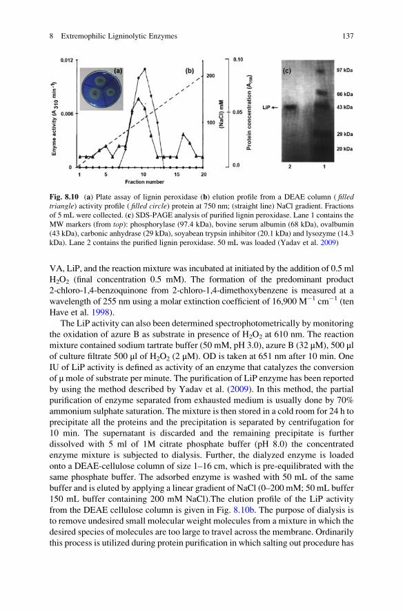
VA, LiP, and the reaction mixture was incubated at initiated by the addition of 0.5 ml
H2O2 (final concentration 0.5 mM). The formation of the predominant product
2-chloro-1,4-benzoquinone from 2-chloro-1,4-dimethoxybenzene is measured at a
wavelength of 255 nm using a molar extinction coefficient of 16,900 M�1 cm�1 (ten
Have et al. 1998).
The LiP activity can also been determined spectrophotometrically by monitoring
the oxidation of azure B as substrate in presence of H2O2 at 610 nm. The reaction
mixture contained sodium tartrate buffer (50 mM, pH 3.0), azure B (32 μM), 500 μlof culture filtrate 500 μl of H2O2 (2 μM). OD is taken at 651 nm after 10 min. One
IU of LiP activity is defined as activity of an enzyme that catalyzes the conversion
of μmole of substrate per minute. The purification of LiP enzyme has been reported
by using the method described by Yadav et al. (2009). In this method, the partial
purification of enzyme separated from exhausted medium is usually done by 70%
ammonium sulphate saturation. The mixture is then stored in a cold room for 24 h to
precipitate all the proteins and the precipitation is separated by centrifugation for
10 min. The supernatant is discarded and the remaining precipitate is further
dissolved with 5 ml of 1M citrate phosphate buffer (pH 8.0) the concentrated
enzyme mixture is subjected to dialysis. Further, the dialyzed enzyme is loaded
onto a DEAE-cellulose column of size 1–16 cm, which is pre-equilibrated with the
same phosphate buffer. The adsorbed enzyme is washed with 50 mL of the same
buffer and is eluted by applying a linear gradient of NaCl (0–200 mM; 50 mL buffer
150 mL buffer containing 200 mM NaCl).The elution profile of the LiP activity
from the DEAE cellulose column is given in Fig. 8.10b. The purpose of dialysis is
to remove undesired small molecular weight molecules from a mixture in which the
desired species of molecules are too large to travel across the membrane. Ordinarily
this process is utilized during protein purification in which salting out procedure has
Fig. 8.10 (a) Plate assay of lignin peroxidase (b) elution profile from a DEAE column ( filledtriangle) activity profile ( filled circle) protein at 750 nm; (straight line) NaCl gradient. Fractions
of 5 mL were collected. (c) SDS-PAGE analysis of purified lignin peroxidase. Lane 1 contains the
MW markers (from top): phosphorylase (97.4 kDa), bovine serum albumin (68 kDa), ovalbumin
(43 kDa), carbonic anhydrase (29 kDa), soyabean trypsin inhibitor (20.1 kDa) and lysozyme (14.3
kDa). Lane 2 contains the purified lignin peroxidase. 50 mL was loaded (Yadav et al. 2009)
8 Extremophilic Ligninolytic Enzymes 137

been employed as the initial step with ammonium sulphate. After the protein is
precipitated from the initial source, it is re-dissolved in buffer and then poured into
a dialysis bag. The homogeneity of the enzyme preparation is checked by SDS-
PAGE. The separating gel contains 12% acrylamide in 0.375M Tris-HCl buffer
(pH 8.8) and the stacking gel remains 5% acrylamide in 0.063M Tris-HCl buffer
(pH 6.8). Proteins are visualized by silver staining as shown in Fig 8.10c.
8.4 Laccase
Laccases (EC 1.10.3.2) are multi copper-containing polyphenol oxidases that are
widely distributed in microorganisms, insects, and plants, showing a specific
function in each of them. From this group, white rot fungi are the most studied
laccases. It catalyze the oxidation of various aromatic compounds, particularly
those with electron-donating groups such as phenols (�OH) and anilines (�NH2),
by using molecular oxygen as an electron acceptor. In nineteenth century, laccases
was first isolated from exudates of the Japanese tree Rhus vernicifera. Laccases usemolecular oxygen to oxidize a variety of aromatic and non-aromatic hydrogen
donors via a mechanism involving radicals. These radicals can undergo further
laccases catalyzed reaction and/or non-enzymatic reaction such as polymerization,
and hydrogen abstraction. Therefore, laccase has also the ability to oxidize phenolic
and non-phenolic substrates. The phenolic substrate oxidation by laccases result in
formation of an aryloxyradicals an active species that is converted to a quinone in
the second stage of the oxidation. Though, typical substrate of laccases known to
be diphenol oxidase, monophenol e.g. sinapic acid or guaiacol can also oxidize
polyamines, aminophenols, lignin, aryl diamine, inorganic ions and it may mitigate
the toxicity of some polycyclic hydrocarbon. However, 2,20-azino-bis(3-ethylbenzothiazoline-6-sulphonic acid) or ABTS is substrates which are most
commonly used, does not form quinone and is not pH dependent. Laccases have
been mostly isolated and characterized from plants and fungi, but only fungal
laccases are used currently in biotechnological applications for the detoxification
of complex industrial wastewater. Unfortunately, these enzymes usually work only
efficiently under mild acidic conditions (pH 4.0–6.0) whereas the temperature range
(30–55 �C) for catalytic activity is suboptimal. In contrast, little is known about
bacterial laccases, which broad range substrate specificity for industrial application.
Most of study has been focused on white-rot fungus in which Phlebia floridensisshowed higher thermostability at pH 4.5 and T. versicolor is useful for dye
decolourization. Recently, in bacterial laccase polyphenol oxidase activity has
been reported in an Azospirillum lipoferum, phenol oxidases, laccase were isolatedfrom cell extracts of the soil bacterium Pseudomonas putida F6. Four strains of thebacterial genus Streptomyces (S. cyaneus, S. ipomoea, S. griseus and
S. psammoticus) and the white-rot fungus T. versicolor were studied for their abilityto produce active extracellular laccase in biologically treated wastewater with
different carbon sources. Similarly, Proteus mirabilis, Bacillus sp., Raoultella
138 R. Chandra et al.

planticola and Enterobacter sakazakii are used for degradation of persistent organicpollutants from biomethanated distillery spent wash, but it is still lacking for
industrial application (Chandra and Singh 2012). Laccases have low substrate
specificity make this interesting for degradation of several compounds with a
phenolic structure because they are extracellular and inducible, do not need a
cofactor, and have low specificity. Therefore, laccases have been employed in
several areas such as bioremediation of aromatic recalcitrant compounds, treatment
of effluents polluted with lignin, chemical synthesis, degradation of a wide number
of textile dyes, and biomass pretreatment for biofuel production.
Due to the properties of their substrate, the enzymes participating in the break-
down of lignin and other related compounds should be exclusively extracellular.
While this is without exception true for the LiP and MnP of white rot fungi and
bacteria, the situation is not the same with laccases. Although most laccases studied
so far are extracellular enzymes, while in wood-rotting fungi and some bacteria
species are usually also found intracellular laccase activity. Blaich and Esser (1975)
has been observed that most white-rot fungal species produced both extracellular
and intracellular laccases with isoenzymes showing similar patterns of activity.
T. versicolor was produced laccases both in extracellular and intracellular fractionswhen grown on glucose, wheat straw, and beech leaves (Schlosser et al. 1997). The
intra and extracellular presence of laccase activity was also detected in
P. chrysosporium and Suillus granulates. The fraction of laccase activity in Neu-rospora crassa, Rigidoporus lignosus and one of the laccase isoenzymes of
P. ostreatus is also probably localized intracellularly or on the cell wall. The
localization of laccase is probably connected with its physiological function and
determines the range of substrates available to the enzyme. It is possible that the
intracellular laccases of fungi as well as periplasmic bacterial laccases could
participate in the transformation of low MW phenolic compounds in the cell. The
cell wall and spores-associated laccases were linked to the possible formation of
melanin and other protective cell wall compounds. However, laccase has been
reported intracellularly as in A. lipoferum, M. mediterranea and in B. subtilis.The bacterial cells must have some strategy to cope with the intracellular presence
of laccase and its toxic by-products. Rearrangement of the electron transport system
has been hypothesized to be one of the ways in which the laccase-positive cells
adapt to endogenous substituted quinones generated as products of laccase cata-
lyzed reaction. Because, in fungi, extracellular localization of the enzymes helps
them circumvent the problem of the reactive species, such as semiquinones and
quinines that are generated by laccases while oxidizing aromatic substrates. These
reactive species are powerful inhibitors of the electron transport system in both
bacteria and mitochondria. The loss of cytochrome c oxidase activity and
acquistion of resistance to quinone analogues has been demonstrated in a laccase-
positive variant of A. lipoferum.
8 Extremophilic Ligninolytic Enzymes 139

8.4.1 Molecular Structure
Laccases are monomeric, dimeric and tetrameric glycoproteins, generally having
fewer saccharide compounds (10–25%) in fungus and bacteria than in plant
enzymes. The carbohydrates, which are 10–45% of the total molecular mass, are
covalently linked, and due to this property of enzymes show high stability. Man-
nose is one of the major components of the carbohydrates attached to laccases. The
molecular weight of a laccase is determined to be in the range of 50–97 kDa from
various experimental reports. The structure of laccase from T. versicolor containingapproximately 500 amino acid residues organized in three β-barrel sequential
domains. The three domains are distributed in a first domain with 150 initial
amino acids, a second domain between the 150 and 300 residue, and a third domain
from the 300 to 500 amino acid. The structure is stabilized by two disulfide bridges
localized between domains I and II and between domains I and III. Figure 8.11a, b
shows the three dimensional ribbon structure of bacterial lacasse from Bacillussubtilis XI based on the homology modelling of B. subtilis MB24 laccase PDB Id:
2x88A) (Guan et al. 2014) and fungal laccase from T. versicolor, respectively (PDBId: 1N68) (Robert et al. 2003).
Laccases of three molecular forms of isozyme have been reported namely, Lac I,
Lac II and Lac III. D’Souza-Ticlo et al. (2009) reported that the MW of Lac II is
56 kDa from Cerrena unicolor. In another study the MW of laccase has been
reported ~97 kDa in T. versicolor. The biochemical properties of spore coat protein
Cot A of B. subtilis has been reported to be similar to multicopper oxidases
including laccases (Hullo et al. 2001). Further, the studies have revealed that Cot
A contains all the structural features of a laccase including the reactive surface-
exposed copper center (T1) and two buried copper centers (T2 and T3). The most
thermostable laccases have been isolated from Streptomyces lavendulae, with half-life of 100 min at 70 �C, and in B. subtilis, for which Cot A reported a half-life of
112 min at 80 �C. The redox potential of any substrate also plays a very important
role in laccase activity. The redox potential of bacterial laccases ranges from 0.4 to
0.5 V, but they are active and stable at high temperatures (66 h at 60 �C), at pH7.0–9.0 and at high salt concentrations. The first gene and c-DNA sequences were
recorded for laccases from the fungi Neurospora crassa, Aspergillus nidulans,Coriolus hirsutus and Phlebia radiata. Since, then the number of sequenced laccase
genes has considerably increased. The sequences mostly encode polypeptides of
approximately 500–600 amino acids in Bacillus subtilis. Cot A is 513 amino acids
long, and its MW is 65 kDa typical eukaryotic signal peptide sequences of about
21 amino acids are found at the N-terminal of the protein sequences. In addition to
the secretion signal sequence, laccase genes from N. crassa, Podospora anserina,Myceliophthora thermophila and Coprinopsis cinerea contain regions that code forN-terminal cleavable propeptides. These laccases also have C-terminal extensions
of controversial function, i.e., the last amino acids from the predicted amino acid
140 R. Chandra et al.
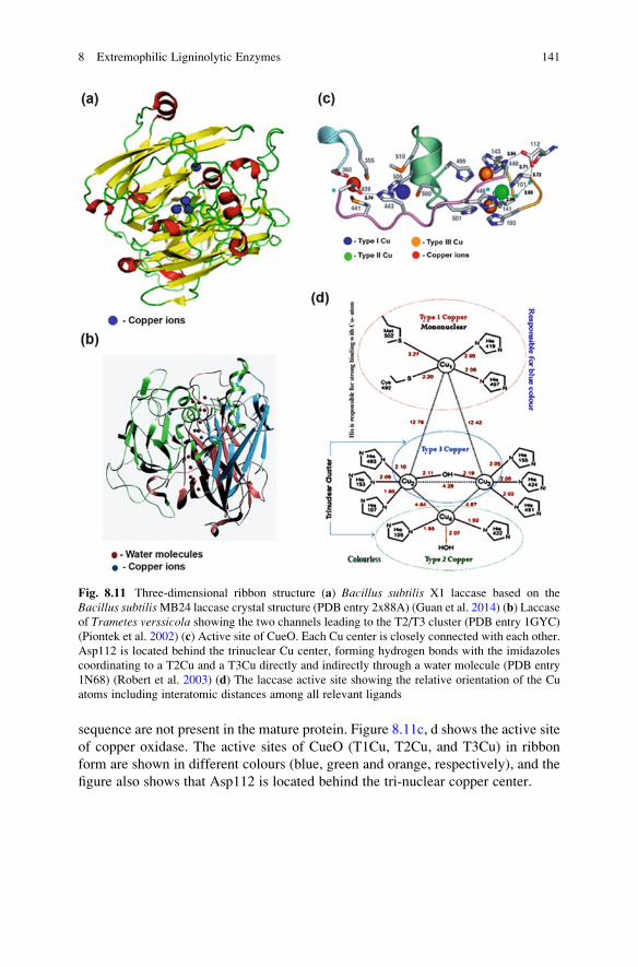
sequence are not present in the mature protein. Figure 8.11c, d shows the active site
of copper oxidase. The active sites of CueO (T1Cu, T2Cu, and T3Cu) in ribbon
form are shown in different colours (blue, green and orange, respectively), and the
figure also shows that Asp112 is located behind the tri-nuclear copper center.
Fig. 8.11 Three-dimensional ribbon structure (a) Bacillus subtilis X1 laccase based on the
Bacillus subtilisMB24 laccase crystal structure (PDB entry 2x88A) (Guan et al. 2014) (b) Laccase
of Trametes verssicola showing the two channels leading to the T2/T3 cluster (PDB entry 1GYC)
(Piontek et al. 2002) (c) Active site of CueO. Each Cu center is closely connected with each other.
Asp112 is located behind the trinuclear Cu center, forming hydrogen bonds with the imidazoles
coordinating to a T2Cu and a T3Cu directly and indirectly through a water molecule (PDB entry
1N68) (Robert et al. 2003) (d) The laccase active site showing the relative orientation of the Cu
atoms including interatomic distances among all relevant ligands
8 Extremophilic Ligninolytic Enzymes 141

8.4.2 Catalytic Cycle and Mode of Action
The catalytic mechanism of a laccase involves the reduction of the oxygen mole-
cule, including the oxidation by one electron of a wide range of aromatic com-
pounds, which include polyphenols, and methoxy-substituted monophenols and
aromatic amines. The use of molecular oxygen as the oxidant and water as gener-
ated by-product are very common catalytic features. Because, laccases have a four
copper (Cu) atom in their active site which participate in oxygen reduction and
water production (Fig. 8.11d). The laccase’s four Cu atoms are disseminated in
three types of cores or places: type 1 Cu (T1Cu), type 2 Cu (T2Cu), and type 3 Cu
(T3Cu). These cores are in two metallic active sites: the mononuclear location T1
and the trinuclear location T2/T3. It is believed that laccase catalysis involves the
following mechanism. Initially reduction of T1Cu by reducing substrate. Further,
Internal electron transfer from the T1Cu to T2Cu and T3Cu. Subsequently, reduc-
tion of oxygen to water at T2Cu and T3Cu site. The T1Cu gives the protein its blue
colour absorbance at about 600 nm depends on the intensity of the Scys–CuII bond
and the ligand field strength. T2Cu does not give any colour, but it is EPR
detectable, and T3Cu contains a pair of atoms in binuclear conformation that
gives a weak absorbance at 330 nm but is not detected by EPR. Spectroscopy
combined with crystallography has provided a detailed description of the active site
in a laccase. T2Cu and T3Cu form a trinuclear center, which is involved in the
catalytic mechanism.
In the catalytic mechanism, an oxygen molecule binds to the trinuclear cluster of
the asymmetric activation site, and it is postulated to restrict access of the oxidizing
agent. During the steady state, laccase catalysis indicates that O2 reduction takes
place. The bonds of the natural substrate, lignin, are cleaved by laccase through Cα–
Cβ cleavage and aryl cleavage. Subsequently, the lignin degradation is caused by
phenoxy radicals leading to the oxidation of the α-carbon or by the cleavage of the
bond between the α-carbon and β-carbon. This oxidation results in oxygen-centeredfree radicals, which can be converted by a second enzyme catalyzed reaction to
quinone. The quinone and the free radicals can then undergo polymerization. The
T2Cu center coordinates with two His ligands and water as ligands. The Type
3 coppers are each 4-coordinate, having three His ligands and a bridging hydroxide
(Fig. 8.11d). The reduction of oxygen by a laccase appears to occur in two 2e�
steps. The first is the rate determining step. In this, the T2/3Cu bridging mode is
reduced by the first 2e�. The peroxide-level intermediate facilitates the second 2e�
reduction (from the T2Cu and T1Cu centers) in which the peroxide is directly
coordinated to the reduced T2Cu, and the reduced T1Cu is coupled to the T3Cu by
covalent Cys–His linkages.
The above mentioned information is described for blue copper oxidases, i.e. blue
laccases, but some authors have reported a small group of laccases that lack the
600 nm band and hence the blue color; some of these non-blue laccases (dubbed
“yellow” or “white”) feature a high redox potential allowing them to oxidize
non-phenolic compounds without any mediators. Both types of laccases (yellow
142 R. Chandra et al.

and blue) have similar MW (70 kDa) and specific activities, and it is observed that
the yellow laccases are obtained from the culture grown on solid state medium,
while the blue forms were isolated from culture grown on liquid medium without
lignin. The yellow laccases are formed by the modification of the blue forms with
low molecular weight lignin decomposition products, and some non-blue laccases
(yellow) have high redox potentials, allowing them to oxidize non-phenolic com-
pounds without any mediator (Pozdnyakova et al. 2006). Therefore, it is assumed
that the yellow form of a laccase is a result of binding of the aromatic product of
lignin degradation to the blue laccase. It has been postulated that a yellow laccase
might contain endogenous mediators derived from lignin, which perform the role of
exogenous mediator in the reaction of non-phenolic compounds. Due to insufficient
information about yellow laccases, more research is required in this field. In
Fig. 8.3a the simplest case is the one in which the substrate molecules are oxidised
to the corresponding radicals by direct interaction with the copper cluster. How-
ever, the substrates of interest cannot be oxidized directly by laccases, either
because they are too large to penetrate into the enzyme active site or because
they have a particularly high redox potential. By mimicking nature, it is possible
to overcome this limitation with the addition of “redox mediators”, which act as
intermediate substrates for laccases, whose oxidized radical forms are able to
interact with the bulky or high redox potential substrate targets Fig. 8.3b
Laccases catalyze the removal of one electron from the phenolic hydroxyl
groups of phenolic lignin model compounds, such as vanillyl glycol, 4,6-di
(t-butyl)guaiacol, and syringaldehyde, to form phenoxy radicals, which generally
undergo polymerization via radical coupling. The reaction is also accompanied by
demethylation, formation of quinone, resulting in ring cleavage. The degradation of
phenolic β-1 lignin substructure models occurs via the formation of phenoxy
radicals, which leads to Cα–Cβ cleavage, Cα oxidation, alkyl–aryl cleavage, and
aromatic ring cleavage. Laccase-catalyzed oxidation of phenols, anilines, and
benzenethiols correlates with the redox potential difference between the TiCu site
of the laccase and the substrate. The presence of electron withdrawing o- and
p-substituents reduces the electron density at the phenoxy group, and thus it is
more difficult to oxidize the phenolic substrate. Bulky substituents, which impose
steric interference with substrate binding, cause a decrease in reactivity.
Laccase has been found to oxidize non-phenolic model compounds and β-1lignin dimers in the presence of a mediator, indicating that the enzyme plays a
significant role in the depolymerization of lignin and pulp delignification. The most
studied mediators for laccases are ABTS, 1-hydroxybenzotriazole (HBT), and
3-hyroxyanthranilic acid (HAA). The oxidation is different for ABTS and HBT,
involving a dication and a benzotriazolyl-1-oxide radical, respectively. Oxygen
uptake by the laccase is faster with ABTS than with HBT, but the oxidation of
non-phenolic substrate is comparable for both the mediators. The ABTS-mediated
oxidation of a non-phenolic substrate proceeds via an electron transfer mechanism.
In the oxidation process, the ABTS is first oxidized to a radical cation (ABTSc+),
and then to a dication (ABTS2+), with redox potentials of 472 mV (ABTS/ABTS•+)
and 885 mV (ABTS•+/ABTS2+), respectively. The dication is the active
8 Extremophilic Ligninolytic Enzymes 143

intermediate, which is responsible for the oxidation of the non-phenolic substrate.
The HBT� mediated oxidation of non-phenolic substrate involves the initial oxi-
dation of HBT to HBT+ by laccase, followed by an immediate deprotonation to
form an N-oxy radical. The latter abstracts the benzylic H-atom from the substrate,
converting it to a radical. The oxidation of VA by laccase-ABTS and by laccase-
HBT (radical Habstraction mechanism) is presented in Fig. 8.12. HBT/HBT+, with
an Eo value of 1.08 V, has a mediator efficiency with laccase that is higher than that
of ABTS. The use of laccase/HBT for the bleaching of paper pulp and for the
removal of lipophilic extractives has been described. Recently, two lignin-derived
phenols, syringaldehyde and acetosyringone, have been shown to act as effective
laccase mediators for the removal of lipophilic compounds from paper pulp.
The degradation of non-phenolic β-O-4 model compounds, which represent the
major substructure in lignin, has been studied using laccase-mediator systems. Four
types of reactions, β-ether cleavage, Cα–Cβ cleavage, Cα-oxidation and aromatic
ring cleavage, are catalyzed by the laccase–BHT (butylated hydroxytoluene)
coupled system. In the oxidation of a non-phenolic β-O-4 lignin model dimer,
1-(4-ethoxy-3-methoxyphenyl)-1,3-dihydroxy-2-(2,6-dimethoxyphenoxy)propane,
the coupled enzyme/HBT system catalyzes the 1e� oxidation of the substrate to
form a β-aryl radical cation or benzylic (Ca) radical intermediates. The electron
density of the aromatic ring affects the 1e� oxidation by the laccase/1-HBT couple.
Substrates containing electron-donating groups favor aromatic ring cleavage prod-
ucts. The β-aryl radical cation is converted to the product via an aromatic ring
cleavage, and the benzylic radical is cleaved at the Cα–Cβ bond, similar to a
Baeyer–Villiger reaction. The β-ether cleavage of the β-O-4 lignin substructure is
caused by reaction with the Ca-peroxy radical intermediate produced from the
benzylic radical. The rate of oxidation depends on the kcat of the laccase for
the mediator and the stability of the enzyme to inactivation by the free radical of
the mediator.
The ability of LiP, MnP and lacasses to degrade lignin has been studied in
diverse industrial processes and bioremediation of contaminated soils and water,
but this ability is non-identical between these three types of enzymes. This may be
due to that enzyme-substrate interactions are different. The study of the interactive
OCH3 OCH3 OCH3
CH2OH CH2OH CHOH
OCH3
CHO
ABTS++ ABTS•+
ABTS•+ ABTS++
ABTS•+ ABTSe- H
ABTS
-O3S -O3S -O3S
SO3- SO3
-
C2H5
C2H5
C2H5
C2H5
SO3-
e-
e-
e-
e-S
+••
•
S
NN N
C2H5
C2H5
S S
SN NN N
NN N
N
S
N
+
•
•
++
+
-
+
-
Fig. 8.12 Radical H-atom abstraction and electron transfer mechanism
144 R. Chandra et al.

mechanisms involved in ligninolytic enzyme and lignin is indeed important in
understanding enzyme reactions and may provide further insights to the develop-
ment of biodegradation technologies. Ligninolytic enzymes are reported
for degradation of lignin by direct interactions of ligninolytic enzymes in terms
of a long-range electron transfer process. However, little is known about the effect
of ligninolytic enzymes structures on the lignin biodegradation at the molecular
level. The molecular docking approach can be used to model the interaction
between a small molecule and a protein at the atomic level, which allow us to
characterize the behavior of small molecules in the binding site of target proteins as
well as to elucidate fundamental biochemical processes. Crystallographic water is a
major challenge in molecular docking. These molecules are strongly bound to the
receptor and observed across several crystallographic structures of a particular
protein. In approximately 65% of the crystallographic protein-ligand complexes,
at least one water molecule is involved in ligand-receptor recognition. The release
of a crystallographic water molecule from its binding site is entropically favorable;
however the process causes a simultaneous loss in enthalpy. To compensate for this
enthalpy loss, a specific moiety of the ligand can be designed to mimic the
interaction network of the displaced water through the formation of equivalent
hydrogen bonds with the protein. Alternatively, structural water can be explicitly
included in the docking experiments, allowing the formation of highly favorable
hydrogen-bonding networks between the ligand and the target binding site. In this
case, a variety of methods are available to evaluate which water molecules are
strongly bound and, therefore, suitable for this purpose. Among these strategies one
can highlight free energy perturbation calculations using Monte Carlo (MC) statis-
tical mechanics simulations, which estimate the binding free energy for a given
water molecule, allowing the discrimination between displaceable and strongly-
bound structural water. MC methods generate poses of the ligand through bond
rotation, rigid-body translation or rotation. The conformation obtained by this
transformation is tested with an energy- based selection criterion. If it passes the
criterion, it will be saved and further modified to generate next conformation. The
main advantage of MC is that the change can be quite large allowing the ligand to
cross the energy barriers on the potential energy surface, a point that isn’t achievedeasily by molecular dynamics based simulation methods.
8.4.3 Common Substrate and Microorganisms
Laccases have a broad range of substrate specificity. Due to this, microorganisms
oxidize a wide range of environmental pollutants as a sole carbon or nitrogen source
for their growth and metabolism. Laccases have been mostly isolated and charac-
terized from plants and fungi, and only fungal laccases are currently used in
biotechnological applications for the detoxification of complex industrial wastewa-
ter. Laccase producing microorganisms and their some common substrate are listed
in Table 8.3.
8 Extremophilic Ligninolytic Enzymes 145

Table 8.3 Some important laccase producing microorganisms in extremophilic environment and
their substrate
Microorganisms Substrate pH Temp. (�C)Fungi
Penicillium pinophilumTERI DB1
Distillery effluent decolourisation 8.5 25
Trametes versicolor Non-phenolic lignin model dimer 5.0 25
Polyporus pinisitus Direct red 28, Acid Blue 74 4–5 50
Myceliophthorathermophila
Direct red 28, Acid Blue 74 6.0 70–80
Trametes trogii Nitrobenzene and anthracene 5.0 25
Trametes versicolor Direct red 28, Acid Blue 74 5.0 65
Pleurotus sp. 2,4 Dichlorophenol, Benzo(a)pyrene [B(a)P] 5.0 28
Trichoderma atroviride Catechol, o-cresol, 4.0–5.0 40–50
Phanerochaetecrysosporium
Lignin 5.0 25
Daedalea flavida Lignin 5.0 25
Pycnoporus coccineus Anthracene, pyrene, fluoranthene, benzo[a]
pyrene, phenanthrene
5.0 28
Phlebia sp. Lignin 5.0 25
Phlebia floridensis Lignin 5.0 25
Pseudochrobactrumglaciale (FJ581024)
Pulp paper mill effluent 5.0 25
Providencial rettgeri(GU193984)
Pulp paper mill effluent 5.0 25
Ganoderma lucidum Antracene, benzo[a]pyrene, fluorine,
acenapthene, acenaphthylene and benzo[a]
anthracene
– –
Bacteria
Serratia marcescens(GU193982)
Black liquor 5.0 25
Citrobactersp. (HQ873619)
Black liquor 5.0 25
Klebsiella pnumoniae(GU193983)
Black liquor 5.0 25
Staphylococcussaprophyticus
Brilliant blue, methyl orange, neutral red 3.0 32
Bacillus SF Mordant black 9, mordant brown 96 &
15, acid blue 74
8.0 60
γ-Proteobacteria JB Caramine 4–10 55
Bacillus subtilis Syringaldazine, ABTS 3.0–7.0 75
146 R. Chandra et al.
8.4.4 Screening of Laccase Producing Microorganisms, ItsSubstrate, Bioassay and Purification
Laccases can oxidize a wide range of molecules more than hundred different types
of compound have been identified as substrate for laccase. There are various natural
and synthetic substrates which are mentioned in Table 8.4, used for laccase assay.
All substrates cannot be directly oxidized by laccases, either because of their large
size which inhibit their penetration into the enzyme active site or because of their
particular high redox potential. To overcome this hindrance, suitable chemical
mediators are used which are oxidized by the laccase and their oxidized forms
are able to interact with high redox potential substrate.
Although polyphenol oxidases copper proteins are able to oxidize aromatic
compounds with molecular oxygen as the terminal electron acceptor. Polyphenol
oxidases are associated with three types of activities:
(a) Catechol oxidase or o-diphenol: oxygen oxidoreductase (EC 1.10.3.1)
(b) Laccases or p-diphenol: oxygen oxidoreductase (EC 1.10.3.2)
(c) Cresolase or monophenols monooxygenase (EC 1.18.14.1)
Faure et al. (1995), compared commercial fungal laccase and catechol oxidase,
purified from Pyricularia oryzae and Agaricus bisporus, respectively, with bacte-
rial laccase from A. lipoferum by using several substrates and phenol oxidase
inhibitors. Five classes of chemical compounds were investigated as substrates
for laccase:
1. L-Tyrosine and several substituted monophenols such as p-coumaric and o-hydroxyphenylacetic or salicylic acids;
Table 8.4 Natural and synthetic substrate of laccases
S. No. Natural substrate Synthetic substrate
1. Acetosyringone 1- Hydroxylbenzotrizole (HBT)
2. Syringaldehyde N- Hydroxyphthalimide (HPI)
3. Vanilin Violuric acid (VLA)
4. Acetovanillone N- Hydroxylacetanlide (NHA)
5. Sinapic acid 2,2,6,6-Tetramethylpiperidine- N-oxyl (TEMPO)
6. Ferulic acid Acetohydroxamic acid
7. p-Coumaric acid 2,2,5,5- Tetramethylpyrrolidine-N-oxyl (PROXYL)
8. Reduced glutathione 2,20-Azinobis-(3-ethylbenzothia- zoline 6- sulfonic acid)(ABTS)
9. Cystine Guaicol
10. Aniline Methyl syringate
11. 4 hydroxybenzyl
alcohol
8 Extremophilic Ligninolytic Enzymes 147

2. o-Diphenols (catechol, pyrogallol, guaiacol, and protocatechic, gallic, and
caffeic acids), L-3, 4- dihydroxyphenylalanine and o-aminophenol, which
could be oxidized by both laccase and catechol oxidase;
3. p-Diphenol and p-substituted aromatic compounds as typical p-phenol oxidasesubstrates such as hydroquinone, p-cresol, p-aminophenol and
p-phenylenediamine;
4. m-Diphenols such as resorcinol, orcinol, 4-hexylresorcinol, and
5-pentadecylresorcinol;
5. Other laccase substrates such as syringaldazine, 1-naphthol, ABTS, and 4- and
5-hydroxyindoles. The range of substrates used by A. lipoferum laccase was
similar to that used by P. oryzae laccase.
The screening methods of laccase producing microorganisms are similar to MnP
as described previously but the minimal media does not contained H2O2. Bioassay
of laccase activity is measured based on the oxidation of various substrate in
presence of suitable buffer (i.e. sodium acetate) in acidic pH (5.0). The oxidation
product is measured by spectrophotometer taking the absorbance at 420 nm or
450 nm depending upon substrate specificity. ABTS has been most commonly used
substrate for laccase bioassay because it acts as cooxidant that can interact with
laccase to accomplish electron transfer and it is chemically oxidized in two steps via
ABTS+ and ABTS2+. Anisyl alcohol and benzyl alcohol can be better oxidized by
ABTS2+ than by ABTS+. Laccase activity is assay through ABTS method. In this
method reaction mixture contained 600 μL sodium acetate buffer (0.1 M, pH 5.0 at
27 �C), 300 μL ABTS (5 mM), 300 μL culture filtrate and 1400 μL distilled water.
The mixture is then incubated for 2 min at 30 �C and the absorbance was measured
immediately in 1-min intervals. One unit of laccase activity has been defined as
activity of an enzyme that catalyzes the conversion of 1 mole of ABTS per minute.
Laccase activity has been measured by another substrate guaiacol. The reaction
mixture contained 10 mM sodium acetate buffer (pH 5.0), 2 mM guaiacol and
0.2 ml of culture supernatant was incubated at 25 �C for 2 h and the absorbance was
read at 450 nm. The relative enzyme activity has been expressed as colorimetric
units/ml (CU/ml).
In general plant lacasses are purified from sap or tissue extracts, whereas fungal
lacasses are purified from culture are purified from culture (fermentation broth) of
the selected organism. Various protein purification techniques are used for purifi-
cation of laccase. Typical purification protocols involve ultrafiltration,
ion-exchange, gel filtration and other electrophoretic and chromatographic tech-
niques. Purification may be single or multi-step process. A single step lacasse
purification from N. crassa are performed by using celite column chromatography
and 54 fold purification with specific activity of 333 U/mg. Laccase from Tversicolor are purified using ethanol precipitation, DEAE–sepharose, phenyl-
sepharose and sephadex G-100 chromatography. T. versicolor 951022 excrete a
single monomeric laccase showing a high specific activity of 91,443 U/mg for
ABTS as substrate. Laccase from T. versicolor is purified with ion exchange
chromatography followed by gel filtration with specificity activity of 101 U mL�1
148 R. Chandra et al.

with 34.8 fold purification. Laccase from Stereum ostrea and obtained up to 70-foldpurification from culture filtrate by a two step protocol-ammonium sulphate (80%
w/v) and sephadex G-100 column chromatography.
8.4.5 Thermostability of Bacterial Laccases
Laccases are highly stable, industrially important enzymes that are capable of
oxidizing a large range of substrates. Thermostability plays an important role in
enzyme catalysis; several sequence and structural factors are involved in this
phenomenon. Thermostable enzymes allow high process temperatures with higher
associated reaction rates and less risk of microbial contamination. Some of the
mechanisms/indicators of increased thermostability include: a more highly hydro-
phobic core, tighter packing (compactness), a deleted loop, greater rigidity
(e.g. through increased proline content in the loop), higher secondary structure
content, greater polar surface area, fewer thermolabile residues, increased
H-bonding, higher pI, a disulfide bridge, more salt bridges and buried polar
interactions. Moreover, the enhanced thermostability of a laccase from Bacillussp. HR03 using site directed mutagenesis of the surface loop was achieved, in which
glutamic acid (Glu188) was substituted with 2 hydrophilic(lysine and arginine) and
1 hydrophobic (alanine) residues. There are some bacterial species that show
thermostability at different temperatures:
(a) Cot A from B. subtilis at 75 �C showed maximum activity and at 80 �C wild
type Cot A has a life of 4 h, whereas recombinant Cot A from E. coli has a half-life of 2 h.
(b) T. thermophilum laccase has optimum activity at 92 �C with a half-life of 4 h at
80 �C and attains 60% of its activity after incubation for 10 min at 100 �C.
8.5 Industrial and Biotechnological Applications of MnP,
LiP and Laccase
Ligninolytic enzymes are involved in the degradation of the complex and recalci-
trant environmental pollutants. This group of enzymes is highly versatile in nature
and they find application in a wide variety of industries. The biotechnological
significance of these enzymes has led to a drastic increase in the demand for
these enzymes in the recent time because of their oxidative ability toward a broad
range of phenolic and non-phenolic compounds.
8 Extremophilic Ligninolytic Enzymes 149

8.5.1 Industrial Waste Detoxification and Bioremediation
MnP, LiP and laccase have used to detoxify or remove various aromatic compounds
found in industrial waste and contaminated soil and water. MnP have capability to
mineralised various PAHs such as anthracene, benzo[a]pyrene, benz[a]anthracene,
phenanthrene, [U-14C]pentachlorophenol, [U-14C]catechol, [U-14C]tyrosine, [U-14
C]tryptophan, [4,5,9,10-14C]pyrene, [ring U-14C]2-amino-4-6-dinitrotolune and
1.1.1-trichloro-2-2-bis-(4-chlorophenyl) ethane (DDT). MnP has also been reported
for the elimination and detoxification of triclosan, an emerging persistent pollutant
with ubiquitous presence in aquatic environment. Further, MnP detoxified afla-
toxins B1, lignite originated humic acid, nylon, polyethylene and amino carbonyl
maillard products (melanoidins). LiP and MnP was effective in decolourizaing kraft
pulp paper mill effluent. LiP was reported for mineralisation of PAHs like naph-
thalene, phenanthrene, benzo[c]phenenthrene, benz[e]pyrene, benz[a]anthracene,
pyrene, chrysene, anthracene, benzo[a] pyrene, perylene. Laccase has been reported
for the degradation and decolourization of chlorophenol- and chlorolignin-
containing black liquor and pulp paper mill wastewater. However, laccase has
been reported to mineralised anthracene, benzo pyrene, fluorene and other
16 PAHs compounds which are listed by USEPA (United States Environmental
Protection Agency) as priority pollutants of environment
8.5.2 Decolourisation of Textile Dye, Pulp Paper Milland Distillery Effluent
MnP decolourise and degrade various types of synthetic dye; azo dye (reactive red
120, congo red, orange G and orange IV, remazol brilliant violet 5R, direct red 5B),
anthraquinone dye (remazol brilliant blue R), indigo dye (Indigo Carmine) and
triphenylmethane dye (Methyl Green) containing textile wastewater. LiP
decolourised various dyes e.g. bromophenol blue, congo red, methylene blue,
methyl green, methyl arrange, poly-R-478, poly S-119, poly T-128. Laccase have
capability to decolourised a wide range of dyes e.g. cibanon red 2B-MD, cibanon
golden yellow PK-MD, cibanon blue GFJ-MD, indanthrene direct black RBS,
remazol brilliant blue R, congo red etc. Laccase catalysed textile dye bleaching
may also be useful in finishing dyed cotton fabric. Under laccase catalysis, soluble
dye precursors could be absorbed, oxidise and polymerised to give the desired
tanning effect. LiP decolourised four distinct classes of dyes, they are-
(a) tryphenylmethane(bromophenol blue), (b) heterocyclic dye (methylene blue
and toluene blue O), (c) azo dye (congo red and methyl orange) and
(d) polymeric dyes (Poly R-478, Poly S-119, AND Poly T-128).
150 R. Chandra et al.

8.5.3 Production of Ethanol and Value Added Products
Ligninolytic enzymes play a central role in lignin degradation. Due to high redox
potential, MnP, LiP and laccase are of high industrial interest for delignification and
production of ethanol and other cellulosed based low molecular weight chemicals
such as vanillin, dimethoxy sulfoxide and phenol. Laccases have been used for the
synthesis of several anticancer drugs such as actinocin, vinablastine and other
pharmaceutical products. Laccase have been employed in several applications
organic synthesis as the oxidation of functional group, the coupling of phenols
and steroids, medical agent (anesthetics, anti-inflammatory, antibiotics and seda-
tives) and synthesis of complex natural products. MnP and LiP have potential to
produce natural aromatic flavours compound e.g. vanillin, β-ionone, β-cyclocitral,dihydroactinidiolide, flavanoids and so one. Laccase can be applied to certain food
processes that enhance or modify the colour appearance of food or beverages for the
elimination of undesirable phenolic compounds, responsible for the browning haze
formation and turbidity in clear fruit juice, beer and wine. The uses of laccase in
baking process increase strength, stability and reduced thickness, thereby improv-
ing the machine ability of dough. Laccase have also been employed to sugar beet
pectin gelation, baking and ascorbic acid determination.
8.5.4 Development of Biosensors
MnP and LiP are known as redox enzyme with efficient direct electron transfer
(DET) properties with electrode. It may enable to use for development of biosensor
based on DET, effective biofuels cell. However, laccase catalysis could be useful as
biosensor for detecting oxygen and a wide variety of reducing substrate (phenols
and anilines). A large number of biosensors containing laccase have been devel-
oped for immunoassay, glucose determination, aromatic amines and phenolic
compounds determination. A carbon paste biosensor modified with a crude extract
of the P ostreatus as a source of laccase source is proposed for catecholamine
determination in pharmaceutical formulation.
8.5.5 Biomechanical Pulping and Pulp Biobleaching
MnP, LiP and laccase are the most important enzyme involved in biomechanical
pulping and kraft pulp bleaching. In the laboratory scale consumption of refining
energy in mechanical pulping was reduced with MnP pre-treatment. However, MnP
degraded residual lignin of kraft pulp and enhanced the pulp bleaching effect. The
laccases have also attracted considerable interest for pulp bio-bleaching. During
lignin degradation, laccases are thought to act on small phenolic lignin fragments,
8 Extremophilic Ligninolytic Enzymes 151
in which the substrate reacts with the lignin polymer, resulting in its degradation.
Laccase-catalyze textile dye bleaching may also be useful in finishing dyed cotton
fabrics.
Take Home Message
• Lignin is amorphous complex polymer of phenylpropane units, which are cross-
linked to each other with a variety of different chemical bonds. It confers rigidity
and recalcitrant nature to the lignocellulosic biomass.
• The common extremophilic ligninolytic enzymes are manganese peroxidase
(MnP), lignin peroxidase (LiP) and laccase. Such enzymes have also proven
their utility in the pollution abatement, especially in the treatment of industrial
waste/wastewater containing hazardous compound like phenols, chlorolignin
synthetic dyes, and polyaromatic hydrocarbons (PAHs) as well as recalcitrant
organic compounds structurally similar to lignin.
• Microorganisms with systems of thermostable enzymes decrease the possibility
of microbial contamination in large scale industrial reactions of prolonged
durations. The mechanisms for many thermotolerant enzymes have been
reported due to their structural properties i.e. presence of Ca2+, saturated fatty
acid, α-helical structure etc.• The ability of extremophilic ligninolytic enzymes to tolerate high temperature,
different metal ions and organic solvents is very important for the efficient
application of this enzyme in the biodegradation and detoxification of industrial
waste.
• The engineering of a disulfide bond (A48C and A63C) near the distal calcium
binding site of MnP by double mutation showed the improvement in thermal
stability as well as pH (pH 8.0) stability in comparison to native enzyme (MnP).
Some of the mechanisms/indicators of increased thermostability of laccases
include: a more highly hydrophobic core, tighter packing (compactness), a
deleted loop, greater rigidity (e.g. through increased proline content in the
loop), higher secondary structure content, greater polar surface area, fewer
thermolabile residues, increased H-bonding, higher pI, a disulfide bridge, more
salt bridges and buried polar interactions.
• Moreover, the enhanced thermostability of a laccase from Bacillus sp. HR03
using site directed mutagenesis of the surface loop was achieved, in which
glutamic acid (Glu188) was substituted with 2 hydrophilic (lysine and arginine)
and 1 hydrophobic (alanine) residues.
• MnP, LiP and laccase have been used to detoxify or remove various aromatic
compounds found in industrial waste and contaminated soil and water. It is used
for the decolourisation of textile dye, pulp paper mill and distillery effluent.
MnP, LiP and laccase are of high industrial interest for delignification and
production of ethanol and other cellulose based low molecular weight chemicals
such as vanillin, dimethoxy sulfoxide and phenol. Laccases have been used for
the synthesis of several anticancer drugs such as actinocin, vinablastine and
other pharmaceutical products. MnP, LiP and laccase are the most important
enzymes involved in biomechanical pulping and kraft pulp bleaching. MnP and
152 R. Chandra et al.

LiP are known as redox enzyme with efficient direct electron transfer (DET)
properties with electrode. It may enable to use for development of biosensor
based on DET, effective biofuels cell.
References
Bao W, Fukushima Y, Jensen KA, Moen MA, Hammel KE (1994) Oxidative degradation of
non-phenolic lignin during lipid peroxidation by fungal manganese peroxidase. FEBS Lett
354:297–300
Bharagava RN, Chandra R, Rai V (2009) Isolation and characterization of aerobic bacteria capable
of the degradation of synthetic and natural melanoidins from distillery effluent. World J
Microbiol Biotechnol 25:737–744
Blaich R, Esser K (1975) Function of enzymes in wood destroying fungi. 2. Multiple forms of
laccase in white rot fungi. Arch Microbiol 103:271–277
Chandra R, Singh R (2012) Decolourisation and detoxification of rayon grade pulp paper mill
effluent by mixed bacterial culture isolated from pulp paper mill effluent polluted site.
Biochem Eng J 61:49–58
D’Souza-Ticlo D, Sharma D, Raghukumar C (2009) A thermostable metal-tolerant laccase with
bioremediation potential from a marine-derived fungus. Mar Biotechnol 11(6):725–737
Faure D, Bouillant ML, Bally L (1995) Comparative study of substrates and inhibitors of
Azospirillum lipoferum and Pyricularia oryzae laccases. Appl Environ Microbiol
61:1144–1146
Glenn JK, Morgan MA, Mayfield MB, Kuwahara M, Gold MH (1983) Anextracellular H2O2-
requiring enzyme preparation involved in lignin biodegradation by the white-rot basidiomycete
Phanerochaete chrysosporium. Biochem Biophys Res Commun 114:1077–1083
Guan ZB, Zhang N, Song CM, Zhou W, Zhou LZ, Zhao H, Xu CW, Cai YJ, Liao XR (2014)
Molecular cloning, characterization, and dye-decolorizing ability of a temperature and
pH-stable laccase from Bacillus subtilis X1. Appl Biochem Biotechnol 172:1147–1157
Hullo MF, Moszer I, Danchin A, Martin-Verstraete I (2001) CotA of Bacillus subtilis is a copper-
dependent laccase. J Bacteriol 183(18):5426–5430
Morita RY (2000) Low temperature environments. Encycl Microbiol 3:93–98
Paszczynski A, Huynh VB, Crawford R (1985) Enzymatic activities of an extracellular,
manganese-dependent peroxidase from Phanerochaete chrysosporium. FEMS Microbiol Lett
29(1–2):37–41
Piontek K, Antorini M, Choinowski T (2002) Crystal structure of a laccase from the fungus
trametes versicolor at 1.90-Å resolution containing a full complement of coppers. J Biol Chem
277:37663–37669
Poulos TL, Edward SL, Wariishi H, Gold MH (1993) Crystallographic refinement of lignin
peroxidase at 2 Ao. J Biol Chem 268:4429–4440
Pozdnyakova NN, Turkovskaya OV, Yudina EN, Rodakiewicz-Nowak Y (2006) Yellow laccase
from the fungus Pleurotus ostreatus D1: Purification and characterization. Appl Biochem
Microbiol 42(1):56–61
Robert SA, Wildner GF, Grass G, Weichse A, Amrus A, Rensing C, Montfort WRA (2003)
Regulatory copper ion lies near the T1 copeer site in the muclticopper oxidase CueO. J Biol
Chem 278:31958–31963
Schlosser D, Grey R, Fritsche W (1997) Patterns of ligninolytic enzymes in T. versicolor.
Distribution of extra- and intracellular enzyme activities during cultivation on glucose,
wheat straw and beech wood. Appl Microbiol Biotechnol 47:412–418
8 Extremophilic Ligninolytic Enzymes 153

Sundaramoorthy M, Gold MH, Poulos TL (2010) Ultrahigh (0.93 Å) resolution structure of
manganese peroxidase from Phanerochaete chrysosporium: implications for the catalytic
mechanism. J Inorg Biochem 104(6):683–690
Sutherland GR, Aust SD (1996) The effects of calcium on the thermal stability and activity of
manganese peroxidase. Arch Biochem Biophys 332(1):128–134
ten Have R, Hartmans S, Teunissen PJM, Field JA (1998) Purification and characterization of two
lignin peroxidase isozymes produced by Bjerkandera sp. strain BOS55. FEBS Lett 422
(3):391–394
Tuor U, Wariishi H, Schoemaker HE, Gold MH (1992) Oxidation of phenolic aryglycerol β- arylether lignin model compounds by manganese peroxidase from Phanerochaete chrysosporium:
Oxidative cleavage of an α-carbonyl model compound. Biochemistry 31:4986–4995
Wariishi H, Akileswaran L, Gold MH (1988) Manganese peroxidase from the basidiomycete
Phanerochaete chrysosporium: spectral characterization of the oxidized states and the catalyticcycle. Biochemistry 27(14):5365–5370
Wariishi H, Valli K, Renganathan V, Gold MH (1989) Thiol-mediated oxidation of nonphenolic
lignin model compounds by manganese peroxidase of Phanerochaete chrysosporium. J BiolChem 264(24):14185–14191
Wong DW (2009) Structure and action mechanism of ligninolytic enzymes. Appl Biochem
Biotechnol 157(2):174–209
Yadav M, Yadav P, Yadav KDS (2009) Purification and characterization of lignin peroxidase from
Loweporus lividus MTCC-1178. Eng Life Sci 9(2):124–129
Yadav S, Chandra R, Rai V (2011) Characterization of potential MnP producing bacteria and its
metabolic products during decolourisation of synthetic melanoidins due to biostimulatory
effect of D-xylose at stationary phase. Process Biochem 46(9):1774–1784
154 R. Chandra et al.

Chapter 9
Extremophilic Pectinases
Prasada Babu Gundala and Paramageetham Chinthala
What Will You Learn From This Chapter?
Most of the harsh environments are filled with extremophiles. These extremophiles
mostly adopted to such extreme conditions by secreting new macromolecules or
enzymes with conformational change. Pectic enzymes are naturally, extremozymes
which acts best at acidic conditions however, there are alkaline pectic enzymes also
prevails and extensively used in fruit juice industry. Most of the pectinase pro-
ducers belongs to fungal populations among them Aspergillus sp. are most notable
pectinase produces. The action of pectin enzymes on pectins results in pectin
oligomers which are good pre and probiotic agents. In addition, pectinases are
extensively used in fruit juice extraction, textile processing, degumming of plant
bast fibres, retting of plant fibres, waste water treatment, tea and coffee fermenta-
tion, paper and pulp industry, animal feed and in oil extraction.
9.1 Diverse Habitats and Their Extremities
Our planet ‘earth’ is a global ecosystem which supports life. An ecosystem includes
all the organisms that live in a particular place, together with their physical
environments. The familiar world we live is normal full of oxygen, never too
cold nor too hot and protected by our atmosphere from most damaging radiations.
The edges of /beyond this normal world is called ‘extreme environment’. These
P.B. Gundala (*) • P. Chinthala
Department of Microbiology, Sri Venkateswara University, Tirupati 517502, India
e-mail: [email protected]; [email protected]
© Springer International Publishing AG 2017
R.K. Sani, R.N. Krishnaraj (eds.), Extremophilic Enzymatic Processing ofLignocellulosic Feedstocks to Bioenergy, DOI 10.1007/978-3-319-54684-1_9
155

harsh environments gives challenging conditions for living organisms and this
could be from ecosystem, climate, landscape or location. The harsh environments
may be with high pH (alkaline), low pH (acidic), extremely hot, cold, hypersaline ,
under pressure, high radiation, without water, oxygen and light, presence of heavy
metals, organic compounds, anthropogenically impacted habitats and astrobiology.
There are some natural extreme environments such as” Monolake” Which is
naturally hyper saline and alkaline ,“Octopus spring” which is alkaline hot spring
with 95 �C, “Rio tinto” extremely acidic river which is full of heavy metals and
yellow stone national park which possess unique geothermal properties (Fig. 9.1).
Many extreme environments are usually dominated by the microbial communities.
The organisms that thrive well in extreme environments are known as
extremophiles (Satyanarayana et al. 2005). The microorganism that resides in
extreme conditions not only tolerate specific extreme condition(s), but usually
requires for survival and growth. The limits of the growth and reproduction of
microbes are �12 �C to more than þ100 �C, pH 0–13, hydrostatic pressure up to
1400 atm and salt concentrations of saturated brines.
In the earth cold environments occurs in fresh and marine waters, glaciers, polar
and high alpine regions. Refrigerators and freezers are man made cold environ-
ments into permanently cold (psychrophilic) and seasonally cold (psychrotrophic)
Fig. 9.1 World famous natural extreme environments. (a) Yellow stone National park, (b) Mono
lake, (c) Octopus spring and (d) RioTinto
156 P.B. Gundala and P. Chinthala

environments. Even though environments with high temperature are not wide
spread as cold habitats, a variety of natural and man made high temperature habitats
such as volcanic, geothermal areas, sun heated litter and soil, sediments, biological
self heated environments such as compost, hay, coal, refuse piles and saw dust
prevails. Most important geothermal areas are in North America, Iceland, Japan,
Italy, Soviet union, India (Manikaran, Himachalpradesh). The presence of large
amounts of sulfur or pyrite creates an environments with pH lower than 3. All such
acidic environments are very low in organic matter and high in heavy metals. Like
wise when large amounts of Na2CO3, NaCl (5–33%) present in environments
creates alkaline environments. There are also alkaline environments created by
industrial process like cement manufacturing mining, furnace slag, electroplating,
food processing, pulp and paper manufacturing. Some environments like open
oceans, seas, fresh water lakes, subsurface areas and desert soils are nutrient poor
environments (oligotrophic). Deep sea, deep soil or sulfur well have high hydro-
static pressures (200–400 bars) and excert some stress. There are also natural hyper
saline lakes, artificial solar lakes, where the salt concentration is about 3–3.5 mol/l
NaCl. These hyper saline bodies may be acidic (Deep sea in the middle coast) or
alkaline (Great salt lake, USA). When the water activity (aw) is below 0.85 creates
highly osmotic pressure environments. These are found in concentrated syrups.
This extreme osmotic stress and low water activity results in dessication and creates
xerophilic environment. Intense sources of radiations near nuclear reactions or by
beams of sterilization also creates stress conditions. The environments with petro-
leum or synthetic organic solvents have high concentration of organic solvents.
9.2 Extremophiles
Microbes have been evolving for nearly 4.0 billion years and are capable of
exploiting a vast range of energy sources and thriving in almost every habitats
including extreme environments. These amazing creatures thriving in extreme
environments are called extremophiles and are found in econiches with extremes
of temperature (Thermophiles and Psychrophiles), pressure (Basophiles), alkalinity
(alkaliphiles), acidity (Acidophiles), salinity (Halophiles), low nutrient conditions
(Oligotrophs), extra dry conditions (Xerophiles), high levels of organic solvents
(Organic solvent tolerant organisms), heavy metals (Metal tolerant organisms) and
radiations (Radio resistant forms) (Table 9.1). These extremophiles have developed
adaptations to survive in such extreme habitats, which include new mechanisms of
energy transduction, regulating intracellular environment and metabolism,
maintaining the structure and functioning of membranes and enzymes and so on.
9 Extremophilic Pectinases 157

9.3 Extremozymes
Extremophiles are adopted at the molecular level to withstand these harsh condi-
tions and it is true that enzymes also shows optimum functional activity at harsh
condition. The enzymes from such extremophiles are called ‘Extremozymes’ withincreased stability and activity at extremes. Due to these extreme stability
extremozymes proffer innovative opportunities for biocatalysis and biotransforma-
tions. The most successful extremozyme ‘Taq polymerase’ from thermophilic
bacteria ‘Thermus aquaticus’ isolated by F.C. Lowyer of the California Institute
of Technology, USA. A plenty of natural extremozymes have been isolated from
thermophiles, halophiles, psychrophiles, acidophiles and alkaliphiles (Table 9.2).
Natural enzymes from mesophiles differs from thermophilic and halophilic
extremozymes by small number of amino acid replacements, without changing
the overall protein confirmation or by possessing excess acid residues on the
Table 9.1 Distribution of extremeophiles in extreme environments
Environmental
condition
Extremophilic
group Natural habitat
Extremes
conditions
Typical
microbes
Low
temperature
Psychrophiles Antarctic sea
water
4 �C Bacillus TA41
High
temperature
Thermophiles Geothermal
marine sediments
100 �C Pyrococcusfuriosus
Low PH Acidophiles Acid mine
drainage
pH 2.0; 75 �C Metallsphaerasedule
High PH Alkaliphiles Sewage sludge pH 10.1; 56 �C Clostridiumparadoxum
High salt Halophiles Hypersaline
waters
4.5M NaCl Halobacteriumhalobacillus
High pressure Basophiles Deep sea hydro
thermal vent
250 atm; 85 �C Methanococcusjanaschii
Osmotic
solvents
Osmophiles Subsurface and
sediments
>0.85 aw Candidapelliculosa
Dessication Xerophiles Soil, rocky areas. – Chloroflexusaurantiacus
Nutrient poor Oligotrophs Soils, subsurface
environments
– RhodococcuserythropolisN9T-4
Ionizing
radiations
Radiation
resistant forms
Ionizing
radiations
Gamma radiations Deinococcusradiodurans
Presence of
heavy metals
Metallo
tolerents
Mine tailings Presence of Cd++.
Co++, Zn++, Ni++,
Cu++
Alcaligeneseutrophicus
Presence of
organic
solvents
Organic sol-
vents resistant
forms
Mud samples of
Kyushu island
Japan
More than 50%
toluene
Pseudomonasputida IA2000
158 P.B. Gundala and P. Chinthala

enzyme surface. Further, using enzyme engineering, it is now possible to design
extremozymes using site directed mutagenesis, random mutagenesis, covalent
modification, multiple covalent immobilization or cross linking, protein-protein
non covalent associations, antibody binding.
9.4 Pectins
9.4.1 Occurrence and Distribution of Pectic Substances
Pectins are high molecular polysaccharides that are formed in all land plants and
many species of Algae. They form the major components of the middle lamella, a
thin layer of adhesive extracellular material found between the primary walls of
adjacent young plant cells. The pectins have multiple functions in plant growth and
development. In plants the pectic substances also found in leaves, bark, roots,
tubers, stalks and fruits of plants. However, large quantities of pectic substances
are found in fruits, berries, flax seeds and root crops in particular sugar beet. Pectic
substances contribute to a plant tissues turgor or increases a plant resistance to
draught and help preserve fruits and vegetables under storage conditions. Pectins
are abundant in citrus fruits (Orange, lemons, grape fruits) and in apples
(Table 9.2). Commercially pectins are derived from citrus peel and apple pomace.
9.4.2 Structure and Their Classification
In nature pectin is the most complex macromolecule and heterogenous in both
chemical structure and molecular weight. Naturally, pectins consists of three major
polysaccharides with a back bone of galacturonic acid residues linked by α(1–4linkages) D-galacturonic acid residues usually referred as galacturonans. These are
homogalacturonan, rhamnogalacturonan-I and rhamnogalacturonan-II (Fig 9.2).
Table 9.2 Pectin content in
selected feedstocksSource Content (%wt)
Orange peel 30
Lemon nuggets 6
Lemon peel 32
Lemon pulp 25
Apple pulp 20.9
Sugar beet pulp 16.2
Peach 18
Mango 21
Pumpkin 22
9 Extremophilic Pectinases 159
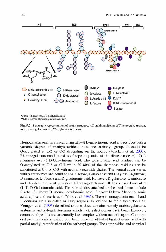
Homogalacturonan is a linear chain α(1–4) D-galacturonic acid and residues with avariable degree of methylesterification at the carboxyl group. It could be
O-acetylated at C-2 or C-3 depending on the source (Vincken et al. 2003).
Rhamnogalacturonan-I consists of repeating units of the disaccharide α(1–2) L
rhamnose α(1–4) D-Galacturonic acid. The galacturonic acid residues can be
O-acetylated at C-2 or C-3 while 20–80% of the rhamnose residues can be
substituted at C-4 or C-3 with neutral sugar side chains. The neutral sugar varies
with plant sources and could be D-Galactose, L-arabinose and D-xylose, D-glucose,
D-mannose, L- fucose and D-glucturonic acid. However, D-galactose, L-arabinose
and D-xylose are most prevalent. Rhamnogalacteronan-II has a back bone of α(1–4) D-Galacturonic acid. The side chains attached to the back bone include
2-keto- 3- deoxy-D mono- octulosonic acid, 3-deoxy-D-lyxo-2-heptulo sonic
acid, apiose and aceric acid (York et al. 1985). These rhamnogalacturonan-I and
II domains are also called as hairy regions. In addition to these three domains.
Voragen et al. (1995) described another three domains namely arabinogalactans,
arabinans and xylogalacturonans which lack galacturonan back bone. However,
commercial pectins are structurally less complex without neutral sugars. Commer-
cial pectins consists mainly of a back bone of α-(1–4)–D-galacturonic acid with
partial methyl esterification of the carboxyl groups. The composition and chemical
Fig. 9.2 Schematic representation of pectin structure. AG arabinogalactan, HG homogalacturonan,
RG rhamnogalacturonan, XG xylogalacturonan)
160 P.B. Gundala and P. Chinthala

structure of the elements of pectin varies with environmental conditions, plant
source and plant development stage etc.
The American chemical society classified pectic substances into four main types
based on the type of modification of back bone chain (Be Miller 1965): (a) Proto
pectin, (b) Pectic acids, (c) Pectinic acids and (d) Pectins.
(a) Proto pectins: Water insoluble pectic substance present plant tissues. Upon
restricted hydrolysis protopectin yields pectin or pectic acids
Protopectinþ H2O !PPase Pectin
Insolubleð Þ Solubleð Þ(b) Pectic acids: These are galacturonans that contains negligible amounts of
methoxyl groups. Normal or acid salts of pectic acids are called pectales.
(c) Pectinic acid: These are the galacturonans that contains methoxy groups
(75%). Pectinotes are normal or acid salts of pectinic acids. Normal or acid
salts of pectinic acids are reffered to as pectinates.
(d) Pectin (Polymethyl galactenate): It is a polymeric material in which 75% of
the carboxyl groups of the galacturonate units are esterified with methanol. It is
located in the cell wall and gives rigidity to the cell wall.
The polygalacturonic acid chain is partly esterified with methyl groups and the
free acid groups may be partly or fully neutralised with sodium, potassium or
ammonium ions. The ratio of esterified GalA groups to total GalA groups is termed
as the DE. Based on the DE pectins are subdivided into two groups (a) High ester
pectins (HM pectins) (>50 DE) and Low esterified pectins (LM) (<50 DE). These
two groups of pectin gel by different mechanisms. To form gels HM-pectin requires
a minimum amount of soluble solids and a pH within a narrow range, around 3.0.
HM-pectin gels are thermally reversible. In general, HM-pectins are hot water
soluble and often contain a dispersion agent such as dextrose to prevent lumping.
LM-pectins produce gels independent of sugar content. They are also are not as
sensitive to pH as the HM-pectins are. LM-pectins require the presence of a
controlled amount of calcium or other divalent cations for gelation.
9.4.3 Degradation of Pectins
Pectins are decomposed instinctively by deesterification as well as by
depolymerisation, the rate of this decomposition depends on pH, water activity,
9 Extremophilic Pectinases 161

Fig. 9.3 Action sites of pectinase and pectinesterase on different pectin subunits
162 P.B. Gundala and P. Chinthala
and temperature. Pectin depolymerization can be obtained by acid hydrolysis,
hydrothermal processing, dynamic high pressure microfluidization (Chen et al.
2013) or photo chemical reaction in media containing TiO2 and partial enzymatic
hydrolysis (Concha and Zuniga 2012; Mandalari et al. 2006). However, enzymatic
degradation has advantage over chemical hydrolysis and other physical methods.
Enzymatic hydrolysis is target specific and produce specific useful oligosaccharides
fragments (Fig 9.3). The enzymatic breakdown of pectins involves mainly two
reactions i.e., hydrolysis (catalyzed by hydrolases) or β-elimination (catalyzed by
lyases). High methoxyl pectins (HMP) or low methoxyl pectins (LMP) can be
breakdown by endo polygalacturonases and yields 95% POS (Pectic oligosaccha-
rides). A mixture of enzymes such as pectin—methyl esterases, endo
polygalacturonases, endo arabinases and endo galactoses can also be employed to
depolymerizing the pectins.
9.4.4 Industrial Importance of Pectin Degradation
Pectin depolymerization results in pectic oligosaccharides (POS). POS are a new
class of prebiotics capable of extending a number of health promoting effects.
Protection to cardiovascular system, colonic cells, stimulates apoptosis in human
colonic adeno carcinoma, reduction of cell damage by heavy metals, anti obesity
effects, anti infection, antitoxic, antibacterial and antioxidant properties. Further,
they are most suitable for use in baby foods as they are not cytotoxic or mutagenic.
Pectic polymer derivatives and oligosaccharides derived from pectins have positive
effects on human health. These included immune regularly effects in the intestine,
lowering the blood cholesterol level and slowing down and absorption of glucose in
the serum of diabetic patients. Moreover, pectin oligosaccharides proved to be good
probiotic components (Table 9.3).
Table 9.3 Applications of pectic oligosaccharides and their source
Source Importance
Citrus pectin Stimulates apoptosis of human colonic adenocarcinoma cells
Carrots Ability to block bacterial adherence of E. Coli to uroepithelial cells
Haw pectin Reduces total cholesterol and triglycerides
Bergamot peal Helps to improve probiotic bacteria and decrease pathogenic
populations
Apple pectins Decreases pathogenic bacteria
Sugar beet pectin Different effects of DP4 and DP5 oligos and shows differential
structure properties
Orange peal Increases butyrate concentration along with increase in
gut-bacteria
Sugar beet and Valencia
oranges
Increased cancellation of acetate, Butyrate, propionate in the media
9 Extremophilic Pectinases 163

To produce pectin oligomers pectin partial degradation or hydrolysis is neces-
sary. Enzymatic degradation has several advantages over chemical hydrolysis
since, enzymes acts on specific target and produce specific useful oligosaccharide
fragments .To meet the demand of specific pectin derived oligosaccharides spe-
cialized mixture of Pectinolytic enzymes are required.
9.5 Pectic Enzymes/Pectin Degrading Enzymes
Enzymes are important biocatalyst for various industrial and biotechnological
purposes and can work in many adverse condition compared to chemical catalysts.
Microorganisms are preferred as a source of enzymes because of their short life
span, high productivity rate, cost effective, and also free of harmful chemicals when
compared to enzymes that are found in plant and animal source. Fifty percent of
available enzymes are originated from fungi and yeast; 35% from bacteria, while
the remaining 15% are either of plant or animal origin. The enzymes hydrolyzing
the pectic substances are broadly known as pectic enzymes or
pectinases. Pectinolytic enzymes or pectinases are a heterogeneous group of related
enzymes that hydrolyze the pectic substances. So far, filamentous microorganisms
are most widely used for pectinase production.
9.5.1 Nomenclature and Classification
Pectinases are a heterogeneous group of related enzymes therefore the classification
of Pectinases is based on their preferred substrate (pectin, pectic acid or oligo
D-galacturonate), the degradation mechanism (transelimination or hydrolysis) and
the type of cleavage (random [endo] or terminal [exo]) (Kashyap et al. 2001).The
three major groups are outlined in Fig 9.4.
Group-I-Protopectinases Protopectinases degrade the insoluble protopectin and
give rise to highly polymerized soluble pectin. Pectinosinase is also synonymous
with protopectinase (PPase). PPases are classified into two types, on the basis of
their reaction mechanism (Sakamoto et al. 1994). A-type PPases react to the inner
site, i.e. the polygalacturonic acid region of protopectin, whereas B-type PPases
react to the outer site, i.e. on the polysaccharide chains that may connect the
polygalacturonic acid chain and cell wall constituents. All three A-type PPases
are similar in biological properties and have a similar molecular weight of 30 kDa.
PPases-F is an acidic protein whereas PPase-L and -S are basic proteins. The
enzymes have pectin releasing effects on protopectin from various origins. PPase-
B, -C and -T have molecular weights of 45, 30, and 55 kDa respectively. PPase-B
and -C have an isoelectric point (pI) of around 9.0 whereas PPase-T has a pI of 8.1
164 P.B. Gundala and P. Chinthala

(Sakai, 1992). PPase-B, -C and -T act on protopectin from various citrus fruit peels
and other plant tissues releasing pectin.
Group-II-Depolymerases Depolymerases catalyze the hydrolytic cleavage of
α-(1–4) glycosidic bonds in the D-galacturonic acid moieties of the pectic
substances. They exert their activity by hydrolyzing the glycosidilic linkages or
by Cleavage. Depolymerases are submerged into hydrolases and cleavages
Sub group-I-Hydrolases The hydrolases include Poly methyl galacturonases
(PMG) and Poly galacturonases (PG) and further classified into Exo and Endo-
PMG and Exo PG or endo PG. Even though reports in literature on
polymethylgalacturonases the existence of this enzyme is still in question (Sakai
et al. 1993). Often, Polymethylgalacturonase preparations contaminated with PE
can be mistaken for PMG containing preparations.
Polygalacturonases (PG) are the pectinolytic enzymes that catalyze the hydro-
lytic cleavage of the polygalacturonic acid chain with the introduction of water
across the oxygen bridge. They are the most extensively studied among the family
of pectinolytic enzymes. The PG involved in the hydrolysis of pectic substances are
endo PG (E.C. 3.2.1.15) and exo PG (E.C. 3.2.1.67). Exo PG can be distinguished
into two types: fungal exo PG which produce monogalacturonic acid as the main
end product and the bacterial exo PG which produce digalacturonic acid as the main
end product. Endo PG occurs in many organisms where as exo PG occurs less
frequently.
Sub group-II-Cleavages Lyases (cleavages or transeliminases) perform non
hydrolytic breakdown of pectates or pectinates, characterized by a trans eliminative
split of the pectic polymer (Sakai et al. 1993). The lyases break the glycosidic
linkages at C-4 and simultaneously eliminate hydrogen from C-5, producing a D
4:5 unsaturated products (Codner 2001) (Fig. 9.5). Lyases can be classified into the
Classification of Pectic Enzyme
Protopectinases Depolymerases Pectin Esterase
A type B Type
Hydrolysases Cleavages
Polymethyl galacturonases (PMG) Polygalactueronase (PG) Polymethylgalactueronate Polygalacturonate
Endo-PMG Exo PMG Endo PG Exo PG Endo PMGL Exo PMGL Exo PGLEnd PGL
Fig. 9.4 Outlines of pectic enzyme classification
9 Extremophilic Pectinases 165
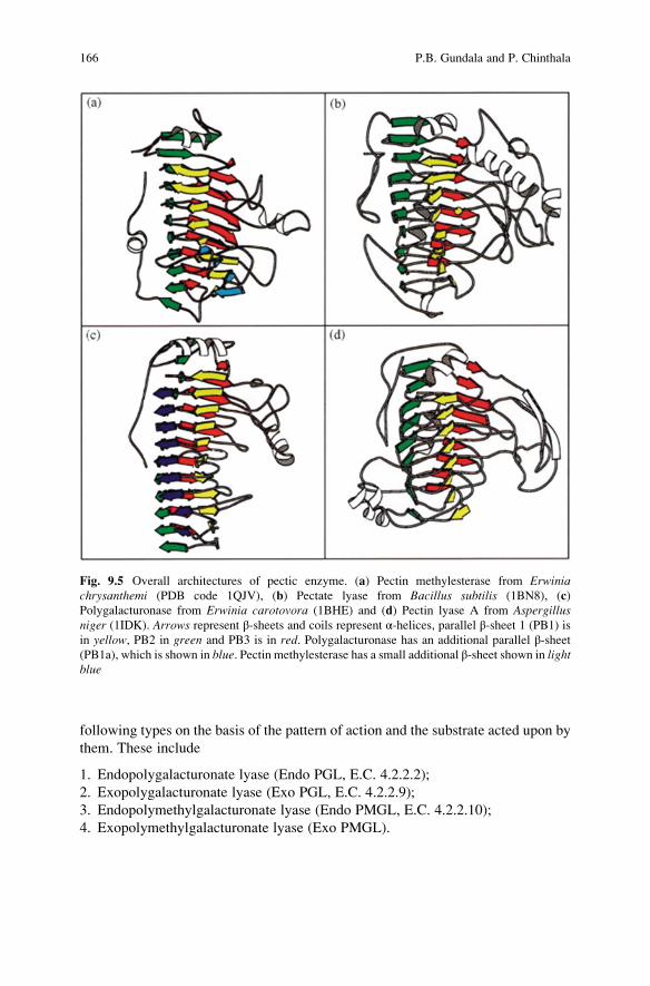
following types on the basis of the pattern of action and the substrate acted upon by
them. These include
1. Endopolygalacturonate lyase (Endo PGL, E.C. 4.2.2.2);
2. Exopolygalacturonate lyase (Exo PGL, E.C. 4.2.2.9);
3. Endopolymethylgalacturonate lyase (Endo PMGL, E.C. 4.2.2.10);
4. Exopolymethylgalacturonate lyase (Exo PMGL).
Fig. 9.5 Overall architectures of pectic enzyme. (a) Pectin methylesterase from Erwiniachrysanthemi (PDB code 1QJV), (b) Pectate lyase from Bacillus subtilis (1BN8), (c)
Polygalacturonase from Erwinia carotovora (1BHE) and (d) Pectin lyase A from Aspergillusniger (1IDK). Arrows represent β-sheets and coils represent α-helices, parallel β-sheet 1 (PB1) is
in yellow, PB2 in green and PB3 is in red. Polygalacturonase has an additional parallel β-sheet(PB1a), which is shown in blue. Pectin methylesterase has a small additional β-sheet shown in lightblue
166 P.B. Gundala and P. Chinthala

Group-III-Esterases Esterases catalyze the de-esterification of pectin by the
removal of methoxy esters. Pectinesterases (PE) (E.C. 3.1.1.11) often referred as
Pectin methylesterases, pectlylhydrolase, pectase, pectin methoxylase, pectin
demethoxylase and pectolipase is a carboxylic acid esterase and belongs to the
hydrolase group of enzymes (Whitaker 1984).
Recently, Schols et al. (1995) described a new group of Rhamnogalacturonan
degrading enzymes. Until now four enzymes with a specific activity towards the RG
I part of the pectin molecule have been reported. These are
(a) Rhamnogalacturonan hydrolase (RG hydrolase)
(b) Rhamnogalacturonan lyase (RG pectin lyase)
(c) Rhamnogalacturonan rhamnohydrolase (RG rhamnohydrolase)
(d) Rhamnogalacturonan galacturonohydrolase (RG galacturonohydrolase).
All enzymes are active towards the subunit of RG I, which contains strictly
alternating galacturonic acid (GalA)—rhamnose (Rha) residues (Fig. 9.6).
Fig. 9.6 A CPK space-filling model of RGase A, with the C-terminal tail shown in blue and the
rest of the molecule shown in purple. All the O-linked glycosylation sites are located at the
C-terminal tail. Mannose carbon atoms are shown in yellow, while the few N-acetylglucosamine
carbons that can be seen are in grey. Carbohydrate oxygens are in red. The α(1–4)-linkedmannoses are linked to threonine residues: 367, 370, 371, 372, 374, 376, 385, 386, 398, 405 and
408, and serine residues 368, 373, 380, 383, 400, 409 and 418. The N-linked glycosylation is found
at Asn32 with the glycan tree: Manα1–6(Manα1–3) Manβ1–4GlcNAcβ1–4GlcNAc and at
Asn299: Manβ1–4GlcNAcβ1–4GlcNAc
9 Extremophilic Pectinases 167

RG-hydrolase and RG-lyase are endo-acting enzymes, the first one splits the GalA-
Rha linkages while the second one cleaves Rha-GalA linkages leaving Δ4,5unsaturated GalA at the non reducing end. RG-rhamnohydrolase and
RG-galacturonohydrolase are exo-acting enzymes which release Rha or saturated
GalA from the non reducing end of RG I respectively. RG-galacturonohydrolase in
not able to split the unsaturated Gal A residue from the non reducing end (Mutter
et al. 1998).
9.5.2 Pectinase Producing Organisms
Proto Pectinase Producers A-type PPases are produced by yeast and yeast-like
fungi however, B-type PPases have been reported in B.subtilis IFO 12113,
B. subtilis IFO 3134 (Sakai and Sakamoto, 1990) and Trametes sp. (Sakai et al.1993). B-type PPases have also been produced by a wide range of Bacillus sp.
Depolymerases Producers Endo PG are generally distributed among fungi, bac-
teria and many types of yeast (Luh and Phaff 1951). They are also found in higher
plants and some plant parasitic nematodes. They have been reported in many
microorganisms, including Aureobasidium pullulans, Rhizoctonia solani Kuhn ,
Rhizopus stolonifer, Fusarium moniliforme, Neurospora crassa, Thermomyceslanuginosus, Aspergillus sp. Mucor flavus ) and Mucor circinelloides. Endo PG
have also been cloned and genetically studied in a large number of microbial
species. In contrast, exo PG occur less frequently. They have been reported in
Bacteroides thetaiotamicron, Erwinia carotovora, Alternaria mali, Fusariumoxysporum , Ralstonia solanacearum Agrobacterium tumefaciens and Bacillus sp.
Polygalacturonate lyases (Pectate lyases or PGLs) are produced by many bacte-
ria and some pathogenic fungi with endo PGLs being more abundant than exo
PGLs. PGLs have been isolated from bacteria and fungi associated with food
spoilage and soft rot. They have been reported in Colletotrichum lindemuthionum,Bacteroides thetaiotaomicron and Erwinia carotovora. Very few reports on the
production of polymethylgalacturonate lyases (pectin lyases or PMGLs) have been
reported in literature. They have been reported to be produced from Aspergillusjaponicus, Pythium splendens, Pichia pinus, Aspergillus sp. and Thermoascusauratniacus.
Pectin Esterase Producers PE is found in plants, plant pathogenic bacteria and
fungi (Hasunuma et al. 2003). It has been reported in Rhodotorula sp. Phytophthorainfestans, Saccharomyces cerevisiae, Lachnospira pectinoschiza, Aspergillusniger, Lactobacillus lactis sp. cremoris, Penicillium frequentans, Aspergillusjaponicus and others. Rhamnogalacturonan degrading enzymes were reported
from A. aculeatus, A. niger, A. aculeatus (Kofod et al. 1994).
168 P.B. Gundala and P. Chinthala

9.5.3 Assay of Pectinases In Vitro
Protopectinase Assay. PPase activity can be assayed by measuring the amount of
pectic substance liberated from protopectin by the carbazole sulphuric acid method
or modified method. The pectin concentration is measured as D-galacturonic acid
from its standard curve. One unit of PPase activity is defined as the enzyme that
liberates pectic substance corresponding to 1 μmol of D-galacturonic acid per ml of
reaction mixture under the assay conditions.
Depolymerase Assay. PG activity is determined on the basis of measuring, during
the course of the reaction: (a) the rate of increase in number of reducing groups and
(b) the decrease in viscosity of the substrate solution (Rexova-Benkova and
Markovic 1976). The amount of reducing sugar can be readily measured by
colorimetric methods like 3,5-dinitrosalicylate reagent method (Miller 1959) and
the arsenomolybdate-copper reagent method (Collmer et al. 1988). One unit of
enzyme activity is defined as the enzyme that releases 1 μmol/ml/min galacturonic
acid under standard assay conditions. Viscosity reduction measurements have also
found widespread use in determining the PG activity. The unit of enzyme activity is
mostly selected as the amount of enzyme required for attaining a certain decrease of
viscosity per unit time. However, this method has met with limited success. There is
no direct correlation between viscosity reduction and the number of glycosidic
bonds hydrolyzed. PG activity can also be determined by the cup-plate method.
Cups are cut out from the solidified agar containing the substrate and are filled with
the enzyme solution. After the lapse of a certain period of time, the zones of
degraded substrate stained with iodine and quantified. PG isolated from different
microbial sources differs markedly from each other with respect to their physico-
chemical and biological properties and their mode of action. Calcium ions influence
the activity of PG. However, in some cases the activity was inhibited. PG can be
produced constitutively and in some cases they are inducible. Induction or stimu-
lation is caused by the low concentration of pectins or oligo and monomeric
fragments. Some PG are sensitive to catabolic repression, others inhibit in vivomostly by a protein present in the host. Sometimes tannins or phenolic compounds
present in the host tissue also inhibit PG activity. Among the PG obtained from
different microbial sources, most have the optimal pH range of 3.5–5.5 and optimal
temperature range of 30–50 �C. The molecular weight of these enzymes falls in the
range of 30–60 kDa. Aspergillus and Botritis sp. produce endo PG of 85 kDa and
69 kDa respectively. Two endo PG (PG I and PG II), isolated from A. niger have anoptimal pH range of 3.8–4.3 and 3.0–4.6 respectively (Singh and Rao 2002).
PE Assay PE activity is most readily followed by gel diffusion assay as described
by Downie et al. (1998). Increased binding of ruthenium red to pectin, as the
number of methyl esters attached to the pectin decreases, is used as the basis of
the assay. The unit of activity in nano or picokatals is calculated based on the
standard curve generated from the log-transformed commercial enzyme activity
versus stained zone diameter. The sensitivity, specificity and simplicity of this PE
9 Extremophilic Pectinases 169

assay are superior to all others. PE activity can also be measured by using a pH stat
because ionization of the carboxyl group of the product releases a proton which
causes a change in pH.
9.5.4 Applications of Pectic Enzymes
Over the years pectinases have been used in several conventional industrial pro-
cesses such as textile, plant fiber processing, tea, coffee, oil extraction and treat-
ment of industrial waste water containing pectinacious material. They have also
been reported to work on the purification of viruses and in making of paper.
Fruit Juice Extraction The chief industrial application of pectinases is in fruit
juice extraction and clarification. Pectin contributes to the fruit juice turbidity and
viscosity. A mixture of pectinases and amylases are used to clarify fruit juices.
Treatment of fruit pulps with pectinases also showed an increase in fruit juice
volume from banana, grapes and apples. Pectinases in combination with other
enzymes viz., cellulases, arabinases and xylanases have been used to increase the
pressing efficiency of the fruits for juice extraction. Vacuum infusion of pectinases
has a commercial application to soften the peel of citrus fruits, to replace hand
cutting for the production of canned segments and to pickle processing where to
avoid excessive softening during fermentation and storage.
Textile Processing and Bioscouring of Cotton Fibers Pectinases have been used
to remove sizing agents from cotton in a safe and eco-friendly manner along with
amylases, lipases, cellulases and hemicellulases with specific enzymes. Pectinases
have been used in the Bioscouring is a novel process for removal of noncellulosic
impurities from the fiber.
Degumming of Plant Bast Fibers Pectinases are used in degumming of textile
fibres in combination with xylanases which is an ecofriendly and economical
alternative to the chemical degumming.
Retting of Plant Fibers Pectinases have been used in retting of flax to separate the
fibers and eliminate pectin.
Waste Water Treatment Food processing industries release waste water that
contains pectin as by-product. Pretreatment of these waste waters with pectinolytic
enzymes removes pectinaceous material and turn into suitable for decomposition by
activated sludge treatment.
Tea and Coffee Fermentation Treatment with Pectinases accelerates tea fermen-
tation and also destroys the foam forming property. They are also used in coffee
fermentation to remove mucilaginous coat from coffee beans.
Paper and Pulp Industry During papermaking pectinase can be employed to
depolymerise pectin and then to lower the peroxide bleaching.
170 P.B. Gundala and P. Chinthala

Animal Feed Pectinases are used in the enzyme cocktail. Pectinases are used in
the production of animal feeds to reduce the feed viscosity thereby to increase the
absorption of nutrients.
Purification of Plant Viruses When the virions are restricted to phloem, to purify
the virion particles alkaline pectinases and cellulases can be used.
Oil Extraction Citrus oils such as lemon oil can be extracted with pectinases.
They destroy the emulsifying properties of pectin which interferes with the collec-
tion of oils from citrus peel extracts.
Improvement of Chromaticity and Stability of Red Wines To improve the
visual characteristics red wine Pectinolytic enzymes are added to during
processing. Enzymatically treated red wines also showed greater stability.
9.6 Extremophilic Pectinases
The stable increase in the number of newly isolated extremophilic microorganisms
and the discovery of their enzymes by academic and industrial institutions under-
lines the enormous potential of extremophiles for application in future biotechno-
logical processes. Enzymes from extremophilic microorganisms offer versatile
tools for sustainable developments in a variety of industrial application because
they show important environmental benefits due to their biodegradability, specific
stability under extreme conditions, improved use of raw materials and decreased
amount of waste products. Even though key advances have been made in the last
decade, our knowledge on physiology, metabolism, enzymology and genetics of
extremophilic microorganisms and their related enzymes is still limited. New
techniques, such as genomics, metanogenomics, DNA evolution and gene shuf-
fling, will lead to the production of enzymes that are highly specific for unbounded
industrial applications. Due to the unusual properties of extremozymes from
extremophiles, there is urgent need to optimize the already existing processes or
to develop new sustainable technologies.
The notion that extremophiles are capable of surviving under non-standard
conditions in non-conventional environments has led to the assumption that the
properties of their enzymes have been optimized for these conditions. Depending
upon the pH requirement for optimum enzymatic activity, pectinase enzyme is
classified into acidic and alkaline pectinase. Acidic pectinases are useful in extrac-
tion, clarification and liquefaction of fruit juices (Kaur et al. 2004) and wines.
Whereas, alkaline pectinases are widely used in the fabric industry, pulp and paper
industry and in improving the quality of black tea.
9 Extremophilic Pectinases 171

9.6.1 Acidic Pectinases
Acidophiles are the organisms that thrive in acidic environments with pH less than
4.0.To survive under extreme pH they develop specific metabolic properties,
genetic features and structural and functional characteristics of their macromole-
cules that are helpful in maintaining pH and distinguishing them from neutrophilic
counter parts. The acidic pectinases are stable at low pH and have extensive
applications in the extraction and clarification of both sparkling clear juices
(apple, pear, grapes and wine) and cloudy (lemon, orange, pineapple and mango)
juices and maceration of plant tissues. Additionally, acidic pectinases are useful in
the isolation of protoplasts and saccharification of biomass. Most commonly these
are isolated from fungal sources, especially from Aspergillus niger because fungi
are potent producers of pectic enzymes and the optimal pH of fungal enzymes is
very close to the pH of many fruit juices, which range from pH 3.0 to 5.5 (Table 9.4)
9.6.2 Alkaline Pectinases
Alkaline pectinases have been used in many industrial and biotechnological pro-
cesses, such as textile and plant fiber processing, coffee and tea fermentation, oil
extraction, treatment of industrial wastewater containing pectinacious material,
purification of plant viruses and paper making. Especially in textile, cotton
bioscouring with the alkaline pectinases would not affect the cellulose backbone
and thus avoid fiber damage without pollution to environment in contrast to drastic
alkaline conditions conventionally used (Klug-Santner et al. 2006). Now, alkaline
pectinases have proved to be the most effective and suitable enzymes for cotton
bioscouring (Wang et al. 2007). Up to now, some microorganisms have been
studied for producing alkaline pectinases. The alkaline pectinases are produced
predominantly from the genus Bacillus sp. and Pseudomonas sp. Although a few
alkaline pectinases from various microbes have also been purified and characterized
by Kobayashi et al. (2001), there is still a demand for the alkaline pectinases with
Table 9.4 Acidic pectinases and their properties
Type of pectinase Producer Optimum pH Properties
PG Saccharomyces pastorionus 4.2 Mol. wt. 43 kDa
pI: 5.4
Km. 0.62 mg/ml
Endo PG Saccharomyces pastorianus 4.2 Mol. 143 kDa
Km 0.59 μg/μlEndo PG Cryptococcus albidus ver albitus 3.75 Mol. wt. 41 kDa
Km. 0.57 μg/μlpI. 8.1
172 P.B. Gundala and P. Chinthala
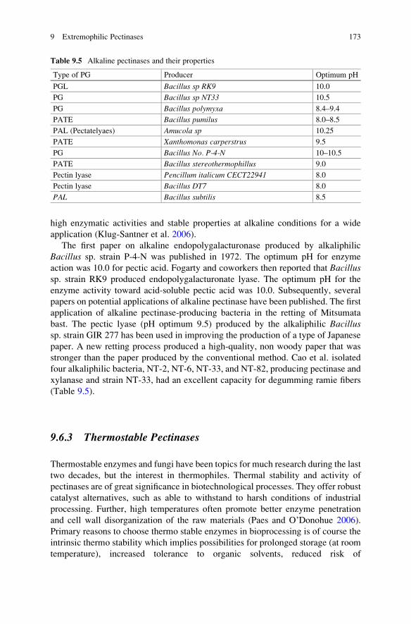
high enzymatic activities and stable properties at alkaline conditions for a wide
application (Klug-Santner et al. 2006).
The first paper on alkaline endopolygalacturonase produced by alkaliphilic
Bacillus sp. strain P-4-N was published in 1972. The optimum pH for enzyme
action was 10.0 for pectic acid. Fogarty and coworkers then reported that Bacillussp. strain RK9 produced endopolygalacturonate lyase. The optimum pH for the
enzyme activity toward acid-soluble pectic acid was 10.0. Subsequently, several
papers on potential applications of alkaline pectinase have been published. The first
application of alkaline pectinase-producing bacteria in the retting of Mitsumata
bast. The pectic lyase (pH optimum 9.5) produced by the alkaliphilic Bacillussp. strain GIR 277 has been used in improving the production of a type of Japanese
paper. A new retting process produced a high-quality, non woody paper that was
stronger than the paper produced by the conventional method. Cao et al. isolated
four alkaliphilic bacteria, NT-2, NT-6, NT-33, and NT-82, producing pectinase and
xylanase and strain NT-33, had an excellent capacity for degumming ramie fibers
(Table 9.5).
9.6.3 Thermostable Pectinases
Thermostable enzymes and fungi have been topics for much research during the last
two decades, but the interest in thermophiles. Thermal stability and activity of
pectinases are of great significance in biotechnological processes. They offer robust
catalyst alternatives, such as able to withstand to harsh conditions of industrial
processing. Further, high temperatures often promote better enzyme penetration
and cell wall disorganization of the raw materials (Paes and O’Donohue 2006).
Primary reasons to choose thermo stable enzymes in bioprocessing is of course the
intrinsic thermo stability which implies possibilities for prolonged storage (at room
temperature), increased tolerance to organic solvents, reduced risk of
Table 9.5 Alkaline pectinases and their properties
Type of PG Producer Optimum pH
PGL Bacillus sp RK9 10.0
PG Bacillus sp NT33 10.5
PG Bacillus polymyxa 8.4–9.4
PATE Bacillus pumilus 8.0–8.5
PAL (Pectatelyaes) Amucola sp 10.25
PATE Xanthomonas carperstrus 9.5
PG Bacillus No. P-4-N 10–10.5
PATE Bacillus stereothermophillus 9.0
Pectin lyase Pencillum italicum CECT22941 8.0
Pectin lyase Bacillus DT7 8.0
PAL Bacillus subtilis 8.5
9 Extremophilic Pectinases 173
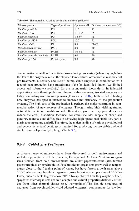
contamination as well as low activity losses during processing (when staying below
the Tm of the enzyme) even at the elevated temperatures often used in raw material
pre treatments. Discovery and use of thermo stable enzymes in combination with
recombinant production have erased some of the first identified hinders (e.g. limited
access and substrate specificity) for use in industrial biocatalysis. In industrial
applications with thermophiles and thermo stable enzymes, isolated enzymes are
today dominating over microorganisms (Turner et al. 2007). In these fields, finding
new enzymes has special interest to improve the efficiency of the production
systems. The high cost of the production is perhaps the major constraint in com-
mercialization of new sources of enzymes. Though, using high yielding strains,
optimal fermentation conditions and efficient enzyme recovery procedures can
reduce the cost. In addition, technical constraint includes supply of cheap and
pure raw materials and difficulties in achieving high operational stabilities, partic-
ularly to temperature and pH. Therefore, the understanding of various physiological
and genetic aspects of pectinase is required for producing thermo stable and acid
stable strains of pectinolytic fungi. (Table 9.6).
9.6.4 Cold-Active Pectinases
A diverse range of microbes have been discovered in cold environments and
include representatives of the Bacteria, Eucarya and Archaea .Most microorgan-
isms isolated from cold environments are either psychrotolerant (also termed
psychrotrophic) or psychrophilic. Psychrotolerant organisms grow well at temper-
atures close to the freezing point of water, but have fastest growth rates above
20 �C, whereas psychrophilic organisms grow fastest at a temperature of 15 �C or
lower, but are unable to grow above 20 �C. Irrespective of how they may be defined,
‘psychro’ microorganisms are cold-adapted and exhibit properties distinctly differ-
ent from other thermal classes (e.g. thermophiles).The flexible structures of
enzymes from psychrophiles (cold-adapted enzymes) compensates for the low
Table 9.6 Thermostable, Alkaline pectinases and their producers
Microorganisms Type of pectinases Optimum pH Optimum temperature (�C)Bacillu sp. NT-33 PG 10.5 75
Bacillus P-4-N PG 10–10.5 65
Bacillus polymyxa PG 8.4–9.4 45
Bacillus sp. PK-9 PGL 10.0 75
Bacillus subtilis PAL 8.5 60–65
Pseudomonas syringe PAL 8.0 40
Bacillus pumulus PATE 8.0–8.5 45
Bacillus stearothermophiles PATE 9.0 70
Bacillus sp DT-7 Pectate lyase 9.0 70
174 P.B. Gundala and P. Chinthala

kinetic energy present in cold environments. Because of their inherent flexible
structure, cold-adapted enzymes show a reduction in activation enthalpy (ΔH#)and a more negative activation entropy (ΔS#) compared with mesophilic and
thermophilic homologues. As a consequence, when temperature is decreased the
reaction rate of enzymes from psychrophiles tends to decrease more slowly com-
pared with equivalent enzymes from thermophiles. This balance of thermodynamic
activation parameters is translated into relatively high catalytic activity (kcat) at
low temperatures and a concomitant low structural stability compared with
enzymes from mesophiles or thermophiles. The gain in enzymatic activity would
be enormous if the reduction in ΔH# was not accompanied by a concomitant
decrease in ΔS#. For example, a decrease in ΔH# of 20 kJ/mol would result in
�50,000-fold increase in kcat at 15 �C at constant ΔS#. However, in practice such avast increase in activity is not observed as a result of enthalpy-entropy compensa-
tion http://onlinelibrary.wiley.com/doi/10.1111/j.1751-7915.2011. 00258.x/full-
b62. This is reflected in the activity-stability-flexibility characteristics of many
thermally adapted enzymes. The compositional and structural features that confer
high flexibility to thermolabile cold-adapted enzymes are generally opposite to that
of more rigid and stable mesophilic and thermophilic homologues. For example,
psychrophilic enzymes tend to possess various combinations of the following
features: decreased core hydrophobicity, increased surface hydrophobicity, lower
arginine/lysine ratio, weaker inter-domain and inter-subunit interactions, more and
longer loops, decreased secondary structure content, more glycine residues, less
proline residues in loops, more proline residues in α-helices, less and weaker metal-
binding sites, a reduced number of disulfide bridges, fewer electrostatic interactions
(H-bonds, salt-bridges, cation–pi interactions, aromatic–aromatic interactions),
reduced oligomerization and an increase in conformational entropy of the unfolded
state. Genomic comparisons of psychrophiles vs. thermophiles have also revealed
that distinct biases in amino acid composition is a trademark of thermal adaptation.
In certain enzymes such as a zinc metalloprotease from an Arctic sea ice bacterium,
the whole structure of the enzyme appears to be uniformly flexible (global flexibil-
ity) as a result of an overall decrease in H-bonding. However, in other enzymes
flexibility has been shown to be localized in the structures surrounding or compris-
ing the active site. Recently, the Psychrophilic and Pectinolytic Yeasts (PPY),
which are able to degrade pectin compounds at low temperature were isolated, in
order to develop the cold-active pectinases applied to food industries. The isolated
PPY strains were identified to Cystofilobasidium capitatum, C. larimarini, Crypto-coccus cylindricus, C. macerans, C. aquaticus and Mrakia frigida and the extra-
cellular fraction of these strains exhibited pectin methylesterase (PME), pectin
lyase (PNL) and polygalacturonase (PG) activities at 5 �C. Further, cold-activepectinases can help to reduce viscosity and clarify fruit juices at low temperatures.
9 Extremophilic Pectinases 175

9.7 Commercial Production/Status of Extremophilic
Pectinases
Cost effective bulk production is required to commercialize any enzyme for
industrial applications. Some researchers reported that Pectinases produced by
solid state fermentation (SSF) should more stable higher stability for pH and
temperature variations and were less affected by catabolic repression than
Pectinases produced by SmF (Submerged fermentation). The Pectinases produced
through SSF not only reduced the production costs but also the method is less
polluting. So far different agricultural and agro industrial residues are used as
substrates such as wheat bran, soybean, Cranberry and straw berry pomace, Coffee
pulp and Coffee, Husk, Cocoa, Orange bagasse, sugarcane bagasse and wheat bran,
apple pomace and from sugarbeet pulp. Due to their multifarious applications
several companies are involved in commercial production of Microbial pectinases
(Table 9.7).
Due to the wide range of applications of pectinases in the food industry the
industrial production of pectinases has drawn world-wide attention. Molds are
primarily used for the production of pectinases on a commercial scale. Suitable
organisms include strains of A. niger, A. wnetii, A. oryzae and Rhizopus sp.Research for additional pectinase producers is hampered by the fact that only a
limited number of microorganisms are approved for application in the food indus-
try. Commercial production may be enhanced by a selection of more productive
mutants which are not subject to catabolic repression or synthesize large quantities
of enzyme without the necessity of an inducer. Although highly productive bacte-
rial strains producing polygalacturonate lyase are known, still pectinases are not
produced commercially from them. There are three different industrial methods
used to produce microbial enzymes; the surface-bran culture (Koji) method, the
deep-tank (submerged) process and the two-stage submerged process. According to
Rombouts and Pilnik (1978) most pectinases are still produced by the surface
method carried out in rotating drums. Although in general the submerged process
is more widely used because of its easier control. A crucial factor in pectinase
production is the composition of the medium. Details about such media are
Table 9.7 List of commercial suppliers of the pectinase enzymea
Product Supplier
Pan enzyme C.H. Boehringer sohn; west Germany
Ultrazyme Ciba-Geigy. A.G. Switzerland
Pectolase Grinsteelvaekatt, Denmark
Solase Kikkoman shoyuco, Japan
Pectinex Schweizerische ferment, A.G. Switzerland
Rapidase: clarizyme Society Rapidase, S.A: France
Klerzyme Wallerstein, Co; USA
Pectinol: Rohament Rohm, Gmb H, West GermanyaAdapted from Kashyap et al. (2001)
176 P.B. Gundala and P. Chinthala

considered to be strictly confidential and are not released by the manufacturers. In
general the medium will be a mixture of carbohydrates (glucose, molasses, and
starch hydrolysates), N sources (NH4þ salts and yeast extract) and minerals. If the
enzyme is not produced constitutively an inducer also has to be added. For reasons
of economy pectin is not used much in production media but it is substituted by
dried sugar cossettes, citrus peel or apple pomace. Control of pH is also very
important. The highest enzyme production is achieved when the pH value drops
from an initial value of about 4.5 to a more or less constant value of 3.5 during the
course of the fermentation which usually takes 3–6 days. At extreme pH (7) a
marked inactivation occurs. At the end of the fermentation the enzymes are
extracted from the semisolid medium and mycelium. The dilute enzyme solution
is concentrated and the enzymes are then precipitated with organic solvents or
inorganic salts. Following precipitation the enzyme cake is centrifuged or filtered
and then dried at low temperatures or spray dried (Sakai et al. 1993). Subsequently,
it is ground to a particular particle size and used to prepare commercial enzyme
formulations. Some preparations are sold as liquid concentrates. Pectinases are
produced by a number of companies in Europe (Novo- Nordisk, Miles Kali-Chemie
and Swiss Ferment Co.),the United States (Miles laboratories and Rohm and Haas
Co.) and in Japan (Kikkoman Shoyu Co.) (Table 9.7).
9.8 Future Prospects of Extremophilic Pectinases
Owing to the importance of extremophilic pectinases there is an urgent need to
develop pectinases with desirable physic chemical characteristics and low cost
production have been the focus of much research. Enzyme producing companies
constantly improves the products for more widespread use. The exploitation of
knowledge of various factors/amino acids residues/non covalent interactions that is
hydrogen bonding, electrostatic interactions, hydrophobic and vander vaals forces
contributing towards the stability of an enzyme could be beneficial for producing
the enzymes with higher stability through protein/enzyme engineering techniques.
More over a better understanding on the correlation of pectic enzyme makeup and
hydrolysis of pectins results in development of novel strategies to produce tailor
made pectinase enzymes for the production of commercial pectin oligosaccharides.
Take Home Message
• Pectin is the most complex macromolecule and a heteropolysaccharide which is
found in the cell wall of plants and fruits. It is composed of heterogenous in both
chemical structure and molecular weight.
• Pectins consists of three major polysaccharides with a back bone of galacturonic
acid residues linked by α (1–4 linkages). These are homogalacturonan,
rhamnogalacturonan-I and rhamnogalacturonan-II. Homogalacturonan is a lin-
ear chain α(1–4)
9 Extremophilic Pectinases 177

D-galacturonic acid and residues with a variable degree of
methylesterification at the carboxyl group. Rhamnogalacturonan-I consists of
repeating units of the disaccharide α(1–2) L rhamnose α(1–4) D-Galacturonicacid. Rhamnogalacteronan-II has a back bone of α (1–4) D-Galacturonic acid.
These rhamnogalacturonan-I and II domains are also called as hairy regions.
Commercial pectins consists mainly of a back bone of α- (1–4)–D-galacturonicacid with partial methyl esterification of the carboxyl groups. Commercial
pectins are structurally less complex without neutral sugars.
• The composition and chemical structure of the elements of pectin varies with
environmental conditions, plant source and plant development stage etc. Based
on the type of modification of back bone chain, the American chemical society
classified pectic substances into four main types: Proto pectin, Pectic
acids. Pectinic acids and Pectins.
• Pectinase producing organisms includes Proto Pectinase producers (yeast, yeast-
like fungi, B.subtilis IFO 12113, B. subtilis IFO 3134 and Trametes sp.),Depolymerases Producers (Aureobasidium pullulans, Rhizoctonia solani Kuhn, Rhizopus stolonifer, Fusarium moniliforme, Neurospora crassa, Thermomyceslanuginosus, Aspergillus sp. Mucor flavus, and Mucor circinelloides), PectinEsterase Producers (Rhodotorula sp. Phytophthora infestans , Saccharomycescerevisiae , Lachnospira pectinoschiza, Aspergillus niger, Lactobacillus lactissp. cremoris, Penicillium frequentans , Aspergillus japonicas).
• The assay for pectinase includes protopectinase assay and depolymerase assay.
Pectic enzymes has wide range of applications including fruit juice extraction,
textile processing and bioscouring of cotton fibers, degumming of plant bast
fibers, retting of plant fibers, waste water treatment, tea and coffee fermentation,
paper making and pulp bleaching, production of animal feeds, oil extraction,
improvement of chromaticity and stability of red wines. The acidic pectinases
are stable at low pH and have extensive applications in the extraction and
clarification of both sparkling clear juices (apple, pear, grapes and wine) and
cloudy (lemon, orange, pineapple and mango) juices and maceration of plant
tissues. Additionally, acidic pectinases are useful in the isolation of protoplasts
and saccharification of biomass. Alkaline pectinases have proved to be the most
effective and suitable enzymes for cotton bioscouring. Cold-active pectinases
can help to reduce viscosity and clarify fruit juices at low temperatures.
References
Be Miller JN (1965) Acid hydrolysis and other hydrolytic reactions of starch. In: Whistler RL,
Paschall EF (eds) Starch chemistry and technology. Academic, New York, p 495
Chen J, Liang RH, Liu W, Li T, Liu CM, Wu S (2013) Pectic-oligosaccharides prepared by
dynamic high-pressure microfluidization and their in vitro fermentation properties. Carbohydr
Polym 91(1):175–182
178 P.B. Gundala and P. Chinthala

Codner RC (2001) Pectinolytic and cellulolytic enzymes in the microbial modification of plant
tissues. J Appl Bacteriol 84:147–160
Collmer A, Ried JL, Mount MS (1988) Assay methods for pectic enzymes. Methods Enzymol
161:329–335
Concha J, Zuniga ME (2012) Enzymatic depolymerization of sugar beet pulp: production and
characterization of pectin and pectic-oligosaccharides as a potential source for functional
carbohydrates. J Chem Eng 192:29–36
Downie B, Dirk LMA, Hadfield KA, Wilkins TA, Bennet AB, Bradford KJ (1998) A gel diffusion
assay for quantification of pectinmethylesterase activity. Anal Biochem 264:149–157
Hasunuma T, Fukusaki EI, Kobayashi A (2003) Methanol production is enhanced by expression of
an Aspergillus niger pectin methylesterase in tobacco cells. J Biotechnol 106:45–52
Kashyap DR, Vohra PK, Chopra S, Tewari R (2001) Applications of pectinases in the commercial
sector: a review. Bioresour Technol 77:215–227
Kaur G, Kumar S, Satyanarayana T (2004) Production, characterization and application of a
thermostable polygalacturonase of a thermophilic mold Sporotrichum thermophile Apinis.
Bioresour Technol 94:239–243
Klug-Santner BG, Schnitzhofer W, Vrsanska M, Weber J, Agrawal PB, Nierstrasz VA, Guebitz
GM (2006) Purification and characterization of a new bio scouring pectate lyase from Bacilluspumilus BK2. J Biotechnol 121:390–401
Kobayashi T, Higaki N, Yajima N, Suzumatsu A, Haghihara H, Kawai S, Ito S (2001) Purification
and properties of a galacturonic acid releasing exopolygalacturonase from a strain of Bacillus.
Biosci Biotechnol Biochem 65:842–847
Kofod LV, Kauppinen S, Christgau S, Andersen LN, Heldt-Hansen HP, Dorreich K, Dalboge H
(1994) Cloning and characterization of two structurally and functionally divergent
rhamnogalacturonases from Aspergillus aculeatus. J Biol Chem 269:29182–29189
Luh BS, Phaff HJ (1951) Studies on polygalacturonase of certain yeasts. Arch Biochem Biophys
33:212–227
Mandalari G, Bennett RN, Kirby AR, Lo Curto RB, Bisignano G, Waldron KW et al (2006)
Enzymatic hydrolysis of flavonoids and pectic oligosaccharides from bergamot (Citrus
bergamia Risso) peel. J Agric Food Chem 54(21):8307e8313
Miller GL (1959) Use of dinitrosalicylic acid reagent for determination of reducing sugars. Anal
Chem 31:426–428
Mutter M, Beldman G, Pitson SM, Schols HA, Voragen AGJ (1998) Rhamnogalacturonan α-Dgalactopyranosylurono hydrolase an enzyme that specifically removes the terminal non reduc-
ing galacturonosyl residue in rhamnogalacturonan regions of pectin. Plant Physiol
117:153–163
Paes G, O’Donohue MJ (2006) Engineering increased thermostability in the thermostable GH-11
xylanase from Thermobacillus xylanilyticus. J Biotechnol 125:338–350Rexova-Benkova L, Markovic O (1976) Pectic enzymes. Adv Carbohydr Chem 33:323–385
Rombouts FM, Pilnik W (1978) Enzymes in fruit and vegetable juice technology. Process
Biochem 13:9–13
Sakai T (1992) Degradation of pectins. In: Winkelmann G (ed) Microbial degradation of natural
products. Weinheim, VCH, pp 57–81
Sakai T, Sakamoto T (1990) Purification and some properties of a proto pectin solubilizing enzyme
that has potent activity on sugar beet protopectin. Agric Biol Chem 53:1213–1223
Sakai T, Sakamoto T, Hallaert J, Vandamme EJ (1993) Pectin, pectinase and protopectinase:
production, properties and applications. Adv Appl Microbiol 39:231–294
Sakamoto T, Hours RA, Sakai T (1994) Purification, characterization and production of two
pectic-transeliminases with protopectinase activity from Bacillus subtilis. Biosci BiotechnolBiochem 58:353–358
Satyanarayana T, Raghukumar C, Shivaji S (2005) Extremophilic microbes: diversity and per-
spective. Curr Sci 89:78–90
9 Extremophilic Pectinases 179

Schols HA, Bakx EJ, Schipper D, AGJ V (1995) A xylogalacturonan subunit present in the
modified hairy regions of apple pectin. Carbohydr Res 279:265–279
Singh SA, Rao AGA (2002) A simple fractionation protocol for, and a comprehensive study of the
molecular properties of two major endopolygalacturonases from Aspergillus niger. BiotechnolAppl Biochem 35:115–123
Turner P, Mamo G, Karlsson EN (2007) Potential and utilization of thermophiles and thermostable
enzymes in biorefining. Microb Cell Fact 6:9
Vincken J-P, Schols HA, Oomen RJ, McCann MC, Ulvskov P, Voragen AG (2003) If
homogalacturonan were a side chain of rhamnogalacturonan I. Implications for cell wall
architecture. Plant Physiol 132:1781–1789
Wang CF, Wang JN, Sheng ZM (2007) Solid-phase synthesis of carbon-encapsulated magnetic
nanoparticles. J Phys Chem C 111(17):6303–6307
Whitaker JR (1984) Pectic substances, pectic enzymes and haze formation in fruit juices. Enzyme
Microb Technol 6:341–349
York WS, Darvill AG, McNeil M, Stevenson TT, Albersheim P (1985) Isolation and character-
ization of plant cell walls and cell wall constituents. Methods Enzymol 118:3–40
Voragen AGJ, Pilnik W, Thibault JF, Axelos MAV, Renard CMGC (1995) Pectins. In: Stephan
AM (ed) Food polysaccharides and their aplications. Dekker, New York, pp 287–339
180 P.B. Gundala and P. Chinthala

Chapter 10
An Overview on Extremophilic Esterases
Roberto Gonzalez-Gonzalez, Pablo Fuci~nos, and Marıa Luisa Rua
What Will You Learn from This Chapter? Extremophiles, organisms that have
evolved to exist in a variety of extreme environments, fall into a number of different
groups, including thermophiles and hyperthermophiles, halophiles, psychrophiles
or piezophiles. Extremophilic microorganisms have the potential to produce valu-
able enzymes able to function under conditions in which usually the enzymes from
non-extremophilic members could not. Many novel enzymes have been isolated
from these microorganisms to date; amongst all of them, hydrolases, and particu-
larly esterases, are experiencing a growing demand. These lipolytic enzymes,
having applications in food, dairy, detergent, biofuel and pharmaceutical industries,
are promising catalysts that may lead towards more efficient and environmentally
friendly processes.
This chapter summarizes the properties and features of esterases from the main
extremophilic groups. As for the interest of hyperthermophilic esterases as
biocatalysts, there are several advantages when performing industrial processes at
high temperature, such as the increased solubility of polymeric substrates, reduction
of viscosity, increased bioavailability, faster reaction rate, and the decreased risk of
mesophilic microbial contamination. Psychrophilic esterases attract great attention
because of their high catalytic efficiency at low temperature. This property not only
saves energy, but it also represents a relevant advantage for processes involving
heat-sensitive compounds. Additionally, their inherent high flexibility, compared to
mesophilic esterases, allows for reactions under extremely low water conditions, in
R. Gonzalez-Gonzalez • M.L. Rua (*)
Department of Food and Analytical Chemistry, University of Vigo, Campus of Ourense, As
Lagoas, 32004 Ourense, Spain
e-mail: [email protected]
P. Fuci~nosInternational Iberian Nanotechnology Laboratory (INL), Avenida Mestre Jose Veiga, 4715-
330 Braga, Portugal
© Springer International Publishing AG 2017
R.K. Sani, R.N. Krishnaraj (eds.), Extremophilic Enzymatic Processing ofLignocellulosic Feedstocks to Bioenergy, DOI 10.1007/978-3-319-54684-1_10
181

which the higher rigidity of mesophilic esterases limits the conversion yields.
Halophilic esterases are stable and active at high salt concentrations, offering
important opportunities in food processing, environmental bioremediation or bio-
synthetic processes.
10.1 Introduction
Carboxylesterase (EC 3.1.1.1, carboxylester hydrolases) and lipase (EC 3.1.1.3,
triacylglycerol hydrolases) belong to a family of carboxylic-ester hydrolases that
catalyze the hydrolysis of ester bonds found throughout the three phylogenetic
domains of life. They catalyze the stereospecific hydrolysis, transesterification, and
conversion of a variety of amines and primary and secondary alcohols. This
property together with the fact that they do not require cofactors and are usually
stable and active in the presence of organic solvents explain numerous biotechno-
logical applications in areas such as food and medical biotechnology, organic
chemical synthesis, paper manufacturing or biodiesel production (Bornscheuer
2002).
It is accepted that carboxylesterases differ from lipases in their preference
toward water-soluble short-chain acylglycerols (�10 carbon atoms) and lack the
interfacial activation. The majority of carboxylesterases belong to the
α/β-hydrolase fold superfamily of enzymes according to conserved motifs (mainly
a central core of anti-parallel beta-sheets surrounded by α-helices) and structural
and biological properties (Ollis et al. 1992). However, other hydrolases have the
α/β-hydrolase fold like serine carboxypeptidases, haloalkane dehalogenase, oxido-
reductase or acetylcholinesterase.
Prokaryote-derived lipolytic enzymes have been classified into eight families
(I–VIII) based on conserved sequence motifs and fundamental biological proper-
ties. Enzymes within family I are true lipases while those from families II–VIII are
carboxylesterases (Arpigny and Jaeger 1999). Although this classification is widely
used as a reference, certain modifications and extensions have been proposed (up to
six new families have been incorporated) in order to accommodate new families
discovered through metagenomics (Lopez-Lopez et al. 2014). The length of the
protein sequences varies and also conserved sequence motifs vary among the
different families. The most characteristic is a pentapeptide sequence motif
GXSXG that is made up of four amino acids in addition to the catalytic serine; X
may represent different amino acids and different conserved pentapeptide
sequences in the existing families.
The discovery of new extremophilic microorganisms and their enzymes have a
great impact on the field of biocatalysis. Extremophiles, that have evolved to exist
in a variety of extreme environments, fall into a number of different classes
including thermophiles, halophiles, acidophiles, alkaliphiles, psychrophiles, and
barophiles (piezophiles). Polyextremophiles are those that can survive in more
182 R. Gonzalez-Gonzalez et al.
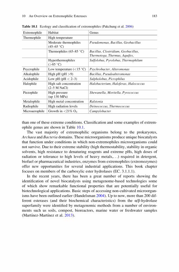
than one of these extreme conditions. Classification and some examples of extrem-
ophile genus are shown in Table 10.1.
The vast majority of extremophilic organisms belong to the prokaryotes,
Archaea and Bacteria domains. These microorganisms produce unique biocatalysts
that function under conditions in which non-extremophiles microorganisms could
not survive. Due to their extreme stability (high thermostability, stability in organic
solvents, high resistance to denaturing reagents and extreme pHs, high doses of
radiation or tolerance to high levels of heavy metals,. . .) required in detergent,
biofuel or pharmaceutical industries, enzymes from extremophiles (extremozymes)
offer new opportunities for several industrial applications. This book chapter
focuses on members of the carboxylic ester hydrolases (EC. 3.1.1.1).
In the recent years, there has been a great number of reports showing the
identification of novel biocatalysts using metagenome-based technologies some
of which show remarkable functional properties that are potentially useful for
biotechnological applications. Basic steps of accessing non-cultivated microorgan-
isms have been outlined earlier (Handelsman 2004). Up to now, more than 200 dif-
ferent esterases (and their biochemical characteristics) from the α/β-hydrolasesuperfamily were identified by metagenomic methods from a number of environ-
ments such us soils, compost, bioreactors, marine water or freshwater samples
(Martınez-Martınez et al. 2013).
Table 10.1 Ecology and classification of extremophiles (Pakchung et al. 2006)
Extremophile Habitat Genus
Thermophile High temperature
Moderate thermophiles
(45–65 �C)Pseudomonas, Bacillus, Geobacillus
Thermophiles (65–85 �C) Bacillus, Clostridium, Geobacillus,Thermotoga, Thermus, Aquifex,
Hyperthermophiles
(>85 �C)Sulfolobus, Pyrolobus, Thermophilum
Psycrophile Low temperature (<15 �C) Psychrobacter, Alteromonas
Alkalophile High pH (pH >9) Bacillus, Pseudoalteromonas
Acidophile Low pH (pH < 2–3) Sulpholobus, Picrophilus
Halophile High salt concentration
(2–5 M NaCl)
Halobacterium, Haloferax, Halococcus
Piezophile High pressure
(up 130 MPa)
Shewanella, Moritella, Pyrococcus
Metalophile High metal concentration Ralstonia
Radiophile High radiation levels Deinococcus, Thermococcus
Microaerophile Growth in <21% O2 Campylobacter
10 An Overview on Extremophilic Esterases 183

10.2 (Hyper)thermophilic Esterases
10.2.1 Biotechnological Interest
There are several advantages in performing industrial bioconversions at high
temperatures, such as the improving of solubility and better accessibility of the
enzyme to the substrate. Thus, in some cases, the use of polluting reagents to
solubilize reactants and products can be avoided (Lagarde et al. 2002). Higher
reaction rates can also be reached due to the reduction of viscosity and increased
diffusion rate. Furthermore, the risk of microbial contamination with mesophiles is
considerably diminished at high temperatures (Gomes and Steiner 2004).
10.2.2 Thermostability and Stability Against Chemicals
Thermal stability is one of the most valuable characteristics in the search for novel
esterases, and extreme thermophiles have been shown to be an interesting source of
them and other stable enzymes. Two types of protein thermostability are of rele-
vance for industrial purposes: (i) thermodynamic stability which refers to the
capability of an enzyme to be functional under denaturing conditions such as
high temperature, presence of an organic solvent, detergents or extreme pH values
and (ii) long-term stability where an enzyme does not lose its activity for prolonged
incubation (Sharma et al. 2012). Structural alignment and comparative modelling
have been applied in order to identify mechanisms related to thermal stability and
resistance to chemical denaturation of several esterases, having being solved many
three-dimensional structures to date. It has been observed that sequence and
structural features that contribute to the stability of (hyper)thermophilic enzymes
include changes in amino acid composition, higher hydrophobic interactions,
increased number of ion pairs and salt bridges, decrease of solvent-exposed surface
and oligomerisation (Levisson et al. 2009a, b; Byun et al. 2007).
Thus, apart from thermo-resistance, thermozymes frequently present an unusual
resistance in the presence of several chemical and physical denaturing agents,
which make them suitable for harsh industrial conditions where mesophilic
enzymes could not function. In addition, their high stability combined with high
retained activity levels at mesophilic temperatures might be valuable in order to cut
down costs in many industrial processes: energy saving with the combination of
lingering useful life of the biocatalysts. Intracellular enzymes from these microor-
ganisms are adapted to operate optimally at (or near) the optimum growth temper-
ature for the organism, being it 60 �C for a moderate thermophile or even 95 �C in
the case of hyperthermophiles. As for extracellular thermozymes, they may be
stable to temperatures considerably higher than the optimum growth temperature
(Cowan and Fernandez-Lafuente 2011). As a biotechnological practical advantage
of thermostability, when cloning and gene expression in a mesophilic host cell,
184 R. Gonzalez-Gonzalez et al.
purification of the protein can be rapidly performed to a high degree by simply
heating the cell-free extract (e.g., 85 �C, 15 min). Almost all of the proteins from the
host cell precipitate when this treatment is applied, while the recombinant thermo-
stable protein remains in a soluble form (Atomi and Imanaka 2004). Most industrial
processes in which esterases are used as biocatalysts are carried out at temperatures
above 45 �C. Specifically, those involving treatment of fats are typically performed
at temperatures up to 70 �C (Fuci~nos et al. 2011).
10.2.3 Finding Lipolitic Activity-Producing (Hyper)thermophiles and Their CarboxylesterasesCharacteristics
As shown in Table 10.2, several esterase-producing (hyper)thermophiles have been
studied (Levisson et al. 2009a, b) and many highly thermotolerant esterases have
been reported either isolated from wild-type strains (e.g. esterase E34Tt from
Thermus thermophilus HB27, with a half-life of 135 min at 85 �C, an optimal
temperature >80 �C and optimal pH of 8.1) (Fuci~nos et al. 2011) or expressed in
heterologous hosts, such as the case of an esterase from Aeropyrum pernix K1
expressed in Escherichia coli, with an optimal temperature of 90 �C and a half-life
over 160 h at 90 �C (Gao et al. 2003). As another remarkable cloning example, a
highly thermostable recombinant esterase from Pyrococcus furiosus was described.The resulting enzyme displayed an optimal temperature of 100 �C and a half-life of
50 min at 126 �C (Ikeda and Clark 1998).
A big number of thermophilic carboxylesterases prefer medium chain (acyl
chain length of around 6) p-nitrophenyl substrates (Table 10.2). Several enzymes
from hyperthermophiles have also been tested for activity toward esters with
various alcoholic moieties other than the standard p-nitrophenyl or
4-methylumbelliferyl esters. Other thermophilic esterases have been characterized
for their ability to resolve racemic mixtures. For example, the kinetic resolution of
the esterase Est3 from Sulfolobus solfataricus P2 was investigated using (R,S)-
ketoprofen methyl ester. The esterase hydrolyzed the (R)-ester of racemic
ketoprofen methylester and displayed an enantiomeric excess of 80% with a
conversion rate of 20% in 32 h (Levisson et al. 2009a, b; Atomi and Imanaka
2004; Kim and Lee 2004).
Regarding to metagenomics approaches, current molecular biology techniques,
direct genome shotgun sequencing, and molecular phylogenetic studies using
metagenomes are nowadays making possible to build total environmental DNA
libraries. They contain the genomes of unculturable microorganisms, which is
allowing us to access to a big number of unknown (hyper)thermophilic and other
enzymes with new and unique properties. Therefore, metagenomic libraries from
thermal environments have been enormously useful over the last few years as for
the aim of screening novel thermostable enzymes, including esterases. As an
10 An Overview on Extremophilic Esterases 185

Table
10.2
Characteristicsofesterasesfrom
(hyper)thermophilic
microorganisms
Microorganism
Enzyme
nam
e
MW
(kDa)
Enzymeproperties
Enzymestability
Rem
arks
References
Aeropyrum
pernixK1
Esterase
63
Bestsubstrate:
p-nitrophenylC8
Optimal
T:90
� COptimal
pH:8
Half-life
over
160h
at90
� CEsteraseandacylaminoacid-releasing
enzymeactivities.Thermostabilitywas
protein
concentration-dependent
(Gao
etal.
2003)
Archaeoglobu
sfulgidus
Esterase
AFEST
35.5
Bestsubstrate:
p-nitrophenylC6
Optimal
T:80
� COptimal
pH:6.5–7.5
Half-life:26min
at
95
� C80%
ofactivityat
110
� C(M
anco
etal.
2000)
Pyroba
culum
calidifontis
VA1
Esterase
Est
34
Bestsubstrate:
p-nitrophenylC6
Optimal
T:90
� COptimal
pH:7.0
Half-life:56min
at
110
� C16%
ofretained
activityat
30
� C.Active
andstable
inthepresence
of80%
water-
miscible
organic
solvents
(Hottaet
al.
2002)
Pyrococcus
furiosus
Esterase
–Bestsubstrate:
4-m
ethylumbelliferyl
C2
Optimal
T:100
� COptimal
pH:7.6
Half-life:50min
at
126
� CHalf-life
of34hoursat
100
� C(Ikedaand
Clark
1998)
Sulfolobus
solfataricusP1
Esterase
34
Bestsubstrate:
p-nitrophenylC6
Optimal:85
� COptimal
pH:8
41%
ofremaining
activityafter5days
at80
� C
Detergentandorganic
solventresistant
(90%
methanol,ethanol,2-propanol,ace-
tone).Activated
bydim
ethylsulfoxide
(Parket
al.
2006)
Thermotoga
maritime
Esterase
(EstA)
267
(hexam
er)
Bestsubstrate:
p-nitrophenylC8
Optimal
T:>95
� COptimal
pH:8.5
Half-life:1.5
hat
100
� CStructure
revealedthepresence
ofan
N-terminal
immunoglobulin(Ig)-like
domain
(Levissonet
al.
2009a,b)
Thermus
thermop
hilus
HB27
Esterase
E34Tt
34
Bestsubstrate:
p-nitrophenylC10
Optimal
T:>80
� COptimal
pH:8.1
Half-life:135min
at
85
� CDetergentessentialto
solubilisetheenzyme
from
cellmem
branes
andformaintaining
activityandstability
(Fuci~ no
set
al.
2011)
186 R. Gonzalez-Gonzalez et al.

Sulfolobu
ssolfataricusP2
Esterase
(SsoPEst)
58.4
Bestsubstrate:
p-nitrophenylC8
Optimal
T:80
� COptimal
pH:5.5
Half-life:1hat
80
� CActivitysignificantlyinhibited
byPMSF
(phenylm
ethylsulfonylfluoride)
(Shanget
al.
2010)
Picroph
ilus
torridus
Esterase
EstA
66
Bestsubstrate:
p-nitrophenylC2
Optimal
T:70
� COptimal
pH:6.5
Half-life:21hat
90
� CRem
arkable
preservationofactivityin
the
presence
ofdetergents,urea,andcommonly
usedorganic
solvents
(Hesset
al.
2008)
From
metagenomic
library
Esterase
29
Bestsubstrate:
p-nitrophenylC5
Optimal
T:70
� COptimal
pH:9
Half-life:30min
at
80
� CStable
at70
� Cforat
least120min
(Tiraw
ongsaroj
etal.2008)
10 An Overview on Extremophilic Esterases 187
example of a novel thermophilic esterase found by means of metagenomics, it is
worth to mention the case of EstE1, mined from a screening of four independent
metagenomic libraries of thermal areas of Indonesia. It displays a typical thermo-
philic profile: extremely stable at 80 �C in the absence of any stabilizer, with a high
optimal temperature of 95 �C. Its activity at lower temperatures is remarkably high:
20% and 30% of its optimal activity is retained at 30 and 40 �C, respectively (Rheeet al. 2005).
10.2.4 Immobilization of Thermophilic Esterases
Enhancing of protein stability, modifications in enzyme functional properties or
even recovery of specific proteins from complex mixtures, have been widely
described and achieved through the development of effective methods for
immobilizing. For example, the immobilization of large multi-subunit proteins
with multiple covalent linkages (multipoint immobilization), results quite interest-
ing for stabilizing proteins where the dissociation of their subunits is the initial step
in enzyme inactivation. A combination of targeted chemistries, for both the support
and the protein, sometimes also combined with chemical or genetic engineering,
has enormously contributed to these purposes (Cowan and Fernandez-Lafuente
2011). As for immobilization studies of thermophilic esterases, many of them
have been reported from genus such as Geobacillus or Anoxybacillus. As an
example, the case study of an esterase from Bacillus stearothermophilusimmobilized by multipoint covalent attachment to glyoxyl agarose. Optimized
immobilization conditions increased the stability of the esterase preparations by
factors up to 600-fold when compared to single-point covalent derivatives; reten-
tion of the initial activity after multipoint covalent attachment was 65%. Multipoint
covalently linked esterase derivatives retained more than 70% of the initial activity
after 1 week in 50% dimethyl sulfide or dimethylformamide at 30 �C. This
structural stabilization was also evident under various denaturing conditions (e.g.,
in the presence of high concentrations of sodium chloride and organic solvents). As
some examples of the importance of immobilizing, the use of enzymes as catalysts
in the industrial biosynthesis of esters, fine chemicals or in order to protect hydroxyl
groups of sugars, usually involves prior immobilization so as to improve the reactor
performance and to facilitate the recovery of both reaction products and enzyme
(Cowan and Fernandez-Lafuente 2011; Fernandez-Lafuente et al. 1995).
10.3 Psychrophilic Esterases
Psychrophilic microorganisms are adapted to live at temperatures below 5 �C. Infact, for some of them, a low temperature environment (<12 �C) is not only
optimal but mandatory for sustained cell growth (Tutino et al. 2010). Psychrophiles
188 R. Gonzalez-Gonzalez et al.

have been isolated from natural terrestrial and aquatic environments such as
Antarctic regions, glaciers, Arctic tundra soil, ocean depths, surfaces of plants
and animals living in such cold environments or even in super-cooled, high-altitude
cloud droplets, and refrigerated appliances (Gomes and Steiner 2004; Maiangwa
et al. 2015).
Psychrophilic microorganisms comprise not only genus from Gram-negative
(e.g. Moraxella, Moritella, Polaribacter, Polaromonas, Pseudoalteromonas, Pseu-domonas, Psychrobacter, Psychroflexus) and Gram-positive bacteria
(e.g. Arthrobacter, Bacillus, Micrococcus), but also archaea (e.g. Halorubrum,Methanococcoides, Methanogenium) and eukaryotes (e.g. Candida, Cladosporium,Cryptococcus, Penicillium) that have developed adaptive mechanisms to overcome
the difficulties of living at reduced temperature (Gomes and Steiner 2004). For
instance, most biological processes in mesophiles show little or no activity at low
temperature, with drops on the reaction rate ranging from 16 to 80-fold when the
temperature is reduced from 37 to 0 �C (Tutino et al. 2010). In contrast,
psychrophiles display unique characteristics in their membranes and enzymes,
which allow them to efficiently perform metabolic functions at low temperature,
in some cases far below 0 �C (Gomes and Steiner 2004; Maiangwa et al. 2015).
Psychrophilic microorganisms have developed very different strategies of adap-
tation to cold environments, although psychrophilic enzymes do possess some
common features enabling high catalytic efficiency at low temperatures
(0–20 �C). Compared to enzymes from mesophiles, cold-adapted enzymes display
a higher structural flexibility, particularly around the active site, for easier accom-
modation of substrates, and lower energy of activation. Such a high molecular
flexibility at low temperature requires weakening the intramolecular forces that
contribute to maintain the three-dimensional structure of the protein, so that the
conformational changes necessary for reaching the activation state have a lower
energy cost (Maiangwa et al. 2015).
Comparative studies using psychrophilic and mesophilic homologues showed
that the increased flexibility of psychrophilic enzymes may be a result of (i) having
fewer ionic interactions and hydrogen bonds; (ii) a decrease in compactness of the
hydrophobic core (iii) a higher number of hydrophobic side chains exposed to the
solvent; (iv) longer and more hydrophilic surface loops; (v) fewer proline and
arginine residues; and (vi) a higher number of glycine residues (Tutino et al.
2010). As a consequence of these adaptive modifications, very frequently, enzymes
from psychrophiles are also more thermolabile than those from mesophiles or
thermophiles. A cold-adapted enzyme can be active (even optimally) at tempera-
tures well above the temperature of the environment where the source microorgan-
ism was isolated. However, the loss of activity is often severe even at moderate
temperatures near or below 50 �C, which is generally recognized as a consequence
of a lack of selective pressure for thermostability in environments where the
temperature is permanently cold (Gerday et al. 1997).
The above mentioned properties make psychrophilic enzymes very attractive for
many industrial applications. Low temperature operation is desired either for
energy saving purposes or due to substrate or product stability. Psychrophilic
10 An Overview on Extremophilic Esterases 189
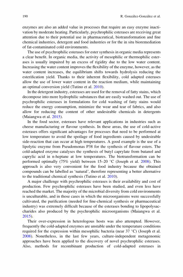
enzymes are also an added value in processes that require an easy enzyme inacti-
vation by moderate heating. Particularly, psychrophilic esterases are receiving great
attention due to their potential use in pharmaceutical, biotransformation and fine
chemical industries, detergent and food industries or for the in situ bioremediation
of fat-contaminated cold environments.
The use of psychrophilic esterases for ester synthesis in organic media represents
a clear benefit. In organic media, the activity of mesophilic or thermophilic ester-
ases is usually impaired by an excess of rigidity due to the low water content.
Increasing the water content improves the flexibility of the enzyme, however, as the
water content increases, the equilibrium shifts towards hydrolysis reducing the
esterification yield. Thanks to their inherent flexibility, cold adapted esterases
allow the use of lower water content in the reaction medium, while maintaining
an optimal conversion yield (Tutino et al. 2010).
In the detergent industry, esterases are used for the removal of fatty stains, which
decompose into more hydrophilic substances that are easily washed out. The use of
psychrophilic esterases in formulations for cold washing of fatty stains would
reduce the energy consumption, minimize the wear and tear of fabrics, and also
allow for reducing the content of other undesirable chemicals in detergents
(Maiangwa et al. 2015).
In the food sector, esterases have relevant applications in industries such as
cheese manufacturing or flavour synthesis. In these areas, the use of cold-active
esterases offers significant advantages for processes that need to be performed at
low temperature to avoid the spoilage of food ingredients caused by undesirable
side-reaction that can occur at high temperatures. A good example is the use of a
lipolytic enzyme from Pseudomonas P38 for the synthesis of flavour esters. The
cold-adapted enzyme catalyses the synthesis of butyl caprylate from butanol and
caprylic acid in n-heptane at low temperatures. The biotransformation can be
performed optimally (75% yield) between 15–20 �C (Joseph et al. 2008). This
approach is also very convenient for the food industry because the obtained
compounds can be labelled as ‘natural’, therefore representing a better alternative
to the traditional chemical synthesis (Tutino et al. 2010).
A major challenge with psychrophilic esterases is their availability and cost of
production. Few psychrophilic esterases have been studied, and even less have
reached the market. The majority of the microbial diversity from cold environments
is unculturable, and in those cases in which the microorganisms were successfully
cultivated, the purification (needed for fine-chemical synthesis or pharmaceutical
industry) was extremely difficult because of the esterases bonding to lipopolysac-
charides also produced by the psychrophilic microorganisms (Maiangwa et al.
2015).
Their over-expression in heterologous hosts was also attempted. However,
frequently the cold-adapted enzymes are unstable under the temperature conditions
required for the expression within mesophilic bacteria (near 37 �C) (Joseph et al.
2008). Nonetheless, in the last few years, culture-independent metagenomic
approaches have been applied to the discovery of novel psychrophilic esterases.
Also, methods for recombinant production of cold-adapted esterases in
190 R. Gonzalez-Gonzalez et al.

Table 10.3 Characteristics of cold-active esterases isolated from psychrophilic microorganisms
Organism Esterase MW
Enzyme
properties
Enzyme
stability References
Alcanivoraxdieselolei B-5(T)
EstB
(expressed
in E. coli)
45.1 kDa Best sub-
strate:
p-nitrophenyl
C6
Optimal pH:
8.5
Optimal T:
20 �C (95%
max at 0 �C)
T: stable at
40 �C.Inactivated
after 3 h at
55 �C
(Zhang et al.
2014)
Uncultured, from a
biogas slurry
metagenomic
library
Est01
(expressed
in E. coli)
44.8 kDa Best sub-
strate:
p-nitrophenyl
C4
Optimal pH:
8.0
Optimal T:
20 �C (43%
max activity
at 10 �C)
pH: stable at
pH 9.0
T: 20% ini-
tial activity
after 1 h at
40 �C.Inactivated
after 20 min
50 �C
(Cheng et al.
2014)
Psychrobactersp. Ant300
PsyEst
(expressed
in E. coli)
43 kDa Best sub-
strate:
p-nitrophenyl
C6
Optimal pH:
7.0–9.
Optimal T:
35 �C (active
at 5 �C)
T:
Inactivated
at 4 �C
(Kulakova
et al. 2004)
Rhodotorulamucilaginosa
Esterase 86 kDa Best sub-
strate: NR
Optimal pH:
7.5
Optimal T:
45 �C (20%
max activity
at 0 �C)
pH: unstable
above pH 9
T: unstable
above 50 �C
(Zimmer
et al. 2006)
Uncultured, from an
activated sludge
metagenomic
library
Lipo1
(expressed
in E. coli)
35.6 kDa Best sub-
strate:
p-nitrophenyl
C4
Optimal pH:
7.5
Optimal T:
10 �C
pH: 70% ini-
tial activity
after 24 h at
28 �C in the
pH range of
6.5–8.5
T: stable at
10 �C.Inactivated
above 30 �C
(Roh and
Villatte
2008)
(continued)
10 An Overview on Extremophilic Esterases 191
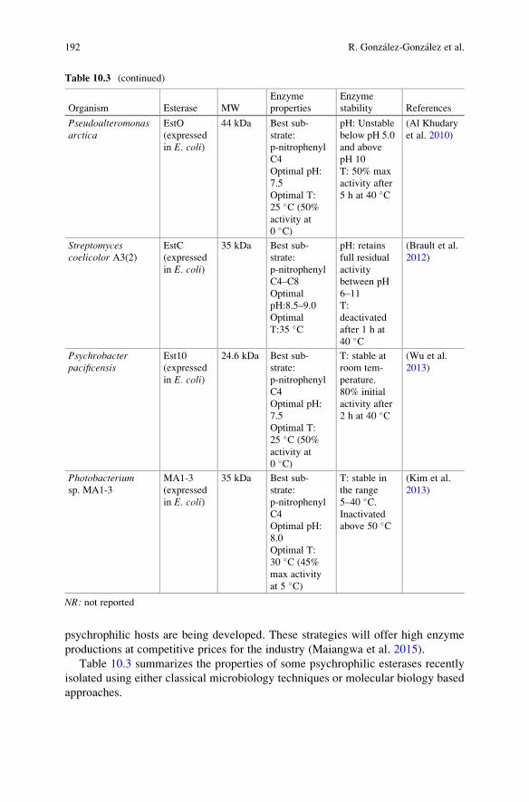
psychrophilic hosts are being developed. These strategies will offer high enzyme
productions at competitive prices for the industry (Maiangwa et al. 2015).
Table 10.3 summarizes the properties of some psychrophilic esterases recently
isolated using either classical microbiology techniques or molecular biology based
approaches.
Table 10.3 (continued)
Organism Esterase MW
Enzyme
properties
Enzyme
stability References
Pseudoalteromonasarctica
EstO
(expressed
in E. coli)
44 kDa Best sub-
strate:
p-nitrophenyl
C4
Optimal pH:
7.5
Optimal T:
25 �C (50%
activity at
0 �C)
pH: Unstable
below pH 5.0
and above
pH 10
T: 50% max
activity after
5 h at 40 �C
(Al Khudary
et al. 2010)
Streptomycescoelicolor A3(2)
EstC
(expressed
in E. coli)
35 kDa Best sub-
strate:
p-nitrophenyl
C4–C8
Optimal
pH:8.5–9.0
Optimal
T:35 �C
pH: retains
full residual
activity
between pH
6–11
T:
deactivated
after 1 h at
40 �C
(Brault et al.
2012)
Psychrobacterpacificensis
Est10
(expressed
in E. coli)
24.6 kDa Best sub-
strate:
p-nitrophenyl
C4
Optimal pH:
7.5
Optimal T:
25 �C (50%
activity at
0 �C)
T: stable at
room tem-
perature.
80% initial
activity after
2 h at 40 �C
(Wu et al.
2013)
Photobacteriumsp. MA1-3
MA1-3
(expressed
in E. coli)
35 kDa Best sub-
strate:
p-nitrophenyl
C4
Optimal pH:
8.0
Optimal T:
30 �C (45%
max activity
at 5 �C)
T: stable in
the range
5–40 �C.Inactivated
above 50 �C
(Kim et al.
2013)
NR: not reported
192 R. Gonzalez-Gonzalez et al.

10.4 Halophilic Esterases
From the Greek roots hals, meaning salt, and phil, meaning loving or friendly with,
halophily indicates that salt is required for function. The molecular ecology of
extremely halophilic archaea and bacteria has been widely described. Halophiles
like Halobacterium, Haloferax, Haloarcula, Halococcus, Natronobacterium and
Natronococcus belong to the archaea group, while Salinibacter ruber is a bacte-
rium. Halophilic organisms have been grouped into five categories according to salt
tolerance: non-halophiles (<0.2 M salt), slight halophiles (0.2–0.5 M salt), moder-
ate halophiles (0.5–2.5 M salt), borderline extreme halophiles (1.5–3.0 M salt), and
extreme halophiles (2.5–5.2 M salt). Halophiles have developed two different
adaptive strategies to cope with the osmotic pressure induced by the high NaCl
concentration of the normal environments they inhabit (Schreck and Grunden
2014).
Some extremely halophilic bacteria accumulate salts such as sodium or potas-
sium chloride (NaCl or KCl), up to concentrations that are isotonic with the
environment (the “salting in” strategy). Enzymes from these halophiles have
adapted to this environmental pressure by acquiring a relatively large number of
negatively charged amino acid residues (Asp, Glu) on their surfaces to prevent
precipitation. These organisms also prefer potassium as counter-ion as it has a much
lower water-binding rate, which helps to maximize the available amount of water
for the enzymes (Schreck and Grunden 2014).
This adaptation might confer additional stability to low water content environ-
ments and actually a number of enzymes from halophilic organisms stable and
active at low water activity (as low as 0.75) have been reported (Ghanem et al.
2000). This makes identifying and using enzymes from this group of halophiles a
clear approach for developing biotechnological processes based on synthesis reac-
tions in non-aqueous media.
In contrast, moderate halophiles accumulate in the cytoplasm high amounts of
specific organic solutes such as ectoine or hydroxyectoine, which function as
osmoprotectants, maintaining low the intracellular salt concentration and
stabilising biological structures without interfering with the normal metabolism
of the cell (the compatible solute strategy) (Dalmaso et al. 2015). Intracellular
enzymes from this group of halophiles do not normally show any particular
adaptation to high salt concentrations but their cell walls, transporters, and peri-
plasmic proteins typically are modified for high salt environments.
As widely described, halophilic esterases constitute an important group of
biocatalysts with different biotechnological applications. Features and some exam-
ples of different properties of carboxylesterases from several halophiles are shown
in Table 10.4.
Apart from being intrinsically stable and active at high salt concentrations,
halophilic enzymes offer important opportunities in biotechnological applications,
such as food processing, environmental bioremediation or any enzymatic transfor-
mation where is required the presence of organic solvents or low water activity.
10 An Overview on Extremophilic Esterases 193

Table
10.4
Characteristicsofcarboxylesterases
from
halophilic
microorganisms
Organism
Esterase
MW
(kDa)
Enzymeproperties
Enzymestability
References
Haloa
rcula
marismortui
Esterase
–Bestsubstrate:p-nitrophenylC5
Optimal
pH:7.5
Optimal
T:45
� COptimal
[NaC
l]or[salt]:4M
pH:NR
T:Loss
ofactivityabove75
� C[N
aCl]or[salt]:50%
activitylost
withoutsalt
(Cam
achoet
al.
2009;
Müller-Santosetal.
2009)
Haloarcula
marism
ortui
Expressed
inE.
coli
34(from
sequence)
Bestsubstrate:vinylbutyrate
Optimal
pH:8.5
Optimal
T:37
� COptimal[N
aCl]or[salt]:2M
NaC
l/
3.0
MKCl.Complete
lack
of
activitywithoutsalt
pH:Stable
between5and9
T:100%
lostactivityat
60
� C[N
aCl]or[salt]:100%
loss
without
salt.Unfolded
insalt-freemedium
(Rao
etal.2009;
Lvet
al.2010)
Haloarcula
marism
ortui
LipCenzyme
overexpressed
in
E.coliBL21
34(from
sequence)
Bestsubstrate:p-nitrophenylC2
Optimal
pH:9.5
Optimal
T:45
� COptimal
[NaC
l]or[salt]:3.4
M
NaC
l/3.0
MKCl
pH:NR
T:stable
atroom
Tat
3.4
MNaC
l
concentration.AsTincreases,salt
additionacceleratesdestabilization
[NaC
l]or[salt]:Circulardichroism
revealedthemaxim
alretentionofthe
α-helical
structure
atthesaltconcen-
trationmatchingtheoptimal
activity
(Müller-Santos
etal.2009)
Tha
lassob
acillus
sp.strain
DF-E4
Extracellular
carboxylesterase
45
Bestsubstrate:p-nitrophenylC4
Optimal
pH:8.5
Optimal
T:40
� COptimal
[NaC
l]or[salt]:0.5
M
NaC
l
pH:Stable
between6.0
and9.5
T:Stable
at45
� Cfor1h
[NaC
l]or[salt]:Stableupto4M
NaC
l;
retained
90%
activityafter12h
incubation
(Lvet
al.2010)
Halobacillus
trueperiwhb2
7Esterase
35
Bestsubstrate:
Optimal
pH:10.0
Optimal
T:42
� COptimal
[NaC
l]or[salt]:2.5
M
NaC
l
–(Y
anet
al.2014)
194 R. Gonzalez-Gonzalez et al.

Bacilluscereus
strain
AGP-03
Extracellular
41kDa
Dim
eric
(25kDa
and
16kDa)
Bestsubstrate:p-nitrophenylC4
(Vmax¼
1654Umg�1
protein;Km
¼52.46μM
)
Optimal
pH:8.5
Optimal
T:55
� COptimal
[NaC
l]or[salt]:4.5%
NaC
l
pH:Stable
between5.5
and10
T:Stable
from
10
� C—
75
� C(activity
retained
from
100to
72%)
[NaC
l]or[salt]:Maxim
um
activityat
4.5%
NaC
l.100%
stable
between
4.5–11%
NaC
l
(GhatiandPaul
2015)
Marinob
acter
lipo
lyticusSM19
LipBLexpressed
inE.coliDH5α
45.3
kDa
Bestsubstrate:p-nitrophenylC6;
tricaproin
OptimalpH:7.0(p-nitrophenylC4)
Optimal
T:80
� COptimal
[NaC
l]or[salt]:Highest
activityin
absence
ofNaC
l
pH:NR
T:5%
decreasein
activityafter1h/
45
� C;80%
decreaseafter2h/50
� C[N
aCl]or[salt]:NR
(Perez
etal.2011)
Marinob
acter
lipo
lyticus
LipBLexpressed
inE.coliBL21
45.3
kDa
Bestsubstrate:p-nitrophenylC4
Optimal
pH:8(p-nitrophenylC6)
Optimal
T:NR
Optimal
[NaC
l]or[salt]:Highest
activityin
absence
ofNaC
l
pH:Stable
between5.5
and10
T:70%
activityretained
afterincuba-
tionat
30
� C/1
h.70%
activitylost
after1hat
50
� C[N
aCl]or[salt]:NR
(Perez
etal.2012)
NR:notreported
10 An Overview on Extremophilic Esterases 195

That is because there is a relation among salt and organic solvent tolerances very
often observed for halophilic enzymes because salt presence has the effect of
reducing water activity (Schreck and Grunden 2014).
The recombinant esterase PE8 (219 aa, 23.19 kDa), from the marine bacterium
Pelagibacterium halotolerans B2T, is an alkaline esterase with an optimal pH of 9.5
and an optimal temperature of 45 �C toward p-nitrophenyl acetate (Wei et al. 2013).
PE8 exhibited activity and enantioselectivity in the synthesis of methyl (R)-3-(4-fluorophenyl)glutarate ((R)-3-MFG), a pharmaceutically important precursor in
the synthesis of the widely used antidepressant (�)paroxetine hydrochloride, from
the prochiral dimethyl 3-(4-fluorophenyl)glutarate (3-DFG). (R)-3-MFG was
obtained in 71.6% ee and 73.2% yield after 36 h reaction under optimized condi-
tions (0.6 M phosphate buffer (pH 8.0) containing 17.5% 1,4-dioxane under 30 �C).
10.5 Piezophiles: Approaches Related to Lipolytic
and Other Enzymes
Piezophiles have been broadly defined as those microorganisms that display opti-
mal growth rates at high pressures. They have been observed among several
prokaryotic genera such as Shewanella, Colwellia, Moritella, Methanococcus,Psycromonas, Photobacterium, Pyrococcus or Thermus, being Earth’s oceans,
with an average pressure of 38 MPa, their main home. Despite the fact that a vast
portion of the world biosphere is a high-pressure environment, deep-sea piezophilic
microorganisms are much less known than other microorganisms. The presence of
hydrothermal vents in the sea floor has allowed knowing habitats where high
pressure and high temperature conditions exist. Many enzymes that are stable at
high pressures have been isolated from a wide variety of extremophilic microor-
ganisms with optimal growth conditions above one atmosphere. Thus, marine
organisms inhabit environments where they might be exposed to a temperature
range of 1–300 �C and pressures that range from 0.1–110 MPa (Gomes and Steiner
2004; Dalmaso et al. 2015; Abe and Horikoshi 2001, Yano and Poulos 2003). It has
not been fully elucidated whether piezophilic adaptation requires the modification
of a few genes, metabolic pathways, or a more profound reorganization of the
genome. Adaptation mechanisms against high pressure include reduction of cell
division, modification of membrane and transport proteins and accumulation of
osmolytes, which stabilize the proteins (Dalmaso et al. 2015).
Enzymes that can operate at high pressures and temperature have great advan-
tages in biotechnological applications. Enzymatic reactions that have a negative
change in activation volume (ΔV<0) are favored by increasing pressure; as an
example, α-chymotrypsin catalyzes the hydrolysis of an anilide when ΔV<0 at
increased pressure and the hydrolysis of an ester when ΔV>0 (Gomes and Steiner
2004). Despite the fact that pressure does not represent a major selective factor for
protein structure and function in piezophiles and that their proteins do not need
196 R. Gonzalez-Gonzalez et al.

specific pressure-related adaptations (Yano and Poulos 2003), there are some
examples of thermophilic protein stabilization by high pressure (Hei and Clark
1994). It has also been described the induction of stabilization and activation of
several lipolytic enzymes and other hydrolases through high pressure mechanisms
(Eisenmenger and Reyes-De-Corcuera 2009).
As already described (Abe and Horikoshi 2001; Yano and Poulos 2003), in spite
of there being many potential biotechnological applications of piezophiles and
piezoenzymes, there are not many known practical applications of piezophiles or
piezophilic enzymes in the market. As a representative application, it is worth to
mention the use of piezophiles in the formation of gels and starch granules
(Demirjian et al. 2001). The main reason of the lack of known applications is the
fact that it is not easy to cultivate piezophiles under their environment high-pressure
conditions using current technology. Thus, investigation through metagenomics
and many other prospections about the properties of these enzymes and other
cellular components of piezophiles need to be continued (Gomes and Steiner 2004).
10.6 Polyextremophilic Esterases
Most of the microorganisms inhabiting extreme environments must thrive under
multiple extreme conditions. For instance, the ocean depths combine high pressure
and cold (in the ocean mud) or high temperatures (in the proximities of hydrother-
mal vents); hot springs frequently combine elevate temperatures and extreme pH
values; and hypersaline areas can combine extreme osmotic pressure and either
high (e.g. the Dead Sea in Israel) or cold temperature (e.g. brines within the sea ice
in Polar regions). Excellent reviews on the poly-extremophilic microbial diversity
of these environments have been reported (Dalmaso et al. 2015; Capece et al. 2013).
Polyextremophilic enzymes produced by these microorganisms display several
unique properties for their applications in the food, detergent, chemical, or paper
industries (Dalmaso et al. 2015). In the case of esterases, simultaneous resistance
towards cold temperature and alkaline pH values (see Table 10.3 for examples) is
greatly appreciated for cold-washing detergent formulations, and resistance
towards extremes of pH and high concentration of organic solvents is suitable for
applications in synthetic chemistry (see Table 10.2 for examples).
10 An Overview on Extremophilic Esterases 197

10.7 Relevance of Esterases to “Lignocellulosic
Feedstocks”
Lignocellulose is the most abundant carbohydrate source in nature and it promises as
an ideal renewable energy source. Second bioethanol generation is obtained mainly
from lignocellulosic materials, which are not a food source in contrast to first
bioethanol generation which derives from starch-based feedstocks (Cragg et al. 2015).
Lignocellulosic feedstocks are mainly composed of cellulose, hemicellulose and
lignin. Hemicellulose is a complex branched polymer composed of different poly-
saccharides, such as pentoses (e.g., xylan and araban), hexoses (mainly mannan,
glucan, glucomannan, and galactan), and pectin. Their backbones have branches
composed of monomers such as D-galactose, D-xylose, L- arabinose, and
D-glucuronic acid, generating various types of hemicelluloses that are strongly
dependent on the tissue and plant species (Van Den Brink and De Vries 2011). All
types of hemicelluloses are partially esterified with acetic acid. Acetylation
increases their solubility but also prevents hydrolysis of glycosidic linkages of
hemicelluloses by the corresponding hydrolases hampering the enzymatic sacchar-
ification to produce fermentable sugars (Cuervo-Soto et al. 2015).
Hemicellulose encases cellulose, i.e. a linear polymer of D-glucose subunits
linked by β-1,4-glycosidic bonds. Both hemicellulose and cellulose are cross-linked
in the plant cell walls with the hydrophobic network of lignin, the third most
abundant component in lignocellulosic materials, which makes the chemical or
enzymatic hydrolysis of cellulose and depolymerization of hemicellulose difficult.
The highly organized crystalline structure of cellulose presents an obstacle to its
hydrolysis. Lignin itself is almost completely resistant against microbial (and
enzymatic) attack. Pretreatment steps (mechanical, chemical, or combinations of
both) are always needed in order to deconstruct those recalcitrant structures and
make them more accessible to enzymes (Bornscheuer et al. 2014).
On the other hand, enzymatic degradation of lignocellulosic materials needs the
combined and synergistic action of several enzymes.
For cellulose to be degraded, three main activities are involved: chain-end-
cleaving cellobiohydrolases, internally chain-cleaving endoglucanases, and
ß-glucosidases, which hydrolyze soluble short-chain glucooligosaccharides to
glucose.
Complete hydrolysis of the hemicellulose fraction requires two groups of
enzymes: (1) endo- xylanase and β-xylosidase, which cleave the xylan main
chain; and (2) accessory enzymes, which remove the side chains and break
crosslinks between xylan and other plant polymers. The latter group consists of
α-L-arabino- furanosidase, α-glucuronidase and a number of carbohydrate esterases
(CE) (Wong 2006).
CE show a great diversity in substrate specificity and structure and are currently
classified in the Carbohydrate-Active Enzymes (CAZymes) database (Cantarel
et al. 2009), in 16 different CE families (CAZy; http://www.cazy.org). CE catalyze
the de-O or de-N-acylation of substituted saccharides, acting on two classes of
198 R. Gonzalez-Gonzalez et al.

substrates: those in which the sugar plays the role of the “acid”, such as pectin
methyl esters and those in which the sugar behaves as the alcohol, such as in
acetylated xylan. A number of possible reaction mechanisms may be involved:
the most common is a Ser-His-Asp catalytic triad catalyzed deacetylation analo-
gous to the action of classical lipase and serine proteases but other mechanisms
such as a Zn2þ catalyzed deacetylation prevails in some families (Lombard et al.
2014). In terms of substrate specificity, the 16 CE families represent acetylxylan
esterases, acetyl esterases, chitin deacetylases, peptidoglycan deacetylases, feruloyl
esterases, pectin acetyl esterases, pectin methylesterases, glucuronoyl esterases and
enzymes catalyzing N-deacetylation of low molecular mass amino sugar deriva-
tives (Biely 2012). Feruloyl esterases (EC 3.1.1.73) do not fit into the established
CE families and have been separately classified based on sequence similarities
(Udatha et al. 2011). Among the accessory enzymes, feruloyl esterases play a key
role in enhancing the accessibility of enzymes to and subsequent hydrolysis of
hemicellulose fibers by removing the ferulic acid side chains and crosslinks. Ferulic
acids are covalently linked to polysaccharides, including glucuronoarabinoxylans,
xyloglucans, and pectins, through ester linkages. Ferulic acid is a cinnamic acid
with the chemical name (3-methoxy- 4-hydroxy)-3-phenyl-2-propenoic acid, or
3-methoxy-4-hydroxy-cinnamic acid (Wong 2006).
Feruloyl esterases belong to a subclass of carboxylic esterases (EC 3.1.1). A
wide range of bacteria and fungi has been reported to secrete these enzymes, which
are highly inducible, depending on the growth substrates (Wong 2006). Feruloyl
esterases have been extensively covered in recent excellent reviews (Wong 2006;
Udatha et al. 2011; Aurilia et al. 2008; Faulds 2010; Fazary and Ju 2007; Koseki
et al. 2009).
Thermostable enzymes that hydrolyze lignocellulose to its component sugars
have significant advantages for improving the conversion rate of biomass over their
mesophilic counterparts. Temperature might be advantageous to accelerate the
lignocellulose deconstruction and facilitate the simultaneous saccharification
prior or during fermentation of the resulting sugars to bioethanol. In an excellent
review (Blumer-Schuette et al. 2014), the possibilities of the thermophilic decon-
struction of lignocellulosic materials are extensively analysed, providing great
information on thermophilic microorganisms and their thermozymes to degrade
plant biomass.
10.8 Conclusions and Perspectives
Extremozymes have a great economic potential in many industrial processes and, in
particular, esterases are one of the most important groups of biocatalysts for
biotechnological applications.
In the recent years, there has been a great increase in the number of reports that,
through metagenomic techniques, have accessed to the genomes of unculturable
10 An Overview on Extremophilic Esterases 199

prokaryotes. This technology has led to the identification and characterization of a
vast number of biocatalysts active under a myriad of conditions that ultimately
reflect the primordial environment from which they were isolated. Esterases are a
good example of that and they are well represented within microbial communities
operating in most environments on the Earth.
Extremozyme libraries are commonly constructed using mesophilic hosts
mainly due to the deepest knowledge of the expression systems and easy cultiva-
tion, but often low-level protein expression is achieved compromising any basic
and biotechnological application where larger amounts of the recombinant protein
would be needed. There has been a consistent effort in the scientific community to
develop genetic systems for robust protein expression, mainly using archaeal
genera of thermophilic organisms, but still more work is needed.
Finally, although some advances have been made, studies on the physiology,
metabolism, enzymology and genetics of extremophiles are still limited. Therefore,
it is expected that this area will experience an important growth, pursuing a better
understanding of the application of esterases and other hydrolases from
extremophiles.
Take Home Message:
• Carboxylesterase and lipase belong to a family of carboxylic-ester hydrolases
that catalyze the hydrolysis of ester bonds. They catalyze the stereospecific
hydrolysis, transesterification, and conversion of a variety of amines and primary
and secondary alcohols. Sequence and structural features that contribute to the
stability of (hyper)thermophilic enzymes include changes in amino acid com-
position, higher hydrophobic interactions, increased number of ion pairs and salt
bridges, decrease of solvent-exposed surface and oligomerisation.
• The use of psychrophilic esterases in formulations for cold washing of fatty
stains would reduce the energy consumption, minimize the wear and tear of
fabrics and also allow for reducing the content of other undesirable chemicals in
detergents.
• The use of cold-active esterases offers significant advantages for processes that
need to be performed at low temperature to avoid the spoilage of food ingredi-
ents caused by undesirable side-reaction that can occur at high temperatures.
• Carboxylesterase can be produced from different extremophilic sources. They
are produced from moderate thermophiles (45–65 �C) such as Pseudomonas,Bacillus, Geobacillus, Thermophiles (65–85 �C) such as Bacillus, Clostridium,Geobacillus, Thermotoga, Thermus, Aquifex, Sulfolobus, Pyrolobus,Thermophilum, Hyperthermophiles (> 85 �C) such as Sulfolobus, Pyrolobus,Thermophilum, Psychrophiles (low temperature< 15 �C) such as Psychrobacter,Alteromonas, Alkalophiles (pH > 9) such as Bacillus, Pseudoalteromonas,Acidophiles (pH< 2–3) such as Sulpholobus, Picrophilus, Halophiles (high
salt concentration, 2–5MNaCl) such asHalobacterium, Haloferax, Halococcus,Piezophiles (high pressure up 130 MPa) such as Shewanella, Moritella,Pyrococcus, Metallophiles (high metal concentration) such as Ralstonia,
200 R. Gonzalez-Gonzalez et al.

Radiophiles (high radiation levels) such as Deinococcus, Thermococcus andMicroaerophiles (growth in <21% O2) such as Campylobacter.
• They have a wide range of applications such as food, detergent, chemical, paper
industries and treatment of biomass (bioremediation).
References
Abe F, Horikoshi K (2001) The biotechnological potential of piezophiles. Trends Biotechnol
19:102–108. doi:10.1016/S0167-7799(00)01539-0
Al Khudary R, Venkatachalam R, Katzer M et al (2010) A cold-adapted esterase of a novel marine
isolate, Pseudoalteromonas arctica: gene cloning, enzyme purification and characterization.
Extremophiles 14:273–285. doi:10.1007/s00792-010-0306-7
Arpigny JL, Jaeger K-E (1999) Bacterial lipolytic enzymes: classification and properties. Biochem
J 343:177–183. doi:10.1042/0264-6021:3430177
Atomi H, Imanaka T (2004) Thermostable carboxylesterases from hyperthermophiles. Tetrahe-
dron: Asymmetr 15:2729–2735. doi:10.1016/j.tetasy.2004.07.054
Aurilia V, Parracino A, D’Auria S (2008) Microbial carbohydrate esterases in cold adapted
environments. Gene 410:234–240
Biely P (2012) Microbial carbohydrate esterases deacetylating plant polysaccharides. Biotechnol
Adv 30:1575–1588. doi:10.1016/j.biotechadv.2012.04.010
Blumer-Schuette SE, Brown SD, Sander KB et al (2014) Thermophilic lignocellulose deconstruc-
tion. FEMS Microbiol Rev 38:393–448. doi:10.1111/1574-6976.12044
Bornscheuer UT (2002) Microbial carboxyl esterases: classification, properties and application in
biocatalysis. FEMS Microbiol Rev 26:73–81
Bornscheuer U, Buchholz K, Seibel J (2014) Enzymatic degradation of (ligno)cellulose. Angew
Chemie—Int Ed 53:10876–10893. doi:10.1002/anie.201309953
Brault G, Shareck F, Hurtubise Y et al (2012) Isolation and characterization of EstC, a new cold-
active esterase from Streptomyces coelicolor A3(2). PLoS One 7:e32041
Byun J, Rhee J, Kim ND et al (2007) Crystal structure of hyperthermophilic esterase EstE1 and the
relationship between its dimerization and thermostability properties. BMC Struct Biol
11:1–11. doi:10.1186/1472-6807-7-47
Camacho R, Mateos J, Gonza lez-Reynoso O et al (2009) Production and characterization of
esterase and lipase from Haloarcula marismortui. J Ind Microbiol Biotechnol 36:901–909
Cantarel BI, Coutinho PM, Rancurel C et al (2009) The Carbohydrate-Active EnZymes database
(CAZy): an expert resource for glycogenomics. Nucleic Acids Res. doi:10.1093/nar/gkn663
Capece MC, Clark E, Saleh JK et al (2013) Polyextremophiles and the constraints for terrestrial
habitability BT—polyextremophiles. In: Polyextremophiles. Springer Netherlands, Dordrecht,
pp 3–59
Cheng X, Wang X, Qiu T et al (2014) Molecular cloning and characterization of a novel cold-
adapted family VIII esterase from a biogas slurry metagenomic library. J Microbiol Biotechnol
24:1484–1489
Cowan D a, Fernandez-Lafuente R (2011) Enhancing the functional properties of thermophilic
enzymes by chemical modification and immobilization. Enzyme Microb Technol 49:326–346.
doi:10.1016/j.enzmictec.2011.06.023
Cragg SM, Beckham GT, Bruce NC et al (2015) Lignocellulose degradation mechanisms across
the Tree of Life. Curr Opin Chem Biol 29:108–119. doi:10.1016/j.cbpa.2015.10.018
Cuervo-Soto LI, Valdes-Garcıa G, Batista-Garcıa R et al (2015) Identification of a novel carbo-
hydrate esterase from Bjerkandera adusta: Structural and function predictions through
10 An Overview on Extremophilic Esterases 201

bioinformatics analysis and molecular modeling. Proteins Struct Funct Bioinf 83:533–546.
doi:10.1002/prot.24760
Dalmaso G, Ferreira D, Vermelho A (2015) Marine extremophiles: a source of hydrolases for
biotechnological applications. Mar Drugs 13:1925–1965. doi:10.3390/md13041925
Demirjian DC, Morıs-Varas F, Cassidy CS (2001) Enzymes from extremophiles. Curr Opin Chem
Biol 5:144–151
Eisenmenger MJ, Reyes-De-Corcuera JI (2009) High pressure enhancement of enzymes: a review.
Enzyme Microb Technol 45:331–347. doi:10.1016/j.enzmictec.2009.08.001
Faulds CB (2010) What can feruloyl esterases do for us? Phytochem Rev 9:121–132. doi:10.1007/
s11101-009-9156-2
Fazary AE, Ju YH (2007) Feruloyl esterases as biotechnological tools: Current and future
perspectives. Acta Biochim Biophys Sin (Shanghai) 39:811–828. doi:10.1111/j.1745-7270.
2007.00348.x
Fernandez-Lafuente R, Cowan DA, Wood ANP (1995) Hyperstabilization of a thermophilic
esterase by multipoint covalent attachment. Enzyme Microb Technol 17:366–372. doi:10.
1016/0141-0229(94)00089-1
Fuci~nos P, Pastrana L, Sanroman a et al (2011) An esterase from Thermus thermophilusHB27 withhyper-thermoalkalophilic properties: Purification, characterisation and structural modelling. J
Mol Catal B: Enzym 70:127–137. doi:10.1016/j.molcatb.2011.02.017
Fuci~nos P, Gonzalez R, Atanes E et al (2012) Lipases and esterases from extremophiles: overview
and case example of the production and purification of an esterase from Thermus thermophilusHB27. Methods Mol Biol 861:239–266. doi:10.1007/978-1-61779-600-5_15
Gao R, Feng Y, Ishikawa K, Ishida H (2003) Cloning, purification and properties of a hyperther-
mophilic esterase from archaeon Aeropyrum pernix K1. J Mol Catal 25:1–8. doi:10.1016/
S1381-1177(03)00064-X
Gerday C, Aittaleb M, Arpigny J et al (1997) Psychrophilic enzymes: a thermodynamic challenge.
Biochim Biophys Acta—Protein Struct Mol Enzymol 1342:119–131
Ghanem EH, Al-Sayed HA, Saleh KM (2000) An alkalophilic thermostable lipase produced by a
new isolate of Bacillus alcalophilus. World J Microbiol Biotechnol 16:459–464
Ghati A, Paul G (2015) Purification and characterization of a thermo-halophilic, alkali-stable and
extremely benzene tolerant esterase from a thermo-halo tolerant Bacillus cereus strain
AGP-03, isolated from “Bakreshwar” hot spring, India. Process Biochem 50:771–781.
doi:10.1016/j.procbio.2015.01.026
Gomes J, Steiner W (2004) The biocatalytic potential of extremophiles and extremozymes. Food
Technol Biotechnol 42:223–235
Handelsman J (2004) Metagenomics: application of genomics to uncultured microorganisms.
Microbiol Mol Biol Rev 68:669–685. doi:10.1128/MMBR.68.4.669-685.2004
Hei DJ, Clark DS (1994) Pressure stabilization of proteins from extreme thermophiles. Appl
Environ Microbiol 60:932–939
Hess M, Katzer M, Antranikian G (2008) Extremely thermostable esterases from the
thermoacidophilic euryarchaeon Picrophilus torridus. Extremophiles 12:351–364. doi:10.
1007/s00792-008-0139-9
Hotta Y, Ezaki S, Atomi H, Imanaka T (2002) Extremely stable and versatile carboxylesterase
from a hyperthermophilic archaeon. Appl Environ Microbiol 68:3925–3931. doi:10.1128/
AEM.68.8.3925-3931.2002
Ikeda M, Clark DS (1998) Molecular cloning of extremely thermostable esterase gene from
hyperthermophilic archaeon Pyrococcus furiosus in Escherichia coli. Biotechnol Bioeng.
doi:10.1002/(SICI)1097-0290(19980305)57:5<624::AID-BIT15>3.0.CO;2-B
Joseph B, Ramteke P, Thomas G (2008) Cold active microbial lipases: some hot issues and recent
developments. Biotechnol Adv 26:457–470
Kim S, Lee SB (2004) Thermostable esterase from a thermoacidophilic archaeon: purification and
characterization for enzymatic resolution of a chiral compound. Biosci Biotechnol Biochem
68:2289–2298. doi:10.1271/bbb.68.2289
202 R. Gonzalez-Gonzalez et al.

Kim YO, Heo YL, Nam BH et al (2013) Molecular cloning, purification, and characterization of a
cold-adapted esterase from Photobacterium sp. MA1-3. Fish Aquat Sci 16:311–318
Koseki T, Fushinobu S, Ardiansyah, et al (2009) Occurrence, properties, and applications of
feruloyl esterases. Appl Microbiol Biotechnol 84:803–810. doi: 10.1007/s00253-009-2148-8
Kulakova L, Galkin A, Nakayama T et al (2004) Cold-active esterase from Psychrobacter
sp. Ant300: Gene cloning, characterization, and the effects of Gly!Pro substitution near the
active site on its catalytic activity and stability. Biochim Biophys Acta—Proteins Proteomics
1696:59–65. doi:10.1016/j.bbapap.2003.09.008
Lagarde D, Nguyen HK, Ravot G et al (2002) High-throughput screening of thermostable esterases
for industrial bioconversions. Org Process Res Dev 6:441–445
Levisson M, van der Oost J, Kengen SW (2009a) Carboxylic ester hydrolases from
hyperthermophiles. Extremophiles 13:567–581
Levisson M, Sun L, Hendriks S et al (2009b) Crystal structure and biochemical properties of a
novel thermostable esterase containing an immunoglobulin-like domain. J Mol Biol
385:949–962. doi:10.1016/j.jmb.2008.10.075
Lombard V, Golaconda Ramulu H, Drula E et al (2014) The carbohydrate-active enzymes
database (CAZy) in 2013. Nucleic Acids Res. doi:10.1093/nar/gkt1178
Lopez-Lopez O, Cerdan ME, Gonzalez-Siso MI (2014) New extremophilic lipases and esterases
from metagenomics. Curr Protein Pept Sci:445–455. doi:10.2174/
1389203715666140228153801
Lv XY, Guo LZ, Song L et al (2010) Purification and characterization of a novel extracellular
carboxylesterase from the moderately halophilic bacterium Thalassobacillus sp. strain DF-E4.Ann Microbiol 61:281–290. doi:10.1007/s13213-010-0135-z
Maiangwa J, Ali MSM, Salleh AB et al (2015) Adaptational properties and applications of cold-
active lipases from psychrophilic bacteria. Extremophiles 19:235–247. doi:10.1007/s00792-
014-0710-5
Manco G, Giosue E, Auria SD et al (2000) Cloning, overexpression, and properties of a new
thermophilic and thermostable esterase with sequence similarity to hormone-sensitive lipase
subfamily from the archaeon Archaeoglobus fulgidus. Arch Biochem Biophys 373:182–192
Martınez-Martınez M, Alcaide M, Tchigvintsev A et al (2013) Biochemical diversity of carboxyl
esterases and lipases from lake Arreo (Spain): a metagenomic approach. Appl Environ
Microbiol 79:3553–3562. doi:10.1128/AEM.00240-13
Müller-Santos M, de Souza EM, Pedrosa F de O et al (2009) First evidence for the salt-dependent
folding and activity of an esterase from the halophilic archaea Haloarcula marismortui.Biochim Biophys Acta—Mol Cell Biol Lipids 1791:719–729. doi:10.1016/j.bbalip.2009.03.
006
Ollis DL, Cheah E, Cygler M et al (1992) The α/β hydrolase fold. Protein Eng 5:197–211
Pakchung A, Simpson P, Codd R (2006) Life on earth. Extremophiles continue to move the goal
posts. Environ Chem 3:77–93
Park Y, Choi SY, Lee H et al (2006) A carboxylesterase from the thermoacidophilic archaeon
Sulfolobus solfataricus P1; purification, characterization, and expression. Biochim Biophys
Acta 1760:820–828. doi:10.1016/j.bbagen.2006.01.009
Perez D, Martın S, Fernandez-Lorente G et al (2011) A novel halophilic lipase, LipBL, showing
high efficiency in the production of eicosapentaenoic acid (EPA). PLoS One 6:1–11. doi:10.
1371/journal.pone.0023325
Perez D, Kovacic F, Wilhelm S et al (2012) Identification of amino acids involved in the hydrolytic
activity of lipase LipBL from Marinobacter lipolyticus. Microbiol (United Kingdom)
158:2192–2203. doi:10.1099/mic.0.058792-0
Rao L, Zhao X, Pan F et al (2009) Solution behavior and activity of a halophilic esterase under
high salt concentration. PLoS One 4:e6980
Rhee JK, Ahn DG, Kim YG, Oh JW (2005) New thermophilic and thermostable esterase with
sequence similarity to the hormone-sensitive lipase family, cloned from a metagenomic
library. Appl Environ Microbiol 71:817–825. doi:10.1128/AEM.71.2.817-825.2005
10 An Overview on Extremophilic Esterases 203
Roh C, Villatte F (2008) Isolation of a low-temperature adapted lipolytic enzyme from
uncultivated micro-organism. Proc Soc Agric Bacteriol 105:116–123. doi:10.1111/j.1365-
2672.2007.03717.x
Schreck SD, Grunden AM (2014) Biotechnological applications of halophilic lipases and
thioesterases. Appl Microbiol Biotechnol 98:1011–1021. doi:10.1007/s00253-013-5417-5
Shang YS, Zhang XE, Wang X De, et al (2010) Biochemical characterization and mutational
improvement of a thermophilic esterase from Sulfolobus solfataricus P2. Biotechnol Lett
32:1151–1157. doi: 10.1007/s10529-010-0274-0
Sharma PK, Singh K, Singh R et al (2012) Characterization of a thermostable lipase showing loss
of secondary structure at ambient temperature. Mol Biol Rep 39:2795–2804. doi:10.1007/
s11033-011-1038-1
Tirawongsaroj P, Sriprang R, Harnpicharnchai P et al (2008) Novel thermophilic and thermostable
lipolytic enzymes from a Thailand hot spring metagenomic library. J Biotechnol 133:42–49.
doi:10.1016/j.jbiotec.2007.08.046
Tutino M, Parrilli E, De Santi C et al (2010) Cold-adapted esterases and lipases: a biodiversity still
under-exploited. Curr Chem Biol 4:74–83
Udatha DB, Kouskoumvekaki I, Olsson L, Panagiotou G (2011) The interplay of descriptor-based
computational analysis with pharmacophore modeling builds the basis for a novel classifica-
tion scheme for feruloyl esterases. Biotechnol Adv 29:94–110. doi: 10.1016/j.biotechadv.2010.
09.003
Van Den Brink J, De Vries RP (2011) Fungal enzyme sets for plant polysaccharide degradation.
Appl Microbiol Biotechnol 91:1477–1492. doi:10.1007/s00253-011-3473-2
Wei X, Jiang X, Ye L et al (2013) Cloning, expression and characterization of a new
enantioselective esterase from a marine bacterium Pelagibacterium halotolerans B2T. J Mol
Catal B: Enzym 97:270–277. doi:10.1016/j.molcatb.2013.09.002
Wong DWS (2006) Feruloyl esterase: a key enzyme in biomass degradation. Appl Biochem
Biotechnol 133:87–112. doi:10.1385/ABAB:133:2:87
Wu G, Wu G, Zhan T et al (2013) Characterization of a cold-adapted and salt-tolerant esterase
from a psychrotrophic bacterium Psychrobacter pacificensis. Extremophiles 17:809–819
Yan S, Lin X, Chen X, Zhang S (2014) Purification and characterization of an esterase from
Halobacillus trueperi whb27. J Pure Appl Microbiol 8:1–9
Yano JK, Poulos TL (2003) New understandings of thermostable and peizostable enzymes. Curr
Opin Biotechnol 14:360–365. doi:10.1016/S0958-1669(03)00075-2
Zhang S, Wu G, Liu Z et al (2014) Characterization of EstB, a novel cold-active and organic
solvent-tolerant esterase from marine microorganism Alcanivorax dieselolei B-5(T).
Extremophiles 18:251–259
Zimmer C, Platz T, Cadez N et al (2006) A cold active (2R,3R)-(-)-di-O-benzoyl-tartrate hydro-
lyzing esterase from Rhodotorula mucilaginosa. Acta Crystallogr Sect F Struct Biol Cryst
Commun 73:132–140. doi:10.1007/s00253-006-0463-x
204 R. Gonzalez-Gonzalez et al.

Chapter 11
Extremophilic Esterases for Bioprocessing
of Lignocellulosic Feedstocks
Juan-Jose Escuder-Rodrıguez, Olalla Lopez-Lopez, Manuel Becerra,
Marıa-Esperanza Cerdan, and Marıa-Isabel Gonzalez-Siso
Abbreviations
AME Acetyl mannan esterase
AXE Acetylxylan esterase
CBM Carbohydrate binding module
CE Carbohydrate esterase
FA Ferulic acid
FAE Ferulic acid esterase
GE Glucuronoyl esterase
GH Glycoside hydrolase
MCA Methyl 3,4-dihydroxycinnamate
MFA Methyl 3-methoxy-4-hydroxycinnamate
MpCA Methyl 4-Hydroxycinnamate
MSA Methyl 3,5-dimethoxy-4-hydroxycinnamate
PMSF Phenylmethylsulfonyl fluoride
SBP Sugar beet pulp
WB Wheat bran
What Will You Learn from This Chapter?
Esterase is the generic name given to any enzyme that catalyzes the hydrolysis (and
formation) of an ester into its alcohol and acid. Esterases acting on lignocellulosic
substrates remove the side chains of hemicelluloses, which are cross-linked with
lignin, therefore favouring hemicellulose degradation, cellulose accessibility, and
J.-J. Escuder-Rodrıguez •O. Lopez-Lopez •M.Becerra •M.-E. Cerdan •M.-I. Gonzalez-Siso (*)
Grupo EXPRELA, Centro de Investigacions Cientıficas Avanzadas (CICA), Departamento de
Bioloxıa Celular e Molecular, Facultade de Ciencias, Universidade da Coru~na, Campus de A
Coru~na, 15071 A Coru~na, Spaine-mail: [email protected]
© Springer International Publishing AG 2017
R.K. Sani, R.N. Krishnaraj (eds.), Extremophilic Enzymatic Processing ofLignocellulosic Feedstocks to Bioenergy, DOI 10.1007/978-3-319-54684-1_11
205

biomass digestibility. Degradation of the complex lignocellulosic polymeric sub-
strate into simple sugars is possible by synergic combination of these esterases with
a series of other enzymes such as hemicellulases, cellulases, and oxidoreductases.
This book chapter covers the enzymes acetyl mannan esterases that catalyze the
deacetylation of 2,3-O-acetyl mannan, acetylxylan esterases that catalyze the
deacetylation of 2,3-O-acetyl xylan and xylo-oligosaccharides, and ferulic acid
esterases that catalyze the hydrolysis of the 4-hydroxy-3-methoxycinnamoyl
(feruloyl) group from arabinose substitutions. Glucuronoyl esterases that degrade
the ester bonds between glucuronic acids of xylan and alcohols of lignin are also
covered. Several industries require the use of robust and stable enzymes for the
degradation of hemicelluloses, with operative activity at high temperature and
extreme pH. An insight focusing on extremophilic (mainly thermophilic) esterases
in the field of lignocellulosic feedstocks bioprocessing is provided.
11.1 Introduction
Esterase is the generic name given to any enzyme (EC class 3.1) that catalyzes the
hydrolysis (and formation in organic media) of an ester into its alcohol an acid.
There is a wide variety of esterase types depending on the nature of their substrates.
This book chapter focuses on members of the carbohydrate esterase (CE) families
which acts on lignocellulosic substrates, i.e., acetyl xylan esterases (AXEs) that
catalyze the deacetylation of 2,3-O-acetyl xylan and xylo-oligosaccharides
(CE families 1–7, 12, 16), acetyl mannan esterases (AMEs) that catalyze the
deacetylation of 2,3-O-acetyl mannan (CE family 1), and ferulic acid esterases
(FAEs) that catalyze the hydrolysis of the 4-hydroxy-3-methoxycinnamoyl
(feruloyl) group from arabinose substituents (CE family 1) (Ulaganathan et al.
2015). Inclusion into CE families follows the classification provided by the Carbo-
hydrate-Active enZymes Server or CAZy (www.cazy.org) (Lombard et al. 2014).
These three types of esterases mentioned above remove the side chains of
hemicelluloses (Fig. 11.1), which are cross-linked with lignin, therefore favouring
cellulose accessibility and biomass digestibility. Degradation of the complex lig-
nocellulosic polymeric substrate into simple sugars is possible by synergic combi-
nation of these esterases with other hydrolases (more than 20 different activities)
mainly of the glycoside hydrolase (GH) families, and also oxidoreductases
(Shallom and Shoham 2003; Ulaganathan et al. 2015).
The new CE family 15 of glucuronoyl esterases (GEs) has recently emerged
(d’Errico et al. 2014). These enzymes degrade ester bonds between glucuronic acids
of xylan and alcohols of lignin (Fig. 11.1). When used in conjunction with other
hemicellulases, cellulases and oxidoreductases, GEs improve the delignification of
lignocellulosic biomass.
206 J.-J. Escuder-Rodrıguez et al.

Fig. 11.1 Carbohydrate esterases acting on hemicelluloses and their complexes with lignin. R1:
OH or arabinofuranosyl group; R2: OH or ferulic acid; R3: OH or acetyl group
11 Extremophilic Esterases for Bioprocessing of Lignocellulosic Feedstocks 207

Hemicelluloses rank second (after cellulose) as most abundant polysaccharides,
constituting about 1/4th of the total biomass on Earth (Ulaganathan et al. 2015). The
breakdown of these polysaccharides is carried out by microorganisms that exploit
plants as nutrient sources, and that live either free in nature or in the digestive tract
of higher animals. Such microorganisms have developed different strategies for
plant cell wall degradation, as reported by Shallom and Shoham (2003), aerobic
fungi such as Trichoderma and Aspergillus produce high concentrations of a varietyof extracellular enzymes that act simultaneously and synergistically to degrade
polysaccharides into monosaccharides or disaccharides. Aerobic bacteria such as
Bacillus subtilis secrete a smaller number of extracellular degrading enzymes
yielding oligosaccharides that continue their breakdown by cell-bound or intracel-
lular enzymes. Anaerobic bacteria such as Clostridium have developed associations
of cellulolytic and hemicellulolytic enzymes in multienzymatic complexes.
The above mentioned types of CE-hemicellulases (AXEs, AMEs, FAEs and
GEs), in concerted and synergic action with other enzymes, can be useful not only
for biofuels production by fermentation of hydrolyzed lignocellulosic feedstocks,
but also for paper and pulp industry, animal food biotechnology, and drink and food
industries (Ghatora et al. 2006). Some CE-hemicellulases commercially available
hitherto together with their origin and mode of action are listed in Table 11.1.
There is a need for robust and stable enzymes capable of the degradation of
hemicelluloses in an industrial context. Enzymes from extremophiles are of partic-
ular interest since these biocatalysts have evolved to be functional under the
extreme conditions where these microorganisms survive. Specifically, it is known
that enzymes from thermophilic microorganisms exhibit a greater degree of stabil-
ity than those from their mesophilic counterparts. These enzymes usually are
resistant to other denaturing agents such as solvents in addition to temperature
(Vieille and Zeikus 2001). Herewith, an overview of the state of extremophilic
(mainly thermophilic) esterases research in the field of lignocellulosic feedstocks
bioprocessing is provided.
Before the advent of metagenomics, the sources of new enzymes were techni-
cally limited to the culturable microorganisms, which have been estimated to
represent less than 1% of the total diversity in most environments (Lopez-Lopez
et al. 2013). Metagenomics consists in the study of the metagenome, the pool of
genomes in an environmental microbial community, including the genomes of
unculturable organisms, thus allowing the discovery of unknown enzymes with
potentially new and unique properties. Genes encoding new enzymes can be mined
in the metagenome sequence or isolated by functional screening of metagenomic
libraries. Metagenomics has been used in the isolation of new thermophilic
esterases, mostly lipolytic (Lopez-Lopez et al. 2013, 2014). Esterases derived
from metagenomic researches and acting on hemicelluloses are included in this
review.
208 J.-J. Escuder-Rodrıguez et al.

Table
11.1
Esterases
commercially
available
inmarket
Carbohydrate
esterase
(CE)families
Sourceorganism
Enzyme
Commission(E.C)
number
Company
Specific
activity
(U/m
g)
Modeofaction
Acetylxylanester-
ases
(AXEs)
Cellvibriojapo
nicas
NCIM
B104
62E.C.3.1.1.72
Prozomix
410
Catalysesthehydrolysisofacetylgroupsfrom
acet-
ylatedxylose,acetylatedglucose,α-napthylacetate,
andp-nitrophenylacetate.
Clostridium
thermocellum
E.C.3.1.1.72
Prozomix
175
Deacetylationofxylansandxylo-oligosaccharides.
Alsocatalysesthehydrolysisofacetylgroupsfrom
acetylatedxylose,acetylatedglucose,α-napthyl
acetate,andp-nitrophenylacetate.
OpitutusterraesPB90
-1E.C.3.1.1.72
Prozomix
NA
Deacetylationofxylansandxylo-oligosaccharides.
Alsocatalysesthehydrolysisofacetylgroupsfrom
acetylatedxylose,acetylatedglucose,α-napthyl
acetate,andp-nitrophenylacetate.
Streptomyces
coelicolor
A3(2)
E.C.3.1.1.72
Prozomix
NA
Deacetylationofxylansandxylo-oligosaccharides.
Alsocatalysesthehydrolysisofacetylgroupsfrom
acetylatedxylose,acetylatedglucose,α-napthyl
acetate,andp-nitrophenylacetate.
Streptom
yces
avermitilis
MA-468
0E.C.3.1.1.72
Prozomix
NA
Deacetylationofxylansandxylo-oligosaccharides.
Catalysesthehydrolysisofacetylgroupsfrom
polymeric
xylan,acetylatedxylose,acetylatedglu-
cose,alpha-napthylacetate,p-nitrophenylacetatebut
notfromtriacetylglycerol.Does
notactonacetylated
mannan
orpectin.
Microbial
E.C.3.1.1.72
Creative
Enzymes
50
Catalysesthehydrolysisofacetylgroupsfrom
acet-
ylatedxylan,ethylacetate,cephalosporinCand
derivatives.
Therm
otog
amaritima
E.C.3.1.1.72
Nzytech
NA
Participates
inthedeacetylationofxylansandxylo-
oligosaccharides.Substrates:Variety
ofacetylated
(continued)
11 Extremophilic Esterases for Bioprocessing of Lignocellulosic Feedstocks 209

Table
11.1
(continued)
Carbohydrate
esterase
(CE)families
Sourceorganism
Enzyme
Commission(E.C)
number
Company
Specific
activity
(U/m
g)
Modeofaction
compounds,includingcephalosporin;
4-nitrophenyl-β-D-xylopyranosidemonoacetates.
Cellvibriojapo
nicus
E.C.3.1.1.72
Nzytech
NA
Participates
inthedeacetylationofxylansandxylo-
oligosaccharides.Substrates:acetylside-chainsof
acetylatedxylans.
Clostridium
thermocellum
E.C.3.1.1.72
Nzytech
NA
Bifunctional
endo-1,4-β-xylanaseandacetylxylan/
glucomannan
esterase.Substrates:xylans,such
as
oat
speltxylanandarabinoxylan(G
H11),and
removes
acetatefrom
acetylatedxylan(CE4).
Bacillussubtilis
E.C.3.1.1.72
Nzytech
NA
Participates
inthedeacetylationofxylansandxylo-
oligosaccharides.Substrates:
7-aminocephalosporanic
acid,cephalosporinC,
p-nitrophenylacetate,β-naphthylacetate,glucose
pentaacetate,andacetylatedxylan.
Rum
inococcus
flavefaciens
E.C.3.1.1.72
Nzytech
NA
Participates
inthedeacetylationofxylansandxylo-
oligosaccharides.Substrates:β-naphthylacetate,
lower
activityagainstα-naphthylacetate.
Ferulicacid
ester-
ases
(FAEs)
Clostridium
thermocellum
E.C.3.1.1.73
Prozomix
0.1
Catalysesthehydrolysisofthe4-hydroxy-3-
methoxycinnam
oyl(feruloyl)groupfrom
anesteri-
fied
sugar.
Acetivibrocellulolyticus
E.C.3.1.1.73
Prozomix
1169
Biological
esterhydrolysis.
Clostridium
thermocellum
E.C.3.1.1.73
Creative
Enzymes
0.5–28
Catalyzesthechem
ical
reaction:feruloyl‐
polysaccharide+H2O¼ferulate+polysaccharide.
Rum
inococcusalbus
E.C.3.1.1.73
Creative
Enzymes
NA
Catalyzesthechem
ical
reaction:feruloyl‐
polysaccharide+H2O¼ferulate+polysaccharide.
Acetivibrio
cellulolyticus
E.C.3.1.1.73
Creative
Enzymes
1169
Thetwosubstratesofthisenzymeareferuloyl-
polysaccharideandH2O,whereasitstwoproducts
areferulate
andpolysaccharide.
210 J.-J. Escuder-Rodrıguez et al.

Rumen
microorganism
E.C.3.1.1.73
Megazyme
40
Catalysesthehydrolysisofthe4-hydroxy-3-
methoxycinnam
oyl(feruloyl)groupfrom
anesteri-
fied
sugar.
Rum
inococcusalbus
E.C.3.1.1.73
Nzytech
NA
Cleaves
theferulate
groupsinvolved
inthe
crosslinkingofhem
icellulosesto
lignin
inplantcell
walls.Substrates:ferulatecrosslinksbetweenxylans
andlignin.
Clostridium
thermocellum
E.C.3.1.1.73
Nzytech
NA
Attackstheferulate
groupsinvolved
inthe
crosslinkingofhem
icellulosesto
lignin
inplantcell
walls.
Glucuronoylester-
ases
(GEs)
Opitutusterrae
PB90
-1(4-O
-Methyl-glucuronyl
methylesterase)
E.C.3.1.1.
Prozomix
NA
Rem
oval
ofmethylesters
from
xylans.
NANotavailable
11 Extremophilic Esterases for Bioprocessing of Lignocellulosic Feedstocks 211

11.2 Structure and Catalytic Mechanism of Esterases
The structure and catalytic mechanism are described as part of a more comprehen-
sive review about extremophilic lipolytic esterases recently published by Lopez-
Lopez et al. (2014).
Esterases and lipases belong to the alpha/beta hydrolase family. Both these
enzymes mediate the hydrolytic cleavage of an ester bond between an alcohol
group and a carboxylic acid. These reactions arouse great interest in diverse
industrial sectors.
Esterases and lipases share little primary sequence similarity but their tertiary
structure is highly conserved. They present a typical alpha/beta hydrolase fold with
eight beta sheets, all parallel except the anti-parallel second, connected through six
surrounding alpha helices. This fold is responsible for positioning the residues of
the catalytic site, which are not contiguous in the primary sequence, in the three-
dimensional structure. The active site contains a catalytic triad of amino acid
residues always arranged in the same order along the sequence: serine (Ser),
aspartate (Asp) or glutamate (Glu), and histidine (His), with the catalytic serine
embedded in the consensus motif Gly-X-Ser-X-Gly (Lopez-Lopez et al. 2014).
The catalytic mechanism is common to these two groups of enzymes. In the first
step, the hydroxyl group of the catalytic serine nucleophilically attacks the carbonyl
carbon of the lipid ester bond. A tetrahedral intermediate is thus formed, stabilized
by the catalytic residues His and Asp/Glu, and by the presence of an oxyanion hole
(Lopez-Lopez et al. 2014). Then, the alcohol component of the ester bond is
cleaved and esterification of the acid component to the catalytic serine –OH
forms a covalent intermediate. In the next step, a water molecule hydrolyzes this
covalent intermediate and forms a new tetrahedral intermediate, releasing the acyl
product (Fig. 11.2). In (trans-) esterification reactions the water molecule is
replaced by an alcohol (or an ester).
Esterases and lipases differ in their biochemical properties, and there are several
criteria for distinguishing between them. One differential characteristic is substrate
preference. Esterases hydrolyze only short-chain (<12 carbon atoms) water-soluble
fatty acid esters, while lipases show preference for long-chain (�12 carbon atoms)
fatty acid esters, with low water solubility. Other differential trait is the phenom-
enon called interfacial activation that occurs in most lipases but not in esterases that
show classical Michaelis–Menten kinetic behavior. Most lipases possess a lid or
loop covering the active site and its opening leads to sudden activation of the
enzyme. This change from closed to open conformation, making the active site
accessible to the substrate so that the enzyme can transform it, is driven by the
lipidic interface of a substrate emulsion (Lopez-Lopez et al. 2014).
212 J.-J. Escuder-Rodrıguez et al.

Fig. 11.2 Molecular
mechanism of the action of
esterases and lipases (For a
description see the text)
11 Extremophilic Esterases for Bioprocessing of Lignocellulosic Feedstocks 213

11.3 Acetyl Xylan Esterases (AXEs)
Xylan consists of beta (1!4)-linked xylose chains that carry acetyl, methyl-
glucuronyl, and arabinosyl substituents (Fig. 11.1). Acetylated arabinoxylan is the
major type of hemicellulose in hardwood and grasses (Schubot et al. 2001).
AXEs (EC 3.1.1.72) deacetylate xylan and deacetylation enhances the hydroly-
sis of xylan by xylanases, and xylan removal in its turn enhances cellulose hydro-
lysis by cellulases (Ulaganathan et al. 2015).
General AXEs 3D-structure comprises three conserved domains, an esterase
domain, a carbohydrate binding module (CBM) and an unknown function domain.
The esterase domain usually has, like in other esterases and lipases, an alpha/beta
hydrolase fold and the catalytic triad Ser-His-Asp (Ulaganathan et al. 2015). Most
of CBMs contain a beta-jelly-roll structure (Shallom and Shoham 2003).
A remarkable AXE, ascribed to CE family 7 and designated AxeA, was isolated
from the anaerobe hyperthermophilic bacteria Thermotoga maritima and charac-
terized by Drzewiecki et al. (2010). It is encoded within a chromosomal gene
cluster for the breakdown and utilization of complex xylans (encoding 22 proteins),
and it is the most thermoactive and thermoresistant AXE currently reported. The
extreme stability of AxeA makes this enzyme particularly valuable for several
biotechnological applications; for example for lignocellulose degradation pro-
cesses, modification of cephalosporin antibiotics, and for the introduction of acetyl
functional groups into polysaccharides The recombinant form, expressed in
Escherichia coli, has an optimum activity at 90 �C with an inactivation half-life
in the absence of substrate of near 67 h at 90 �C. Moreover, AxeA showed broad
substrate specificity, being active on the synthetic substrate p-nitrophenyl-acetate,
several acetylated sugars, and xylan of different origins, as well as cephalosporin
C. The AxeA monomer is structurally similar to cephalosporin C deacetylases.
Therefore AxeA can be useful not only in lignocellulose degradation processes, but
also in the modification of cephalosporin antibiotics.
AxeA is an intracellular enzyme whose physiological function would be the
deacetylation of xylooligosaccharides produced by extracellular xylanases from com-
plex xylans and entering into the cells through transporters. All proteins involved in
the process being encoded by the same chromosomal gene cluster. AxeA is active in
homodimeric and homohexameric forms, while crystallographic data suggest an
oligomeric state of 12 monomers arranged as two homohexamers in the asymmetric
unit of the protein crystal. AxeA shows 42% identity and 58% similarity in amino acid
sequence to the multifunctional xylo-oligosaccharide/cephalosporin C deacetylase
from Bacillus subtilis (Drzewiecki et al. 2010). Crystallography of this enzyme also
shows a hexameric quaternary structure, in this case formed by a trimer of dimers, with
the active sites pointing towards the center of the hexameric ring (Vincent et al. 2003).
The thermophilic intracellular acetyl-xylo-oligosaccharide esterase fromGeobacillusstearothermophilus Axe2 belongs to the lipase GDSL family (characterized by a
modified pentapeptide containing the nucleophilic Ser) and has a “doughnut-shaped”
homo-octameric structure; the eight active sites are organized in four closely situated
pairs, facing the internal cavity (Lansky et al. 2014).
214 J.-J. Escuder-Rodrıguez et al.

There is a diversity of other thermophilic microorganisms producing AXEs with
different characteristics that in addition to high temperature are active in acid or
alkaline reaction media (Ghatora et al. 2006). Recently, the Aspergillus niger acetylxylan esterase (AnAXE1) was expressed and targeted to the apoplast in Arabidopsisthaliana which caused reduced plant xylan acetylation (Pawar et al. 2015). Xylans
of transgenic lines resulted to be easily enzymatically digested and extracted by hot
water, acids or alkali. In fact, the fermentation by the mushroom Trametesversicolor of tissue hydrolysates from a transgenic plant with 30% less xylan
acetylation resulted in about 70% more ethanol than from a wild type plant, while
transgenic plants grew and developed normally. Therefore, endogenous xylan
deacetylation in woody tissues of transgenic plants is an alternative way to facilitate
degradation of lignocellulosic biomass and its fermentation to biofuels.
11.4 Acetyl Mannan Esterases (AMEs)
Mannans are made of beta-1,4-linked D-mannose backbone combined with glucose
and galactose residues (Fig. 11.1). They constitute the main type of hemicellulose
in softwood and plant structures like seeds and fruits (Chauhan et al. 2012). Those
including 1,3 and 1,4 linked beta-D-glucans are mostly found in Poales
(Ulaganathan et al. 2015). AMEs (EC 3.1.1.6) act on galacto-glucomannans and
release the acetyl group. In the same way like xylan, the mannan structure demands
the synergistic action of a variety of main- and side-chain-cleaving glycosyl
hydrolases and esterases (Moreira and Filho 2008). Although some scientific
literature is available about other enzymes degrading mannan, specific information
about AMEs is very scarce, and they are much lesser known than the counterpart
AXEs. Several AXEs and AMEs are unspecific and active on both xylan and
mannan as substrates. Tenkanen (1998) reported two extracellular AMEs: an acetyl
esterase from T. reesei, which is only active towards short oligomeric and mono-
meric acetates but both derived from xylan and glucomannan, and the acetyl
glucomannan esterase of A. oryzae, mostly active towards polymeric glucomannan,
but able to remove acetyl groups from xylan. High enzymatic AME activity is
detected in the filtrate of a culture of the mushroom Schizophyllum commune(Tenkanen et al. 1993). The characterization of remarkable thermophilic AMEs
has not been reported up to our knowledge.
11.5 Ferulic Acid Esterases (FAEs)
Alpha-L-arabinofuranosyl residues of xylan are esterified with ferulic acid (FA) at
O-5 or O-2 positions (Fig. 11.1). Dimers of FA cross-link the xylan chains and also
attach them to lignin in gramineaceous plants (Schubot et al. 2001;
Rakotoarivonina et al. 2011).
11 Extremophilic Esterases for Bioprocessing of Lignocellulosic Feedstocks 215

FAEs (EC 3.1.1.73), also known as feruloyl esterases, cinnamoyl esterases or
cinnamic acid hydrolases, cleave ester bonds between arabinose and FA, or
hydroxycinnamic acids in general, sidegroups of xylan. FAEs also catalyze the
release of the hydroxycinnamic acids sterified with pectin. They enhance the action
of xylanases and cellulases by increasing the accessibility of these hydrolases to
hemicelluloses and cellulose after catalyzing the removal of FA (Ulaganathan et al.
2015). And vice versa, FAEs alone usually produce only small amounts of free FA
from natural substrates, being more effective in cooperation with xylanases
(Debeire et al. 2012). A particular biotechnological application of FAEs is the
release of FA from crop residues to use it as an effective antioxidant in food,
cosmetics and pharmaceutical industries (Sang et al. 2011). The majority of FAEs
shows also AXE activity, although rather low.
Crepin et al. (2004) classified FAEs into four types (A–D) based on their
substrate specificity towards mono- and diferulates, substitutions on the phenolic
ring, and on their amino acid sequence identity (Table 11.2). Topakas et al. (2005)
compared FAEs of the four types, two mesophilic and two thermophilic from
Sporotrichum thermophile and found that the substrate specificity was in accor-
dance with their classification over a wide range of phenylalkanoate substrates.
Moreover, thermophilic FAEs showed a lower catalytic efficiency than their
mesophilic counterparts, but released more FA from plant cell walls within a
shorter time interval at comparable temperatures.
Ghatora et al. (2006) analyzed the production of plant cell wall acting esterases
by a series of 16 thermophilic and thermotolerant fungal strains isolated from
composting soil and observed that, in general, the thermophilic strains were better
producers of esterases than the thermotolerant strains, and also that most of the
esterase isoforms produced by thermophilic fungi were FAEs with high affinity for
p-nitrophenyl-ferulate. A few esterases with novel properties were identified in
this work.
The active site triad and reaction mechanism of FAEs is thought to be similar to
lipases and cutinases (Andersen et al. 2002). The crystal structures of the FAE
modules from two xylanases (XynY, XynZ) of Clostridium thermocellum display a
typical (beta/alpha)8 fold, with a classical Ser–His–Asp catalytic triad (Shallom and
Shoham 2003). Nonetheless, none of the fungal esterases analyzed by Ghatora et al.
(2006) were active against p-nitrophenyl myristate, i.e., none showed lipolytic
activity.
Thermobacillus xylanilyticus is a strict aerobic hemicellulolytic thermophilic
spore-forming bacterium that produces a single domain FAE, characterized by
Rakotoarivonina et al. (2011). The enzyme, named Tx-Est1, showed 52% amino
acid sequence similarity to the previously characterized FAE domains of the
bifunctional enzyme XynY from C. thermocellum. Tx-Est1 contained, in its
C-terminal part, the conserved putative lipase catalytic triad residues Ser202,
Asp287, and His322. The putative catalytic nucleophile Ser202 belongs to the
Gly-X-Ser-X-Gly lipase pentapeptide consensus. The conserved putative residues
forming the oxyanion hole in the N-terminal part of the protein are probably
constituted by the consensus Gly-Val-Gly-Gly-Asp at position 91–95. Optimum
216 J.-J. Escuder-Rodrıguez et al.

Table
11.2
TheclassificationofFAEsaccordingto
Crepin
etal.(2004)
Param
eter
TypeA
Aspergillus
nigerFaeA
TypeB
Penicillium
funiculosum
FaeB,
Neurosporacrassa
Fae-1
TypeC
A.nigerFaeB,
Talarom
yces
stipitatus
FaeC
TypeD
Pseud
omon
asflu
orescens
XylD
Preferentialindu
ctionmedium
WB(W
heat
Bran)
SBP(Sugar
BeetPulp)
SBP-W
B(Sugar
Beet
Pulp-W
heatBran)
WB(W
heatBran)
Hydrolysisof
methylesters
MCA(M
ethyl
3,4-dihydroxycinnam
ate)
NO
YES
YES
YES
MFA(M
ethyl3-m
ethoxy-4-
hydroxycinnam
ate)
YES
YES
YES
YES
MpCA(M
ethyl
4-H
ydroxycinnam
ate)
YES
YES
YES
YES
MSA(M
ethyl3,5-dim
ethoxy-
4-hydroxycinnam
ate)
YES
NO
YES
YES
Release
ofdiferulicacid
Yes
(5–50 )
No
No
Yes
(5–50 )
Sequence
similarity
Lipase
26–40%
Carbohydrate
esterase
family
1acetylxylanesterase
45–46%
Chlorogenateesterase
tannase
43–60%
Xylanase
91%
11 Extremophilic Esterases for Bioprocessing of Lignocellulosic Feedstocks 217
pH and temperature of Tx-Est1 expressed in E. coli were 8.5 and 65 �C respec-
tively, maintaining an 80% of maximal activity at 80 �C and a thermostability
above 24 h at 50 �C. The enzyme can therefore be considered thermoalkaliphilic.
Tx-Est1 activity was strongly inhibited by phenylmethylsulfonyl fluoride (PMSF),
a serine protease inhibitor, and by diethylpyrocarbonate, a histidine modifier,
showing the importance of Ser and His in its activity. Tx-Est1 displayed a very
high affinity for feruloylated arabino-xylotetraose, which could be correlated with
the physiological role of this intracellular enzyme, whose preferential substrates
might be oligosaccharides. The enzyme was also able to efficiently release phenolic
acids from complex agricultural by-products of different compositions. The char-
acteristics of Tx-Est1 are presumably advantageous for industrial or biotechnolog-
ical applications based on lignocellulosic feedstocks bioconversion.
Metagenomics has been used to search for novel FAEs from unculturable
microorganisms in several environments. EstF27 is a FAE found by metagenomic
techniques, isolated from agricultural soil with treatment of mechanized straw
returning; 4-methylumbelliferyl p-trimethylammonio cinnamate chloride was
used as specific substrate for FAE activity detection (Sang et al. 2011). EstF27
showed one conserved active site with the pentapeptide motif G-X-S-X-G (amino
acid positions from 149 to 153) and a putative catalytic triad comprising His73,
Asp123 and Ser151, in a distribution similar to that present in lipases. However, it
was inactive towards p-nitrophenyl laurate (C12), indicating that it exhibited no
lipase activity. According to its substrate specificity, EstF27 was classified as a type
A FAE. The enzyme showed highly soluble expression in E. coli at 37 �C, highactivity and stability in alkaline conditions and remarkable stability in the presence
of high salt concentrations, but was neither active nor stable at thermophilic (higher
than 55 �C) temperatures.
Other than soils, hemicellulases with FAE activity have also been isolated from
the rumen of several animals. Cheng et al. (2012) isolated a protease-insensitive
FAE from cow rumen, the enzyme was named FAE-SH1, expressed in E. coli andpurified. The recombinant enzyme showed broad specificity against the four methyl
esters of hydroxycinnamic acids (Table 11.2) and optimum activity conditions at
pH 8 and 40 �C. It retained 45% of activity after 3 h of incubation at 50 �C and over
70% of activity after 3 h of incubation at 4 �C and pH from 2 to 9. FAE-SH1
improved the cleavage of FA from wheat straw in combination with cellulase,
β-1,4-endoxylanase, β-1,3-glucanase, and pectinase that converts pectin into pectic
acid. Recently, a library of the termite enteric flora metagenome was screened for
FAE activity (Rashamuse et al. 2014) and seven positive fosmid clones were found.
Six of the seven FAE genes were expressed in E. coli, and the purified enzymes
exhibited optimal conditions for activity from 40 to 70 �C and pH from 6.5 to 8.0.
The analysis of substrate specificity corroborated the requirement for at least one
methoxy group on the aromatic ring of the hydroxycinnamic acid ester substrate for
optimal FAE activity. The six new FAEs contained the classical G-X-S-X-G
pentapeptide sequence with the catalytic Ser and also the other classical lipase
conserved regions; although they showed no lipase activity.
218 J.-J. Escuder-Rodrıguez et al.

11.6 Glucuronoyl Esterases (GEs)
GEs (EC 3.1.1.-) belong to CE family 15 and break ester bonds between glucuronyl
substituents in xylan and lignin alcohols, whereas other ester bonds including esters
of galacturonic acid are not recognized by these enzymes (d’Errico et al. 2014). The4-O-methyl substituent in the glucuronic acid residue is the key structural determi-
nant for the specificity of GEs. Eight GEs have been characterized hitherto. They
belong to serine type esterases requiring no metal ion co-factors for catalytic
activity and their typical structure is modular with a catalytic core and a
N-terminal CBM linked by a serine and threonine rich region prone to
O-glycosylation. The active site of GEs is exposed to the surface of the enzyme,
therefore potentially providing access to large substrates such as lignin ester
carbohydrate complexes (d’Errico et al. 2014). Among the GEs characterized so
far there are several from thermophilic (Myceliophthora thermophila,Sporotrichum thermophile) and thermotolerant (Phanerochaete chrysosporium)microorganisms.
11.7 Complexed Hemicellulases
A xylanosome is an extracellular multi-enzymatic complex that synergistically
degrades xylan. Xylanosomes contain xylanases and side-chain acting enzymes
like ferulic acid esterases or acetyl xylan esterases.
The organization of the multi-enzyme complexes cellulosome or xylanosome
offers a number of advantages for the effective hydrolysis of polysaccharides. It
allows optimum concerted activity and synergism of the enzymes, avoiding
non-productive adsorption of the enzymes, limiting competition between the
enzymes for the sites of adsorption on the substrates, and facilitating the
processivity of the exo-enzymes along the polysaccharides.
The cellulosome and xylanosome of Clostridium sp. have been recently
reviewed with a focus on strain improvement for bioethanol and biobutanol pro-
duction from lignocellulosic agro-industrial residues (Thomas et al. 2014). Much of
our understanding of these multiprotein complexes: catalytic components, archi-
tecture, and mechanisms of attachment to the bacterial cell and to substrate, has
been derived from the study of Clostridium thermocellum. Clostridia contain
multifunctional enzymes that consist of cellulases with mannanase and xylanase
units. Cellulosome from Clostridium sp. contains a non-catalytic scaffolding pro-
tein complexed with a number of cellulosomal enzymes (multiple endo-glucanases,
cellobiohydrolases, xylanases and other degradative enzymes). Man-designed
cellulosomes have been constructed and, for example, enhanced synergistic activity
was observed on wheat straw (a natural recalcitrant substrate) by engineering
tetravalent cellulosome complexes containing two different types of cellulases
and two distinct xylanases. The cellulosome complex is constructed extracellularly,
11 Extremophilic Esterases for Bioprocessing of Lignocellulosic Feedstocks 219

probably at the cell surface and during the log phase of growth, being released to the
medium and attached to cellulose in the stationary phase of growth. Clostridiumsp. lacks true xylanosomes; instead xylanases are often found associated with the
cellulosome.
Streptomyces olivaceoviridis xylanosomes have been characterized. Strikingly,
a scaffoldin subunit has not been found and therefore the mechanism of xylanases
aggregation is unknown (Jiang et al. 2006). A scaffoldin is a large non-catalytic
glycoprotein, which integrates the various subunits into the cohesive complex
(cellulosome or xylanosome), by combining its “cohesin” domains with a typical
“dockerin” domain present on each of the subunit enzymes. The high-affinity of the
cohesin-dockerin interactions determines the structure of the multienzymatic com-
plex. Scaffoldins usually contain also a CBM and may contain a module for
anchoring to the bacterial cell. S. olivaceoviridis xylanosomes include eight sub-
units, with two major xylanases (named FXYN and GXYN), two truncated forms
that are products of proteolytic activity on FXYN and the xylan-binding domain of
FXYN. FXYN contains both xylanase and endoglucanase activities but cellulase
activity of the xylanosome is very low (Jiang et al. 2006).
Other characterized xylanosomes are those from Butyrivibrio fibrisolvens, Bacil-lus subtilis, and the fungus Penicillium purpurogenum. The subunit composition is
different depending on the species and the carbon source used for their growth.
Also, one fosmid clone was isolated from a metagenomic library of the termite gut
microbial community, which carried three contiguous xylanase genes putatively
comprising a xylanosome operon (Nimchua et al. 2012).
11.8 Conclusions and Future Perspectives
Pretreatment processes precede most bioprocesses used for biotechnological valu-
ation of lignocellulosic feedstocks. Their aim is to make the very recalcitrant
supramolecular structure of these materials more accessible to the hydrolytic
enzymes. One added difficulty is the different composition of lignocelluloses
depending on their source. The conversion of polysaccharides into simple ferment-
able sugars needs the synergistic concerted action of several GHs, hemicellulases,
oxidoreductases and accessory enzymes that break the interactions among cellu-
lose, hemicellulose and lignin; and in some cases also the interactions with pectin.
These accessory enzymes are carbohydrate esterases with different substrate spec-
ificity (AXEs, AMEs, FAEs and GEs). Their 3D structure and mechanism of
catalysis are similar to those described for lipolytic esterases, although they usually
lack lipolytic activity. These enzymes are associated in extracellular complexes,
such as xylanosomes and cellulosomes, in anaerobic bacteria.
New enzymes with improved characteristics for industrial applications are still
needed to create a highly efficient enzymatic cocktail that degrades lignocellulosic
feedstocks for further bioprocessing. These improved characteristics include,
among others, stability, resistance to high temperature and to extreme pH. Such
220 J.-J. Escuder-Rodrıguez et al.
new enzymes can be constructed modifying the ones existing in nature by protein
engineering, or can be searched in the microbial population present in
extremophilic environments by metagenomics, allowing the discovery of
completely new enzymes with unique features even from unculturable microorgan-
isms. Metagenomics and protein engineering can also be combined in the search for
a biocatalyst with the desired performance. Development of suitable substrates and
high-throughput screening methods (like fluorescent substrates together with
microfluidic devices that are promising systems) is a challenge to take the maxi-
mum profit from the possibilities offered by the metagenomics tools.
Take Home Message
• Esterases are those enzymes which hydrolyses the esters to acids and alcohols.
There are different types of esterases acting on lignocellulosic substrates, namely
acetyl xylan esterases, acetyl mannan esterases, feruloyl esterases and glucuronoyl
esterases. The enzyme acetyl mannan esterases catalyzes the deacetylation of 2/3-
O linked acetyl mannan. Acetyl xylan esterases mediates the catalysis of the
deacetylation of 2/3-O linked acetyl xylan and xylo-oligosaccharides. The ferulic
acid esterases catalyzes the hydrolysis of the 4-hydroxy-3-methoxycinnamoyl
(feruloyl) group from arabinose substitutions. Glucuronoyl esterases mediated
the degradation of the ester bonds between glucuronic acids of xylan and alcohols
of lignin.
• Esterases and lipases belong to the alpha/beta hydrolase family. Both these
enzymes mediate the hydrolytic cleavage of an ester bond between an alcohol
group and a carboxylic acid.
• Esterases and lipases share little primary sequence similarity but their tertiary
structure is highly conserved. Esterases and lipases differ in their biochemical
properties and there are several criteria for distinguishing between them. Ester-
ases hydrolyze only short-chain (<12 carbon atoms) water-soluble fatty acid
esters, while lipases show preference for long-chain (�12 carbon atoms) fatty
acid esters, with low water solubility. Other differential trait is the phenomenon
called interfacial activation that occurs in most lipases but not in esterases that
show classical Michaelis–Menten kinetic behavior.
• A xylanosome is an extracellular multi-enzymatic complex that synergistically
degrades xylan. Xylanosomes contain xylanases and side-chain acting enzymes
like ferulic acid esterases or acetyl xylan esterases.
• Metagenomics consists in the study of the metagenome, the pool of genomes in
an environmental microbial community, including the genomes of unculturable
organisms, thus allowing the discovery of unknown enzymes with potentially
new and unique properties. Metagenomics has been used in the isolation of new
thermophilic esterases.
Acknowledgement Funding both from the European Union Seventh Framework Programme
(FP7/2007-2013) under Grant Agreement n� 324439, and from Xunta de Galicia (Consolidacion
D.O.G. 10-10-2012. Contract Number: 2012/118) co-financed by FEDER.
11 Extremophilic Esterases for Bioprocessing of Lignocellulosic Feedstocks 221

References
Andersen A, Svendsen A, Vind J et al (2002) Studies on ferulic acid esterase activity in fungal
lipases and cutinases. Colloids Surf B 26:47–55
Chauhan PS, Puri N, Sharma P, Gupta N (2012) Mannanases: microbial sources, production,
properties and potential biotechnological applications. Appl Microbiol Biotechnol
93:1817–1830
Cheng F, Sheng J, Dong R et al (2012) Novel xylanase from a Holstein cattle rumen metagenomic
library and its application in xylooligosaccharide and ferulic acid production from wheat straw.
J Agric Food Chem 60:12516–12524
Crepin VF, Faulds CB, Connerton IF (2004) Functional classification of the microbial feruloyl
esterases. Appl Microbiol Biotechnol 63:647–652
Debeire P, Khoune P, Jeltsch J-M, Phalip V (2012) Product patterns of a feruloyl esterase from
Aspergillus nidulans on large feruloyl-arabino-xylo-oligosaccharides from wheat bran.
Bioresour Technol:425–428
d’Errico C, Jørgensen JO, Krogh KBRM et al (2014) Enzymatic degradation of lignin-
carbohydrate complexes (LCCs): model studies using a fungal glucuronoyl esterase from
Cerrena unicolor. Biotechnol Bioeng 112: 914-922
Drzewiecki K, Angelov A, Ballschmiter M et al (2010) Hyperthermostable acetyl xylan esterase.
Microb Biotechnol 3:84–92
Ghatora SK, Chadha BS, Saini HS et al (2006) Diversity of plant cell wall esterases in thermo-
philic and thermotolerant fungi. J Biotechnol 125:434–445
Jiang Z, Deng W, Yan Q, Zhai Q, Li L, Kusakabe I (2006) Subunit composition of a large
xylanolytic complex (xylanosome) from Streptomyces olivaceoviridis E-86. J Biotechnol
126:304–312
Lansky S, Alalouf O, Vered H et al (2014) A unique octameric structure of Axe2, an intracellular
acetyl-xylooligosaccharide esterase from Geobacillus stearothermophilus. Acta Crystallogr D70:261–278
Lombard V, Ramulu HG, Drula E et al (2014) The carbohydrate-active enzymes database in 2013.
Nucleic Acids Res 42:D490–D495
Lopez-Lopez O, Cerdan ME, Gonzalez Siso MI (2013) Hot spring metagenomics. Life 2:308–320
Lopez-Lopez O, Cerdan ME, Gonzalez Siso MI (2014) New extremophilic lipases and esterases
from metagenomics. Curr Protein Pept Sci 15:445–455
Moreira LRS, Filho EXF (2008) An overview of mannan structure and mannan degrading enzyme
systems. Appl Microbiol Biotechnol 79:165–178
Nimchua T, Thongaram T, Uengwetwanit T et al (2012) Metagenomic analysis of novel
lignocellulose-degrading enzymes from higher termite guts inhabiting microbes. J Microbiol
Biotechnol 22:462–469
Pawar PM-A, Derba-Maceluch M, Chong S-L et al (2015) Expression of fungal acetyl xylan
esterase in Arabidopsis thaliana improves saccharification of stem lignocellulose. Plant
Biotechnol J. doi:10.1111/pbi.12393
Rakotoarivonina H, Hermant B, Chabbert B et al (2011) A thermostable feruloyl-esterase from the
hemicellulolytic bacterium Thermobacillus xylanilyticus releases phenolic acids from
non-pretreated plant cell walls. Appl Microbiol Biotechnol 90:541–552
Rashamuse K, Ronneburg T, Sanyika W et al (2014) Metagenomic mining of feruloyl esterases
from termite enteric flora. Appl Microbiol Biotechnol 98:727–737
Sang SL, Li G, Hu XP, Liu YH (2011) Molecular cloning, overexpression and characterization of a
novel feruloyl esterase from a soil metagenomic library. J Mol Microbiol Biotechnol
20:196–203
Shallom D, Shoham Y (2003) Microbial hemicellulases. Curr Opin Microbiol 6:219–228
Schubot FD, Kataeva IA, Blum DL et al (2001) Structural basis for the substrate specificity of the
feruloyl esterase domain of the cellulosomal xylanase Z from Clostridium thermocellum.Biochemistry 40:12524–12532
222 J.-J. Escuder-Rodrıguez et al.

Tenkanen M (1998) Action of Aspergillus oryzae and Trichoderma reesei esterases in the
deacetylation of hemicelluloses. Biotechnol Appl Biochem 27:19–24
Tenkanen M, Puls J, Ratt€o M, Viikari L (1993) Enzymatic deacetylation of galactoglucomannans.
Appl Microbiol Biotechnol 39:159–165
Thomas L, Joseph A, Gottumukkala LD (2014) Xylanase and cellulase systems of Clostridium sp.:
an insight on molecular approaches for strain improvement. Bioresour Technol 158:343–350
Topakas E, Christakopoulosa P, Fauldsb CB (2005) Comparison of mesophilic and thermophilic
feruloyl esterases: characterization of their substrate specificity for methyl phenylalkanoates. J
Biotechnol 115:355–366
Ulaganathan K, Goud BS, Reddy MM et al (2015) Proteins for breaking barriers in lignocellulosic
bioethanol production. Curr Protein Pept Sci 16:100–134
Vieille C, Zeikus GJ (2001) Hyperthermophilic enzymes: sources, uses, and molecular mecha-
nisms for thermostability. Microbiol Mol Biol Rev 65:1–43
Vincent F, Charnock SJ, Verschueren KHG et al (2003) Multifunctional xylooligosaccharide/
cephalosporin C deacetylase revealed by the hexameric structure of the Bacillus subtilisenzyme at 1.9 A resolution. J Mol Biol 330:593–606
11 Extremophilic Esterases for Bioprocessing of Lignocellulosic Feedstocks 223

Chapter 12
An Overview on Extremophilic Chitinases
Mohit Bibra, R. Navanietha Krishnaraj, and Rajesh K. Sani
What Will You Learn from This Chapter?
Cellulose, hemicellulose and chitin are the three most abundant polysaccharides on
the earth. Due to the increase in the energy demand chitinases have become
relatively important in the past few decades. Chitinases, produced by bacteria,
fungi, insects, and plants, have been used in several applications ranging from
anti-phytopathogen to antitumor cancer agents. Evolution in molecular biology and
genetic engineering has given new prospects in understanding the structure of
chitinases’ functionality and catalytic mechanisms at molecular level. Owing to
their applicability in different fields, studies related to chitinases have become very
important. Synergism between the cellulases and chitinases has supported in under-
standing the catalytic mechanism and developing a deep understanding of the both
types of the enzymes. Literature suggest that there are few reports on extremophilic
chitinases production. Molecular biology and genetic engineering have only helped
in the expression and understanding of extremophilic chitinases so far. This chapter
focuses on the various sources of chitinases, their production and catalytic mech-
anisms, extremophilic chitinases, molecular studies and their potential applications.
This chapter also provides an insight on the various aspects of chitinases with
emphasis on both research and industry.
M. Bibra • R. Navanietha Krishnaraj • R.K. Sani (*)
Department of Chemical and Biological Engineering, South Dakota School of Mines and
Technology, 501 East St. Joseph Street, Rapid City, SD 57701-3995, USA
e-mail: [email protected]
© Springer International Publishing AG 2017
R.K. Sani, R.N. Krishnaraj (eds.), Extremophilic Enzymatic Processing ofLignocellulosic Feedstocks to Bioenergy, DOI 10.1007/978-3-319-54684-1_12
225
12.1 Introduction
In the past 150 years agriculture, pharmaceutical, transport, health sectors etc. have
developed at a faster rate providing the mankind with greater life expectancy, better
food sources, increased standard of living and other innumerable advantages. Most
of these developments have involved chemical processes which over the course of
time have polluted the natural resources drastically. The extensive use of chemicals
for various processes and post effluent management not only requires huge capital
input but also poses threat for the environment due to improper handling. Human
activities have further resulted in a profound impact on the local, regional and
global environment. A vision of greener world for coming generations has neces-
sitated development of bioprocesses to replace chemicals being used for the
production of pharmaceuticals, plastics, rubber, paper, oils etc. Microorganisms
and their enzymes have emerged as a viable option for the change that is required.
Several key industrial processes example production and development of biofuels ,
acetic acid, lactic acid, milk based products (Lemes et al. 2016), biological control
(Herrera-Estrella and Chet 1999), baking (Bueno et al. 2016) etc. involve use of
microorganisms and their enzymes. Extremophilic enzymes have further enhanced
the opportunities of their use in several chemical processes for which biological
option was an outlier for last few decades. Cellulose and chitin are most abundant
materials on the earth, and are being extensively investigated for their use in
different fields. Chitin, has made a huge impact in several industrial sectors. The
chapter discusses about the sources for chitinases and their related functions in the
respective sources. It also provides different aspects of production, catalysis,
developments, and applications of various chitinases including extremophilic
chitinases.
12.2 Chitin and Its Derivatives
Chitin is the most abundant polysaccharide in marine environment and second most
abundant polysaccharide in the terrestrial environments after cellulose. It is a
crystalline, water insoluble, and recalcitrant cellulose derived homopolymer
where the 2-OH group is substituted by an acetamido group on the β-1, 4-linkedN-acetylglucosamine units (Eijsink et al. 2008; Bhattacharya et al. 2007).
Naturally, chitin exists in two conformations: α chitin and β chitin. In α config-
uration, the individual polymeric chains are arranged in antiparallel fashion
whereas in β configuration these are arranged in parallel fashion (Hamid et al.
2013). Chitosan, a water-soluble chitin derivative, is derived from chitins by
removing the N-acetyl groups which render in less bulky amino groups on the
polymer. The solubility of chitosan in water makes it a favorable substrate in many
different applications e.g., gels, fibers, and films (Rinaudo 2006).
226 M. Bibra et al.

It is commonly found as a key component in the structural make up of insects,
fungi, yeast, algae, and in the internal structures of vertebrates where it functions.
As per estimates, the amount of chitin observed are of the order of 1010–1011 tons
on an annual basis (Tanaka et al. 1999).
Production, and processing of sea food, exoskeleton shedding, and production of
other products from chitinaceous organisms generates huge quantities of chitin that
pose a threat for the marine, and terrestrial environment as a potent source of
pollution. Hence, the degradation of chitin materials is not only important for
recycling of nutrients, but also to prevent any potential environmental hazards
(Chakrabortty et al. 2012). Conventional treatment methods employed in industry
involve pretreatment of chitin with HCl for demineralization, and NaOH for
deproteinization (Jagadeeswari et al. 2011; Oku and Ishikawa 2006). However,
due to the various hazards associated with the use of chemicals, biological control
(e.g. chitinase producing organisms or direct application of enzymes) offers a
potential sustainable, and green solution for the chitin waste disposal. Enzymes,
such as lysozyme, some glucanases, and chitinase can hydrolyze this linear chitin
polymer and among these chitinases can specifically degrade the chitin and chitin
based materials.
12.3 Chitinases
Chitinases (EC 3.2.1.114) are extracellular, inducible, enzymes which hydrolyze
the β-(1-4) glycosidic bond between the C1 and C4 of two consecutive
N-acetylglucosamine groups producing chitooligomers (Liu et al. 2003). They are
members of glycosyl hydrolase group, which is classified into 136 families (http://
www.cazy.org/Glycoside-Hydrolases.html, accessed on June 16, 2016), based on
amino acid sequence similarities. Most of the chitinases belong to glycosyl hydro-
lase family 18 and 19 (GH 18 and GH 19). GH 18 includes chitinases from bacteria,
fungi, viruses, animals, and few plants. They are non-catalytic endo-acting and
catalytic with exo and endo binding preferences producing chitobiose as the main
product. However certain endoacting chitinases are not able to cleave trimers and
tetramers yielding longer products. On the other hand, GH 19 includes almost
exclusively plant chitinases (Tanaka et al. 1999).
Chitin-hydrolyzing enzymes are classified into three categories (endochitinases,
exochitinases, and N-acetyl-β-glucosaminidases-GlcNAc) on the basis of cleavage
mechanism (Neeraja et al. 2010; Dahiya et al. 2006) (Fig 12.1).
1. Endochitinases randomly cleave β-(1-4) glycosidic bonds of chitin2. Exochitinases cleave the chain from the non-reducing end to form diacetyl-
chitobiose (GlcNAc2).
3. N-Acetyl-β-glucosaminidases hydrolyze GlcNAc2 into GlcNAc or produce
GlcNAc from the non-reducing end of N-acetyl-chitooligosaccharides.
12 An Overview on Extremophilic Chitinases 227

Fig. 12.1 Mechanistic action of chitinases on chitin (A) acts on non reducing ends of chitin giving
oligochitomers; (B) acts on non reducing end to form chitobioside (GlcNAc)2 and (C) acts in the
(GlcNAc)2 to produce GlcNAc
228 M. Bibra et al.

12.4 Sources of Chitinases
Chitinases can be obtained from a wide range of organisms including bacteria, fungi,
insects, plants and animals. Depending on the source of their production, they perform
different functions ranging from acting as an agent in anti-phytopathogenic to that in
cell differentiation. The chitinase producing organisms can be isolated from the sites
having sufficient amount of chitin that includes chitin waste sites, crab and shrimp food
industries, trees infected with fungi and others. A chitinase producing gene Chi18H8
isolated from soil and was considered to be suppressive towards club root disease of
cabbage (Hjort et al. 2014). A thermostable and alkaline chitinase producing strain
Bacillus thruingiensis subsp. kurstaki HBK-51 was isolated from a mixture of crabs,
campus soil, and compost (Kuzu et al. 2012). Chitinase BoCHI3-1 was obtained from
the suspension cultured bamboo cells (Onaga et al. 2011). Chitinase producing
Paenibacillus sp. D1 was obtained from a common effluent plant (Singh and Chhatpar
2011). A broad classification of the chitinases on the basis of their source is discussed in
the following sections.
12.4.1 Bacterial Chitinases
Bacterial chitinases are produced both by all three domains—Bacteria, Archaea,
and Eukarya. Chitinases from bacterial origin belong to GH (glycosyl hydrolase)
family 18 with an exception of chitinase from Streptomyces griseus HUT 6037
which belongs to GH family 19 (Ohno et al. 1996). Bacterial chitinases have a
molecular weight of about 20–60 kDa comparable to that of chitinases obtained
from plant species (40–85 kDa). Bacterial chitinases have been reported from
various genera including Serratia, Thermococcus, Pyrococcus, Streptomyces,Bacillus, and Aeromonas (Bhattacharya et al. 2007).
The optimum temperature andpH for bacterial chitinases can varywith the bacterial
species in the same genera. An endochitinase from Streptomyces violaceusniger hasoptimum temperature of 28 �C whereas the optimum temperature of 80 �C has been
reported for a thermostable chitinase produced by Streptomyces thermoviolaceusOPC-520 (Shekhar et al. 2006; Tsujibo et al. 1993). Similarly chitinase from Baciluscereus 6E1 had optimum temperature of 35 �C as compared to 50 �C of Bacillussp. BG-11 (Bhushan and Hoondal 1998; Wang et al. 2001). Extremophilic chitinases,
which can withstand extreme conditions for example extreme temperature and pH,
high salt concentrations etc., are obtained mostly from archaea and actinomycetes.
There are few reports of extremophilic chitinases from fungi as well as from plants.
Table 12.1 shows the list of different extremophilic chitinases available.
The chitinase from hyperthermophile archaeon, Pyrococcus furiosus, is active at90 �C and pH 6.0–7.5. This chitinase gene has two catalytic domains and two substrate-
binding domains, which is uncommon in bacterial chitinases. There is only one report of
chitinase having more than one catalytic domain from a bacterium Thermococcuskodakaraensis (Oku and Ishikawa 2006). A thermostable chitinase found in Bacillus
12 An Overview on Extremophilic Chitinases 229

Table
12.1
Extrem
ophilic
chitinases
producedbyvariousorganisms
Organisms
Optimum
temperature
Tmax
Tem
perature
range
Residual
activity
Optimum
pH
pH
range
References
Anan
ascomosus
70
� C85
� C20–85
� C90%
at80
� Cafter1h
3.0
3.0–12.0
Onagaetal.(2011)
Bacillussp
BG-11
50
� C90
� C45–85
� C50%
at80
� Cafter20min
7.5–9.0
6.0–9.0
Bhushan
and
Hoondal
(1998)
Baciluscereus
6E1
35
� C70
� C4–70
� C70%
at65
� Cafter1h
5.8
2.5–8.0
Wanget
al.(2001)
Bacilluspum
ilusSG2
55
� C60
� C30–70
� C60%
at60
� Cand78%
at0.5M
Naclafter1h
7.0
4.0–10.0
Vahed
etal.(2013)
Bacillusthuring
iensissubsp.
kurstaki
HBK-51
110
� C110
� C30–120
� C96%
after3h
9.0
3.0–12.0
Kuzu
etal.(2012)
Cha
etom
ium
thermoph
illum
60
� C60
� C30–80
� C96.7%
at60
� Cafter1h
5.5
4.0–8.0
Liet
al.(2010)
Paenibacillussp
D1
50
� C60
� C40–60
� C86.07�
2.04%
at60
� C8.0
4.0–8.0
Singhand
Chhatpar
(2011)
Rhizop
usoryzae
60
� C60
� C40–60
� C50%
at70
� Cafter
5.5–6.0
5.0–8.5
Chen
etal.(2013)
Streptomyces
thermoviolaceus
OPC-520
80
� C80
� CNA
Activeat
50
� Cfor14days
9.0
4.0–12.0
Tsujiboet
al.
(1993)
Talaromyces
emersonii
65
� C76
� C20–80
� CNA
5.5–6.5
3.0–7.0
McC
ourm
acketal.
(1991)
Therm
oascus
aurantiacus
var.
levisporus
50
� C60
� C30–80
� C95.6%
at50
� Cafter1h
8.0
4.0–12.0
Liet
al.(2010)
Therm
ococcuskoda
karaensis
KOD
1ChiAΔ4a
90
� CNA
NA
Activeat
100
� Cfor>7h
4.5
4.0–9.0
Tanakaet
al.
(2001)
Therm
ococcuskoda
karaensis
KOD
1ChiAΔ5a
85
� C100
� CNA
Inactivated
at100
� Cin
1min
5.0
4.5–8.0
Tanakaet
al.
(2001)
Therm
omyces
lanu
ginosus
55
� C70
� CNA
24%
at70
� Cafter20min
4.5
NA
Guoet
al.(2005)
aTwodifferentcatalyticdomainsofsameenzymehavingdifferentoperational
conditions
230 M. Bibra et al.

thuringiensis subsp. KustakiHBK-51 is isolated from chitin wastes (crabs, campus soil
and compost) on chitinase detection agar. The chitinase produced has an optimal activity
at 110 �C and at pH 9.0. The enzyme could retain about 75–98% of activity over a wide
range of temperature 30–120 �C and pH 3–12 after 3 h incubation (Kuzu et al. 2012).
Another thermostable chitinase Tk-ChiA obtained from Thermococcuskodakarensis has optimal temperature 85 �C and pH 5.0 for colloidal chitin. This
enzyme has two catalytic domains and three substrate binding domains. Mutation
studies with deletion of certain sequences showed that the two catalytic sites in the
enzyme function independent of each other. Their combined hydrolytic effect is
additive instead of being synergistic (Tanaka et al. 1999). Paenibacillus sp., a
ubiquitously found bacterial species, not only produce chitinases but also aid in
the plant growth by nitrogen fixation, mineral solubilization and production of
siderophores and phytohormones. Paenibacillus sp. D1 isolated from a common
effluent treatment plant produced chitinase which could withstand the pH 8.0 at a
temperature of 45–60 �C in the presence of fungicides (Singh and Chhatpar 2011).
12.4.2 Fungal Chitinases
Chitinases have been found in several fungi including Trichoderma, Oenicillium,Penicillium, Lecanicillium, Neurospora, Mucor, Beauveria, Lycoperdon, Aspergil-lus, Myrothecium, Conidiobolus, Metharhizium, Stachybotrys, Agaricus (Karthik
et al. 2014; Hamid et al. 2013). The cell wall in certain fungi is composed of chitin,
which is water insoluble. Thus it is highly essential to break down the chitin into its
precursor components, which then can be used by fungi for growth. Chitinases can
effectively break down the chitin into precursor components, which can then be
utilized by fungi for hyphal growth, hyphal extension, hyphal fusion, autolysis etc.
Fungal chitinases mostly belong to GH 18 family and show high amino acid
homology with class III plant chitinases (Dahiya et al. 2006; Takaya et al. 1998).
Chitinases have vital physiological and biological roles including morphogenetic,
autolytic, nutritional, and parasitic roles. A chitinase gene (CTS1) is required for the
cell separation after division and cell clumping in Saccharomyces cerevisiae, whilefunctional expression of chitosanase and chitinase have been reported to influence
morphogenesis in the yeast (Schizosaccharomyces pombe) (Shimono et al. 2002).
Optimum temperature for most fungal chitinases is 40–50 �C. Certain thermophilic
fungal species are known to produce chitinases, which can function at high temperature
and pH (Li et al. 2010; Karthik et al. 2014). Chitinase from Rhizopus oryzae is active attemperature of 60 �C (Chen et al. 2013). Two thermophilic fungal species,Thermoascusaurantiacus var. levisporus, and Chaetomium thermophillum, have been reported to
produce two novel thermostable chitinases TaCHIT1 and CtCHIT1 which can with-
stand a temperature of 60�C (Li et al. 2010). Chitinase from Talaromyces emersonii hasan optimum activity at a temperature of 65�C and pH 5.5–6.5 (Mccormack et al. 1991).
A chitinase of Thermomyces lanuginosus exhibits optimum catalytic activity at 55 �Cand the half-life time of the enzyme at 65 �C is 25 min (Guo et al. 2005).
12 An Overview on Extremophilic Chitinases 231
12.4.3 Plant Chitinases
Chitinases are found in higher plants monocotyledons and dicotyledons. They play
a imperative role in the defensive mechanism of plants and are also included in the
class of pathogen related (PR) proteins. On the basis of the amino acid sequences,
plant chitinases have been categorized into 5 or 6 classes. The key structure of the
class I, II and IV enzymes includes a main structural unit consisting of two α-richglobular domains. While 8 α-helices and 8 β-strands form the class III and V plant
chitinases. The former carries out the hydrolysis of the β-1, 4-glycosidic linkage bymeans of an inverting mechanism, and the latter through a retaining mechanism
(Tamo et al. 2003). Chitinases from plant origin generally have molecular weight
from 25 to 36 kDa and may be either acidic or basic depending on the amino acids
present (Punja and Zhang 1993).
Many plant endochitinases, especially those with a high isoelectric point, exhibit
an additional lysozyme or lysozyme like activity (Brunner et al. 1998). Plant
chitinases have shown inhibitory activity towards the fungal spore germination
and mycelial growth in disc plate diffusion against common fungal pathogens
Trichoderma, Fusarium, and Alternaria (Hamid et al. 2013; Onaga et al. 2011;
Dahiya et al. 2006). In addition to their roles with the pathogen management in the
plants, these enzymes also play a significant role in certain physiological activities
including embryogenesis and ethylene synthesis, legume nodulation (Fukamizo
et al. 2003; Kasprzewska 2003; Punja and Zhang 1993). They degrade and deac-
tivate the bacterial chitooligosaccharides when the bacterial-plant interaction is not
compatible. In mycorrhizal fungi, the chitinases degrade the inducers in compatible
interactions and bind to receptors during non-compatible interactions chitinases
inducing an immune reaction, which in turn enhances the production of pathogen
related proteins (Kasprzewska 2003; Collinge et al. 1993).
The constitutive production of chitinase is very low but it is triggered and
enhanced along with other PR proteins in presence of various, abiotic agents
(ethylene, salicylic acid, salt solutions, ozone, and UV light) and by biotic factors
(fungi, bacteria, viruses, viroids, fungal cell wall components, and oligosaccha-
rides) in their environment (Kasprzewska 2003). Two chitinase genes FaChi2-1 and
FaChi2-2, from strawberry plants were effective against Colletotrichum fragariaeor Colletotrichum acutatum, which are known to cause severe strawberry disease
anthracnose crown rot in southeastern US and in many parts of the world (Khan and
Shih 2004). Most of the chitinases that are obtained from plants are active in
moderate temperature and pH range. Only a few reports of extremophilic chitinases
from plant origin are available in literature. A thermostable chitinase PLChi A
obtained from Ananas comosus has half-life of more than 5.2 days at 70 �C and pH
4.5. Even at 80 �C, the half-life is 65 min at pH 4.0 (Onaga et al. 2011). Two
different isoforms of chitinases obtained from cultured cells of Bambusa oldhamiiwere stable at 70 �C and 80 �C at a pH of 3.0 and 4.0 respectively. The two isoforms
were observed to effectively control the growth of Scolecobasidium longiphorumand stable after storing for 1 year at 4 �C (Kuo et al. 2008).
232 M. Bibra et al.

12.4.4 Insect Chitinases
Chitin forms a crucial component of insect endoskeleton and its appropriate level
should be maintained for the upholding of insect growth. Insect chitinases have a
molecular weight range of 40–85 kDa. These play important roles as degradative
enzymes during ecdysis where endochitinases randomly break the cuticle to
chitooligosaccharides which are subsequently hydrolyzed by exoenzymes to N-ace-
tyl-glucosamine. The monomers are reused for new cuticle synthesis (Koga et al.
1997; Kramer et al. 1993). As the new cuticle is produced it is protected from the
chitinolytic activity of chitinases by a cuticle organizing protein Chaudhari et al.
(2011). Hormones regulate the enzyme production during the transformation of the
larvae. Two main sources of insect chitinases areManduca sexta and Bombyx mori(Koga et al. 1997; Kramer et al. 1993). The expression and levels of the chitinase
enzyme in Manduca sexta is very tightly regulated during the morphogenesis of
insect. The chitinase is active for very short time during the larval-larval, larval-
pupal and pupal-adult molting (Kramer et al. 1993). Insect chitinases have been
found to possess transglycosylation activity that can produce oligosaccharides of
pharmaceutical significance (Lee et al. 2002; Shen et al. 2009). No extremophilic
chitinases have been reported from insects so far.
12.5 Catalysis Mechanism and Molecular Insights
of Chitinases
Chitinases from different sources including microbes, fungi, plants and insects
belong to either family GH 18 or GH 19. Different isoforms of chitinases are
found in different organisms e.g. Serratia marcecens, Aeromona, Bacilluslicheniformis X-74, and Streptomyces griseus etc. These isoforms are believed to
be the product of post-translational modification when glycosylation or proteolysis
occurs (Dahiya et al. 2006). Chitin and cellulose share common similarities in terms
of abundance, water insolubility, β-1, 4 glycosidic bonds, and crystalline structure.
A similar correlation has been found in the chitinases studied so far showing the
presence of catalytic and substrate binding domains in a similar fashion to the
cellulases. Reese et al. (1950) first demonstrated that an accessory protein (now
called as carbohydrate binding module -CBM) is involved in the cellulose hydro-
lysis by cellulases, which makes the cellulose more accessible for the hydrolytic
enzymes. It was only in 2005 after the discovery of an accessory protein CBP 21 in
chitinase from Serratia marscens that this hypothesis was accepted (Eijsink et al.
2008). However, the number of catalytic and substrate binding domains varies in
different chitinases.
The chitinases have CBMs which can adhere and disrupt the surface of poly-
saccharide (Merzendorfer 2013). The substrate-binding domain accumulates cata-
lytic sites on the surface of substrates and also disrupts the hydrogen bonds in the
12 An Overview on Extremophilic Chitinases 233

crystalline region of substrates and thereby facilitating subsequent hydrolysis by the
catalytic domains (Tanaka et al. 2001). Chitin-binding domain of ChiB, a chitinase
from Serratia marcecens, interacts with the reducing end of substrates that extend
beyond subsites in the active-site cleft (Zakarlaseen et al. 2009). During hydrolysis,
chitinases show processive action in which they remain attached to the substrate in
subsequent hydrolysis. This processive action is energetically favorable for
chitinases as the individual polymer chains do not attach to the insoluble material
during hydrolysis.
The substrate-binding sites in processive chitinases have more aromatic (tryp-
tophan) residues. These residues function as a flexible and hydrophobic sheath
along which the polymer chain can slide during the processive mode of action
(Zakarlassen et al. 2009). Point mutations in the tryptophan residues near the
catalytic center resulted in loss of processivity of ChiB for water insoluble sub-
stances (e.g. chitin) but a 29-fold increase in activity towards the water soluble
oligomeric and polymeric substances (e.g. chitosan) (Horn et al. 2006). Thus
processivity is a requirement for hydrolyzing insoluble substrates but can reduce
the efficiency of the enzyme toward more accessible substrates.
CBP 21, a binding protein, helps in binding to β-chitin while certain other CBP21like proteins aid in binding to the α chitin. CBP has been observed as a member of
CBM family 33 (Levasseur et al. 2013). It has been observed that CBP21 like proteins
can enhance the infectivity of insect virus suggesting its possible role in substrate
binding. Several crystal structure and site-directed mutagenesis studies confirm the
above made suggestion regarding the chitinases activity towards insoluble substrates.
It has been found that CBP21 contains conserved polar residues that are crucial for its
synergistic effect. Catalytic efficiency of SpChiD, chitinase D from Serratiaproteamaculans, in degradation of insoluble chitin substrates was improved by fusing
polycystic kidney disease (PKD) domain and chitin binding protein 21 (CBP21)
(Madhuprakash et al. 2015). Pointmutation in an aromatic residue near to the catalytic
center in chitobiohydrolase chitinase B (ChiB) from Serratia marcescens resulted inloss of activity against the water insoluble chitin but enhanced activity for polymeric
chitin and chitin hexamer oligosaccharide. Thus it was established that enzyme speed
for chitin processivity is compromised at the expense of processing speed for chitosan
and other related chitin isomers (Horn et al. 2006; Katouno et al. 2004).
The chitinases carry out their function by acid catalyzed glucose hydrolysis,
which could be achieved by two methods. Stereochemistry retention of the
anomeric oxygen at C-1 relative to the initial configuration (i) and inversion of
stereochemistry (ii). Stereochemistry retention involves the double displacement
mechanism in which the β-1, 4 glycosidic oxygen is protonated to produce an
oxocarbenium intermediate. This oxocarbenium intermediate is stabilized by a
second carboxylate from nearby sugar residue by covalent or electrostatic interac-
tions. The nucleophilic attack by water produces the hydrolysis products that retain
the stereochemistry. This type of mechanisms is commonly found in family
19 chitinases. During stereochemistry inversion for family 18 chitinases a bound
water molecule acts as a nucleophile. The reaction initiates with the protonation of
glycosidic oxygen by a protonated residue. N-acetyl moiety of the -1 amino acid
234 M. Bibra et al.

residue near the catalytic center carries a nucleophilic attack, which results in
cleavage of the sugar chain and formation of an oxazolinium ion intermediate.
Subsequent hydrolysis of this ion commences the reaction completion
(Merzendorfer 2013; Dahiya et al. 2006; Horn et al. 2006; Brameld and Goddard
1998a, b; Tews et al. 1997).
Molecular insights have demonstrated that the stearic movement of residues
Asp140, Asp142, and Glu144 is very critical in bringing the water molecule close
to the reaction center and further nucleophilic attack. Ser 93 and Tyr 10 aid in the
catalysis of the chitinases by stabilizing Asp 140 and Asp 142. Conformational
changes are observed in Trp-97 and Trp-220 as soon as the chitin binds the protein
creating a hydrophobic sandwich between sugar residues at +1 and +2. In double
displacement reaction, the anomeric carbon is positively polarized due to the electron
withdrawing effect of the oxazolinium ring. Glu-144 polarizes the water molecule
and two other water-mediated hydrogen bonds to the protein and place the water
molecule in vicinity of the catalytic site to favor the nucleophilic attack on the
anomeric carbon C1, retaining the β-anomeric stereochemistry (Zakarlassen et al.
2009). The activity of the chitinases is also dependent on the amount of chitin present
and the ease of accessibility of it to the enzymes. Studies on a rice chitinase cloned in
Pichia pastoris showed that the chitinase activity on fungi R. stolonifer, B. squamosa,A. niger, and P. aphanidermatum was dependent on exposure of the chitin to the
chitinase and amount of chitin present in the fungal cell wall (Yan et al. 2008).
12.6 Chitinase Production
Fermentation, e.g. solid state, submerged, is the main process involved in the
chitinase production. Chitinases are extracellular enzymes and their production is
governed by a cumulative effect of several physical and biochemical factors
e.g. carbon and nitrogen sources, agitation rate, pH, dissolved oxygen (DO),
temperature, media composition, and inoculum levels. These affect the success
and efficiency of a fermentation process and hence enzyme production. Table 12.2
provides information regarding the various conditions maintained for chitinase
production in different organisms. Maximum chitinase production has been
observed when chitin is used as a sole carbon and nitrogen source. Easily metab-
olizable sugars e.g. glucose supported growth but reduced the chitinase production
(Jholapara et al. 2013; Chakrabortty et al. 2012; Faramarzi et al. 2009; Patidar et al.
2005). No significant change in chitinase production by Masilla tominae was
observed with different organic nitrogen sources (Faramarzi et al. 2009).
The chitinases production is also influenced by agitation rate. Instead of a low
(75 RPM) and high (225 RPM) a moderate (150 RPM) agitation rate enhanced the
chitinase production and activity in Verticillium lecanii F091 (Liu et al. 2003). Highagitation rates can cause negative effects such as rupture of cells, change in
morphological state, decrease in productivity, vacuolation, and autolysis whereas
at lower agitation rates there is not enough mixing of the components. pH not only
12 An Overview on Extremophilic Chitinases 235

Table
12.2
Variousphysicalandbiochem
ical
param
etersobserved
forchitinaseproduction
Organism
Fermentationtype
Reactor
pH
Tem
pRPM
Volume:
working/total
Activity
References
Bacillusalvei
NRC-14
Submerged
ferm
entation
Erlenmeyer
flasks
6.0
28
� C130
100ml/250ml
1.5
U/m
lAbdel-A
zizet
al.
(2012)
Bacilluscereus
6E1
Submerged
ferm
entation
NA
5.8
35
� CNA
600ml
84Units/gsubstrate
Wanget
al.
(2001)
Bacilluspum
ilus
Submerged
ferm
entation
Erlenmeyer
flasks
6.5
30
� C150
100ml/500ml
97.67U/100ml
Tasharrofiet
al.
(2011)
IsariaFum
osorosea
Submerged
ferm
entation
NA
5.7
25
� CNA
NA
186.34�
3.81mU/m
lAliet
al.(2010)
Massila
timon
aeSubmerged
ferm
entation
Erlenmeyer
flasks
6.5
30
� C150
100ml/500ml
10.1
U/m
lFaram
arzi
etal.
(2009)
Penicillium
chrysogenum
PPCS1
Solidsubstrate
ferm
entation
Erlenmeyer
flasks
5.0
24
� C150
5g/150ml
3809units/ginitialdry
substrate)
Patidar
etal.
(2005)
Penicillium
chrysogenum
PPCS2
Solidsubstrate
ferm
entation
Erlenmeyer
flasks
4.0
24
� C150
5g/150ml
2516units/ginitialdry
substrate)
Patidar
etal.
(2005)
Serratiamarscenes
CBC5
Submerged
ferm
entation
Erlenmeyer
flasks
7.0
30
� C200
50ml/250ml
10.1
U/m
lChakrabortty
etal.(2012)
Trichod
ermaha
rzianum
TUBF966
Submerged
ferm
entation
Erlenmeyer
flasks
5.0
30
� C180
40ml/NA
14.7
U/m
lSandhyaet
al.
(2004)
Verticillium
lecanii
F091
Submerged
ferm
entation
Erlenmeyer
flasks
4.0
24
� C200
200ml/500ml
9.95mU/m
lLiu
etal.(2003)
Verticillium
lecanii
F091
Submerged
ferm
entation
Stirred-tank
bioreactor
4.0
24
� C150
3l/5l
18.2
mU/m
lLiu
etal.(2003)
Verticillium
lecanii
F091
Submerged
ferm
entation
Airlift
bioreactor
4.0
24
� C150
15l/30l
19.9
mU/m
lLiu
etal.(2003)
236 M. Bibra et al.

decides the growth of the organism and chitinase production but also the secretion
of chitinase from the organism into the medium. Optimum pH and temperature
range varies with the different organisms. Usually chitinases from organisms
functional at high temperatures have also shown wide range of favorable pH
range (Table 12.2). Effect of inoculum size on chitinase production depends on
the species type. Two different isolates of Penicillium PPCS1 and PPCS2 showed
different effects of inoculum size on the chitinase production. In PPCS1 the
chitinase production increased up to a point and then started decreasing while in
PPCS2 the production continuously was increasing with increase in inoculum size
(Patidar et al. 2005).
Optimum levels of MgSO4 enhance the chitinase production in submerged and
submerged substrate fermentation both. As observed the chitinase production
increases to a level with increasing concentrations of MgSO4 after which it
becomes constant (Jholapara et al. 2013; Bhushan and Hoondal 1998). Addition
of non-ionic detergents e.g. Tween 80, Tween 20, Triton X-100 also increase the
chitinase production (Patidar et al. 2005). These non-ionic detergents disrupt the
cell walls and aid in extracellular secretion of the chitinases that in turns results in
higher chitinase production. Lab scale to pilot scale production of chitinases from
Verticillium lecanii F091 has resulted in higher chitinase activity (Liu et al. 2003).
Following production chitinases can be purified using different methods ammo-
nium chloride precipitation, ammonium sulfate precipitation, ion exchange chro-
matography and ethanol-glycol etc. (Faramarzi et al. 2009).
12.7 Chitinase Assay
Enzyme assays help in finding the amount, type, and various factors affecting the
activity. To determine the chitinase activity, different methods have been proposed
using the colloidal chitin, carboxymethylchitin-Remazol Brilliant Violet 5R
(CM-chitin- RBV; Loewe Biochemica GmbH, Otterfing, Germany), chitin-azure
or p-nitrophenol based substrate (Chakrabortty et al. 2012; Dahiya et al. 2005;
Tanaka et al. 1999). Insoluble colloidal chitin has been most commonly used as a
substrate for determining the chitinase activity. Colloidal chitin is prepared by
processing the chitin obtained from of shells crabs, oysters, shrimps etc. thorough
different steps that involve the use of acids and precipitation. As per method
developed by Rodriguez-Kabana et al. (1983), 50 gm chitin is finely crushed with
pestle and mortar and followed by grinding in mixer. This chitin is partially
hydrolyzed with 400 ml 10 N HCl for 2–3 h with continuous shaking at room
temperature. After hydrolysis the chitin appears in colloidal form. The colloidal
chitin is washed several times with large volumes of distilled water to adjust pH 7.0
(Khan and Khan 2014). It is very important during preparation of colloidal chitin to
ground it to a fine powder in order to separate the chitin chunks from precipitated
chitin.
12 An Overview on Extremophilic Chitinases 237

A modified approach to prepare colloidal chitin given by Murthy and Bleakly
(2012) is economical and less time consuming. Crab shell flakes, are grinded in a
mortar and pestle for 5 min and sieved through 130 mm two piece polypropylene
Buchner filter. Twenty grams of sieved flakes are treated with 150 ml of ~12 M
HCl, which is added and stirred continuously with a glass pipette for 5 min and later
is done at an interval of 5 min for 60 min at room temperature. The mixture is then
passed through 8 layers of cheesecloth to remove large chitin chunks. The filtrate
obtained is treated with 2 l of ice-cold water to allow colloidal chitin precipitation.
The filtrate with ice water can be kept overnight at 4 �C to get better precipitation.
The mixture is then filtered through two layers of coffee filter paper under vacuum
and is washed with 3 l of tap water to raise the pH to 7.0. The colloidal chitin
obtained is then pressed between the coffee filter papers to remove the moisture and
placed in glass beaker and autoclaved at 121 �C, 15 psi (STP) for 20 min. The
autoclaved colloidal chitin has cake like texture and can be stored at 4 �C until
further use (Murthy and Bleakley 2012).
For chitinase assay the cultures (microbial or fungal) are grown to obtain an
extract by filtration, separation, or centrifugation. The cultural filtrate serves as the
source for the extracellular chitinase enzymes. This cultural filtrate is incubated
with colloidal chitin at ambient temperature and pH depending upon the organism
from which they are obtained. Chitinase hydrolyze the colloidal chitin into
chitooligosaccharides, which can be measured by different methods used to esti-
mate the reducing sugars. Common method for reducing sugar measurement is by
using dinitrosalicyclic acid (DNS). For this method 1 ml of the supernatant obtained
after centrifuging the cultures at 5000 rpm for 20 min is added to 1 ml of 1% (w/v)
colloidal chitin in citrate phosphate buffer pH 5.5 and incubated at 50 �C for 30 min.
Incubating the reaction mixtures in boiling water bath for 3–5 min stops the
reaction. The solutions are then centrifuged at 5000 rpm for 10 min. To 1 ml of
the supernatant obtained 1.5 ml of dinitrosalycilic acid (DNS) is added and kept in
boiling water bath for 5 min. The change in color is observed depending on the
amount of reducing present in the solution. Absorbance at 540 nm using a UV-VIS
spectrophotometer is taken to compare the amount of reducing sugars in the
solution against the standard. Using different concentrations of the reducing sugars
with dinitrosalycilic acid (DNS) a standard curve can be plotted following the same
protocol as for the enzyme assays. One chitinase enzyme unit is defined as the
amount of enzyme which catalyzes the release of one μg of reducing sugar per ml
per minute under the reaction conditions (Chakrabortty et al. 2012).
Estimation of chitinase activity can also be done using a chromogenic substance.
A chromogenic substrate on being incubated with the enzyme solutions releases
chromogenic products, which are measured spectrophotometrically at different
wavelengths. Measuring the chitinolytic activity by using p-nitrophenyl-N-acetyl-β-D-glucosaminide ( pNP-GlcNAc) is based on estimating the amount of released
p-nitrophenol ( pNP). The reaction mixture consists of 0.5 ml enzyme solution,
0.5 ml 10 mm pNP-GlcNAc solution, and 0.5 ml 0.1 M citrate-phosphate buffer pH
5.5. The mixture is incubated at 60 �C for 30 min and the reaction is stopped by adding
0.5 ml 1 M Na2CO3 to the mixture. The release of pNP is spectrophotometrically
238 M. Bibra et al.
measured at 400 nm, and enzyme activity is calculated using a standard curve for
known concentrations of pNP. One chitinase enzyme unit is defined as the amount of
enzyme that can release 1 μmole pNP per hour under assay conditions (Tasharrofi et al.2011).
The chitinase activity can also be measured using the SDS-PAGE, which gives
an advantage of molecular weight determination along with enzyme activity. In gel
chitinase activity measurement is carried in polyacrylamide gel electrophoresis
(PAGE) having 5% stacking gel, and 12% resolving gel, which has 0.66 mg/ml
carboxymethylchitin-Remazol Brilliant Violet 5R (CM-chitin-RBV). After electro-
phoresis the gel is incubated with 100 mM sodium phosphate buffer and 0.1% of
Triton X-100 for 2 h, at room temperature, which helps in the renaturation of the
proteins. Clear zones are developed after the incubation due to the in-gel degrada-
tion of CM-chitin- RBV by chitinases. The gel is stained by Coomassie Brilliant
Blue R-250 or silver nitrate, which terminates the enzyme reaction and aids in the
appearance of protein bands (Wang et al. 2001).
12.8 Genetic Engineering and Molecular Biology
Techniques of protein engineering and directed protein evolution have been con-
tinuously used to maximize the effectiveness and efficiency of hydrolytic enzymes.
Multiple approaches allow the identification of mutant enzymes possessing desir-
able qualities such as increased activity, modified specificity, selectivity, or cofac-
tor binding. Site directed mutagenesis or deletions give an insight about the
functioning of the individual amino acids and their role in the catalysis or substrate
binding in the chitinases.
Recombinant technology has been critical in modification of chitinases in-terms
of stability and overexpression. Pyrococcus furiosus and Thermococcuskodakarensis share gene homology among the two chitinases genes. Although no
chitinase activity was observed in culture supernatant of Pyrococcus furiosusgrown in media with chitin as the sole carbon source. Genetic engineering of the
sequence encoding chitinases protein into E. coli after single nucleotide deletion at
position 1006 resulted in recombinant strains able to show chitinase activity in cell
culture extract. The cell culture extract from the recombinant E. coli strains showedsignificant chitinase activity with optimum temperature and pH 90 �C and 6.0–7.5
respectively (Oku and Ishikawa 2006). cDNA for two chitinase genes Tachit1 and
Ctchit1 from thermophilic fungi Thermoascus aurantiacus var. levisporus andChaetomium thermophilum, respectively was prepared and genetically engineered
in a yeast Pichia pastoris. The genes were added to a shuttle vector pPIC9K which
has AOX1 promoter and the Saccharomyces cerevisiae a-factor secretion signal
located immediately upstream of the multiple cloning site. Restriction endonucle-
ase digestion and later ligation of the amplified products to above mentioned
plasmids resulted in new plasmids having both the chitinase genes. The new
12 An Overview on Extremophilic Chitinases 239

recombinant plasmids were cut with restriction endonuclease SacI and incorporated
into competent Pichia pastoris cells by electroporation (Li et al. 2010).
Mostly chitinase reported are extracellular enzymes but both extracellular and
intracellular enzymes were obtained in two plant origin chitinase genes LbCHI31and LbCHI32 from Limonium bicolor on transformation in E. coli. However, theextracellular counterparts exrCHI31 and exrCHI32 of these two chitinase genes
showed more activity than the intracellular counterparts inrCHI31 and inrCHI32(Liu et al. 2013). Sometimes the recombination of the chitinase gene sequences into
other organism does not result in chitinase activity. Molecular biology studies can
be very helpful in studying the machinery for the synthesis and secretion of
chitinases. Tanaka et al. (2001) observed that incorporation of Chi A gene isolated
did not result in the periplasmic secretion of enzyme in recombinant E. coli. It wasonly after the signal sequence of ChiA was replaced by a bacterial signal sequence,
periplasmic secretion of enzymes could be achieved. Thus a signal peptide, which
directs the secretion of enzymes, can affect the success of the recombination. The
wild type organism might have the suitable signal peptide for chitinase secretion
but it might not be suited for the recombinant species. The recombinant species
might be able to produce the gene but not able to secrete it in absence of proper
signal peptide.
Genetic engineering and molecular biology studies can also give insights for the
catalysis and substrate binding efficiency of the enzymes. Site directed and point
mutations in the amino acids provide information about their role during catalysis
and substrate binding. Mutants obtained by point mutations in wild-type chitinase A
DNA from Vibrio carchariae helped in studying the catalysis mechanism of
chitinases. Mutations in Trp 275 and Trp 397 emphasized their role in the binding
of soluble substrates. Mutations of Trp168, Tyr171, Trp570, and D392N resulted in
loss of the hydrolyzing activity against colloidal chitin, and reduced the hydrolyz-
ing activity against the pNP substrate. Mutations in Trp 168 and Trp 171 to glycine
indicated their importance in bringing the chitin chain to the binding cleft (Suginta
et al. 2007).
Site directed mutation in the exposed aromatic amino acids of chitinase B gene
from Serratia marcescens 2170 also provided information about the significant role
of these residues in the chitin hydrolyzing and binding activity. Replacement of
tyrosine and tryptophan with alanine residues resulted in reduction in substrate
binding and chitin hydrolysis establishing their significance in these activities.
Although no change was observed when an exposed phenylalanine residue was
replaced with alanine. Thus the difference in substrate binding and catalysis of two
different chitinases Chi A and Chi B from Serratia marcescens were studied with
the help of these mutational studies (Katouno et al. 2004).
Mutation studies also help us to understand the difference in activity of several
isoforms and hence differentiate them form one another. Deletion mutants in
isoforms Thermococcus kodakarensis showed that the two isoforms work indepen-
dently of each other. Mutant studies confirmed that the activity of Tk-ChiA is due to
the additive effect of activities in region A (Tk-ChiAA3) and region B (Tk-ChiAA2).Only mutants Tk-ChiAA2 and ChiAA4 with region B., exhibited high
240 M. Bibra et al.

thermostability and retain more than 70% activity even after heat treatment at
100 �C for 3 h (Tanaka et al. 1999, 2001).
Interspecies genetic engineering of chitinase genes helps in the enhancement of
desirable characteristics of species. Transformation of a chitinase gene pGL2 from
rice enhanced the antifungal property of grape vine. Somatic embryos obtained
from grape vine were transformed with Agarobacterium tumifaciens strain
LBA4404 having a vector pGL2 with chitinase coding region from rice. Up to
two folds higher chitinase activity was observed among the transformed plants.
Reduced rate of lesion formation was observed in the transformed plants as
compared to the non-transformed plants that correlated with the increase of
chitinase activity in the transformed plants (Nirala et al. 2010). Recombinant and
genetic engineering studies assist in modification of chitinase producers at intra and
interspecies level. Biochemical and molecular biology studies have helped us to
understand the catalytic, secretory, and binding processes of chitinases.
12.9 Applications
Research over the years have identified several commercial applications for the
chitinase enzymes. Use of biological measures (e.g. microorganism or microbial
products) to control the plant pathogens offers sustainable solution without posing
any threats to the natural soil, water and air resources. As an antiphytopathogenic
agents role of chitinases have been well researched and established. Chitinase
Chi18H8 isolated from soil showed antiphytopathogenic activity against Alternariaalternata, Colletotrichum gloeosporiodies, Fusarium graminarium and Fusariumoxysporum (Hjort et al. 2014). Due to the absence of chitin in plant tissues the
chitinases are better suited for phytopathogenic control as compared to other
glucanases (Neeraja et al. 2010).
The chitinase produced by Enterobacter sp. NRG4 shows antifungal activity
towards Fusarium moniliforme, Aspergillus niger, Mucor rouxi, and Rhizopusnigricans (Dahiya et al. 2005). Recombinant rice chitinase from Pichia pastorisexhibited antifungal property against Rhizopus stolonifer (Ehrenb. et Fr.) Vuill,
Botrytis squamosa Walker, Pythium aphanidermatum (eds.) Fitzp, and Aspergillusniger van Tiegh. The antifungal activity of the chitinase was affected by the ease ofchitin availability to enzyme and chitin amount in the fungal cell wall (Yan et al.
2008). Culture filtrate of Streptomyces hygroscopicus strain SRA14 possess anti-
fungal properties because of extracellular chitinase enzyme (Prapagdee et al. 2008).
Chitinase from Paenibacillus sp. D1 has high tolerance towards commonly used
fungicides (example Captan, Carbendazim, and Mancozeb) in the fields. In pres-
ence of Captan half-life of chitinase was 119.17 min at 80 �C and was able to
withstand wide range of temperature (40–60 �C) and pH (pH 4.0–8.0). Thus it is a
suitable candidate for application in field where huge variations can be found. The
chitinase from Paenibacillus sp. D1 thus can be used in integrated pest management
to control of soil-borne fungal phytopathogens (Singh and Chhatpar 2011).
12 An Overview on Extremophilic Chitinases 241

Chitinases can be very influential in studying the growth patterns in fungi. They
along with other hydrolyzing enzymes can hydrolyze the chitin in the fungal cell
wall giving access to the protoplast. Several fungal studies related to cell wall
synthesis, enzyme synthesis and secretion have been done with the help of
chitinases. Chitinase from Enterobacter sp. NRG4 was used to obtain protoplasts
from Trichoderma reesei, Pleurotus florida, Agaricus bisporus, and Aspergillusniger (Dahiya et al. 2005; Hamid et al. 2013).
Chitinases have also found application in controlling the morphogenesis in
mosquito and hence controlling the diseases transmitted by them. Chitinases
obtained from a saprophytic fungusMyrothecium verrucaria can control the spreadof Aedes aegypti, a vector of yellow fever and dengue (Mendonsa et al. 1996). A
numerous medicinal applications of chitinases have been found. Chitinases can
augment the activity in antifungal ointments and drugs for against several fungal
diseases. Solid waste (CaCO3, chitin, and protein) from shellfish processing has
been used to produce single cell proteins with the help of chitinases. Chitinase from
Serratia marcescens is used in combination with yeast, Pichia kudriavzevii, toproduce SCP where the chitinase hydrolyzes the chitinous material and yeast
produces the single cell protein. In a similar fashion the chitinase from
Myrothecium verrucaria and Saccharomyces cerevisiae has been used to produce
SCP from chitinous waste. Myrothecium verrucaria chitinase preparation is used
for chitin hydrolysis, and Saccharomyces cerevisiae for SCP (Dahiya et al. 2006;
Wang and Hawang 2001; Hamid et al. 2013).
Studies on Acid mammalian chitinase (AMCase) revealed that the chitinases are
important in mediating several inflammatory responses in human beings example
asthma, allergic diseases, atopic dermatitis etc. AMCase has been found to be
involved in T helper cells 2 mediated inflammatory response responsible for
mediating the onset of asthma. This was confirmed by the fact that administration
of anti-AMCase antibody leads to a decrease of T helper type 2 (Th2)-
inflammation, tissue eosinophilia and lymphocyte accumulation (Zhu et al. 2004).
Certain medical applications for chitin have also been developed. Chitin film and
fiber can be used as materials for wound dressing and controlled drug release
(Kanke et al. 1989; Kato et al. 2003). Chitin is also used as an excipient and drug
carrier in film, gel or powder form for applications involving mucoadhesivity.
Chitin derivative hydroxyapatite-chitin-chitosan (composite bone-filling material)
forms a self-hardening paste for guided tissue regeneration in treatment of peri-
odontal bony defects (Ito et al. 1999).
Products of chitin hydrolysis chitooligosaccharides, glucosamines, and GlcNAc
are used in different pharmaceuticals. Chitopentose and Chitoheptose have shown
antitumor activities. Hydrolysate produced by crude enzyme solution from Bacillusamyloliquefaciens V656 had (GlcNAc)6 showed higher antitumor activity. Hydro-
lysates of water soluble chitosan inhibited the growth rate of CT26 cells and
survival rate to 34% in 1 day (Liang et al. 2007).
242 M. Bibra et al.

12.10 Conclusions
Chitinases have been studied for more than 40 years now and research is still being
carried on these enzymes because of their several applications in various areas.
They have found their roles in plant pathogenesis, morphogenesis, growth related
studies, single cell protein formations, pharmaceutical industries and biofuels. Very
few of the chitinases being used are from extremophilic species which can tolerate
high temperature or wide range of pH’s. Finding new robust enzymes capable of
withstanding extreme conditions will definitely enhance their applications in
already proved commercial aspects. Studies related to the structure, catalysis and
substrate binding can aid us in better understanding of certain unearthed concepts
which might create new milestones in research. Use of protein engineering and
molecular biology can confer certain desirable characteristics to the existing
chitinases. Thus in the future, there is a great possibility and opportunity for
generating chitinases with novel functions.
Take Home Message
• Chitin is a crystalline, water insoluble, and recalcitrant cellulose derived homo-
polymer. Chitin exists in two conformations namely α chitin and β chitin. The
individual polymeric chains are arranged in antiparallel fashion in α configura-
tion. The individual polymeric chains are arranged in parallel fashion in βconfiguration.
• Chitosan, a water-soluble chitin derivative, is derived from chitins by removing
the N-acetyl groups which render in less bulky amino groups on the polymer.
The solubility of chitosan in water makes it a favorable substrate in many
different applications e.g., gels, fibers, and films. It is commonly found as a
key component in the structural make up of insects, fungi, yeast, algae, and in the
internal structures of vertebrates.
• Enzymes, such as lysozyme, some glucanases, and chitinase can hydrolyze this
linear chitin polymer and among these chitinases can specifically degrade the
chitin and chitin based materials. Chitinases are classified into three categories
namely endochitinases, exochitinases, and N-acetyl-β-glucosaminidases based
on the cleavage mechanism. The enzyme endochitinase randomly mediates the
cleave β-(1-4) glycosidic bonds of chitin, the enzyme exochitinases catalyzes
cleave the chain from the non-reducing end to form diacetyl-chitobiose and the
enzyme N-Acetyl-β-glucosaminidases hydrolyzes diacetyl-chitobiose into
N-Acetyl-D-glucosamine or produce N-Acetyl-D-glucosamine from the
non-reducing end of N-acetyl-chitooligosaccharides. The chitinolytic activity
of the enzyme can be assessed by using p-nitrophenyl-N-acetyl-β-D-glucosaminide ( pNP-GlcNAc) based on estimating the amount of released p-nitrophenol ( pNP) using spectrophotometric technique.
• Thermostable chitinases can be obtained from bacterial sources such as
Thermococcus kodakarensis, Pyrococcus furiosus and Bacillus thuringiensissubsp., Paenibacillus sp and fungal species such Thermoascus aurantiacus
12 An Overview on Extremophilic Chitinases 243

var. levisporus, Chaetomium thermophillum, Talaromyces emersonii, and
Thermomyces lanuginosus.
References
Abdel-Aziz SM, MoharamME, Hamed HA, Mouafi FE (2012) Extracellular metabolites produced
by a novel strain, Bacillus alvei NRC-14:1. Some properties of the chitinolytic system.
New York Sci J 5:53–62
Ali S, Wu J, Huang Z, Ren SX (2010) Production and regulation of extracellular chitinase from
the entomopathogenic fungus Isaria fumosorosea. Biocontrol Sci Tech 20(7):723–738
Bhattacharya D, Nagpure A, Gupta RK (2007) Bacterial chitinases: properties and potential. Crit
Rev Biotechnol 27:21–28. doi:10.1080/07388550601168223
Bhushan B, Hoondal GS (1998) Isolation, purification and properties of a thermostable chitinase
from an alkalophilic Bacillus sp. BG-11. Biotechnol Lett 20:157–159. doi:10.1023/
A:1005328508227
Brameld KA, Goddard WA III (1998a) Substrate distrortion to a boat conformation at subsite-1 is
critical in the mechanism of family 18 chitinases. J Am Chem Soc 120:3571–3580
Brameld KA, Goddard WA III (1998b) The role of enzyme distortion in the single displacement
mechanism of family 19 chitinases. Proc Natl Acad Sci U S A 120:4276–4281
Brunner F, Stintzi A, Fritig B, Legrand M (1998) Substrate specificities of tobacco chitinases.
Plant J 14:225–234
Chakrabortty S, Bhattacharya S, Das A (2012) Optimization of process parameters for chitinase
production by a marine isolate of Serratia marcescens. Int J Pharm Biol Sci 2:8–20
Chaudhari SS, Arakane Y, Specht CA et al (2011) Knickkopf protein protects and organizes chitin
in the newly synthesized insect exoskeleton. Proc Natl Acad Sci U S A 108:17028–17033
Chen W, Chen C, Jiang S (2013) Purification and characterization of an extracellular chitinase
from the entomopathogen Metarhizium anisopliae. J Mar Sci Technol 21:361–366. doi:10.
6119/JMST-012-0518-2
Collinge DB, Kragh KM, Mikkelsen JD et al (1993) Plant chitinases. Plant J 3:31–40
Dahiya N, Tewari R, Hoondal GS (2006) Biotechnological aspects of chitinolytic enzymes: a
review. Appl Microbiol Biotechnol 71:773–782. doi:10.1007/s00253-005-0183-7
Dahiya N, Tewari R, Tiwari RP et al (2005) Production of an antifungal chitinase from
Enterobacter sp. NRG4 and its application in protoplast production. World J Microbiol
Biotechnol 21(8–9):1611–1616
Eijsink VGH, Vaaje-Kolstad G, Varum KM, Horn SJ (2008) Towards new enzymes for biofuels:
lessons from chitinase research. Trends Biotechnol 26:228–235. doi:10.1016/j.tibtech.2008.02.
004
Faramarzi MA, Fazeli M, Yazdi MT et al (2009) Optimization of culture conditions for production
of chitinase by a soil isolate of Massilia Timonae. Biotechnology 8(1):93–99
Fukamizo T, Sakai C, Tamoi M (2003) Plant chitinases: structure-function relationships and their
physiology. Foods Food Ingredients J Jpn 208:631–632
Hamid R, Khan MA, Ahmad M et al (2013) Chitinases: an update. J Pharm Bioallied Sci 5
(1):21–29. doi:10.4103/0975-7406.106559
Herrera-Estrella A, Chet I (1999) Chitinases in biological control. EXS 87:171–184
Horn SJ, Sikorski P, Cederkvist JB et al (2006) Costs and benefits of processivity in enzymatic
degradation of recalcitrant polysaccharides. Proc Natl Acad Sci U S A 103(48):18089–18094
Guo RF, Li DC, Wang R (2005) Purification and properties of a thermostable chitinase from
thermophilic fungus Thermomyces lanuginosus. Acta Microbiol Sin 45:270–274
244 M. Bibra et al.

Hjort K, Prest I, Elvang A et al (2014) Bacterial chitinase with phytopathogen control capacity
from suppressive soil revealed by functional metagenomics. Appl Microbiol Biotechnol
98:2819–2828. doi:10.1007/s00253-013-5287-x
Ito M, Hidaka Y, Nakajima M, Yagasaki H, Kafrawy AH (1999) Effect of hydroxyapatite
content on physical properties and connective tissue reactions to a chitosan–hydroxyapatite
composite membrane. J Biomed Mater Res 45:204–208
Jholapara RJ, Mehta RS, Bhagwat AM et al (2013) Exploring and optimizing the potential of
chitinase production by isolated Bacillus spp. Int J Pharm Pharm Sci 5(4):412
Kanke M, Katayama H, Tsuzuki S, Kuramoto H (1989) Appilcation of chitin and chitosan to
pharmaceutical preparations. I.: film preparation and in vitro evaluation. Chem Pharm Bull 37
(2):523–525
Karthik N, Akanksha K, Binod P, Pandey A (2014) Production, purification and properties of
fungal chitinases – a review. Indian J Exp Biol 52:1025–1035
Kasprzewska A (2003) Plant chitinases-regulation and function. Cell Mol Biol Lett 8:809–824
Kato A, Kano E, Adachi I, Molyneux RJ, Watson AA, Nash RJ et al (2003) Australine and
related alkaloids: easy structural confirmation by 13 C NMR spectral data and biological
activities. Tetrahedron Asymmetry 14(3):325–331
Katouno F, Taguchi M, Sakurai K et al (2004) Importance of exposed aromatic residues in
Chitinase B from Serratia marcescens 2170 for crystalline chitin hydrolysis. J Biochem
136:163–168. doi:10.1093/jb/mvh105
Khan AA, Shih DS (2004) Molecular cloning, characterization, and expression analysis of two
class II chitinase genes from the strawberry plant. Plant Sci 166:753–762. doi:10.1016/j.
plantsci.2003.11.015
Khan RS, Khan ZH (2014) Studies on chitinase isolation and thermostability between Bacilluscirculance strain L2 and Bacillus Licheniformis strain 2J-1. Sci Res Report 4:1–7
Koga D, Sasaki Y, Uchiumi Y et al (1997) Purification and characterization of Bombyx morichitinases. Insect Biochem Mol Biol 27:757–767
Kramer KJ, Corpuz LM, Choi H, Muthukrishnan S (1993) Sequence of a c-DNA and expression
of the gene encoding epidermal and gut chitinases ofManduca sexa. Insect Biochem Mol Biol
23:691–701
Kuo CJ, Liao YC, Yang JH et al (2008) Cloning and characterization of an antifungal class III
chitinase from suspension-cultured bamboo (Bambusa oldhamii) cells. J Agric Food Chem
56:11507–11514. doi:10.1021/jf8017589
Kuzu SB, Güvenmez HK, Denizci AA (2012) Production of a thermostable and alkaline chitinase
by Bacillus thuringiensis subsp. kurstaki strain HBK-51. Biotechnol Res Int 2012:135498.
doi:10.1155/2012/135498
Lee HW, Park YS, Jung JS, Shin WS (2002) Chitosan oligosaccharides, dp 2–8, have prebiotic
effect on the Bifidobacterium bifidium and Lactobacillus sp. Anaerobe 8:319–324Levasseur A, Drula E, Lombard V et al (2013) Expansion of the enzymatic repertoire of the
CAZy database to integrate auxiliary redox enzymes. Biotechnol Biofuels 6:41. doi:10.1186/
1754-6834-6-41. PMID: 23514094
Li AN, Yu K, Liu HQ et al (2010) Two novel thermostable chitinase genes from thermophilic
fungi: cloning, expression and characterization. Bioresour Technol 101:5546–5551. doi:10.
1016/j.biortech.2010.02.058
Liang TW, Chen YJ, Yen YH, Wang SL (2007) The antitumor activity of the hydrolysates of
chitinous materials hydrolyzed by crude enzyme from Bacillus amyloliquefaciens V656.
Process Biochem 42(4):527–534
Liu BL, Kao PM, Tzeng YM, Feng KC (2003) Production of chitinase from Verticillium lecaniiF091 using submerged fermentation. Enzyme Microb Technol 33:410–415. doi:10.1016/
S0141-0229(03)00138-8
Liu Z, Huang Y, Zhang R et al (2013) Chitinase genes lbchi31 and lbchi32 from Limoniumbicolor were successfully expressed in Escherichia coli and exhibit recombinant chitinase
activities. Sci World J 2013:648382. doi:10.1155/2013/648382
12 An Overview on Extremophilic Chitinases 245

Madhuprakash J, El Gueddari NE, Moerschbacher BM, Podile AR (2015) Catalytic efficiency of
chitinase-D on insoluble chitinous substrates was improved by fusing auxiliary domains. PLoS
One 10:e0116823. doi:10.1371/journal.pone.0116823
Mccormack J, Hackett TJ, Tuohy MG, Coughlan MP (1991) Chitinase production by
Talaromyces emersonii. Biotechnol Lett 13:677–682Mendonsa ES, Vartak PH, Rao JU, Deshpande MV (1996) An enzyme from Myrothecium
verrucaria that degrades insect cuticles for biocontrol of Aedes aegypti mosquito. Biotechnol
Lett 18(4):373–376
Merzendorfer H (2013) Insect-derived chitinases. Adv Biochem Eng Biotechnol 136:19–50.
doi:10.1007/10_2013_207
Murthy N, Bleakley B (2012) Simplified method of preparing colloidal chitin used for screening
of chitinase-producing microorganisms. Int J Microbiol 10:7
Neeraja C, Anil K, Purushotham P et al (2010) Biotechnological approaches to develop bacterial
chitinases as a bioshield against fungal diseases of plants. Crit Rev Biotechnol 30:231–241.
doi:10.3109/07388551.2010.487258
Nirala NK, Das DK, Srivastava PS et al (2010) Expression of a rice chitinase gene enhances
antifungal potential in transgenic grapevine (Vitis vinifera L.) Vitis 49:181–187
Oku T, Ishikawa K (2006) Analysis of the hyperthermophilic chitinase from Pyrococcus furiosus:activity toward crystalline chitin. Biosci Biotechnol Biochem 70:1696–1701. doi:10.1271/bbb.
60031
Ohno T, Armand S, Hata T, Nikaidou N et al (1996) A modular family 19 chitinase found in the
prokaryotic organism Streptomyces griseus HUT 6037. J Bacteriol 178:5065–5070
Onaga S, Chinen K, Ito S, Taira T (2011) Highly thermostable chitinase from pineapple: cloning,
expression, and enzymatic properties. Process Biochem 46:695–700. doi:10.1016/j.procbio.
2010.11.015
Patidar P, Agrawal D, Banerjee T, Patil S (2005) Optimisation of process parameters for
chitinase production by soil isolates of Penicillium chrysogenum under solid substrate fermen-
tation. Process Biochem 40:2962–2967. doi:10.1016/j.procbio.2005.01.013
Prapagdee B, Kuekulvong C, Mongkolsuk S (2008) Antifungal potential of extracellular
metabolites produced by Streptomyces hygroscopicus against phytopathogenic fungi. Int J
Biol Sci 4:330–337
Punja ZK, Zhang YY (1993) Plant chitinases and their roles in resistance to fungal diseases. J
Nematol 25:526–540
Reese ET et al (1950) The biological degradation of soluble cellulose derivatives and its relation-
ship to the mechanism of cellulose hydrolysis. J Bacteriol 59:485–497
Rinaudo M (2006) Chitin and chitosan: properties and applications. Prog Polym Sci 31:603–632.
doi:10.1016/j.progpolymsci.2006.06.001
Rodriguez-Kabana R, Godoy G, Morgan-Jones G, Shelby RA (1983) The determination of soil
chitinase activity: conditions for assay and ecological studies. Plant and Soil 75(1):95–106
Sandhya C, Adapa LK, Nampoothiri KM, Binod P, Szakacs G, Pandey A (2004) Extracellular
chitinase production by Trichoderma harzianum in submerged fermentation. J Basic Microbiol
44(1):49–58
Shekhar N, Bhattacharya D, Kumar D, Gupta RK (2006) Biocontrol of wood- rotting fungi using
Streptomyces violaceusniger XL-2. Can J Microbiol 52:805–808
Shen KT, Chen MH, Chan HY et al (2009) Inhibitory effects of chitooligosaccharides on tumor
growth and metastasis. Food Chem Toxicol 47:1864–1871
Shimono K, Matsuda H, Kawamukai M (2002) Functional expression of chitinase and
chitosanase, and their effects on morphologies in the yeast Schizosaccharomyces pombe.Biosci Biotechnol Biochem 2002(66):1143–1147
Singh AK, Chhatpar HS (2011) Purification and characterization of chitinase from Paenibacillussp. D1. Appl Biochem Biotechnol 164:77–88. doi:10.1007/s12010-010-9116-8
246 M. Bibra et al.

Suginta W, Songsiriritthigul C, Kobdaj A et al (2007) Mutations of Trp275 and Trp397 altered the
binding selectivity of Vibrio carchariae chitinase A. Biochim Biophys Acta Gen Subj
1770:1151–1160. doi:10.1016/j.bbagen.2007.03.012
Takaya N, Yamazaki D, Horiuchi H et al (1998) Cloning and characterization of a chitinase-
encoding gene (ChiA) from Aspergillus nidulans, disruption of that decreases germination
frequency and hyphal growth. Biosci Biotechnol Biochem 62:60–65
Tamo F, Chiye S, Masahiro T (2003) Structure-plant chitinases: structure – function relationships
and their physiology. Foods Food Ingredients J Jpn 208:631–632
Tanaka T, Fujiwara S, Nishikori S (1999) A unique chitinase with dual active sites and triple
substrate binding sites from the hyperthermophilic archaeon Pyrococcus kodakar. Appl Envi-ron Microbiol 65:5338–5344
Tanaka T, Fukui T, Imanaka T (2001) Different cleavage specificities of the dual catalytic
domains in chitinase from the hyperthermophilic archaeon Thermococcus kodakaraensisKOD1. J Biol Chem 276:35629–35635. doi:10.1074/jbc.M105919200
Tasharrofi N, Adrangi S, Fazeli M et al (2011) Optimization of chitinase production by Bacilluspumilus using Plackett-Burman design and response surface methodology. Iran J Pharm Res
10:759–768
Tews I, Scheltinga AC, Terwisscha V et al (1997) Substrate assisted catalysis unifies two families
of chitinolytic enzymes. J Am Chem Soc 119:7954–7959
Tsujibo H, Minoura K, Miyamoto K et al (1993) Purification and properties of a thermostable
chitinase from Streptomyces thermoviolaceus OPC-520. Appl Environ Microbiol 59:620–622.
doi:10.1271/bbb.64.96
Vahed M, Motalebi E, Rigi G et al (2013) Improving the chitinolytic activity of Bacillus pumilusSG2 by random mutagenesis. J Microbiol Biotechnol 23:1519–1528. doi:10.4014/jmb.1301.
01048
Wang SL, Hwang JR (2001) Microbial reclamation of shellfish wastes for the production of
chitinases. Enzyme Microb Technol 28(4):376–382
Wang S-Y, Moyne A-L, Thottappilly G et al (2001) Purification and characterization of a Bacilluscereus exochitinase. Enzyme Microb Technol 28:492–498. doi:10.1016/S0141-0229(00)
00362-8
Yan R, Hou J, Ding D et al (2008) In vitro antifungal activity and mechanism of action of
chitinase against four plant pathogenic fungi. J Basic Microbiol 48:293–301. doi:10.1002/
jobm.200700392
Zakarlassen H, Aam BB, Hom SJ et al (2009) Aromatic residues in the catalytic center of chitinase
A from Serratia marcescens affect processivity, enzyme activity, and biomass converting
efficiency. J Biol Chem 284:10610–10617. doi:10.1074/jbc.M900092200
Zhu Z, Zheng T, Homer RJ, Kim YK et al (2004) Acidic mammalian chitinase in asthmatic Th2
inflammation and IL-13 pathway activation. Science 304(5677):1678–1682
12 An Overview on Extremophilic Chitinases 247

Chapter 13
Extremophilic Lipases
Marcelo Victor Holanda Moura, Rafael Alves de Andrade, Leticia Dobler,
Karina de Godoy Daiha, Gabriela Coelho Breda, Cristiane Dinis AnoBom,
and Rodrigo Volcan Almeida
What Will You Learn from This Chapter?
Lipases, or triacylglycerol ester hydrolases (E.C. 3.1.1.3), are one of the most
important classes of biocatalysts. Their versatility allows them to be used in various
applications, such as organic and fine chemical synthesis, and the production of
biofuels, food, beverages, and cleaning products. Several industrial processes are
carried out under specific conditions that may be too hostile to allow biocatalysis with
known mesophilic enzymes, such that new and more stable enzymes are still required
to better fulfill the different industrial requirements. Extremophilic organisms—
organisms that inhabit harsh environments—have certain adaptations in their enzy-
matic machinery that enable them to support extreme conditions. These organisms
have yielded enzymes with attractive features, such as greater specific activity in
M.V.H. Moura • L. Dobler • K. de Godoy Daiha • G.C. Breda • R.V. Almeida (*)
Departamento de Bioquımica, Instituto de Quımica, Laboratorio de Microbiologia Molecular e
Proteınas, Programa de Pos-graduac~ao em Bioquımica, Universidade Federal do Rio de
Janeiro, Av. Athos da Silveira Ramos, 149, Centro de Tecnologia, Bloco A, sala 541, Cidade
Universitaria, 21941-909 Rio de Janeiro, RJ, Brazil
e-mail: [email protected]; [email protected]
R.A. de Andrade
Departamento de Bioquımica, Instituto de Quımica, Laboratorio de Microbiologia Molecular e
Proteınas, Programa de Pos-graduac~ao em Bioquımica, Universidade Federal do Rio de
Janeiro, Av. Athos da Silveira Ramos, 149, Centro de Tecnologia, Bloco A, sala 541, Cidade
Universitaria, 21941-909 Rio de Janeiro, RJ, Brazil
Departamento de Bioquımica, Instituto de Quımica, Laboratorio de Biologia Estrutural de
Proteınas, Programa de Pos-graduac~ao em Bioquımica, Universidade Federal do Rio de
Janeiro, 21941-909 Rio de Janeiro, RJ, Brazil
C.D. AnoBom
Departamento de Bioquımica, Instituto de Quımica, Laboratorio de Biologia Estrutural de
Proteınas, Programa de Pos-graduac~ao em Bioquımica, Universidade Federal do Rio de
Janeiro, 21941-909 Rio de Janeiro, RJ, Brazil
© Springer International Publishing AG 2017
R.K. Sani, R.N. Krishnaraj (eds.), Extremophilic Enzymatic Processing ofLignocellulosic Feedstocks to Bioenergy, DOI 10.1007/978-3-319-54684-1_13
249

lower or higher temperatures, different optimal pH ranges, and a high tolerance to salt
concentrations. This chapter provides a review of the evolution of scientific publica-
tions on extremophilic lipases (e.g. thermophilic, alkaliphilic, psychrophilic, halo-
philic, and acidophilic lipases), as well as a review of the structural characteristics of
these biocatalysts, some molecular reasons to explain their stability in such diverse
and extreme conditions, and a few examples of their industrial applications.
13.1 Introduction
Lipases are triacylglycerol ester hydrolases (E.C. 3.1.1.3). They are one of the most
important classes of biocatalysts that act in a range of different substrates, catalyz-
ing hydrolysis, esterification, alcoholysis, acidolysis, interesterification,
aminolysis, and other reactions (Daiha et al. 2015). Some important markets for
lipases include the food, beverage, and cleaning products industries and applica-
tions that involve organic synthesis. The world demand for these enzymes is
forecast to grow 6.2% per year through 2017, approaching US$345 million
(Freedonia Group 2015). Considering that some industrial processes require harsh
conditions in order to increase the production rate and substrate solubility and/or
minimize the formation of undesirable by-products, enzymes capable of acting
under such unfavorable circumstances are of great interest. In this context, the
present chapter provides a review of the literature concerning extremophilic lipases.
There is no strict definition in the literature for an extremophilic lipase. Enzymes
may be classified as extremophilic or not depending on their microorganism of
origin, or the optimal conditions for their activity (i.e. temperature, pH, salinity,
etc.). Indeed, some lipases not originated from extremophilic organisms may still
present extremophilic properties. In this review, the criteria used to define
extremophilic enzymes are based mainly on the microorganism of origin and follow
the definitions provided by Madigan et al. (2000) and Schreck and Grunden (2014),
as shown in Table 13.1. However, some of the examples given do not fall into this
specific classification, but have been included because of their great commercial
importance and their capacity to withstand harsh reaction conditions.
13.2 Structure and Catalytic Mechanism of Lipases
The first structure of a lipase was determined in 1990. Since then, more lipase
structures have been determined, and 147 structures classified as triacylglycerol
lipases (E.C. 3.1.1.3) are currently deposited in the Protein Data Bank (PDB), all of
them using crystallography and X-ray diffraction methodologies. Despite this
apparently large number, there is still little structural information available at an
atomic level on lipases from extremophilic organisms. Only 55 such structures are
to be found in the PDB, corresponding to ten different lipases from extremophile
organisms (Table 13.2).
250 M.V.H. Moura et al.

From these crystal structures, lipases were classified as belonging to the struc-
tural family of α/β hydrolases. In addition to lipases, the other members of this
family are esterases, proteases, dehalogenases, peroxidases, and epoxide hydrolases
(Anobom et al. 2014).
Table 13.1 Definitions of extremophilic lipases used in this chapter
Types of extremophilic lipases Optimal growth conditionsa
Alkaliphilic pH of between 10 and 11
Acidophilic pH of 5 or less
Thermophilic Temperatures between 45 and 80 �C (organisms that grow at
temperatures above 80 �C are considered hyperthermophilic)
Psychrophilic Temperature below 15 �CHalophilic NaCl concentration 1–6%: low
6–15%: moderate
15–30%: extremeaCriteria based on Madigan et al. (2000) and Schreck and Grunden (2014)
Table 13.2 Triacylglycerol lipase (EC 3.1.1.3) structures from extremophiles deposited in the
Protein Data Bank (PDB)
Lipases from extremophiliceukaryotes
PDB code
Lipase from Thermomyceslanuginosus
4ZGB, 4S0X, 4N8S, 4KJX, 4GHW, 4GWL, 4GI1, 4GLB,
4GBG, 4FLF, 4DYH, 4EA6, 1GT6, 1DT3, 1DT5, 1DTE,
1DU4, 1EIN, 1TIB
Lipase A from Pseudozymaantarctica
3GUU, 2VEO
Lipase B from Pseudozymaantarctica
3W9B, 4K5Q, 4K6G, 4K6H, 4K6K, 3ICV, 3ICW, 1LBS,
1LBT, 1TCA, 1TCB, 1TCC, 4ZV7, 5A6V, 5A71
Lipases from extremophilicbacteria
PDB code
T1 Lipase from Geobacilluszalihae
3UMJ, 2Z5G, 2DSN
Lipase from Geobacillusstearothermophillus
4FMP, 1JI3, 1KU0, 4X6U, 4X71, 4X7B, 4X85
Lipase 42 from Bacillus sp. 4FKB
Lipase L2 from Bacillus sp. 4FDM
Lipase from Geobacillusthermocatenulatus
2W22, 5CE5
Lipase from Geobacillus sp.SBS-4S
3AUK
Lipases from extremophilicarchaea
PDB code
Lipase from Archaeglobusfulgidus
2ZYH, 2ZYI, 2ZYR, 2ZYS
13 Extremophilic Lipases 251

13.2.1 α/β-Hydrolase Fold
The α/β domains are composed of a parallel or mixed β-sheet surrounded by an
α-helix and are widespread in nature. A subclass of this fold was identified in 1992
by comparing the structures of different hydrolytic enzymes. Although these
enzymes do not have sequence similarity, and show specificity for different types
of substrates, they have structural similarity and conserved catalytic residues,
suggesting that they evolved from a common ancestor. The canonical α/β-hydrolasefold presents a central β-sheet composed of seven parallel β-strands (β1, β3, β4, β5,β6, β7) and one antiparallelβ-strand (β2). Strands β3 to β8 are connected by six
β-helices that surrounding this central β-sheet. On one side are helices αA and αC,and on the other are helices αB, αD, αE, and αF (Fig. 13.1a) (Nardini and Dijkstra
1999; Anobom et al. 2014).
The active site is composed of a nucleophilic residue, an acid residue (Asp or
Glu), and a histidine residue, which form the catalytic triad. In lipases, the nucle-
ophilic residue has so far been characterized as a serine. This residue is located in a
highly conserved pentapeptide called the nucleophilic elbow, which displays the
sequence Sm-X-S-X-Sm (Sm is a small residue, usually a glycine; X is any residue;
and S is serine) (Nardini and Dijkstra 1999; Anobom et al. 2014).
The pentapeptide forms a tight γ turn after the strand-β5, in a strand-loop-helix
motif, and induces the nucleophilic residue to adopt an energetically unfavorable
conformation of angles ϕ and ψ in the main chain, imposing steric constraints on
Fig. 13.1 (a) Crystallographic structure of lipase B from Candida antarctica (Uppenberg et al.
1994—PDB ID 1TCC) showing the characteristic α/β-hydrolase fold. The α-helices and unstruc-
tured loops are in light gray, the β-strands are dark gray, and the catalytic triad residues are black(Ser 105, Asp 187, His 224). (b) Detail showing the catalytic serine (represented as a black stick) inthe nucleophilic elbow, in a strand-turn-helix motif
252 M.V.H. Moura et al.

the residues located in its proximity (Fig. 13.1b). The acid residue may be an
aspartic acid or glutamic acid residue, and is usually found after strand-β7, but insome structures this residue has been found after strand-β6. The histidine residue
has been found in the loop region after the last β-strand (Nardini and Dijkstra 1999;Anobom et al. 2014).
The conformation of the nucleophilic elbow contributes to the formation of the
oxyanion hole, required to stabilize the negatively charged transition state that
occurs during hydrolysis. The oxyanion hole is normally formed by two nitrogen
atoms located at the protein backbone. The first one is always located at the residue
immediately after the nucleophile, while the second is typically found between
strand β3 and helix αA (Nardini and Dijkstra 1999).
The crystallographic structures of enzymes belonging to the α/β hydrolase
family indicate that this fold may exhibit considerable variability. However, the
presence of the catalytic triad, the nucleophilic elbow after the canonical strand β5,and the presence of at least five parallel strands in the central β-sheet are charac-
teristics common to all these structures (Nardini and Dijkstra 1999).
Several lipases can present a mobile amphipathic subdomain called a lid, which
controls the access of substrate molecules to the active site. In the presence of a
water-lipid interface, the lid opens and enzymatic activity is increased—a phenom-
enon called interfacial activation. The complexity and size of the lid of the different
lipases whose structures have been determined varies greatly, and may be formed
by a loop region, or by one or more α-helices (Anobom et al. 2014).
13.3 Literature Search: An Overview
With the aim of analyzing the evolution of the scientific work being published in
relation to extremophilic lipases, a literature search was carried out of the Web of
Science®, a research platform maintained by Thomson Reuters covering more than
12,000 journals in all subject areas.
The search covered publications filed between 1985 and July 2016. A specific
strategy using keywords was set up for five major groups of extremophilic lipases:
halophilic, psychrophilic, alkaliphilic, acidophilic, and thermophilic. The publica-
tions retrieved in the search had their title and abstract carefully examined to select
only those that referred to one or more of the five groups (Table 13.3).
Extremophilic lipases are attracting increasing scholarly attention. More scien-
tific documents were published in the last 8 years (from 2009 to 2016) than in the
first 24 years of the analysis (Fig. 13.2). This increase may be associated with the
acknowledged potential of using these enzymes in a variety of industrial processes.
The literature search revealed that thermophilic lipases seem to be the most
abundant group of extremophiles lipases described in literature. Two other groups
that are also gaining researchers’ attention, mainly in recent years, are alkaliphilic
and psychrophilic lipases. In comparison, fewer documents on halophilic and
acidophilic lipases were retrieved. Table 13.3 depicts the search strategies used
for each of the extremophilic lipase groups.
13 Extremophilic Lipases 253

The countries of origin of the research groups were analyzed, as were any
specificities related to the authorship of the publications. India and China are in the
top five countries withmost research in all types of extremophilic lipases. Researchers
fromMalaysia are third in the ranking of publications related to thermophilic lipases;
South Korea is the third country in number of documents on psychrophilic and
alkaliphilic lipases, and fourth in the thermophilic ranking; and Brazil ranks fifth in
the number of publications on alkaliphilic lipases and second in the ranking of
publications on acidophilic lipases (Table 13.4).
Table 13.3 Search strategies used for each of the extremophilic lipase groups
Extremophilic
lipase groups Search strategies
Number of
documents (after
analysis)
Halophilic
lipases
(lipase* and (haloph* or halotolerant* or “salt
tolerant*”))
50
Psychrophilic
lipases
(lipase* and (psychroph* or “cold adapt*” or “cold
activ*”)) or (“low temperature” near/10 lipase*)
128
Alkaliphilic
lipases
(lipase* and (alkaliphil* or “alkali* stab*”)) or “alkaline
lipase*”
213
Acidophilic
lipases
(lipase* and (acidophilic or acidophile or “acid stable”))
or “acidic lipase*” or (“acidic pH” near/10 lipase*)
50
Thermophilic
lipases
(lipase* near/10 (*thermophil* or *thermostab* or
“thermal stab*”))
366
“*” is a wildcard representing any group of characters, including no character; “near/n” is an
operator indicating that two terms are within n words of each other
Fig. 13.2 Number of scientific documents found for each of the extremophilic lipase groups per
publication year (grouped into triennia). The asterisk marks the current triennium, 2015–2017,
which is not yet over
254 M.V.H. Moura et al.

Table
13.4
Fivetopcountriesin
publicationsper
typeofextrem
ophilic
lipase—
from
1985to
2016
Thermophilic
lipases
Psychrophilic
lipases
Alkaliphilic
lipases
Acidophilic
lipases
Halophilic
lipases
Country
Number
Country
Number
Country
Number
Country
Number
Country
Number
China
50
China
30
India
66
India
15
China
10
India
49
India
19
China
41
Brazil
4India
8
Malaysia
35
S.Korea
12
S.Korea
15
China
3Iran
7
S.Korea
25
Japan
12
Brazil
13
Japan
3Spain
7
Spain
25
Italy
8Taiwan
13
France
3Brazil
3
13 Extremophilic Lipases 255

Finally, it is important to point out that the searches conducted in this study
recovered scientific publications containing the target keywords in the title and/or
abstract. Therefore, they were not designed to identify all the documents published
in the field of extremophilic lipases, but to map out this field of inquiry and compare
the different groups.
13.4 Thermophilic Lipases
The literature search carried out (Fig. 13.1) reveals that the more sought-after
extremozymes are the ones with the capacity to withstand and perform catalytic
reactions at high temperatures. The interest in finding new thermophilic enzymes,
particularly lipases, is due to the fact that industrial processes are generally
conducted at temperatures above 45 �C, which rules out the use of mesophilic
enzymes. There are a number of advantages in carrying out industrial bioconver-
sions at higher temperatures, such as the thermodynamic increase in reaction rates,
reduced contamination with foreign mesophilic microorganisms, reduced viscosity
rates, and increased diffusion and solubility (Lopez-Lopez et al. 2014).
When thermophilic enzymes are used as biocatalysts, the temperature range at
which the reactions can be performed is considerably higher, as their optimal
reaction temperatures vary from 45 to 80 �C. This higher reaction temperature
has some major impacts, affecting, for instance, the physico-chemical qualities of
some lipid compounds, which need higher temperatures to be processed. However,
this temperature range is still considerably lower than the temperatures that can be
reached using inorganic catalysts—as high as 200 or 300 �C. Nonetheless, pro-cesses catalyzed by thermophilic enzymes have the further advantage of being
cleaner, as they consume less energy and form fewer by-products (Lopez-Lopez
et al. 2014).
Two of the most important genera of microorganisms utilized as sources of
thermophilic lipases are Bacillus and Geobacillus (Haki and Rakshit 2003).
Another important source of extremophilic enzymes are microorganisms from the
Archaea domain, which are capable of surviving in very extreme conditions. These
include Pyrococcus furiosus, whose optimal temperature of growth is 100 �C.Handelsman and Shoham (1994) published an interesting study about the isola-
tion of thermostable lipases. A bacterial, lipase-producing strain, H1, later identi-
fied as Bacillus sp., was identified, which had an optimal growth temperature of
65 �C. This isolated lipase’s maximum activity was at 70 �C, pH 7.0, and it
maintained at least 50% of its activity for 50 h at 60 �C.A recent study by Mahadevan and Neelagund (2014) reported structural insights
and characterization of a thermostable lipase from Geobacillus sp. Iso5, a typical
thermophilic bacterium with optimal growth at 50 �C. The lipase was produced andpurified was found to have an optimal temperature of 70 �C and optimal pH of 8.0.
The biocatalyst was found to be thermostable (the ability to retain enzymatic
activity) when 90% of its activity was maintained after 2 h incubation at 70 �C.
256 M.V.H. Moura et al.

When studying hyperthermophilic microorganisms as sources of thermostable
lipases, it is not easy to use the microorganisms themselves to produce the enzymes,
since they often require extreme and costly cultivation conditions. With the devel-
opment of molecular biology techniques, these hyperthermophilic enzymes are now
being cloned and expressed in heterologous hosts, such as Escherichia coli andPichia pastoris (Alqueres et al. 2011).
This technology was used by Almeida et al. (2006) to clone and express lipase
PF2001Δ60 from Pyrococcus furiosus in Escherichia coli. Hyperthermophilic
archaea P. furiosus was isolated from geothermally heated marine sediment of
the coast of Italy, and presented an anaerobic metabolism with optimal growth
occurring at 100 �C. When expressed in E. coli without its 60 initial base pairs,
which hypothetically coded for a signal peptide, the lipase showed an optimal
temperature of 80 �C and pH of 7.0. Furthermore, this lipase presented 100%
thermostability after 6 h of incubation at 70 �C (Alqueres et al. 2011).
Lipase PF2001Δ60 was further covalently immobilized by Branco et al. (2015).
The lipase, immobilized on glyoxyl-agarose, was found to have an optimal tem-
perature of up to 90 �C, and around 80% residual activity after 48 h at 70 �C.Thermophilic fungi are another important source of thermostable lipases. One
remarkable example is Thermomyces lanuginosus, which produces a lipase widely
known as TLL. Avila-Cisneros et al. (2014) obtained TLL from T. lanuginosususing solid-state fermentation. This study showed that maximum lipase activity was
achieved between 60 and 85 �C at pH 10. In thermostability studies, lipase activity
rose from 30 to 50 �C, and stayed constant between 50 and 60 �C after 4 hours
incubation. These characteristics show that TLL is a promising extremozyme for
industrial applications. Indeed, it is already being marketed by Novozymes® under
the trade name Lipozyme® TL. This product is widely used in studies related to the
application of biocatalysis in the pharmaceutical field, such as the production of the
anticonvulsant and antiepileptic drug pregabalin, and for producing biodiesel by
transesterification reactions (Avila-Cisneros et al. 2014).
The paper industry is one of the sectors that benefits from thermostable lipases.
In the papermaking process, some impurities from the processing of lignocelullosic
material tend to aggregate and form a substance called pitch. This substance is
responsible for clogging machines and causing problems in subsequent process
steps, such as bleaching. Many esters are present in the composition of pitch, and
since the whole papermaking process is conducted at high temperatures, thermo-
philic lipases are used to control pitch formation (Gutierrez et al. 2009). The first
strategies for controlling pitch enzymatically were developed in the 1980s and
continue to be used to this day (Gutierrez et al. 2009). In the 1990s, Novozymes
(Bagsvaerd, Denmark) launched a product called Resinase®A2X, a lipase from the
fungus Aspergillus oryzae. This product’s optimal temperature was 70 �C, and it
was successfully used in pitch control in the paper industry in Asia. Since then,
Novozymes has developed a variant of Resinase® that is more thermophilic than its
predecessor, with an optimal temperature of 85 �C.The studies mentioned above are just a few examples of the potential industrial
applications of thermophilic lipases. There are many other opportunities that are yet
13 Extremophilic Lipases 257
to be explored, and the continuous search for new enzymes and technologies is
likely to increase the use of these extremozymes as biocatalysts, offering excellent
prospects for the implementation of cleaner and environmentally friendlier
processes.
13.5 Psychrophilic Lipases
Psychrophiles are organisms that inhabit the cold environments of the Earth. They
are able to live and thrive in temperatures between �20 �C and �0 �C. Since theyare adapted to cold, they cannot normally grow at temperatures above 30 �C(Siddiqui and Cavicchioli 2006). Psychrophilic microorganisms are relatively
abundant in nature, due to the large quantity living in the oceans. Early estimates
indicated there were about 3.5 � 1030 cells in the subseafloor environment, which
corresponded to 27–33% of the Earth’s living biomass. Later, this number was
estimated to constitute 0.6% of the Earth’s total biomass (Kallmeyer et al. 2012).
Other environments suitable for cold biospheres are the high altitudes of mountains,
underground caves, and the polar areas of the globe. All three domains of life—
Bacteria, Archaea, and Eukarya—have psychrophilic representatives. Many
psychrophiles live in biotopes with more than one stress factor, such as low
temperature and high pressure in deep underwater environments (piezo-
psychrophiles), or high salt concentration and low temperature in sea ice (halo-
psychrophiles). Cell-specific adaptation strategies related to their ability to with-
stand such extreme conditions have been identified (Gomes and Steiner 2004).
Psychrophilic organisms present several features that compensate for the slow
metabolic rates that would occur at low temperatures. The majority of cold-adapted
enzymes are characterized by having lower optimal activity temperatures, some-
times with a concomitant decrease in stability at higher temperatures. Moreover,
they tend to exhibit a high reaction rate (up to ten times higher kcat than heat-stablehomologs) by decreasing the activation barrier (measured in Gibbs free energy, ΔG)between the substrate and the transition state (Siddiqui and Cavicchioli 2006).
Psychrophilic lipases have become consolidated for their excellent potential use
in biotechnological processes. This is mainly because of their high catalytic activity
at low temperatures and high enantioselectivity. These features render the use of
such biocatalysts possible in a variety of industries, including detergent production
(cold washing), the food industry (e.g., fermentation, cheese manufacture, bakery,
meat tenderizing), environmental bioremediations (digesters, composting, oil or
xenobiotic biology applications), and fine chemicals synthesis (e.g., organic syn-
thesis of chiral intermediates) (Gomes and Steiner 2004; Joseph et al. 2008).
Lipases A and B from the psychrophile Candida antarctica (CalA and CalB,
respectively) are amongst the most widely used catalysts in the literature. Although
isolated in the cold environment of Antarctica, they are remarkably stable and even
have optimal activity at 40–45 �C. Indeed, most of the uses reported for these
enzymes are at non-cold temperatures.
258 M.V.H. Moura et al.

CalB, when used in organic solvents, has proved to be an excellent catalyst for
the preparation of chiral drugs and fine chemicals precursors, due to its high
stereoselectivity and yields in kinetic resolutions or desymmetrizations. One of
the many uses of this catalyst is in the resolution of myo-inositol derivatives. An
experimental design to optimize the enzymatic transformation of myo-inositol
derivatives in hexane made with Novozyme 435 with vinyl acetate led to 15-fold
increased productivity over the original protocol, with a high conversion rate
(50% � 1) and high selectivity (enantiomeric excess ¼ 99%,
enantioselectivity > 100) after about 20 h, with the catalyst being reused for
seven cycles (Manoel et al. 2012).
In another example of psychrophilic lipases being used in kinetic resolution, Xu
et al. (2010) cloned and expressed a Proteus sp. lipase (LipK107) in E. coli. Theyperformed an in silico analysis of the 3D structure of the protein based on two
homologous models of LipK107 made from X-ray structures of Burkholderiaglumae lipase (BGL) and Pseudomonas aeruginosa lipase (PAL). By comparing
the data from the in silico analysis with the experimental characterization of the
recombinant protein, they determined the existence of a lid domain in LipK107,
which was further proved by the interfacial activation assay of the recombinant
enzyme. The structural model also predicted the enantioselectivity of LipK107
when the enzyme was used to catalyze the resolution of racemic 1-phenylethanol.
The lid-open model of LipK107 identified the R-enantiomer as the preferred
enantiomer, while the lid-closed model showed that the S-enantiomer was the
most abundant.
The most commercially important field of application for lipases is their addition
to detergents. Enzymes can reduce the environmental load of detergent products,
since they save energy by enabling a lower wash temperature to be used, are
biodegradable, leaving no harmful residues, and have no negative impact on
sewage treatment processes (Joseph et al. 2008). Cold active lipases are expected
to represent a larger share of industrially applicable enzymes in the coming years.
These enzymes offer great industrial and biotechnological potential due to their
capability to catalyze reactions at low temperatures. This would reduce the energy
consumption and wear and tear of textile fibers, while minimizing the addition of
toxic compounds used for the same purposes (Gomes and Steiner 2004).
13.6 Alkaliphilic Lipases
Madigan et al. (2000) define alkaliphiles as microorganisms that can successfully
grow at pHs from 10 to 11 (Table 13.1). Nevertheless, the term “alkaline lipase” is
widely used in the literature to refer to lipases whose optimal activity is above of
pH 8. The literature search performed for this chapter therefore included pH 8- and
pH 9-optimal enzymes in the group called alkaliphilic lipases, which were the
subject of most of the publications on this group of lipases retrieved in the search.
Most lipases prefer to catalyze reactions in alkaline pHs (Ohara et al. 2014).
13 Extremophilic Lipases 259

Alkaline enzymes are widely used, especially in the cleaning industry, and are
included in approximately 50% of detergent formulations produced in developed
countries. Their industrial use started in the early 1900s, with a patent filed by Otto
Rohm. Many products, such as soap powder and products for degreasing surfaces
and cleaning glass, include lipases in their formulations. Some examples of the
commercial detergents that contain lipases are: Ariel, Sunlight, Tide, Dixan,
Nadhif, Surf, Wheel, Nirma, and Henko (Niyonzima and More 2015). All lipases
used in detergent formulation have an optimal pH in the alkaline region, between
pH 8.0 and 12.0. Since the main aim of adding lipases to these products is oil and fat
stain removal, the main targets of the lipases tend to be carboxylic acids, which are
more soluble in alkaline pHs. Moreover, the final products are generally formula-
tions containing surfactants and salts (Niyonzima and More 2015).
In 2012, the cleaning industry accounted for around 27% of global lipase
demand (Freedonia Group 2015). Novozymes offers some of the most important
enzymes used in detergent formulations. Lipolase®, Lipoclean®, and Lipex® are
genetically engineered variants of lipase from the fungus Thermomyceslanuginosus (Jurado-Alameda et al. 2012). These catalysts are regarded as signif-
icant enzymatic products.
A recent study called attention to the production, in solid-state fermentation, of a
lipase from Thermomyces lanuginosus (TLL), which has an optimal pH of 10. What
makes it remarkable is the fact that besides being active in alkaline pHs, the lipase
also has other extremophilic characteristic, such as thermostability and high opti-
mal temperature (85 �C) (Avila-Cisneros et al. 2014).Although the laundry industry accounts for a significant share of the industrial
applications of lipases, there are many other applications for those enzymes that are
already a reality in industry, such as the preparation of enantiopure compounds
from racemates, the synthesis of esters, polyunsaturated fatty acid (PUFA) enrich-
ment, and biodiesel synthesis (Liu et al. 2014).
Due to the great diversity of alkaline lipases, it is also possible to find research
that investigates lipases that are both alkaline and psychrotrophic. Ji et al. (2015)
produced and characterized an extracellular cold-adapted alkaline lipase from the
psychrotrophic bacterium Yersinia enterocolitica. They found it had excellent
activity at pH 9.0 and between 0 and 60 �C.In view of the high number of known alkaline lipases, they are often found in
combination with other extremophile characteristics, making them suitable for a
variety of different bioprocesses.
13.7 Acidophilic Lipases
Acidophiles are organisms that thrive under highly acidic conditions, usually
pH 2.0 or below (Madigan et al. 2000). They are found in all the domains of
life—Archaea, Bacteria, and Eukaryotes—but their capacity to flourish in such
environments is due to their capacity to pump protons out of the internal cellular
260 M.V.H. Moura et al.
space, thus keeping the internal pH close to neutral. Therefore, “acid lipases”—
meaning enzymes that catalyze optimally at low pHs—are not easy to find even in
the large group of hydrolases/lipases (EC 3.1.1.3). Consequently, it is more com-
mon to find research investigating neutral enzymes with high stability at low pHs.
The main problem affecting the viability of acid enzymes has to do with their
structure. The most common catalytic triad that is seen in the (E.C. 3.1.1.3) enzyme
family is the Ser-Asp-His triad, where the histidine has the role of proton acceptor
from the catalytic serine, making the enzymes naturally alkaline (Ohara et al. 2014).
Several studies have been conducted to better understand the structure of such
enzymes. The aim is to produce information to enable the application of techniques
such as site-directed mutation and then to achieve more successful catalysis, as in
Ohara et al. (2014).
Ohara et al. made four changes to obtain SshEstI (an alkaline carboxylesterase
from Sulfolobus shibatae DSM5389) with optimal activity at lower pHs. The
strategy was based on literature that discovered one enzyme in the sedolisin family
that has a Ser-Glu-Asp triad instead of the common catalytic triad (Ser-His-Asp).
Since sedolisins are known to be acidic enzymes, and the His residue is related to
the protonation of the catalytic triad, it was supposed that site-directed mutagenesis
at the His might reduce the optimal pH of serine hydrolases (Ohara et al. 2014).
Although the group has managed to decreased the optimal pH of enzyme activity
from 8 to 6 (with maintenance of 80% activity at a pH of around 5), it has not been
possible to effectively classify the enzyme as an acid enzyme. This work stands out
for the novelty of using genetic engineering in the construction of acidic enzymes.
Other researchers have endeavored to transform naturally alkaline enzymes into
acidic enzymes, but we are not aware of any that have yet achieved this goal. Other
strategies used to decrease optimal pH or increase stability at low pH are directed
evolution and immobilization on supports with acid micro-environments.
According to the findings of our literature search, the most intensively studied
acidophilic lipase is the castor bean acid lipase, which was first discovered more
than a decade ago. The enzyme has interesting features, such as the potential to
purify an enzyme from mature castor bean seeds (prior to germination). Its optimal
pH is 4.2, and when expressed in E. coli it conserved its acidophilic characteristics
and hydrolyzed triolein at an optimal pH of 4.5 (Eastmond 2004).
Another successful case of the obtainment of an acid lipase from solid-state
fermentation was the production of a lipase with an optimal pH of 2.5 that
maintained 75% activity under extreme acidity (pH 1.5) (Mahadik et al. 2002).
It is clear, then, that although acidic lipases have many possible applications,
they have not yet been fully explored. Their main uses are in medical studies for the
treatment of lysosomal acid lipase deficiencies, like Wolman disease (first
described by Patrick and Lake 1969) and cholesterol ester storage disease.
The former pathology, named after the first physician to describe it, is modernly
called lysosomal acid lipase deficiency (LAL-D). Individuals with this disease
cannot produce enough or any active lysosomal acid lipase, which is responsible
for breaking down fatty material like cholesteryl esters and triglycerides (Patrick
and Lake 1969) in the human body.
13 Extremophilic Lipases 261

The enzyme replacement for these diseases was approved by the FDA in 2015
under the trade name Kanuma™. This drug, administrated intravenously, is com-
posed of a hydrolytic lysosomal cholesteryl ester and triacylglycerol-specific
enzyme and is produced from the egg white of transgenic hens (Gallus gallus).The production platform was chosen in order to achieve a more suitable glycosyl-
ation pattern for the enzyme (Sheridan 2016).
13.8 Halophilic Lipases
Halophilic organisms are defined by their ability to thrive in high concentrations of
salt. They can be found in each of the three domains of life—Archaea, Bacteria, and
Eukarya—and can be separated into three categories: slightly halophilic (0.2–0.5 M
salt), moderately halophilic (0.5–2.5 M salt), and extremely halophilic (above
2.5 M salt) (Schreck and Grunden 2014). To withstand high salt conditions, some
organisms use active transporters to pump salt out of the cell, while importing
compatible solutes to the intracellular space, such as glycine, betaine, sugars,
polyols, amino acids, and ectoines, which help them to maintain a more isotonic
environment (Gomes and Steiner 2004; Schreck and Grunden 2014). Other organ-
isms, in particular of the Archaea domain, do not have these mechanisms, but their
proteins have some characteristic traits that explain their stability in such environ-
ments, such as a higher concentration of acidic amino acid residues and fewer
aliphatic and hydrophobic short-chain amino acids.
Halophilic lipases started to be explored more widely in the last decade. Since
the early 2000s, several metagenomic works have been published using isolates
from highly saline environments, such as the Great Salt Lake in North America, and
salt lakes in South American deserts and Asia (Babavalian et al. 2013).
Halophilic lipases are of industrial interest due to their high stability. Their
stability is observed not only in high salt concentrations, but in various other
respects, such as stability in high and low temperatures and towards organic
solvents (Schreck and Grunden 2014). Such is the case of a crude preparation
obtained by Boutaiba et al. (2006). In a metagenomic approach, they isolated
54 strains of an archaea from the Algerian desert. One of the strains, characterized
as Natronococcus sp., was cultivated in Gibbons medium, generating a crude
preparation that presented activity towards p-nitrophenyl palmitate (pNPP), indi-
cating the presence of a lipase. This preparation showed optimal activity towards
pNPP in the presence of 4 M NaCl, and showed no activity at all in the absence of
salt. The preparation had maximum activity at 50 �C, and was highly thermostable,
with more than 90% of original activity being retained when incubated for 60 min at
50 �C. The preparation was also stable at 4 �C, with no loss of function after
6 months of storage at 4 �C.Kumar et al. (2012) isolated 108 halophilic strains with hydrolytic activity from
various saline habitats in India, such as coastal regions of Gujarat, Goa, Kerala and
Sambhar Salt Lake, Rajashthan. Twenty-three of them presented lipase activity;
262 M.V.H. Moura et al.

nine showed protease activity; and one had protease, amylase, and lipase activity.
Seven of the lipases were further characterized. Six of the isolates presented
optimal temperatures above 50 �C, and five of them showed an optimal temperature
of 65 �C. Four of the isolates also showed thermostability, being stable for 1 h when
incubated at 50 �C or higher. Another common feature was stability in organic
solvents, such as hexane, decane, and toluene. Of the seven lipases characterized,
five were stable in 25% organic reaction conditions. These combined features
showed that halophilic enzymes are extremely resilient and capable of withstanding
several types of detrimental habitats.
Perez et al. (2012) used a strain of Marinobacter lipolyticus, a moderate halo-
phile isolated from Cadiz, Spain, to isolate and express lipase LipBL in Escherichiacoli. This lipase, although not very stable in saline media, showed some interesting
features when characterized. It was found to have an optimal temperature of 80 �C,the ability to hydrolyze olive oil and fish oil, and high stability in various organic
solvents, such as DMSO (30%), N,N-dimethylformamide (30%), ethanol (30%),
2-propanol (30%), diethylether (30%), toluene (5%), and hexane (5%), when
incubated for 30 min at room temperature. Due to its high industrial potential, the
lipase was purified and immobilized on different inorganic supports, including octyl
agarose (hydrophobic support), dextran-sulfate (anionic support), and CNBr-
activated support, in which the enzyme was covalently linked. Immobilization
was designed to enhance the enzyme’s stability and facilitate its reuse. In this
immobilized form, LipBL was also active towards different chiral and prochiral
esters, such as butyroyl mandelic acid, methyl mandelate, dimethyl phenyl
glutarate, and 4-phenyl-2-hydroxy ethyl butyrate (Perez et al. 2011).
Although not widely explored, halophilic lipases are potentially good candidates
for a wide range of industrial processes, constituting versatile, resilient catalysts.
13.9 Structural Characteristics
13.9.1 Structural Features that Contribute to Stability
The structural mechanisms responsible for the high stability of extremozymes is a
matter of great biotechnological interest, as it allows the design of more stable
proteins for industrial uses. The structures of enzymes from extremophilic organ-
isms do not differ significantly from those of enzymes from other organisms
(Vieille and Zeikus 2001). Several studies have been performed, mainly by com-
paring the structures of stable proteins with their non-stable counterparts, and by
increasing the stability of some proteins by performing mutations.
Proteins from halophilic organisms, in particular those classified in the Archaea
domain, have some characteristic traits that explain their stability in high salt
conditions. In general, they have a higher concentration of acidic amino acid
residues, which increases the negative charges on the protein. They also have
13 Extremophilic Lipases 263

fewer aliphatic and hydrophobic short-chain amino acids, such as Val, Gly, and
Ala, thus diminishing their hydrophobicity. The reduced hydrophobicity decreases
the tendency to aggregate at high salt concentrations and also minimizes detrimen-
tal electrostatic interactions between proteins (Madern et al. 2000; Gomes and
Steiner 2004).
Some of these features have been demonstrated by Müller-Santos et al. (2009).In order to investigate the structure of halophilic lipases and esterases, they
expressed gene lipC from the halophilic archaea Haloarcula marismortui in
E. coli, naming the enzyme Hm EST. The enzymatic activity of Hm EST depended
greatly on the salt concentration in the reaction conditions, with its optimal activity
occurring at 2–3 M KCl. After purification, the protein was submitted to circular
dichroism, showing that in environments with a lower salt concentration, it lost its
structure, being unfolded. A 3D model was also constructed based on the surface
properties of this enzyme. The model pointed to a structure enriched in acidic
amino acids and depleted in basic residues. This result further indicates a “salting”
adaptation, which granting the protein improved stability in high-salt environments.
Psychrophilic lipases are able to effectively function at cold temperatures and
have higher rates of catalysis than lipases from mesophilic or thermophilic organ-
isms, which show little activity at low temperatures. Psychrophilic lipases show
decreased ionic interactions and hydrogen bonds, and longer surface loops, causing
the increased flexibility of the polypeptide chain and enabling the easier accom-
modation of the substrates at low temperature (Gomes and Steiner 2004; Joseph
et al. 2008). They also have fewer hydrophobic groups and more charged groups on
their surface. Their primary structures include fewer arginine than lysine residues
and a few proline residues, thereby decreasing the number of salt bridges, especially
the Arg-mediated ones. The number of aromatic–aromatic interactions is also
somewhat lower in this type of enzyme, which reduces the number of interactions
and enables greater flexibility of the structure. These features are the explanations
currently available for the adaptation to cold shown by these lipases (Siddiqui and
Cavicchioli 2006; Joseph et al. 2008).
A strain of Pseudomonas fragi, the main spoiling agent of refrigerated meat and
raw milk, has been isolated and one of its lipases has been cloned and expressed in
E. coli (Alquati et al. 2002). The structures obtained by molecular modeling of this
lipase were compared to lipases from Pseudomonas aeruginosa and Burkholderiacepacia in order to elucidate differences in their behavior. A large quantity of
charged residues exposed at the protein surface was detected in the cold-active
lipase, as well as fewer disulphide bridges and proline residues in loop structures.
Arginine residues were distributed differently than in mesophilic enzymes, with
only a few residues involved in stabilizing intramolecular salt bridges and a large
proportion of them exposed at the protein surface that may contribute to the
increased conformational flexibility of the cold-active lipase.
Most of the knowledge gathered so far was acquired in the study of thermophilic
proteins. Nevertheless, many of the structural features responsible for high temper-
ature stability can be extrapolated to explain stability in other extreme conditions.
264 M.V.H. Moura et al.
In general, thermostable proteins have a relative lack of flexibility. Several
strategies are used to increase protein rigidity, each modestly contributing to the
stabilization energy. Stabilizing factors include: increased surface charge networks,
hydrogen bonds, disulfide bonds, and secondary structure content, stabilization of
helix dipoles, more proline residues, fewer glycine residues, fewer labile residues
(e.g., Asn, Gln, Met, Cys), reduced surface area and volume, and stabilization by
ligands such as metal. Most thermostable proteins exhibit some but not all of these
properties (Tyndall et al. 2002).
In the following topic, the structure of lipase P1 from Bacillusstearothermophilus is shown as an example of an extremophilic bacterial lipase.
The structure of the lipase from Archaeoglobus fulgidus, the only archael lipase
determined so far, is also discussed.
13.9.2 Lipase P1 from Bacillus stearothermophilus
Lipase P1 from Bacillus stearothermophilus was the first structure determined for a
thermophilic lipase. The structure consists of seven parallel β strands surrounded byα-helices 1 (αA) and 13 (αF) on one side, and α-helices 2 (αB), 4 (αC) 5, 9, 10 (αD)11 and 12 (αE) on the other side (Tyndall et al. 2002).
As seen in the structures of various bacterial lipases, lipase P1 from Bacillusstearothermophilus lacks strands β1 and β2 of the canonical α/β-hydrolase fold. Thecatalytic triad consists of Ser113, Asp317, and His358. The catalytic serine in the
nucleophilic elbow is located between the β5-strand and deep α4 helix inside the
core of the structure. In the Bacillus lipase, the serine residue is incorporated into
the consensus sequence Ala-Xaa-Ser-Xaa-Gly (where Xaa represents His and Gln,
respectively). The α6-helix and the adjacent loop region constitute the flexible lid,
which, in its closed conformation, isolates the substrate-binding cleft from solvent.
The active site is also bounded by α11 and α12 (αE) helix, which are separated onlyby Cys295 residue. Lipase P1 was also the first structure determined for a lipase
containing a Zn2+ binding site, besides the Ca2+ binding site normally found in
bacterial lipases. The Zn2+ coordination provides a stabilizer mechanism to keep
the structural elements together at the catalytic domain. The site consists of two
histidine residues and two aspartic acid residues. His81 and His87 are located in a
large insertion (approximately 25 residues) after the canonical helix αB, composed
on the helix α3 and an antiparallel β-sheet (strands β1 and β2). Asp 61 and Asp
238 are located in helix αB and in the loop after helix α8, respectively. Anotherimportant deviation from the canonical α/β-hydrolase fold is the inclusion of a helix(α5) between canonical helix αC (α4) and strand β6. Helix α5 is near the zinc-
binding region and makes hydrogen bonds with it. After helix α6 there is a
significant deviation from the canonical fold: the insertion of about 50 residues
composed of a large loop, helixes α7 and α8 (separated by two residues, Arg and
Ser), and helix α9. This insertion allows the lid domain great mobility and is also
potentially associated with the specificity of the enzyme (Tyndall et al. 2002).
13 Extremophilic Lipases 265

The calcium binding site is formed by Gly286, located in the loop region after
strand β7, and by Gly 360, Asp 365, and Pro366 in the loop situated after strand β9(Tyndall et al. 2002).
The lipase P1 from Bacillus stearothermophilus is an example of an
extremophilic lipase stabilized by ligands (Zn2+). Furthermore, this lipase shows
other stabilizing mechanisms, such as a higher number of salt bridges, and helix-
forming, hydrophobic, and proline residues, than its mesophilic homolog (Tyndall
et al. 2002).
13.9.3 Lipase from Archeoglobus fulgidus
Archaeoglobus fulgidus lipase shows optimal activity at 90 �C and pH 10–11,
putting it in the most alkaline pH range detected for hydrolases. The overall
structure presents a bipartite architecture consisting of an N-terminal α/β hydrolasedomain and a C-terminal domain with β-barrel structure, which distinguishes it
from all other lipases with determined structures (Chen et al. 2009).
The N-terminal domain (residues 1–237) is formed by a central β-sheetcontaining six parallel β-strands, surrounded by seven α-helixes: four on one side
and three on the other. The lid is also found in the N-terminal domain, which
comprises three α-helixes—α3, α4, and α5—and two hinge residues (Lys 61 and
Asp 101) (Chen et al. 2009).
The C-terminal domain (residues 238–474) is of the β-sandwich type, consistingof two layers of seven β-strands. The antiparallel β-sheet front consists of strands Gto M and the back β-sheet consists of strands N to T. A substrate-covering motif on
top of the β-sandwich consists of α12, α13, α14, and α15, and forms part of the
hydrophobic substrate binding tunnel. This motif was not recognized as a second
lid, since it does not cover the active site of the enzyme and hinges have not been
identified in this region (Chen et al. 2009).
The active site is at the bottom of a hydrophobic cleft covered by the lid. The
catalytic triad comprises Ser136, Asp163 and His210, located in their canonical
positions of the α/β-hydrolase fold. The oxyanion hole is formed by the backbone
nitrogen atoms of Leu31 (βA-strand and α1 helix) and Met137 (after the nucleo-
phile). Ser136 in the active site is at the entrance of a deep hydrophobic tunnel, 20Ålong and 7Åwide, formed between the N-terminal catalytic domain and C-terminal
domain. There is space for about 18 hydrocarbon units to be accommodated in this
tunnel (Chen et al. 2009).
The hydrophobic substrate binding tunnel is covered by the lid domain and by
the substrate-covering motif, which is directly above the hydrophobic tunnel.
Access to the substrate-binding pocket is only possible through the lid opening in
the upper part of the N-terminal domain (Chen et al. 2009).
The two hinge residues (Asp61 and Lys101) are located at the two ends of the
lid. The opening of the lid is induced by the hinge and increases the hydrophobic
266 M.V.H. Moura et al.

surface exposed to the solvent. Lys101 and Asp61 interact with Ser64 via hydrogen
bond under acidic conditions (Chen et al. 2009). However, in more basic conditions
(pH above 8.5) Lys101 undergoes a 90� rotation to form a hydrogen bond with
Glu109. This conformational change causes the lid to have a greater range of
motions and makes it more easily opened for substrate binding to the active site.
This is a possible explanation for the alkalophilic nature of the lipase from
Archaeoglobus fulgidus (Chen et al. 2009).The N- and C-terminal domains interact through a highly hydrophobic region
and complementary charges. Four ion pairs are formed (Glu39-Arg317, Arg44-
Asp472, Glu59-Arg328, and Lys184-Asp370) in this interaction and play an
important role in the domain-domain interactions, forming a tight bipartite structure
that results in a highly thermostable structure (Chen et al. 2009).
The C-terminal domain is essential for the substrate specificity and catalytic
efficiency of the lipase from Archaeoglobus fulgidus. When it is deleted, the
enzyme loses its function to hydrolyze long-chain esters (C16) and presents very
low activity for medium-chain esters (C6). The C-terminal domain also contributes
to the thermophilicity, alkalophilicity, thermostability, and pH stability of the
enzyme (Chen et al. 2009).
13.10 Conclusions and Perspectives
There are many advantages and disadvantages of using enzymes in traditional
chemical processes. The cost associated with the use of enzymes is still the
principal obstacle to the widespread application of biocatalysis, as enzymes are
more unstable than traditional catalysts. Consequently, extremophilic enzymes
constitute an alternative to circumvent this bottleneck and increase the range of
processes capable of being catalyzed by enzymes. Many extremophilic lipases—
enzymes with extreme characteristics—are already on the market (e.g. CalB, CalA,
TLL) and the number of publications on the subject is growing exponentially.
Given the diversity of extremophilic organisms and their habitats, and the spread
of molecular biology techniques and reduction of their costs, this technology has
great as yet untapped potential. New structural research is needed to understand the
molecular reasons behind the great stability of these organisms in such harsh
conditions and develop ways of manipulating them by enzyme engineering.
Take Home Message
• Lipases are triacylglycerol ester hydrolases that act on different substrates and
mediates the catalysis of wide range of reactions such as hydrolysis, esterifica-
tion, alcoholysis, acidolysis, interesterification, aminolysis, and other reactions.
Lipases has several applications such as in food, beverage, and cleaning products
industries and applications that involve organic synthesis.
• The extremophilic lipases have five major groups namely halophilic, psychro-
philic, alkaliphilic, acidophilic, and thermophilic. Thermophilic lipases have
13 Extremophilic Lipases 267

been produced from bacterial sources such as Geobacillus sp. Iso5, Pyrococcusfuriosus, Bacillus, Geobacillus, Escherichia coli and fungal sources such asThermomyces lanuginosus and Aspergillus oryzae. Psychrophilic lipases can be
produced from microbial sources such as Candida antarctica, E.coli,Burkholderia glumae lipase (BGL) and Pseudomonas aeruginosa. Alkaliphiliclipases can be produced from Thermomyces lanuginosus and Yersiniaenterocolitica.Acidophilic lipases have been produced from Sulfolobus shibataeDSM5389 and E. coli. Halophilic lipases have been produced from
Marinobacter lipolyticus, Escherichia coli and Natronococcus sp.• Thermophilic lipases have applications in the paper industry, pharmaceutical
field, such as the production of the anticonvulsant and antiepileptic drug
pregabalin, and for producing biodiesel by transesterification reactions. Psychro-
philic lipases have applications in a variety of industries, including detergent
production (cold washing), the food industry (e.g., fermentation, cheese manu-
facture, bakery, meat tenderizing), environmental bioremediations (digesters,
composting, oil or xenobiotic biology applications), and fine chemicals synthe-
sis. Alkaliphilic lipases have applications in detergent industry, the synthesis of
esters, polyunsaturated fatty acid (PUFA) enrichment, and biodiesel synthesis.
References
Almeida RV, Alqueres SMC, Larentis AL, R€ossle SC, Cardoso AM, Almeida WI, Bisch PM,
Alves TLM, Martins OB (2006) Cloning, expression, partial characterization and structural
modeling of a novel esterase from Pyrococcus furiosus. Enzym Microb Technol 39
(5):1128–1136
Alquati C, De Gioia L, Santarossa G, Alberghina L, Fantucci P, Lotti M (2002) The cold-active
lipase of Pseudomonas fragi. Eur J Biochem 269(13):3321–3328
Alqueres SMC, Branco RV, Freire DMG, Alves TLM, Martins OB, Almeida RV (2011) Charac-
terization of the recombinant thermostable lipase (Pf2001) from Pyrococcus furiosus: effects ofthioredoxin fusion Tag and triton X-100. Enzym Res 2011:1–7
Anobom CD, Pinheiro AS, De-Andrade RA, Aguieiras EC, Andrade GC, Moura MV, Almeida
RV, Freire DM (2014) From structure to catalysis: recent developments in the biotechnological
applications of lipases. Biomed Res Int 2014:1–11
Avila-Cisneros N, Velasco-Lozano S, Huerta-Ochoa S, Cordova-Lopez J, Gimeno M, Favela-
Torres E (2014) Production of thermostable lipase by Thermomyces lanuginosus on solid-statefermentation: selective hydrolysis of sardine oil. Appl Biochem Biotechnol 174(5):1859–1872
Babavalian H, Amoozegar MA, Pourbabaee AA, MoghaddamMM, Shakeri F (2013) Isolation and
identification of moderately halophilic bacteria producing hydrolytic enzymes from the largest
hypersaline playa in Iran. Microbiology 82(4):466–474
Boutaiba S, Bhatnagar T, Hacene H, Mitchell DA, Baratti JC (2006) Preliminary characterization
of a lipolytic activity from an extremely halophilic archaeon, Natronococcus sp. J Mol Catal B
Enzym 41:21–26
Branco RV, Gutarra ML, Guisan JM, Freire DM, Almeida RV, Palomo JM (2015) Improving the
thermostability and optimal temperature of a lipase from the hyperthermophilic ArchaeonPyrococcus furiosus by covalent immobilization. Biomed Res Int 2015:1–8
268 M.V.H. Moura et al.

Chen CKM, Lee GC, Ko TP, Guo RT, Huang LM, Liu HJ, Ho YF, Shaw JF, Wang AHJ (2009)
Structure of the alkalohyperthermophilic Archaeoglobus fulgidus lipase contains a unique
C-terminal domain essential for long-chain substrate binding. J Mol Biol 390(4):672–685
Daiha KG, Angeli R, Oliveira SD, Almeida RV (2015) Are lipases still important biocatalysts? A
study of scientific publications and patents for technological forecasting. PLoS One 10:
e0131624
Eastmond PJ (2004) Cloning and characterization of the acid lipase from castor beans. J Biol
Chem 279(44):45540–45545
Freedonia Group (2015) World enzymes report
Gomes J, Steiner W (2004) The biocatalytic potential of extremophiles and extremozymes. Food
Technol Biotechnol 42(4):223–235
Gutierrez A, Jose C, Martınez AT (2009) Microbial and enzymatic control of pitch in the pulp and
paper industry. Appl Microbiol Biotechnol 82(6):1005–1018
Haki GD, Rakshit SK (2003) Developments in industrially important thermostable enzymes: a
review. Bioresour Technol 89(1):17–34
Handelsman T, Shoham Y (1994) Production and characterization of an extracellular thermostable
lipase from a thermophilic Bacillus sp. J Gen Appl Microbiol 40(5):435–443
Ji X, Chen G, Zhang Q, Lin L, Wei Y (2015) Purification and characterization of an extracellular
cold-adapted alkaline lipase produced by psychrotrophic bacterium Yersinia enterocoliticastrain KM1. J Basic Microbiol 65(9):3969–3975
Joseph B, Ramteke PW, Thomas G (2008) Cold active microbial lipases: some hot issues and
recent developments. Biotechnol Adv 26(5):457–470
Jurado-Alameda E, Roman MG, Vaz DA, Perez JLL (2012) Fatty soil cleaning with ozone and
lipases. Household Pers Care Today 7:49–52
Kallmeyer J, Pockalny R, Adhikari RR, Smith DC, D’Hondt S (2012) Global distribution of
microbial abundance and biomass in subseafloor sediment. PNAS 109(40):16213–16216
Kumar S, Karan R, Kapoor S, Singh SP, Khare SK (2012) Screening and isolation of halophilic
bacteria producing industrially important enzymes. Braz J Microbiol 43(4):1595–1603
Liu Y, Zhang R, Lian Z,Wang S, Wright AT (2014) Yeast cell surface display for lipase whole cell
catalyst and its applications. J Mol Catal B Enzym 106:17–25
Lopez-Lopez O, Cerdan ME, Siso MIG (2014) New extremophilic lipases and esterases from
metagenomics. Curr Protein Pept Sci 15(5):445
Madern D, Ebel C, Zaccai G (2000) Halophilic adaptation of enzymes. Extremophiles 4(2):91–98
Madigan MT, Martinko JM, Dunlap PV, Clark DP (2000) Bock biology of microorganisms, 9th
edn. Prentice Hall, Upper Saddle River, NJ
Mahadevan GD, Neelagund SE (2014) Thermostable lipase from Geobacillus sp. Iso5:
bioseparation, characterization and native structural studies. J Basic Microbiol 54(5):386–396
Mahadik ND, Puntambekar US, Bastawde KB, Khire JM, Gokhale DV (2002) Production of acidic
lipase by Aspergillus niger in solid state fermentation. Process Biochem 38(5):715–721
Manoel EA, Pais KC, Cunha AG, Simas AB, Coelho MAZ, Freire DM (2012) Kinetic resolution
of 1, 3, 6-tri-O-benzyl-myo-inositol by Novozym 435: optimization and enzyme reuse. Org
Process Res Dev 16(8):1378–1384
Müller-Santos M, de Souza EM, Pedrosa FDO, Mitchell DA, Longhi S, Carriere F, Canaan S,
Krieger N (2009) First evidence for the salt-dependent folding and activity of an esterase from
the halophilic archaea Haloarcula marismortui. Biochim Biophys Acta Mol Cell Biol Lipids
1791(8):719–729
Nardini M, Dijkstra BW (1999) α/β hydrolase fold enzymes: the family keeps growing. Curr Opin
Struct Biol 9(6):732–737
Niyonzima FN, More SS (2015) Microbial detergent compatible lipases. J Sci Ind Res 74:105–113
Ohara K, Unno H, Oshima Y, Hosoya M, Fujino N, Hirooka K, Takahashi S, Yamashita S,
Kusunoki M, Nakayama T (2014) Structural insights into the low pH adaptation of a unique
carboxylesterase from Ferroplasma altering the pH optima of two carboxylesterases. J Biol
Chem 289(35):24499–24510
13 Extremophilic Lipases 269
Patrick AD, Lake BD (1969) Deficiency of an acid lipase in wolman’s disease. Nature 222:1067–1068. doi:10.1038/2221067a0
Perez D, Martın S, Fernandez-Lorente G, Fillice M, Guisan JC, Ventosa A, Garcıa MT, Mellado E
(2011) A novel halophilic lipase, LipBL, showing high efficiency in the production of
eicosapentaenoic acid (EPA). PLoS One 6(8):e23325
Perez D, Kovacic F, Wilhelm S, Jaeger KE, Garcıa MT, Ventosa A, Mellado E (2012) Identifi-
cation of amino acids involved in the hydrolytic activity of lipase LipBL from Marinobacterlipolyticus. Microbiology 158(8):2192–2203
Schreck SD, Grunden AM (2014) Biotechnological applications of halophilic lipases and
thioesterases. Appl Microbiol Biotechnol 98(3):1011–1021
Sheridan C (2016) FDA approves farmaceutical drug from transgenic chickens. Nat Biotechnol 34
(2):117–119
Siddiqui KS, Cavicchioli R (2006) Cold-adapted enzymes. Annu Rev Biochem 75:403–433
Tyndall JD, Sinchaikul S, Fothergill-Gilmore LA, Taylor P, Walkinshaw MD (2002) Crystal
structure of a thermostable lipase from Bacillus stearothermophilus P1. J Mol Biol 323
(5):859–869
Uppenberg J, Hansen MT, Patkar S, Jones TA (1994) The sequence, crystal structure determina-
tion and refinement of two crystal forms of lipase B from Candida antarctica. Structure 2
(4):293–308
Vieille C, Zeikus GJ (2001) Hyperthermophilic enzymes: sources, uses and molecular mecha-
nisms for thermostability. Microbiol Mol Biol Rev 65:1–43
Xu T, Gao B, Zhang L, Lin J, Wang X, Wei D (2010) Template-based modeling of a psychrophilic
lipase: conformational changes, novel structural features and its application in predicting the
enantioselectivity of lipase catalyzed transesterification of secondary alcohols. Biochim
Biophys Acta 1804:2183–2190
270 M.V.H. Moura et al.

Chapter 14
Bioprospection of Extremozymes
for Conversion of Lignocellulosic Feedstocks
to Bioethanol and Other Biochemicals
Felipe Sarmiento, Giannina Espina, Freddy Boehmwald, Rocıo Peralta,
and Jenny M. Blamey
What Will You Learn from This Chapter?
Microbial enzymes are playing a preponderant role for diverse industrial applica-
tions, including second generation biofuels production. Because of the intrinsic
properties of microbial enzymes such as consistency and versatility, they represent
interesting environmentally-friendly additions or alternatives to current chemical
biofuel production processes. In this context, extremozymes, or enzymes derived
from extremophiles, play an even bigger role, because they are stable and able to
catalyze reactions optimally under the harsh conditions of industrial processing. In
the case of bioethanol production, extremophilic enzymes could be applied under
the acidic or basic conditions of pretreatment, and also under the high temperatures
of complete cellulose/hemicellulose hydrolysis.
So far, most industrial enzymes used in bioethanol production correspond to
recombinant versions of mesophilic bacteria or fungi enzymes, thus, there is a real
need for novel extremozymes to improve current chemical processes, or to develop
new cost-efficient and sustainable production processes. To unlock the microbial
diversity and discover novel extremophilic biocatalysts, classic enzymatic
bioprospection over culturable microorganisms and novel molecular techniques
such as metagenomics and genomics are being applied. In addition, these
F. Sarmiento
Swissaustral USA, 111 Riverbend Rd., Office #271, Athens, GA 30602, USA
G. Espina • F. Boehmwald • R. Peralta
Fundacion Cientıfica y Cultural Biociencia, Jose Domingo Ca~nas, 2280 Nu~noa, Santiago, Chile
J.M. Blamey (*)
Swissaustral USA, 111 Riverbend Rd., Office #271, Athens, GA 30602, USA
Fundacion Cientıfica y Cultural Biociencia, Jose Domingo Ca~nas, 2280 Nu~noa, Santiago, Chile
Faculty of Chemistry and Biology, University of Santiago, Santiago, Chile
e-mail: [email protected]
© Springer International Publishing AG 2017
R.K. Sani, R.N. Krishnaraj (eds.), Extremophilic Enzymatic Processing ofLignocellulosic Feedstocks to Bioenergy, DOI 10.1007/978-3-319-54684-1_14
271

biocatalysts are being further improved by using different enzyme engineering
techniques. A review of these different approaches with a focus on extremozymes
is presented in this chapter.
14.1 Introduction
It is widely known that there is a limited reservoir of fossil fuels in our planet. After
reaching its extraction peak, its production will enter terminal decline, threatening
the international energy security system and geopolitical stability. In addition, the
excessive use of fossil fuels and their combustion have caused substantial environ-
mental damage, pollution, acid rain and global warming.
These issues have started to seriously affect our society, and the spiraling
environmental impact as well as the uncertain long term financial cost of energy
has raised serious concerns in the public and governments across the world.
Consequently, in response to the energy crisis alert and immediate environmental
deterioration associated with conventional non-renewable fuel usage, there is a
strong drive to find viable alternatives to the use of fossil fuels. Novel renewable
sources of energy are required to decrease climate change, reduce environmental
pollution and to sustainably satisfy the increasing demand for energy. In this
context, lignocellulosic (plant) biomass is gaining increased industrial interest
and, in the form of biofuels, is considered to be one of the most auspicious energy
alternatives available in the short-term.
Lignocellulose is the most abundant renewable natural resource on earth and is
mainly composed of three types of polymers: cellulose, hemicellulose and lignin,
which are connected in a complex matrix. Cellulose represents 35–50% of the plant
biomass by weight and is a linear polysaccharide composed of β-1,4 linked glucoseunits aggregated into microfibrils. These are composed of approximately 30–36
glucan chains aggregated laterally by means of non-covalent interactions to finally
produce a stable crystalline lattice structure, insoluble in water and most organic
solvents (Arantes and Saddler 2010). Hemicelluloses, such as xylan, xyloglucan,
mannan and glucomannan, represents 20–35% of plant biomass and are highly-
branched heteropolysaccharides composed of pentoses, hexoses and/or uronic
acids. Their exact composition and structure varies strongly among different plant
species, tissues and cell types (Scheller and Ulvskov 2010). On the other hand,
lignin is a complex organic aromatic heteropolymer, which represents 10–25% of
plant biomass. It consists of three methoxylated monolignols incorporated into
lignin in the form of guaiacyl, syringyl and p-hydroxyphenyl present in diverse
amounts, depending on the source of lignin (Chen and Dixon 2007). In addition to
the main polymers of the lignocellulosic matrix, other minor components are
present such as pectin, proteins, lipids, soluble sugars and minerals.
Plant cell walls are organized in a highly-ordered and tightly-packed intricate
structure, where the crystalline cellulose is coated with hemicelluloses via hydro-
gen bonds (maintaining cell wall flexibility by preventing microfibrils adhering to
272 F. Sarmiento et al.

each other), and both polysaccharides are embedded in lignin (adding strength and
rigidity to the cell walls), which is cross-linked to hemicellulose via ferulic acid
ester linkages (Viikari et al. 2012).
Plant biomass degradation is a fundamental process for the life of a myriad of
microorganisms. During the course of evolution, fungi and bacteria have developed
different enzymatic mechanisms to depolymerize plant cell walls in order to
harness energy from lignocellulosic material.
Since lignocellulose is a very complex and recalcitrant material there are a great
variety of cooperatively acting enzymes involved in its degradation. Among these
enzymes the most notable are: Lignin-modifying enzymes (LMEs) [e.g. LigninPeroxidases (EC 1.11.1.14), Manganese peroxidase (EC 1.11.1.13), Laccases
(EC 1.10.3.2), Versatile peroxidase (EC 1.11.1.16), Glucose oxidase (EC 1.1.3.4),
Glyoxal oxidase (EC 1.1.3.-), Aryl alcohol oxidase (EC 1.1.3.7)], Cellulases [e.g.endo-1,4-β-D-glucanases (EC 3.2.1.4), exo-1,4-β-D-glucanases (EC 3.2.1.74), exo-
1,4-β-D-glucan cellobiohydrolase (EC 3.2.1.91) and β-D-glucosidases(EC 3.2.1.21)] and Hemicellulases (e.g. Endo-1,4-β-D-xylanases (EC 3.2.1.8),
β-D-xylosidases (EC 3.2.1.37), α-L-arabinofuranosidases (EC 3.2.1.55), α-D-glu-curonidases (EC 3.2.1.139), Acetyl xylan esterases (EC 3.1.1.72), Feruloyl ester-
ases (EC 3.1.1.73), p-Coumaroyl esterases (EC 3.1.1.), Mannan endo-
1,4-β-mannosidase (EC 3.2.1.78), exo-β-D-mannanase (EC 3.2.1.25), exo-
1,4-β-mannobiohydrolase (EC 3.2.1.100), Acetyl esterase (EC 3.1.1.6),
Xyloglucan-specific endo-β-1,4-Glucanase (EC 3.2.1.151), Glucuronoarabinoxylan
endo-1,4-β-Xylanase (EC 3.2.1.136).
In the case of lignocellulosic feedstocks with a high content of pectin (e.g. sugarbeet pulp) pectinases [Endo-pectin lyases (EC 4.2.2.10), Exo-pectin lyases
(EC 4.2.2.9), Endo-pectate lyases (EC 4.2.2.2), Rhamnogalacturonan lyase
(EC 4.2.2.24), Endo-polygalacturonase (EC 3.2.1.15), Exo-polygalacturonase
(EC 3.2.1.67), endo-1,4-β-galactanase (EC 3.2.1.89), β-galactosidase (EC 3.2.1.23),
Arabinan Endo-1,5-α-L-arabinanase (EC 3.2.1.99), α-L-arabinofuranosidases(EC 3.2.1.55), pectin esterase (EC 3.1.1.11), acetyl esterase (EC 3.1.1.6),
rhamnogalacturonan acetyl esterase (EC 3.1.1.86)] enzymes are also required.
In the present chapter selected and relevant technological aspects of the discov-
ery and development of novel enzymes for application in the production of biofuels
from lignocellulosic feedstocks will be discussed with a focus on enzymes derived
from extremophiles or extremozymes.
14.2 Extremozymes for the Production of Bioethanol from
Lignocellulosic Feedstocks
To date, bioethanol is the most widely used liquid biofuel and lignocellulosic
bioethanol represents a much better and environmentally acceptable alternative,
in terms of long term sustainability than first generation biofuels made from
14 Bioprospection of Extremozymes for Conversion of Lignocellulosic. . . 273

agricultural crops. The global production and use of bioethanol have increased
dramatically in the past few years. The main drivers of this market the increasing
investments in research and technology development in the field and the govern-
ment support of various countries that have set ambitious goals to substitute a part
of the fossil fuels used for transportation (which represent about 34% of the total
world energy consumption) with liquid biofuels. This includes those countries of
the European Union that aim to reach a quota of 10% biofuels in the transport sector
by 2020 and the US Department of Energy Office with a scenario for supplying
30% of the 2004 petrol demand with biofuels by the year 2030.
Nonetheless, the production of bioethanol from lignocellulosic biomass is a
complex task which requires a multi-step process that includes: (i) pretreatment
for the breakdown of lignin and subsequent recovery of cellulose and hemicellu-
lose, (ii) enzymatic hydrolysis of cellulose and hemicellulose into fermentable
mono sugars, and (iii) fermentation of the resulting monosaccharides by
ethanologenic microorganisms to produce high yields of bioethanol.
The first step, pretreatment of the plant biomass, is strictly required in order to
reduce its size and open its structure to facilitate rapid and efficient hydrolysis of
carbohydrates to fermentable sugars. Several pretreatment methods are used,
including physical pretreatments (e.g. microwave irradiation, pyrolysis, extrusion
and freezing), chemical pretreatments (e.g. using alkali, concentrated or diluted
acids, organic solvents, ethylene diamine, hydrogen peroxide, ionic liquids or
ozonolysis) and physico-chemical pretreatments (e.g. steam explosion, ammonia
fiber explosion (AFEX), wet oxidation, liquid hot water and super-critical carbon
dioxide explosion) (Viikari et al. 2012). Most of these methods employ harsh
conditions (e.g. high temperature, high pressure, extreme pHs, protein denaturing
solvents) while mild biological pretreatments utilize microorganisms (mainly
white-, brown- and soft-rot fungi, actinomycetes, and bacteria) that produce several
of the enzymes involved in the lignin degradation mentioned above. To date it is
still not very common to pretreat biomass only through the action of lignin-
degrading enzymes; however, a combination of traditional physico-chemical pre-
treatments followed by enzymes adapted to harsh conditions may become an
interesting alternative.
A second step of enzymatic hydrolysis is necessary for the complete deconstruc-
tion of the plant cell wall polysaccharides to their constituent fermentable hexose
and pentose sugars. To date, the efficient saccharification of lignocelluloses into
fermentable sugars requires a whole set of biocatalysts, which brings substantial
economic implications due to the cost of these commercial enzymes. The world-
wide bioethanol enzymes market in 2013 was worth $360.5 million and is expected
to grow up to $548.6 million by the end of 2018 with a CAGR of 8.8% (Dewan
2014).
The third step of this process, fermentation of the C6 and C5 sugars, is out of the
scope of this chapter because it depends on the specific fermentative microorganism
selected as well as its specific metabolic pathways and constituent enzymes.
For the first two steps, it has been suggested that the utilization of a mixture of
enzymes derived from extremophilic microorganisms can potentially increase
274 F. Sarmiento et al.

efficiency and reduce process costs during the bioconversion of plant cell wall to
biofuels. Therefore, there has been growing interest in the use of extremozymes
(e.g. thermophilic, acidophilic or alkaliphilic enzymes) as they are tolerant to
otherwise harsh industrial conditions for biological systems. Extremozymes and
their unique characteristics allow them to be superior in their hydrolytic activity to
their mesophilic counterparts currently used in most bioethanol production pro-
cesses, having the potential to enhance plant biomass degradation and its use and
conversion into bioethanol. See Table 14.1 in order to find a list of the main
enzymes involved during the pretreatment and enzymatic hydrolysis of lignocellu-
lose that could benefit from having an extremophilic version.
14.3 Classic Microbial/Enzymatic Search
of Extremozymes
Classic enzymatic screening over cultured microorganisms is still an efficient
methodology to find novel biocatalysts that are active under the extreme conditions
of temperature, pH and other physico-chemical parameters of many industrial
processes (Table 14.2). Typically, this methodology involves the following steps:
first, environmental samples are collected from particular sites with conditions that
match the characteristics of the target enzyme to be developed. For example, if the
goal is to develop an enzyme with optimal activity at high temperatures, the best
place to collect samples are hot environments such as hot springs or hydrothermal
vents, where there are higher chances to isolate thermophilic and hyperthermophilic
microorganisms that may harbor thermoresistant enzymes. In addition to natural
environments, man-made systems such as industrial waste streams or mining
effluents are of high interest to isolate particular extremophiles and extremozymes.
Second, microorganisms are isolated from the environmental samples by applying
specific selective pressures such as pH, temperature, substrate composition, and
presence/absence of metals. These selective pressures are chosen to match the
target characteristics of the biocatalyst to be developed. Third, a functional screen-
ing where enzymatic assays are performed over the supernatant and/or the crude
extract of each selected microorganism is conducted to find the enzymatic activity
of interest (Fig. 14.1). Through this “functional approach”, the presence and the
functionality of the target enzyme are confirmed under a particular set of selected
conditions that can match industrial requirements. Also, enzymatic properties such
as specific activity and thermal stability of the target protein can be determined
early on a project without the need of first going through a cloning and expression
phase of the candidate gene. Therefore, early confirmation of these enzymatic
parameters under the target industrial conditions is a powerful asset to discover
optimal biocatalysts for industrial applications, and it represents a strong advantage
over molecular and metagenomic approaches for discovering novel enzymes.
14 Bioprospection of Extremozymes for Conversion of Lignocellulosic. . . 275

Table
14.1
Mainextrem
ozymes
ofinterestforlignocellulosicbioethanolproduction
Processing
steps
Catalytic
requirem
entsa
Enzymetype
EC
number
Brief
description
CAZyfamily(Lombardet
al.
2014)
Pretreatm
ent
Acidophilic
Thermophilic
Solvent-toler-
antenzymes
Ligninases
Lacasses
1.10.3.2
Copper-containingoxidase
enzymes,actonboth
o-and
p-quinols,andoften
actingalso
onam
inophenolsand
phenylenediamine.The
semiquinonemay
reactfurther
either
enzymically
or
non-enzymically
AA1,NC
Lignin
Peroxidases
1.11.1.14
Hem
oprotein,involved
inthe
oxidativedegradationoflignin
AA2
Manganese
peroxidase
1.11.1.13
Hem
oprotein,involved
inthe
oxidativedegradationoflignin
AA2
Versatile
peroxidase
1.11.1.16
Hem
oprotein
that
combines
the
substrate-specificity
characteris-
tics
ofthetwoother
ligninolytic
peroxidases,EC1.11.1.13,man-
ganeseperoxidaseandEC
1.11.1.14,lignin
peroxidase.Itis
also
able
tooxidizephenols,
hydroquinones
andboth
low-and
high-redox-potential
dyes,dueto
ahybridmoleculararchitecture
that
involves
multiple
binding
sitesforsubstrates
AA2
276 F. Sarmiento et al.

Enzymatic
hydrolysis
Acidophilic
Thermophilic
Cellulase
Endo-1,4-β-D
-
glucanases
3.2.1.4
Randomly
cleaveinternal
β-1,4-glucosidic
bondsat
amor-
phoussitesin
thecellulose
poly-
saccharidechain,generating
oligosaccharides
ofvarious
lengthsandconsequentlynew
chainends.Endohydrolysisof
(1!
4)-β-D-glucosidic
linkages
incellulose,lichenin
andcereal
β-D-glucans
GH5,GH6,GH7,GH8,GH9,
GH10,GH12,GH26,GH44,
GH45,GH48,GH51,GH74,
GH124,NC
Exo-1,4-β-D
-
glucanases
3.2.1.74
Attackthenon-reducingendof
cellulose
toyield
cellobiose
as
theprimaryproduct,andliberate
D-glucose
from
β-glucanand
cellodextrins
GH1,GH3,GH5,GH9
Exo-1,4-β-D
-glucan
cellobiohydrolase
3.2.1.91
Release
cellobiose
either
from
thereducingornon-reducingend
ofcellulose
andliberate
D-cellobiose
from
β-glucanin
a
processivemanner
GH5,GH6,GH9
β-D-glucosidases
3.2.1.21
Release
D-glucose
unitsfrom
soluble
cello-oligosaccharides,
cellodextrinsandavariety
of
glycosides
GH1,GH2,GH3,GH5,GH9,
GH30,GH116,NC
Hem
icellulases
XylanEndo-
1,4-β-D
-xylanases
3.2.1.8
Randomly
hydrolyse
theβ-1,4
bondin
thexylanbackbone,
yieldingshortxylo-oligomers
GH5,GH8,GH9,GH10,GH11,
GH12,GH16,GH26,GH30,
GH43,GH44,GH51,GH62,
GH98
Exo-1,4-β-D
-
xylosidases
3.2.1.37
Exo-typeglycosidases
that
catal-
yse
thesuccessiveremoval
of
xylosylresidues
from
the
non-reducingterm
iniof
xylobiose
andlinearxylo-
oligosaccharides
GH1,GH3,GH5,GH30,GH39,
GH43,GH51,GH52,GH54,
GH116,GH120
(continued)
14 Bioprospection of Extremozymes for Conversion of Lignocellulosic. . . 277

Table
14.1
(continued)
Processing
steps
Catalytic
requirem
entsa
Enzymetype
EC
number
Brief
description
CAZyfamily(Lombardet
al.
2014)
α-L-
arabinofuranosidases
3.2.1.55
Exo-typeglycosidases
that
catal-
yse
thesuccessiveremoval
of
arabinose
residuefrom
the
non-reducingterm
iniofα-1,2-,
α-1,3-andα-4,6-linked
arabinofuranosylresidues
GH2,GH3,GH10,GH43,GH51,
GH54,GH62
α-D-glucuronidases
3.2.1.139
Catalyze
thehydrolysisofα-1,2
glycosidic
bondsbetween4-O
-
methyl-D-glucuronic
acid
and
xylan
GH4,GH67
Acetylxylan
esterases
3.1.1.72
Hydrolyze
theesterlinkages
betweenxylose
unitsofxylanand
acetic
acid
CE1,CE2,CE3,CE4,CE5,
CE6,CE7,CE12,CE15
Feruloylesterases
3.1.1.73
Hydrolyze
theesterlinkages
betweenarabinofuranosylside-
chainresidues
andferulicacid
CE1,NC
p-Coumaroyl
esterases
3.1.1.-
Hydrolyze
theesterlinkages
betweenarabinofuranosylside-
chainresidues
andp-coumaric
acid
–
aDependingonthereactioncondition
278 F. Sarmiento et al.

Table
14.2
Summaryofthetwodifferentapproaches
forthesearch
anddiscoveryofnovel
extrem
ozymes
Search
approach
Definition
Typeof
screening
Methodology
Challenges
Exam
ples
Classic
micro-
bial/enzymatic
screening
Efficienttechniqueto
find
novel
biocatalystsover
cul-
turedmicroorganisms
Functional
Environmentalsamplesare
collectedfrom
particular
sites.Then,microorganisms
areisolatedfrom
theenvi-
ronmentalsamplesbyapply-
ingspecificselective
pressureschosento
match
thetarget
characteristicsof
thebiocatalystto
bedevel-
oped.Finally,functional
screeningwhereenzymatic
assaysareperform
edover
thesupernatantand/orthe
crudeextractofeach
selected
microorganism
isconducted
tofindtheenzymaticactivity
ofinterest(Fig.14.1)
–Itsapplicationislimited
only
tomicroorganismsthat
canbecultivated
under
lab-
oratory
conditions(1%)
–Robustenzymatic
assays
areneeded
toscreen
for
activitiesover
microorgan-
isms,andtheassaysmustbe
adaptedto
work
under
extrem
econditions
–High-throughputsystem
s
forscreeningmultiple
sam-
plessimultaneouslymustbe
further
developed
anditisa
requirem
entthat
theequip-
mentandtoolscancopewith
theextrem
econditions
required
byextrem
ophiles
–Threehyperthermophilic
archaeagrowingas
acon-
sortium
oncrystallinecellu-
lose
at90
� C(G
raham
etal.
2011)
–Athermophilic
xylanase
cocktailfoundin
Geoba
cillus
sp.strain
WSUCF1when
grownon
xylan,(Bhalla
etal.2015)
Metagenomics
Cultivation-independent
techniquethat
consistsin
the
extractionofallmicrobial
DNAsin
acertainenviron-
mentalsample,constructing
metagenomicslibraries,and
screeningto
seek
novel
functional
genes
Sequence-
based
Sequence-based
screeningis
perform
edbyusingthe
polymerasechainreaction
(PCR),hybridizationand/or
byusinghigh-throughput
sequencingwhichdoes
not
requirecloningorPCR
amplification,andcanpro-
duce
hugenumbersofDNA
readsat
anaffordable
cost
Meaningfulinform
ationis
–Itseffectivenessdepends
ontheconcentration,quality,
purity
andfragmentlength
of
thestartingDNA
material
–Novel
sequencesmay
not
be(properly)annotated
especiallyin
thecase
of
extrem
ophilic
genes
–Databases
does
notalways
provideinform
ationabout
–Functional
expressionof
psychrophilic
β-glucosidases
and
endoxylanases
from
metagenomes
derived
from
enrichmentsfrom
subseafloorsedim
ents
(Klippel
etal.2014)
–Hydrolysiscarriedoutat
50
� Concellulose,xylan
andcorncobbyfour
(continued)
14 Bioprospection of Extremozymes for Conversion of Lignocellulosic. . . 279

Table
14.2
(continued)
Search
approach
Definition
Typeof
screening
Methodology
Challenges
Exam
ples
annotatedbymeansofbio-
inform
aticsanalysisbased
on
sequence
homologyderived
from
knownandcharacter-
ized
sequences
importantcatalyticand
enzymatic
properties
lignocellulose
foundin
a
metagenomicslibrary
ofa
long-term
dry
thermophilic
methanogenic
digester
community(W
anget
al.
2015)
Functional
Function-based
screening
consistsin
thefunctional
expressionofmetagenomics
libraries
inorder
toidentify
gene(s)orgeneclustersof
interestthat
displaythe
desired
activitiesin
aspecific
industrial
context
Themostcommonscreening
approaches
aredirectpheno-
typical
detection,heterolo-
gouscomplementation,
inducedgeneexpressionand
directenzymatic
activity
detection
–Dependsontheconcen-
tration,quality,purity
and
fragmentlength
ofthe
startingDNA
material
–Averylargenumber
of
samplesneedto
bescreened
–Robustenzymatic
screen-
ingassaysshould
be
developed
280 F. Sarmiento et al.

When searching for enzymes with potential applications for bioethanol produc-
tion, the main targets correspond to microorganisms, mainly fungi and bacteria,
living in decaying plant biomass where enzymes for the degradation of lignocellu-
losic material are probably active. There are many well-known bacterial genera that
present cellulolytic activity such as Clostridium, Butyrivibrium and Cellulomonas.However, when the plant biomass is rich in lignin, fungus such as white-rot
basidiomycetes are predominant because of their excellent production of ligninases.
Also, some of the most studied microorganisms for the production of cellulolytic
enzymes at industrial level (cellulases and hemicellulases) correspond to
Trichoderma reesei and Aspergillus niger, two fungi which belong to the phylum
Ascomycota (Stricker et al. 2008).
Nonetheless, the current trend in bioethanol production is to perform hydrolysis
of lignocellulose under temperature close to 50 �C ensuring high yields of sugars
(complete hydrolysis) and avoiding microbial contamination (Bhalla et al. 2013). In
this context, the exploration of extreme environments for the search of enzymes for
bioethanol production have yielded some interesting microbial resources that have
helped to redefine the range of natural conditions under which cellulolytic organ-
isms exist. For example, the work of Graham and collaborators described three
hyperthermophilic archaea growing as a consortium on crystalline cellulose at
90 �C, which correspond to the first reported archaea able to optimally deconstruct
lignocellulose above 90 �C (Graham et al. 2011). On the other hand, thermostable
enzymes derived from thermophiles and hyperthermophiles are playing an impor-
tant role, not only because of their robustness and their thermal stability, but also
because of their high specific activity which allows reduced hydrolysis times and a
reduction on the required amount of enzyme. Few examples of thermophiles and
hyperthermophiles of interest for the production of thermostable cellulases,
Fig. 14.1 Functional screening diagram
14 Bioprospection of Extremozymes for Conversion of Lignocellulosic. . . 281

hemicellulases and ligninases belong to the genera Bacillus, Geobacillus,Caldibacillus, Acidobacillus, Thermotoga, Anaerocellum and Rhodothermus.
Even though classical bioprospection and functional screenings of enzymes in
microorganisms is a straightforward methodology and it has been used for decades
to develop novel biocatalysts with proven results, its application is limited only to
microorganisms that can be cultivated under laboratory conditions, which corre-
sponds to just 1% of the total number of prokaryotic species present in any given
sample (Vartoukian et al. 2010). Classic laboratory techniques to grow microor-
ganisms, such as liquid culture enrichment and plating, represent controlled sys-
tems that favor the microorganisms best suited for the particular tested conditions.
This may result in biased sampling, where even the most dominant species in an
environmental sample might not be represented. Many physicochemical variables
affect the cultivation of microorganisms, such as temperature, pH, nutrient avail-
ability and oxygen levels. Biological factors, like the diverse interactions between
different microorganisms and with other biological species, also play an important
role. To take into account all of these different variables using classic culturing
techniques is a complex task, so only a limited number of them are controlled when
isolating novel microorganisms. Under these selected conditions, the majority of
the microbes in an environmental sample cannot grow. This translates into the low
diversity of cultured microorganisms. As a result, the majority of bacterial phyla
identified so far have no cultured representatives.
Novel culturing approaches have been developed in recent years to advance the
study of the unculturable microorganisms, often called “Microbial Dark Matter”, in
an effort to increase our knowledge of microbial diversity. These new approaches
are largely based on growing the microorganisms in situ in the environment or by
bringing the natural environment into the laboratory. In the latter approach, micro-
organisms grow with access to nutrients and chemicals from their natural environ-
ment but without the exposure to other external environmental forces. Few
examples of these novel techniques are: diffusion chambers, gel microdroplets,
dilution to extinction, and hollow-fiber membrane chambers (Vester et al. 2015).
Another successful example corresponds to the iChip, a high-throughput in situcultivation system based on miniature diffusion chambers which are inoculated
with just one environmental cell (Nichols et al. 2010). The further development of
novel culturing techniques like the iChip towards application for the cultivation of
extremophilic microorganisms is needed to speed-up the discovery of novel
extremozymes.
Additional technical challenges need to be addressed to successfully apply
classic bioprospection towards developing novel extreme biocatalysts for biofuel
applications. For example, robust enzymatic assays are needed to screen for
activities over microorganisms, and the assays must be adapted to work under
extreme conditions. Also, to expedite the discovery of novel enzymes, high-
throughput systems for screening multiple samples simultaneously must be further
developed. It is a requirement that the equipment and tools used in these systems
can cope with the extreme conditions required by extremophiles. Hence,
282 F. Sarmiento et al.
developing automated systems for working with extremophiles and extremozymes
is a scientific and an engineering challenge.
Using extremophiles as producing strains for novel extremozymes is an ideal
situation, however, normally they are not suitable for fermentation at large-scale in
bioreactors, which translates in low cell mass yields. Indeed, certain
hyperthermophiles do not grow optimally in bioreactors, potentially because of
the accumulation of toxic compounds as result of Maillard reactions (Kim and Lee
2003). In addition, operating bioreactors under extreme conditions of temperature,
pH and salt concentration shortens the life time of sensors and seals, which again
exposes the need for novel tools to work with extremophiles. To avoid these issues,
the current strategy is to clone and express the genes derived from extremophiles
into well-known mesophilic hosts. Indeed, 90% of the enzymes currently used on
industry correspond to recombinant versions (Adrio and Demain 2014). However,
recombinant expression of genes that code for extremophilic enzymes in E. coli andother common bacterial and fungi hosts is also problematic. For example, a
common issue during heterologous expression of genes in strains of E. coli is theincorrect folding of the expressed polypeptide and subsequent protein aggregation
which translates into the formation of insoluble inclusion bodies (Rosano and
Ceccarelli 2014). Other common issues correspond to protein toxicity, the presence
or absence of chaperones, codon bias and poor secretion ability. There is a clear
need for understanding and developing novel easy-to-use hosts which are able to
express extremophilic enzymes. Recent efforts are reported for the use of
Talaromyces cellulolyticus, a high cellulolytic enzyme-producing fungus, as a
host for the expression of hyperthermophilic cellulases from the archaea
Pyrococcus sp. (Kishishita et al. 2015). By using a glucoamylase promoter the
recombinant expression yields in T. cellulolyticus were over 100 mg/L for two
different hyperthermophilic cellulases. Other interesting hosts for industrial
enzymes recombinant expression, including extremophilic enzymes for biofuels,
correspond to Bacillus subtilis, Saccharomyces cerevisiae, Pichia pastoris, Asper-gillus niger and Trichoderma ressei (Adrio and Demain 2014).
In spite of the above mentioned technical limitations, bioprospection of
extremozymes from culturable microorganisms through the application of activity
based enzymatic screenings is still an effective methodology to find novel
biocatalysts for bioethanol production from plant biomass and other industrial
applications. Recent research successfully reports the discovery and testing of
novel extremophilic enzymes from isolated extremophiles for application in the
production of bioethanol such as thermophilic xylanase cocktail found in
Geobacillus sp. strain WSUCF1. When this thermophilic bacterium was grown
on xylan or various inexpensive untreated and pretreated lignocellulosic biomasses,
the enzyme cocktail with xylanase activity played an important role in hydrolyzing
the hemicellulose component of lignocellulose to xylooligosaccharides and xylose
at pH 6.5 and 70 �C, showing a half-life of 18 days. It was also reported that rates ofhydrolysis on lignocellulosic material were better with the WSUCF1 secretome
than those with commercial enzymes (Bhalla et al. 2015).
14 Bioprospection of Extremozymes for Conversion of Lignocellulosic. . . 283

Other successful examples of thermostable enzymes for bioethanol production
are the extracellular and intracellular hemicellulases and cellulases found in
Caldicellulosiruptor owensensis. These enzymes deconstruct hemicellulose
increasing the surface area and porosity of lignocellulose structure allowing the
access for its cellulases to degrade cellulose. Through this enzymatic system
C. owensensis was able to perform two-step hydrolysis to bioethanol production.
Furthermore, the hydrolysis carried out by C. owensensis enzymes had a notable
performance where commercial cellulases such as Cellic CTec2 (Novozymes) were
utilized for lignocellulosic biomass hydrolysis. In this process, hyperthermal
enzymolysis was performed at 70 or 80 �C by enzymes of C. owensensis followedby mesothermal enzymolysis (50–55 �C) with commercial cellulases. The advan-
tages of this process are no sugar loss and few inhibitors generation (Peng et al.
2015).
Thermophilic laccases are also sought in thermophilic microorganisms because
of their capacity in removing lignin from biomass, which enhances the efficiency of
cellulose and hemicellulose hydrolysis and facilitates the utilization of carbohy-
drates in the production of lignocellulosic ethanol and other biofuels.
Myceliophthora thermophila laccase–methyl syringate is an example of laccase-
mediator system (LMS) applied to de-lignify unbleached eucalyptus kraft pulp. The
potential of LMS to remove lignin from plant biomass could be exploited as an
enzymatic pretreatment method in lignocellulosic ethanol production because the
typical generation of inhibitory compounds such as furfural and phenols during
thermochemical pretreatment is prevented (Christopher et al. 2014).
14.4 Metagenomics for the Discovery of Extreme
Biocatalysts
In order to accelerate the process of enzyme discovery two factors should be taken
in consideration: (1) efficiency and sensitivity of the screening strategy, and (2) the
genetic diversity of candidate genes (Xing et al. 2012). Despite the fact that
isolation and culture methods for the discovery of new microorganisms, as
discussed above, are used as successful strategies for novel enzyme discovery,
this approach imposes several constrains on the development of new enzymes,
since nearly 99% of microorganisms from natural environments cannot be effi-
ciently cultivated using current available methods (Vartoukian et al. 2010).
Metagenomics is a cultivation-independent technique that consists in the extrac-
tion of all microbial DNAs existing in a certain environmental sample, constructing
metagenomic libraries, and screening to seek novel functional genes (Ferrer et al.
2005). This approach takes advantage of the rich diversity of genes and biochemical
reactions of the millions of non-cultivated and uncharacterized microorganisms and
offers an alternative to culturable-dependent approaches by greatly broadening the
range of microbial resources that can be of use in the process of enzyme
284 F. Sarmiento et al.
development. Metagenomics as a resource of great amount of genetic information
emerged in the mid-late 1990s but it was back in 1995 when the first applications
were reported, searching for cellulases encoding genes in microbial consortia from
anaerobic digesters (Healy et al. 1995).
Metagenomic is a challenging technology with a great potential not only for
industrial applications but also for the understanting of microbial adaptation and
evolution. Following this approach several enzymes for biofuel production have
been characterized from diverse extreme environments such as deep sea sediment,
cold environments, alkaline lakes and volcanoes, among others.
In this section, research strategies and tools for the development of new
biocatalysts from metagenomes will be analyzed; special emphasis will be on
approaches for accessing novel biocatalysts from complex extremophilic environ-
ments and communities.
14.4.1 Sequence and Functional Screening Approaches
Screening using a metagenomic methodology can be performed on a sequence-
based or a function-based approach (Table 14.2). Sequence-based screening is
performed by using the polymerase chain reaction (PCR), hybridization and/or by
using high-throughput sequencing (454 pyrosequencing, Ilumina, AB solid) which
does not require cloning or PCR amplification, and can produce huge numbers of
DNA reads at an affordable cost. Meaningful functional information is annotated by
means of bioinformatic analysis based on sequence homology derived from known
and characterized sequences. It is important to emphasize that using this approach
novel sequences are possibly not to be annotated properly or simply not annotated
at all (Vester et al. 2015). This is especially critical regarding the study of
unexplored extremophilic environments, mainly due to the relative low amount of
available extremophilic genes in annotated databases and the possible low phylo-
genetic affiliation of those extremophilic genes with their annotated mesophilic
counterparts. This may create a bias and hinders the discovery of truly new enzymes
since databases do not always give accurate information about important catalytic
and enzymatic properties, such as substrate affinity and/or efficiency, optimal
temperature, thermostability among others.
Function based screening consist in the functional expression of metagenomic
libraries in order to identify gene(s) or gene clusters of interest that display the
desired activities in a specific industrial context. Most common screening
approaches are direct phenotypical detection, heterologous complementation,
induced gene expression and direct enzymatic activity detection (Li et al. 2012).
Functional-based screening holds the key to discover new biocatalysts and novel
versions of known enzymes with potential biofuel applications. With this approach
it is possible to solve problems such as substrate/product inhibition, stability,
14 Bioprospection of Extremozymes for Conversion of Lignocellulosic. . . 285
narrow substrate specificity or enantioselectivity by searching the right enzyme for
a specific industrial setting. However, it is not that simple; since in order to find the
right enzyme able to catalyze a specific transformation of industrial interest,
commonly, a large number of samples need to be screened and robust enzy-
matic screening assays should be developed.
Recent examples for this activity-based approach are the functional expression
of psychrophilic β-glucosidases and endoxylanases from metagenomes derived
from enrichments from subseafloor sediments (Klippel et al. 2014). Functional
analysis of the obtained enzymes revealed discrepancies and additional variability
for the recombinant enzymes as compared to the sequence-based predictions.
Another example is the hydrolysis carried out at 50 �C on cellulose, xylan and
corncob by four lignocellulose hydrolases (a cellulose, a xylanase, a β-xylosidaseand a β-glucosidase) found in a metagenomic library of a long-term dry thermo-
philic methanogenic digester community (Wang et al. 2015). Optimal temperatures
of these enzymes were found to be between 60� and 75 �C with more than 80%
residual activities after 2 h at 50 �C. This work showed that screening of thermo-
stable enzymes from microorganisms belonging to the same ecosystem could be a
convenient strategy to degrade lignocellulose biomass.
Another effective functional approach is the use of heterologous complementa-
tion by foreign genes that are required for the growth of the host under specific
selective conditions. Using this approach, it is possible to select the recombinant
clones containing and producing the gene product in an active form.
14.4.2 DNA Extraction and Strategies for SampleEnrichment
The effectiveness of the metagenomic approaches is highly dependent of the
concentration, quality, purity and fragment length of the starting DNA material.
However, the yield and quality of DNA from environmental samples can be
significantly affected by their chemical composition and degradation state, the
type and abundance of the microbial community and the presence of interferents
(e.g. humic compounds). In this context, direct DNA extraction from extremophilic
environments is usually not a trivial task due to their inherent harshness. For
example, high concentration of salts, metals, sulfur, and/or humic acids, and
extreme pHs are common features of extremophilic environments. These extreme
conditions may be difficult for DNA recovery and may affect several molecular
methods such as downstream steps of PCR amplification, restriction, digestion and
transformation. In some cases, extensive dilution of the crude DNA extract will
allow direct PCR amplification, but this cannot fundamentally solve the problem.
Therefore, further purification of DNA extracted from extreme environments is
frequently mandatory for downstream processing. On the other hand, because of the
difficulties to lyse some types of extremophilic microorganisms (e.g.
286 F. Sarmiento et al.

hyperthermophiles), biases may be introduced in the representation of individual
genomes into the final obtained metagenome.
Despite the fact that many specific methods for the isolation of metagenomic
DNA have been described, none of the methods reported hitherto are universally
applicable and every type of sample requires optimization of DNA extraction
methods. If the resultant concentration of metagenomic DNA is too low for
downstream processing, as it is often the case when dealing with extremophilic
samples, the metagenomic DNA can be greatly amplified by multiple displacement
amplification (MDA) (Taupp et al. 2011). Even though this technique is biased and
may yield short DNA fragments producing artificial sequences, this method can be
considered as being able to deal with very dilute DNA samples for consequent PCR
typing.
As metagenomes are large collections of genetic material, usually the target gene
(s) may be problematic to identify, because they represent a very small portion of
the total sequences contained in the metagenomic sample. To overcome this issue,
several culture enrichment techniques have been developed, where microbial
communities are exposed to physical, chemical and nutritional selective pressures,
which increment the representation of desired phenotypes and dramatically
enhance the gene hit rate. For example, a study from Grant and collaborators
demonstrated that by using DNA isolated from enriched cultures grown on cellu-
lose as their major carbon source, the cellulase activity found on the metagenomic
samples increased three to four times when compared with metagenomic libraries
isolated and prepared directly from total environmental DNA (Grant et al. 2009).
However, culture enrichment techniques will promote the selection of fast-growing
species, which is translated in the loss of a large proportion of the microbial
diversity. This issue can be partially minimized by reducing the selection pressure
to a mild level after a short period of stringent treatment.
14.4.3 Vectors and Host Selection
Appropriate vector selection plays an important role in metagenomic technology to
successfully clone and express functional genes. The selection of vector systems
depends on several factors such as the quality of the extracted DNA sample, the size
of insert fragments, the needed number of copies of the vector, the host used, and
the screening method. Plasmid, bacterial artificial chromosomes (BAC), cosmid,
and fosmid are examples of the most frequently used vectors.
As important as choosing an appropriate expression vector, the selection of the
host plays a major role on the successful functional expression of genes obtained by
a metagenomic approach. Recombinant expression of genes can be biased, because
of the large differences that can be found in the gene expression machineries among
different taxonomic groups. Many times, the expression host does not recognize the
sequence information and the enzyme expression is truncated or fails completely.
As an example, E. coli is the most commonly used host due to the substantial
14 Bioprospection of Extremozymes for Conversion of Lignocellulosic. . . 287

genetic toolbox available. It has been suggested that only about 40% of the
enzymatic activities found in metagenomic samples may be obtained by random
cloning in E. coli (Gabor et al. 2004).Alternative hosts for library construction and screening with different expression
properties are under development and include Bacillus subtilis, Pseudomonasputida, Streptomyces lividans or Rhizobium leguminosarum (Wexler et al. 2005)
Nonetheless, many improvements are still required for the expression of phyloge-
netically distant groups (e.g. Archaea).Additionally, is important to consider that due to the phylogenetic/evolutionary
distance between extremophiles and the available heterologous hosts from
mesophilic origin, recombinant expression of extremophilic genes faces several
additional challenges (e.g. toxicity of the gene product, breakdown of the gene
product, protein misfolding leading to improper secretion and/or formation of
inclusion bodies) based on the differences in their codon usage, recognition of
promoters, missing initiation factors and/or cofactors, among others, which might
affect the recombinant expression of extremophilic genes dramatically (Ekkers
et al. 2012). Furthermore, it seems very difficult to predict any of these beforehand
as the complete composition of metagenomics DNA is unknown and each chal-
lenge may vary from gene to gene and depends on the expression host used.
The efficiency of active enzyme expression derived from extremophilic
metagenomes will be greatly improved as the technology continues to mature and
new host bacteria are developed specifically for the expression of extremophilic
genes (e.g. Thermus thermophilus or Sulfolobus sulfataricus). Nonetheless, in orderto obtain a comprehensive solution, this should be complemented by developing
optimal and broad cloning and expression vectors along with an improvement of
fast functional screening methods for the detections of active extremozymes.
Recently a new thermostable screening system has been developed by
Biomethodes (Evry, France; http://www.biomethodes.com) that relies on a thermo-
philic microorganism that allows plate selection of thermostable mutants after
overnight cultivation. This methodology is being potentially applicable in
metagenome library screening for biofuel enzymes with improved thermal activity
and/or stability.
14.5 Protein Engineering of Extremozymes
Regardless of the approach used for the enzyme discovery and development, there
is always room for improvement of the properties of a determined enzyme through
protein engineering. Even enzymes from extremophiles, which are much more
suitable for industrial processes, can benefit from protein engineering. For instance,
enzymes such as cellulases, from either mesophilic or extremophilic origin, are
known to have low degradation efficiency, and low enzyme activity, which tend to
constrain their wider applications in industrial production.
288 F. Sarmiento et al.

Protein engineering is a powerful tool that allows the development of enzymes
with new desirable properties such as thermostability, thermoactivity, specificity,
enantioselectivity, pH adaptation, etc. It is based on the use of recombinant DNA
technology and a number of different approaches have been developed for modi-
fying proteins by mutagenesis of the parent gene (Antikainen and Martin 2005).
The two main approaches are rational design and directed evolution although
proteins can also be engineered by a combination of both.
Rational protein design involves site-directed mutagenesis for altering a gene or
vector sequence at a selected location. Point mutations, insertions, or deletions are
introduced by incorporating primers containing the desired modification(s) in a
PCR reaction. Therefore, a vast and detailed knowledge and understanding of the
three-dimensional structure, function and even the catalytic mechanism of an
enzyme is needed in order to obtain an improvement in protein functionality from
a point mutation (Arnold 1993).
There are two well-established methods to achieve site-directed mutagenesis:
overlap extension and whole plasmid single round PCR (Antikainen and Martin
2005), and both offer a relatively inexpensive and rapid preparation of mutants with
new/improved activities. However, one of the major drawbacks of protein engi-
neering through rational design is that in many cases there is limited amount of
structural or functional information of the enzymes, or this is simply unavailable.
This situation is even more complex when studying enzymes from extremophilic
microorganisms, where the information available is even more limited.
On the other hand, directed evolution works by mimicking evolution by natural
selection and involves iterative rounds of random mutagenesis, thus, one of the
main advantages is that it requires little prior knowledge of the target protein
structure, function or mechanism. With random mutagenesis, point mutations are
introduced at random positions in a target gene, usually through PCR employing an
error-prone DNA polymerase (error-prone PCR), chemical mutagens, or saturation
mutagenesis (Neylon 2004). The randomly mutated sequences are then cloned into
an adequate expression vector, and the resulting mutant libraries are then screened
with a highly sensitive assay, and pass through a suitable selection process that
favors the desired protein properties to identify mutants with altered or improved
properties. An additional technique known as DNA shuffling mimic the recombi-
nation that occurs naturally during sexual reproduction, mixing and matching
pieces of successful variants in order to produce better results (Stemmer 1994;
Antikainen and Martin 2005). For best results, both of these processes involve
iterative rounds of evolution and selection to engineer a particular enzyme property.
However, one of the disadvantages of directed evolution is that the screening and
selection of variants with the desired properties, among the great majority of
mutants that are negative or neutral, is time-consuming and in order to automate
this process, expensive robotic equipment is needed. Therefore, one of the major
challenges of using random methods to engineer proteins is to find or develop an
efficient high throughput screening (HTS) method for obtaining desired mutants,
and it is also important to take into consideration that not all desired activities can
be easily assessed by HTS (Liu et al. 2014).
14 Bioprospection of Extremozymes for Conversion of Lignocellulosic. . . 289
It should be noted that currently effective pretreatment followed by a high load
of industrial hydrolytic enzymes is needed to achieve an efficient, rapid and
complete saccharification of biomass. The high cost of the required commercial
enzymes constitutes one of the major technical and economical bottlenecks in the
overall bioconversion process, hindering the use and commercialization of ligno-
cellulosic bioethanol. Therefore, it is suggested that any improvement, either
through rational design or directed evolution, in the activity and/or stability of
any of the enzymes involved in the process might have the potential to effectively
enhance plant biomass degradation and therefore, its utilization and conversion into
bioethanol. Some examples of protein engineering of extremophilic carbohydrate-
degrading enzymes associated to cellulose and hemicellulose hydrolysis are
described below.
14.5.1 Rational Design of Extremophilic GlycosideHydrolases
The Carbohydrate-Active Enzymes (CAZy) database classifies glycoside hydro-
lases in different families according to their specific characteristics such as amino
acid sequence, mechanism, and structure (Lombard et al. 2014). This may suggest
that if the three-dimensional structure of a glycoside hydrolase within a family has
been solved, other members of the same family will probably share the same protein
fold, and as protein function is intimately linked to three-dimensional structure, this
offers some clues for engineering proteins through rational design.
One example is the hemicellulase α-L-arabinofuranosidase (HiAXHd3) belong-ing to glycoside hydrolase family 43 (GH43). This enzyme from the thermophilic
fungus Humicola insolens was modified by the mutation of Tyr166 to Ala166,
generating a thermophilic hydrolase with both, arabinofuranosidase and xylanase
activity. This promising work suggests that any glycoside hydrolase family 43 (all
sharing a five-bladed β-propeller fold), provides a structural platform for generating
multifunctional enzymes able to hydrolyze complex substrates through different
action modes (McKee et al. 2012).
Another example is glycoside hydrolase family 52, where the hemicellulose
β-xylosidase (GSxyn) from the thermophilic bacterium Geobacillusstearothermophilus 1A05585, has been successfully modified (by the mutation of
Tyr509 to Glu509) to introduce a new catalytic xylanase activity, while conserving
its β-xylosidase activity (Huang et al. 2014). In this case, rational design was not
made based on the three-dimensional structure of the protein, as at the time that
Huang and collaborators performed the experiments, there was no structure avail-
able for glycoside hydrolase family 52. They did site-directed mutagenesis based on
the amino acid sequence alignment of GSxyn and on the information of two other
G. stearothermophilus xylanases where, in the same position, they have a glutamic
acid instead of a tyrosine. GSxyn wild type displayed β-xylosidase activity using
290 F. Sarmiento et al.

pNP-XP (KM ¼ 0.48 mM and kcat ¼ 36.6 s�1), but no activity against beechwood
xylan; on the other hand, Y509E mutant shows xylanolytic activity whilst retaining
β-xylosidase activity (KM¼ 0.51 mM and kcat¼ 20.6 s�1) (Huang et al. 2014). The
introduction of broadened substrate acceptance into GSxyn has verified the possi-
bility for engineering (through a single amino acid substitution) additional catalytic
functions, without losing the original enzymatic activity. As most of the residues,
including this tyrosine, are strongly conserved across the majority of GH52 (Espina
et al. 2014), it is suggested that the members of this family have also the potential to
provide a structural scaffold for generating bifunctional enzymes (Huang et al.
2014).
Both examples represent an improvement of thermophilic hemicellulases that
might have the potential to diminish the commercial enzyme load required during
the enzymatic hydrolysis step of lignocellulosic bioethanol production as one
enzyme would be performing the activities of two different enzymes.
To date there are several site-directed mutagenesis kits available on the market
(e.g. QuickChange II Site-Directed Mutagenesis Kit from Agilent, Q5® Site-
Directed Mutagenesis Kit from New England Biolabs, Phusion Site-Directed
Mutagenesis Kit from Thermo Fisher) and there are also companies that offer
site-directed mutagenesis services. Genewiz Inc., (South Plainfield, New Jersey,
US) and GenScript (Piscataway, New Jersey, US) are just two examples of com-
panies that can increase the efficiency of enzymes by performing site-directed
mutagenesis, including deletion, insertion, and point mutations to obtain mutant
constructs in a fast manner. PEACCEL is another service company specialized in
protein engineering and synthetic biology that offer rational design of industrial
enzymes. The approaches employed by PEACCEL for rational design are based on
evolutionary and functional analysis of protein families, protein structure analysis
and modeling, and it employs its unique expertise and robust in-silico methods for
guiding design of mutant libraries in site directed mutagenesis, but also offers
saturation mutagenesis and directed evolution experiments.
14.5.2 Directed Evolution of Extremophilic GlycosideHydrolases
One example of successful directed evolution is the highly active Cel5A
endoglucanase from Thermoanaerobacter tengcongensis MB4, where five variants
with improved activities were identified using the Congo Red screening method
(Teather and Wood 1982) from 4700 mutants generated after three rounds of error-
prone PCR (Liang et al. 2011). When compared with the wild type Cel5A, the two
best variants, 3F6 and C3–13, showed 135 and 193% of its specific activity against
carboxymethyl cellulose (CMC) substrate (Liang et al. 2011).
Another example is the work of Wang and collaborators (2012) that
mutagenized through directed evolution the cellobiohydrolase II (CBHII) encoding
14 Bioprospection of Extremozymes for Conversion of Lignocellulosic. . . 291
gene (cbh2) from the thermophilic fungus Chaetomium thermophilum. In their
work, two mutants, CBHIIX16 and CBHIIX305, showed an optimum temperature
of 60 �C and pH level 5 or 6, while the wild-type CBHII, has an optimum reaction
temperature of 50 �C and pH 4. Moreover, after 1 h at 80 �C both mutants retained
more than 50% of their activities while CBHII lost it all. A possible explanation for
this enhanced characteristics of the mutants is given by the sequence analysis that
revealed that CBHIIX16 contained five mutated amino acids while CBHIIX305
contained six (Wang et al. 2012).
These examples indicate an improvement of thermophilic cellulases that might
also have the potential to diminish the commercial enzyme load required during the
enzymatic hydrolysis step of lignocellulosic bioethanol production.
Furthermore, there are several companies dedicated to protein design and the
improvement of enzymes through directed evolution. Codexis, Inc. (Redwood City,
California, US) is one of the largest companies and has applied its protein engi-
neering platform, CodeEvolver® and their ProSAR™ and MOSAIC® directed
evolution technologies to improve biocatalysts such as a carbonic anhydrase
(Alvizo et al. 2014). Another interesting company working in protein engineering
is Novici Biotech (Vacaville, California, US) that have developed a proprietary
gene shuffling technology, called Genetic ReAssortment by MisMatch Resolution
(GRAMMR®), that overcomes the limitations of conventional variant gene library
construction approaches to produce large (103–106) and high-quality shuffled gene
libraries with exceptional crossover frequencies (10–20 per kb) and extremely
granular crossover resolution (Padgett et al. 2010). This technology streamlines
directed evolution workflows with rapid construction of initial libraries and pro-
duction of subsequent libraries of re-shuffled hits at much lower per-gene cost and
higher diversity content than can be achieved by other shuffling methods or by gene
synthesis.
14.5.3 Semi-Rational Protein Engineering and Design
Even though engineering proteins through the classical directed evolution approach
has been proven to be successful, it is widely known that it can also be very
challenging due to various reasons, including the intrinsic disadvantages mentioned
above, along with the very low coverage of the immense sequence space possible
for any average protein, even when generating protein libraries with millions of
members (Lutz 2010). In addition, library design is constrained by the degeneracy
of the genetic code and the experimental method bias. Therefore, many researchers
are now inclined to move towards new strategies for designing more efficient
libraries (smaller and of higher quality) through a more rational design rather
than larger libraries and more screening and selection methods (Lutz 2010).
Often referred to as semi-rational, smart or knowledge-based library design,
these novel approaches utilize information on protein sequence, structure and
function and also computational predictive algorithms to preselect promising target
292 F. Sarmiento et al.

sites and restrict amino acid diversity for protein engineering. As a result, the size of
the library is dramatically reduced and, when considering evolutionary variability,
topological constraints and mechanistic features to weigh-in on amino acid identity
libraries with higher functional content are produced (Lutz 2010). Furthermore, as
these smaller high-quality libraries require less iteration to identify variants with the
desired phenotype, they may possibly soon reduce the need for high-throughput
methods (as well as their limitations) during library analysis (Lutz 2010).
A critical component to the success of this emerging engineering strategy is the
development and advances in computational tools for the evaluation of protein
sequence datasets and the analysis of conformational variations of amino acids in
protein (Lutz 2010). For instance, RosettaDesign server identifies low energy
amino acid sequences for target protein structures (http://rosettadesign.med.unc.
edu). After providing the backbone coordinates of the target structure and specify-
ing which residues to design, the server returns the sequences, coordinates and
energies of the designed protein (Liu and Kuhlman 2006). Another server is FoldX,
(http://foldx.embl.de/) which is an empirical force field that was developed for the
rapid evaluation of the effect of mutations on the stability, folding and dynamics of
proteins and nucleic acids through the calculation of the free energy of a macro-
molecule based on its high-resolution three-dimensional structure (Schymkowitz
et al. 2005). Both of these strategies can be also used for computational design of
protein thermostability by FRESCO (Framework for Rapid Enzyme Stabilization
by Computation), which is a computational protocol that employs molecular
dynamics simulations of up to 500 designed variants to eliminate poor designs,
which increases the relative abundance of improved variants among the experi-
mentally tested designs (Wijma et al. 2014)
These are promising predictors for altering protein features such as substrate
specificity, stereoselectivity and stability by enzyme redesign, as well as the
creation of new functions by de novo design (a bottom-up approach that entails
designing an entirely new protein, one amino acid at a time).
14.6 Conclusions
The production of bioethanol from plant biomass is a complex task which requires a
multi-step process. For the first two steps, pretreatment and enzymatic hydrolysis,
the finding of novel biocatalysts adapted to some of the harsh conditions employed
is becoming increasingly important.
Extremophiles appear as a rich source of extremozymes, which are better
adapted to the process conditions required for biofuel production, and it is
suggested that their use can potentially reduce process costs associated to the
bioconversion of plant cell wall to biofuels. Extremozymes are superior to their
mesophilic counterparts currently used during bioethanol production, their uses
have the potential to effectively enhance plant biomass degradation and its utiliza-
tion and conversion into bioethanol. Among these, thermostable enzymes are
14 Bioprospection of Extremozymes for Conversion of Lignocellulosic. . . 293

playing an important role because of their robustness, thermal stability, and higher
specific activity.
In order to accelerate the process of enzymes discovery and development,
classical functional approach for the identification of extremozymes along with
the use of more sophisticated technologies such as metagenomics and bioinformat-
ics for searching extreme environments is required. As previously discussed,
currently only 1% of microorganisms in an environmental sample are culturable.
The other vast majority of microbial diversity must be reached either by improving
culturing techniques as this certainly holds the key for obtaining better industrial
biocatalysts, or through cultivation-independent techniques, although DNA does
not always represent a reliable indicator of novel industrial enzymes. In both cases,
cloning and recombinant expression is necessary, and appropriate molecular tools,
such as novel expression vectors, as well as selection of suitable hosts play a major
role on the successful functional expression of genes obtained from extremophiles
or DNA isolated from extreme environments. Currently, the majority of the molec-
ular tools available are designed for expression of mesophilic enzymes, so the
development of novel cloning and expression tools is one of the biggest challenges
faced to advance further the use of extremozymes on industrial settings.
Directed evolution and rational design for protein engineering are currently the
most used techniques for improving the performance of new extremozymes
allowing the development of improved enzymes with better thermostability, activ-
ity, specificity, enantioselectivity, pH adaptation, etc. The most suitable technique
to use will mostly depend on the existing structural knowledge of the target enzyme
as both methods have been proven to be successful at engineering cellulases and
hemicellulases. Semi-rational protein engineering and design offers a good alter-
native to the most common methods since it does not strictly require all the protein
information to rationalize and improve the quality of an enzyme.
The use of novel extreme biocatalysts is certainly required for the generation of
new forms of energy based on renewable resources such as lignocellulosic
feedstock.
Take Home Message
• Lignocellulosic biomass is the most abundant renewable natural resource and
produced from municipal, agricultural and industrial sources. Lignocellulose is
chiefly composed of three types of polymers namely cellulose, hemicellulose
and lignin. Cellulose is a linear polysaccharide composed of β-1,4 linked glucoseunits aggregated into microfibrils and it comprises of 35–50% of the plant
biomass. Hemicelluloses constitutes 20–35% of plant biomass and is composed
of xylan, xyloglucan, mannan and glucomannan. It is a highly-branched
heteropolysaccharides composed of pentoses, hexoses and/or uronic acids.
• Lignin is a complex organic aromatic heteropolymer and it constitutes 10–25%
of plant biomass. It consists of three methoxylated monolignols incorporated
into lignin in the form of guaiacyl, syringyl and p-hydroxyphenyl present in
diverse amounts, depending on the source of lignin. In addition to the main
294 F. Sarmiento et al.

polymers of the lignocellulosic matrix, other minor components are present such
as pectin, proteins, lipids, soluble sugars and minerals.
• Lignocellulose is a very complex and recalcitrant material. Lignin is degraded
by Lignin-modifying enzymes (LMEs) such as Lignin Peroxidases, Manganese
peroxidase, Laccases, Versatile peroxidase, Glucose oxidase, Glyoxal oxidase,
Aryl alcohol oxidase, Cellulases (e.g. endo-1,4-β-D-glucanases, exo-1,4-β-D-glucanases, exo-1,4-β-D-glucan cellobiohydrolase and β-D-glucosidases) and
Hemicellulases (e.g. Endo-1,4-β-D-xylanases, β-D-xylosidases, α-L-arabinofuranosidases, α-D-glucuronidases, Acetyl xylan esterases, Feruloyl
esterases, p-Coumaroyl esterases, Mannan endo-1,4-β-mannosidase, exo-β-D-mannanase, exo-1,4-β-mannobiohydrolase, Acetyl esterase, Xyloglucan-spe-
cific endo-β-1,4-Glucanase and Glucuronoarabinoxylan endo-1,4-β-Xylanase.Lignocellulosic feedstock containing high content of pectin can be treated
with the pectinases such as Endo-pectin lyases, Exo-pectin lyases, Endo-pectate
lyases, Rhamnogalacturonan lyase, Endo-polygalacturonase,
Exo-polygalacturonase, endo-1,4-β-galactanase, β-galactosidase, Arabinan
Endo-1,5-α-L-arabinanase, α-L-arabinofuranosidases, pectin esterase, acetyl
esterase and rhamnogalacturonan acetyl esterase.
• Extremozymes are the best starting point in search of new biocatalysts for
industrial applications and biofuels generation. They can also be improved in
their performance through the use of protein engineering leading to the devel-
opment of new extremozymes with desirable properties as thermostability,
thermoactivity, specificity, enantioselectivity, pH adaptation, required by
industry.
References
Adrio JL, Demain AL (2014) Microbial enzymes: tools for biotechnological processes. Biomol-
ecules 4:117–139
Alvizo O, Nguyen LJ, Savile CK et al (2014) Directed evolution of an ultrastable carbonic
anhydrase for highly efficient carbon capture from flue gas. Proc Natl Acad Sci USA
111:16436–16441
Antikainen NM, Martin SF (2005) Altering protein specificity: techniques and applications.
Bioorg Med Chem 13:2701–2716
Arantes V, Saddler JN (2010) Access to cellulose limits the efficiency of enzymatic hydrolysis: the
role of amorphogenesis. Biotechnol Biofuels 3:4
Arnold FH (1993) Protein engineering for unusual environments. Curr Opin Biotechnol 4:450–455
Bhalla A, Bansal N, Kumar S et al (2013) Improved lignocellulose conversion to biofuels with
thermophilic bacteria and thermostable enzymes. Bioresour Technol 128:751–759
Bhalla A, Bischoff KM, Sani RK (2015) Highly thermostable xylanase production from a
thermophilic Geobacillus sp. strain WSUCF1 utilizing lignocellulosic biomass. Front Bioeng
Biotechnol 3:84
Chen F, Dixon RA (2007) Lignin modification improves fermentable sugar yields for biofuel
production. Nat Biotechnol 25:759–761
Christopher LP, Yao B, Ji Y (2014) Lignin biodegradation with laccase-mediator systems. Front
Energy Res 2:12
14 Bioprospection of Extremozymes for Conversion of Lignocellulosic. . . 295

Dewan SS (2014) Global markets for enzymes in industrial applications. BCC Research, Welles-
ley, MA
Ekkers DM, Cretoiu MS, Kielak AM et al (2012) The great screen anomaly a new frontier in
product discovery through functional metagenomics. Appl Microbiol Biotechnol
93:1005–1020
Espina G, Eley K, Pompidor G et al (2014) A novel β-xylosidase structure from Geobacillusthermoglucosidasius: the first crystal structure of a glycoside hydrolase family GH52 enzyme
reveals unpredicted similarity to other glycoside hydrolase folds. Acta Crystallogr D Biol
Crystallogr 70:1366–1374
Ferrer M, Golyshina OV, Chernikova TN et al (2005) Novel hydrolase diversity retrieved from a
metagenome library of bovine rumen microflora. Environ Microbiol 7:1996–2010
Gabor EM, Alkema WB, Janssen DB (2004) Quantifying the accessibility of the metagenome by
random expression cloning techniques. Environ Microbiol 6:879–886
Graham JE, Clark ME, Nadler DC et al (2011) Identification and characterization of a multidomain
hyperthermophilic cellulase from an archaeal enrichment. Nat Commun 2:375
Grant S, Sorokin DY, Grant WD et al (2009) A phylogenetic analysis of Wadi el Natrun soda lake
cellulase enrichment cultures and identification of cellulase genes from these cultures.
Extremophiles 8:421–429
Healy FG, Ray RM, Aldrich HC et al (1995) Direct isolation of functional genes encoding
cellulases from the microbial consortia in a thermophilic, anaerobic digester maintained on
lignocellulose. Appl Microbiol Biotechnol 43:667–674
Huang Z, Liu X, Zhang S et al (2014) GH52 xylosidase from Geobacillus stearothermophilus:characterization and introduction of xylanase activity by site-directed mutagenesis of Tyr509. J
Ind Microbiol Biotechnol 41:65–74
Kim KW, Lee SB (2003) Inhibitory effect of Maillard reaction products on growth of the aerobic
marine hyperthermophilic archaeon Aeropyrum pernix. Appl Environ Microbiol
69:4325–4328
Kishishita S, Fujii T, Ishikawa K (2015) Heterologous expression of hyperthermophilic cellulases
of archaea Pyrococcus sp. by fungus Talaromyces cellulolyticus. J Ind Microbiol Biotechnol
42:137–141
Klippel B, Sahm K, Basner A et al (2014) Carbohydrate-active enzymes identified by
metagenomic analysis of deep-sea sediment bacteria. Extremophiles 18:853–863
Li S, Yang X, Yang S (2012) Technology prospecting on enzymes: application, marketing and
engineering. Comput Struct Biotechnol J 2:1–11
Liang C, Fioroni M, Rodrıguez-Ropero F et al (2011) Directed evolution of a thermophilic
endoglucanase (Cel5A) into highly active Cel5A variants with an expanded temperature
profile. J Biotechnol 154:46–53
Liu Y, Kuhlman B (2006) RosettaDesign server for protein design. Nucleic Acids Res 34:W235–
W238
Liu M, Xie W, Xu H et al (2014) Directed evolution of an exoglucanase facilitated by a
co-expressed β-glucosidase and construction of a whole engineered cellulase system in
Escherichia coli. Biotechnol Lett 36:1801–1807Lombard V, Ramulu HG, Drula E et al (2014) The carbohydrate-active enzymes database (CAZy)
in 2013. Nucleic Acids Res 42:D490–D495
Lutz S (2010) Beyond directed evolution-semi-rational protein engineering and design. Curr Opin
Biotechnol 21:734–743
McKee LS, Pe~na MJ, Rogowski A et al (2012) Introducing endoxylanase activity into an
exo-acting arabinofuranosidase that targets side chains. Proc Natl Acad Sci USA
109:6537–6542
Neylon C (2004) Chemical and biochemical strategies for the randomization of protein encoding
DNA sequences: library construction methods for directed evolution. Nucleic Acids Res
32:1448–1459
296 F. Sarmiento et al.

Nichols D, Cahoon N, Trakhtenberg EM et al (2010) Use of ichip for high-throughput in situ
cultivation of “uncultivable” microbial species. Appl Environ Microbiol 76:2445–2450
Padgett HS, Lindbo JA, Fitzmaurice WP (2010) Method of increasing complementarity in a
heteroduplex. USPTO. United States, Novici Biotech LLC. US7833759 B2
Peng X, Qiao W, Mi S et al (2015) Characterization of hemicellulase and cellulase from the
extremely thermophilic bacterium Caldicellulosiruptor owensensis and their potential appli-
cation for bioconversion of lignocellulosic biomass without pretreatment. Biotechnol Biofuels
8:131
Rosano GL, Ceccarelli EA (2014) Recombinant protein expression in Escherichia coli: advancesand challenges. Front Microbiol 5:172
Scheller HV, Ulvskov P (2010) Hemicelluloses. Annu Rev Plant Biol 61:263–289
Schymkowitz J, Borg J, Stricher F et al (2005) The FoldX web server: an online force field.
Nucleic Acids Res 33:W382–W388
Stemmer WP (1994) DNA shuffling by random fragmentation and reassembly: in vitro recombi-
nation for molecular evolution. Proc Natl Acad Sci USA 91:10747–10751
Stricker AR, Mach RL, De Graaff LH (2008) Regulation of transcription of cellulases- and
hemicellulases-encoding genes in Aspergillus niger and Hypocrea jecorina (Trichodermareesei). Appl Microbiol Biotechnol 78:211–220
Taupp M, Mewis K, Hallam SJ (2011) The art and design of functional metagenomic screens. Curr
Opin Biotechnol 22:465–472
Teather RM, Wood PJ (1982) Use of Congo red-polysaccharide interactions in enumeration and
characterization of cellulolytic bacteria from the bovine rumen. Appl Environ Microbiol
43:777–780
Vartoukian SR, Palmer RM, Wade WG (2010) Strategies for culture of ‘unculturable’ bacteria.FEMS Microbiol Lett 309:1–7
Vester JK, Glaring MA, Stougaard P (2015) Improved cultivation and metagenomics as new tools
for bioprospecting in cold environments. Extremophiles 19:17–29
Viikari L, Vehmaanpera J, Koivula A (2012) Lignocellulosic ethanol: from science to industry.
Biomass Bioenergy 46:13–24
Wang M, Lai GL, Nie Y et al (2015) Synergistic function of four novel thermostable glycoside
hydrolases from a long-term enriched thermophilic methanogenic digester. Front Microbiol
6:509
Wang XJ, Peng YJ, Zhang LQ et al (2012) Directed evolution and structural prediction of
cellobiohydrolase II from the thermophilic fungus Chaetomium thermophilum. Appl Microbiol
Biotechnol 95:1469–1478
Wexler M, Bond PL, Richardson DJ et al (2005) A wide range-host metagenomic library from a
waste water treatment plant yields a novel alcohol/aldehyde dehydrogenase. Environ
Microbiol 7:1917–1926
Wijma HJ, Floor RJ, Jekel PA et al (2014) Computationally designed libraries for rapid enzyme
stabilization. Protein Eng Des Sel 27:49–58
Xing MN, Zhang XZ, Huang H (2012) Application of metagenomic techniques in mining enzymes
from microbial communities for biofuel synthesis. Biotechnology Adv 30:920–929
14 Bioprospection of Extremozymes for Conversion of Lignocellulosic. . . 297
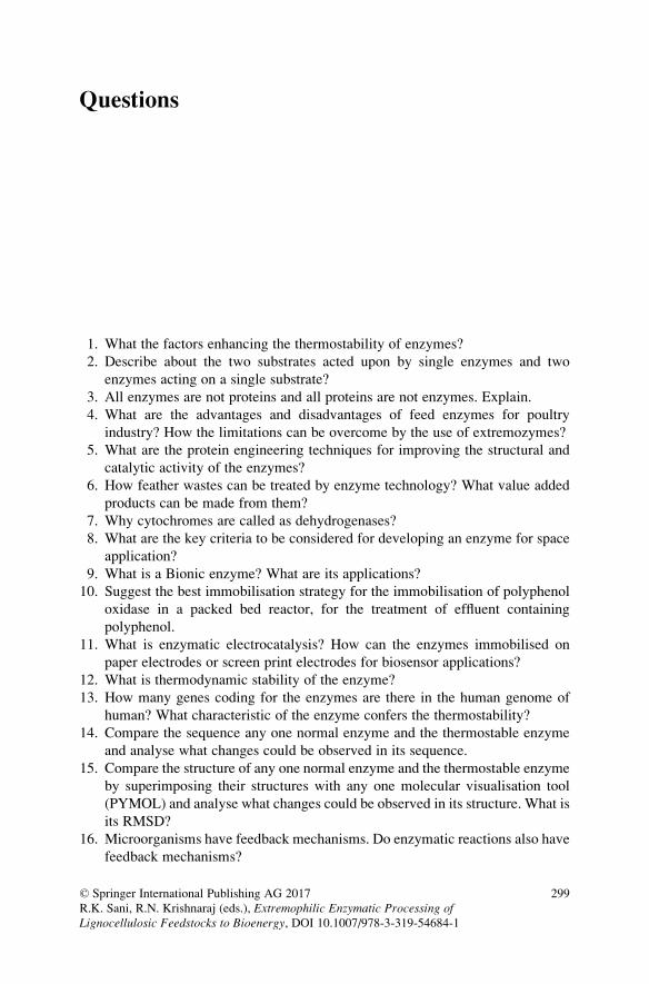
Questions
1. What the factors enhancing the thermostability of enzymes?
2. Describe about the two substrates acted upon by single enzymes and two
enzymes acting on a single substrate?
3. All enzymes are not proteins and all proteins are not enzymes. Explain.
4. What are the advantages and disadvantages of feed enzymes for poultry
industry? How the limitations can be overcome by the use of extremozymes?
5. What are the protein engineering techniques for improving the structural and
catalytic activity of the enzymes?
6. How feather wastes can be treated by enzyme technology? What value added
products can be made from them?
7. Why cytochromes are called as dehydrogenases?
8. What are the key criteria to be considered for developing an enzyme for space
application?
9. What is a Bionic enzyme? What are its applications?
10. Suggest the best immobilisation strategy for the immobilisation of polyphenol
oxidase in a packed bed reactor, for the treatment of effluent containing
polyphenol.
11. What is enzymatic electrocatalysis? How can the enzymes immobilised on
paper electrodes or screen print electrodes for biosensor applications?
12. What is thermodynamic stability of the enzyme?
13. How many genes coding for the enzymes are there in the human genome of
human? What characteristic of the enzyme confers the thermostability?
14. Compare the sequence any one normal enzyme and the thermostable enzyme
and analyse what changes could be observed in its sequence.
15. Compare the structure of any one normal enzyme and the thermostable enzyme
by superimposing their structures with any one molecular visualisation tool
(PYMOL) and analyse what changes could be observed in its structure. What is
its RMSD?
16. Microorganisms have feedback mechanisms. Do enzymatic reactions also have
feedback mechanisms?
© Springer International Publishing AG 2017
R.K. Sani, R.N. Krishnaraj (eds.), Extremophilic Enzymatic Processing ofLignocellulosic Feedstocks to Bioenergy, DOI 10.1007/978-3-319-54684-1
299
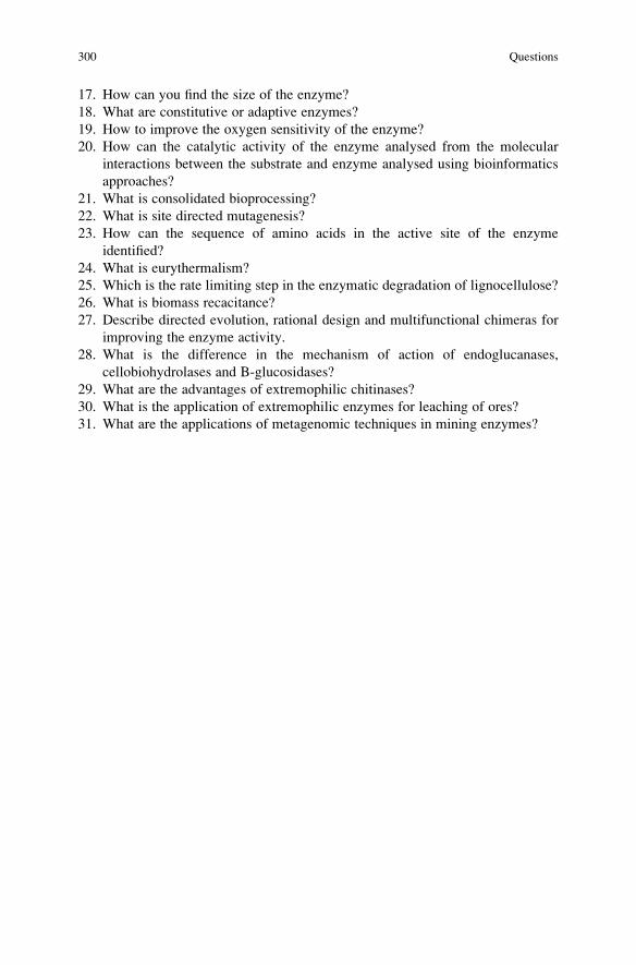
17. How can you find the size of the enzyme?
18. What are constitutive or adaptive enzymes?
19. How to improve the oxygen sensitivity of the enzyme?
20. How can the catalytic activity of the enzyme analysed from the molecular
interactions between the substrate and enzyme analysed using bioinformatics
approaches?
21. What is consolidated bioprocessing?
22. What is site directed mutagenesis?
23. How can the sequence of amino acids in the active site of the enzyme
identified?
24. What is eurythermalism?
25. Which is the rate limiting step in the enzymatic degradation of lignocellulose?
26. What is biomass recacitance?
27. Describe directed evolution, rational design and multifunctional chimeras for
improving the enzyme activity.
28. What is the difference in the mechanism of action of endoglucanases,
cellobiohydrolases and B-glucosidases?
29. What are the advantages of extremophilic chitinases?
30. What is the application of extremophilic enzymes for leaching of ores?
31. What are the applications of metagenomic techniques in mining enzymes?
300 Questions

Index
AAbsolute substrate specificity, 10, 27
Accessory enzymes, 94, 198, 199, 220
Acetyl esterase, 215, 273, 295
Acetyl mannan esterases (AMEs), 206, 215
Acetyl xylan esterases, 76, 273
Acid hydrolysis, 101, 163
Acid mammalian chitinase (AMCase), 242
Acid-based pretreatments, 35, 38–39, 46, 50
Acidophiles, 2, 4, 26, 100, 117, 118, 157, 158,
172, 182, 200, 260
Acidophilic enzyme, 26
Activation energy, 6, 16
Active site, 6, 9, 14, 15, 20, 21, 27, 54, 56, 92,
93, 97, 102, 120, 123, 141–143, 147,
216, 218, 219, 252, 265, 266
Adsorption, 2, 22, 23, 27, 219
Adverse conditions, 2, 4, 164
Alanine aminotransferase (ALT), 7
Alcohol dehydrogenase, 11
Algae, 2, 22, 117, 159, 227, 243
Alkaline pretreatments, 39
Alkaliphiles, 2, 4, 26, 100, 105, 106, 117, 118,
157, 158, 182, 259
Alkalophilicity, 81, 82, 267
Alkanine protease (ALP), 7
Allosteric enzymes, 15
Alpha-L-iduronidase, 7
Amino acid modification, 74, 80, 82, 84
Amino acid sequence, 12, 54, 55, 93, 97, 101,
108, 111, 140, 216, 227, 232, 290, 293
Aminophenols, 138, 276
Ammonia fiber explosion, 36, 39, 40, 42, 44,
48, 274
Ammonia recycle percolation (ARP), 37, 41
Amylase, 99, 100
α-Amylase, 7, 100–111
Amylopectin, 96, 97, 101, 102, 111
Amylose, 96, 97, 101, 107, 111
Antibiotics, 24, 59, 151, 214
Antibody binding, 159
Apoenzyme, 6, 11, 12
Arabinan Endo-1,5-α-L-arabinanase, 273Arabinans, 160, 273, 295
Arabinogalactans, 160
Arabinoxylan, 76, 77, 79, 83, 214
Arginase, 10
Aromatic ring cleavage, 144
Arrhenius equation, 16
Aryl alcohol oxidase, 273
Asparaginase, 7
Asparagine, 7, 82
BBacteria, 2, 199, 208, 214, 219, 220, 225, 227,
229, 232, 258, 260, 262, 265, 271, 273,
274, 281, 288
Bacterial artificial chromosomes (BAC), 59,
287
Bacterial chitinases, 229–231
Barophiles, 117, 182
Bilirubin oxidase, 7
Biobleaching, 74, 77, 119, 151–153
Biocatalysts, 2, 6, 89, 107, 183–185, 193, 199,
200, 208, 249, 250, 256, 258, 271, 274,
275, 279, 282–288, 292–294
Biochemical reaction, 6
Biofuels, 26, 62, 95, 110, 151, 215, 226, 243,
249, 271–275, 283, 284, 293
© Springer International Publishing AG 2017
R.K. Sani, R.N. Krishnaraj (eds.), Extremophilic Enzymatic Processing ofLignocellulosic Feedstocks to Bioenergy, DOI 10.1007/978-3-319-54684-1
301

Biological pretreatment, 37, 39, 46, 47, 49–50,
274
Biomass accessibility, 43, 46, 48, 49
Biomass degradation, 35–37, 39, 41, 43, 47, 49,
50, 275, 290, 293
Biomass digestibility, 44, 45, 206
Biomass fractionation, 35, 36, 38, 48
Bioprocess engineering, 1
Biorefineries, 32, 33, 41, 47, 48, 50, 51, 119
Bioremediation, 2, 119, 139, 144, 150, 190,
193, 201, 258, 268
Biosensors, 3, 7, 10, 24, 151, 153
Biotechnology, 1, 4, 24–26, 182, 208
Bleaching, 77
Bond specificity, 10, 27, 218
Broad specificity, 10, 27, 218
CCarbohydrate-active enzymes (CAZymes), 54,
69, 119, 198, 290
Carbohydrate binding modules, 56
Carbohydrate esterases (CE), 54, 69, 198, 220
Carbohydrates, 41, 54, 56, 69, 131, 140, 177,
274, 284
Carbonic anhydrase, 6, 10, 137, 292
Carboxylesterase, 182, 185–188, 193, 194,
200, 261
Carboxypeptidase, 10, 182
Carriers, 2, 22, 23, 27
Catalysts, 256, 258–260, 263
Catalytic activity, 8, 12, 15, 16, 22, 23, 77, 82,
138, 175, 219, 231, 258
Catalytic domains, 55, 56, 58, 229–231, 234
Catalytic efficiency, 18, 82, 189, 234, 267
Catalytic reaction, 6, 16, 17, 77, 256
Cellobiohydrolases (CBH), 56, 57, 90, 94, 95,
198
Cellobiose, 56, 58, 63, 64, 69, 90, 93–95, 277
Cellobiose dehydrogenase, 93
Cellotetraose, 56, 69
Cellotriose, 56, 69
Cellulase activity, 41, 42, 62–65, 287
Cellulases, 3, 24, 27, 32, 39–42, 44, 54–62, 64,
65, 67, 69, 74, 83, 91–96, 170, 171, 216,
219, 225, 233, 281, 283–285, 288, 292,
294, 295
Cellulolytic enzyme, 7, 56
Cellulolytic fungi, 56, 61, 69
Cellulose binding motif (CBM), 93
Cellulose crystallinity index (CrI), 34, 35, 38,
39, 42, 44
Cellulose degradation, 56, 58, 60–62, 69
Cellulosome, 7, 61, 219, 220
Cheese making, 8
Chemical hydrolysis, 109, 163, 164
Chemical processes, 1, 4, 226
Chitin, 3, 22, 58, 91–94, 96, 199, 225–227
α Chitin, 226, 243
β Chitin, 226, 243
Chitin degrading organisms, 93
Chitinase assay, 237–239
Chitinase production, 235–237
Chitinases, 91, 225–244
Chitinolytic activity, 233, 238, 243
Chitobiose, 93, 227
Chitosan, 22, 226, 234, 242, 243
Chymotrypsinogen, 6, 15
Cloning, 59, 68, 69, 100, 184, 185
Coenzymes, 2, 6, 11, 16
Cofactors, 2, 11, 12, 14, 16, 182
Cofactor specificity, 11
Cold-adapted microorganisms, 105
Collagenase, 7
Competitive inhibition, 20, 21
Conserved carboxylic residues, 93
Conserved domains, 108, 214
Cooperativity, 15
Copolymerization, 22
Copper dependent enzymes, 91, 96
Corn stover, 39, 41, 43, 44, 46, 47, 53
Cosmid, 59, 287
Covalent bonding, 2, 14, 22, 23
Covalent modification, 159
Cross linking, 24, 27, 159
Crystallization, 9, 109
Cyanide poisoning, 7
Cyclodextrin glucanotransferase, 101, 111
Cyclodextrinase, 101, 111
DD- Galactose, 160
Degree of enzyme purity, 108
Degree of polymerization, 63, 76, 83
Deletions, 58, 68, 231, 239, 240, 289, 291
Delignification, 124, 131, 143, 151, 152
Demethylation, 134, 135, 143
Denaturation, 16, 17, 83, 108, 184
Denaturation temperature midpoint, 83
Depolymerase, 165, 168, 178
Depolymerase assay, 169, 178
Depolymerization, 101, 131, 143, 163, 198
Deprotonation, 135, 144
Detoxification of lignin, 45, 116
302 Index

Dextrins, 109–111
D-glucose, 127, 160, 198
β-D-glucosidases, 64, 273α-D-glucuronidases, 273D-glucturonic acid, 160
Dialyzed enzyme, 137
Dimethylphenylenediamine, 135
Diphenol oxidase, 138
Directed evolution, 80–84, 261, 289–292, 294
Distal domain, 131
Disulfide bond, 82, 120, 130, 152, 265
D-mannose, 160, 215
DNA libraries, 58, 74, 185
DNA polymerase, 3, 6, 25, 289
DNA shuffling, 74, 81, 82, 289
Double displacement mechanism, 234
Drug metabolizing enzyme, 12
D-xylose, 77, 83, 160, 198
β-D-xylosidases, 273Dye decolurisation, 119
Dynamic high pressure microfluidization, 163
EEadie–Hofstee plot, 19
Effectiveness, 2, 22, 33, 35, 36, 39, 42, 45, 49,
50, 110, 239, 286
Effectors, 15, 21, 122
Electron transport system, 139
Encapsulation, 2, 22, 24, 27
Endo arabinases, 163
Endo galactoses, 163
Endo polygalacturonases, 163
Endo- xylanase, 198
Endo-1,4-β-D-glucanases, 273Endo-1,4-β-D-xylanases, 273Endo-1,4-β-galactanase, 273Endoamylases, 101, 111
Endoglucanases, 56, 61, 63, 64, 69, 90, 95, 198
Endo-pectate lyases, 273
Endo-pectin lyases, 273
Endopeptidase, 10
Endo-polygalacturonase, 273
Endopolygalacturonate lyase, 166
Endopolymethylgalacturonate lyase, 166
Endozym, 12
Energy, 13, 16, 17, 22, 25, 32, 34, 37, 38, 41,
45, 46, 48–50, 67, 77, 90, 99
Entrapment, 2, 21–24, 27
Enzymatic degradation, 59, 111, 124, 163, 164,
198
Enzymatic hydrolysis, 32, 33, 37, 39, 41–45,
50, 54, 96, 163, 198, 274, 275, 291–293
Enzyme Commission, 13, 27
Enzyme kinetics, 9, 17–20
Enzymes, 12–15
Enzyme-substrate complex, 9, 20, 21
Error prone PCR, 74, 81, 82
Esterases, 83, 163, 167, 181–201
Exo-1,4-β-D-glucan cellobiohydrolase, 273,
295
Exo-1,4-β-D-glucanases, 32, 273Exo-1,4-β-mannobiohydrolase, 273
Exoamylases, 90, 102, 111
Exoglucanases, 56, 63, 64, 69
Exo-pectin lyases, 273
Exo–polygalacturonase, 273
Exopolygalacturonate lyase, 166
Exopolymethylgalacturonate lyase, 166
Exozymes, 12
Exo-β-D-mannanase, 273
Extreme radiations, 2, 4
Extremozymes, 2, 4, 25–27, 100, 118,
158–159, 171, 199, 256, 258, 263, 271,
273–284, 288–294
Extrusion, 35, 38, 49, 50, 274
FFeedstocks, 31–50, 159, 198–199, 208, 218,
220, 273
Fermentation, 8, 10, 66, 110, 148, 170, 172,
174, 176–178, 199, 208, 215, 235–237,
257, 258, 260, 261, 268, 274, 283
Ferulic acid (FA), 127, 199, 215
Ferulic acid esterase (FAE), 216, 218
Feruloyl esterases, 199, 273
Fibrinolytics, 7
Fluorimetric assays, 107
Food processing, 25, 27, 117, 157, 170, 193
Fosmid, 59, 218, 220, 287
Functionalization, 2
Fungal chitinases, 231
Fungi, 2, 22, 56, 60, 61, 65, 66, 68, 69, 74, 78,
83, 90–92, 97, 119, 120, 122, 124, 125,
131, 134, 138–140, 145, 146, 164, 168,
172, 173, 199, 216, 227, 229, 231–233,
235, 239, 242, 243, 257, 271, 273, 274,
281, 283
Gβ-Galactosidase, 26, 273Galacturonic acid, 159, 169, 177, 219
Gene expression, 107, 184, 285, 287
Genetic engineering, 2, 7, 225, 239–241, 261
Index 303

Genome sequencing, 65, 69
Genomic analysis, 108
Genomics, 171, 271
Geometric specificity, 11
Glucoamylase, 3, 101, 102, 104, 111, 283
Glucocerebrosidase, 7
Glucose, 10, 24, 58, 63, 64, 67, 69, 90, 93
Glucose oxidase, 63, 64, 273, 295
α-Glucosidase, 101, 111Glucuronoarabinoxylan endo-1,4-β-xylanase,
273, 295
Glucuronoyl esterases (GEs), 199, 219
Glutaminase, 7
Glycoprotein, 131, 140
α- Glycosidase, 11Glycoside hydrolase (GH), 60, 67, 92, 206, 290
Glycosides, 8
Glycosidic bonds, 11, 56, 69, 91, 96, 101, 102,
111, 165, 169, 198, 227, 233, 243
β-Glycosidic bonds, 11Glycosyl hydrolases (GH), 3, 54, 56, 58, 60, 65,
69
Glycosyl transferases (GT), 54, 69
Glycosylation, 55, 167, 233, 262
Glyoxal oxidase, 273
Grinding, 237
Group specificity, 10, 27
Guaiacol, 127, 128, 134, 135, 138, 148
HH2O2-dependent oxidative depolymerization,
131
Half life, 25, 75, 78, 80–82, 108, 140, 149, 185,
186, 214, 231, 232, 241, 283
Halophiles, 2, 4, 100, 106, 118, 157, 158, 182,
193, 200
Hanes-Woolf plot, 19
Hemicellulases, 74, 83, 170, 218, 220, 273,
281, 284, 291, 294, 295
Hemicellulose, 25, 32, 33, 35–37, 39–47, 49,
50, 53, 54, 65, 69, 74, 76, 83, 92, 96,
198, 199, 206, 208, 215, 216, 271–274,
283, 284, 290, 294
Heterologous expression, 107, 108, 130, 283
Heterologous host-vector systems, 2
Hexokinase, 10
High fructose corn syrup, 109, 111
High methoxyl pectins (HMP), 163
High-throughput–omics, 108
Hill equation, 19, 20
Holoenzyme, 6
Homogalacturonan, 159, 177
Homology modelling, 140
Horseradish peroxidases, 134
Host proteolysis, 108
Host-vector systems, 2, 107
Hydrogen abstraction, 124, 138
Hydrogen bonding, 9
Hydrolases, 3, 13
Hydrolysis, 54–58, 61, 63, 64, 69, 73, 83,
94–97, 109
Hydrolysis of cellulose, 63, 90
Hydrolytic capability, 83
Hydrostatic pressure, 117, 156, 157
Hydrothermal pretreatment, 36, 43, 49
Hydrothermal processing, 163
Hyperthermophiles, 103, 184, 185, 200, 281,
283, 286
Hyperthermophilic microorganisms, 107, 257,
275
Hyperureamia, 7
IImmobilization processes, 2
Immobilized enzymes, 7, 24
Induced fit hypothesis, 14
Inhibition, 15, 20, 21, 27, 64, 94, 285
Insect chitinases, 233
Insertions, 68, 289, 291
Internatinal unit (IU), 16, 27, 129, 137
Invertin, 8
Ionic liquid, 35, 40, 41, 274
Iron protoporphyrin, 131
Irreversible inhibition, 15
Isomaltose, 9, 101
Isozymes, 12, 120, 131
KKatal, 16, 27
km, 9, 12, 18, 21, 116, 291
LLaccase, 46, 118, 123, 128, 134, 138–153, 273,
284, 295
Lactase, 10
Lactate dehydrogenase, 12
L-arabinofuranose, 76
L-arabinofuranose residues, 76
α-L-arabinofuranosidases, 32, 273L-arabinose, 160
Leukemia, 7
L- fucose, 160
304 Index

Ligases, 13
Lignin, 32–37, 39–48
Lignin content, 33, 39, 42, 44
Lignin-modifying enzymes (LMEs), 273, 295
Ligninolytic enzymes, 115–153
Lignin oxidation, 37, 45
Lignin peroxidase (LiP), 118, 132–135, 137,
152, 273, 276, 295
Lignin sulfonation, 37
Lignocellulose, 54, 60, 64, 73, 78, 83, 90, 91,
96, 199, 214, 272–275, 281, 283, 286,
294, 295
Lignocellulosic feedstocks, 31–50, 198, 208,
218, 220, 271–295
Lignocellulosic fractionation, 36, 40
Lineweaver–Burk plot, 18
Lipase, 27, 170, 182, 199, 200, 212, 214, 218,
249–254
Liquefaction, 25, 100, 101, 109, 110, 171
Liver cirrhosis, 7
Lock and key theory, 14, 27
Low methoxyl pectins (LMP), 163
Lytic polysaccharide monoxygenases, 91, 96
MMaltodextrins, 101
Maltose, 105, 109
Manganese peroxidase (MnP), 118–130, 132,
134, 139, 144, 149–153, 295
β-mannanase, 57
Mannan endo-1,4-β-mannosidase, 273, 295
Mannose, 55, 121, 140, 167
Mass transfer, 2, 22, 24
Maximum velocity, 9, 18
Mediator, 123, 124, 133, 142–144, 147
Metabolic channelling, 15
Metabolic pathway, 2, 196, 274
Metagenome, 59, 108, 183, 285–288
Metagenome library, 288
Metagenomic approaches, 59, 83, 190, 262,
275, 286, 287
Metagenomics, 58–60, 77, 80, 108, 182, 185,
188, 197, 208, 218, 221, 271, 284–288,
294
Metal activated enzymes, 12
Metalloenzymes, 12
Metal-resistant microorganisms, 2
Methoxy esters, 167
Methoxyl groups, 161
Methyl 3,4-Dihydroxycinnamate (MCA), 217
Methyl 3,5-Dimethoxy-4-Hydroxycinnamate
(MSA), 217
Methyl 3-Methoxy-4-Hydroxycinnamate
(MFA), 217
Methyl 4-Hydroxycinnamate (MpCA), 217
Michaelis Menten constant, 9
Microbial bioprocesses, 1
Microbial dark matter, 282
Microorganisms, 59–62, 69, 78–80, 83, 92, 95,
97, 99, 100, 103, 105–107, 111, 119,
125–129, 131, 135–138, 145–149, 152,
164, 168, 171, 172, 174, 176, 182–185,
188, 190, 196
Milling, 34, 38
Modulators, 15, 56
Moisture content, 33
Molecular biology, 3, 111, 185, 192, 225, 240,
241, 243, 257, 267
Molecular docking, 145
Molecular flexibility, 189
Molecular geometry, 11, 20
Molecular scissors, 7
Molecular structure, 120–122, 131–132,
140–141
Molecular weight (MW), 6, 24, 93, 101, 107,
110, 119, 137, 139, 140, 143, 151, 159,
164, 169, 177, 229, 232, 233, 239
Monooygenases, 94
Monophenol, 138, 142, 147
Monte Carlo (MC), 145
Multi-component cellulolytic enzyme, 56
Multi-enzyme complex, 15
Multiple covalent immobilization, 159
Mutation, 67, 130, 152, 231, 234, 240, 261,
263, 289–291, 293
Myocardial infarction, 7
NNegative cooperativity, 15
Negative modulators, 15
Nomenclature, 11, 13, 164–168
Non-competitive inhibition, 21
Normal enzymes, 2, 3, 12, 15
OOligosaccharides, 8, 63, 90, 91, 101, 102, 110,
163, 164, 177, 208, 232
O-linked glycosylation, 55, 167
Organic acids, 24, 32, 39, 47, 119, 122
Organic solvents, 2, 36, 37, 40, 41, 46, 100,
105, 118, 130, 152, 157, 158, 173, 177,
182–184, 188, 193, 197, 259, 262, 263,
272, 274
Organosolv pretreatment, 40
Index 305
Oxidation, 37, 42, 45, 47, 48, 94, 120, 122–124,
128, 131–134, 136–138
Oxidative cleavage, 91, 96, 124, 131
Oxidoreductases, 3, 13, 27, 66, 206
Ozonolysis, 36, 40, 42, 48, 274
PPaper industry, 25, 74, 83, 171, 257, 268
Pattern-mimicking, 108
p-Coumaroyl esterases, 273, 278, 295
PCR primers, 108
Pectales, 161
Pectase, 167
Pectic acid, 161, 164, 173, 178, 218
Pectic oligosaccharides, 163
Pectin – methyl esterases, 163
Pectin degrading enzymes, 164–171
Pectin demethoxylase, 167
Pectin depolymerization, 163
Pectin esterase, 168, 178, 273, 295
Pectin methoxylase, 167
Pectin methylesterases, 167, 199
Pectinase, 24, 68, 164, 168–178, 273, 295
Pectinesterases (PE), 167
Pectinic acids, 161, 178
Pectinotes, 161
Pectins, 159–164, 169, 177, 178, 199
Pectlylhydrolase, 167
Pectolipase, 167
Pelleting process, 74
Pepsin, 6, 9, 10, 12, 15, 17
Pepsinogen, 6, 15
Peptidases, 3, 10
Peptide mass fingerprinting (PMF), 68
Peroxidases, 66, 119, 120, 131, 132, 251
Phenolic aromatic compounds, 134
Phenylmethylsulfonyl fluoride (PMSF), 187,
218
Physicochemical pretreatment processes, 34,
42, 45
Piezophiles, 196–197, 200
Piezophilic adaptation, 196
Plant chitinases, 231, 232
Plants, 2, 6, 25, 53, 66, 76, 77, 83, 92, 99, 101,
110, 138, 145, 159, 168, 232, 233, 241
Plasmid, 59, 65, 239, 287, 289
Plate assay, 62, 127, 136, 137
Polyamines, 138
Polycyclic hydrocarbon, 138
Polyextremophilic esterases, 197
Polyextremophilic microorganisms, 103
Polymerase chain reaction (PCR), 3, 7, 25, 67,
74, 108, 285–287, 289, 291
Polymerization, 33, 127, 134, 138, 142, 143
Polymethyl galactenate, 161
Polyphenol oxidase, 138
Polysaccahridelyases (PL), 54, 69
Positive cooperativity, 15
Positive modulators, 15
Post-translational modification, 233
Precipitation, 117, 129, 137, 148, 177, 193,
237, 238
Pressure, 1, 2, 4, 6, 16, 17, 25–27, 34, 41–45,
47, 100, 106, 109, 116–118, 130, 156,
157, 163, 189, 193, 196, 197, 200, 258,
274, 275, 279, 287
Pressure resistant enzymes, 109
Pretreatment, 274, 275, 284, 290, 293
Product, 2, 6, 8, 10, 11, 13, 14, 17, 18, 20, 21,
27, 32, 34, 38, 40–44, 46–50, 54, 56, 58,
63, 64, 73, 77, 93, 96, 97, 101, 102, 108,
109, 111, 123, 124, 127–129, 133–135,
137, 139, 143, 144, 148, 150–152, 165,
170, 171, 176, 177, 184, 188, 189, 212,
218, 227, 233, 234, 238, 239, 241, 242,
250, 256, 257, 259, 260, 277, 285, 286,
288
Promoters, 69, 108, 239, 283, 288
Prosthetic group, 6, 11, 121, 132
Protease, 3, 6, 10, 12, 13, 25–27, 55, 65, 74,
199, 251, 263
Protein engineering, 2, 239, 288–290,
292–293
Protein folding, 54, 283, 293
Protein misfolding, 288
Protein-protein non covalent
associations, 159
Proteins, 2, 3, 6, 8, 9, 11, 13, 15–17, 25–27,
59–61, 63, 65–69, 77, 80, 81, 91–93, 97,
108, 118, 120, 129, 130, 137, 140–142,
145, 147, 148, 158, 164, 169, 177, 182,
184, 185, 188, 189, 193, 196, 200, 214,
219, 221, 232–235, 239, 242, 243, 253,
259, 262–265, 275, 283, 288–295
Proteolysis, 2, 108, 233
Proteomics, 65, 66
Proto pectin, 161, 178
Protopectinase assay, 169, 178
Protoplast fusion, 66, 68
Proximal domain, 131
Psychrophiles, 100, 105, 118, 157, 158, 174,
175, 182, 188, 189, 258
Pullulan, 102, 107
Pullulanase, 3, 101, 102, 111
Pulping, 74, 119, 151–153
Purification, 136–138, 147–149, 170–172, 190,
264
306 Index

QQuinone, 135, 138, 139, 142, 143
RRandom amplified polymorphic
DNA analysis, 68
Random mutagenesis, 80, 159, 289
Reaction, 2, 3, 6–13
Reaction catalysis, 6
Reaction mechanisms, 101, 111, 123, 164
Reactive specificity, 126
Recalcitrance of lignocellulose, 37, 45
Recalcitrant structure, 32
Recombinant enzyme, 107, 108, 286
Recombinant expression, 283, 287, 288, 294
Reducing agent, 91, 93, 94
Reductases, 12
Restriction digestion pattern, 68
Restriction enzymes, 7, 9
Reversible inhibition, 20
Rhamnogalacturonan acetyl
esterase, 273, 295
Rhamnogalacturonan
galacturonohydrolase, 167
Rhamnogalacturonan hydrolase, 167
Rhamnogalacturonan lyase, 167, 273, 295
Rhamnogalacturonan rhamnohydrolase, 167
Rhamnogalacturonan-I, 159, 160, 177, 178
Rhamnogalacturonan-II, 159, 177
Rhodonase, 7
Ribozyme, 2, 6, 27
Rosettazyme, 7
SSaccharification, 34–36, 38, 40–42, 44, 46, 47,
77, 96, 100, 101, 172, 178, 198, 199,
274, 290
Salt-tolerant α-amylase, 108
β-Sandwich structure, 93
Scaffoldin, 220
Secretome, 56, 65–67, 283
Sensitivity selectivity, 2
Severity factor (SF), 42, 43
Signal peptide, 108, 140, 240, 257
Site directed mutagenesis, 2, 74, 80, 149, 152,
159, 239, 291
Skin ulcers, 7
Soaking in aqueous ammonia (SAA), 36, 40,
41, 128
Sodium dodecyl sulfate-polyacrylamide gel
electrophoresis (SDS-PAGE), 68, 129,
137, 138, 239
Solid state fermentation, 66
Specific activity, 3, 16, 104, 105, 129, 148, 167,
275, 281, 291, 294
Specific mediator, 143
Specificity, 4, 6, 8–11, 14, 27, 103, 198, 199,
214, 216, 219, 239, 252, 265, 267, 286,
289, 293, 294
Stability, 104, 106, 110, 118, 130, 140, 144,
151, 152, 158, 171, 173
Starch, 101–102, 104, 106, 107, 109
Starch degrading enzymes, 101–102
Starch hydrolysis, 109
Starch liquefaction, 25, 100, 109
Starch-degrading enzymes, 109
Steam explosion, 36, 42–44, 49, 50, 274
Stereochemistry, 234, 235
Stereochemistry retention, 234
Sterochemical specificity, 27
Streptokinase, 7
Structural homology, 91, 93
Structural stability, 2, 175
Substance, 6, 8, 16, 125, 159, 161, 164, 165,
169, 178, 190, 234, 238, 257
Substrate, 9–18
Substrate concentration, 9, 16–21, 27
Sugar beet pulp (SBP), 159, 273
Sugar biorefinery platform
Sugarcane bagasse, 38, 39, 42, 43, 53, 176
Sulfite pretreatment, 37, 45, 46
Supercritical fluids, 37, 46
Surface area, 23, 33–35, 38, 62, 149, 152, 265,
284
Surface immobilization, 2
Syringic acid, 134–136
TTaq polymerase, 7
Telomerase, 9
Temperature, 104, 108, 110, 116–118, 127,
130, 138, 140, 149, 152, 157, 163, 169,
173–178, 184, 185, 188–190, 196, 197,
200, 208, 215, 216, 229, 231
The alcohol dehydrogenase, 25
Thermal denaturation, 108
Thermoactive, 108, 214
Thermophiles, 83, 100, 103–105, 117, 118,
157, 158, 173–175, 182–184, 189, 200,
281
Index 307

Thermophilic fungi, 83, 216, 239, 257
Thermophilicity, 81, 82, 267
Thermostability, 26, 75, 77, 80–83, 100, 101,
105, 107–109, 138, 149, 152, 183–185,
189, 218, 240, 257, 260, 263, 267, 285,
289, 293, 294
Thermostable enzymes, 3, 25, 77, 100, 119,
130, 149, 152, 173, 185, 199, 281, 284,
286, 293
Thermostable xylanases, 74, 77, 82
Total molecular mass, 140
Transaminases, 13
Transcription factor, 108
Transcriptional engineering, 108
Transferases, 13
Transglycosylation activity, 26, 233
Transition-state complex, 9
Trypsinogen, 6, 15
Turnover number, 9, 27
UUltrasound pretreatment, 37, 45, 46
Uncompetitive inhibition, 21
Urease, 7, 9, 10
Uricase, 7, 10
Urokinase, 7
VVan der Waals interactions, 9
Vander Walls Forces, 2
Vanillic acid, 135
Veratraldehyde, 124, 134, 135
Versatile peroxidase, 273, 295
Viscosity, 25, 63, 100, 102, 109, 169–171, 175,
184, 256
Vital factor, 8
Vitamins, 11
WWater treatment, 170, 178
Western blot, 67
Wet oxidation, 37, 42, 45, 47, 48, 274
Wheat bran, 78, 79, 176
Wheat straw, 38–40, 42, 47, 53, 78, 139, 218
XXenophiles, 2, 4
Xylan, 25, 76–78, 83, 198, 206, 214, 215, 219,
283, 286, 291, 294, 295
Xylanases, 75, 77–84, 170, 214, 216, 220, 290
Xylanolytic enzymes, 76, 78, 83
Xylogalacturonans, 160
Xyloglucan-specific endo-β-1,4-Glucanase,273, 295
β-Xylosidase, 57, 75, 76, 78, 80, 83, 198, 290,291
YYeast, 8, 9, 58, 68, 164, 168, 177, 178, 231,
239, 242, 243
ZZinc finger protein, 67
Zymase, 8
Zymogens, 6, 15
Zymogram, 67
308 Index


Developments in Fungal Biologyand Applied Mycology

Tulasi SatyanarayanaSunil K. Deshmukh • B. N. JohriEditors
Developments in FungalBiology and AppliedMycology
123

EditorsTulasi SatyanarayanaBiological Sciences and EngineeringNetaji Subhas Institute of Technology(University of Delhi)
New DelhiIndia
Sunil K. DeshmukhTERI-Deakin Nano-Biotechnology CentreThe Energy and Resources Institute (TERI)New DelhiIndia
B. N. JohriDepartment of BiotechnologyBarkatullah UniversityBhopal, Madhya PradeshIndia
ISBN 978-981-10-4767-1 ISBN 978-981-10-4768-8 (eBook)https://doi.org/10.1007/978-981-10-4768-8
Library of Congress Control Number: 2017958608
© Springer Nature Singapore Pte Ltd. 2017, corrected publication 2020This work is subject to copyright. All rights are reserved by the Publisher, whether the whole or partof the material is concerned, specifically the rights of translation, reprinting, reuse of illustrations,recitation, broadcasting, reproduction on microfilms or in any other physical way, and transmissionor information storage and retrieval, electronic adaptation, computer software, or by similar or dissimilarmethodology now known or hereafter developed.The use of general descriptive names, registered names, trademarks, service marks, etc. in thispublication does not imply, even in the absence of a specific statement, that such names are exempt fromthe relevant protective laws and regulations and therefore free for general use.The publisher, the authors and the editors are safe to assume that the advice and information in thisbook are believed to be true and accurate at the date of publication. Neither the publisher nor theauthors or the editors give a warranty, express or implied, with respect to the material contained herein orfor any errors or omissions that may have been made. The publisher remains neutral with regard tojurisdictional claims in published maps and institutional affiliations.
Printed on acid-free paper
This Springer imprint is published by Springer NatureThe registered company is Springer Nature Singapore Pte Ltd.The registered company address is: 152 Beach Road, #21-01/04GatewayEast, Singapore 189721, Singapore
To late Prof. S. B. Saxena, whosebirth centenary has beencelebrated during 2016–2017
Preface
Fungal biology deals with the study of fungi, including their growth and devel-opment, their genetic and biochemical characteristics, their taxonomy and geno-mics, and their use to humans. The current research focuses on mushrooms whichmay have hypoglycemic activity, anticancer activity, anti-pathogenic activity, andimmune system-enhancing activity. A recent research has found that the oystermushroom naturally contains the cholesterol-lowering drug, lovastatin, thatmushrooms produce large amounts of vitamin D when exposed to UV light, andthat certain fungi may be a future source of taxol. To date, penicillin, lovastatin,cyclosporine, griseofulvin, cephalosporin, ergometrine, and statins are the mostfamous pharmaceuticals which have been isolated from fungi.
Fungi are fundamental for life on earth in their roles as symbionts (e.g., in theform of mycorrhizae, insect symbionts, and lichens). Many fungi are able to breakdown complex organic biomolecules such as lignin, and pollutants such as xeno-biotics, petroleum, and polycyclic aromatic hydrocarbons. By decomposing thesemolecules, fungi play a critical role in the global carbon cycle.
The kingdom fungi encompasses an enormous diversity of taxa with variedecologies, life cycle strategies, and morphologies ranging from unicellular aquaticchytrids to large mushrooms. Little is, however, known about their true biodiver-sity, which has been estimated at 1.5 to 5 million species, with about 5% of thesehaving been formally classified. Advances in molecular genetics have opened theway for DNA analysis to be incorporated into taxonomy, which has sometimeschallenged the historical groupings based on morphology and other traits. Phylo-genetic studies published in the last decade have helped reshape the classificationwithin kingdom fungi, which is divided into one subkingdom, seven phyla, and tensubphyla.
The human use of fungi for food preparation or preservation and other purposesis extensive and has a long history. Mushroom farming and mushroom gatheringare large industries in many countries. The study of the historical uses and socio-logical impact of fungi is known as ethnomycology. Because of the capacity of thisgroup to produce an enormous range of natural products with antimicrobial or otherbiological activities, many species have long been used or are being developed forindustrial production of antibiotics, vitamins, and anticancer andcholesterol-lowering drugs. More recently, methods have been developed for
vii
genetic engineering of fungi, enabling metabolic engineering of fungal species. Forexample, genetic modification of yeast species, which are easy to grow at fast ratesin large fermentation vessels, has opened the way for pharmaceutical productionthat are potentially more efficient than production by the original source organisms.
Several pivotal discoveries in biology have been made by researchers usingfungi as model organisms, which grow and sexually reproduce rapidly in the lab-oratory. For example, the one gene–one enzyme hypothesis was formulated byscientists using the bread mold Neurospora crassa to test their biochemical theo-ries. Other important model fungi are Aspergillus nidulans and the yeasts Sac-charomyces cerevisiae and Schizosaccharomyces pombe, each with a long historyof use to investigate issues in eukaryotic cell biology and genetics, such as cellcycle regulation, chromatin structure, and gene regulation. Other fungal modelshave more recently emerged that address specific biological questions relevant tomedicine, plant pathology, and industrial uses; examples include Candida albicans,a dimorphic, opportunistic human pathogen;Magnaporthe grisea, a plant pathogen;and Pichia pastoris, a yeast widely used for eukaryotic protein production. Fungiare used extensively to produce industrial chemicals such as citric, gluconic, lactic,and malic acids, and industrial enzymes such as lipases used in biological deter-gents, cellulases used for making cellulosic ethanol and stonewashed jeans, andamylases, invertases, proteases, and xylanases. Several fungi such as Psilocybemushrooms (colloquially known as magic mushrooms) are ingested for their psy-chedelic properties, both recreationally and religiously.
We are grateful to Prof. G. P. Mishra, Prof. R. S. Mehrotra, and Dr. Shashi Raifor their constant encouragement in bringing out this book on the occasion of thebirth centenary of late Prof. S. B. Saksena.
The book is an attempt in collating recent developments in fungi from variousenvironments: their diversity and potential applications. We greatly appreciate theefforts of experts in contributing on various aspects of fungi. The opinionsexpressed by the authors are their own. We wish to thank all the contributors forreadily accepting our invitation and Springer for publishing the book.
New Delhi, India Tulasi SatyanarayanaNew Delhi, India Sunil K. DeshmukhBhopal, India B. N. Johri
viii Preface
Contents
1 Significant Contributions of Prof. S.B. Saksena to IndianMycology . . . . . . . . . . . . . . . . . . . . . . . . . . . . . . . . . . . . . . . . . . . . . . . 1R. S. Mehrotra, M. R. Siddiqui and Ashok Aggarwal
2 Biology and Significance of Saksenaea vasiformis . . . . . . . . . . . . . . . 19Itisha Singh and R. K. S. Kushwaha
3 History of Mycology from India—Some Glimpses . . . . . . . . . . . . . . 29C. Manoharachary
4 Various Aspects of Ammonia Fungi. . . . . . . . . . . . . . . . . . . . . . . . . . 39Akira Suzuki
5 Marine Filamentous Fungi: Diversity, Distribution andBioprospecting . . . . . . . . . . . . . . . . . . . . . . . . . . . . . . . . . . . . . . . . . . . 59K. R. Sridhar
6 Keratinophilic Fungi Distribution, Pathogenicity andBiotechnological Potentials . . . . . . . . . . . . . . . . . . . . . . . . . . . . . . . . . 75Shilpa A. Verekar and Sunil K. Deshmukh
7 RETRACTED CHAPTER: Fungal World of Cave Ecosystem . . . .. . . . 99Seema Rawat, Rachna Rautela and B. N. Johri
8 Anaerobic Gut Fungi . . . . . . . . . . . . . . . . . . . . . . . . . . . . . . . . . . . . . 125D. N. Kamra and Birbal Singh
9 Fungal Endophytes Representing Diverse Habitats and TheirRole in Plant Protection . . . . . . . . . . . . . . . . . . . . . . . . . . . . . . . . . . . 135Satish K. Verma, Surendra K. Gond, Ashish Mishra,Vijay K. Sharma, Jitendra Kumar, Dheeraj K. Singh, Anuj Kumarand Ravindra N. Kharwar
10 Fusarium oxysporum: Genomics, Diversity and Plant–HostInteraction . . . . . . . . . . . . . . . . . . . . . . . . . . . . . . . . . . . . . . . . . . . . . . 159Anjul Rana, Manvika Sahgal and B. N. Johri
ix
11 Yeast Genome Sequencing: Basic Biology, Human Biology, andBiotechnology. . . . . . . . . . . . . . . . . . . . . . . . . . . . . . . . . . . . . . . . . . . . 201Krishna Kant Sharma
12 Fungal Differentiation: A Model Phenomenon to ScreenAntifungal Drugs . . . . . . . . . . . . . . . . . . . . . . . . . . . . . . . . . . . . . . . . . 227E. K. Pathan, S. G. Tupe and M. V. Deshpande
13 Candida Albicans Biofilm as a Clinical Challenge . . . . . . . . . . . . . . . 247Ashwini Jadhav and Sankunny Mohan Karuppayil
14 Characteristics and Multifarious Potential Applications of HAPPhytase of the Unconventional Yeast Pichia anomala . . . . . . . . . . . . 265Swati Joshi and Tulasi Satyanarayana
15 Fungal Inulinolytic Enzymes: A Current Appraisal . . . . . . . . . . . . . 279Hemant Kumar Rawat, Hemant Soni and Naveen Kango
16 Fungal Tannase: Recent Advances and IndustrialApplications . . . . . . . . . . . . . . . . . . . . . . . . . . . . . . . . . . . . . . . . . . . . . 295Sunny Dhiman, Gunjan Mukherjee, Anu Kumar, Papiya Mukherjee,Shilpa A. Verekar and Sunil K. Deshmukh
17 Mycoremediation: An Alternative Treatment Strategy for HeavyMetal-Laden Wastewater . . . . . . . . . . . . . . . . . . . . . . . . . . . . . . . . . . 315Tuhina Verma, Annapurna Maurya, Manikant Tripathiand Satyendra Kumar Garg
18 Treatment of Landfill Leachate Using Fungi: An Efficientand Cost-Effective Strategy. . . . . . . . . . . . . . . . . . . . . . . . . . . . . . . . . 341Pooja Ghosh and Indu Shekhar Thakur
19 Studies on Mycorrhiza in Pinus gerardiana Wall. ex D. Don,a Threatened Pine of the NW Himalaya . . . . . . . . . . . . . . . . . . . . . . 359Sunil Kumar, Vaneet Jishtu, J. S. Thakur and T. N. Lakhanpal
20 Role of Phosphate-Solubilizing Fungi in Sustainable Agriculture . . .. . . . 391Gurdeep Kaur and M. Sudhakara Reddy
21 Biotechnological Advancements in Industrial Productionof Arbuscular Mycorrhizal Fungi: Achievements, Challenges,and Future Prospects . . . . . . . . . . . . . . . . . . . . . . . . . . . . . . . . . . . . . 413Ankit Kumar, Reena Singh and Alok Adholeya
22 Role of Fungicides in Crop Health Management: Prospectsand Challenges . . . . . . . . . . . . . . . . . . . . . . . . . . . . . . . . . . . . . . . . . . 433T. S. Thind
23 Bioherbicides: Strategies, Challenges and Prospects . . . . . . . . . . . . . 449K. R. Aneja, S. A. Khan and A. Aneja
x Contents

24 Characterization of Lamellate Mushrooms—An Appraisal . . . . . . . 471N. S. Atri, Munruchi Kaur and Samidha Sharma
25 Occurrence and Distribution of Mushrooms in Semi-evergreenSal (Shorea robusta) Forest Chhattisgarh, Central India . . . . . . . . . 501Kamlesh Shukla, Bhoopander Giri and R. V. Shukla
26 Fungal Pigments: An Overview . . . . . . . . . . . . . . . . . . . . . . . . . . . . . 525Gunjan Mukherjee, Tulika Mishra and Sunil K. Deshmukh
27 Ex situ Conservation of Fungi: A Review . . . . . . . . . . . . . . . . . . . . . 543Sanjay K. Singh
28 Camouflaged Mycotoxins in Some Field Crops and Forages:A Review . . . . . . . . . . . . . . . . . . . . . . . . . . . . . . . . . . . . . . . . . . . . . . . 563Skarma Nonzom and Geeta Sumbali
Index . . . . . . . . . . . . . . . . . . . . . . . . . . . . . . . . . . . . . . . . . . . . . . . . . . . . . . 601
Contents xi

Editors and Contributors
About the Editors
Prof. Tulasi Satyanarayana became a faculty fellow at the Division of Biological Sciences andEngineering, Netaji Subhas Institute of Technology (affiliated to the University of Delhi), NewDelhi, after retiring from Department of Microbiology, University of Delhi South Campus, NewDelhi, in 2016. He has over 270 scientific papers and reviews, six edited books, and three patent tohis credit. He is a fellow of the National Academy of Agricultural Sciences (NAAS), the Asso-ciation of Microbiologists of India (AMI), the Biotech Research Society (I), and the MycologicalSociety of India (MSI). He has 40 years of research and teaching experience and was the presidentof the AMI and MSI. His research has focused on understanding the diversity and applications ofyeasts, thermophilic fungi, and bacteria and their enzymes as well as carbon sequestration usingextremophilic bacterial carbonic anhydrases.
Dr. Sunil K. Deshmukh is a fellow and area convenor at the Nano-Biotechnology ResearchCentre, The Energy and Resources Institute (TERI), New Delhi. He was an assistant director(natural products) at Piramal Enterprises Ltd., Mumbai. He has broad industrial experience in thefield of applied microbiology. He is now the president of the Mycological Society of India. He has100 publications and eight patent to his credit. He has also edited seven books.
Dr. B. N. Johri is a professor and NASI senior scientist at the Department of Biotechnology,Barkatullah University, India. He has been the recipient of many academic awards, including theIndian National Science Academy’s Young Scientist Medal, Rafi Ahmad Kidwai MemorialAward, and Acharya PC Ray Fellowship (MPCST). He is a fellow of the National Academy ofSciences (I), National Academy of Agricultural Sciences, and National Institute of Ecology. Hehas extensive teaching and research experience and has 148 research publications and three editedbooks to his credit.
Contributors
Alok Adholeya Biotechnology & Bioresources Division, Centre for MycorrhizalResearch, The Energy & Resources Institute (TERI), New Delhi, India
Ashok Aggarwal Department of Botany, Kurukshetra University, Kurukshetra,India
A. Aneja University Health Center, Kurukshetra University, Kurukshetra,Haryana, India
xiii

K. R. Aneja Formerly at Department of Microbiology, Kurukshetra University,Kurukshetra, Haryana, India
N. S. Atri Department of Botany, Punjabi University, Patiala, Punjab, India
Sunil K. Deshmukh TERI-Deakin Nano Biotechnology Centre, The Energy andResources Institute (TERI), New Delhi, India
M. V. Deshpande Biochemical Sciences Division, CSIR-National ChemicalLaboratory, Pashan, Pune, India
Sunny Dhiman University Institute of Biotechnology, Chandigarh University,Mohali, Punjab, India
Satyendra Kumar Garg Department of Microbiology, Centre of Excellence,Dr. Ram Manohar Lohia Avadh University, Faizabad, India
Pooja Ghosh Centre for Rural Development and Technology, Indian Institute ofTechnology, New Delhi, India
Bhoopander Giri Department of Botany, Swami Shraddhanand College,University of Delhi, Delhi, India
Surendra K. Gond Botany Section, MMV, Banaras Hindu University, Varanasi,India
Ashwini Jadhav School of Life Sciences, S.R.T.M. University, Nanded, MH,India
Vaneet Jishtu Himalayan Forest Research Institute Conifer Campus, Panthaghati,Shimla, H.P, India
B. N. Johri Department of Biotechnology, Barkatullah University, Bhopal,Madhya Pradesh, India
Swati Joshi School of Life Sciences, Central University of Gujarat, Gandhinagar,India
D. N. Kamra ICAR National Professorial Chair, Animal Nutrition Division,Indian Veterinary Research Institute, Izatnagar, India
Naveen Kango Department of Microbiology, Dr. Harisingh GourVishwavidyalaya, Sagar, MP, India
Sankunny Mohan Karuppayil School of Life Sciences, S.R.T.M. University,Nanded, MH, India
Gurdeep Kaur Department of Agricultural Sciences, Chandigarh University,Gharuan, Mohali, Punjab, India
Munruchi Kaur Department of Botany, Punjabi University, Patiala, Punjab, India
xiv Editors and Contributors

S. A. Khan Department of Biotechnology and Microbiology, Bhagwati College ofManagement and Technology, Meerut, India
Ravindra N. Kharwar Department of Botany, Banaras Hindu University,Varanasi, India
Ankit Kumar Biotechnology & Bioresources Division, Centre for MycorrhizalResearch, The Energy & Resources Institute (TERI), New Delhi, India
Anu Kumar University Institute of Biotechnology, Chandigarh University,Mohali, Punjab, India
Anuj Kumar Departments of Botany, Buddha PG College, Kushinagar, India
Jitendra Kumar Centre of Advanced Study in Botany, Banaras Hindu University,Varanasi, India
Sunil Kumar Department of Biosciences, Himachal Pradesh University, Shimla,H.P, India
R. K. S. Kushwaha Department of Botany, Shri Shakti College, Ghatampur,Kanpur, India
T. N. Lakhanpal Department of Biosciences, Himachal Pradesh University,Shimla, H.P, India
C. Manoharachary Department of Botany, Osmania University, Hyderabad,Telangana, India
Annapurna Maurya Department of Microbiology, Centre of Excellence, Dr. RamManohar Lohia Avadh University, Faizabad, India
R. S. Mehrotra Kurukshetra University, Kurukshetra, India
Ashish Mishra Centre of Advanced Study in Botany, Banaras Hindu University,Varanasi, India
Tulika Mishra Department of Biotechnology, Chandigarh University, Mohali,Punjab, India
Gunjan Mukherjee University Institute of Biotechnology, Chandigarh Univer-sity, Mohali, Punjab, India
Papiya Mukherjee Department of Botany, Panjab University, Chandigarh, India
Skarma Nonzom Department of Botany, University of Jammu, Jammu, India
E. K. Pathan Biochemical Sciences Division, CSIR-National Chemical Labora-tory, Pashan, Pune, India
Anjul Rana Department of Microbiology, G.B. Pant University of Agriculture andTechnology, Pantnagar, Uttarakhand, India
Editors and Contributors xv
Hemant Kumar Rawat Department of Microbiology, Dr. Harisingh GourVishwavidyalaya, Sagar, MP, India
Manvika Sahgal Department of Microbiology, G.B. Pant University ofAgriculture and Technology, Pantnagar, Uttarakhand, India
Tulasi Satyanarayana Biological Sciences and Engineering, Netaji SubhasInstitute of Technology (University of Delhi), Dwarka, New Delhi, India
Krishna Kant Sharma Laboratory of Enzymology and Recombinant DNATechnology, Department of Microbiology, Maharshi Dayanand University, Rohtak,Haryana, India
Samidha Sharma Department of Botany, Arya College Ludhiana, Ludhiana,Punjab, India
Vijay K. Sharma Centre of Advanced Study in Botany, Banaras HinduUniversity, Varanasi, India
Kamlesh Shukla School of Studies in Biotechnology, Pt. Ravishankar ShuklaUniversity, Raipur, Chhattisgarh, India
R. V. Shukla Department of Botany, C.M.D. College Bilaspur, Bilaspur,Chhattisgarh, India
M. R. Siddiqui Division of Seed Science & Technology, IARI, New Delhi, India
Birbal Singh Indian Veterinary Research Institute, Regional Station, Palampur,Himachal Pradesh, India
Dheeraj K. Singh Centre of Advanced Study in Botany, Banaras HinduUniversity, Varanasi, India
Itisha Singh Department of Microbiology, Saaii College Medical Sciences andTechnology, Chobepur, Kanpur, India
Reena Singh Biotechnology & Bioresources Division, Centre for MycorrhizalResearch, The Energy & Resources Institute (TERI), New Delhi, India
Sanjay K. Singh National Fungal Culture Collection of India, Biodiversity andPalaeobiology Group, MACS’ Agharkar Research Institute, Pune, India
Hemant Soni Department of Microbiology, Dr. Harisingh Gour Vishwavidyalaya,Sagar, MP, India
K. R. Sridhar Department of Biosciences, Mangalore University, Mangalore,Karnataka, India
M. Sudhakara Reddy Department of Biotechnology, Thapar University, Patiala,Punjab, India
Geeta Sumbali Department of Botany, University of Jammu, Jammu, India
xvi Editors and Contributors

Akira Suzuki Department of Natural Sciences, Faculty of KnowledgeEngineering, Tokyo City University, Setagaya-ku, Tokyo, Japan
Indu Shekhar Thakur School of Environmental Sciences, Jawaharlal NehruUniversity, New Delhi, India
J. S. Thakur Govt. College Banjar, Kullu, H.P, India
T. S. Thind Department of Plant Pathology, Punjab Agricultural University,Ludhiana, India
Manikant Tripathi Department of Microbiology, Centre of Excellence, Dr. RamManohar Lohia Avadh University, Faizabad, India
S. G. Tupe Biochemical Sciences Division, CSIR-National Chemical Laboratory,Pashan, Pune, India
Shilpa A. Verekar Mérieux NutriSciences, Mahape, Navi Mumbai, Maharashtra,India
Satish K. Verma Centre of Advanced Study in Botany, Banaras HinduUniversity, Varanasi, India
Tuhina Verma Department of Microbiology, Centre of Excellence, Dr. RamManohar Lohia Avadh University, Faizabad, India
Editors and Contributors xvii