rage-dependent sensitization of thoracic sensory …
TRANSCRIPT

i
RAGE-DEPENDENT SENSITIZATION OF THORACIC SENSORY NEURONS
DURING INFLAMMATORY CONDITIONS
A Thesis Submitted to the College of Graduate Studies and Research
In Partial Fulfillment of the Requirements for the Degree of Master of Science
In the Department of Physiology
University of Saskatchewan, Saskatoon, SK
By
Manoj Nair G Baskaran
Copyright Manoj Nair G Baskaran, December, 2016. All Rights Reserved.

i
PERMISSION TO USE
By presenting this thesis in partial fulfilment of the requirements for a postgraduate degree from
the University of Saskatchewan, I permit the libraries of this university may make it freely
available for appraisal. I further agree that permission for copying of this thesis in any manner,
either wholly or partially for scholarly purposes may be granted by the supervisors or committee
members who supervised my work, or in their absence, by the Head of the Department or Dean of
the College in which my thesis was completed. It is understood that the copying or publication of
this thesis wholly or parts thereof for financial gain will not be allowed without my written
permission. It is also understood that due recognition will be given to me and to the University of
Saskatchewan in any scholarly use which may be produced from any material contained in my
thesis.
Requests for permission to copy or make other use from material in this thesis either wholly or
partially must be addressed to:
Head of the Department of Physiology
College of Medicine
University of Saskatchewan
Saskatoon, Saskatchewan S7N 0W5

ii
ACKNOWLEDGEMENTS
I would like to express my utmost gratitude to my supervisors, Dr. Verónica Campanucci and Dr.
Juan Ianowski. This work would have not been possible without your mentorship, knowledge and
guidance. Thank you very much!
To Dr. George Katselis and his team (Paulos and Brooke), thank you for your support and guidance
on the mass spectrometry work.
To my thesis committee members Dr. Francisco Cayabyab, Dr. Jennifer Chlan-Fourney and Dr.
Sean Mulligan, thank you for the invaluable discussions and feedback that have greatly improved
the value of this thesis.
To the members of the Campanucci (Zeinab, Andy and Rylan) and Ianowski Lab (Noman,
Santosh, Jay, Nikolai and Paula) thank you for your friendship, support and thought-provoking
discussions.
To the College of Medicine, thank you for the opportunity and support throughout my graduate
program.
Last but not least, to my mom and dad, and friends, thank you for your solid support and
encouragement!

iii
TABLE OF CONTENTS
PERMISSION TO USE ................................................................................................................. i
ACKNOWLEDGEMENTS ........................................................................................................ ii
TABLE OF CONTENTS ............................................................................................................ iii
LIST OF TABLES ........................................................................................................................ v
LIST OF FIGURES ..................................................................................................................... vi
LIST OF ABBREVIATIONS .................................................................................................... vii
CHAPTER 1: INTRODUCTION.................................................................................................1
1.1 Neural innervation of airway SG......................................................................................3
1.1.1 Neurogenic inflammation and its contribution to ASL hypersecretion ...................6
1.2 Cooperative immunogenicity of the peripheral nervous and immune systems ...........8
1.2.1 RAGE and RAGE isoform expression ....................................................................9
1.2.2 Role of RAGE in neurogenic inflammation ..........................................................11
1.3 Rationale and Hypothesis ................................................................................................13
CHAPTER 2: MATERIALS AND METHODS
2.1 Animals .............................................................................................................................14
2.2 Primary tDRG cultures ...................................................................................................14
2.3 Whole-cell Patch Clamp Electrophysiology ..................................................................15
2.4 Mass spectrometry (MS)-based proteomic analyses .....................................................17
2.4.1 In-solution Digestion .............................................................................................17
2.4.2 Strong Cation Exchange (SCX)-based Fractionation ............................................18
2.4.3 MS Workflow ........................................................................................................19
2.4.4 Protein Identification .............................................................................................20
2.4.5 Manual Identification of Unique Modified RAGE isoforms .................................21
2.5 Statistical Analyses...........................................................................................................23

iv
CHAPTER 3: RESULTS
3.1 Determination of working concentration for CAP-evoked currents ..................................24
3.2 Exposure of sensory neurons to cytokines cocktail induces sensitization of CAP-evoked
currents ...............................................................................................................................24
3.3 RAGE expression is required for the potentiation of CAP-evoked currents in tDRG
neurons .......................................................................................................................................... 27
3.4 RAGE expression is required to increase neuronal excitability in tDRG neurons ............29
3.5 Expression profile of RAGE and its isoforms in tDRG neurons exposed to LPS .............32
CHAPTER 4: DISCUSSION AND CONCLUSION
4.1 LPS-mediated sensitization of tDRG neuron .....................................................................35
4.2 Expression of RAGE and its isoforms in tDRG neurons ...................................................39
4.3 Challenges ..........................................................................................................................42
4.4 Conclusions and Future Directions ....................................................................................43
REFERENCES ....................................................................................................................................... 45

v
LIST OF TABLES
Table 3.1 Mean resting potentials of WT and RAGE KO tDRG neurons under control and
inflammatory conditions ........................................................................................31

vi
LIST OF FIGURES
Figure 1.1 Neural innervation of airway SGs ............................................................................4
Figure 2.1 Processing of MS/MS spectra in the identification of unique post-translationally
modified RAGE isoform peptides in WT tDRG neurons under control and LPS
treatment conditions ...............................................................................................22
Figure 3.1 Dose-response of CAP-evoked currents in WT and RAGE KO DRG neurons ....25
Figure 3.2 CC exposure increases CAP-evoked currents in tDRG neurons from WT neonatal
mice ........................................................................................................................26
Figure 3.3 LPS exposure increases CAP-evoked currents in tDRG neurons from WT
neonatal mice .........................................................................................................28
Figure 3.4 LPS exposure increases cell excitability in DRG neurons from WT mice ............30
Figure 3.5 MS-based identification of peptide signal intensities of full length RAGE (Tv1-
RAGE) ...................................................................................................................33
Figure 3.6 MS-based peptide identification of RAGE isoforms 1-2 (MmusRAGEv1-2
abbreviated as ‘v1’ and ‘v2’, respectively) ............................................................34

vii
LIST OF ABBREVIATIONS
a.u. arbitrary unit
ABC ammonium bicarbonate
ACN acetonitrile
ADAM10 a disintegrin and metalloproteinase domain-containing protein 10
AGE advanced glycation end-products
ANOVA analysis of variance
ASL airway surface liquid
cAMP cyclic adenosine monophosphate
CC cytokine cocktail
CGRP calcitonin gene-related peptide
COPD chronic obstructive pulmonary disease
cRAGE cleaved RAGE
Dia-1 diaphanous-1
DNA deoxyribonucleic acid
DRG dorsal root ganglion / ganglia
DTT dithiothreitol
EC50 half maximal effective concentration
EGTA ethylene glycol-bis(β-aminoethyl ether)-N,N,N',N'-tetraacetic acid
ERK 1/2 extracellular signal–regulated kinases 1 and 2
ERK extracellular signal–regulated kinase
esRAGE endogenous secretory RAGE
ET-1 endothelin-1
FA formic acid
HBSS Hanks’ balanced salt solution
HEPES N-2-hydroxyethylpiperazine-N-2-ethane sulfonic acid
HMGB1 high mobility group-box protein 1

viii
IAA iodoacetamide
IB4 isolectin B4
Ig immunoglobulin domain
IgC2-1 constant immunoglobulin domain 2, exon 4-6
IgC2-2 constant immunoglobulin domain 2, exon 7-8
IgV variable immunoglobulin domain
IL-4 interleukin 4
IL-6 interleukin 6
IL-8 interleukin 8
LC-MS liquid chromatography-mass spectrometry
LC-MS/MS liquid chromatography-tandem mass spectrometry
LPS lipopolysaccharide
MAPK mitogen-activated protein kinases
mRNA messenger ribonucleic acid
MgATP adenosine 5'-triphosphate magnesium salt
MHC major histocompatibility complex
MmusRAGEv1 Mus musculus receptor for advanced glycation-end products isoform 1
MmusRAGEv2 Mus musculus receptor for advanced glycation-end products isoform 2
MS mass spectrometry
MS/MS tandem mass spectrometry
MYD88 myeloid differentiation primary response 88
mβCD methyl β-cyclodextrin
NADPH nicotinamide adenine dinucleotide phosphate
NaV1.8 voltage-gated sodium channel subtype 1.8
NCBI National Center for Biotechnology Information
NF-κB nuclear factor κ B
NGF nerve growth factor
PAMP pathogen-associated molecular patterns

ix
PCR polymerase chain reaction
PKA cyclic adenosine monophosphate-dependent protein kinase
PKC protein kinase C
PKCβII protein kinase C beta type II
PRR pattern recognition receptor
qPCR quantitative polymerase chain reaction
QTOF quadrupole time-of-flight
RAGE KO receptor for advanced glycation end-products-knock out
RAGE receptor for advanced glycation end-products
ROS reactive oxygen species
SCX strong cation-exchange
SEM standard error of the mean
Ser307 amino acid residue serine 307
Ser377 amino acid residue serine 377
SG submucosal gland
siRNA small interfering RNA
SP substance P
sRAGE soluble RAGE
Src proto-oncogene tyrosine-protein kinase
tDRG thoracic dorsal root ganglia
TFE trifluororethanol
Th2 T helper type 2
Thr12 threonine 12 amino acid residue
Thr5 threonine 5 amino acid residue
TLR toll-like receptor
TLR4 toll-like receptor 4
TNF-α tumor necrosis factor α
TRPV1 transient receptor potential vanniloid 1

x
Tv1-RAGE full-length receptor for advanced glycation end-products
VRM resting membrane potential
WT wild-type

1
CHAPTER 1
INTRODUCTION
Airway diseases such as asthma, chronic obstructive pulmonary disease (COPD), cystic fibrosis,
and bronchitis may arise from different etiologies but typically progress as a result of chronic
inflammation (Hargreave and Parameswaran, 2006; Widdicombe and Wine, 2015). Inflammatory
reaction to pathogens, irritants, and other stress-related substances is part of a normal protective
physiological response of the airways which keeps the lung free of pathogens and noxious stimuli.
Foreign particles inhaled with each breath must be detected and removed to protect the
airway. It is not surprising then, that a number of systems have evolved to detect and react to
inhaled particles. Epithelial cells, immune cells and sensory neurons in the airway express
receptors for noxious stimuli (such as bacterial products, irritants such as capsaicin (CAP), and
many others). The detection of any of these signals trigger the release of paracrine factors or the
release of neurotransmitters from nerve endings that in turn initiate an innate immune reaction. A
central component of the innate airway defence against inhaled pathogens is the airway surface
liquid (ASL; includes mucus) that facilitates mucociliary clearance.
During a bacterial infection, the airway epithelia, innate immune cells, and nerve endings
detect bacterial products, such as lipopolysaccharide (LPS), and trigger the release of various
inflammatory mediators including cytokines, chemokines, histamines, and eicosanoids that serve
to recruit innate and adaptive immune responses (Talbot et al., 2016) and to produce copious
volumes of ASL among other protective functions (Knowles and Boucher, 2002).
In this thesis, I investigated the effect of inflammatory signals on sensory neurons
innervating the upper airways. Under inflammatory conditions these sensory neurons increase

2
ASL secretion from airway submucosal glands (SG), the source of ~95% of the ASL in upper
airways (Wine, 2007). However, the underlying mechanisms that take place in sensory neurons
under inflammatory conditions remain poorly understood. Therefore, I investigated the cellular
mechanisms of sensory neuron sensitization in the dorsal root ganglia (DRG) of mice and the role
of Receptor for Advanced Glycation End-products (RAGE). RAGE expression has been linked to
chronic airway inflammatory pathologies (Sukkar et al., 2012), thus I have concentrated my
research on the role of RAGE in mediating the response to bacterial products in modulating
sensory neuron electrophysiological changes triggered in response to the bacterial product LPS. I
also investigated changes in RAGE expression and the expression of RAGE variants, generated
by alternative splicing, triggered by LPS in our preparation. Previous work has shown that in
humans, murine and canine tissues RAGE undergoes alternative splicing which produces large
transcriptomic and proteomic diversity of RAGE tissue-specific isoforms, however the
significance of the spliced variants is unclear (Lopez-Diez et al., 2013).
Our results show that DRG neurons exposed to inflammatory conditions (i.e. LPS) develop
a sensitivity to stimulation and produce stronger action potential firing. This response is absent in
RAGE KO (receptor for advanced glycation end-products-knock out) cells, suggesting that the
response to LPS is mediated by RAGE. Using mass spectrometry (MS)-based global proteomics,
I identified the expression of the RAGE isoforms MmusRAGEv1 and v2 (Lopez-Diez et al., 2013)
specific to airway peripheral neurons under pathological inflammatory conditions (i.e. LPS). Thus,
taken together, my results may contribute to the understanding of the role of neuronal stimulation
of ASL hypersecretion in pathological conditions.
3
1.1 Neural innervation of airway SG
Neuronal stimulation is a major driver of ASL secretion during disease (Luan et al., 2014;
Widdicombe and Wine, 2015). Most of the ASL produced in the upper airways emanates from
airway SGs (Wine, 2007), which secretion is modulated/regulated by the airway intrinsic plexus.
The airways intrinsic plexus is formed by sensory and autonomic fibers responsible for regulating
airway function and maintaining homeostasis by compensatory reflexes (Fisher, 1964; Wine,
2007). Thus, the neural control of the airways is carried by parasympathetic (cholinergic) and
sympathetic (adrenergic) reflex responses (Fig. 1.1) (Fischer et al., 2009; Hiemstra and Zaat,
2013).
ASL secretion by SG is under afferent control. The cell bodies of vagal afferent neurons
are located in the nodose ganglion, and these neurons relay sensory information to the brain stem
from which parasympathetic reflexes are initiated (Bautista et al., 2006; Springall et al., 1987;
Widdicombe and Wine, 2015). In addition, sympathetic afferent and efferent fibers are also found
in the vicinity of the SGs (Widdicombe and Wine, 2015). Sympathetic afferents travel with their
efferent counterpart and their cell bodies reside in DRG from the thoracic (T1-T6) regions of the
spinal cord (Dinh et al., 2004; Qin et al., 2007). Sympathetic and parasympathetic afferent neurons
display many similarities. Other than their different anatomical locations, they both express typical
sensory markers such as the transient receptor potential vanniloid 1 (TRPV1), substance P (SP),
calcitonin gene related peptide (CGRP) and IB4 (Dinh et al., 2004; Plato et al., 2006; Zhang et al.,
2008). It is generally accepted that the parasympathetic afferents have a stronger contribution since
most of the known airway reflexes can be significantly suppressed by bilateral vagotomy.
However, it has been recently postulated that these two afferent systems interact synergistically
during inflammation, and thus, they can play a significant role in the regulation of the airways

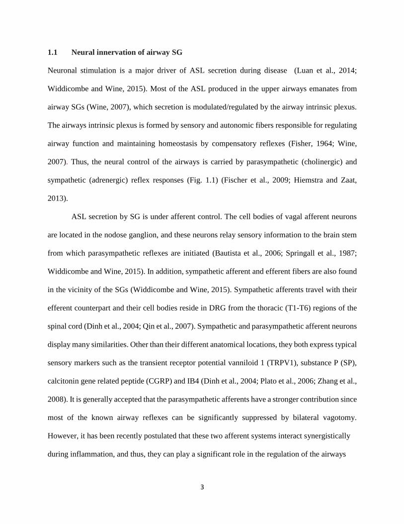
4
Figure 1.1 Neural innervation of airway SGs. Airway SGs receive parasympathetic (left) and
sympathetic (right) inputs. Afferent fibers carry sensory information from the airways via the
nodose and jugular ganglia to the brainstem nucleus tractus solitarius. Here, interneurons synapse
with vagal preganglionic fibers that in turn excite intrinsic neuron-mediated SG ASL secretion.
Sympathetic afferents transmit signals through DRGs from the thoracic segment T1-T4 whose
axons terminate in Rexed Lamina II (substantia gelatinosa) of the dorsal horn. Interneurons of the
substantia gelatinosa synapse with Rexed Lamina V efferent fibers that then stimulate SG ASL
secretion. Reproduced from Widdicombe and Wine (2015).

5
under pathological conditions (Lee and Yu, 2014).
DRGs are nodule-like structures located in the intervertebral foramina that are dorsolateral
to the spinal cord which relay information from the external environment to the central nervous
system (CNS). Neurons within the DRGs are pseudo-unipolar cells with two bifurcations, which
in the case of thoracic ganglia (tDRG), one projects peripherally to the lungs and heart; and the
second projects centrally to the dorsal horn of spinal cord. Immunohistological studies on mouse
lumbar DRGs showed heterogeneous populations of neuronal cell bodies with either unmyelinated
C- (~70%) or myelinated Aβ-/Aδ- (~30%) fibers (Ruscheweyh et al., 2007) however, these
proportions are uncertain in tDRGs. Aβ-fibers carry information related to touch and movement
whereas Aδ-fibers are sensitive to chemical stimuli, noxious gases and inhaled particles, and
inflammatory and immunologic mediators. C-fibers are sensitive to CAP, cigarette smoke,
bradykinin, ozone, hypertonic saline and various other chemical stimuli. Both fiber types when
stimulated produce reflex bronchoconstriction and increased cough to curb further noxious stimuli
exposure, however, C-fibers can also increase vascular permeability and ASL production (Hamid
et al., 2005). In the upper airways, afferent nerve endings arborize into the epithelial layer where
depolarizations from various stimuli are transmitted through the neuronal cell body to vertical and
radial interneurons (Todd, 2010) in the dorsal horn (Rexed Lamina II) of the spinal cord (Ordovas-
Montanes et al., 2015). Subsequently, interneurons from these laminae dedicated to the modulation
of noxious and non-noxious stimuli synapse onto Rexed Lamina VII efferent fibers (Todd, 2010).
The activation of these efferent fibers contribute to the stimulation of ASL secretion from SG and
also vasodilatation, plasma exudation, neuropeptide release and bronchoconstriction collectively
termed as ‘neurogenic inflammation’ (Barnes, 2001; Undem and Carr, 2002).

6
1.1.1 Neurogenic inflammation and its contribution to ASL hypersecretion
In addition to orthodromic inputs to the spinal cord from sensory neurons, action potentials in
sensory neurons can also be transmitted antidromically at branch points back down to the
periphery. This constitutes the axon reflex, which together with sustained local depolarizations,
lead to a rapid and local release of neural mediators from both peripheral axons and terminals
(Sauer et al., 2001). Classic experiments by Goltz (1874) and Bayliss (1901) showed that
electrically stimulating DRGs induced skin vasodilation, which led to the concept of a ‘neurogenic
inflammation’, independent of that produced by the immune system. Neurogenic inflammation is
mediated mainly by the release of the neuropeptides CGRP and SP from nerve endings, among
other pro-inflammatory peptides, which act directly on vascular endothelial and smooth muscle
cells (Brain and Williams, 1989; Edvinsson et al., 1987; McCormack et al., 1989; Saria, 1984). In
addition to acting on the vasculature, these signals can act directly attracting and activating innate
immune cells (mast cells, dendritic cells) and adaptive immune cells (T lymphocytes) (Ansel et
al., 1993; Cyphert et al., 2009; Ding et al., 2008; Hosoi et al., 1993; Mikami et al., 2011; Rochlitzer
et al., 2011) (see section 1.2). In the acute settings, neurogenic inflammation is protective,
facilitating physiological wound healing and immune defense against pathogens by activating and
recruiting immune cells. However, it is also likely to play major roles in the pathophysiology of
allergic and autoimmune diseases by amplifying pathological or maladaptive immune responses.
For example, in allergic airway inflammation sensory neurons have been shown to play a central
role in initiating and augmenting the activation of innate and adaptive immunity (Caceres et al.,
2009; Engel et al., 2011; Ostrowski et al., 2011).
Some studies suggest that during chronic inflammatory conditions, the DRG neurons of
the sympathetic afferent pathway, including C-fibers, undergo sensitization (de Groat and

7
Yoshimura, 2009; Janig et al., 1996) which could potentially play a role in SG hypersecretion.
DRG neurons become sensitized when exposed to pathogens known to affect the airways, such as
the bacterial LPS. LPS binds to toll-like receptor 4 (TLR4), a member of the pattern-recognition
receptor (PRR) family, that induces potentiation of TRPV1 currents, increased intracellular Ca2+
concentration (Diogenes et al., 2011) and the activation of signaling cascades that result in the
activation of nuclear factor kappa B (NF-κB) and mitogen-activated protein kinases (MAPKs) (Tse
et al., 2014). Thus, sensitized efferent sensory C-fibers, that innervate the SGs, can amplify
inflammation and result in ASL hypersecretion in the airways by axon reflex through the release
of neuropeptides on the airways during neurogenic inflammation (Barnes, 2001; Barnes et al.,
1998; Maggi et al., 1995). During neurogenic inflammation, nociceptive C-fibers release various
neuropeptides such as SP, neurokinin-A and CGRP as observed in guinea pig, rat and ferret studies
through activation with the chilli pepper alkaloid, CAP (Hamid et al., 2005; Millqvist, 2000).
The role of sensory inflammation in the overproduction of ASL in airway diseases is
supported by multiple pieces of evidence. For instance, SP is able to stimulate SG secretion in
human airways in vitro and also goblet cell secretion in guinea pig airways through the activation
of NK1 (neurokinin 1)-receptors. Other factors that stimulate SG secretion are VIP (vasoactive
intestinal peptide), cholinergic agonists, elevated cyclic adenosine monophosphate (cAMP),
elevated Ca2+ and SP-induced acetylcholine release through enhancement of ganglionic
transmission (Barnes, 2001). Despite evidences of neuropeptidergic factors as the key instigator
of neurogenic inflammation and subsequent increased SG secretion in animal models, observations
in in vivo human studies are poorly supportive of this hypothesis. This deviation may in part be
due to species-specific pulmonary differences. For example, intra-epithelial SP- and CGRP-
positive fibers make up more than 60% in guinea pigs as opposed to 1% in human airways

8
(Bowden and Gibbins, 1992). Some studies indicated increased SP in bronchial nerves from
biopsies of fatal asthmatics while other studies were contradictory. Other considerations include
plasticity-driven increased peripheral nerve innervation during inflammatory states mediated by
neurotrophins secreted from inflammatory cells that may account for varied observations (Barnes,
2001). Given such inconsistencies, newer hypotheses have reappraised neurogenic inflammation
to be the result of both immunological- and neurogenic-driven factors that could contribute to ASL
hypersecretion. This neuro-immune hypothesis stems from the idea that although neurogenic
inflammation is protective in the acute phase, chronic states tend to result in pathological or
maladaptive immune responses (Chiu et al., 2012) which may enhance SG secretions in the airway.
1.2 Cooperative immunogenicity of the peripheral nervous and immune systems
The early understanding of immunogenicity involved responses of the immune system, excluding
a role of the peripheral nervous system. However, emerging evidence strongly supports a role of
the peripheral nervous system, since it shares identical and integrative protective mechanisms with
the immune system. For instance, both systems express members of the PRR family that recognize
a variety of highly conserved pathogen-associated molecular patterns (PAMP) (Santoni et al.,
2015; Srikrishna and Freeze, 2009). These receptors, i.e. RAGE and toll-like receptors (TLR), are
classified under the immunoglobulin (Ig) superfamily of cell surface molecules that initiate and
amplify responses to inflammation, infection and injury in a tightly-regulated manner (Sukkar et
al., 2012). However, in pathophysiological scenarios such as in chronic airway inflammation, over-
active PRRs as a result of perturbed endogenous regulatory mechanisms can sensitize sensory
fibers leading to unwanted consequences (Chiu et al., 2012). Sensory fibers are inherently capable
of transmitting danger signals more readily than the mobilization of the innate immune system.

9
The high level of organization between neurons and airway structural cells suggests the likelihood
of direct modification of inflammatory processes in the pulmonary system (McGovern and
Mazzone, 2014).
In fact, research has shown that transient receptor potential cation channel, subfamily A,
member 1 (TRPA1)-knockout mice that were exposed to the allergen ovalbumin, had no leukocyte
infiltration in the airways, decreased cytokine and ASL production, and greatly attenuated airway
hyper-reactivity (Caceres et al., 2009). Moreover, ablation of both TRPA1- and TRPV1-positive
vagal mouse sensory neurons through genetic or pharmacological means caused a marked
reduction in airway hyper-reactivity (Ordovas-Montanes et al., 2015). Such evidence suggests an
intimate link between the peripheral nervous and immune systems. Thus, understanding the
molecular mechanisms that underlie the coordinated interaction of peripheral neurons and immune
cells may offer better therapies for airway inflammatory diseases.
1.2.1 RAGE and RAGE isoform expression
RAGE is a mammalian multiligand transmembrane receptor classified under the Ig superfamily.
It contains one variable (V)- and two constant (C)-type extracellular domains, a transmembrane
domain and a cytoplasmic tail (Lopez-Diez et al., 2013). The V-domain which contains two N-
glycosylation sites is the primary binding site for most extracellular ligands. Intracellular signalling
is mediated by the cytoplasmic tail through a common interaction with Dia-1 (diaphanous-1). Ager,
the gene that codes for RAGE, is found on the locus for the MHC (major histocompatibility
complex) Class III region on chromosome 6p21.31 in humans. Gene ancestry and protein mapping
studies have indicated that Ager evolved from early Metazoans and its encoded protein contains

10
sequences that closely resemble cell adhesion proteins. The latter suggests that RAGE was first a
cell adhesion protein before evolving into an inflammation-related receptor (Sessa et al., 2014).
RAGE was initially described as the receptor for advanced glycation end-products (AGEs),
which form as the result of the non-enzymatic glycation of proteins in the presence of reducing
sugars (Goldin et al., 2006). However, it is now evident that it also serves as a receptor for the
calcium binding protein S100/calgranulin superfamily, high mobility group-box protein 1
(HMGB1), an amyloidogenic isoform of serum amyloid A and amyloid-β peptide (Lopez-Diez et
al., 2013), and LPS (Rong et al., 2004). These interactions occur in cis, however in trans
homophilic reactions with RAGE and other unbound RAGE forms, respectively, are also known
to occur during cell-cell interaction (Sessa et al., 2014). RAGE has been linked to several
inflammatory-related diseases such as diabetes and metabolic disorders, cardiovascular disorders,
pulmonary disorders, neurologic disorders, including the aggressiveness of cancers. Cellular
RAGE expression is readily detectable during embryonic development and decreases significantly
upon cellular differentiation. Uniquely, this decrease is absent in lung tissue where its expression
remains high under normal conditions and decreases in pathological states. Non-pulmonary
differentiated tissues express low levels of RAGE and only increase under inflammatory
conditions (Sparvero et al., 2009).
Several RAGE isoforms reported in human, murine and canine studies have revealed that
variations of the Ig-V-domain can affect extracellular-ligand binding. Moreover, the lack of the
transmembrane domain can result in a soluble form of RAGE that can be secreted. Isoforms that
have a signal peptide can also be secreted or inserted into cell membranes. Secreted forms
generated either through alternative messenger ribonucleic acid (mRNA) splicing or proteolytic
cleavage of full-length RAGE (Tv1-RAGE) can play dual roles (Lopez-Diez et al., 2013). For

11
example, it can act as a “decoy receptor” in moderating inflammatory responses and can also
promote inflammatory responses through interaction with Mac-1 (macrophage-1 antigen)
(Sparvero et al., 2009). Bioinformatics has shown that there are 17 different RAGE isoforms (13
of which were recently identified – MmusRAGE v1-13) in mice that are expressed in different
levels in the brain, heart, kidney, liver, lung and pancreas. Tv1-RAGE mRNA contain exons 1
(SP), 2/3 (IgV domain), 4-6 (IgC2-1), 7-8 (IgC2-2), 9 (non-structural), 10 (transmembrane
domain) and 11 (cytoplasmic tail). Other membrane-bound (have transmembrane domain)
isoforms include MmusRAGEv1 and mRAGE_v4. Conversely, likely soluble isoforms include
MmusRAGEv2 to v6.
1.2.2 Role of RAGE in neurogenic inflammation
RAGE has been recognized to play a central role in the airway inflammatory process based on
molecular, biochemical and genomic studies (Mulrennan et al., 2015; Repapi et al., 2010; Sukkar
et al., 2012). RAGE is ubiquitously expressed, and its up-regulation was firstly implicated in
diabetes (Hofmann et al., 1999; Neeper et al., 1992; Schmidt et al., 1992; Yan et al., 2010),
however, it has now been included as part of the pathological mechanisms active in multiple airway
diseases, such as asthma, COPD, and cystic fibrosis (Buckley and Ehrhardt, 2010; Milutinovic et
al., 2012). Among the multiple RAGE ligands, LPS, is known to induce ASL secretion through
the innate immune and peripheral nervous systems (Alexander and Rietschel, 2001; Yamamoto et
al., 2011; Yanagihara et al., 2001). Brochoalveolar lavage collected from ovalbumin-sensitized
mice challenged with aerosolized LPS had higher IL-4 (interleukin 4) and tumor necrosis factor α
(TNF-α) compared to controls (Qiao and Zhang, 2014). Airway epithelia of LPS-treated mice also
displayed airway cell hyperplasia that is characteristic of airway inflammation (Toward and

12
Broadley, 2002; Vernooy et al., 2002). LPS cultured neonatal rat DRG neurons acutely exposed
to LPS showed PKC (protein kinase C)- and PKA (cAMP-dependent protein kinase)-mediated
CGRP release and increased intracellular Ca2+ (Hou and Wang, 2001). Interestingly, septic shock
experiments involving RAGE KO mice that were intraperitoneally injected with LPS had
attenuated serum levels of TNF-α, interleukin 6 (IL-6) and ET-1 (endothelin-1) compared to wild-
type controls. In addition, the same group also showed through binding essays that LPS binds to
the RAGE (Yamamoto et al., 2011). In humans, increased levels of RAGE have been observed in
the alveolar walls of patients with COPD, and the up-regulation of RAGE also correlates with the
expression of HMGB1, a transcription enhancer protein, in asthma patients (Sukkar et al., 2012;
Zhou et al., 2012). The latter is consistent with the decreased antigen-induced airway inflammation
and hyporesponsiveness in RAGE KO mice model of asthma (Lee et al., 2013). HMGB1 can bind
to RAGE and other TLRs to initiate a cascade of events that activate NF-κB which in turn induces
cytokine production and promotes inflammation. It has been hypothesized that RAGE and TLRs
may have collaborative signalling through myD88-dependent pathways that tend to elicit stronger
inflammatory responses than TLR-exclusive pathways (Hall and Agrawal, 2015). The interaction
between RAGE and its ligands induces reactive oxygen species (ROS) accumulation, probably
through NADPH (nicotinamide adenine dinucleotide phosphate) oxidase (Ruderman et al., 1992;
Schmidt et al., 1994; Vincent et al., 2007; Wautier and Schmidt, 2004). Thus, the activation of NF-
κB perpetuates oxidative stress and together with other pro-inflammatory factors generate the
inflammatory state in neurons (Tobon-Velasco et al., 2014). Alternatively, sensitized RAGE KO
mice challenged with ovalbumin displayed reduced eosinophilic inflammation and goblet cell
metaplasia, decreased Th2 (T helper type 2) cytokine production, and lower numbers of pulmonary
innate lymphoid cells compared to controls (Taniguchi et al., 2015).

13
1.3 Rationale and Hypothesis
During inflammatory conditions, sensory neurons innervating the SGs in the upper airways
contribute to ASL secretion. It is not clear however, whether sympathetic afferent neurons play a
role, and if so, to what extent in the regulation of ASL secretion under inflammatory conditions.
My working hypothesis is that under inflammatory conditions, RAGE expression and signalling
mediates tDRG neuron sensitization. In particular, I propose that exposure to LPS will cause an
increase in CAP-evoked current parameters in neurons from WT mice, but these changes will not
be expected in neurons from RAGE KO mice. Similarly, we propose that evoked action potential
generation will be increased in neurons from WT, but not from RAGE KO, mice when exposed to
LPS. Furthermore, I propose that changes in the expression pattern of RAGE isoforms underlie
the sensitization of tDRG neurons during LPS exposure. Here, I expect to see an increase in full
length RAGE (Tv1-RAGE) expression, however the expression pattern of RAGE isoforms in
membrane and cytosolic fractions is unknown and cannot be speculated.

14
CHAPTER 2
MATERIALS & METHODS
2.1 Animals. A colony of RAGE KO mice on a C57BL/6 background was maintained by
breeding heterozygous mice as previously described (Chandna et al., 2015). Heterozygous mice
were generated by back-crossing RAGE KO (homozygous) mice (Myint et al., 2006) with
C57BL/6 wild type (WT) mice. All experiments were based on tDRGs (T1-T4) from homozygous
(RAGE KO) mice and their C57BL/6 (wild type) littermates. Mice were genotyped using genomic
DNA (deoxyribonucleic acid) and PCR (polymerase chain reaction) as previously described
(Myint et al., 2006) but with slight modifications. Briefly, ~2 mm of mouse tail was digested for
30 mins at 95oC in 300 µL digestion buffer (0.5 M EDTA (ethylenediaminetetraacetic acid) and
50% NaOH dissolved in distilled water). Samples were chilled on ice for 5 mins before the addition
of 75 µL neutralization buffer (40 mM Tris-HCl (tris hydrochloride)). Samples were vortexed
briefly and 2 µL was used for PCR (20 µL reaction) (Truett et al., 2000). PCR reactions were run
on 2% agarose gels containing 0.5 µg/mL ethidium bromide and visualized on a UV (ultraviolet)-
illuminated gel box. All in vitro experiments were done with neonatal pups (P0–P4). This work
was approved by the University of Saskatchewan’s Animal Research Ethics Board (Campanucci:
protocol 20090082) and adhered to the Canadian Council on Animal Care guidelines for humane
animal use.
2.2 Primary tDRG cultures. Bilateral tDRG (T1-T4) neurons were harvested and cultured
from neonatal (P0-P4) mice as previously described for peripheral neurons (Campanucci et al.,
2008). tDRG neurons were selected as they are known to innervate the upper airways

15
(Widdicombe and Wine, 2015). Briefly, ganglia were removed under sterile conditions and
enzymatically dissociated at 37°C in HBSS (Hanks’ balanced salt solution) containing trypsin
(180–200 U/mL; Worthington, Freehold, NJ, USA) and buffered with HEPES (N-2-
hydroxyethylpiperazine-N-2-ethane sulfonic acid; pH 7.4). The resulting cell suspension was
washed twice in serum-containing Leibovitz's L-15 medium to inactivate the trypsin and plated on
laminin-coated glass bottom petri dishes (35 mm) made in-house. The neurons were grown in L-
15 medium supplemented with vitamins, cofactors, penicillin–streptomycin, 5 mM glucose, 5%
rat serum and NGF (nerve growth factor; 10 ng/mL). Cultures were maintained at 37°C in a
humidified atmosphere of 95 % air-5 % CO2 and fed every 4 days with growth media. To eliminate
non-neuronal cells, cultures were treated with cytosine arabinoside (10 μM; Sigma-Aldrich, St.
Louis, MO, USA) from days 2 to 4. Established DRG cultures were incubated for 24h at 37oC;
(5% CO2; humidified) in growth media alone (control), with a cytokine cocktail (CC) consisting
of 1.5 nM IL-6 (Murata et al., 2011; Suto et al., 1993), 1.5 nM IL-8 (interleukin 8) (Dong et al.,
2012), 3 nM TNF-α (Hensellek et al., 2007) and 0.6 µM IL1-β (interleukin 1 beta) (Saleh et al.,
2013) or with 1µg/mL LPS (Sigma-Aldrich)-supplemented growth media. Both CC and LPS pro-
inflammatory inducers were tested in WT tDRGs for their ability to produce robust and
reproducible CAP-evoked currents that were indicative of inflammation (increased peak
amplitude, current density and charge). Of these two inducers, I chose LPS for this study as it not
only met the requirements mentioned but was also easier to prepare.
2.3 Whole-cell Patch Clamp Electrophysiology. Medium-sized tDRG neurons (~25-35 µm)
were selected for whole-cell recording. Membrane currents were recorded with an Axopatch 200B
amplifier (Molecular Devices, Palo Alto, CA, USA) equipped with a 1 GΩ cooled head-stage

16
feedback resistor and a Digidata 1400A analog-to-digital converter (Molecular Devices), and
stored on a personal computer. Current- and voltage-clamp protocols, data acquisition, and
analysis were performed using pClamp 10 (Molecular Devices) and Origin 9.0 software
(OriginLab Corporation, Northampton, MA, USA). Patch pipettes were made using thin-wall
borosilicate glass capillaries (World Precision Instruments, FL, USA) using a vertical puller (PC
10; Narishige Scientific Instrument Lab., Tokyo, Japan) and polished with a microforge
(Narishige) to a final resistance of 3–8 MΩ when filled with intracellular recording solution. In
most experiments, 75% of the series resistance was compensated, and junction potentials were
cancelled at the beginning of the experiment. Recording electrodes were filled with the following
intracellular solution (in mM): 65 KF, 55 KC2H3O2, 5 NaCl, 0.2 CaCl2, 1 MgCl2, 10 EGTA
(ethylene glycol-bis(β-aminoethyl ether)-N,N,N',N'-tetraacetic acid), 2 MgATP (adenosine 5'-
triphosphate magnesium salt) and 10 HEPES, and pH was adjusted to 7.2 with KOH (all from
Sigma-Aldrich). Cultured neurons were perfused continuously at 1 mL/min with control perfusion
solution consisting of (in mM): 140 NaCl, 5.4 KCl, 0.33 NaH2PO4, 0.44 KH2PO4, 2.8 CaCl2, 0.18
MgCl2, 10 HEPES, 5.6 glucose, 2 glutamine, 0.001% atropine and 5 μg/mL phenol red; pH was
adjusted to 7.4 with NaOH (all from Sigma-Aldrich). Whole-cell patched neurons were allowed
to stabilize for 5 min before recording. Action potentials were generated in current-clamp mode
by injection of a series of depolarizing current steps at 100 pA increments for 500 ms. All other
experiments were carried out under voltage-clamp mode. A fast-step perfusion system was used
to deliver either control or CAP (1µM)-containing extracellular solution at 1 mL/min perfusion
rate.

17
2.4 MS-based proteomic analyses. MS-based proteomic analysis was used to study global
protein changes in tDRG neurons from WT mice in control or LPS conditions. This method was
preferred over Western Blotting or qPCR (quantitative polymerase chain reaction) because no
RAGE isoform-specific antibodies are available and qPCR is unable to detect posttranslational
modifications, respectively.
Suspensions of cultured tDRG neurons from WT and RAGE KO mice were prepared as
described above, and plated onto specialized cell culture dishes (Sarstedt, Nümbrecht, Germany).
We used approximately 80,000 cells per experimental group (control or LPS), each plating is
generated from 20 pups. Membrane and soluble fractions from cultured tDRG neurons were
obtained using the ProteoExtract Native Membrane Protein Extraction Kit (EMD Millipore, MA,
USA) and processed as per the manufacturer’s instructions and stored at -80oC. Protein
concentrations were determined by Nanodrop analysis with a BioTek Synergy HY multi-detection
plate reader (Winooski, VT, USA).
2.4.1 In-solution Digestion. Protein samples were concentrated by acetone precipitation. To
precipitate proteins, 4x sample volume of cold (-80⁰C) acetone was added to an aliquot from lysate,
vortexed, and incubated overnight at -80⁰C. Then samples were centrifuged 13000 x g at 4⁰C for
15 min. After washing protein pellets twice with cold (-80⁰C) 80% acetone/20% water, protein
pellets were air dried and re-suspended in 55 µL trifluoroethanol (TFE) buffer (10% TFE, 100 mM
ammonium bicarbonate (ABC)). Proteins were digested in-solution using an in-house developed
protocol. Briefly, 45 µL of each protein sample was placed in 1.5 mL tube and diluted with 5 µl
of 100 mM ABC buffer (Fisher Scientific, Fair Lawn, NJ, USA) and 50 µL TFE (Fisher Scientific)
to denature proteins. The samples were treated with 1 µL of 1M DTT (dithiothreitol) (MP

18
Biomedicals, Solon, OH, USA) while shaking at 300 RPM (Eppendorf Thermomixer, Eppendorf,
Mississauga, ON, Canada) at 60oC for 60 min to reduce disulfide bonds formation. Next, samples
were alkylated with 100 µL of 110 mM iodoacetamide (IAA; Fisher Scientific) at 37oC for 30 min
on a shaker, covered with aluminum foil, to prevent further disulfide bond formation. The samples
were dried in a speedvac (Labconco, Kansas City, MO, USA). Proteins in the samples were treated
with 1 mL cold acetone followed by refrigeration at -80oC for 60 min to eliminate salts and other
interfering compounds (e.g. detergents), which can prevent digestion. The samples were
centrifuged twice at 18000 x g for 30 min and acetone was carefully removed. Next, samples were
dried in a speedvac and a buffer-containing trypsin (Promega Corporation, Madison, WI, USA)
solution (50 ng/µL trypsin in 1 mM HCl (hydrochloric acid) /100 mM ABC) was added to the
samples in a 40:1 protein:trypsin. The samples were incubated in a shaker at 300 RPM overnight
at 37oC. Trypsin buffer at the same ratio was added again in the morning to ensure complete
digestion of proteins into peptides. After 2 hrs of further incubation at 37oC, digested peptides
were dried in speedvac and stored at -80oC until further analysis.
2.4.2 Strong Cation Exchange (SCX)-based Fractionation. SCX-based fractionation was
performed using SCX SpinTips sample prep kit (Protea Biosciences, Morgantown, WV, USA).
The digested protein samples were dissolved in 200 µL of SCX reconstitution solution, a pH of 3
was adjusted with formic acid (FA). The samples were then loaded on to SCX SpinTip and
centrifuged at 2000 x g for 6 min. To enhance peptide binding to SCX SpinTip samples were
centrifuged 3 times. The flow through at the end of the step was transferred to a 1.5 mL tube for
MS analysis. Peptides bound to SCX column were eluted using stepwise concentrations (in M):
20, 40, 60, 80, 100, 150, 250, 500 of ammonium formate (Sigma-Aldrich) in 10% acetonitrile

19
(ACN; Fisher Scientific) at pH 3. 150 µL of ammonium formate (in increasing concentrations)
was added to the SpinTip and centrifuged at 2000 x g for 6 min. The flow through was collected
after every centrifugation and transferred to 1.5 mL tube. All flow through were dried in speedvac
and stored at -80oC.
2.4.3 MS Workflow. All SCX fractions containing tryptic peptide were reconstituted in 20 µl
of MS grade water:ACN:FA (97:3:0.1 v/v) followed by vortexing for 1-2 min. The resulting
solutions were centrifuged at 18000 x g for 10 min at 4ºC. 15 µL aliquot of each sample was
transferred to a mass spectrometry vial (Agilent Technologies Canada Ltd., Mississauga, ON, CA)
for liquid chromatography-tandem mass spectrometry (LC-MS/MS) analysis. All MS analyses
were performed on an Agilent 6550 iFunnel quadrupole time-of-flight (QTOF) mass spectrometer
equipped with an Agilent 1260 series liquid chromatography instrument and a Chip Cube LC-MS
(liquid chromatography-mass spectrometry) interface (Agilent Technologies). Chromatographic
peptide separation was accomplished using a high-capacity high performance liquid
chromatography (HPLC)-Chip II: G4240-62030 Polaris-HR-Chip_3C18 consisting of a 360 nL
enrichment column and a 75 µm × 150 mm analytical column, which were both packed with
Polaris C18-A, 180Å, 3 µm stationary phase. Samples were loaded onto the enrichment column
with 50% solvent A (0.1% FA in water) and 50% solvent B (0.1% ACN:FA) at a flow rate of 2.0
µL/min. Samples loaded to enrichment column were transferred onto analytical column, and
peptides were separated with linear gradient solvent system. The linear gradient program was
employed for peptide separation with solvent A and solvent B on an analytical column. The linear
gradient is 3–25% solvent B for 50 min and then 25–90% solvent B for 10 min at a flow rate of
0.3 µL/min. Positive-ion electrospray MS data was acquired using a capillary voltage set at 1900
20
V, the ion fragmentor set at 360 V, and the drying/collision gas (nitrogen) set at 225ºC with a flow
rate of 12.0 L/min. Spectral results were collected over a mass range of 250–1700 mass/charge
(m/z) at a scan rate of 8 spectra/sec. Tandem mass spectrometry (MS/MS) data were collected over
a range of 100–1700 m/z and a set isolation width of 4 atomic mass units. The top 20 most intense
precursor ions for each MS scan were selected for MS/MS with active exclusion for 0.25 min. To
clarify, precursor ions are of a specific m/z ratio in the first stage (MS) of MS/MS. These ions will
then undergo collision-induced dissociation using nitrogen gas in the second stage (MS/MS) of
MS/MS.
2.4.4 Protein Identification. Spectral data were converted to a m/z data format using Agilent
MassHunter Qualitative Analysis Software (Agilent Technologies) and were processed against the
NCBI (National Center for Biotechnology Information) non-redundant Mus musculus database,
using SpectrumMill (Agilent Technologies) as the database search engine. Search parameters
included a fragment mass error of 50 parts per million (ppm), a parent mass error of 20 ppm,
trypsin cleavage specificity, and carbamidomethyl as a fixed modification of cysteine. In addition,
four stages of database search in variable modification mode were carried out with different sets
of variable modifications. In first stage, carbamylated lysine, oxidized methionine, pyroglutamic
acid, deamidated asparagine and phosphorylated serine, threonine, and tyrosine were set as
variable modifications. In the second stage, validated hits from the first stage were searched using
the following variable modifications: acetyl lysine, oxidized methionine, pyroglutamic acid,
deamidated asparagine, and phosphorylated serine, threonine, and tyrosine. The validated hits from
the second stage were searched using semi-trypsin non-specific C-terminus, yielding the third
stage validated hits, which were subsequently searched using semi-trypsin non-specific N-

21
terminus (fourth stage), with no other variable modifications specified. After each stage, Spectrum
Mill validation was performed at peptide and protein levels (1% false discovery rate).
Protein samples were reduced, alkylated and digested with trypsin in-solution in
accordance with a modified version of a previously published protocol (Zhang et al., 2011). Tryptic
peptides were analyzed by LC-MS/MS using an Agilent HPLC Chip/6550 iFunnel QTOF mass
spectrometer (Agilent Technologies). MS/MS were referenced against the mouse NCBI non-
redundant database and a custom database (containing mouse RAGE protein isoforms) using
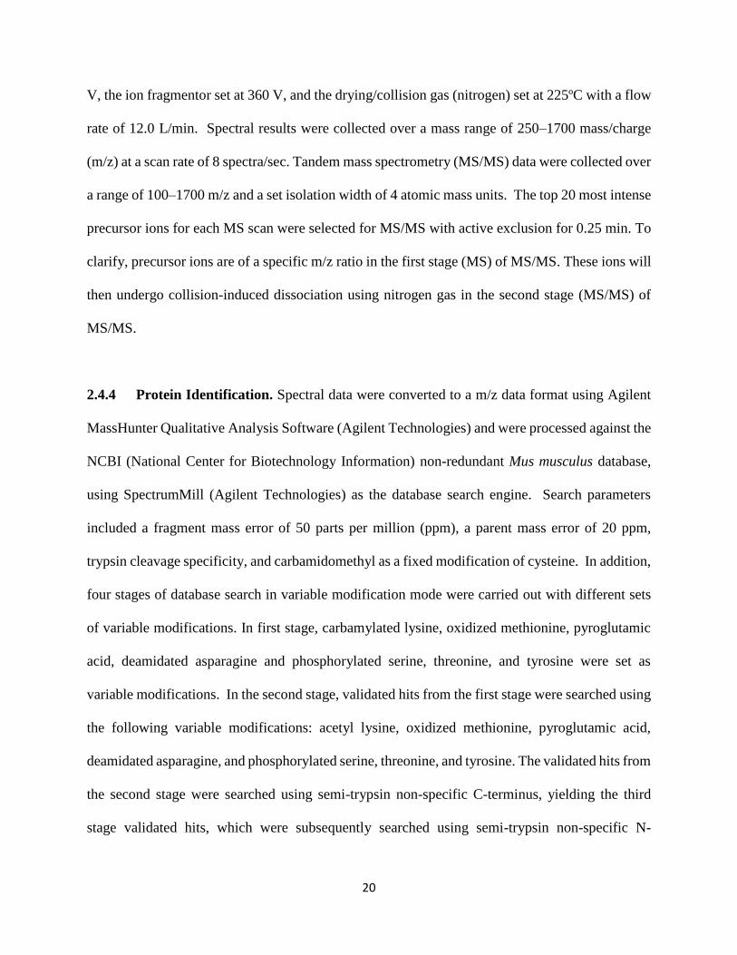
Spectrum Mill (Fig. 2.1). Spectral counts were used to report relative quantitation of proteins.
2.4.5 Manual Identification of Unique Modified RAGE isoforms (see Fig. 2.1). For either
genotype, tryptic peptides identified in each RAGE isoform from the custom database in each
treatment group were sorted according to their sequences. Then, common and duplicate peptides
found in 2 or more RAGE isoforms were discarded leaving behind unique peptides that were
specific to a particular RAGE isoform. Finally, these peptides were further specified according to
their post-translational modifications.

22
Figure 2.1 Processing of MS/MS spectra in the identification of unique post-
translationally modified RAGE isoform peptides in WT tDRG neurons under control and
LPS treatment conditions.
MS/MS Spectra referenced against
mouse NCBI non-redundant database
Result: Canonical RAGE isoform
identified. No isoforms identified
MS/MS Spectra referenced against
mouse custom database
Result: Several RAGE isoforms identified
Manually sorted peptides from each
isoform by amino acid sequence.
Duplicates and common peptides from 2
or more RAGE isoforms eliminated.
Result: Peptides specific to a particular
RAGE isoform identified. However, some
of these peptides had post-translational
modifications specific to either
membrane or cytosolic, and control or
LPS treatment
Manually sorted peptides by post-
translational modification
Result: Identified peptides specific to a
particular RAGE isoform, fraction and
treatment.

23
2.5 Statistical Analyses. We compared the EC50 (half maximal effective concentration)
obtained from the dose response curves using tDRG neurons from either WT and RAGE KO, by
unpaired t-test with Welch correction. In voltage-clamp experiments, raw values of peak
amplitude, current density and charge of CAP-evoked current were compared using the unpaired
t-test with Welch correction. In current-clamp experiments, action potential counts were compared
using Mann-Whitney test. Resting membrane potentials (VRM) were compared using the Kruskal-
Wallis test (non-parametric analysis of variance, ANOVA). The significance threshold was set at
0.05 for all statistical tests performed.

24
CHAPTER 3
RESULTS
3.1 Determination of working concentration for CAP-evoked currents
In order to test the possible sensitization of sensory neurons exposed to inflammatory conditions
(i.e. LPS-treatment or cytokines), we studied neuronal excitability in response to the injection of
depolarizing current and to applications of the agonist CAP. Thus, to determine a submaximal
working concentration of CAP, that would allow us to observe potential increases or decreases in
whole-cell currents, we generated a dose-response curve to increasing concentrations of CAP in
tDRG cultured neurons from WT and RAGE KO mice (Fig. 3.1). Cultured neurons were briefly
exposed to CAP (0.1, 0.5, 1, 2.5, 5 and 10 µM; 1s) and the data were fitted with a logistic Hill
function (R2=0.93746 for WT, and R2=0.94002 for RAGE KO). The EC50 of CAP obtained in
cultures from WT and RAGE KO mice was 1.50 ± 0.44 µM and 2.25 ± 0.81 µM, respectively. No
significant difference (p>0.05) was found in the EC50 values between the two genotypes.
Subsequently, 1 µM CAP was selected as the submaximal working concentration throughout the
study.
3.2 Exposure of sensory neurons to cytokines cocktail induces sensitization of CAP-
evoked currents
Cultured tDRG neurons from WT mice were incubated in either control or cytokine cocktail (CC)-
containing growth media (for 24 hr) before whole-cell recording of CAP-evoked currents. The
mean peak amplitude of CAP-evoked currents in CC-treated neurons (-1063.51 ± 170.34 pA) was
significantly higher (p<0.05) than in control neurons (-554.04 ± 64.01 pA) (Fig. 3.2A, B). Mean

25
Figure 3.1 Dose-response of CAP-evoked currents in WT and RAGE KO DRG neurons.
Dose dependency of CAP-evoked currents from cultured tDRG neurons from wild-type (WT) and
RAGE-knockout (RAGE KO) mice (P0-P4) obtained by whole-cell patch clamp
electrophysiology. DRGs neurons were clamped at -60mV and exposed to CAP for 1s. Data points
indicate mean current density (pA/pF) at 0.1 (n = 6 for each genotype), 0.5 (WT=7; RAGE KO=6),
1 (WT=11; RAGE KO=10), 2.5 (WT=6; RAGE KO=7), 5 (WT=5; RAGE KO=8) and 10 µM CAP
(WT=5; RAGE KO=7). The data were fitted with a logistic Hill function – WT R2=0.93746;
RAGE KO R2=0.94002. Error bars indicate mean ± SEM. The EC50 of CAP in wild-type and
RAGE KO was 1.50 ± 0.44 µM and 2.25 ± 0.81 µM, respectively.

26
Figure 3.2 CC exposure increases CAP-evoked currents in tDRG neurons from WT
neonatal mice. The CC consisted of 1.5 nM IL-6 (Murata et al., 2011; Suto et al., 1993), 1.5 nM
IL-8 (Dong et al., 2012), 3 nM TNF-α (Hensellek et al., 2007) and 0.6 µM IL1-β (Saleh et al.,
2013). (A) Representative CAP (1 µM; 1 s)-evoked current traces (VH = -60 mV) from cultured
wild type (WT) tDRGs. (B) Mean peak amplitude (pA), (C) current density (pA/pF) and (D)
charge (1 x 10⁶ pC) graphs of WT DRG neurons exposed to either control or CC-containing media
(24h). Treatment groups (mean peak amplitude, mean current density, mean charge): WT control
(13); WT CC (10). Error bars indicate mean ± SEM. Letter ‘a’ denotes p<0.01 and ‘c’ denotes
p<0.05 based on unpaired t-test with Welch correction. Control group is abbreviated as ‘CTRL’.

27
current density was significantly higher (p < 0.05) in the CC group (-31.73 ± 6.13 pA/pF)
compared to controls (-16.19 ± 2.16 pA/pF) (Fig. 3.2A, C). The mean charge was significantly
higher (p < 0.01) in the CC group (2.70 ± 0.35 x 10⁶ pC) compared to controls (1.26 ± 0.22 x 10⁶
pC) (Fig. 3.2A, D). The results indicate that DRG neurons exposed to proinflammatory cytokines,
modeling the conditions of a diseased airway, develop sensitization and become hyperresponsive
to stimulation.
3.3 RAGE expression is required for the potentiation of CAP-evoked currents in tDRG
neurons
To generate inflammatory conditions that are physiologically more relevant to airways pathology,
we expossed cultured tDRG neurons to the bacterial endotoxin LPS. Thus, cultured neurons from
WT and RAGE KO mice were incubated in either control or LPS-containing growth media (for
24 hr) before the recording of whole-cell CAP-evoked currents. Consistent with our results with
the CC-containing media, the mean peak amplitude of CAP-evoked currents in LPS-treated
neurons from WT mice (-1188.54 ± 177.76 pA) was significantly higher (p<0.01) than in control
neurons (-554.04 ± 64.01 pA) (Fig. 3.3A, B). To account for neuronal size we calculated the mean
current density, which was significantly higher (p < 0.005) in the LPS group (-49.49 ± 7.38 pA/pF)
compared to controls (-16.19 ± 2.16 pA/pF) (Fig. 3.3A, C). Consistently, the mean charge was
significantly higher (p < 0.005) in the LPS group (2.70 ± 0.35 x 10⁶ pC) compared to controls (1.26
± 0.22 x 10⁶ pC) (Fig. 3.3 A, D). Contrasting with our results in neurons from WT mice, tDRG
neurons from RAGE KO mice did not exhibit significant differences in CAP-evoked currents in
the LPS group compared to controls (p>0.05) (Fig. 3.3A-D). Mean peak amplitude for control

28
group was 282.13 ± 43.79 pA and LPS-treated group was 349.77 ± 41.71 pA (Fig 3.3A, B). Mean
current density for control (-9.00 ± 0.88 pA/pF) and LPS-treated (–10.94
Figure 3.3 LPS exposure increases CAP-evoked currents in tDRG neurons from WT
neonatal mice. (A) Representative CAP (1 µM; 1 s)-evoked current traces (VH = -60 mV) from
cultured wild type (WT) and RAGE-knockout (RAGE KO) tDRGs. (B) Mean peak amplitude
(pA), (C) current density (pA/pF) and (D) charge (1 x 10⁶ pC) graphs of WT and RAGE KO DRG
neurons exposed to either control or LPS-containing media (1 µg/mL; 24h). Treatment groups
(mean peak amplitude, mean current density & mean charge): WT control (13); WT LPS (9);
RAGE KO control (8); RAGE KO LPS (9; Exception: n=8 for Mean Charge in RAGE KO LPS
group). Error bars indicate mean ± SEM. Letter ‘a’ denotes p<0.01 and ‘b’ denotes p<0.005 based
on unpaired t-test with Welch correction. Control group is abbreviated as ‘CTRL’.

29
± 1.14 pA/pF) groups (Fig. 3.3 A, C) were not significantly different (p>0.05). Similarly, the mean
charges for the control group (0.86 ± 0.20 x 10⁶ pC) was not significantly different (p>0.05) from
the LPS-treated group (1.07 ± 0.18 x 10⁶ pC) (Fig. 3.3A, D).
3.4 RAGE expression is required to increase neuronal excitability in tDRG neurons
To test neuronal excitability we generated action potentials in cultured tDRG neurons from WT
and RAGE KO mice, mantained in either in control or LPS conditions. We quantified action
potentials generated by the injection of depolarizing current steps (0 to 1000 pA, at 100pA
increments; Fig. 3.4). Cultured tDRG neurons from WT mice treated with LPS displayed a
significant (p<0.05) increase in mean action potential counts at 200pA (5.08 ± 0.96 counts)
compared to controls (2.42 ± 0.56 counts; Fig. 3.4A, B). In contrast, there were no significant
(p>0.05) differences in action potential counts between the control (2.63 ± 0.60 counts) and LPS
groups (2.45 ± 0.58 counts) in tDRG neurons from RAGE KO mice (Fig. 3.4A, C). There were no
significant differences (p>0.05) in mean resting membrane potentials in WT and RAGE KO tDRG
neurons under control and inflammatory conditions (Table 3.1).

30
Figure 3.4 LPS exposure increases cell excitability in DRG neurons from WT mice. (A)
Representative action potential traces in response to injection of depolarizing current steps (0 to
1000 pA, at 200 pA increments). Mean action potential counts generated in tDRG neurons exposed
to either control or from (B) WT and (C) RAGE KO groups (control and LPS). Error bars represent
mean ± SEM. Letter ‘c’ denotes p<0.05 based on Mann-Whitney Test. (D) Step current injection
protocol used in whole-cell current-clamp electrophysiology experiments. Treatment groups: WT
control (12); WT LPS (12); RAGE KO control (16); RAGE KO LPS (11). Control group is
abbreviated as ‘CTRL’.

31
TREATMENT GROUP
WT CTRL WT LPS WT CC
RAGE KO
CTRL
RAGE KO
LPS
Mean VRM ± SEM (mV) -44.65 ± 2.43 -48.20 ± 2.32 -44.37 ± 2.25 -47.28 ± 1.56 -47.66 ± 1.88
Table 3.1 Mean VRM of WT and RAGE KO tDRG neurons under control and
inflammatory conditions. Combined mean resting membrane potential of WT and RAGE KO
experimental groups recorded in voltage- (excluding dose-response experiments) and current-
clamp experiments. Treatment groups: WT CTRL (25); WT LPS (21); WT CC (10); RAGE KO
CTRL (24) and RAGE KO LPS (20). Raw values from each treatment group were compared using
the Kruskal-Wallis Test (non-parametric ANOVA) and found to be not significantly different
(p>0.05). Control group is abbreviated as ‘CTRL’.

32
3.5 Expression profile of RAGE and its isoforms in tDRG neurons exposed to LPS
In this study, we detected an increase in expression levels of full length RAGE (Tv1-RAGE; 41.1
arbitrary units, a.u.) in tDRG neurons from WT mice exposed to LPS, with respect to control (26.3
a.u.) (Fig 3.5). Next, I investigated the expression of specific RAGE isoforms under different
experimental conditions. This was done by identifying and mapping specific peptides to regions
of the respective RAGE isoforms (Table 3.2). Peptides mapped to the N-terminus of
MmusRAGEv1 (membrane-bound) and C-terminus of MmusRAGEv2 (soluble), confirmed the
expression of these two RAGE isoforms in tDRG neurons from WT mice. However, other peptides
failed to map uniquely to other isoforms, and further bioinformatics analysis will be needed to
identify their presence. Therefore, only data for isoforms MmusRAGEv1 and v2, and only those
with the highest intensity were included (Fig 3.6A, B). In WT tDRG neurons (Fig. 3.6A) under
control conditions, the membrane fraction revealed MmusRAGEv1 and v2 levels to be 0.10 a.u.
and 2.03 a.u., respectively. In the membrane fraction of LPS-treated neurons, MmusRAGEv1 was
not detected and MmusRAGEv2 decreased (0.75 a.u.) relative to controls. In the cytosolic fraction,
5.38 a.u. MmusRAGEv1 was detected in control, and no MmusRAGEv2 was detected. The
cytosolic fraction of LPS-treated neurons showed a decrease in MmusRAGEv1 (2.74 a.u.) and an
increase in MmusRAGEv2 (1.35 a.u.), relative to controls. In addition, peptides mapped to the
MmusRAGEv1 and v2 revealed posttranslational modifications (Figure 3.6B). Our findings show
that Thr5 (threonine 5 residue) of MmusRAGEv1 was phosphorylated in the membrane fractions
while Thr12 (threonine 12 residue) was found to be phosphorylated in the cytosolic fraction.
Additionally, in the cytosolic fraction, Ser307 (serine 307 residue) of MmusRAGEv2 was found
to be phosphorylated.

33
Figure 3.5 MS-based identification of peptide signal intensities of full length RAGE (Tv1-
RAGE). Data were collected from cultured tDRG neurons from WT mice exposed to control and
LPS conditions. Arbitrary Unit is abbreviated ‘a.u.’.

34
A
B
Figure 3.6 MS-based peptide identification of RAGE isoforms 1-2 (MmusRAGEv1-2
abbreviated as ‘v1’ and ‘v2’, respectively). (A) Bar graph membrane and cytosolic fractions of
MmusRAGEv1 and v2 under control and LPS-treated conditions in WT tDRGs from neonatal
mice. Top row indicates control and LPS membrane fractions. Bottom row indicates control and
LPS cytosolic fractions. Arbitrary Unit is abbreviated ‘a.u.’. (B) Total intensities of unique RAGE
isoform peptides identified by MS-based proteomics from WT tDRGs.
Accession #
WT MEM CTL
WT MEM CTL Modifications Total
Intensity Total Intensity
(x106)
322718476 isoform 1 mETAtGIVDEGTFR
(N-terminal) m:Oxidized methionine
t:Phosphorylated T 98700 0.10
322718478 isoform 2 ALWVSLGWVR
(C-terminal) none 2030000 2.03
Accession #
WT MEM LPS
WT MEM LPS Modifications Total
Intensity Total Intensity
(x106)
322718476 isoform 1 Not detected - - -
322718478 Isoform 2 ALWVSLGWVR
(C-terminal) none 746000 0.75
Accession #
WT CYT CTL WT CYT CTL Modifications Total
Intensity Total Intensity
(x106)
322718476 isoform 1 mETATGIVDEGtFR
(N-terminal) m:Oxidized methionine
t:Phosphorylated T 5380000 5.38
322718478 isoform 2 Not detected - - -
Accession #
WT CYT LPS WT CYT LPS Modifications Total
Intensity Total Intensity
(x106)
322718476 isoform 1 mETATGIVDEGtFR
(N-terminal) m:Oxidized methionine
t:Phosphorylated T 2740000 2.74
322718478 isoform 2 GQLKALWVsLGWVR
(C-terminal) s:Phosphorylated S 1345500 1.35

35
CHAPTER 4
DISCUSSION & CONCLUSION
Parasympathetic (vagal) and sympathetic (DRG) sensory afferents are known to trigger protective
reflexes in the upper airways in response to particulate matter and chemical irritants. Under chronic
inflammatory conditions, these systems have been proposed to become hyperactive and contribute
to the overproduction of ASL by SGs in the upper airways. At a later stage, airway tissues undergo
remodelling which drastically worsens mucociliary clearance and enhances bacterial colonization.
Protective reflex responses in the upper airways have been mostly studied as the result of activation
of the vagal parasympathetic branch innervating the airways (Bessac and Jordt, 2008; Kollarik et
al., 2010). In contrast, much less is known about the role of sympathetic afferent DRG neurons in
these reflex responses. In my research, I studied the sensitization of sympathetic afferent tDRG
neurons, which innervate SGs in the upper airways (Ianowski et al., 2007; Rogers, 2001; Wine,
2007). LPS exposure caused sensitization of cultured tDRG neurons, manifested as potentiation of
CAP-evoked currents and increased action potential generation. These effects require RAGE, and
the LPS treatment failed to induce sensitization of tDRG neurons from RAGE KO mice (Myint et
al., 2006). LPS-induced sensitization of tDRG neurons correlates with changes in the expression
pattern and posttranslational modifications of at least two RAGE isoforms, MmusRAGEv1 and v2
(Lopez-Diez et al., 2013).
4.1 LPS-mediated sensitization of tDRG neurons
In the current study, I concentrated on TRPV1-evoked currents, together with evoked action
potentials, as a tool to monitor excitability in tDRG neurons. TRPV1 sensory transduction plays a

36
major role in sensory signaling in the airways, being expressed in sympathetic (DRG) and
parasympathetic (vagal) afferents, and contributing not only to nociception but also to the initiation
of protective reflexes as cough and mucus secretion in response to irritants (Lee and Yu, 2014).
Here, I investigated the effect of LPS on tDRG neurons known to innervate the upper airways. My
data revealed that LPS treatment induced the potentiation of TRPV1-evoked currents and
increased action potential firing in tDRG neurons from wild type mice, where neither of these
effects were based on changes in resting potential. In contrast, LPS failed to induce these
sensitization-related changes in tDRG neurons from RAGE KO mice. The lack of effect of LPS
on neurons from RAGE KO mice suggests that either LPS leads to the expression/production of
inflammatory mediators that rely on RAGE signalling, or that LPS itself triggers RAGE activation.
The latter is consistent with the report by Yamamoto et al. (2011) showing that LPS can bind to
RAGE. LPS is known to act through TLR4 receptors, however, our results suggest that in tDRG
neurons from RAGE KO mice this pathway may not be active. In fact, it has been recently
suggested that RAGE and TLRs can cooperate to produce an inflammatory response to some
shared ligands, such as HMGB1 (Ibrahim et al., 2013). It was previously reported that when TLR4,
which usually co-localizes with TRPV1 in sensory neurons, is activated by LPS it sensitizes
TRPV1, resulting in increased pain signaling (Diogenes et al., 2011; Li et al., 2015a). My data
suggest that RAGE may be required for this effect to take place in tDRG neurons. Activation of
TLR4 and RAGE lead to shared downstream signaling pathways involving activation of adapter
proteins MYD88 (myeloid differentiation primary response 88)-NF-κB and MAPK signaling
cascades (Li et al., 2015b; Yu et al., 2011) supporting my findings of required cooperation or co-
activation of these receptors in tDRG neurons.

37
My study revealed that expression of RAGE during LPS treatment is required for TRPV1
potentiation and increased excitability of tDRG neurons. It is well accepted that the interaction of
RAGE with its ligands leads to the production of cytosolic ROS (Ott et al., 2014; Vincent et al.,
2007), which has been particularly studied in the context of diabetes and arteriosclerosis (Coughlan
et al., 2009; Giacco and Brownlee, 2010). In addition, cytosolic ROS have been shown to
potentiate CAP-evoked currents by cysteine modification of the TRPV1 channels recovering the
receptor from agonist-induced desensitization, which was postulated as an underlying mechanisms
of pain abnormalities (Chuang and Lin, 2009). Thus, tDRG neuron sensitization during LPS
exposure could be linked to RAGE-induced oxidative stress. A comparison of TRPV1-evoked
currents in tDRG neurons from WT vs RAGE KO mice showed similar sensitivity to CAP, as the
EC50 were not significant different. However, neurons from RAGE KO mice had significantly
smaller peak amplitudes (as well as current density and charge) with respect to those of neurons
from WT mice at 1, 2.5 and 5 µM CAP. Although, it is unclear at this point the bases of this
difference, a report from our lab has shown that autonomic neurons from RAGE KO mice have
lower basal ROS levels than neurons from WT mice (Chandna et al., 2015), which may contribute
to smaller TRPV1-evoked currents. In addition, it has been reported that TRPV1 channels require
proto-oncogene tyrosine-protein kinase (Src) function in colonic DRG neurons (Jin et al., 2004),
therefore, considering that Src is a kinase downstream from RAGE (Sorci et al., 2010), it is
possible that lack of RAGE expression may have an effect on the basal activity of the TRPV1
channels. In addition, the basal sensitivity of TRPV1 to chemicals and toxins is thought to be
modulated by protein kinase C beta type II (PKCβII). RAGE-dependent activation of PKCβII
allows for the phosphorylation of TRPV1 channels thus increasing its activity and consequent
sensitization (Mandadi et al., 2011).

38
In the current study we showed that RAGE is required for the increase in neuronal
excitability during LPS exposure. Previously reported experiments involving LPS-treated colonic
mouse DRGs also showed increased action potential spiking (Ochoa-Cortes et al., 2010).
Inflammation at the level of the peripheral terminal is thought to increase action potential spiking
through changes in voltage-gated sodium channels, potassium channels, chloride inward rectifier
and calcium channels, however, sodium and potassium channels have been regarded with higher
importance to this effect. NaV1.8 has been attributed to repetitive firing in DRGs, specifically
because of its rapid repriming kinetics. Knockout models of NaV1.8 showed normal responses to
noxious stimuli but exhibited blunted visceral sensitization after topical application of irritants
(Beyak and Vanner, 2005). RAGE may modulate membrane depolarization through NaV1.8
(voltage-gated sodium channel subtype 1.8) (Gold and Gebhart, 2010), however, the potential link
between NaV1.8 and RAGE remains to be addressed. In addition to voltage-gated sodium
channels, Kv1.4 and 4.2 among other expressed potassium channels are believed to play a role in
regulating excitability through the modulation of the activation threshold and/or firing rate or
sustained action potential discharge. These channels have been shown to co-localize with DRGs
that express TRPV1 and CGRP and appear to influence transient IA type currents. In a model of
visceral inflammation, a reduction in peak IA current resulted in a hyperpolarizing shift in the
inactivation curve, indicative of less IA contributing channels available at or near the resting
membrane potential (Beyak and Vanner, 2005).
Lipid raft clustering is a long standing phenomena of chronic inflammation, sepsis and
various other diseases. Lipid rafts, composed of sphingolipids and cholesterol, are dynamic
assemblies of proteins and lipids that float freely within the lipid bilayer of the cell membrane.
Ligand binding or oligomerization can cluster or disrupt proteins in or out of rafts. Moreover,

39
certain ligand-activated proteins display increased raft affinity which then initiate downstream
signalling cascades (Simons and Ehehalt, 2002). One study showed that the binding of AGEs to
RAGE can increase its trafficking to lipid rafts (Munesue et al., 2013). Coincidentally, cholesterol
depletion by methyl β-cyclodextrin (mβCD) of TRPV1-containing lipid rafts exhibited reduced
Ca2+ influx in rat trigeminal sensory neurons following CAP exposure (Szoke et al., 2010). Similar
outcomes were observed in another study where neuronal excitability was diminished in NaV1.8-
containing lipid rafts of rat DRG neurons that were dissociated with mβCD treatment (Pristerà et
al., 2012). These evidences point to the possibility that RAGE activation may increase its affinity
and trafficking to clustered TRPV1- and NaV1.8-contaning lipid rafts that result in increased Ca2+
uptake and neuronal excitability. To ascertain if this is due to gene regulation or the inhibition of
translocation to the cell surface, transcription and expression analyses would need to be performed
in future experiments.
4.2 Expression of RAGE and its isoforms in tDRG neurons
Data gathered from the MS analysis identified changes in levels of RAGE and some of its isoforms
in tDRG neurons from WT mice exposed to LPS. Expression levels of full length RAGE (Tv1-
RAGE) is expected to increase under inflammatory conditions (Chakraborty et al., 2013; Li et al.,
2014), however changes in the expression patterns of RAGE isoforms has not been previously
explored. From the 17 alternative splice isoforms of RAGE described in mice, two isoforms were
identified in tDRG, MmusRAGEv1 and v2 (Lopez-Diez et al., 2013). Both isoforms contain the
first two Ig domains, IgV and IgC2-1 and -2, which form the ligand binding site of the receptor.
As described by Lopez-Diez et al. (2013), isoform MmusRAGEv1 lacks exon 1, including the
signal peptide region, and parts of exons 2 and 3, but the rest of the transcript is unaltered including

40
the membrane domain. The detection of MmusRAGEv1 in tDRG samples was surprising, first of
all, our samples were collected at postnatal days 1-4 and this particular isoform was only found
before in 11 day old embryonic mice and was not detected in later embryonic stages. The latter, is
intriguing and may suggest that this protein is expressed in postnatal life. In fact, our MS data
analyses of posttranslational changes revealed phosphorylation changes in MmusRAGEv1, these
modifications have not been investigated before and may be associated to changes in the proteome
that occurs in postnatal life. Second, the transcript for MmusRAGEv1 lacks the signal peptide
region, which indicates that the translated protein will not be able to enter the intracellular vesicular
transport system at the endoplasmic reticulum, and therefore, will not insert into the plasma
membrane (Boron and Boulpaep, 2008). Perhaps this is the reason why we observed higher levels
of this protein in the cytosol versus membrane, still the presence of a membrane domain is puzzling
and further studies need to be conducted to better understand the expression and role of this
isoform. MmusRAGEv2 (Lopez-Diez et al., 2013), which contains the signal peptide domain and
the first two Ig domains, and a stop codon before exon 10, could generate soluble versions of
RAGE. The generation of RAGE soluble protein versions have been discussed before, Harashima
et al. (2006) addressed this possibility by intron 9 retention, and Kang et al. (2014) revealed the
role of soluble intracellular RAGE isoforms participating downstream of the extracellular signal–
regulated kinases (ERK) in pancreatic cancer cells. The detection of this isoform in tDRG neurons
is particularly interesting, since its increase in the cytosolic fraction after LPS treatment is
consistent with possible cytosolic signalling, as shown in pancreatic cancer cells (Kang et al.,
2014). Furthermore, the expression of MmusRAGEv2 in the cytosolic fraction after LPS treatment
shows phosphorylation in amino acid residue serine 307 (Ser 307), which is reminiscent of the
serine phosphorylation site identified in full length RAGE (amino acid residue serine 377; Ser

41
377). In pancreatic cancer cells, Ser377 is a particularly important site that undergoes
phosphorylation by ERK1/2 (extracellular signal–regulated kinases 1 and 2), phosphorylated
RAGE is been proposed to accumulate in the mitochondria and modulate ATP biosynthesis (Kang
et al., 2014). Thus, a serine phosphorylation site in the vicinity of Ser 377 opens new questions of
modulation of this isoform that will be addressed in future experiments. At this point, we cannot
explain the detection of MmusRAGEv2 in the membrane fraction of tDRG neuron samples. Since
this isoform does not contain a membrane domain, we may be detecting another similar isoform
or contamination with the cytosolic fraction.
Different biological processes have been previously reported to remove the transmembrane
domain and allow the generation of soluble RAGE (sRAGE), either by alternative splicing
(esRAGE; endogenous secretory RAGE) or proteolytic cleavage (Maillard-Lefebvre et al., 2009)
of carboxyl-terminal truncation of membrane-bound isoforms (cRAGE; cleaved RAGE) (Hanford
et al., 2004) by the sheddase ADAM10 (a disintegrin and metalloproteinase domain-containing
protein 10) (Raucci et al., 2008). The functional association of MmusRAGEv1 and v2 to
neurogenic inflammation are unknown as this is the first study to identify the isoforms in
sympathetic afferent tDRG neurons. Most of these isoforms have no physiological function yet.
Although preliminary, it is interesting to note that MmusRAGEv2 was detected in the tDRG
membrane fraction, without any changes after LPS treatment. We cannot explain this observation
at this point, however, this suggests further studies are required to look for residual expression of
potential cytosolic RAGE isoforms in the RAGE KO mouse.

42
4.3 Challenges
This work has established RAGE as a key mediator in LPS-induced sensitization of afferent tDRG
neurons of the upper airway in mice. This was demonstrated from electrophysiological data
showing the failure of LPS to potentiate CAP-evoked TRPV1 currents and increase action
potential generation in RAGE KO relative to WT tDRG neurons. MS analysis for protein
identification represents a step forward in biochemistry, allowing to obtain a global picture of the
proteins expressed in a cell, including isoforms, post-translational modifications and downstream
signalling pathways. However, MS also presents multiple limitations and pitfalls, many of which
were evident during my study. The main reasons why we decided to explore MS as a protein
identification technique for the expression of RAGE and its isoforms was due to the lack of
commercially available antibodies for RAGE isoforms and the interest of exploring post-
translational modifications. However, as reviewed by Lubec and Afjehi-Sadat (2007), multiple
factors can hamper the power of MS, such as the low abundance of protein in the samples tested
and the complexity of bioinformatic analysis. The expression level of RAGE isoforms (detected
and not detected) vary significantly among tests, which was likely due to the low abundance of
protein. In addition, we may have been “blind” to changes in expression levels of RAGE isoforms
that were not detected by the used MS analysis. The major limiting factor here was the ability to
obtain high cell numbers given that this study employed primary and not immortalized cell
cultures. The latter forced us to combined samples collected over a period of 5 months and to
perform sensitive steps multiple times increasing our chances of cross-contamination. Thus, we
were not only faced with very low protein concentrations but also we encountered intriguing
results, such as detecting a cytosolic version of RAGE in the membrane fractions. To confirm the
findings of this study, qPCR could be performed to not only evaluate the presence and expression

43
of MmusRAGEv1 and v2, but other RAGE isoforms that may be present in tDRGs and undetected
by MS analysis. The results presented in this thesis were generated as a preliminary analysis of the
vast amount of data obtained by MS of tDRG neurons exposed to either control or LPS treatment.
This was the stepping stone that triggered follow up experiments that are currently underway to
explore the full potential of our samples. Unfortunately, the latter escapes the scope of this thesis
and have not been included. Thus, the pitfalls listed here, although have affected and limited the
findings included in this thesis, have also helped us to better design and to optimize the MS analysis
of the tDRG samples.
4.4 Conclusions and Future Directions
Given the complexity of neurogenic inflammation and the multi-ligand promiscuity of RAGE,
further examination of RAGE will need to be done to confirm its interaction (direct or indirect)
with LPS in vivo and in vitro. This is because it is known that other PRRs such as TLR4 do interact
with LPS. As yet, there is no compelling evidence to suggest that LPS binds to RAGE in vitro or
in vivo in sympathetic afferent tDRGs. Although the purpose of this thesis was not to explore the
interaction of LPS with RAGE or its cascade of mechanisms, the magnitude of response from this
interaction should be assessed in WT tDRGs with RAGE and TLR4 antagonists. This would help
confirm the cognate experiments done using RAGE KO tDRGs. The caveat with using a knock-
out model such as the tDRG neurons from RAGE KO mice is that its tissues are biochemically
different. For instance, RAGE KO peripheral neurons inherently have markedly lower redox states
under control conditions and such differences may offset responses from LPS-induced
sensitization.

44
MS analyses have identified isoforms MmusRAGEv1-2 in afferent tDRG neurons
following LPS treatment. To this effect, siRNA (small interfering RNA) could be designed to
strategically block the expression of these isoforms before and after LPS exposure in WT tDRGs.
Calcium imaging to reflect neuronal excitability and electrophysiological experiments to assess
CAP-evoked TRPV1 potentiation and action potential generation can be studied. Information
gathered from these in vitro experiments could be extended to ex vivo studies using mouse airway
explants to study the contribution of tDRG to mucus secretion. Mouse tracheal explants could be
treated with powdered siRNA (Lam et al., 2012) and observed for changes to ASL levels by in situ
recording of airway plexuses.
In vivo experiments manipulating the expression of specific RAGE isoforms by powdered
siRNA could potentially be used to treat live mice that have been exposed to LPS and their tissues
assessed for biochemical and other changes. For example, brochoalveolar lavage and tissue
samples can be obtained and assessed for gross morphological changes, leukocyte counts and
airway diameter clearance. Mice tracheal explants could also be used to study secretion rates and
ASL levels. Additionally, MS analysis revealed changes in the expression of two RAGE isoforms
in WT tDRGs treated with LPS. These isoforms would now require further examination and could
potentially serve as a targeted strategy in RAGE-associated airway inflammatory diseases.

45
REFERENCES
Alexander, C., Rietschel, E.T., 2001. Bacterial lipopolysaccharides and innate immunity.
J Endotoxin Res 7, 167-202.
Allette, Y.M., Due, M.R., Wilson, S.M., Feldman, P., Ripsch, M.S., Khanna, R., White,
F.A., 2014. Identification of a functional interaction of HMGB1 with Receptor for Advanced
Glycation End-products in a model of neuropathic pain. Brain Behav Immun 42, 169-177.
Ansel, J.C., Brown, J.R., Payan, D.G., Brown, M.A., 1993. Substance P selectively
activates TNF-alpha gene expression in murine mast cells. J Immunol 150, 4478-4485.
Barnes, P.J., 2001. Neurogenic inflammation in the airways. Respir Physiol 125, 145-
154.
Barnes, P.J., Rodger, I.W., Thomson, N.C., 1998. Asthma, basic mechanisms and clinical
management, 3rd ed. Academic Press, San Diego.
Bautista, D.M., Jordt, S.E., Nikai, T., Tsuruda, P.R., Read, A.J., Poblete, J., Yamoah,
E.N., Basbaum, A.I., Julius, D., 2006. TRPA1 mediates the inflammatory actions of
environmental irritants and proalgesic agents. Cell 124, 1269-1282.
Bayliss, W.M., 1901. On the origin from the spinal cord of the vaso-dilator fibres of the
hind-limb, and on the nature of these fibres. J Physiol 26, 173-209.
Bessac, B.F., Jordt, S.E., 2008. Breathtaking TRP channels: TRPA1 and TRPV1 in
airway chemosensation and reflex control. Physiology (Bethesda) 23, 360-370.
Beyak, M.J., Vanner, S., 2005. Inflammation-induced hyperexcitability of nociceptive
gastrointestinal DRG neurones: the role of voltage-gated ion channels. Neurogastroenterol Motil
17, 175-186.
Boron, W.F., Boulpaep, E.L., 2008. Medical Physiology. Elsevier Health Sciences.
Bowden, J.J., Gibbins, I.L., 1992. Vasoactive intestinal peptide and neuropeptide Y
coexist in non-noradrenergic sympathetic neurons to guinea pig trachea. J Auton Nerv Syst 38,
1-19.
Brain, S.D., Williams, T.J., 1989. Interactions between the tachykinins and calcitonin
gene-related peptide lead to the modulation of oedema formation and blood flow in rat skin. Br J
Pharmacol 97, 77-82.
Buckley, S.T., Ehrhardt, C., 2010. The receptor for advanced glycation end products
(RAGE) and the lung. J Biomed Biotechnol 2010, 917108.

46
Caceres, A.I., Brackmann, M., Elia, M.D., Bessac, B.F., del Camino, D., D'Amours, M.,
Witek, J.S., Fanger, C.M., Chong, J.A., Hayward, N.J., Homer, R.J., Cohn, L., Huang, X.,
Moran, M.M., Jordt, S.E., 2009. A sensory neuronal ion channel essential for airway
inflammation and hyperreactivity in asthma. Proc Natl Acad Sci U S A 106, 9099-9104.
Campanucci, V.A., Krishnaswamy, A., Cooper, E., 2008. Mitochondrial reactive oxygen
species inactivate neuronal nicotinic acetylcholine receptors and induce long-term depression of
fast nicotinic synaptic transmission. J Neurosci 28, 1733-1744.
Chakraborty, R., Bhatt, K.H., Sodhi, A., 2013. High mobility group box 1 protein
synergizes with lipopolysaccharide and peptidoglycan for nitric oxide production in mouse
peritoneal macrophages in vitro. Mol Immunol 54, 48-57.
Chandna, A.R., Nair, M., Chang, C., Pennington, P.R., Yamamoto, Y., Mousseau, D.D.,
Campanucci, V.A., 2015. RAGE mediates the inactivation of nAChRs in sympathetic neurons
under high glucose conditions. Eur J Neurosci 41, 341-351.
Chiu, I.M., von Hehn, C.A., Woolf, C.J., 2012. Neurogenic inflammation and the
peripheral nervous system in host defense and immunopathology. Nat Neurosci 15, 1063-1067.
Chuang, H.H., Lin, S., 2009. Oxidative challenges sensitize the capsaicin receptor by
covalent cysteine modification. Proc Natl Acad Sci U S A 106, 20097-20102.
Coughlan, M.T., Thorburn, D.R., Penfold, S.A., Laskowski, A., Harcourt, B.E., Sourris,
K.C., Tan, A.L., Fukami, K., Thallas-Bonke, V., Nawroth, P.P., Brownlee, M., Bierhaus, A.,
Cooper, M.E., Forbes, J.M., 2009. RAGE-induced cytosolic ROS promote mitochondrial
superoxide generation in diabetes. J Am Soc Nephrol 20, 742-752.
Cyphert, J.M., Kovarova, M., Allen, I.C., Hartney, J.M., Murphy, D.L., Wess, J., Koller,
B.H., 2009. Cooperation between mast cells and neurons is essential for antigen-mediated
bronchoconstriction. J Immunol 182, 7430-7439.
de Groat, W.C., Yoshimura, N., 2009. Afferent nerve regulation of bladder function in
health and disease. Handb Exp Pharmacol, 91-138.
Ding, W., Stohl, L.L., Wagner, J.A., Granstein, R.D., 2008. Calcitonin gene-related
peptide biases Langerhans cells toward Th2-type immunity. J Immunol 181, 6020-6026.
Dinh, Q.T., Groneberg, D.A., Peiser, C., Mingomataj, E., Joachim, R.A., Witt, C., Arck,
P.C., Klapp, B.F., Fischer, A., 2004. Substance P expression in TRPV1 and trkA-positive dorsal
root ganglion neurons innervating the mouse lung. Respir Physiol Neurobiol 144, 15-24.
Diogenes, A., Ferraz, C.C., Akopian, A.N., Henry, M.A., Hargreaves, K.M., 2011. LPS
sensitizes TRPV1 via activation of TLR4 in trigeminal sensory neurons. J Dent Res 90, 759-764.
Dong, F., Du, Y.R., Xie, W., Strong, J.A., He, X.J., Zhang, J.M., 2012. Increased
function of the TRPV1 channel in small sensory neurons after local inflammation or in vitro
exposure to the pro-inflammatory cytokine GRO/KC. Neurosci Bull 28, 155-164.

47
Edvinsson, L., Ekman, R., Jansen, I., McCulloch, J., Uddman, R., 1987. Calcitonin gene-
related peptide and cerebral blood vessels: distribution and vasomotor effects. J Cereb Blood
Flow Metab 7, 720-728.
Engel, M.A., Leffler, A., Niedermirtl, F., Babes, A., Zimmermann, K., Filipovic, M.R.,
Izydorczyk, I., Eberhardt, M., Kichko, T.I., Mueller-Tribbensee, S.M., Khalil, M., Siklosi, N.,
Nau, C., Ivanovic-Burmazovic, I., Neuhuber, W.L., Becker, C., Neurath, M.F., Reeh, P.W.,
2011. TRPA1 and substance P mediate colitis in mice. Gastroenterology 141, 1346-1358.
Fischer, A.J., Goss, K.L., Scheetz, T.E., Wohlford-Lenane, C.L., Snyder, J.M., McCray,
P.B., Jr., 2009. Differential gene expression in human conducting airway surface epithelia and
submucosal glands. Am J Respir Cell Mol Biol 40, 189-199.
Fisher, A.W., 1964. The Intrinsic Innervation of the Trachea. J Anat 98, 117-124.
Giacco, F., Brownlee, M., 2010. Oxidative stress and diabetic complications. Circ Res
107, 1058-1070.
Gold, M.S., Gebhart, G.F., 2010. Nociceptor sensitization in pain pathogenesis. Nat Med
16, 1248-1257.
Goldin, A., Beckman, J.A., Schmidt, A.M., Creager, M.A., 2006. Advanced glycation
end products: sparking the development of diabetic vascular injury. Circulation 114, 597-605.
Goltz, F., 1874. Über gefässerweiternde Nerven. Pflueger Arch Ges Physiol. 9.
Hall, S.C., Agrawal, D.K., 2015. Toll-like Receptors, Triggering Receptor Expressed on
Myeloid Cells Family Members and Receptor for Advanced Glycation End-products in Allergic
Airway Inflammation. Expert Rev Respir Med.
Hamid, Q., Shannon, J., Martin, J., 2005. Physiologic basis of respiratory disease. BC
Decker, Inc., Hamilton.
Hanford, L.E., Enghild, J.J., Valnickova, Z., Petersen, S.V., Schaefer, L.M., Schaefer,
T.M., Reinhart, T.A., Oury, T.D., 2004. Purification and characterization of mouse soluble
receptor for advanced glycation end products (sRAGE). J Biol Chem 279, 50019-50024.
Harashima, A., Yamamoto, Y., Cheng, C., Tsuneyama, K., Myint, K.M., Takeuchi, A.,
Yoshimura, K., Li, H., Watanabe, T., Takasawa, S., Okamoto, H., Yonekura, H., Yamamoto, H.,
2006. Identification of mouse orthologue of endogenous secretory receptor for advanced
glycation end-products: structure, function and expression. Biochem J 396, 109-115.
Hargreave, F.E., Parameswaran, K., 2006. Asthma, COPD and bronchitis are just
components of airway disease. Eur Respir J 28, 264-267.
Hensellek, S., Brell, P., Schaible, H.G., Brauer, R., Segond von Banchet, G., 2007. The
cytokine TNFalpha increases the proportion of DRG neurones expressing the TRPV1 receptor
via the TNFR1 receptor and ERK activation. Mol Cell Neurosci 36, 381-391.

48
Hiemstra, P.S., Zaat, S.A.J., 2013. Antimicrobial peptides and innate immunity. Springer,
Basel ; New York.
Hofmann, M.A., Drury, S., Fu, C., Qu, W., Taguchi, A., Lu, Y., Avila, C., Kambham, N.,
Bierhaus, A., Nawroth, P., Neurath, M.F., Slattery, T., Beach, D., McClary, J., Nagashima, M.,
Morser, J., Stern, D., Schmidt, A.M., 1999. RAGE mediates a novel proinflammatory axis: a
central cell surface receptor for S100/calgranulin polypeptides. Cell 97, 889-901.
Hosoi, J., Murphy, G.F., Egan, C.L., Lerner, E.A., Grabbe, S., Asahina, A., Granstein,
R.D., 1993. Regulation of Langerhans cell function by nerves containing calcitonin gene-related
peptide. Nature 363, 159-163.
Hou, L., Wang, X., 2001. PKC and PKA, but not PKG mediate LPS-induced CGRP
release and [Ca(2+)](i) elevation in DRG neurons of neonatal rats. J Neurosci Res 66, 592-600.
Ianowski, J.P., Choi, J.Y., Wine, J.J., Hanrahan, J.W., 2007. Mucus secretion by single
tracheal submucosal glands from normal and cystic fibrosis transmembrane conductance
regulator knockout mice. J Physiol 580, 301-314.
Ibrahim, Z.A., Armour, C.L., Phipps, S., Sukkar, M.B., 2013. RAGE and TLRs: relatives,
friends or neighbours? Mol Immunol 56, 739-744.
Janig, W., Levine, J.D., Michaelis, M., 1996. Interactions of sympathetic and primary
afferent neurons following nerve injury and tissue trauma. Prog Brain Res 113, 161-184.
Jin, X., Morsy, N., Winston, J., Pasricha, P.J., Garrett, K., Akbarali, H.I., 2004.
Modulation of TRPV1 by nonreceptor tyrosine kinase, c-Src kinase. Am J Physiol Cell Physiol
287, C558-563.
Kang, R., Hou, W., Zhang, Q., Chen, R., Lee, Y.J., Bartlett, D.L., Lotze, M.T., Tang, D.,
Zeh, H.J., 2014. RAGE is essential for oncogenic KRAS-mediated hypoxic signaling in
pancreatic cancer. Cell Death Dis 5, e1480.
Knowles, M.R., Boucher, R.C., 2002. Mucus clearance as a primary innate defense
mechanism for mammalian airways. J Clin Invest 109, 571-577.
Kollarik, M., Ru, F., Brozmanova, M., 2010. Vagal afferent nerves with the properties of
nociceptors. Auton Neurosci 153, 12-20.
Lam, J.K., Liang, W., Chan, H.K., 2012. Pulmonary delivery of therapeutic siRNA. Adv
Drug Deliv Rev 64, 1-15.
Lee, C.C., Lai, Y.T., Chang, H.T., Liao, J.W., Shyu, W.C., Li, C.Y., Wang, C.N., 2013.
Inhibition of high-mobility group box 1 in lung reduced airway inflammation and remodeling in
a mouse model of chronic asthma. Biochem Pharmacol 86, 940-949.
Lee, L.Y., Yu, J., 2014. Sensory nerves in lung and airways. Compr Physiol 4, 287-324.

49
Li, Y., Adamek, P., Zhang, H., Tatsui, C.E., Rhines, L.D., Mrozkova, P., Li, Q.,
Kosturakis, A.K., Cassidy, R.M., Harrison, D.S., Cata, J.P., Sapire, K., Zhang, H., Kennamer-
Chapman, R.M., Jawad, A.B., Ghetti, A., Yan, J., Palecek, J., Dougherty, P.M., 2015a. The
Cancer Chemotherapeutic Paclitaxel Increases Human and Rodent Sensory Neuron Responses to
TRPV1 by Activation of TLR4. J Neurosci 35, 13487-13500.
Li, Y., Wu, R., Tian, Y., Yu, M., Tang, Y., Cheng, H., Tian, Z., 2015b. RAGE/NF-
kappaB signaling mediates lipopolysaccharide induced acute lung injury in neonate rat model.
Int J Clin Exp Med 8, 13371-13376.
Li, Y., Wu, R., Zhao, S., Cheng, H., Ji, P., Yu, M., Tian, Z., 2014. RAGE/NF-kappaB
pathway mediates lipopolysaccharide-induced inflammation in alveolar type I epithelial cells
isolated from neonate rats. Inflammation 37, 1623-1629.
Lopez-Diez, R., Rastrojo, A., Villate, O., Aguado, B., 2013. Complex tissue-specific
patterns and distribution of multiple RAGE splice variants in different mammals. Genome Biol
Evol 5, 2420-2435.
Luan, X., Campanucci, V.A., Nair, M., Yilmaz, O., Belev, G., Machen, T.E., Chapman,
D., Ianowski, J.P., 2014. Pseudomonas aeruginosa triggers CFTR-mediated airway surface liquid
secretion in swine trachea. Proc Natl Acad Sci U S A 111, 12930-12935.
Lubec, G., Afjehi-Sadat, L., 2007. Limitations and pitfalls in protein identification by
mass spectrometry. Chem Rev 107, 3568-3584.
Maggi, C.A., Giachetti, A., Dey, R.D., Said, S.I., 1995. Neuropeptides as regulators of
airway function: vasoactive intestinal peptide and the tachykinins. Physiol Rev 75, 277-322.
Maillard-Lefebvre, H., Boulanger, E., Daroux, M., Gaxatte, C., Hudson, B.I., Lambert,
M., 2009. Soluble receptor for advanced glycation end products: a new biomarker in diagnosis
and prognosis of chronic inflammatory diseases. Rheumatology (Oxford) 48, 1190-1196.
Mandadi, S., Armati, P.J., Roufogalis, B.D., 2011. Real-Time Translocation and Function
of PKCβII Isoform in Response to Nociceptive Signaling via the TRPV1 Pain Receptor.
Pharmaceuticals 4, 1503.
McCormack, D.G., Mak, J.C., Coupe, M.O., Barnes, P.J., 1989. Calcitonin gene-related
peptide vasodilation of human pulmonary vessels. J Appl Physiol (1985) 67, 1265-1270.
McGovern, A.E., Mazzone, S.B., 2014. Neural regulation of inflammation in the airways
and lungs. Auton Neurosci 182, 95-101.
Mikami, N., Matsushita, H., Kato, T., Kawasaki, R., Sawazaki, T., Kishimoto, T.,
Ogitani, Y., Watanabe, K., Miyagi, Y., Sueda, K., Fukada, S., Yamamoto, H., Tsujikawa, K.,
2011. Calcitonin gene-related peptide is an important regulator of cutaneous immunity: effect on
dendritic cell and T cell functions. J Immunol 186, 6886-6893.

50
Millqvist, E., 2000. Cough provocation with capsaicin is an objective way to test sensory
hyperreactivity in patients with asthma-like symptoms. Allergy 55, 546-550.
Milutinovic, P.S., Alcorn, J.F., Englert, J.M., Crum, L.T., Oury, T.D., 2012. The receptor
for advanced glycation end products is a central mediator of asthma pathogenesis. Am J Pathol
181, 1215-1225.
Mulrennan, S., Baltic, S., Aggarwal, S., Wood, J., Miranda, A., Frost, F., Kaye, J.,
Thompson, P.J., 2015. The Role of Receptor for Advanced Glycation End Products in Airway
Inflammation in CF and CF related Diabetes. Sci Rep 5, 8931.
Munesue, S., Yamamoto, Y., Urushihara, R., Inomata, K., Saito, H., Motoyoshi, S.,
Watanabe, T., Yonekura, H., Yamamoto, H., 2013. Low-molecular weight fractions of Japanese
soy sauce act as a RAGE antagonist via inhibition of RAGE trafficking to lipid rafts. Food Funct
4, 1835-1842.
Murata, Y., Rydevik, B., Nannmark, U., Larsson, K., Takahashi, K., Kato, Y., Olmarker,
K., 2011. Local application of interleukin-6 to the dorsal root ganglion induces tumor necrosis
factor-alpha in the dorsal root ganglion and results in apoptosis of the dorsal root ganglion cells.
Spine (Phila Pa 1976) 36, 926-932.
Myint, K.M., Yamamoto, Y., Doi, T., Kato, I., Harashima, A., Yonekura, H., Watanabe,
T., Shinohara, H., Takeuchi, M., Tsuneyama, K., Hashimoto, N., Asano, M., Takasawa, S.,
Okamoto, H., Yamamoto, H., 2006. RAGE control of diabetic nephropathy in a mouse model:
effects of RAGE gene disruption and administration of low-molecular weight heparin. Diabetes
55, 2510-2522.
Neeper, M., Schmidt, A.M., Brett, J., Yan, S.D., Wang, F., Pan, Y.C., Elliston, K., Stern,
D., Shaw, A., 1992. Cloning and expression of a cell surface receptor for advanced glycosylation
end products of proteins. J Biol Chem 267, 14998-15004.
Ochoa-Cortes, F., Ramos-Lomas, T., Miranda-Morales, M., Spreadbury, I., Ibeakanma,
C., Barajas-Lopez, C., Vanner, S., 2010. Bacterial cell products signal to mouse colonic
nociceptive dorsal root ganglia neurons. Am J Physiol Gastrointest Liver Physiol 299, G723-732.
Ordovas-Montanes, J., Rakoff-Nahoum, S., Huang, S., Riol-Blanco, L., Barreiro, O., von
Andrian, U.H., 2015. The Regulation of Immunological Processes by Peripheral Neurons in
Homeostasis and Disease. Trends Immunol 36, 578-604.
Ostrowski, S.M., Belkadi, A., Loyd, C.M., Diaconu, D., Ward, N.L., 2011. Cutaneous
denervation of psoriasiform mouse skin improves acanthosis and inflammation in a sensory
neuropeptide-dependent manner. J Invest Dermatol 131, 1530-1538.
Ott, C., Jacobs, K., Haucke, E., Navarrete Santos, A., Grune, T., Simm, A., 2014. Role of
advanced glycation end products in cellular signaling. Redox Biol 2, 411-429.

51
Plato, M., Kummer, W., Haberberger, R.V., 2006. Structural and neurochemical
comparison of vagal and spinal afferent neurons projecting to the rat lung. Neurosci Lett 395,
215-219.
Pristerà, A., Baker, M.D., Okuse, K., 2012. Association between Tetrodotoxin Resistant
Channels and Lipid Rafts Regulates Sensory Neuron Excitability. PLoS ONE 7, e40079.
Qiao, M., Zhang, J., 2014. [Effects of inhaled LPS on inflammation and mucus
hypersecretion in the airway of asthmatic mice]. Sichuan Da Xue Xue Bao Yi Xue Ban 45, 432-
436.
Qin, C., Foreman, R.D., Farber, J.P., 2007. Characterization of thoracic spinal neurons
with noxious convergent inputs from heart and lower airways in rats. Brain Res 1141, 84-91.
Raucci, A., Cugusi, S., Antonelli, A., Barabino, S.M., Monti, L., Bierhaus, A., Reiss, K.,
Saftig, P., Bianchi, M.E., 2008. A soluble form of the receptor for advanced glycation
endproducts (RAGE) is produced by proteolytic cleavage of the membrane-bound form by the
sheddase a disintegrin and metalloprotease 10 (ADAM10). FASEB J 22, 3716-3727.
Repapi, E., Sayers, I., Wain, L.V., Burton, P.R., Johnson, T., Obeidat, M., Zhao, J.H.,
Ramasamy, A., Zhai, G., Vitart, V., Huffman, J.E., Igl, W., Albrecht, E., Deloukas, P.,
Henderson, J., Granell, R., McArdle, W.L., Rudnicka, A.R., Wellcome Trust Case Control, C.,
Barroso, I., Loos, R.J., Wareham, N.J., Mustelin, L., Rantanen, T., Surakka, I., Imboden, M.,
Wichmann, H.E., Grkovic, I., Jankovic, S., Zgaga, L., Hartikainen, A.L., Peltonen, L.,
Gyllensten, U., Johansson, A., Zaboli, G., Campbell, H., Wild, S.H., Wilson, J.F., Glaser, S.,
Homuth, G., Volzke, H., Mangino, M., Soranzo, N., Spector, T.D., Polasek, O., Rudan, I.,
Wright, A.F., Heliovaara, M., Ripatti, S., Pouta, A., Naluai, A.T., Olin, A.C., Toren, K., Cooper,
M.N., James, A.L., Palmer, L.J., Hingorani, A.D., Wannamethee, S.G., Whincup, P.H., Smith,
G.D., Ebrahim, S., McKeever, T.M., Pavord, I.D., MacLeod, A.K., Morris, A.D., Porteous, D.J.,
Cooper, C., Dennison, E., Shaheen, S., Karrasch, S., Schnabel, E., Schulz, H., Grallert, H.,
Bouatia-Naji, N., Delplanque, J., Froguel, P., Blakey, J.D., Team, N.R.S., Britton, J.R., Morris,
R.W., Holloway, J.W., Lawlor, D.A., Hui, J., Nyberg, F., Jarvelin, M.R., Jackson, C., Kahonen,
M., Kaprio, J., Probst-Hensch, N.M., Koch, B., Hayward, C., Evans, D.M., Elliott, P., Strachan,
D.P., Hall, I.P., Tobin, M.D., 2010. Genome-wide association study identifies five loci
associated with lung function. Nat Genet 42, 36-44.
Rochlitzer, S., Veres, T.Z., Kuhne, K., Prenzler, F., Pilzner, C., Knothe, S., Winkler, C.,
Lauenstein, H.D., Willart, M., Hammad, H., Muller, M., Krug, N., Lambrecht, B.N., Braun, A.,
2011. The neuropeptide calcitonin gene-related peptide affects allergic airway inflammation by
modulating dendritic cell function. Clin Exp Allergy 41, 1609-1621.
Rogers, D.F., 2001. Mucus hypersecretion in chronic obstructive pulmonary disease.
Novartis Found Symp 234, 65-77; discussion 77-83.
Rong, L.L., Yan, S.F., Wendt, T., Hans, D., Pachydaki, S., Bucciarelli, L.G., Adebayo,
A., Qu, W., Lu, Y., Kostov, K., Lalla, E., Yan, S.D., Gooch, C., Szabolcs, M., Trojaborg, W.,
Hays, A.P., Schmidt, A.M., 2004. RAGE modulates peripheral nerve regeneration via
recruitment of both inflammatory and axonal outgrowth pathways. FASEB J 18, 1818-1825.

52
Ruderman, N.B., Williamson, J.R., Brownlee, M., 1992. Glucose and diabetic vascular
disease. FASEB J 6, 2905-2914.
Ruscheweyh, R., Forsthuber, L., Schoffnegger, D., Sandkuhler, J., 2007. Modification of
classical neurochemical markers in identified primary afferent neurons with Abeta-, Adelta-, and
C-fibers after chronic constriction injury in mice. J Comp Neurol 502, 325-336.
Saleh, A., Chowdhury, S.K., Smith, D.R., Balakrishnan, S., Tessler, L., Schartner, E.,
Bilodeau, A., Van Der Ploeg, R., Fernyhough, P., 2013. Diabetes impairs an interleukin-1beta-
dependent pathway that enhances neurite outgrowth through JAK/STAT3 modulation of
mitochondrial bioenergetics in adult sensory neurons. Mol Brain 6, 45.
Santoni, G., Cardinali, C., Morelli, M.B., Santoni, M., Nabissi, M., Amantini, C., 2015.
Danger- and pathogen-associated molecular patterns recognition by pattern-recognition receptors
and ion channels of the transient receptor potential family triggers the inflammasome activation
in immune cells and sensory neurons. J Neuroinflammation 12, 21.
Saria, A., 1984. Substance P in sensory nerve fibres contributes to the development of
oedema in the rat hind paw after thermal injury. Br J Pharmacol 82, 217-222.
Sauer, S.K., Reeh, P.W., Bove, G.M., 2001. Noxious heat-induced CGRP release from
rat sciatic nerve axons in vitro. Eur J Neurosci 14, 1203-1208.
Schmidt, A.M., Hasu, M., Popov, D., Zhang, J.H., Chen, J., Yan, S.D., Brett, J., Cao, R.,
Kuwabara, K., Costache, G., et al., 1994. Receptor for advanced glycation end products (AGEs)
has a central role in vessel wall interactions and gene activation in response to circulating AGE
proteins. Proc Natl Acad Sci U S A 91, 8807-8811.
Schmidt, A.M., Vianna, M., Gerlach, M., Brett, J., Ryan, J., Kao, J., Esposito, C.,
Hegarty, H., Hurley, W., Clauss, M., et al., 1992. Isolation and characterization of two binding
proteins for advanced glycosylation end products from bovine lung which are present on the
endothelial cell surface. J Biol Chem 267, 14987-14997.
Sessa, L., Gatti, E., Zeni, F., Antonelli, A., Catucci, A., Koch, M., Pompilio, G., Fritz, G.,
Raucci, A., Bianchi, M.E., 2014. The receptor for advanced glycation end-products (RAGE) is
only present in mammals, and belongs to a family of cell adhesion molecules (CAMs). PLoS
One 9, e86903.
Simons, K., Ehehalt, R., 2002. Cholesterol, lipid rafts, and disease. J Clin Invest 110,
597-603.
Sorci, G., Bianchi, R., Riuzzi, F., Tubaro, C., Arcuri, C., Giambanco, I., Donato, R.,
2010. S100B Protein, A Damage-Associated Molecular Pattern Protein in the Brain and Heart,
and Beyond. Cardiovasc Psychiatry Neurol 2010.
Sparvero, L.J., Asafu-Adjei, D., Kang, R., Tang, D., Amin, N., Im, J., Rutledge, R., Lin,
B., Amoscato, A.A., Zeh, H.J., Lotze, M.T., 2009. RAGE (Receptor for Advanced Glycation
Endproducts), RAGE ligands, and their role in cancer and inflammation. J Transl Med 7, 17.

53
Springall, D.R., Cadieux, A., Oliveira, H., Su, H., Royston, D., Polak, J.M., 1987.
Retrograde tracing shows that CGRP-immunoreactive nerves of rat trachea and lung originate
from vagal and dorsal root ganglia. J Auton Nerv Syst 20, 155-166.
Srikrishna, G., Freeze, H.H., 2009. Endogenous damage-associated molecular pattern
molecules at the crossroads of inflammation and cancer. Neoplasia 11, 615-628.
Sukkar, M.B., Ullah, M.A., Gan, W.J., Wark, P.A., Chung, K.F., Hughes, J.M., Armour,
C.L., Phipps, S., 2012. RAGE: a new frontier in chronic airways disease. Br J Pharmacol 167,
1161-1176.
Suto, N., Ikura, K., Sasaki, R., 1993. Expression induced by interleukin-6 of tissue-type
transglutaminase in human hepatoblastoma HepG2 cells. J Biol Chem 268, 7469-7473.
Szoke, E., Borzsei, R., Toth, D.M., Lengl, O., Helyes, Z., Sandor, Z., Szolcsanyi, J.,
2010. Effect of lipid raft disruption on TRPV1 receptor activation of trigeminal sensory neurons
and transfected cell line. Eur J Pharmacol 628, 67-74.
Talbot, S., Foster, S.L., Woolf, C.J., 2016. Neuroimmunity: Physiology and Pathology.
Annu Rev Immunol 34, 421-447.
Taniguchi, A., Miyahara, N., Waseda, K., Kurimoto, E., Fujii, U., Tanimoto, Y.,
Kataoka, M., Yamamoto, Y., Gelfand, E.W., Yamamoto, H., Tanimoto, M., Kanehiro, A., 2015.
Contrasting roles for the receptor for advanced glycation end-products on structural cells in
allergic airway inflammation vs. airway hyperresponsiveness. Am J Physiol Lung Cell Mol
Physiol 309, L789-800.
Tobon-Velasco, J.C., Cuevas, E., Torres-Ramos, M.A., 2014. Receptor for AGEs
(RAGE) as mediator of NF-kB pathway activation in neuroinflammation and oxidative stress.
CNS Neurol Disord Drug Targets 13, 1615-1626.
Todd, A.J., 2010. Neuronal circuitry for pain processing in the dorsal horn. Nat Rev
Neurosci 11, 823-836.
Toward, T.J., Broadley, K.J., 2002. Goblet cell hyperplasia, airway function, and
leukocyte infiltration after chronic lipopolysaccharide exposure in conscious Guinea pigs: effects
of rolipram and dexamethasone. J Pharmacol Exp Ther 302, 814-821.
Truett, G.E., Heeger, P., Mynatt, R.L., Truett, A.A., Walker, J.A., Warman, M.L., 2000.
Preparation of PCR-quality mouse genomic DNA with hot sodium hydroxide and tris
(HotSHOT). Biotechniques 29, 52, 54.
Tse, K.H., Chow, K.B., Leung, W.K., Wong, Y.H., Wise, H., 2014. Lipopolysaccharide
differentially modulates expression of cytokines and cyclooxygenases in dorsal root ganglion
cells via Toll-like receptor-4 dependent pathways. Neuroscience 267, 241-251.
Undem, B.J., Carr, M.J., 2002. The role of nerves in asthma. Curr Allergy Asthma Rep 2,
159-165.

54
Vernooy, J.H., Dentener, M.A., van Suylen, R.J., Buurman, W.A., Wouters, E.F., 2002.
Long-term intratracheal lipopolysaccharide exposure in mice results in chronic lung
inflammation and persistent pathology. Am J Respir Cell Mol Biol 26, 152-159.
Vincent, A.M., Perrone, L., Sullivan, K.A., Backus, C., Sastry, A.M., Lastoskie, C.,
Feldman, E.L., 2007. Receptor for advanced glycation end products activation injures primary
sensory neurons via oxidative stress. Endocrinology 148, 548-558.
Wautier, J.L., Schmidt, A.M., 2004. Protein glycation: a firm link to endothelial cell
dysfunction. Circ Res 95, 233-238.
Widdicombe, J.H., Wine, J.J., 2015. Airway Gland Structure and Function. Physiol Rev
95, 1241-1319.
Wine, J.J., 2007. Parasympathetic control of airway submucosal glands: central reflexes
and the airway intrinsic nervous system. Auton Neurosci 133, 35-54.
Yamamoto, Y., Harashima, A., Saito, H., Tsuneyama, K., Munesue, S., Motoyoshi, S.,
Han, D., Watanabe, T., Asano, M., Takasawa, S., Okamoto, H., Shimura, S., Karasawa, T.,
Yonekura, H., Yamamoto, H., 2011. Septic shock is associated with receptor for advanced
glycation end products ligation of LPS. J Immunol 186, 3248-3257.
Yan, S.F., Ramasamy, R., Schmidt, A.M., 2010. The RAGE axis: a fundamental
mechanism signaling danger to the vulnerable vasculature. Circ Res 106, 842-853.
Yanagihara, K., Seki, M., Cheng, P.W., 2001. Lipopolysaccharide Induces Mucus Cell
Metaplasia in Mouse Lung. Am J Respir Cell Mol Biol 24, 66-73.
Yu, X., Shen, N., Zhang, M.L., Pan, F.Y., Wang, C., Jia, W.P., Liu, C., Gao, Q., Gao, X.,
Xue, B., Li, C.J., 2011. Egr-1 decreases adipocyte insulin sensitivity by tilting PI3K/Akt and
MAPK signal balance in mice. EMBO J 30, 3754-3765.
Zhang, L., Katselis, G.S., Moore, R.E., Lekpor, K., Goto, R.M., Lee, T.D., Miller, M.M.,
2011. Proteomic analysis of surface and endosomal membrane proteins from the avian LMH
epithelial cell line. J Proteome Res 10, 3973-3982.
Zhang, X., Douglas, K.L., Jin, H., Eldaif, B.M., Nassar, R., Fraser, M.O., Dolber, P.C.,
2008. Sprouting of substance P-expressing primary afferent central terminals and spinal
micturition reflex NK1 receptor dependence after spinal cord injury. Am J Physiol Regul Integr
Comp Physiol 295, R2084-2096.
Zhou, Y., Jiang, Y.Q., Wang, W.X., Zhou, Z.X., Wang, Y.G., Yang, L., Ji, Y.L., 2012.
HMGB1 and RAGE levels in induced sputum correlate with asthma severity and neutrophil
percentage. Hum Immunol 73, 1171-1174.