radionuclide methods in oncology otto lang, md, phd otakar bělohlávek, md, csc dept nucl med...
TRANSCRIPT
Radionuclide Radionuclide methods in methods in oncologyoncology
Otto Lang, MD, PhDOtto Lang, MD, PhD
Otakar Bělohlávek, MD, CScOtakar Bělohlávek, MD, CSc
Dept Nucl MedDept Nucl Med
Charles Univ, 3rd Med FacCharles Univ, 3rd Med Fac
Materials for medical students
Role for Nuclear Role for Nuclear MedicineMedicine
DiagnosisDiagnosis Specific or non-specificSpecific or non-specific
Staging Staging Important for proper therapyImportant for proper therapy
Follow-upFollow-up Early detection of recurrensEarly detection of recurrens
TreatmentTreatment Specific or non-specificSpecific or non-specific
Tumors Tumors
Metabolically active tissues – many Metabolically active tissues – many similar properties as inflammationsimilar properties as inflammation Increased vascularizationIncreased vascularization Increased capillary permeabilityIncreased capillary permeability NNewly proliferated capillaries ewly proliferated capillaries Increased blood flowIncreased blood flow MMetabolically active cellsetabolically active cells Increased energy demandIncreased energy demand
Tumor cellsTumor cells
HHigh density of some common igh density of some common receptorsreceptors
Expression of sExpression of several specific everal specific receptors receptors
Expression of some Expression of some specific specific tumor tumor antigenesantigenes
All these properties could be used All these properties could be used for imaging and therapyfor imaging and therapy

Diagnostic Diagnostic radiopharmaceuticals radiopharmaceuticals
Non-specific - Non-specific - demonstrate tumor sites demonstrate tumor sites but are not specific for malignancybut are not specific for malignancy
PET or PET-CTPET or PET-CT F-18 FDG – anaerobic metabolismF-18 FDG – anaerobic metabolism
Planar, SPECT or SPECT-CTPlanar, SPECT or SPECT-CT Diphosphonates – bone scanDiphosphonates – bone scan Ga-67 citrate – similar to FDG – localising Ga-67 citrate – similar to FDG – localising
agentagent Colloids – liver-spleen scanColloids – liver-spleen scan Leukocytes – bone marrow scanLeukocytes – bone marrow scan MIBI – several tumorsMIBI – several tumors
Diagnostic Diagnostic radiopharmaceuticalsradiopharmaceuticals
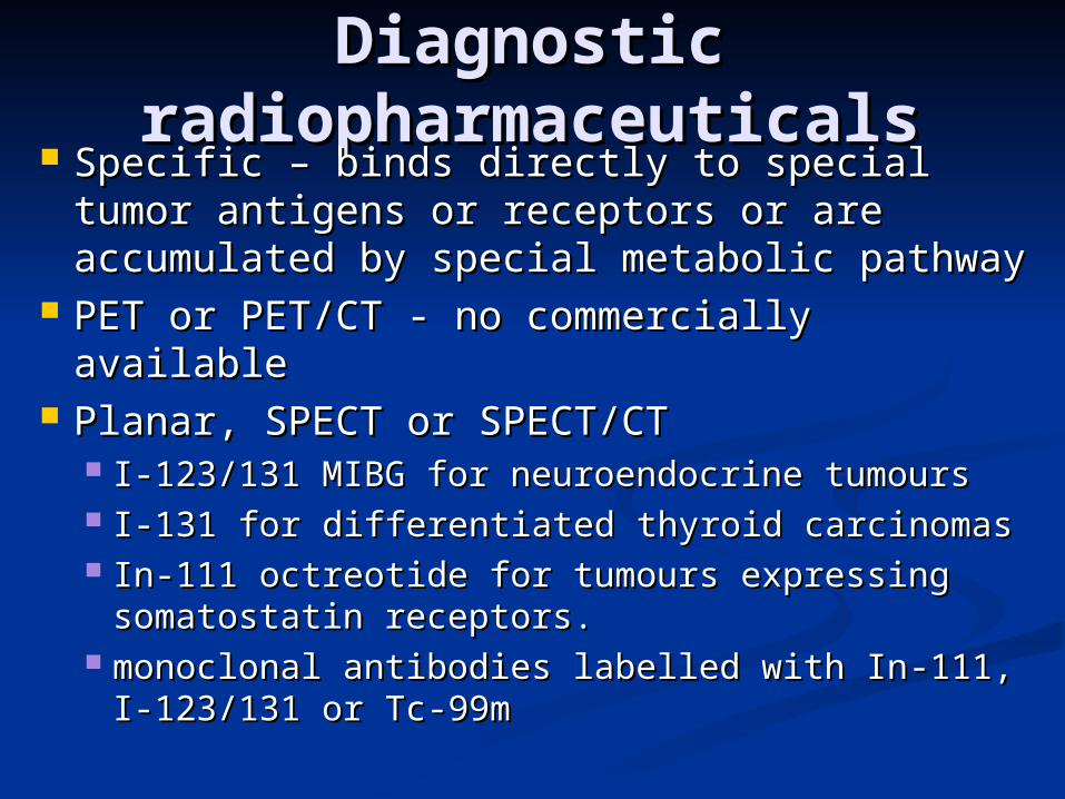
Specific – binds directly to special tumor Specific – binds directly to special tumor antigens or receptors or are accumulated antigens or receptors or are accumulated by special metabolic pathwayby special metabolic pathway
PET or PET/CT - no commercially PET or PET/CT - no commercially availableavailable
Planar, SPECT or SPECT/CTPlanar, SPECT or SPECT/CT I-I-123/131 MIBG for neuroendocrine tumours123/131 MIBG for neuroendocrine tumours II--131 for 131 for differentiateddifferentiated thyroid carcinomas thyroid carcinomas InIn-111-111 octreotide for tumours expressing octreotide for tumours expressing
somatostatin receptors.somatostatin receptors. monoclonal antibodies labelled with Inmonoclonal antibodies labelled with In-111-111, I, I--
123/131 or Tc123/131 or Tc--99m 99m
Therapeutic Therapeutic radiopharmaceuticalsradiopharmaceuticals
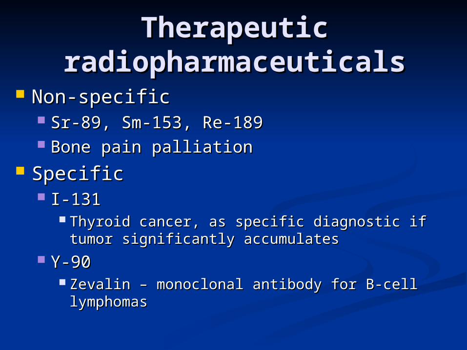
Non-specificNon-specific Sr-89, Sm-153, Re-189Sr-89, Sm-153, Re-189 Bone pain palliationBone pain palliation
SpecificSpecific I-131I-131
Thyroid cancer, as specific diagnostic if tumor Thyroid cancer, as specific diagnostic if tumor significantly accumulatessignificantly accumulates
Y-90Y-90 Zevalin – monoclonal antibody for B-cell Zevalin – monoclonal antibody for B-cell
lymphomaslymphomas

Ga-67 scanGa-67 scan
Introduced in seventies of 20th Introduced in seventies of 20th century for lymphomas (prof. century for lymphomas (prof. Dienstbier)Dienstbier)
Mechanisms of accumulationMechanisms of accumulation tumour viabilitytumour viability blood flowblood flow capillary permeabilitycapillary permeability lymphatic drainagelymphatic drainage transferrin receptors on the tumour cellstransferrin receptors on the tumour cells

Ga-67 scanGa-67 scan Procedure Procedure Patient preparationPatient preparation Laxatives for bowel preparationLaxatives for bowel preparation post post
injectioninjection, nothing else, nothing else Several weeks post tumor therapy (FN)Several weeks post tumor therapy (FN)
radiation radiation therapy therapy and chemotherapy can and chemotherapy can alter the normal pattern of gallium alter the normal pattern of gallium distributiondistribution
180 MBq is usually administered180 MBq is usually administered imaging follows after 48 – 72 hoursimaging follows after 48 – 72 hours WB + SPECT, middle-energy collimatorWB + SPECT, middle-energy collimator

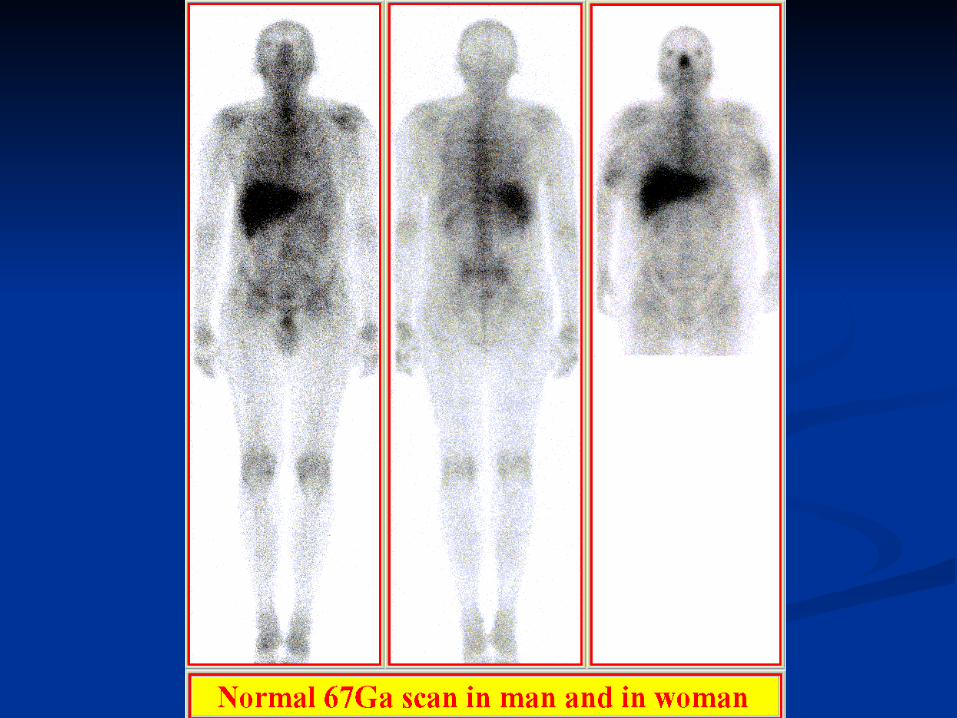
Ga-67 scanGa-67 scan
Normal scanNormal scan Accumulates in bone marrow and liver. Accumulates in bone marrow and liver. Splenic uptake is variable. Splenic uptake is variable. The kidneys are usually visualized and also The kidneys are usually visualized and also
lacrimal, salivary, nasopharyngeal and lacrimal, salivary, nasopharyngeal and genital activity is often present. genital activity is often present.
Female breasts can be visualized, but Female breasts can be visualized, but accumulation is physiologically accumulation is physiologically symmetrical. symmetrical.
Radioactivity is commonly seen in the colonRadioactivity is commonly seen in the colon
Ga-67 scanGa-67 scan
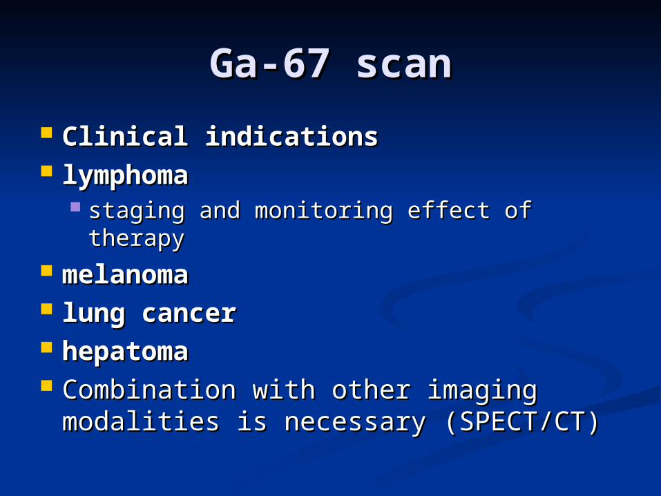
Clinical indicationsClinical indications lymphoma lymphoma
staging and monitoring effect of therapystaging and monitoring effect of therapy mmelanomaelanoma lung cancerlung cancer hepatomahepatoma CCombination with other imaging ombination with other imaging
modalities is necessary modalities is necessary (SPECT/CT)(SPECT/CT)
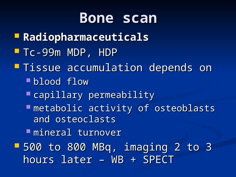
Bone scanBone scan RadiopharmaceuticalsRadiopharmaceuticals TcTc--99m MDP99m MDP,, HDP HDP TTissue accumulation depends onissue accumulation depends on
blood flowblood flow capillary permeabilitycapillary permeability metabolic activity of osteoblasts and metabolic activity of osteoblasts and
osteoclasts osteoclasts mineral turnovermineral turnover
500 to 800 MBq500 to 800 MBq,, imaging 2 to 3 hours imaging 2 to 3 hours laterlater – WB + SPECT – WB + SPECT

Bone scan Bone scan Clinical indications:Clinical indications: DDiagnosis of metastasesiagnosis of metastases of different of different
tumorstumors – staging and follow-up – staging and follow-up PPositivity many months before an ositivity many months before an
abnormality can be detected on abnormality can be detected on X rayX ray - - method of choice to method of choice to seek forseek for bone bone metastasesmetastases
MMainlyainly BBronchogenous carcinoma, prostate, ronchogenous carcinoma, prostate,
breast, thyroid, and renal tumours breast, thyroid, and renal tumours
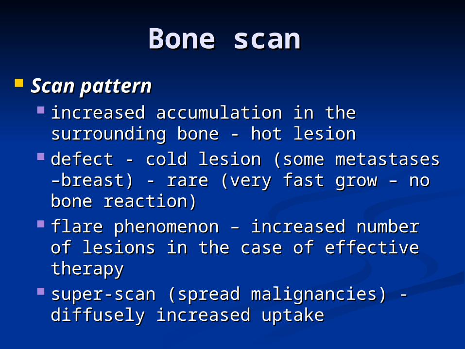
Bone scan Bone scan Scan patternScan pattern
increased accumulation in the increased accumulation in the surrounding bone - hot lesion surrounding bone - hot lesion
defect - cold lesion (some metastases –defect - cold lesion (some metastases –breast) - rare (very fast grow – no bone breast) - rare (very fast grow – no bone reaction)reaction)
flare phenomenon – increased number of flare phenomenon – increased number of lesions in the case of effective therapylesions in the case of effective therapy
super-scan (spread malignancies) - super-scan (spread malignancies) - diffusely increased uptakediffusely increased uptake
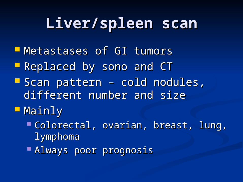
Liver/spleen scanLiver/spleen scan
Metastases of GI tumorsMetastases of GI tumors Replaced by sono and CTReplaced by sono and CT Scan pattern – cold nodules, Scan pattern – cold nodules,
different number and sizedifferent number and size MainlyMainly
Colorectal, ovarian, breast, lung, Colorectal, ovarian, breast, lung, lymphomalymphoma
Always poor prognosisAlways poor prognosis
Thyroid scanThyroid scan
Non-specific test with pertechnetateNon-specific test with pertechnetate Mainly cold nodules – especially in Mainly cold nodules – especially in
children – must be biopsied!!!children – must be biopsied!!!

Bone marrow scanBone marrow scan
Colloids or leukocytesColloids or leukocytes Similar as bone scanSimilar as bone scan Better sensitivityBetter sensitivity

FDG PETFDG PEThttp://www.homolka.cz/nm/http://www.homolka.cz/nm/
For several tumors – staging and follow-For several tumors – staging and follow-upup
Mainly lymphomas, lung cancers, Mainly lymphomas, lung cancers, melanoma, colorectal cancers and othersmelanoma, colorectal cancers and others
Not suitable for prostate cancerNot suitable for prostate cancer Patient preparationPatient preparation
At least 1 w post chemo, 3 m radiotherapyAt least 1 w post chemo, 3 m radiotherapy One hour before injection physical restOne hour before injection physical rest Fasting, no milk, no sugarFasting, no milk, no sugar

Specific methodsSpecific methods
Binding to receptors or antigensBinding to receptors or antigens I-123 MIBG – pheochromocytoma, I-123 MIBG – pheochromocytoma,
neuroblastoma in childrenneuroblastoma in children In-111 Octreoscan – neuroendocrine In-111 Octreoscan – neuroendocrine
tumors (insulinoma, vipoma, tumors (insulinoma, vipoma, carcinoid), SCLCcarcinoid), SCLC
I-131 – thyroid cancer – follow-up I-131 – thyroid cancer – follow-up and treatmentand treatment
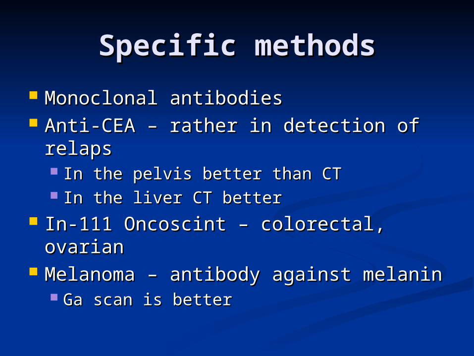
Specific methodsSpecific methods
Monoclonal antibodiesMonoclonal antibodies Anti-CEA – rather in detection of relapsAnti-CEA – rather in detection of relaps
In the pelvis better than CTIn the pelvis better than CT In the liver CT betterIn the liver CT better
InIn--111 Oncoscint – colorectal, ovarian111 Oncoscint – colorectal, ovarian Melanoma – antibody against melaninMelanoma – antibody against melanin
Ga scan is betterGa scan is better

Bone Bone scan – scan –
multiple multiple metastasmetastas
eses

Bone scan – multiple Bone scan – multiple metastasesmetastases
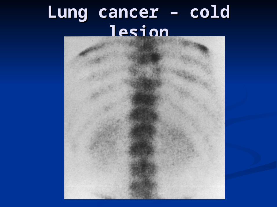
Lung cancer – cold lesionLung cancer – cold lesion

Breast cancerBreast cancernormal X ray with hot spot on scintigraphynormal X ray with hot spot on scintigraphy
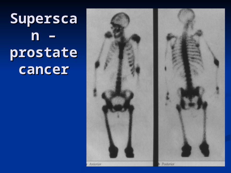
SuperscSuperscan – an –
prostate prostate cancercancer

Bone scan - prostate Bone scan - prostate cancercancer
progressionprogression
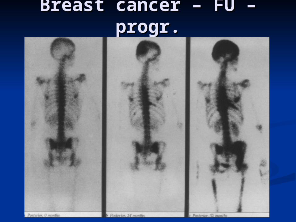
Breast cancer – FU – Breast cancer – FU – progr.progr.

Thyroid – folicular caThyroid – folicular caon sonography solid noduleon sonography solid nodule

Thyroid cancer - Thyroid cancer - anaplasticanaplastic

Thyroid cancerThyroid cancer
Tc-99m Tc-99m post surgery I-131

Thyroid cancer –I-131 - Thyroid cancer –I-131 - metameta

early late
Tc-99mTc-99m sestamibi sestamibiparathyroid adenomaparathyroid adenoma

Neuroblastoma Neuroblastoma liver and bone involvmentliver and bone involvment
Bone scan
I-131 MIBG scan
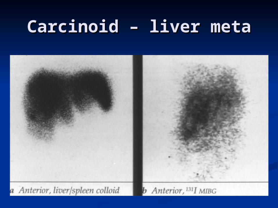
Carcinoid – liver metaCarcinoid – liver meta
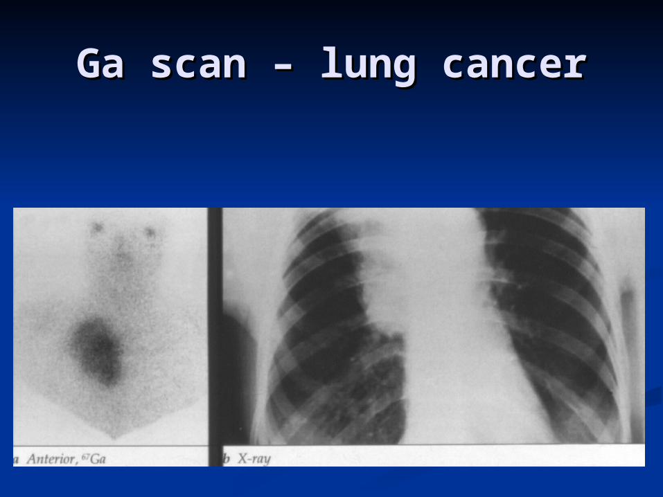
Ga scan – lung cancerGa scan – lung cancer
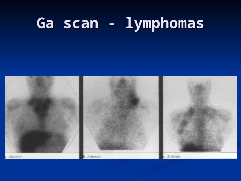
Ga scan - lymphomasGa scan - lymphomas

Palpable mass on the Palpable mass on the neckneck
lymphomalymphomaTc-99m pertechnetate
Ga-67 citrate
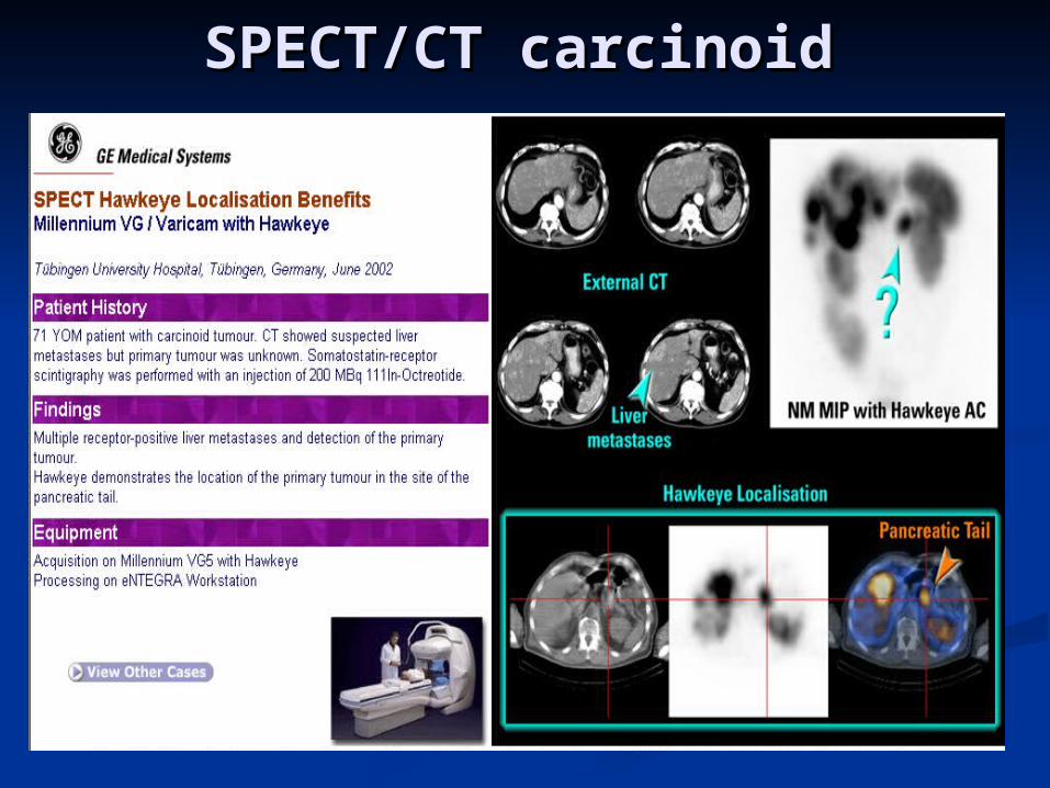
SPECT/CT carcinoid SPECT/CT carcinoid
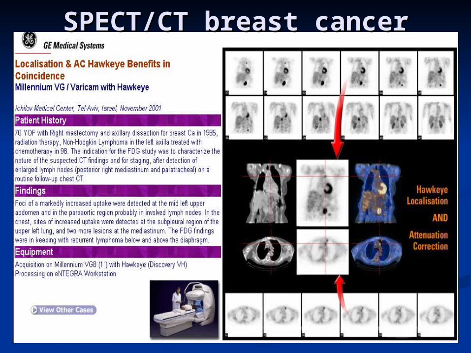
SPECT/CT breast cancerSPECT/CT breast cancer
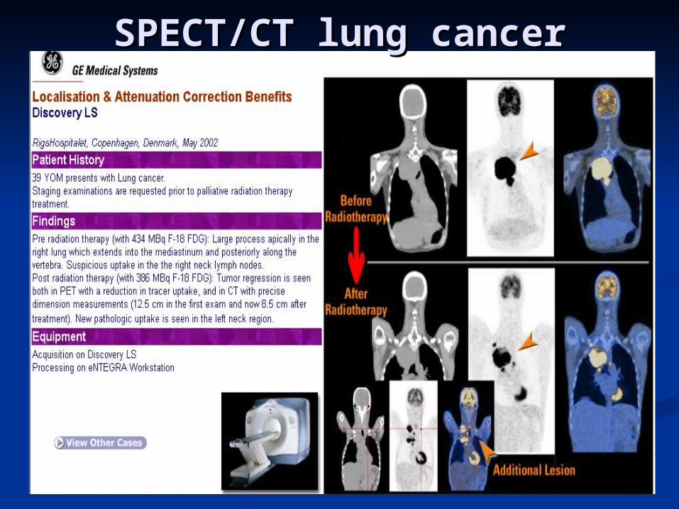
SPECT/CT lung cancerSPECT/CT lung cancer

SPECT/CT lung cancerSPECT/CT lung cancer

FDG PET - normalFDG PET - normal

FDG PET melanomaFDG PET melanoma
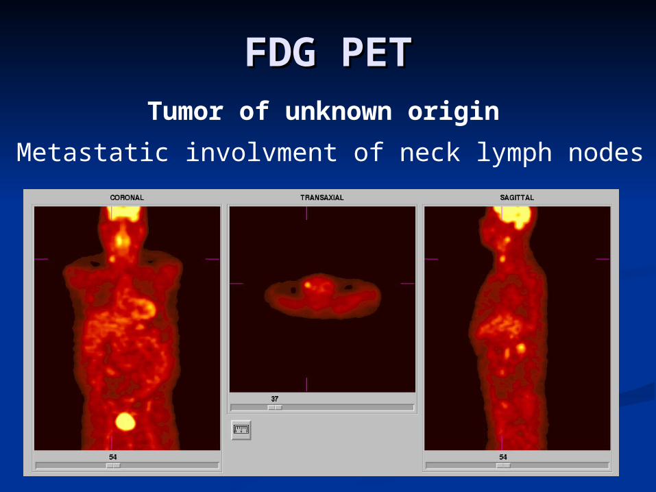
Metastatic involvment of neck lymph nodes
Tumor of unknown origin
FDG PETFDG PET
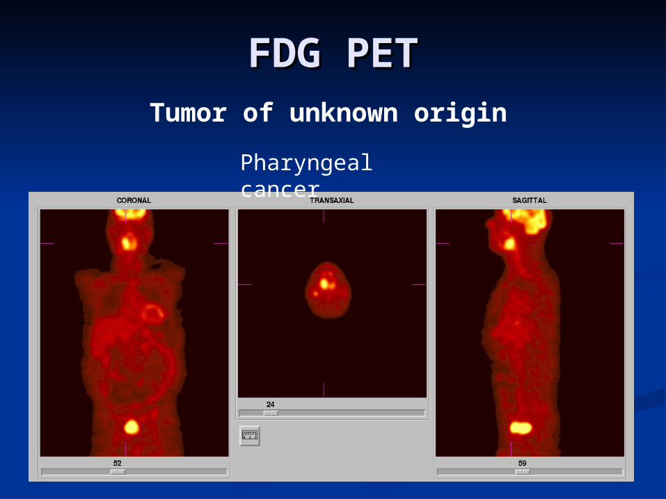
FDG PETFDG PETTumor of unknown origin
Pharyngeal cancer
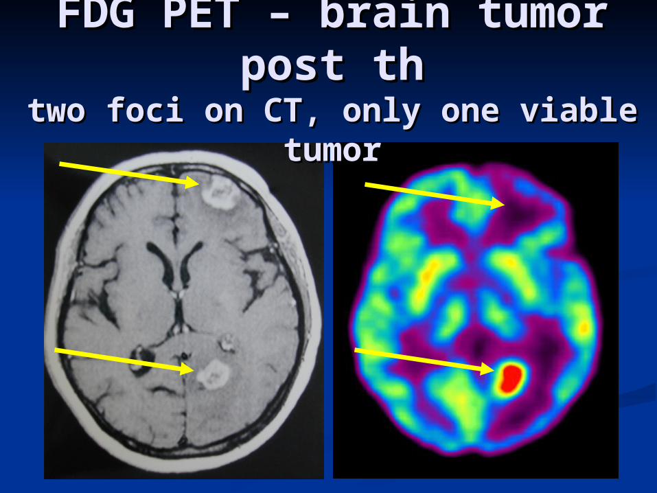
FDG PET – brain tumor FDG PET – brain tumor post thpost th
two foci on CT, only one viable two foci on CT, only one viable tumortumor
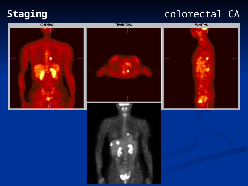
Staging colorectal CA

Effect of therapy Lung cancer
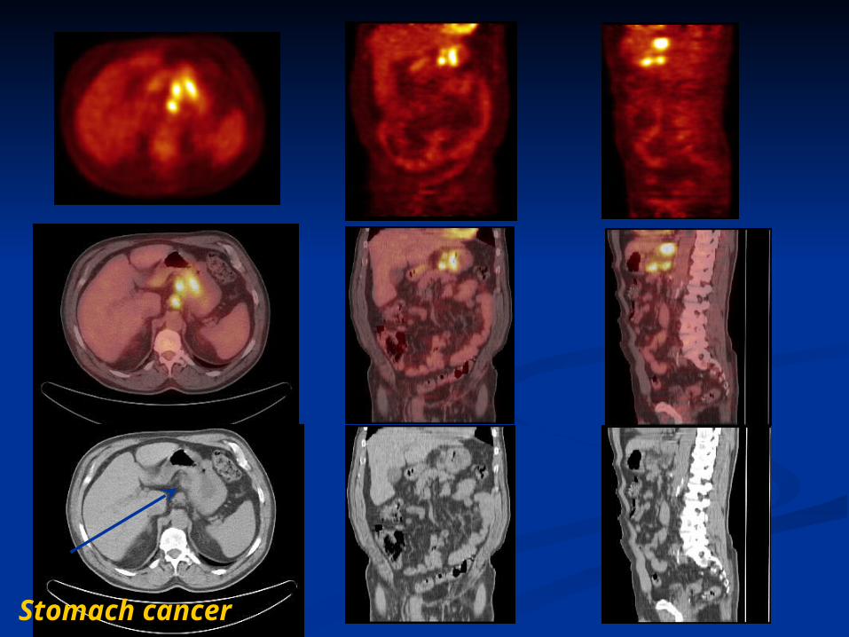
Stomach cancer
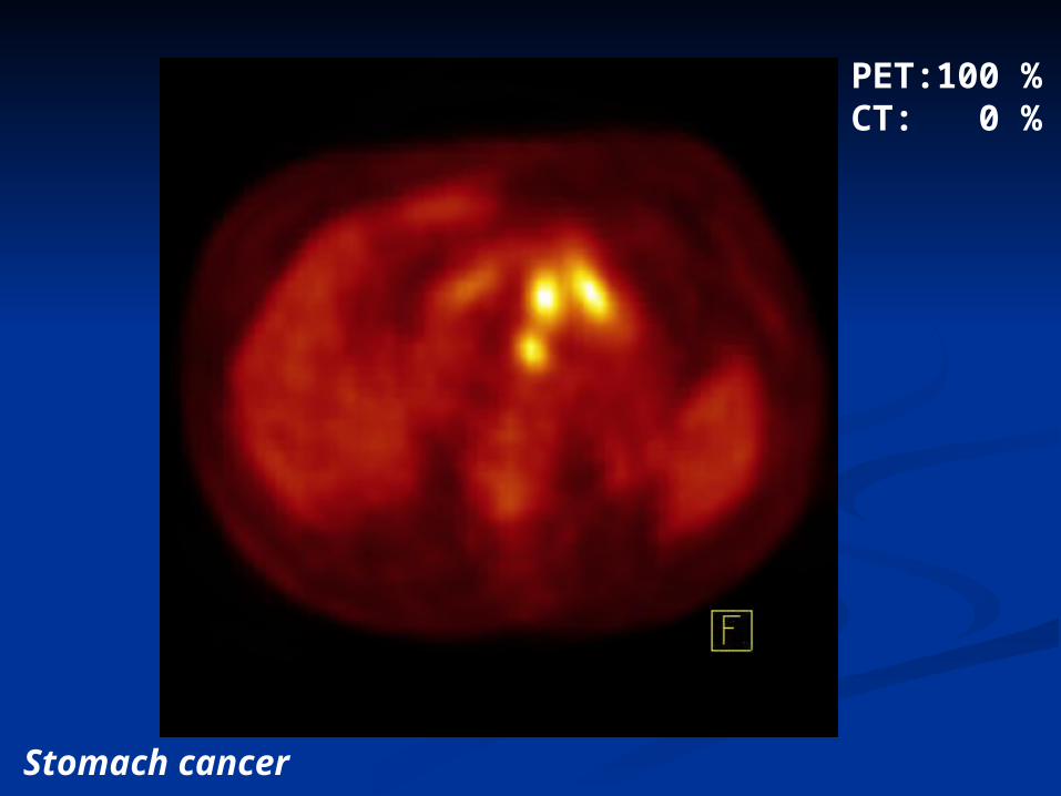
PET:100 %CT: 0 %
Stomach cancer

PET: 80 %CT: 20 %
Stomach cancer

PET: 60 %CT: 40 %
Stomach cancer

PET: 40 %CT: 60 %
Stomach cancer

PET: 20 %CT: 80 %
Stomach cancer

PET: 0 %CT: 100 %
Stomach cancer
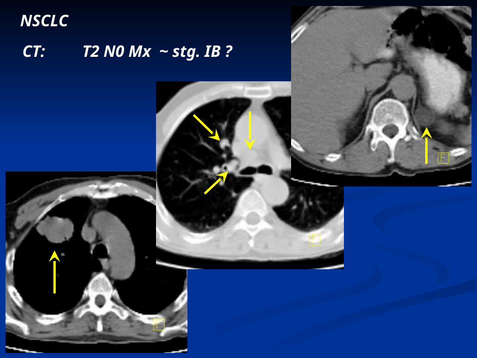
NSCLC
CT: T2 N0 Mx ~ stg. IB ?

NSCLC
CT: T2 N0 Mx ~ stg. IB ?
PET: T2 N2 M0 ~ stg. II
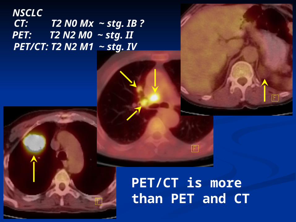
NSCLCCT: T2 N0 Mx ~ stg. IB ?PET: T2 N2 M0 ~ stg. IIPET/CT: T2 N2 M1 ~ stg. IV
PET/CT is more than PET and CT