radiologi bone tumour
DESCRIPTION
Presentasi dari Pemeriksaan radiologi Bone tumor. referat, makalah,TRANSCRIPT

Dr. Rosalina,SpRad


Normal Anatomy
epiphysis
metaphysisdiaphysis
physis
Childhood Adult
cortex
Medullary spaceMedullary space
Physeal scarPhyseal scar
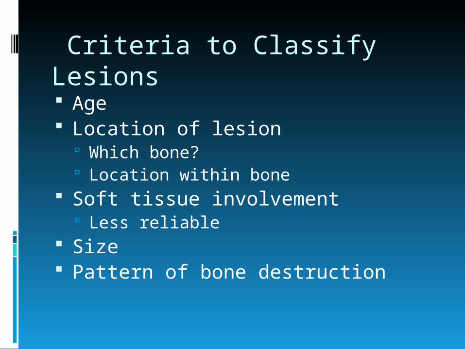
Criteria to Classify Lesions Age Location of lesion
Which bone? Location within bone
Soft tissue involvement Less reliable
Size Pattern of bone destruction

Criteria to Classify Lesions Zone of transition
Margin of lesion
Visible tumor matrix
Polyostotic or monostotic

Malignant Bone Tumors by Age 1-30
Ewing’s Osteosarcoma
30-40 Fibrosarcoma and MFH Malignant Giant Cell Tumor Reticulum Cell Sarcoma

AGE 20Location metaphysisMargins 3
Periosteal Reaction irregularMatrix boneOther
DX osteosarcoma
Osteosarcoma

Malignant Bone Tumors by Age
40+ Mets
Myeloma
(Chondrosarcoma)
Location
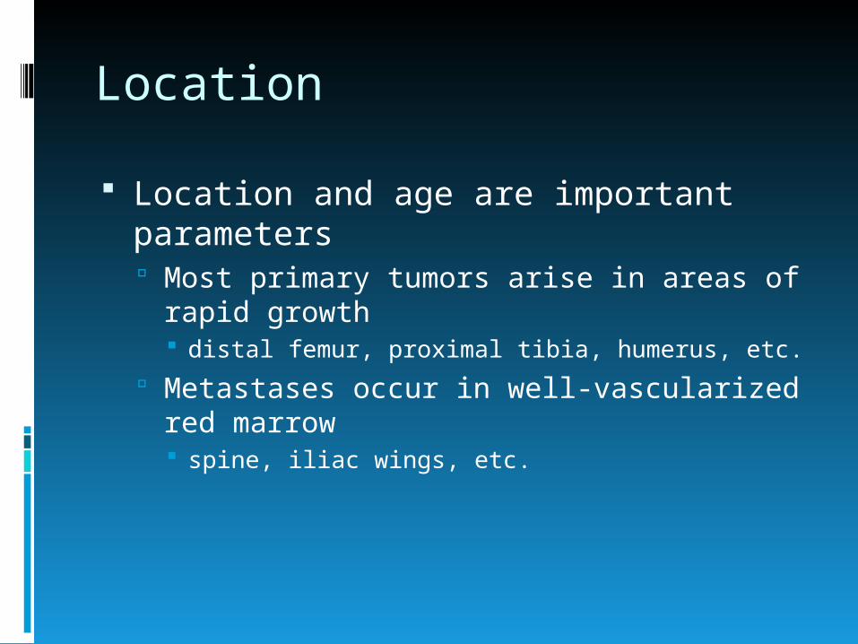
Location and age are important parameters Most primary tumors arise in areas of rapid
growth distal femur, proximal tibia, humerus, etc.
Metastases occur in well-vascularized red marrow spine, iliac wings, etc.

Location
Enchondroma: phalanges
Osteosarcoma & giant cell tumor: around the knee
Hemangioma: skull and spine
Chordoma: sacrum and clivus
Adamantinoma: mid-tibia

Location

Pattern of Bone Destruction Geographic
Well-defined margin
Least aggressive
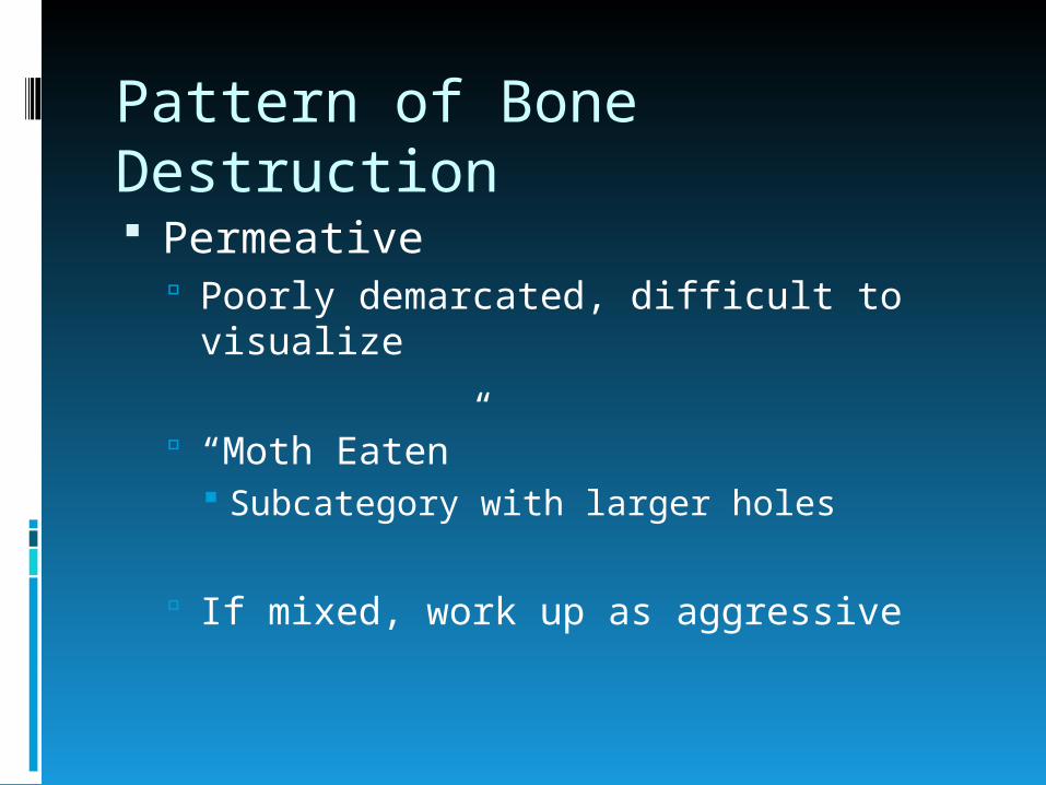
Pattern of Bone Destruction Permeative
Poorly demarcated, difficult to visualize
“Moth Eaten” Subcategory with larger holes
If mixed, work up as aggressive

“Permeative”
Ewing Eosinophilic
Granuloma Infection Myeloma,
metastasis Lymphoma Osteosarcoma

“Motheaten”
Myeloma, metastases
Infection Eosinophilic
Granuloma Osteosarcoma Chondrosarcoma Lymphoma

Reaction of Bone to Tumor Margin between tumor and native bone
can be visible on the plain radiograph
Slowly progressive process is “walled-off” by native bone, producing distinct margins
Rapidly progressive process destroys bone, producing indistinct margins

Zone of transition
Wide Aggressive
Narrow Less aggressive

Non-ossifying fibroma-Narrow Zone

Margin Sclerosis
Sclerotic margin Generally non-aggressive
Lack of sclerotic margin Suggests more aggressive Exceptions

Radiographic Margins
Margin types 1A, 1B, 1C, 2, and 3 Least aggressive 1A, to most aggressive
3

Margins: 1A,1B,1C
increasing aggressiveness

1A: Sclerotic margin
Simple cyst (UBC) Enchondroma Fibrous Dysplasia Chondroblastoma Giant Cell Tumor Chondrosarcoma

1B: Well-defined, non-sclerotic
Giant Cell Tumor Enchondroma Chondroblastoma Myeloma,
Metastatsis Fibrous Dysplasia Chondrosarcoma

Giant Cell Tumor:Well-defined, Non-sclerotic
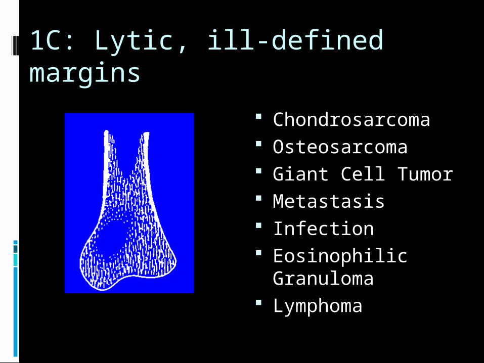
1C: Lytic, ill-defined margins
Chondrosarcoma Osteosarcoma Giant Cell Tumor Metastasis Infection Eosinophilic
Granuloma Lymphoma

Periosteal Reaction
Limited usefulness
Thick, uninterrupted Long standing process, often non-aggressive
Stress fracture, Chronic infection, Osteoid osteoma
Spiculated, lamellated Aggressive process Tumor likely

Types of PeriostealReaction
Malignant type reaction

Codman Triangle
Periosteal reaction
Tumor
Advancing tumor margin destroys periosteal new bone before it ossifies
CodmanTriangle

Tumor Matrix
Chondroid matrix Calcified rings, arcs, dots (stippled) Enchondroma, osteochondroma,
chondroblastoma, chondrosarcoma
Osteoid matrix Dense, homogenous, cloudlike Osteoid osteoma, bone island,
osteosarcoma
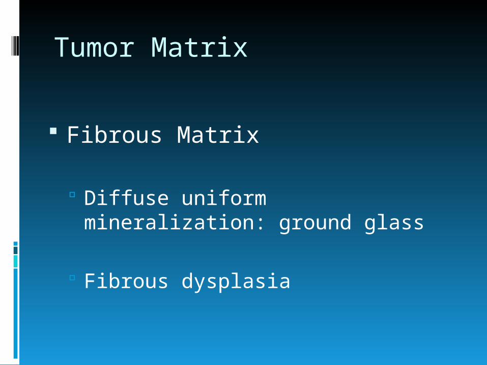
Tumor Matrix
Fibrous Matrix
Diffuse uniform mineralization: ground glass
Fibrous dysplasia

Matrix

AGE 56Location metaphysealMargins 1A
Periosteal Reaction noneMatrix chondroidOther
DX enchondroma
Enchondroma

Polyostotic vs. Monostotic Benign polyostotic
Fibrous Dysplasia, Paget’s, histiocytosis, multiple exostosis, multiple enchondromatosis
Malignant polyostotic Mets, Myeloma, Ewing’s with mets,
Osteosarc with mets and MFH.

Work up
Radiograph Aggresive STOP
+
-
Will information regarding polyostoticvs. monostotic be valuable?
Bone Scan Polyostotic - Chest film and CT
Can distinguish benign, mets, myeloma from other
-
+
+
-+
STOP

Workup
Chest film and CT-
+MR (Less likely CT)For site evaluation
Plan to resect lung mets
Biopsy
+
Palliative Care
-

Biopsy
Sample away from necrotic or non-aggressive area
Avoid contaminating compartments Knee- suprapatellar bursa is large Pelvis- avoid gluteal musculature which
will need for coverage

AGE 13Location metadiaphysisMargins 1A-1B
Periosteal Reaction noneMatrix noneOther trabecular struts
DX UBC
Unicameral Bone Cyst

Aneurysmal Bone Cyst

AGE adultLocation metaphysisMargins 1B
Periosteal Reaction noneMatrix noneOther fx
DX ABC
Aneurysmal Bone Cyst

Non-ossifying fibroma

Giant Cell Tumor

Giant Cell Tumor
AGE 45Location metaphysisMargins 1B
Periosteal Reaction noneMatrix noneOther epi involvement
DX GCT

Osteoid Osteoma

Osteoid Osteoma
AGE 45Location diaphysisMargins 1B
Periosteal Reaction thickMatrix faintOther
DX osteoid osteoma

AGE 66Location diaphysealMargins 1A
Periosteal Reaction minimal, thickMatrix noneOther 2nd lesion
DX wait…..
Multiple Myeloma

AGE 66Location diaphysealMargins 2
Periosteal Reaction noneMatrix noneOther
DX wait…..
Multiple Myeloma

Multiple Myeloma
AGE 66Location flat boneMargins 1B
Periosteal Reaction noneMatrix noneOther multiple
DX myeloma

AGE 12Location diaphysisMargins 3
Periosteal Reaction lamellatedMatrix noneOther
DX Ewing
Ewing’s Sarcoma

Ewing’s sarcoma
“onion-skin”

Osteosarcoma

AGE 16Location diaphysisMargins 3
Periosteal Reaction spiculatedMatrix boneOther fx
DX osteosarcoma
Osteosarcoma

Osteomyelitis

Primary lymphoma

Primary lymphoma

Metastatic Adenocarcinoma

THANK YOU