quantitative analysis of intra-golgi transport shows...
TRANSCRIPT

Quantitative analysis of intra-Golgi transport showsintercisternal exchange for all cargoSerge Dmitrieffa,1, Madan Raob,c, and Pierre Sensa,2
aLaboratoire Gulliver, Centre National de la Recherche Scientifique-Ecole Supérieure de Physique et de Chimie Industrielles, Unité Mixte de Recherche7083, 75231 Paris Cedex 05, France; bRaman Research Institute, Bangalore 560 080, India; and cNational Centre for Biological Sciences (Tata Institute ofFundamental Research), Bangalore 560 065, India
Edited by Jennifer Lippincott-Schwartz, National Institutes of Health, Bethesda, MD, and approved August 13, 2013 (received for review February 21, 2013)
The mechanisms controlling the transport of proteins through theGolgi stack of mammalian and plant cells is the subject of intensedebate, with two models, cisternal progression and intercisternalexchange, emerging as major contenders. A variety of transportexperiments have claimed support for each of these models. Wereevaluate these experiments using a single quantitative coarse-grained framework of intra-Golgi transport that accounts for bothtransport models and their many variants. Our analysis makesa definitive case for the existence of intercisternal exchange bothfor small membrane proteins and large protein complexes––thisimplies that membrane structures larger than the typical protein-coated vesicles must be involved in transport. Notwithstanding,we find that current observations on protein transport cannotrule out cisternal progression as contributing significantly tothe transport process. To discriminate between the differentmodels of intra-Golgi transport, we suggest experiments andan analysis based on our extended theoretical framework thatcompare the dynamics of transiting and resident proteins.
Golgi apparatus | secretory pathway | quantitative transport model |resident golgi enzymes | convection-diffusion
The Golgi apparatus, a complex cellular organelle responsiblefor lipid and protein maturation and sorting, has attracted
a great deal of attention, with many conflicting viewpoints re-garding its mechanisms of transport. The Golgi of plant andanimal cells consists of a stack of 5–20 cisternae (1), possiblyinterconnected by membrane tubules (2), which exchange ma-terial by vesicle budding and fusion (3, 4) (Fig. 1). Each cisternahas a distinct chemical identity, allowing progressive proteinmaturation from the cis to the trans face (5).There is a long-standing argument about the way proteins are
transported through the Golgi, an issue intimately tied to thestructure and dynamics of the organelle itself. The Golgi couldbe a rather static structure, in which cisternae keep constantpositions and identities, and exchange proteins by vesiculartransport. Alternatively, cisternae could progress from the cisend to the trans end without exchanging their cargo (6). Bio-chemical maturation of individual cisterna is known to occur inyeast (Saccharomyces cerevisiae) Golgi, which is not stacked butmade of dispersed cisternae (7, 8). The cisternal progressionmodel posits that this maturation translates into a physical pro-gression of the cisternae (and their content) along the stack. It issupported by the observation that large molecules such as pro-collagen aggregates, presumably unable to enter conventionaltransport vesicles, nonetheless progress through the stack, sug-gesting that cisternae are created at the cis face and destroyed atthe trans face (9). This picture was recently challenged (10) bythe observation that proteins do not exit the Golgi linearly withtime (as a model purely based on cisternal progression wouldpredict) but exponentially, as can be explained by intercisternalexchange. These are however two extreme models, and cisternalprogression and intercisternal exchange could act concomitantly.This is clear even in the cisternal progression model, whichrequires that resident Golgi enzymes (which are found in par-ticular location in the Golgi stack) undergo specific retrograde(vesicular) transport.
Existing quantitative models are often tailored to support(11) or disprove (10) the cisternal progression model, and theircomparison with quantitative data involves a large number offitting parameters (10).However, the relevance of each mode of transport can only be
identified by an unbiased quantitative model based on the gen-eral formalism of transport phenomena (12). We report herethat all available quantitative data on a variety of cargo, in-cluding large procollagen aggregates, can be reproduced bya combination of (i) global protein translation from the cis- tothe trans-Golgi, (ii) diffusive-like protein exchange between cis-ternae, and (iii) protein exit throughout the stack. As shownbelow, the diffusive component implies that intercisternal ex-change is not restricted to small protein-coated vesicles, andinvolves large transport carriers. We rigorously establish thattransport data based on tagging a single molecular species can beargued to be consistent with many different models of transportand therefore cannot provide an unequivocal picture of intra-Golgi transport. To reach this goal, we propose experimentalstrategies based on dynamical correlations between transitingand resident Golgi proteins. A useful virtue of our formalism isthat it can include the influence of the local biochemical andphysical environment within the different cisternae as an energylandscape through which proteins diffuse, and thus permitsa description of transiting proteins and resident Golgi enzymeswithin the same mathematical framework.
ModelTransport Equations for Intercisternal Exchange. Treating the Golgistack as composed of distinct cisternae, we analyze protein
Significance
Two models compete to explain the way proteins transitthrough the Golgi, a cellular organelle consisting of stackedmembrane-bound compartments (cisternae) and responsiblefor protein maturation and sorting. The cisternal maturationmodel proposes that cisternae created de novo move throughthe stack, carrying their content with them. The vesiculartransport model views cisternae as static structures betweenwhich proteins are exchanged by vesicular transport. We havedeveloped a transport model that can quantify the importanceof intercisternal exchange by analyzing the spatiotemporalevolution of a protein distributionwithin the Golgi. Intercisternalexchange is confirmed both for small membrane proteins andlarge protein complexes. This suggests the involvement of mem-brane carriers much larger than typical protein-coated vesicles.
Author contributions: S.D., M.R., and P.S. designed research, performed research, contrib-uted new reagents/analytic tools, analyzed data, and wrote the paper.
The authors declare no conflict of interest.
This article is a PNAS Direct Submission.1Present address: Nédélec Group, Cell Biology and Biophysics, European Molecular BiologyLaboratory, 69115 Heidelberg, Germany.
2To whom correspondence should be addressed. E-mail: [email protected].
This article contains supporting information online at www.pnas.org/lookup/suppl/doi:10.1073/pnas.1303358110/-/DCSupplemental.
15692–15697 | PNAS | September 24, 2013 | vol. 110 | no. 39 www.pnas.org/cgi/doi/10.1073/pnas.1303358110

transport along its axis of polarity (the cis–trans axis), for whichthe cisterna number n, varying between 1 (the cis-most) and N(the trans-most) plays the role of a discrete spatial coordinate.The distribution of a chemical species A within the Golgi may becharacterized by its concentration An!t" in cisterna n at time t.Intercisternal exchange is restricted to “jumps” between adjacentcisternae (with rates kn for n! n+ 1 and k!n for n! n! 1 ; Fig.1). We emphasize that the rates kn; k!n, and rn characterizing thecoarse-grained dynamics may be used regardless of the mi-croscopic details of the exchange process. For vesicular trans-port, they are the product of the rates of fission, translocation,and fusion of vesicles carrying A, and include the waiting timeof A within a cisterna. They are not restricted to processesinvolving protein-coated vesicles, and may include transportthrough connecting membrane tubules and contributions fromany fragment that detaches from one cisterna and fuses witha neighboring cisterna. These rates may depend on the localconcentration An. A master equation (12) can be written forthe concentration An!t" :
"t An!t"= kn! 1An! 1 ! k!nAn|!!!!!!!!!!!!!!{z!!!!!!!!!!!!!!}
net flux: n!1!n
!"knAn ! k!n+1An+ 1
#
|!!!!!!!!!!!!!!!!{z!!!!!!!!!!!!!!!!}net flux: n!n+1
: [1]
A straightforward generalization of the model could includetransport between distant cisternae. This however does not bringnew insight, nor does it improve the comparison with availableexperimental data on transiting proteins.We will rewrite Eq. 1 in a continuous formalism, because this
allows for a better description of cisternal progression. Thecoordinate n (the cisterna number) can be written as a contin-uous variable, and spatial variations are then written as a de-rivative: "nAn = !An+1 !An!1"=2, with distances normalized bythe intercisternal distance (the connection between the discreteand continuous models is described in detail in SI Appendix). Ifthe different exchange rates do not depend too drastically onposition !"nkn # kn", Eq. 1 can be transformed into a Fokker-Planck equation (12). In this continuous description, inter-cisternal exchange amounts to an effective translation withvelocity vt, combined with an effective diffusion with a diffusionconstant Dt :
"An
"t=
""n
$Dt"An
"n! vtAn
%
with Dt =kn + k!n+1
2and vt = kn ! k!n+1 :
[2]
This illustrates that intercisternal exchange always yields aneffective diffusion coefficient, even if all transport steps areanterograde !kn>0; k!n = 0", as we discuss below.
Including Cisternal Progression and External Fluxes. Proteins may betransported toward the Golgi trans face by cisternal progression,defined as the process by which the entire content of a cisternamoves from position n to position n+ 1 in the stack over a timeΔt. The progression velocity is thus defined as vp # 1=Δt, and isthe same for all cisternae. Furthermore, the species A may inprinciple be imported to or exported from any cisterna along thestack. These processes, which include direct recycling to theendoplasmic reticulum (ER), may be expressed as an externalflux Jn = Jnin ! rnAn composed of an influx Jnin to cisterna n, thatcould come from outside the Golgi or from distant cisternae,and a rate of exit rn from cisterna n. Eq. 2 becomes
"An
"t=
""n
0
BBB@Dt"An
"n
z!!}|!!{diffusion
!"vp + vt
#An
z!!!!!!!}|!!!!!!!{net translation
1
CCCA+ Jnz}|{external flux
: [3]
The influx is not expected to contribute significantly to thedynamics of transiting proteins coming from the ER. It is ignoredfor now but is reintroduced in Results, where we derive the distri-bution of resident Golgi enzymes. Fluxes entering at the cis faceand exiting from the trans face of the stack are included in themodel as boundary fluxes (see below).Eq. 3 illustrates three fundamental mechanisms governing the
temporal evolution of a protein distribution within the Golgi: (i)protein exchange between neighboring cisternae introduces aneffective diffusion of the concentration along the Golgi stack,characterized by a diffusion coefficient Dt, (ii) directed proteintransport from the cis- to the trans-Golgi leads to proteintranslation at a velocity v= vt + vp; this accounts both for cisternalprogression (at velocity vp) and for a bias for anterograde !vt > 0" orretrograde !vt < 0" intercisternal exchange, and (iii) proteins mayin principle exit from any Golgi cisterna to join other organelles(the ER or lysosomes) at a rate rn, which may be zero. Note thatbecause the spatial coordinate is a dimensionless number, allthree parameters have units of rates !min!1".Because it does not depend on the microscopic processes re-
sponsible for transport, Eq. 3 constitutes the most rigorousquantification of an arbitrary transport process, and should beused as a first approach to characterize Golgi transport. Theimpact of the three main parameters on the distribution ofproteins throughout the Golgi is best seen when analyzing thepropagation of an initially localized protein distribution (pulse-chase experiments, Fig. 1). The translation velocity displaces theconcentration peak (linearly in time if v is constant), diffusionbroadens the peak (its width increases as the square root of timeif Dt is constant), and protein exit decreases the total proteinconcentration (exponentially with time if r is constant). The variousrates could vary for different proteins, possibly transported bydifferent mechanisms, and should in particular be very differentfor transiting proteins and resident Golgi enzymes.Cisternal progression only affects the translation velocity in
Eq. 3, whereas anterograde intercisternal exchange affects boththe velocity and the diffusion coefficient. Our formalism thusreadily shows a fundamental qualitative difference between thetwo contending models. Within the cisternal progression model,the movement of transiting proteins may occur in the absence of
A
C
B
Fig. 1. Sketch of the Golgi apparatus as a polarized stack of connectedcisternae exchanging material. (A) Proteins synthesized in the ER gothrough the ER-Golgi intermediate compartment (ERGIC) before enter-ing the Golgi through its cis face. After biochemical maturation andsorting, they exit the Golgi through the trans face to join the trans-Golginetwork (TGN). (B) Relevant transport processes, including cisternal pro-gression (translation), diffusion through connecting membrane tubules,vesicular transport, and exit. (C ) Spatiotemporal evolution of an initiallynarrow protein distribution (as produced by a pulse of secretion from theER); pure convection produces a uniform translation of the peak (dashedline), diffusion broadens the peak, and exit exponentially decreases theprotein content.
Dmitrieff et al. PNAS | September 24, 2013 | vol. 110 | no. 39 | 15693
BIOPH
YSICSAND
COMPU
TATIONALBIOLO
GY

intercisternal exchange, thus vp > 0, kn = k!n = 0. This amounts toa perfect translation, without broadening, of a peak of concen-tration, i.e., Dt = 0. Intercisternal exchange, on the other hand,necessarily involves some broadening, with an apparent diffusioncoefficient directly related to the translation velocity (Dt = vt=2 inthe absence of retrograde transport, i.e., when k!n = 0, andDt > vt=2 if k!n $ 0). This immediately leads to a powerful con-clusion: if the analysis of the pulse-chase data using Eq. 3 sug-gests that v> 2Dt, then we can unambiguously conclude that thedata are incompatible with a transport based purely on inter-cisternal exchange and must allow for some cisternal progression.This illustrates how a quantitative analysis based on generic trans-port equations may shed light on the nature of intra-Golgi trans-port, without requiring the knowledge of microscopic detailsof individual transport steps. We show in the next section thatall available data for transiting proteins are well fitted by as-suming constant exchange rates. Spatial variations of thetransport rates are then introduced to study the dynamics ofresident Golgi enzymes.
Boundary Fluxes. Eq. 3 must be supplemented by boundary con-ditions at the cis !n= 1" and trans !n=N" faces of the stack. Atthe cis face, the influx of material J1in from the ER is taken as aparameter (possibly varying with time), imposed by the experi-mental procedure (e.g., in pulse-chase or incoming wave proto-cols, see below). The rate of protein exit at the cis face is taken asa fitting parameter k!!= k!n=1". The outflux of material at thetrans face JNout includes contributions both from vesicles secretedat the trans-Golgi and from the maturation of the trans cisterna:JNout = !vp + kN"AN . As can be seen from Eq. 3, these two con-tributions may not be easily distinguished, as the net fluxthroughout the Golgi involves the net velocity v= vp + vt. We thuswrite the exit flux JNout = !v+ k+"AN , where k+ = kN ! vt is the
fitting parameter of trans-Golgi exit. In addition to the transportparameters (v and Dt) and the exit rate r, there are thus twoadditional boundary parameters k! and k+ in the model.Boundary conditions do affect the spatiotemporal distribution ofproteins inside the Golgi, but we show below that the (bulk)parameters Dt and v, which control the actual transport throughthe Golgi, can nevertheless be determined with reasonable accuracy.
ResultsConfrontation with Experimental Data on Transiting Proteins. Ourtheoretical framework was used to analyze different experimentalobservations, collectively illustrated in Fig. 2. Fluorescence re-covery after photobleaching experiments (FRAP) performed onthe whole Golgi gives access to the total concentration of taggedproteins inside the Golgi. An exponential recovery dynamics isreported in ref. 10, both for small membrane proteins (glycopro-tein of vesicular stomatitis virus, VSVG) and for large cytosolicprotein complexes (procollagen). This was used as an argumentagainst pure cisternal progression, for which a linear recoverydynamics is expected.Our analysis shows (Fig. 2A) that the recovery profile is rather
insensitive to the mode of intra-Golgi transport, and in particularto the effective diffusion coefficient Dt, the only parameter thatsolely depends on intercisternal exchange. We fit the data witha single exponential decay of characteristic time 16 min, whichcould be accounted for by any one of the following: protein exitthroughout the Golgi (parameter r), early exit from the cis face(parameter k!), late exit via the trans face (parameter k+), or anycombination of the three. The dynamics of small inert solublecargo molecule reported in ref. 10 follows a similar, althoughslightly faster, exponential recovery, with similar conclusionsregarding its means of transport. When fluorescent VSVG pro-teins were only allowed to enter the Golgi for a short time, the
A
B
C
D
Fig. 2. Quantitative analysis of data from different experimental protocols using a numerical solution of Eq. 3. (A and B) Optical microscopy assays. A wholeGolgi FRAP experiment probing the exit of tagged proteins from the Golgi following (A) a steady influx, abruptly stopped at t = 0, of a small transmembraneprotein (VSVG) and a large soluble protein aggregate (procollagen), and (B) a short influx, starting at t =0 and stopping at t = 5 min, of VSVG (10). k+ was setto zero in the fits because it does not influence the early relaxation. (C and D) EM assays. (C) Pulse-chase experiment for VSVG (13). Setting either convection(gray curve) or diffusion (dashed curve) to zero cannot reproduce the data. Fits are constrained so that the total protein concentration matches the data att = 14 min. k! was set to zero because it has the same effect as r. (D) Evolution of the concentration of procollagen aggregates in the cis (black) and trans(gray) face of the Golgi upon sudden blockage of ER secretion (exiting wave experiment) (9). Data are in percentage of the concentration in normal conditions(steady ER secretion), and are not sensitive to exit rate. More information on the fitting procedure and experimental uncertainty is given in SI Appendix.
15694 | www.pnas.org/cgi/doi/10.1073/pnas.1303358110 Dmitrieff et al.
exponential recovery started immediately after the cessation ofthe fluorescence influx (Fig. 2B). This shows that proteins do notneed to reach the trans face to exit the Golgi, because recoverywould otherwise show a delay (gray curve in Fig. 2B), and suggestthat proteins can exit at the cis face (parameter k!) or throughoutthe stack (parameter r). Although these experiments give impor-tant information concerning the rate at which proteins are ex-ported from the Golgi, such average measures of Golgi dynamicsdo not yield any clear-cut conclusion on the dominant means oftransport across the Golgi stack. For instance, the exponentialfluorescence decay of both FRAP experiments is consistent with atransport solely based on cisternal progression !Dt = 0", providedproteins are allowed to exit throughout the Golgi at a sufficientrate !r $ v=N". A quantitative assessment of intra-Golgi trans-port, which is tantamount to obtaining numerical values for v andDt, requires the knowledge of the protein distribution inside theentire organelle.Following the transport of a pulse of protein (pulse-chase
protocol, Fig. 1C), or the evolution of the protein distributionacross the Golgi after ER secretion has been suddenly blocked(exiting wave protocol), could in principle yield independentmeasurements of the various parameters. Our analysis of pulse-chase data on small membrane proteins (VSVG, Fig. 2C) (13)clearly shows a combination of translation !v$ 0", broadening!Dt $ 0", and decay (at least one nonvanishing parameter amongfr; k!; k+g) of the peaked concentration distribution. The best fit(black curve in Fig. 2C) suggests that all transport rates havesimilar values (v%Dt % k+ ’ 0:2! 0:3 min!1). The value of thevelocity corresponds to a transit time across the Golgi of orderttransit =N=v ’ 15 min (where N ’ 6 is the number of cisternae).More importantly, the high value of the diffusion coefficientindicates that VSVG is exchanged between cisternae during itstransport through the Golgi.For large cytosolic procollagen aggregates, the exiting wave
protocol reported in ref. 9 shows that concentration differencesbetween the cis- and trans-Golgi relax rather smoothly after se-cretion is stopped, unlike what would be expected within a purecisternal progression model (solid lines compared with dashedlines in Fig. 2D). Our analysis of the (rather scarce) data suggeststhat, just as VSVG, procollagen undergoes intercisternal exchange
with a fairly large diffusion coefficient, Dt ’ v. This large value ofDt is rather surprising for such large protein complexes and offundamental significance.Experimental limitations, such as variability within and be-
tween cells or the finite amount of time needed to set up transportblock, could be argued to smoothen concentration gradients in away similar to intercisternal exchange. We show in SI Appendixthat given the experimental error (below 10% for data of Fig. 2D,ref. 9), a finite diffusion coefficient must be invoked to explainthe procollagen exiting wave data provided ER export ceaseswithin 10 min of the initiation of the block. For a 5-min block, wefind Dt ’ v=2 for procollagen (SI Appendix).The analysis of Fig. 2 C and D provides compelling evidence
that the two cargo molecules studied undergo retrograde trans-port during their journey through the Golgi apparatus. Indeed,our formalism enables us to determine the average number ofintercisternal exchange steps experienced by a protein. In a stackwith N cisternae, it is equal to k+ k! times the average time!N=v" spent in the Golgi, or equivalently to 2NDt=v. We thuspredict an average of 2N ’ 10 exchange steps for VSVG, and 5–10 steps for procollagen. Because v> vt and using k=Dt + vt=2and k!=Dt ! vt=2, we find that at least one-fourth of thesetransport steps is backward (toward the ER).
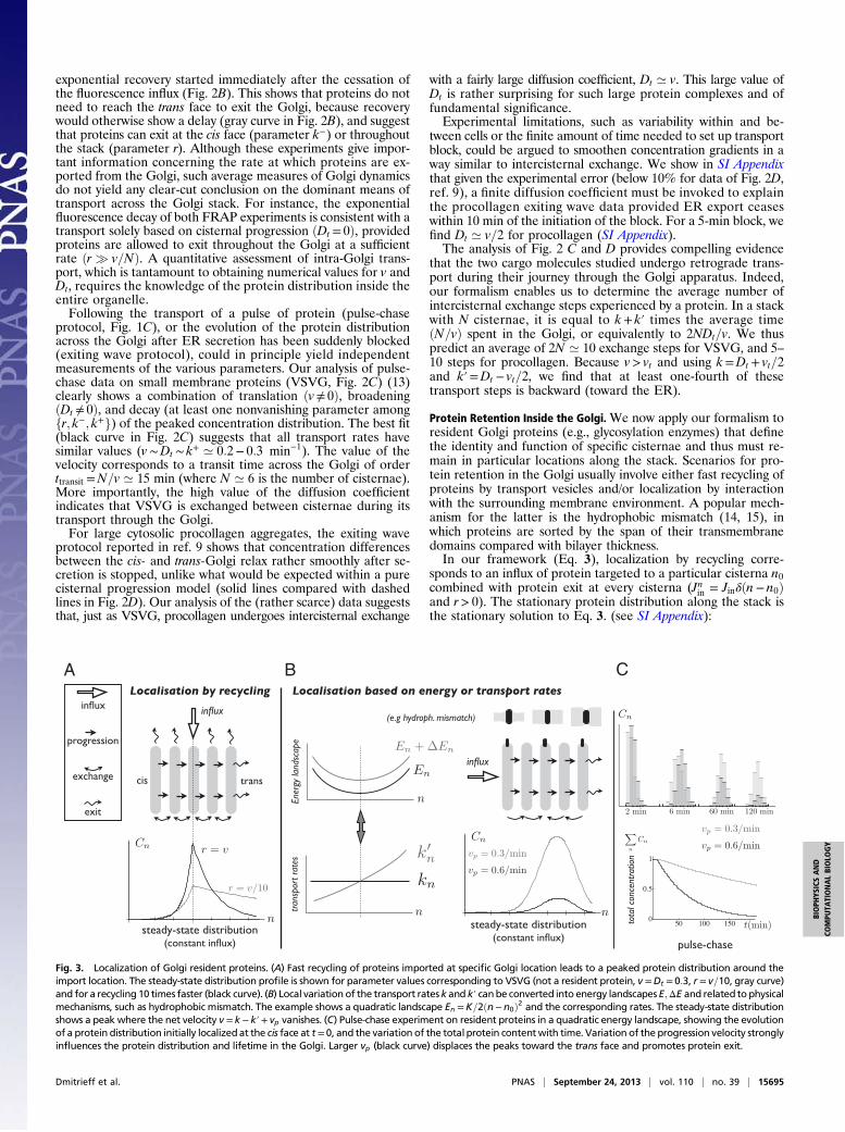
Protein Retention Inside the Golgi.We now apply our formalism toresident Golgi proteins (e.g., glycosylation enzymes) that definethe identity and function of specific cisternae and thus must re-main in particular locations along the stack. Scenarios for pro-tein retention in the Golgi usually involve either fast recycling ofproteins by transport vesicles and/or localization by interactionwith the surrounding membrane environment. A popular mech-anism for the latter is the hydrophobic mismatch (14, 15), inwhich proteins are sorted by the span of their transmembranedomains compared with bilayer thickness.In our framework (Eq. 3), localization by recycling corre-
sponds to an influx of protein targeted to a particular cisterna n0combined with protein exit at every cisterna (Jnin = Jin!!n! n0"and r> 0). The stationary protein distribution along the stack isthe stationary solution to Eq. 3. (see SI Appendix):
A B C
Fig. 3. Localization of Golgi resident proteins. (A) Fast recycling of proteins imported at specific Golgi location leads to a peaked protein distribution around theimport location. The steady-state distribution profile is shown for parameter values corresponding to VSVG (not a resident protein, v =Dt = 0:3, r = v=10, gray curve)and for a recycling 10 times faster (black curve). (B) Local variation of the transport ratesk and k! canbe converted into energy landscapesE;ΔE and related to physicalmechanisms, such as hydrophobic mismatch. The example shows a quadratic landscape En =K=2!n!n0"2 and the corresponding rates. The steady-state distributionshows a peak where the net velocity v = k! k!+ vp vanishes. (C) Pulse-chase experiment on resident proteins in a quadratic energy landscape, showing the evolutionof a protein distribution initially localizedat the cis face at t = 0, and the variation of the total protein contentwith time. Variation of the progression velocity stronglyinfluences the protein distribution and lifetime in the Golgi. Larger vp (black curve) displaces the peaks toward the trans face and promotes protein exit.
Dmitrieff et al. PNAS | September 24, 2013 | vol. 110 | no. 39 | 15695
BIOPH
YSICSAND
COMPU
TATIONALBIOLO
GY

A± !n"=Jine"± !n!n0"&&&&&&&&&&&&&&&&&&v2 + 4rDt
p ; "± =v
2Dt±
&&&&&&&&&&&&&&&&&&v2
4D2t+
rDt
s
: [4]
The protein distribution is peaked at n0 and is asymmetric, A!corresponding to n> n0 and A+ to n< n0. It is spread over 1="!(respectively, 1="+) cisternae toward the trans (respectively, cis)Golgi face, and is broader toward the trans face due to protein con-vection. Accurate protein localization requires r& v, as illustratedin Fig. 3A, a much faster rate than the one we measured forthe transiting protein VSVG !r ’ v=10". The stationary distribution(Eq. 4 and Fig. 3A) requires that the influx is balanced by theoutflux Jin = r
RN1 dnA!n", but is not sensitive to details of the re-
cycling pathway. Whether proteins leaving the Golgi are recycled tocisterna n0 directly or via a more complex pathway (e.g., involvingthe ER or lysosomes) does not modify the steady-state profile.The effect of the biochemical environment on protein re-
tention corresponds to a variation of the transport coefficients vtand Dt along the stack. Generically, protein movement in theGolgi can be written as a diffusion in an effective energy land-scape E!n" characterizing the protein’s energy in the differentcisternae, supplemented by an activation energy ΔE!n" associ-ated with transport intermediates. In SI Appendix, we show that
Dt =kn2"1+ e"nE
#and vt = kn
"1! e"nE
#
with kn = k0e!ΔE!n" and k!n+1 = kne"nE!n" :[5]
A landscape that promotes localization near a particular cis-terna n0 can locally be written as a quadratic potential: E!n"=12K!n! n0"2, where K is the coupling strength. About half theproteins moving through such a landscape would be localized ator near the minimum n0 with a spread Δn= 1=
&&&&K
pcisternae. A
bulk flow (e.g., due to cisternal progression) with velocity vpdisplaces the energy minimum by an amount !n' vp=!KDt"(Fig. 3B and SI Appendix). Thus, precise and robust localizationrequire a large coupling strength K ≳ vp=Dt.The landscape approach allows us to test the relevance of the
hydrophobic mismatch mechanism, for which the energy E!n"can be computed. The membrane thickness of organelles is knownto continuously increase along the secretory pathway from about3.7 nm in the ER to 4.2 nm at the plasma membrane (16), andproteins could be confined to membranes that best match thelength of their hydrophobic domains. The energy of hydro-phobic mismatch leads to a quadratic energy landscape withK %Ks!h2 ’ 0:25kBT (17) (!h ’ 0:1 nm is the mismatch be-tween adjacent cisternae and Ks ’ 0:1 J=m2 is the bilayer stretch-ing modulus). Hydrophobic mismatch can thus in principlelocalize proteins against thermal fluctuations with an accuracyof about Δn% 1=
&&&&K
p= 2 cisternae, and protein localization is in-
deed known to be affected by the length of its transmembranedomain (18, 19). It is however not robust against variation ofthe anterograde flux because K < v=Dt ’ 1, consistent with theobservation that the transmembrane domain length was notthe sole factor affecting protein localization in the Golgi (18).The two mechanisms above (localization by recycling and by
an energy landscape) were used to analyze the distribution of theresident enzyme Man I in Arabidopsis thaliana Golgi stacks; SIAppendix, Fig. S2. This enzyme is localized to cisternae 3 and 4 ofthe stack with a 90% accuracy (20). Such strong confinementrequires either fast recycling !r ’ 2:6v" or a deep energy well!K ’ 2:2". Such large value of K is inconsistent with retentionsolely based on hydrophobic mismatch !K ’ 0:25".
DiscussionCisternal Progression or Vesicular Transport. Our framework pro-duces two strong predictions: (i) the level of intercisternal ex-change (although not its directionality) can be directly quantifiedby measuring the coarse-grained diffusion coefficient Dt, and (ii)
measuring a convection velocity v> 2Dt would necessarily implysome level of cisternal progression. We stress that cisternal pro-gression cannot be disproved in case v< 2Dt, because this couldcorrespond to progression combined with retrograde vesiculartransport. Our analysis of the data clearly shows the existenceof some degree of diffusion, including significant backward trans-port steps, both for the small membrane protein VSVG and forthe large protein complex procollagen (Fig. 2). Furthermore, wefind that v ’ Dt for both species. This implies that (i) inter-cisternal exchange is confirmed in both cases, and (ii) cisternalprogression cannot be proved or disproved by the existing trans-port data. We emphasize that this follows from a strict applicationof general transport principles, and reflects the inadequacy of theexisting experimental data to be more discriminating. Detailedmicroscopic models used to interpret coarse-grained experimentaltransport data (10) should be viewed with caution, confirmingboth the utility and necessity of our coarse-grained approach.Our analysis shows that procollagen is exchanged between
cisternae despite its size. This is at odds with packaging andtransport in conventional small protein-coated transport vesicles,and implies that transport is at least partly mediated by large“pleiomorphic membrane carriers” (PMCs), as sketched in Fig.4. PMCs containing procollagen aggregates could take the formof “megavesicles” such as those involved in the transport of large(engineered) protein complexes (21), or of large tubulo–vesicu-lar connections such as those connecting the Golgi to sur-rounding organelles (22). Such large transport intermediateshave not yet been seen, but recent experiments suggest indirectlythat largo cargo can indeed be exchanged between cisternae(23). A mechanism based on lateral segregation in the cisternalmembranes caused by a Rab cascade, the cisternal progenitormodel (24), has recently linked the formation of large intra-Golgi transport carriers to the maturation of membrane com-ponents. The present work directly infers their involvement inintra-Golgi transport from quantitative transport data. Note thatintercisternal exchange could be quite fast, so a given procolla-gen aggregate could only spend a very short fraction of its transittime outside cisternae. If an exchange step takes %1 s and thereare 10 such steps for a transit time of %15 min, an aggregatespends about 99% of its time inside cisternae. This suggests thatthe formation of megavesicles could be a rare event.Finally, it is intriguing that diffusion and convection are found
to occur at similar rates !v ’ Dt" for both cargoes. This couldindicate that these two processes share the same underlyingmechanisms and/or that one process is coupled to the other, as
procollagen
VSVG
coated vesicle
PMC
distension
cisterna
Fig. 4. Given the evidence for intercisternal exchange during the transit oflarge protein complexes through themammalian Golgi apparatus (procollagenis exchanged on average 6 times during its journey according to our analysis),we propose that PMCs could be involved in intercisternal exchange, possiblytriggered by distensions in procollagen-containing cisternae. The sketchpresents a snapshot of a dynamic process, showing a PMC being exchangedbetween two cisternae: according to our analysis, a procollagen complex isexchanged an average of six times between cisternae during its journeythrough the Golgi (%20 min). This may not lead to a net progression alongthe stack, and does not invalidate cisterna progression as the main causefor anterograde protein transport.
15696 | www.pnas.org/cgi/doi/10.1073/pnas.1303358110 Dmitrieff et al.
suggested by the cisternal progenitor model (24). More insightcould be gained by comparing values for v and Dt in differentorganisms. Scale-forming algae such as Scherffelia dubia havea regularly stacked Golgi of 15–20 cisternae. Proteoglycan scalesreadily identifiable by EM and too large to fit in conventionaltransport vesicles transit through the stack without ever beingseen outside cisternae (20). The absence of scale-containingmegavesicles would imply that these scales undergo pure con-vection in the Golgi. A quantitative incoming wave experiment,yet unavailable to our knowledge, should produce data alongthe dashed lines of Fig. 2D, corresponding to the absenceof diffusion.
Experimental Proposal. We have shown that all available data onthe transport of two very different types of cargo through theGolgi (the small membrane protein VSVG and the large colla-gen complex procollagen) are reproduced by a model of intra-Golgi transport involving constant anterograde and retrogradetransport rates, corresponding to a net constant velocity v andconstant effective diffusion coefficient Dt. Models involvingmore than these two or equivalent parameters for intra-Golgitransport are not falsifiable by current transport experimentsand should be treated with caution.Our analysis shows that diffusion, a signature of intercisternal
exchange, contributes to the transport of both types of cargo. Thisis rather surprising for the large protein complex procollagenand should therefore be confirmed by additional transport datawith high statistical significance, and using a fast !<10 min"transport block protocol. We advocate the use of high-resolutionmicroscopy instead of low resolution optical assays (FRAP),because the latter are dominated by the boundary conditions(Fig. 2 A and B) and do not give sufficient insight into the intra-Golgi dynamics.Direct evidence for cisternal progression may be obtained only
if v> 2Dt ; however, our analysis of the transport data showedv ’ Dt for both types of cargo. More information on the natureof protein transport could be gained by studying correlation inthe transport dynamics of different protein species. A promisingtechnique is the newly developed Retention Using SelectiveHooks method (25), which allows one to precisely control therelease of proteins from the ER into the Golgi, following whichtheir progression and export can be monitored by optical orelectron microscopy.
More insight on the interplay between progression and ex-change could be gained by comparing the dynamics of transitingand resident Golgi proteins. Monitoring the distribution anddynamics of resident proteins under conditions that affect thetransport of transiting proteins could be a promising strategy, asthe localization of resident proteins is affected by cisternal pro-gression. One first needs to identify the mechanism by whichparticular resident proteins are localized, fast recycling (Fig. 3A)and localized retrograde transport related to an energy land-scape (Fig. 3B) being the two generic ones. The distribution ofresident proteins within the stack should then be determined, byhigh-resolution microscopy, under conditions affecting the transittime of proteins putatively transported by cisternal progression,such as drugs targeting the cytoskeleton. According to our pre-diction, this distribution should correlate with the transport rateof transiting proteins in one of two ways if transit is due to cis-ternal progression. The distribution of proteins localized by fastrecycling should broaden, whereas the distribution of proteinslocalized by retrograde transport should be displaced (and notbroaden) toward the trans-Golgi face, under conditions thatdecrease the Golgi transit time (Fig. 3). Finding such correlationswould bring support to the cisternal progression mechanism.We close by recalling that transport solely based on cisternal
progression cannot be reconciled with existing transport data(10). Exchange mediated by large membrane structures (the PMCs)seems the most reasonable compromise, and can be linked to bio-chemical maturation by the cisternal progenitor model (24). Infact, the distinction between cisternal progression and inter-cisternal exchange becomes less clear if transport involves largePMCs (Fig. 4) of size possibly close to the cisterna size, thatundergo frequent scission and fusion. A more crucial question israther whether there exists a bulk anterograde flow of material inthe Golgi, or whether transport is mainly protein-specific. Dynam-ical correlation between different transiting proteins could in-form us of the extent to which they use the same carrier. It wouldin particular be very interesting if the transport of VSVG, for in-stance, was increased by the presence of procollagen. That wouldsuggest that procollagen can create its PMCs and that VSVG canbe exchanged between cisternae by riding along these structures.
ACKNOWLEDGMENTS. We acknowledge stimulating discussions with BrunoGoud, Vivek Malhotra, Satyajit Mayor, and Franck Perez. This work wassupported by the foundation Pierre-Gilles de Gennes pour la Recherche.
1. Polishchuk RS, Mironov AA (2004) Structural aspects of Golgi function. Cell Mol LifeSci 61(2):146–158.
2. Marsh BJ, Volkmann N, McIntosh JR, Howell KE (2004) Direct continuities betweencisternae at different levels of the Golgi complex in glucose-stimulated mouse isletbeta cells. Proc Natl Acad Sci USA 101(15):5565–5570.
3. Orci L, Malhotra V, Amherdt M, Serafini T, Rothman JE (1989) Dissection of a singleround of vesicular transport: Sequential intermediates for intercisternal movement inthe Golgi stack. Cell 56(3):357–368.
4. Malhotra V, Orci L, Glick BS, Block MR, Rothman JE (1988) Role of an N-ethyl-maleimide-sensitive transport component in promoting fusion of transport vesicleswith cisternae of the Golgi stack. Cell 54(2):221–227.
5. Wilson C, Ragnini-Wilson A (2010) Conserved molecular mechanisms underlying ho-meostasis of the Golgi complex. Int J Cell Biol 2010:758230.
6. Marsh BJ, Howell KE (2002) The mammalian Golgi—complex debates. Nat Rev MolCell Biol 3(10):789–795.
7. Matsuura-Tokita K, Takeuchi M, Ichihara A, Mikuriya K, Nakano A (2006) Live imagingof yeast Golgi cisternal maturation. Nature 441(7096):1007–1010.
8. LosevE, etal. (2006)Golgimaturationvisualized in livingyeast.Nature441(7096):1002–1006.9. Bonfanti L, et al. (1998) Procollagen traverses the Golgi stack without leaving the
lumen of cisternae: Evidence for cisternal maturation. Cell 95(7):993–1003.10. Patterson GH, et al. (2008) Transport through the Golgi apparatus by rapid parti-
tioning within a two-phase membrane system. Cell 133(6):1055–1067.11. Glick BS, Elston T, Oster G (1997) A cisternal maturation mechanism can explain the
asymmetry of the Golgi stack. FEBS Lett 414(2):177–181.12. Van Kampen N (2007) Stochastic Processes in Physics and Chemistry (Elsevier, Amsterdam).13. Trucco A, et al. (2004) Secretory traffic triggers the formation of tubular continuities
across Golgi sub-compartments. Nat Cell Biol 6(11):1071–1081.
14. Webb RJ, East JM, Sharma RP, Lee AG (1998) Hydrophobic mismatch and the in-corporation of peptides into lipid bilayers: A possible mechanism for retention in theGolgi. Biochemistry 37(2):673–679.
15. Füllekrug J, Nilsson T (1998) Protein sorting in the Golgi complex. Biochim BiophysActa 1404(1-2):77–84.
16. Mitra K, Ubarretxena-Belandia I, Taguchi T, Warren G, Engelman DM (2004) Modu-lation of the bilayer thickness of exocytic pathway membranes by membrane proteinsrather than cholesterol. Proc Natl Acad Sci USA 101(12):4083–4088.
17. Phillips R, Ursell T, Wiggins P, Sens P (2009) Emerging roles for lipids in shapingmembrane-protein function. Nature 459(7245):379–385.
18. Machamer CE (1993) Targeting and retention of Golgi membrane proteins. Curr OpinCell Biol 5(4):606–612.
19. Rayner JC, Pelham HRB (1997) Transmembrane domain-dependent sorting of proteinsto the ER and plasma membrane in yeast. EMBO J 16(8):1832–1841.
20. Donohoe BS, et al. (2013) Cis-Golgi cisternal assembly and biosynthetic activationoccur sequentially in plants and algae. Traffic 14(5):551–567.
21. Volchuk A, et al. (2000) Megavesicles implicated in the rapid transport of intra-cisternal aggregates across the Golgi stack. Cell 102(3):335–348.
22. Luini A, Ragnini-Wilson A, Polishchuck RS, De Matteis MA (2005) Large pleiomorphictraffic intermediates in the secretory pathway. Curr Opin Cell Biol 17(4):353–361.
23. Lavieu G, Zheng H, Rothman JE (2013) Stapled Golgi cisternae remain in place ascargo passes through the stack. eLIFE, 2:e00558.
24. Pfeffer SR (2010) How the Golgi works: A cisternal progenitor model. Proc Natl AcadSci USA 107(46):19614–19618.
25. Boncompain G, et al. (2012) Synchronization of secretory protein traffic in pop-ulations of cells. Nat Methods 9(5):493–498.
Dmitrieff et al. PNAS | September 24, 2013 | vol. 110 | no. 39 | 15697
BIOPH
YSICSAND
COMPU
TATIONALBIOLO
GY