pulmonary vascular and cardiac effects of peroxynitrite decomposition in newborn rats
TRANSCRIPT
Free Radical Biology & Medicine 49 (2010) 1306–1314
Contents lists available at ScienceDirect
Free Radical Biology & Medicine
j ourna l homepage: www.e lsev ie r.com/ locate / f reeradb iomed
Original Contribution
Pulmonary vascular and cardiac effects of peroxynitrite decomposition innewborn rats
Jaques Belik a,b,c,⁎, Danielle Stevens d, Jingyi Pan a, Brendan A.S. McIntyre a, Crystal Kantores e,Julijana Ivanovska e, Emily Z. Xu c, Christine Ibrahim e, Brian K. Panama c, Peter H. Backx c,Patrick J. McNamara a,b,c, Robert P. Jankov b,c,e
a Physiology and Experimental Medicine Program, The Hospital for Sick Children Research Institute, University of Toronto, Toronto, ON, Canada M5S 1A8b Division of Neonatology, Department of Pediatrics, University of Toronto, Toronto, ON, Canada M5S 1A8c Department of Physiology, University of Toronto, Toronto, ON, Canada M5S 1A8d Maastricht University, 6202 Maastricht, The Netherlandse Clinical Integrative Biology, Sunnybrook Research Institute, University of Toronto, Toronto, ON, Canada M5S 1A8
Abbreviations: AAAT, aortic arterial acceleration tim(4-sulfonatophenyl)porphyrinato iron(III); LVET, left vemedial wall thickness; ONOO−, peroxynitrite anion; Opulmonary arterial acceleration time; PASMC, pulmonaPDE 5, phosphodiesterase 5; PVR, pulmonary vascularassociated phosphoprotein; RV/LV+ S, right ventricularRVET, right ventricular ejection time; sGC, soluble guavascular resistance.⁎ Corresponding author. Physiology and Experime
Hospital for Sick Children Research Institute, UniverCanada M5S 1A8. Fax: +1 416 813 5245.
E-mail address: [email protected] (J. Belik).
0891-5849/$ – see front matter © 2010 Elsevier Inc. Adoi:10.1016/j.freeradbiomed.2010.07.021
a b s t r a c t
a r t i c l e i n f oArticle history:Received 9 February 2010Revised 25 June 2010Accepted 26 July 2010Available online 2 August 2010
Keywords:NeonateCardiomyocyteSmooth muscleEchocardiographyNitrateFree radicals
Evidence implicates oxidative stress as playing a prominent role in the pathogenesis of pulmonaryhypertension, to which peroxynitrite anion (ONOO−) may make a major contribution. Hypothesizing thatremoval of ONOO− would attenuate chronic neonatal pulmonary hypertension, we examined the effects of aONOO− decomposition catalyst (FeTPPS) on pulmonary arteries in vitro, on primary cultured pulmonaryartery smooth muscle cell (PASMC) and cardiomyocyte survival and growth, and on central hemodynamicsin rat pups exposed to hypoxia (13% O2) for 7 days from birth. Daily FeTPPS (30 mg/kg ip) reduced lungnitrotyrosine content, attenuated vascular remodeling, and normalized pulmonary vascular resistance inhypoxia-exposed animals. FeTPPS attenuated proliferation and increased apoptosis of neonatal PASMCs invitro. Isolated neonatal pulmonary arteries treated with FeTPPS showed reduced agonist-induced forcedevelopment and enhanced endothelium-dependent and -independent relaxation, possibly via increasednitrate. However, we observed endothelial dysfunction, enhanced lung tissue phosphodiesterase 5 activity,and biventricular cardiac hypertrophy in air-exposed animals receiving FeTPPS. Further, in contrast toPASMCs, FeTPPS enhanced survival of newborn cardiomyocytes. We conclude that decomposition of ONOO−
with FeTPPS attenuates chronic hypoxia-induced pulmonary hypertension; however, it may negativelyinfluence the modulation of normal pulmonary arterial relaxation function, cell survival, and growth.
e; FeTPPS, 5,10,15,20-tetrakisntricular ejection time; MWT,2•−, superoxide anion; PAAT,
ry artery smooth muscle cell;resistance; VASP, vasodilator-and left ventricular + septum;nylate cyclase; SVR, systemic
ntal Medicine Program, Thesity of Toronto, Toronto, ON,
ll rights reserved.
© 2010 Elsevier Inc. All rights reserved.
Superoxide anion (O2•−) is a reactive oxygen species generated in
increased amounts by the injured pulmonary vasculature and isbelieved to be involved in the pathogenesis of pulmonary arterialhypertension. A potentially major source of O2
•− is endothelial nitricoxide synthase (eNOS), which under normal conditions is primarilyresponsible for nitric oxide (NO) generation and vasomotor toneregulation in the pulmonary vasculature, but when “uncoupled”
generates O2•− in addition to NO. Peroxynitrite anion (ONOO−) is a
relatively stable product of the nearly diffusion-limited reactionbetween NO and O2
•−, which occurs almost 10 times faster than thereaction between superoxide dismutase (SOD) and O2
•− [1]. Chronicexposure of newborn rats to hypoxia (13% O2) from birth inducespulmonary ONOO− generation as evidenced by increased nitrotyrosineformation in the lung [2]. Newborn lambs with severe pulmonaryhypertension treatedwith intratracheal SODhad improvedoxygenationand decreased lung oxidative stress [3]; both effects may have derivedfrom decreased generation of ONOO−.
We have previously shown that ONOO− is a vasoconstrictor innewborn rat pulmonary arteries [4], in contrast to its dual vasocon-stricting and vasodilatory effects on pulmonary vessels derived fromadult animals [5,6]. At high concentrations, ONOO− (10−4 M)reduced endothelium-dependent but not -independent vasorelaxa-tion in vitro. Themechanism accounting for this apparent direct effectof ONOO− on eNOS function is currently unclear, but others haveshown that this molecule is capable of oxidizing the enzyme cofactortetrahydrobiopterin to dihydrobiopterin, thus resulting in uncoupling
1307J. Belik et al. / Free Radical Biology & Medicine 49 (2010) 1306–1314
and reduced NO generation. Finally, we found a temporal association(maximal at day 4) between immunoreactive nitrotyrosine on thewalls of pulmonary resistance arteries and eNOS uncoupling inchronic hypoxia-exposed newborn rats [2], strongly suggesting thatONOO−-mediated eNOS uncoupling contributes to the pulmonaryhypertension observed in these animals.
The goal of this study was therefore to evaluate the effect of aONOO− decomposition catalyst, 5,10,15,20-tetrakis(4-sulfonatophe-nyl)porphyrinato iron(III) (FeTPPS) on structural and functionalchanges in chronic hypoxia-induced pulmonary hypertension innewborn rats. We hypothesized that ONOO− decomposition wouldreduce pulmonary arterial muscle contraction and enhance endothe-lium-dependent relaxation in vitro. We further hypothesized thatdaily FeTPPS administration would normalize pulmonary vascularresistance and attenuate the structural changes of chronic hypoxia-induced pulmonary hypertension.
Experimental procedures
Chemicals and reagents
Except where otherwise indicated, all chemicals and reagents wereobtained from Sigma–Aldrich (Oakville, ON, Canada).
Animal exposures and interventions
All procedures were conducted according to criteria established bythe Canadian Council on Animal Care andwere approved by the AnimalCare Committees of the Sunnybrook and The Hospital for Sick ChildrenResearch Institutes. On their anticipated day of delivery, pathogen-freepregnant Sprague–Dawley dams (Taconic, Germantown,NY, USA)wereplaced in sealed 100×80×60-cm exposure Plexiglas chambers (Bio-Spherix, Redfield, NY, USA) with a 12/12-h light/dark cycle, with thetemperaturekept at 25±1 °Candhumidity at 50%. Each litter, limited to10–12 pups to control for nutritional effects, was nursed in eithernormoxia (21% O2) or hypoxia (13% O2) from birth until 7 days of life.The O2 concentration, temperature, and humidity were continuouslymonitored, recorded, and regulated by computer using customizedhardware (OxyCycler Model A84XOC; BioSpherix) and software(AnaWin2 Run-Time, 2.2.18; Watlow–Anafaze, St. Louis, MO, USA).Dams were exchanged daily between paired normoxia and hypoxiachambers to prevent any maternal toxicity and consequent nutritionaleffects on the pups. FeTPPS (30 mg/kg) in 0.9% saline vehicle or vehiclealonewas administered ip (injected volume: 5 μl/g bodywt) just beforecommencement of hypoxic exposure and daily thereafter. The dose ofFeTPPS usedwas the sameas thatwhichhaspreviously been reported tobe effective inmicewith ONOO−-mediated spinal cord injury [7]. At theend of each exposure period, pups were killed either by pentobarbitalsodium overdose or by exsanguination after anesthesia.
Preparation of tissue extracts
Tissue extracts were prepared in lysis buffer consisting of 50 mMHepes (4-(2-hydroxyethyl)-1-piperazineethanesulfonic acid) bufferedwith sodiumhydroxide topH7.4, 150 mMsodiumchloride, 1% TritonX-100, 10% glycerol, 1.5 mM magnesium chloride, 2 mM EDTA, 1 mMphenylmethanesulfonyl fluoride, 2 μg/ml leupeptin, and 2 μg/mlpepstatin. Lung tissue was homogenized in a rotor/stator-typehomogenizer, and pulmonary arteries and bronchi were frozen in liquidnitrogen and then ground with mortar and pestle before ice-cold lysisbufferwas added. After 1 h on ice, the homogenateswere centrifuged at14,000 rpm for 20 min and the supernatant was collected and stored at−80 °C. Total protein concentration was measured using the Bradfordmethod [8]; extracts were diluted to a final concentration of 4 mg/ml.Given the small size of pulmonary vessels, tissueswere pooled such thateach sample was derived from three animals.
Organ bath studies
Third-generation lung intralobar pulmonary artery ring segments(average diameter 80–100 μm and length 2 mm) were dissected freeand mounted in a wire myograph (Danish Myo Technology A/S,Aarhus, Denmark). Isometric changes were digitized and recordedonline (Myodaq; Danish Myo Technology A/S). Tissues were bathedin Krebs–Henseleit buffer (NaCl, 115 mM; NaHCO3, 25 mM; NaHPO4,1.38 mM; KCl, 2.51 mM; MgSO4 · 7H2O, 2.46 mM; CaCl2, 1.91 mM;and dextrose, 5.56 mM) bubbled with air/6% CO2 and kept at 37 °C.After 1 h of equilibration, the optimal tissue resting tension wasdetermined by repeated stimulation with 128 mM KCl until maxi-mum active tension was reached. All subsequent force measurementswere obtained at optimal resting tension.
Pulmonary vascular muscle force generation was evaluated bystimulating with the thromboxane A2 mimetic U46619. Contractileresponses were normalized to the tissue cross-sectional area using theequation (width×diameter)×2 and expressed as mN/mm2. Relaxa-tion was induced with the respective endothelium-dependent and-independent agonists acetylcholine and sodium nitroprusside, afterprecontraction with U46619 at concentrations equivalent to the EC75.
Histological studies
Animals were anesthetized ip with ketamine (80 mg/kg) andxylazine (20 mg/kg). After the thoracic cavity was opened and thetrachea cannulated, the pulmonary veins were divided. The pulmo-nary circulationwas flushedwith 1 ml PBS containing 1 U/ml heparin,via a needle inserted in the right ventricle, to clear the lungs of bloodwhile the lungs were inflated at a constant pressure of 20 cmH2O. Thelungs were then perfusion-fixed at 100 cmH2O pressure with ice-cold4% (wt/vol) paraformaldehyde in PBS, excised en bloc, dehydrated,cleared in xylene, and embedded in paraffin. Sections (5 μm) wereimmunostained using an avidin–biotin–peroxidase method, as previ-ously described in detail [1]. Slides were incubated with anti-nitrotyrosine rabbit polyclonal antiserum overnight at 4 °C at adilution of 1/500 (2 μg/ml; Upstate Biotechnology, Lake Placid, NY,USA) followed by an anti-rabbit biotin-conjugated secondary anti-body (Santa Cruz Biotechnology, Santa Cruz, CA, USA) for 1 h at roomtemperature. For assessment of percentage arterial medial wallthickness (%MWT), pulmonary arterioles were identified by thepresence of both inner and outer elastic lamina using Weigert'sresorcin–fuchsin (Rowley Biochemical, Danvers, MA, USA), digitallycaptured (Pixera Penguin 600CL; San Jose, CA, USA), and measured byan observer blinded to group identity, as previously described indetail [2]. Mean external diameter was calculated frommeasurementsof round and ovoid vessels in two perpendicular planes (to account forany irregularities in vessel shape), excluding vessels that were cuttangentially (greater than threefold difference in MWT betweenperpendicular planes). Results are shown as mean values from threeor four animals representing two litters per group.
Measurement of PASMC proliferation and apoptosis
Primary culture PASMCswere obtained from Sprague–Dawley ratsduring the first week of life (generally days 2–4). After dissection andremoval under sterile conditions, the third-generation intrapulmon-ary arterial tissue was cut into small pieces and incubated inDulbecco's modified Eagle's medium (DMEM) containing papain(0.5 mg/ml), albumin (1 mg/ml), and dithiothreitol (1 mg/ml) on icefor 15 min and at 37 °C for a further 15 min and then centrifuged at1200 rpm for 5 min. The pellet was resuspended in DMEM with 10%fetal bovine serum (FBS) and maintained in a humid incubator on air/5% CO2 at 37 °C. The cells were passaged by trypsinizing with 0.25%trypsin–EDTA (GIBCO, ON, Canada) and used for experiments atpassages 2–4. The specificity of the cultured cells was confirmed by
1308 J. Belik et al. / Free Radical Biology & Medicine 49 (2010) 1306–1314
immunostaining with mouse monoclonal antibody raised againstsmooth muscle myosin heavy chain (SMMMS-1; Neomarkers) andsmooth muscle calponin. PASMCs were treated with DMEM alone(control), DMEM with the apoptosis inducer paclitaxel (10 μM;positive control for apoptosis), 10% (vol/vol) fetal bovine serum(positive control for proliferation), or varying doses of FeTPPS for 24 hat a gas phase of 21% O2, 5% CO2, and 74% N2. Proliferation wasquantified using a WST-8 colorimetric assay kit (Cayman Chemical,Ann Arbor, MI, USA), according to the manufacturer's instructions.Apoptosis was quantified by measuring histone-complexed DNAfragments, using a commercially available colorimetric ELISA kit (CellDeath Detection ELISAPLUS; Roche, Laval, QC, Canada), according to themanufacturer's instructions. Values are expressed as a proliferation orapoptosis index, where the mean OD change in control cells wasassigned a value of 1, and all other valueswere expressed as amultipleor fraction of the control value.
Estimation of cardiomyocyte survival
Primary rat neonatal cardiomyocytes were isolated and culturedfrom postnatal day 1 or 2 pups according to published protocols [9]. Thecells were characterized by positive staining against mousemonoclonalcardiac α-actin and the presence of beating colonies, clumps, andindividual cells. Because of slow growth and the potential forovergrowth of contaminating myocardial fibroblasts when culturedfor prolonged periods, insufficient cell numbers could be obtained forquantifying proliferation and apoptosis, as was performed for PASMCs.Instead, cardiomyocyte survival in the presence of varying doses ofFeTPPSwas examined using the Live/Dead Viability/Cytotoxicity kit formammalian cells (Invitrogen, Carlsbad, CA, USA). The assay uses two-color fluorescence to differentially stain live and dead cells. The greencalcein-AM is a general cellular stain for live cells, and the red ethidiumhomodimer-1 (EthD-1) intercalates into the DNA of dead and dyingcells, specifically staining their nuclei. Cellswere cultured for 2–3 days inDMEM with 1% FBS in the presence or absence of FeTPPS in 96-wellblack Optilux clear-bottom plates (BD Biosciences). On the day of theLive/Dead assay, cells were washed in 1× HBSS and incubated in 4 μMcalcein-AM and 2 μM EthD-1 for 20 min at 37 °C and visualized on aNikon TE2000 inverted microscope equipped with an environmentalchamber set to 37 °C.
To evaluate the peroxynitrite effect on cardiomyocyte apoptosis thefollowing assaywas conducted. The cardiomyocyteswere plated in a 12-well dish at 200,000 cells perwell and kept in DMEM/F-12 1/1 (vol/vol)containing 5% FBS for 48 h. For the apoptosis assay, cardiomyocytesweremaintained in DMEM/F-12 (without FBS) in the absence (control) orpresenceof peroxynitrite (10−4 M) for 24 h. The cellswere subsequentlytrypsinized and resuspended in 100 μl of binding buffer (140 mM NaCl,2.5 mM CaCl2, 10 mM Hepes, pH 7.4). Next, 3.5 μl of annexin V–FITC(Pharmingen, San Diego, CA, USA) and 3 μl of propidium iodide wereadded and incubated for 15 min on ice. The myocytes were centrifuged(10,000 rpm for 20 s) and the pelleted myocytes were resuspended in200 μl of binding buffer and analyzed on a FACSCalibur flow cytometer(Becton–Dickinson, San Jose, CA, USA).
Nitrotyrosine ELISA
Nitrotyrosine, a “footprint” of ONOO−-mediated reactions, wasmeasured in standards and total lung protein samples using acommercially available competitive ELISA kit (Cayman Chemical).
Western blot analysis of vasodilator-stimulated phosphoprotein (VASP)phosphorylation
Lung tissue was lysed in RIPA buffer containing protease andphosphatase inhibitors, fractionated by SDS–PAGE, transferred topolyvinylidene difluoride membranes, and blotted and band densities
were measured as previously described [3]. VASP phosphorylation,used as a marker of cGMP activity, was quantified by Phos-Tagacrylamide SDS–PAGE [4], according to a method previously reportedin detail [5]. Membranes were blotted with anti-VASP (dilution1:1000; Santa Cruz Biotechnology), yielding two bands, an upperband representing phosphorylated and a lower band representingunphosphorylated VASP (only one band was found when Phos-Tagwas omitted from the resolving gel).
Phosphodiesterase (PDE) 5 activity assay
PDE 5 activity was assayed as previously described [6]. Briefly, lungtissue (approximately 100 mg per sample) was homogenized in lysisbuffer supplemented with protease and phosphatase inhibitors.Samples were immediately placed on ice and assayed the same day.Protein was purified over a Centri-Spin 10 column to remove anyphosphate contamination (Princeton Separations, Adelphia, NJ, USA),because the assay is dependent on free phosphate. Total protein wasassayed for cGMP hydrolytic activity using a commercially availablecolorimetric cyclic nucleotide phosphodiesterase assay kit (Biomol,PlymouthMeeting, PA, USA)with or withoutMY-5445 (40 μM), a PDE5-specific inhibitor, to determine PDE 5-specific cGMP hydrolyticactivity. Results are shown as PDE 5-specific picomoles of cGMPhydrolyzed per milligram of total protein per minute for each sample.
Cardiac ventricular weight
The right-to-left ventricular + septum weight ratio (RV/LV + S)was used as a surrogate marker for right ventricular hypertrophyresulting from pulmonary hypertension, as previously reported [10].
Echocardiography
Evaluation of PVR was performed on anesthetized animals sponta-neously breathing roomair, as previously described in detail [7]. Briefly, ashort-axis view at the level of the aortic valve was obtained and thepulmonary artery was identified using color-flow Doppler. The pulmo-nary arterial acceleration time(PAAT)wasmeasuredas the time fromtheonset of systolic flow to peak pulmonary outflow velocity and the rightventricular ejection time (RVET) as the time from onset to completion ofsystolic pulmonary flow. A surrogate of PVR was calculated according tothe formula 1/(PAAT/RVET). For measurements of systemic hemody-namics, a short-axis viewat the level of the aortic valvewas obtained andthe aorta was identified using color-flow Doppler. The aortic arterialacceleration time (AAAT) and left ventricular ejection time (LVET) werethen calculated from the aortic Doppler profile. AAAT was measured asthe time from the onset of systolic flow to peak aortic outflow velocityand LVET wasmeasured as the time from onset to completion of systolicaortic flow. A surrogate of systemic vascular resistance (SVR) wascalculated according to the formula 1/(AAAT/LVET). Data from a caninemodel suggest that Doppler measurement of aortic blood flow velocityand acceleration can be used for the noninvasive assessment of changesin inotropy and afterload [11].
Data analysis
Data were evaluated by one- or two-way analysis of variance(ANOVA) with multiple comparisons obtained by the Tukey–Kramertest, or the unpaired Student t test when appropriate. Statisticalsignificance was accepted at Pb0.05. All statistical analyses wereperformed with the Number Cruncher Statistical System (NCSS;Kaysville, UT, USA). Data are presented as means±SEM.
1309J. Belik et al. / Free Radical Biology & Medicine 49 (2010) 1306–1314
Results
Acute in vitro effects of FeTPPS on normal pulmonary arteries
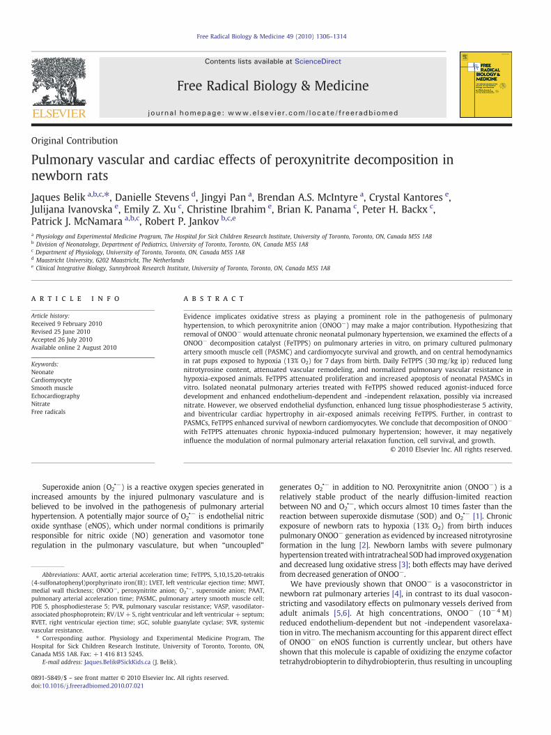
Newborn pulmonary arterial muscle contraction in response to thethromboxane A2 analogue U46619 was significantly reduced in thepresence of FeTPPS (Fig. 1A). The right-shifted forcedose response in thepresence of FeTPPS was also observed when the vessels werepreincubated with the soluble guanylate cyclase (sGC) inhibitor ODQ,suggesting that the lower force in the FeTPPS-treated vessels was notsGC-mediated (Fig. 1B). Uric acid, an in vitroONOO− scavenger [12], hadeffects similar to those of FeTPPS (Fig. 1C). Furthermore, no changes indose response were observed when uric acid and FeTPPS werecoadministered. Together, these findings are consistent with FeTPPS-induced changes in force being secondary to ONOO− decomposition(Fig. 1C).
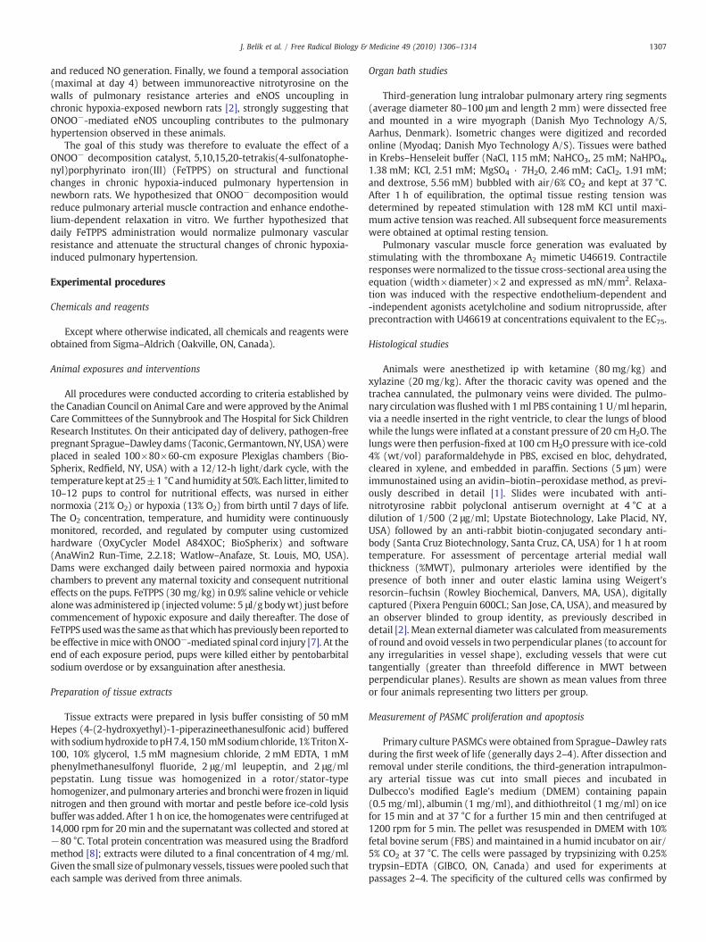
To evaluate the effects of FeTPPS on vasorelaxation, precontracted(U46619; EC75) newborn pulmonary arteries were treated withacetylcholine (endothelium-dependent relaxation) or sodium nitro-prusside (endothelium-independent relaxation). At the tested con-centrations, FeTPPS induced a maximal twofold increase in theendothelium-dependent and -independent pulmonary arterial vasor-elaxation responses (Fig. 2).
Decomposition of ONOO− by FeTPPS leads to a relative reductionin nitrite and increase in nitrate formation [13]. We thereforeevaluated the newborn rat pulmonary arterial muscle tone response
Fig. 1. Thromboxane A2 analogue (U46619)-induced force generation of pulmonaryarteries obtained from newborn animals (N=24). Vessels were studied in the absence(control) or presence of (A) FeTPPS (10−4 M), (B) the soluble guanylate cyclaseinhibitor ODQ (10−5 M), and (C) uric acid (5×10⁻⁵ M). **Pb0.01 compared withcontrol values by two-way ANOVA and Tukey–Kramer multiple-comparison test.
to nitrite and nitrate in precontracted (U46619; EC75) pulmonaryarteries. Nitrite (10−6 M) induced a 10±4% increase (Pb0.01) inpulmonary arterial muscle contraction, whereas nitrate was associ-ated with a maximum reduction in force at 3×10⁻⁷ M (14±1%;Pb0.01). These findings are consistent with FeTPPS mediating arterialrelaxation at least partly through increased generation of nitrate.
Effects of FeTPPS on nitrotyrosine, arterial wall remodeling, cardiacventricular weight, and pulmonary hemodynamics
The effects of daily FeTPPS administration on pulmonary arterial(Fig. 3A) and total lung (Fig. 3B) ONOO− content were evaluated bynitrotyrosine immunostaining and ELISA, respectively, in rat pupsexposed from birth to either air or hypoxia. After 4 days of chronichypoxia exposure immunoreactive nitrotyrosine in the pulmonaryarterial wall was greatly increased by exposure to hypoxia, aspreviously reported [2]. This increase in nitrotyrosine was completelyabrogated by treatment with FeTPPS (Fig. 3A). When total lungnitrotyrosine content was measured by ELISA, no increase in hypoxia-exposed animals was observed (Fig. 3B). However, treatment withFeTPPS greatly reduced nitrotyrosine in the lungs of both air- andhypoxia-exposed animals (Fig. 3B).
Effects of treatment with FeTPPS (30 mg/kg) on pulmonaryvascular remodeling after 7 days of chronic hypoxia exposure (medial
Fig. 2. (A) Endothelium-dependent (ACh—acetylcholine) and (B) -independent (SNP—sodium nitroprusside) relaxation response of U46619-precontracted (EC75) pulmonaryarteries obtained from newborn rats (N=25). Vessels were studied in the absence(control) or presence of FeTPPS (10−4 M). **Pb0.01 compared with control values bytwo-way ANOVA and Tukey–Kramer multiple-comparison test.
Air+Vehicle80
95
110
125
140
Nit
roty
rosi
ne
(mg
/ml o
f lu
ng
ho
mo
gen
ate)
*
*
A
B
Hypoxia+FeTPPSAir+FeTPPS Hypoxia+Vehicle
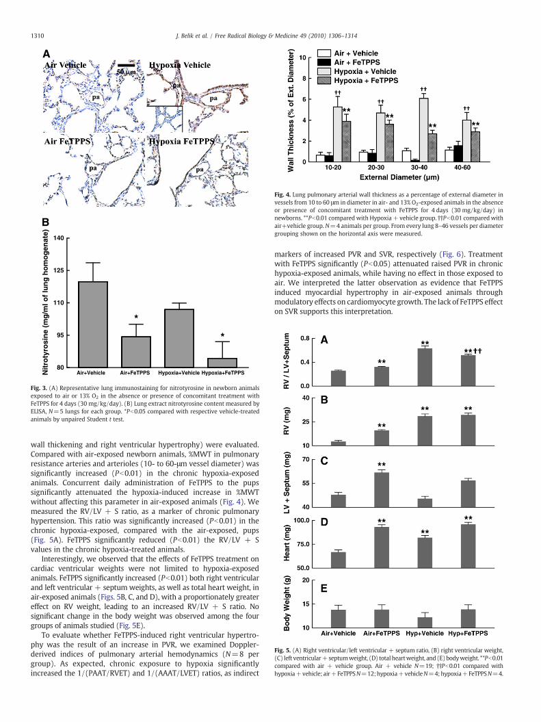
Fig. 3. (A) Representative lung immunostaining for nitrotyrosine in newborn animalsexposed to air or 13% O2 in the absence or presence of concomitant treatment withFeTPPS for 4 days (30 mg/kg/day). (B) Lung extract nitrotyrosine content measured byELISA, N=5 lungs for each group. *Pb0.05 compared with respective vehicle-treatedanimals by unpaired Student t test.
Fig. 4. Lung pulmonary arterial wall thickness as a percentage of external diameter invessels from 10 to 60 μm in diameter in air- and 13% O2-exposed animals in the absenceor presence of concomitant treatment with FeTPPS for 4 days (30 mg/kg/day) innewborns. **Pb0.01 comparedwith Hypoxia+ vehicle group. ††Pb0.01 comparedwithair+vehicle group. N=4 animals per group. From every lung 8–46 vessels per diametergrouping shown on the horizontal axis were measured.
Fig. 5. (A) Right ventricular/left ventricular + septum ratio, (B) right ventricular weight,(C) left ventricular+ septumweight, (D) total heartweight, and (E)bodyweight. **Pb0.01compared with air + vehicle group. Air + vehicle N=19; ††Pb0.01 compared withhypoxia+vehicle; air+FeTPPSN=12;hypoxia+vehicleN=4;hypoxia+FeTPPSN=4.
1310 J. Belik et al. / Free Radical Biology & Medicine 49 (2010) 1306–1314
wall thickening and right ventricular hypertrophy) were evaluated.Compared with air-exposed newborn animals, %MWT in pulmonaryresistance arteries and arterioles (10- to 60-μm vessel diameter) wassignificantly increased (Pb0.01) in the chronic hypoxia-exposedanimals. Concurrent daily administration of FeTPPS to the pupssignificantly attenuated the hypoxia-induced increase in %MWTwithout affecting this parameter in air-exposed animals (Fig. 4). Wemeasured the RV/LV + S ratio, as a marker of chronic pulmonaryhypertension. This ratio was significantly increased (Pb0.01) in thechronic hypoxia-exposed, compared with the air-exposed, pups(Fig. 5A). FeTPPS significantly reduced (Pb0.01) the RV/LV + Svalues in the chronic hypoxia-treated animals.
Interestingly, we observed that the effects of FeTPPS treatment oncardiac ventricular weights were not limited to hypoxia-exposedanimals. FeTPPS significantly increased (Pb0.01) both right ventricularand left ventricular + septum weights, as well as total heart weight, inair-exposed animals (Figs. 5B, C, and D), with a proportionately greatereffect on RV weight, leading to an increased RV/LV + S ratio. Nosignificant change in the body weight was observed among the fourgroups of animals studied (Fig. 5E).
To evaluate whether FeTPPS-induced right ventricular hypertro-phy was the result of an increase in PVR, we examined Doppler-derived indices of pulmonary arterial hemodynamics (N=8 pergroup). As expected, chronic exposure to hypoxia significantlyincreased the 1/(PAAT/RVET) and 1/(AAAT/LVET) ratios, as indirect
markers of increased PVR and SVR, respectively (Fig. 6). Treatmentwith FeTPPS significantly (Pb0.05) attenuated raised PVR in chronichypoxia-exposed animals, while having no effect in those exposed toair. We interpreted the latter observation as evidence that FeTPPSinduced myocardial hypertrophy in air-exposed animals throughmodulatory effects on cardiomyocyte growth. The lack of FeTPPS effecton SVR supports this interpretation.
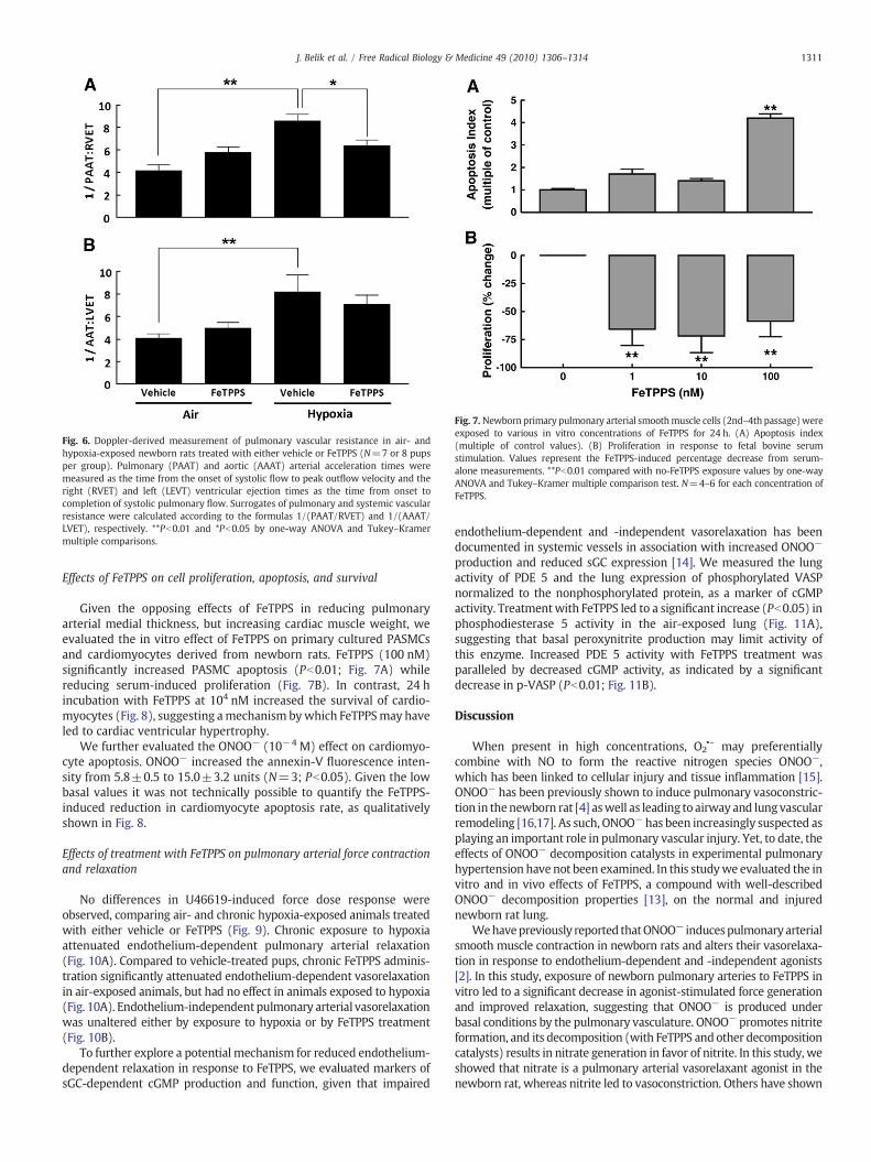
Fig. 6. Doppler-derived measurement of pulmonary vascular resistance in air- andhypoxia-exposed newborn rats treated with either vehicle or FeTPPS (N=7 or 8 pupsper group). Pulmonary (PAAT) and aortic (AAAT) arterial acceleration times weremeasured as the time from the onset of systolic flow to peak outflow velocity and theright (RVET) and left (LEVT) ventricular ejection times as the time from onset tocompletion of systolic pulmonary flow. Surrogates of pulmonary and systemic vascularresistance were calculated according to the formulas 1/(PAAT/RVET) and 1/(AAAT/LVET), respectively. **Pb0.01 and *Pb0.05 by one-way ANOVA and Tukey–Kramermultiple comparisons.
Fig. 7. Newborn primary pulmonary arterial smoothmuscle cells (2nd–4th passage)wereexposed to various in vitro concentrations of FeTPPS for 24 h. (A) Apoptosis index(multiple of control values). (B) Proliferation in response to fetal bovine serumstimulation. Values represent the FeTPPS-induced percentage decrease from serum-alone measurements. **Pb0.01 compared with no-FeTPPS exposure values by one-wayANOVA and Tukey–Kramer multiple comparison test. N=4–6 for each concentration ofFeTPPS.
1311J. Belik et al. / Free Radical Biology & Medicine 49 (2010) 1306–1314
Effects of FeTPPS on cell proliferation, apoptosis, and survival
Given the opposing effects of FeTPPS in reducing pulmonaryarterial medial thickness, but increasing cardiac muscle weight, weevaluated the in vitro effect of FeTPPS on primary cultured PASMCsand cardiomyocytes derived from newborn rats. FeTPPS (100 nM)significantly increased PASMC apoptosis (Pb0.01; Fig. 7A) whilereducing serum-induced proliferation (Fig. 7B). In contrast, 24 hincubation with FeTPPS at 104 nM increased the survival of cardio-myocytes (Fig. 8), suggesting amechanism bywhich FeTPPSmay haveled to cardiac ventricular hypertrophy.
We further evaluated the ONOO− (10−4 M) effect on cardiomyo-cyte apoptosis. ONOO− increased the annexin-V fluorescence inten-sity from 5.8±0.5 to 15.0±3.2 units (N=3; Pb0.05). Given the lowbasal values it was not technically possible to quantify the FeTPPS-induced reduction in cardiomyocyte apoptosis rate, as qualitativelyshown in Fig. 8.
Effects of treatment with FeTPPS on pulmonary arterial force contractionand relaxation
No differences in U46619-induced force dose response wereobserved, comparing air- and chronic hypoxia-exposed animals treatedwith either vehicle or FeTPPS (Fig. 9). Chronic exposure to hypoxiaattenuated endothelium-dependent pulmonary arterial relaxation(Fig. 10A). Compared to vehicle-treated pups, chronic FeTPPS adminis-tration significantly attenuated endothelium-dependent vasorelaxationin air-exposed animals, but had no effect in animals exposed to hypoxia(Fig. 10A). Endothelium-independentpulmonary arterial vasorelaxationwas unaltered either by exposure to hypoxia or by FeTPPS treatment(Fig. 10B).
To further explore a potential mechanism for reduced endothelium-dependent relaxation in response to FeTPPS, we evaluated markers ofsGC-dependent cGMP production and function, given that impaired
endothelium-dependent and -independent vasorelaxation has beendocumented in systemic vessels in association with increased ONOO−
production and reduced sGC expression [14]. We measured the lungactivity of PDE 5 and the lung expression of phosphorylated VASPnormalized to the nonphosphorylated protein, as a marker of cGMPactivity. Treatmentwith FeTPPS led to a significant increase (Pb0.05) inphosphodiesterase 5 activity in the air-exposed lung (Fig. 11A),suggesting that basal peroxynitrite production may limit activity ofthis enzyme. Increased PDE 5 activity with FeTPPS treatment wasparalleled by decreased cGMP activity, as indicated by a significantdecrease in p-VASP (Pb0.01; Fig. 11B).
Discussion
When present in high concentrations, O2•– may preferentially
combine with NO to form the reactive nitrogen species ONOO−,which has been linked to cellular injury and tissue inflammation [15].ONOO− has been previously shown to induce pulmonary vasoconstric-tion in thenewborn rat [4] aswell as leading to airway and lung vascularremodeling [16,17]. As such, ONOO− has been increasingly suspected asplaying an important role in pulmonary vascular injury. Yet, to date, theeffects of ONOO− decomposition catalysts in experimental pulmonaryhypertension have not been examined. In this studywe evaluated the invitro and in vivo effects of FeTPPS, a compound with well-describedONOO− decomposition properties [13], on the normal and injurednewborn rat lung.
Wehavepreviously reported thatONOO− inducespulmonary arterialsmooth muscle contraction in newborn rats and alters their vasorelaxa-tion in response to endothelium-dependent and -independent agonists[2]. In this study, exposure of newborn pulmonary arteries to FeTPPS invitro led to a significant decrease in agonist-stimulated force generationand improved relaxation, suggesting that ONOO− is produced underbasal conditions by the pulmonary vasculature. ONOO− promotes nitriteformation, and its decomposition (with FeTPPS andother decompositioncatalysts) results in nitrate generation in favor of nitrite. In this study, weshowed that nitrate is a pulmonary arterial vasorelaxant agonist in thenewborn rat, whereas nitrite led to vasoconstriction. Others have shown
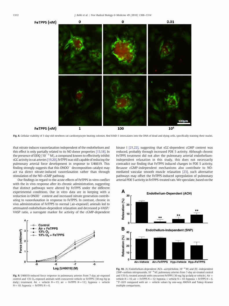
Fig. 8. Cellular viability of 1-day-old newborn rat cardiomyocyte beating colonies. Red EthD-1 intercalates into the DNA of dead and dying cells, specifically staining their nuclei.
1312 J. Belik et al. / Free Radical Biology & Medicine 49 (2010) 1306–1314
that nitrate induces vasorelaxation independent of the endothelium andthis effect is only partially related to its NO donor properties [13,18]. Inthe presence of ODQ (10−5 M), a compound known to effectively inhibitsGCactivity in rat arteries [19,20], FeTPPSwas still capableof reducing thepulmonary arterial force development in response to U46619. Thisfinding strongly suggests that this ONOO− decomposition catalyst mayact via direct nitrate-induced vasorelaxation rather than throughstimulation of the NO–cGMP pathway.
Our findings in regard to the acute effects of FeTPPS in vitro conflictwith the in vivo response after its chronic administration, suggestingthat distinct pathways were altered by FeTPPS under the differentexperimental conditions. Our in vitro data are in keeping with areduction in ONOO− content and increased nitrate generation contrib-uting to vasorelaxation in response to FeTPPS. In contrast, chronic invivo administration of FeTPPS to normal (air-exposed) animals led toattenuated endothelium-dependent relaxation and decreased p-VASP/VASP ratio, a surrogate marker for activity of the cGMP-dependent
Fig. 9. U46619-induced force response in pulmonary arteries from 7-day air-exposedcontrol and 13% O2-exposed animals with concurrent vehicle or FeTPPS (30 mg/kg ipdaily) treatment. Air + vehicle N=13; air + FeTPPS N=12); hypoxia + vehicleN=10; hypoxia + FeTPPS N=6.
kinase I [21,22], suggesting that sGC-dependent cGMP content wasreduced, probably through increased PDE 5 activity. Although chronicFeTPPS treatment did not alter the pulmonary arterial endothelium-independent relaxation in this study, this does not necessarilycontradict our finding that FeTPPS induced changes in PDE 5 activity.Because cGMP-independent mechanisms also contribute to NO-mediated vascular smooth muscle relaxation [23], such alternativepathways may offset the FeTPPS-induced upregulation of pulmonaryarterial PDE 5 activity in FeTPPS-treated rats.We speculate, based on the
Fig. 10. (A) Endothelium-dependent (ACh—acetylcholine, 10−4 M) and (B) -independent(SNP—sodium nitroprusside, 10−4 M) pulmonary arteries from 7-day air-treated controland 13% O2-treated animals with concurrent FeTPPS (30 mg/kg ip daily or vehicle). Air +vehicle N=16; air + FeTPPSN=12; hypoxia + vehicleN=10; hypoxia+ FeTPPS N=6.**Pb0.01 compared with air + vehicle values by one-way ANOVA and Tukey–Kramermultiple comparisons.
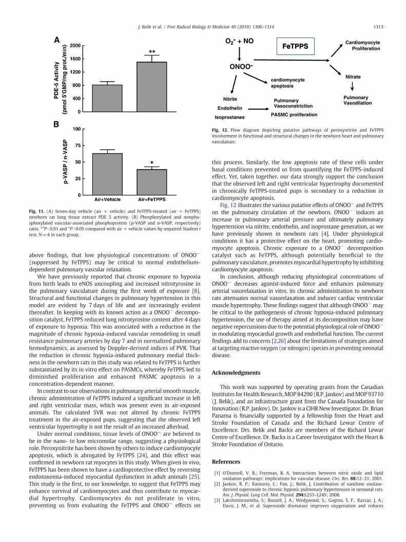
Fig. 11. (A) Seven-day vehicle (air + vehicle) and FeTPPS-treated (air + FeTPPS)newborn rat lung tissue extract PDE 5 activity. (B) Phosphorylated and nonpho-sphorylated vascular-associated phosphoprotein (p-VASP and n-VASP, respectively)ratio. **Pb0.01 and *Pb0.05 compared with air + vehicle values by unpaired Student ttest. N=4 in each group.
O2- + NO
ONOO-
Nitrite
Endothelin
Isoprostanes
cardiomyocyte apoptosis
Nitrate
Cardiomyocyte Proliferation
PASMC proliferation
Pulmonary Vasoconstriction
Pulmonary Vasodilation
FeTPPS
Fig. 12. Flow diagram depicting putative pathways of peroxynitrite and FeTPPSinvolvement in functional and structural changes in the newborn heart and pulmonaryvasculature.
1313J. Belik et al. / Free Radical Biology & Medicine 49 (2010) 1306–1314
above findings, that low physiological concentrations of ONOO−
(suppressed by FeTPPS) may be critical to normal endothelium-dependent pulmonary vascular relaxation.
We have previously reported that chronic exposure to hypoxiafrom birth leads to eNOS uncoupling and increased nitrotyrosine inthe pulmonary vasculature during the first week of exposure [8].Structural and functional changes in pulmonary hypertension in thismodel are evident by 7 days of life and are increasingly evidentthereafter. In keeping with its known action as a ONOO− decompo-sition catalyst, FeTPPS reduced lung nitrotyrosine content after 4 daysof exposure to hypoxia. This was associated with a reduction in themagnitude of chronic hypoxia-induced vascular remodeling in smallresistance pulmonary arteries by day 7 and in normalized pulmonaryhemodynamics, as assessed by Doppler-derived indices of PVR. Thatthe reduction in chronic hypoxia-induced pulmonary medial thick-ness in the newborn rats in this study was related to FeTPPS is furthersubstantiated by its in vitro effect on PASMCs, whereby FeTPPS led todiminished proliferation and enhanced PASMC apoptosis in aconcentration-dependent manner.
In contrast to our observations in pulmonary arterial smoothmuscle,chronic administration of FeTPPS induced a significant increase in leftand right ventricular mass, which was present even in air-exposedanimals. The calculated SVR was not altered by chronic FeTPPStreatment in the air-exposed pups, suggesting that the observed leftventricular hypertrophy is not the result of an increased afterload.
Under normal conditions, tissue levels of ONOO− are believed tobe in the nano- to low micromolar range, suggesting a physiologicalrole. Peroxynitrite has been shown by others to induce cardiomyocyteapoptosis, which is abrogated by FeTPPS [24], and this effect wasconfirmed in newborn rat myocytes in this study. When given in vivo,FeTPPS has been shown to have a cardioprotective effect by reversingendotoxemia-induced myocardial dysfunction in adult animals [25].This study is the first, to our knowledge, to suggest that FeTPPS mayenhance survival of cardiomyocytes and thus contribute to myocar-dial hypertrophy. Cardiomyocytes do not proliferate in vitro,preventing us from evaluating the FeTPPS and ONOO− effects on
this process. Similarly, the low apoptosis rate of these cells underbasal conditions prevented us from quantifying the FeTPPS-inducedeffect. Yet, taken together, our data strongly support the conclusionthat the observed left and right ventricular hypertrophy documentedin chronically FeTPPS-treated pups is secondary to a reduction incardiomyocyte apoptosis.
Fig. 12 illustrates the various putative effects of ONOO− and FeTPPSon the pulmonary circulation of the newborn. ONOO− induces anincrease in pulmonary arterial pressure and ultimately pulmonaryhypertension via nitrite, endothelin, and isoprostane generation, as wehave previously shown in newborn rats [4]. Under physiologicalconditions it has a protective effect on the heart, promoting cardio-myocyte apoptosis. Chronic exposure to a ONOO− decompositioncatalyst such as FeTPPS, although potentially beneficial to thepulmonaryvasculature, promotesmyocardial hypertrophyby inhibitingcardiomyocyte apoptosis.
In conclusion, although reducing physiological concentrations ofONOO− decreases agonist-induced force and enhances pulmonaryarterial vasorelaxation in vitro, its chronic administration to newbornrats attenuates normal vasorelaxation and induces cardiac ventricularmuscle hypertrophy. These findings suggest that although ONOO− maybe critical to the pathogenesis of chronic hypoxia-induced pulmonaryhypertension, the use of therapy aimed at its decomposition may havenegative repercussions due to the potential physiological role of ONOO−
in modulating myocardial growth and endothelial function. The currentfindings add to concerns [2,26] about the limitations of strategies aimedat targeting reactive oxygen (or nitrogen) species in preventing neonataldisease.
Acknowledgments
This work was supported by operating grants from the CanadianInstitutes for Health Research,MOP84290 (R.P. Jankov) andMOP93710(J. Belik), and an infrastructure grant from the Canada Foundation forInnovation (R.P. Jankov). Dr. Jankov is a CIHRNew Investigator. Dr. BrianPanama is financially supported by a fellowship from the Heart andStroke Foundation of Canada and the Richard Lewar Centre ofExcellence. Drs. Belik and Backx are members of the Richard LewarCentre of Excellence. Dr. Backx is a Career Investigator with the Heart &Stroke Foundation of Ontario.
References
[1] O'Donnell, V. B.; Freeman, B. A. Interactions between nitric oxide and lipidoxidation pathways: implications for vascular disease. Circ. Res. 88:12–21; 2001.
[2] Jankov, R. P.; Kantores, C.; Pan, J.; Belik, J. Contribution of xanthine oxidase-derived superoxide to chronic hypoxic pulmonary hypertension in neonatal rats.Am. J. Physiol. Lung Cell. Mol. Physiol. 294:L233–L245; 2008.
[3] Lakshminrusimha, S.; Russell, J. A.; Wedgwood, S.; Gugino, S. F.; Kazzaz, J. A.;Davis, J. M., et al. Superoxide dismutase improves oxygenation and reduces
1314 J. Belik et al. / Free Radical Biology & Medicine 49 (2010) 1306–1314
oxidation in neonatal pulmonary hypertension. Am. J. Respir. Crit. Care Med. 174:1370–1377; 2006.
[4] Belik, J.; Jankov, R. P.; Pan, J.; Tanswell, A. K. Peroxynitrite inhibits relaxation andinduces pulmonary artery muscle contraction in the newborn rat. Free Radic. Biol.Med. 37:1384–1392; 2004.
[5] Iesaki, T.; Gupte, S. A.; Kaminski, P. M.; Wolin, M. S. Inhibition of guanylate cyclasestimulation by NO and bovine arterial relaxation to peroxynitrite and H2O2. Am. J.Physiol. 277:H978–H985; 1999.
[6] Chabot, F.; Mitchell, J. A.; Quinlan, G. J.; Evans, T. W. Characterization of thevasodilator properties of peroxynitrite on rat pulmonary artery: role of poly(adenosine 5′-diphosphoribose) synthase. Br. J. Pharmacol. 121:485–490; 1997.
[7] Genovese, T.; Mazzon, E.; Esposito, E.; Muia, C.; Di, P. R.; Bramanti, P., et al.Beneficial effects of FeTSPP, a peroxynitrite decomposition catalyst, in a mousemodel of spinal cord injury. Free Radic. Biol. Med. 43:763–780; 2007.
[8] Bradford, M. M. A rapid and sensitive method for the quantitation of microgramquantities of protein utilizing the principle of protein–dye binding. Anal. Biochem.72:248–254; 1976.
[9] Wickenden, A. D.; Kaprielian, R.; Parker, T. G.; Jones, O. T.; Backx, P. H. Effects ofdevelopment and thyroid hormone on K+ currents and K+ channel geneexpression in rat ventricle. J. Physiol. 504 (Pt 2):271–286; 1997.
[10] Le Cras, T. D.; Markham, N. E.; Tuder, R. M.; Voelkel, N. F.; Abman, S. H. Treatmentof newborn rats with a VEGF receptor inhibitor causes pulmonary hypertensionand abnormal lung structure. Am. J. Physiol. Lung Cell. Mol. Physiol. 283:L555–L562;2002.
[11] Sohn, S.; Kim, H. S.; Han, J. J. Doppler flowvelocitymeasurement to assess changes ininotropy and afterload: a study inhealthydogs. Echocardiography19:207–213; 2002.
[12] Turan, N. N.; Yildiz, G.; Gumusel, B.; Demiryurek, A. T. Ischemic and peroxynitritepreconditioning effects in chronic hypoxic rat lung. Exp. Lung Res. 34:325–341; 2008.
[13] Misko, T. P.; Highkin, M. K.; Veenhuizen, A. W.; Manning, P. T.; Stern, M. K.; Currie,M. G., et al. Characterization of the cytoprotective action of peroxynitritedecomposition catalysts. J. Biol. Chem. 273:15646–15653; 1998.
[14] Kagota, S.; Tada, Y.; Nejime, N.; Nakamura, K.; Kunitomo, M.; Shinozuka, K.Chronic production of peroxynitrite in the vascular wall impairs vasorelaxationfunction in SHR/NDmcr-cp rats, an animal model of metabolic syndrome. J.Pharmacol. Sci. 109:556–564; 2009.
[15] Sugiura, H.; Liu, X.; Kobayashi, T.; Togo, S.; Ertl, R. F.; Kawasaki, S., et al. Reactivenitrogen species augment fibroblast-mediated collagen gel contraction, mediatorproduction, and chemotaxis. Am. J. Respir. Cell Mol. Biol. 34:592–599; 2006.
[16] DeMarco, V. G.; Habibi, J.; Whaley-Connell, A. T.; Schneider, R. I.; Sowers, J. R.;Andresen, B. T., et al. Rosuvastatin ameliorates the development of pulmonaryarterial hypertension in the transgenic (mRen2)27 rat. Am. J. Physiol. Heart Circ.Physiol. 297:H1128–H1139; 2009.
[17] Ichikawa, T.; Sugiura, H.; Koarai, A.; Yanagisawa, S.; Kanda, M.; Hayata, A., et al.Peroxynitrite augments fibroblast-mediated tissue remodeling via myofibroblastdifferentiation. Am. J. Physiol. Lung Cell. Mol. Physiol. 295:L800–L808; 2008.
[18] Kleschyov, A. L.; Oelze, M.; Daiber, A.; Huang, Y.; Mollnau, H.; Schulz, E., et al. Doesnitric oxide mediate the vasodilator activity of nitroglycerin? Circ. Res. 93:e104–e112; 2003.
[19] Tseng, C. M.; Tabrizi-Fard, M. A.; Fung, H. L. Differential sensitivity among nitricoxide donors toward ODQ-mediated inhibition of vascular relaxation. J.Pharmacol. Exp. Ther. 292:737–742; 2000.
[20] Teixeira, C. E.; Priviero, F. B.; Todd Jr., J.; Webb, R. C. Vasorelaxing effect of BAY 41-2272 in rat basilar artery: involvement of cGMP-dependent and independentmechanisms. Hypertension 47:596–602; 2006.
[21] Mulsch, A.; Oelze,M.;Kloss, S.;Mollnau,H.; Topfer,A.; Smolenski, A., et al. Effects of invivo nitroglycerin treatment on activity and expression of the guanylyl cyclase andcGMP-dependent protein kinase and their downstream target vasodilator-stimu-lated phosphoprotein in aorta. Circulation 103:2188–2194; 2001.
[22] Oelze, M.; Mollnau, H.; Hoffmann, N.; Warnholtz, A.; Bodenschatz, M.; Smolenski,A., et al. Vasodilator-stimulated phosphoprotein serine 239 phosphorylation as asensitive monitor of defective nitric oxide/cGMP signaling and endothelialdysfunction. Circ. Res. 87:999–1005; 2000.
[23] Weisbrod, R. M.; Griswold, M. C.; Yaghoubi, M.; Komalavilas, P.; Lincoln, T. M.;Cohen, R. A. Evidence that additional mechanisms to cyclic GMP mediate thedecrease in intracellular calcium and relaxation of rabbit aortic smooth muscle tonitric oxide. Br. J. Pharmacol. 125:1695–1707; 1998.
[24] Klassen, S. S.; Rabkin, S. W. The metalloporphyrin FeTPPS but not by cyclosporin Aantagonizes the interaction of peroxynitrate and hydrogen peroxide oncardiomyocyte cell death. Naunyn Schmiedebergs Arch. Pharmacol. 379:149–161;2009.
[25] Lauzier, B.; Sicard, P.; Bouchot, O.; Delemasure, S.; Moreau, D.; Vergely, C., et al. Aperoxynitrite decomposition catalyst: FeTPPS confers cardioprotection duringreperfusion after cardioplegic arrest in a working isolated rat heart model.Fundam. Clin. Pharmacol. 21:173–180; 2007.
[26] Jankov, R. P.; Negus, A.; Tanswell, A. K. Antioxidants as therapy in the newborn:some words of caution. Pediatr. Res. 50:681–687; 2001.