pterygopalatine fossa - judoctors · back into the pterygopalatine fossa. the veins coalesce in the...
TRANSCRIPT
PTERYGOPALATINE
FOSSA
Prof. Dr.Mohammed Hisham
Al-Muhtaseb
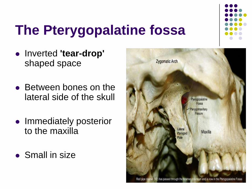
The Pterygopalatine fossa
Inverted 'tear-drop'shaped space
Between bones on the lateral side of the skull
Immediately posterior to the maxilla
Small in size
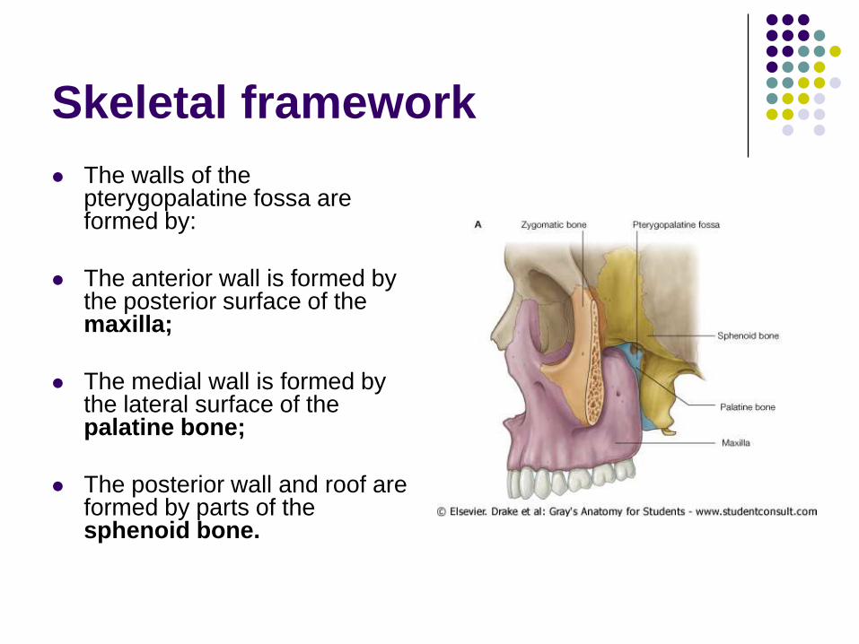
Skeletal framework
The walls of the pterygopalatine fossa are formed by:
The anterior wall is formed by the posterior surface of the maxilla;
The medial wall is formed by the lateral surface of the palatine bone;
The posterior wall and roof are formed by parts of the sphenoid bone.
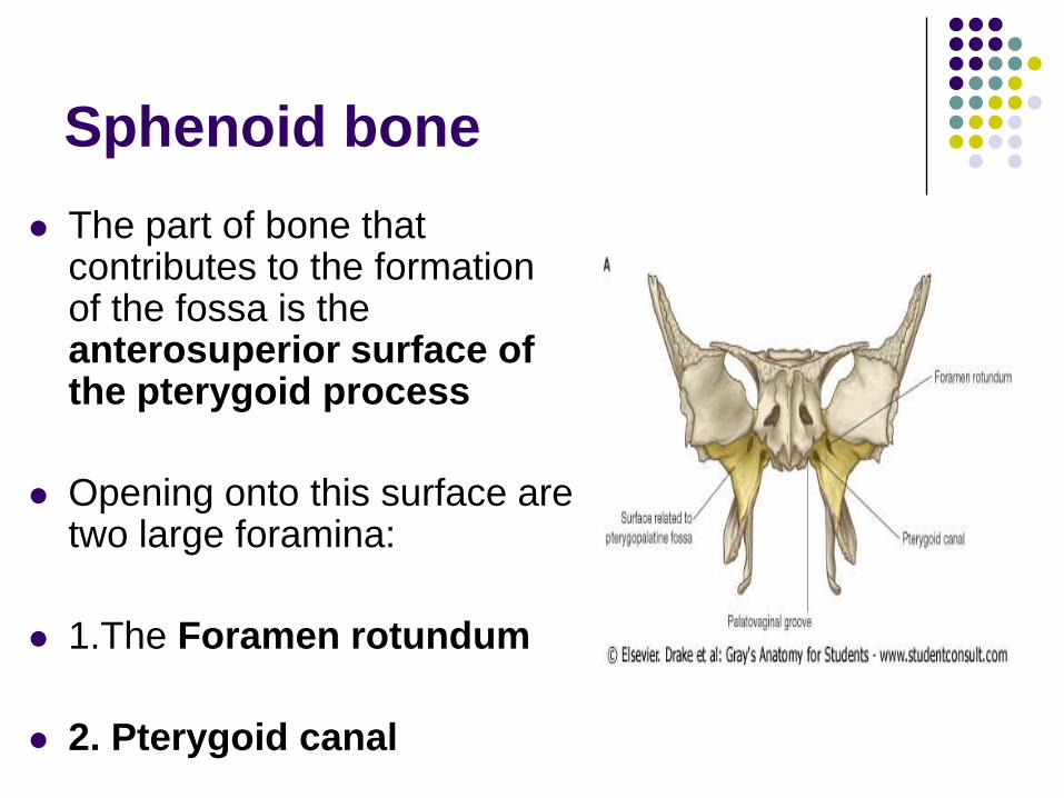
Sphenoid bone
The part of bone that contributes to the formation of the fossa is the anterosuperior surface of the pterygoid process
Opening onto this surface are two large foramina:
1.The Foramen rotundum
2. Pterygoid canal
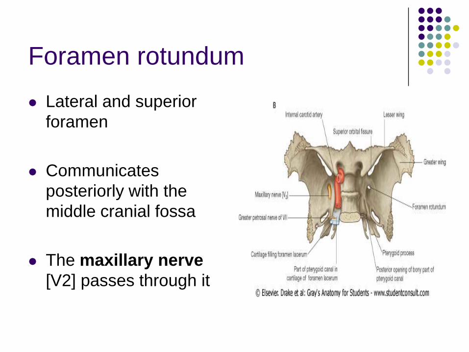
Foramen rotundum
Lateral and superior
foramen
Communicates
posteriorly with the
middle cranial fossa
The maxillary nerve
[V2] passes through it
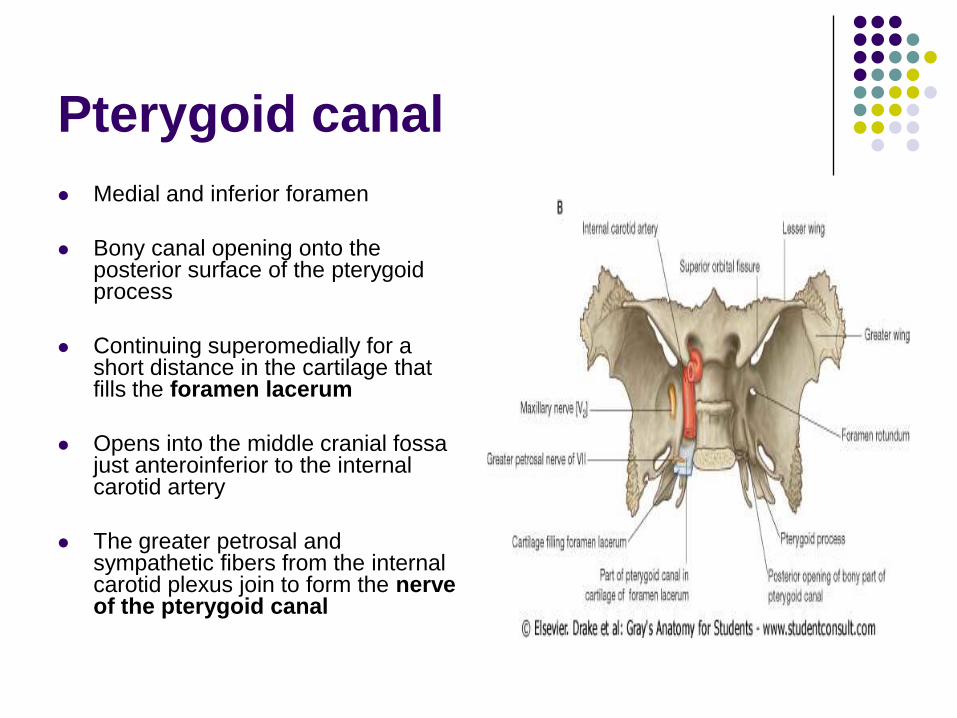
Pterygoid canal
Medial and inferior foramen
Bony canal opening onto the posterior surface of the pterygoid process
Continuing superomedially for a short distance in the cartilage that fills the foramen lacerum
Opens into the middle cranial fossa just anteroinferior to the internal carotid artery
The greater petrosal and sympathetic fibers from the internal carotid plexus join to form the nerve of the pterygoid canal

Gateways
Seven foramina and fissures provide apertures through which structures enter and leave the pterygopalatine fossa
1. Foramen rotundum and pterygoid canal communicate with the middle cranial fossa
2. Palatovaginal canal opens onto the posterior wall and leads to the nasopharynx;
3. Palatine canal leads to the roof of the oral cavity (hard palate) and opens inferiorly;
4 Sphenopalatine foramen opens onto the lateral wall of the nasal cavity and is in the medial wall;
Gateways
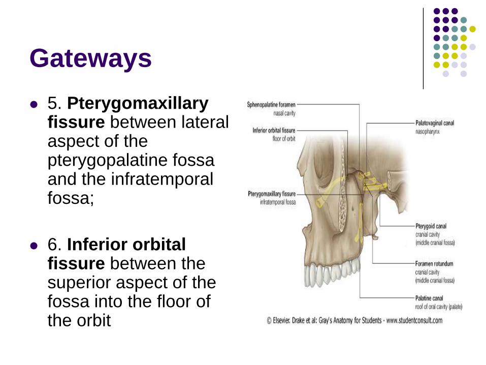
5. Pterygomaxillary fissure between lateral aspect of the pterygopalatine fossa and the infratemporal fossa;
6. Inferior orbital fissure between the superior aspect of the fossa into the floor of the orbit
Contents
1. The maxillary nerve [V2]
2. Terminal part of the maxillary artery
3. Nerve of the pterygoid canal
4. The pterygopalatine ganglion
5. Veins and lymphatics also pass through the pterygopalatine fossa.
Pterygopalatine
ganglion
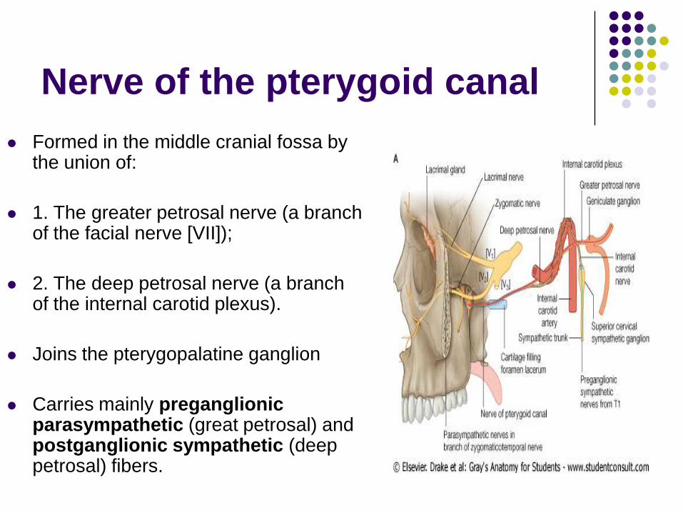
Nerve of the pterygoid canal
Formed in the middle cranial fossa by the union of:
1. The greater petrosal nerve (a branch of the facial nerve [VII]);
2. The deep petrosal nerve (a branch of the internal carotid plexus).
Joins the pterygopalatine ganglion
Carries mainly preganglionic parasympathetic (great petrosal) and postganglionic sympathetic (deep petrosal) fibers.
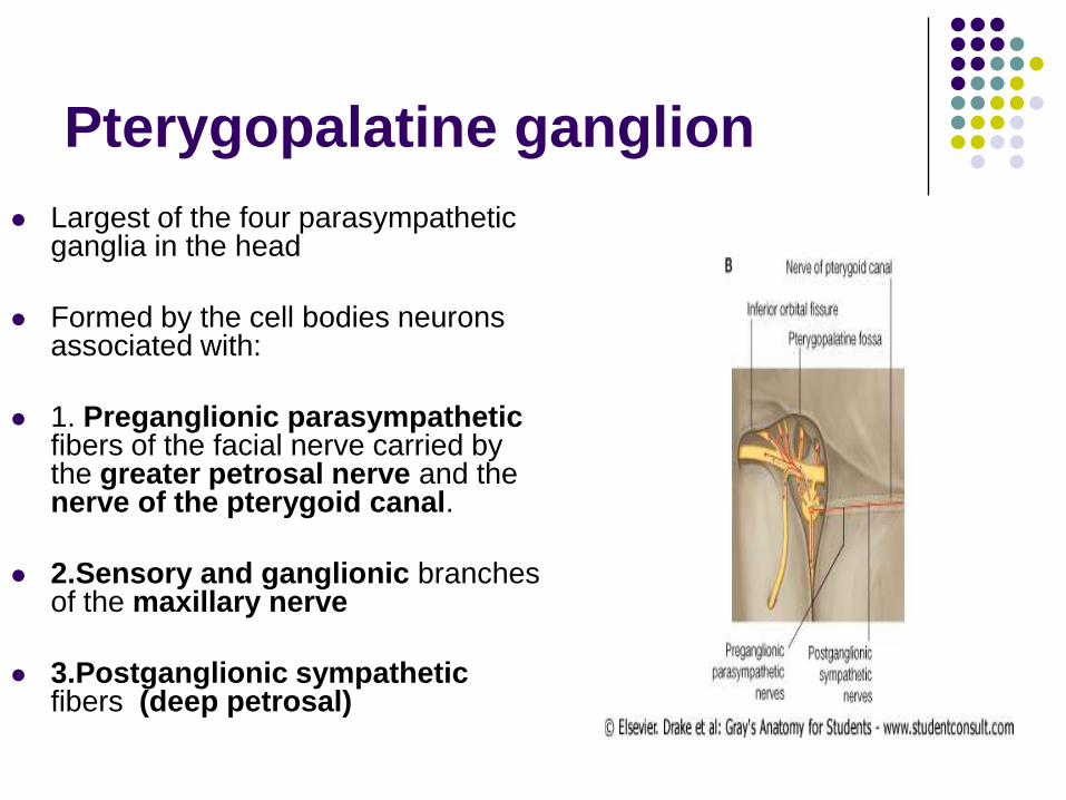
Pterygopalatine ganglion
Largest of the four parasympathetic ganglia in the head
Formed by the cell bodies neurons associated with:
1. Preganglionic parasympatheticfibers of the facial nerve carried by the greater petrosal nerve and the nerve of the pterygoid canal.
2.Sensory and ganglionic branches of the maxillary nerve
3.Postganglionic sympatheticfibers (deep petrosal)
Pterygopalatine ganglion
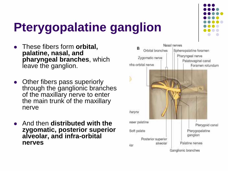
These fibers form orbital, palatine, nasal, and pharyngeal branches, which leave the ganglion.
Other fibers pass superiorly through the ganglionic branches of the maxillary nerve to enter the main trunk of the maxillary nerve
And then distributed with the zygomatic, posterior superior alveolar, and infra-orbital nerves
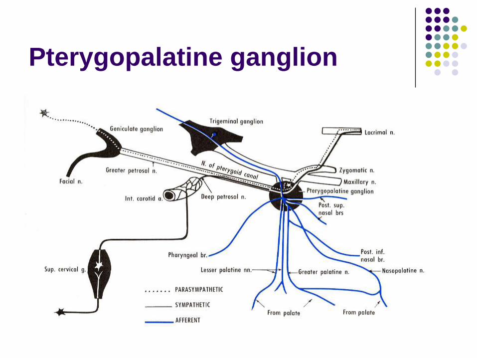
Pterygopalatine ganglion

Orbital branches
Pass through the inferior orbital fissure
Supply of the orbital wall (periosteum) and
lacrimal gland
Supply the sphenoidal and ethmoidal
sinuses.
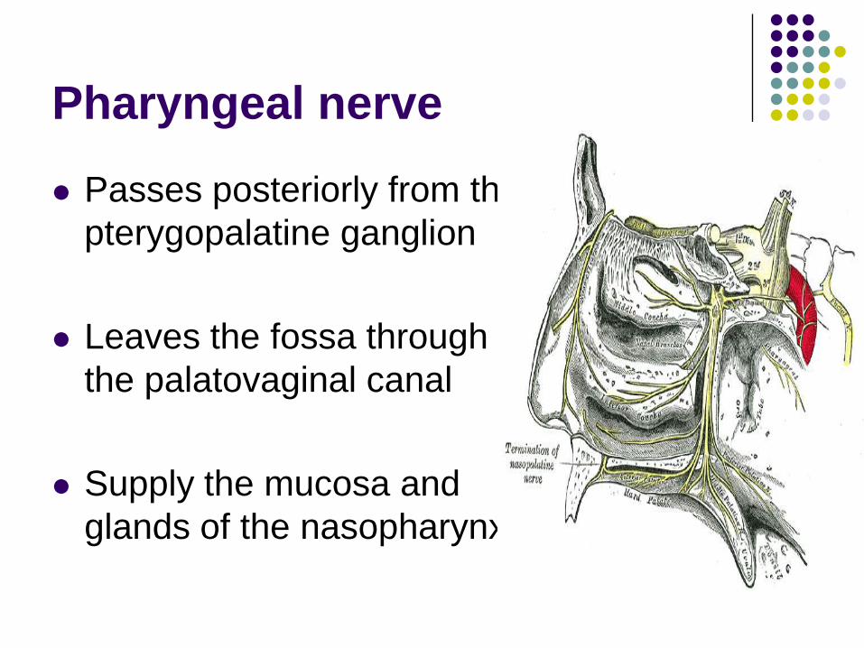
Pharyngeal nerve
Passes posteriorly from the
pterygopalatine ganglion
Leaves the fossa through
the palatovaginal canal
Supply the mucosa and
glands of the nasopharynx.
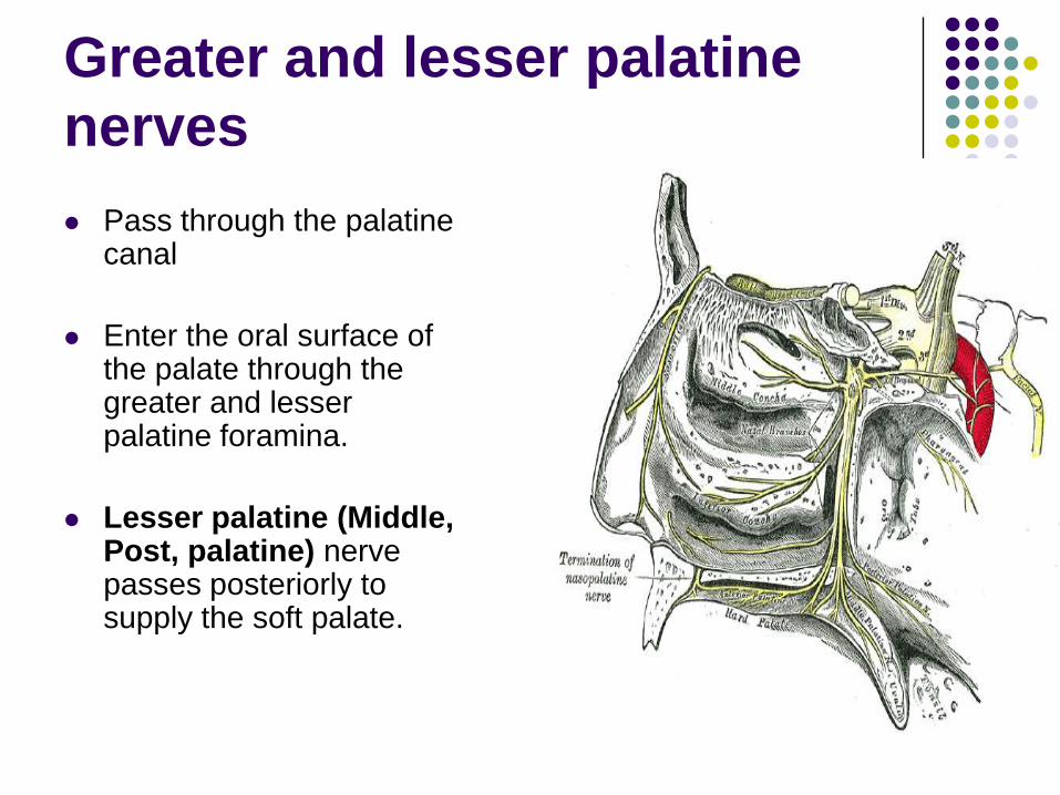
Greater and lesser palatine
nerves
Pass through the palatine canal
Enter the oral surface of the palate through the greater and lesser palatine foramina.
Lesser palatine (Middle, Post, palatine) nerve passes posteriorly to supply the soft palate.
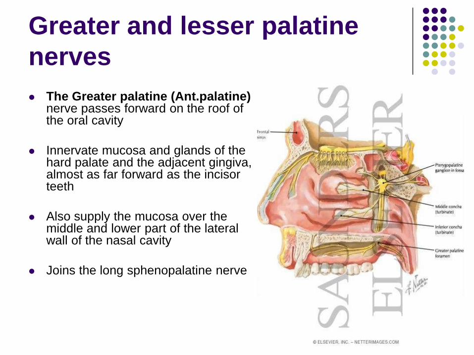
Greater and lesser palatine
nerves
The Greater palatine (Ant.palatine)nerve passes forward on the roof of the oral cavity
Innervate mucosa and glands of the hard palate and the adjacent gingiva, almost as far forward as the incisor teeth
Also supply the mucosa over the middle and lower part of the lateral wall of the nasal cavity
Joins the long sphenopalatine nerve
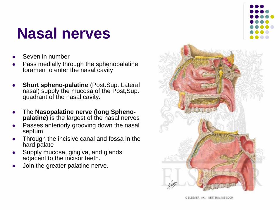
Nasal nerves
Seven in number
Pass medially through the sphenopalatine foramen to enter the nasal cavity
Short spheno-palatine (Post.Sup. Lateral nasal) supply the mucosa of the Post,Sup. quadrant of the nasal cavity.
The Nasopalatine nerve (long Spheno-palatine) is the largest of the nasal nerves
Passes anteriorly grooving down the nasal septum
Through the incisive canal and fossa in the hard palate
Supply mucosa, gingiva, and glands adjacent to the incisor teeth.
Join the greater palatine nerve.
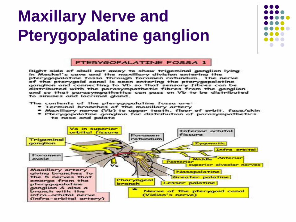
Maxillary Nerve
Maxillary nerve [V2]
Purely sensory
Originates from the trigeminal ganglion in the cranial cavity
Exits the middle cranial fossa, and enters the pterygopalatine fossa through the foramen rotundum
It terminates as the infra-orbital nerve through the inferior orbital fissure.
Maxillary nerve
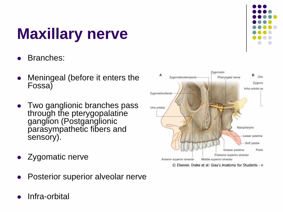
Branches:
Meningeal (before it enters the Fossa)
Two ganglionic branches pass through the pterygopalatine ganglion (Postganglionic parasympathetic fibers and sensory).
Zygomatic nerve
Posterior superior alveolar nerve
Infra-orbital
Zygomatic nerve
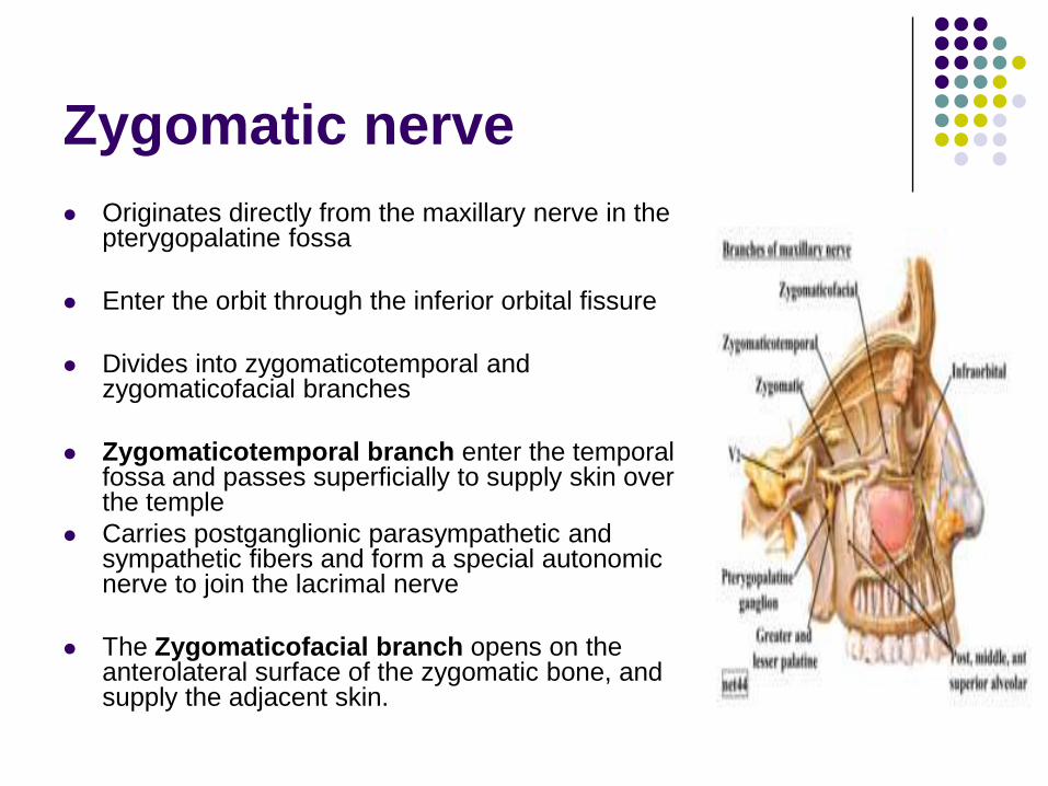
Originates directly from the maxillary nerve in the pterygopalatine fossa
Enter the orbit through the inferior orbital fissure
Divides into zygomaticotemporal and zygomaticofacial branches
Zygomaticotemporal branch enter the temporal fossa and passes superficially to supply skin over the temple
Carries postganglionic parasympathetic and sympathetic fibers and form a special autonomic nerve to join the lacrimal nerve
The Zygomaticofacial branch opens on the anterolateral surface of the zygomatic bone, and supply the adjacent skin.

Posterior superior alveolar
nerve
Passes laterally out of the fossa through the pterygomaxillary fissure
Enter the posterior surface of the maxilla approximately midway between the last molar tooth and the inferior orbital fissure
Supplies the molar teeth and adjacent buccal gingivae
contributes to the supply of the maxillary sinus
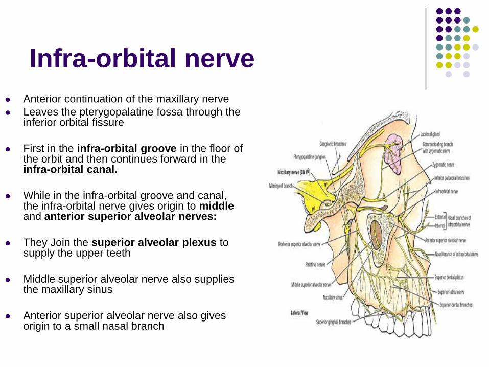
Infra-orbital nerve
Anterior continuation of the maxillary nerve
Leaves the pterygopalatine fossa through the inferior orbital fissure
First in the infra-orbital groove in the floor of the orbit and then continues forward in the infra-orbital canal.
While in the infra-orbital groove and canal, the infra-orbital nerve gives origin to middleand anterior superior alveolar nerves:
They Join the superior alveolar plexus to supply the upper teeth
Middle superior alveolar nerve also supplies the maxillary sinus
Anterior superior alveolar nerve also gives origin to a small nasal branch
Infra-orbital nerve
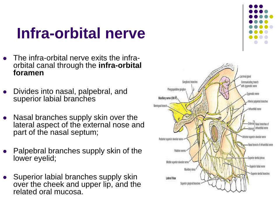
The infra-orbital nerve exits the infra-orbital canal through the infra-orbital foramen
Divides into nasal, palpebral, and superior labial branches
Nasal branches supply skin over the lateral aspect of the external nose and part of the nasal septum;
Palpebral branches supply skin of the lower eyelid;
Superior labial branches supply skin over the cheek and upper lip, and the related oral mucosa.
Maxillary Nerve and
Pterygopalatine ganglion
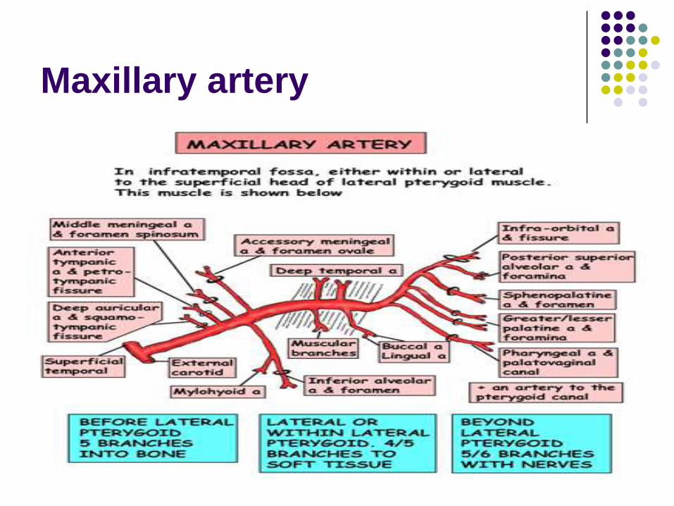
Maxillary Artery
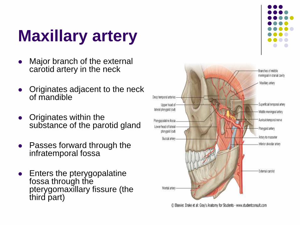
Maxillary artery
Major branch of the external carotid artery in the neck
Originates adjacent to the neck of mandible
Originates within the substance of the parotid gland
Passes forward through the infratemporal fossa
Enters the pterygopalatine fossa through the pterygomaxillary fissure (the third part)
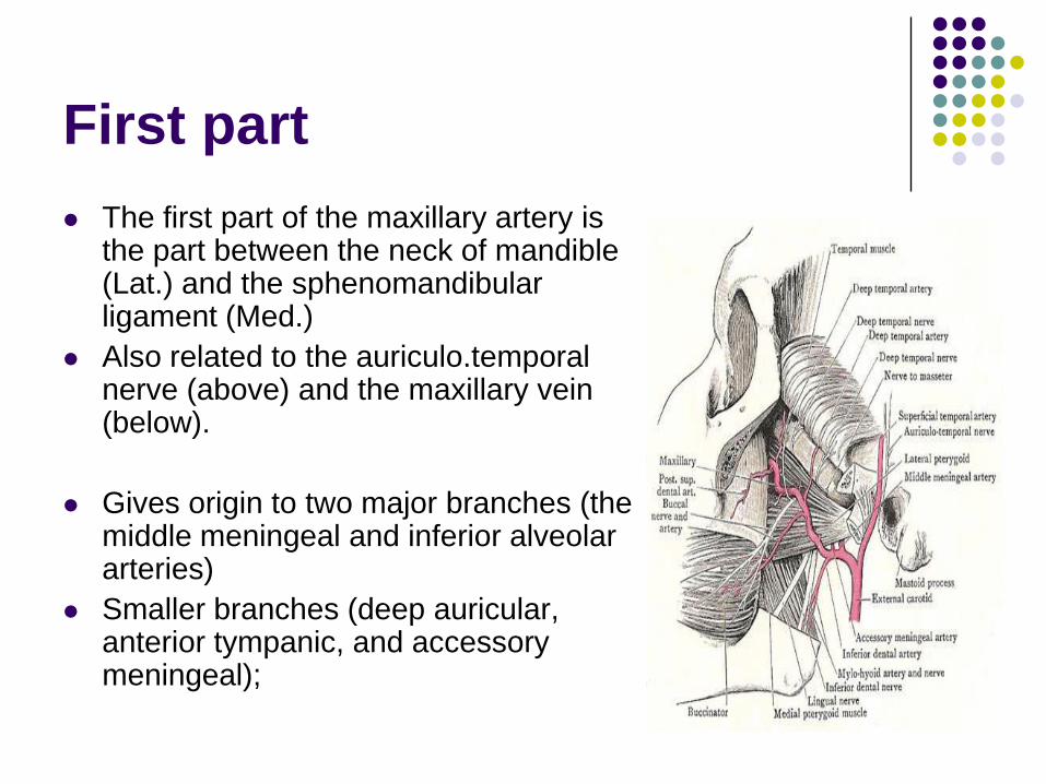
First part
The first part of the maxillary artery is the part between the neck of mandible (Lat.) and the sphenomandibular ligament (Med.)
Also related to the auriculo.temporal nerve (above) and the maxillary vein (below).
Gives origin to two major branches (the middle meningeal and inferior alveolar arteries)
Smaller branches (deep auricular, anterior tympanic, and accessory meningeal);

Second part
The second part of the maxillary artery the part related to the lateral pterygoid muscle
Gives origin to deep temporal, masseteric, buccal, and pterygoid branches (muscles of mastication)
Course with branches of the mandibular nerve
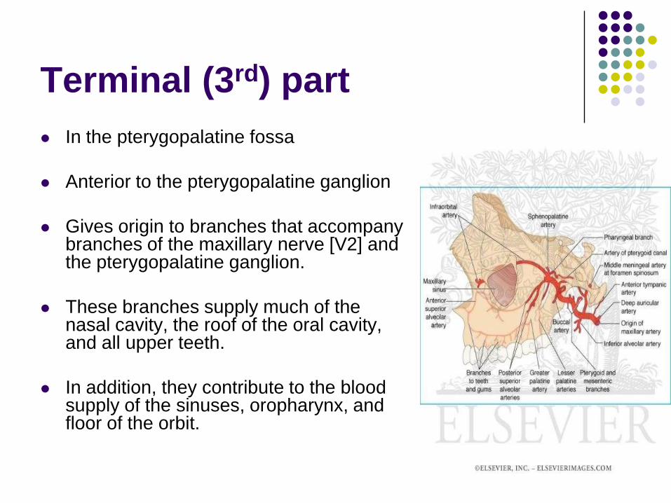
Terminal (3rd) part
In the pterygopalatine fossa
Anterior to the pterygopalatine ganglion
Gives origin to branches that accompany branches of the maxillary nerve [V2] and the pterygopalatine ganglion.
These branches supply much of the nasal cavity, the roof of the oral cavity, and all upper teeth.
In addition, they contribute to the blood supply of the sinuses, oropharynx, and floor of the orbit.
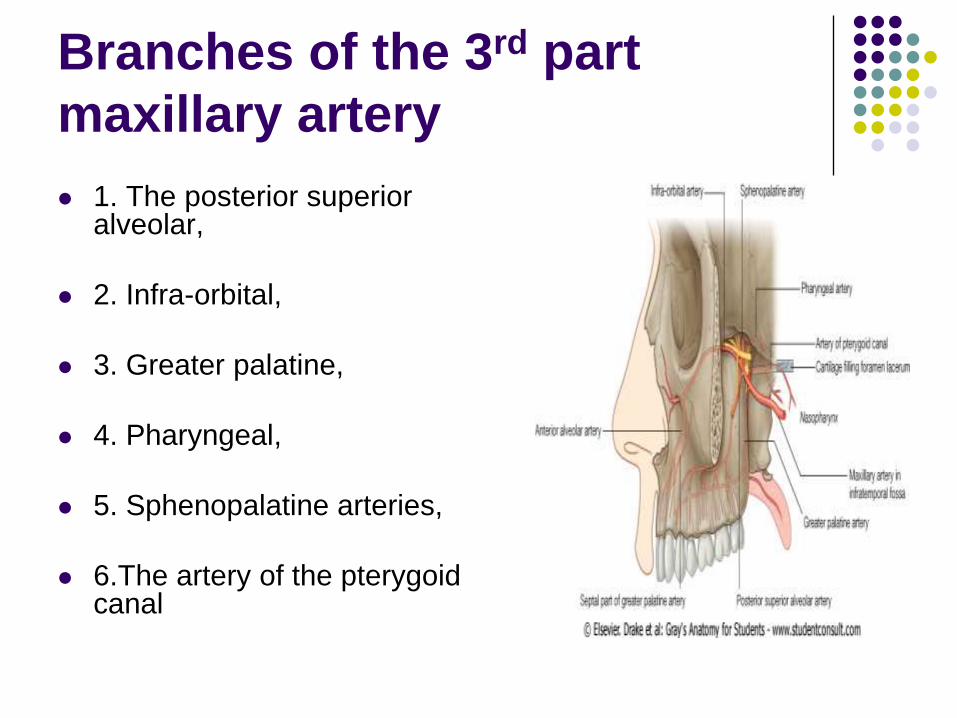
Branches of the 3rd part
maxillary artery
1. The posterior superior alveolar,
2. Infra-orbital,
3. Greater palatine,
4. Pharyngeal,
5. Sphenopalatine arteries,
6.The artery of the pterygoid canal
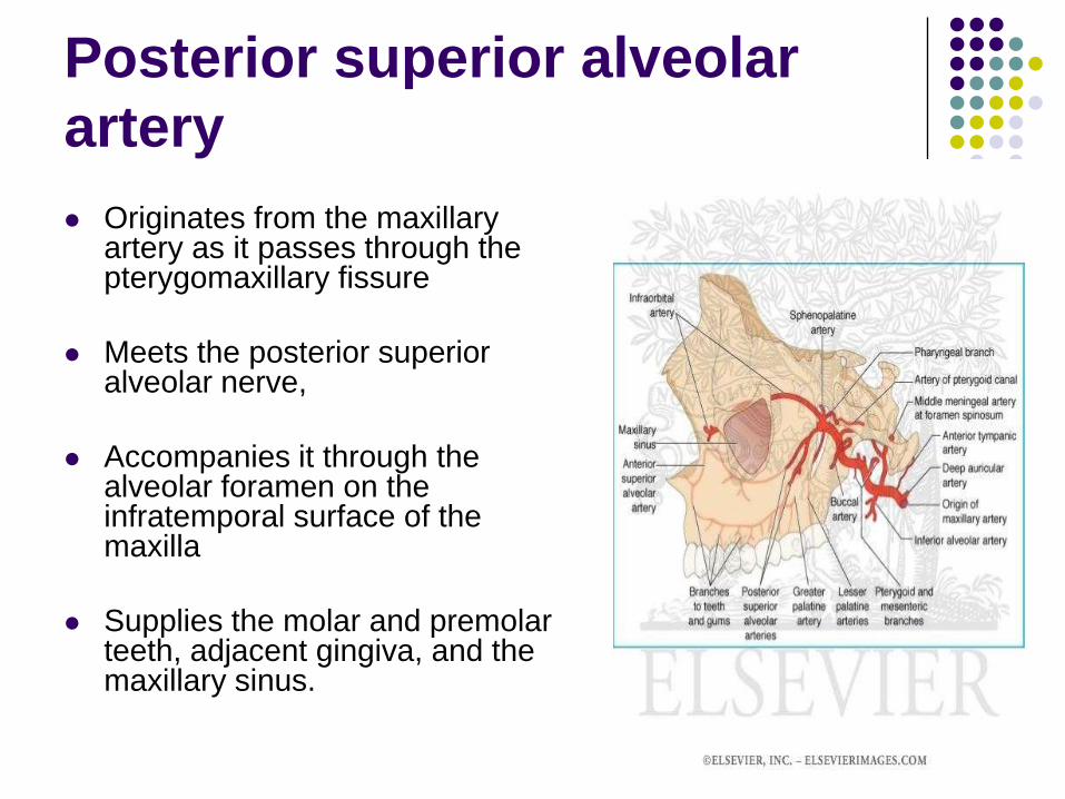
Posterior superior alveolar
artery
Originates from the maxillary artery as it passes through the pterygomaxillary fissure
Meets the posterior superior alveolar nerve,
Accompanies it through the alveolar foramen on the infratemporal surface of the maxilla
Supplies the molar and premolar teeth, adjacent gingiva, and the maxillary sinus.
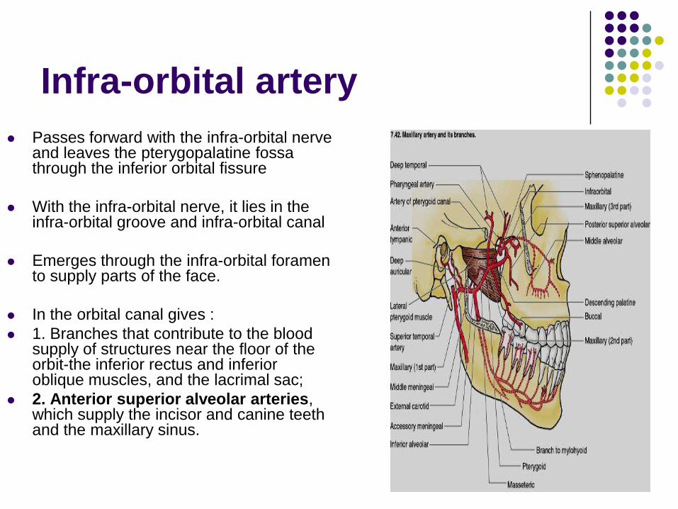
Infra-orbital artery
Passes forward with the infra-orbital nerve and leaves the pterygopalatine fossa through the inferior orbital fissure
With the infra-orbital nerve, it lies in the infra-orbital groove and infra-orbital canal
Emerges through the infra-orbital foramen to supply parts of the face.
In the orbital canal gives :
1. Branches that contribute to the blood supply of structures near the floor of the orbit-the inferior rectus and inferior oblique muscles, and the lacrimal sac;
2. Anterior superior alveolar arteries, which supply the incisor and canine teeth and the maxillary sinus.
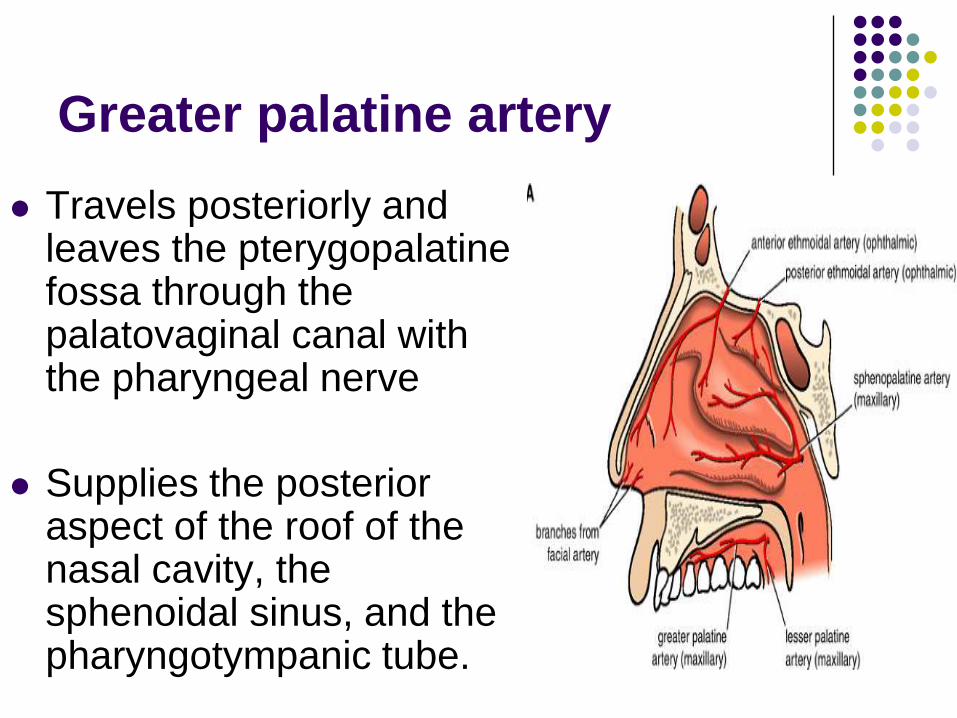
Greater palatine artery
Travels posteriorly and leaves the pterygopalatine fossa through the palatovaginal canal with the pharyngeal nerve
Supplies the posterior aspect of the roof of the nasal cavity, the sphenoidal sinus, and the pharyngotympanic tube.
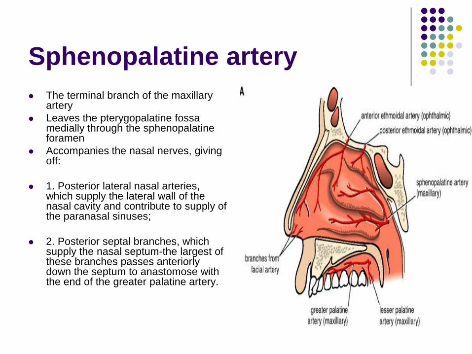
Sphenopalatine artery
The terminal branch of the maxillary artery
Leaves the pterygopalatine fossa medially through the sphenopalatine foramen
Accompanies the nasal nerves, giving off:
1. Posterior lateral nasal arteries, which supply the lateral wall of the nasal cavity and contribute to supply of the paranasal sinuses;
2. Posterior septal branches, which supply the nasal septum-the largest of these branches passes anteriorly down the septum to anastomose with the end of the greater palatine artery.
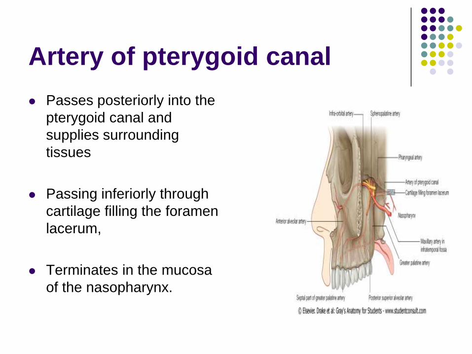
Artery of pterygoid canal
Passes posteriorly into the
pterygoid canal and
supplies surrounding
tissues
Passing inferiorly through
cartilage filling the foramen
lacerum,
Terminates in the mucosa
of the nasopharynx.
Maxillary artery

Veins
Drain areas supplied by branches of the terminal part of the maxillary artery
Generally travel with these branches back into the pterygopalatine fossa.
The veins coalesce in the fossa and then pass laterally through the pterygomaxillary fissure to join the pterygoid plexus of veins in the infratemporal fossa
The infra-orbital vein, drains the inferior aspect of the orbit,
May pass directly into the infratemporal fossa, so bypassing the pterygopalatine fossa

Thank you