anatomical and clinical appraisal of the pterygopalatine ganglion
TRANSCRIPT

Anatomical and Clinical Appraisal of the Pterygopalatine Ganglion
Karin Petra Quirina Oomen

This thesis was financially supported by: Stichting ORLU, Merck Sharp & Dohme B.V., Specsavers
International B.V., Stallergenes, EmiD audiologische apparatuur, Daleco Pharma B.V.
ISBN: 978-90-39355558
© 2011 K.P.Q. Oomen, The Netherlands
All rights reserved. No part of this publication may be reproduced or transmitted in any form
or by any means, electronic or mechanical, including photocopy, recording or otherwise,
without the written permission of the author.
Lay out: Chris Bor, Medische fotografie en Illustratie, AMC, Amsterdam
Printed by: Buijten & Schipperheijn, Amsterdam
Anatomical and Clinical Appraisal of the Pterygopalatine Ganglion
Een anatomische en klinische studie van het ganglion pterygopalatinum(met een samenvatting in het Nederlands)
Proefschrift
ter verkrijging van de graad van doctor aan de Universiteit Utrecht op gezag van de
rector magnificus, prof. dr. G. J. van der Zwaan, ingevolge het besluit van het college voor
promoties in het openbaar te verdedigen op vrijdag 27 mei 2011, des ochtends te 10.30
uur
door
Karin Petra Quirina Oomen
geboren op 13 augustus 1977 te De Bilt

Promotor: Prof. dr. G. J. Hordijk
Co-promotoren: Dr. R. L. A. W. Bleys
Dr. J. A. de Ru

CONTENTS
Chapter 1 General introduction 7
Chapter 2 A previously undescribed branch of the pterygopalatine ganglion 19
Chapter 3 Neurochemical characterization of pterygopalatine ganglion
branches in humans
31
Chapter 4 Improved depiction of pterygopalatine fossa anatomy using ultra
high resolution magnetic resonance imaging at 7 Tesla
45
Chapter 5 Sluder’s neuralgia; a trigeminal autonomic cephalalgia? 59
Chapter 6 Effects of radiofrequency thermocoagulation of the sphenopalatine
ganglion on facial pain; correlation with diagnosis
69
Chapter 7 Microvascular decompression of the pterygopalatine ganglion in
patients with refractory cluster headache
79
Chapter 8 General discussion 91
Summary 107
Dutch summary 107
Color figures 113
Dankwoord 125
Curriculum Vitae 129

1C h a p t e r
General introduction

GENERAL INTRODUCTION
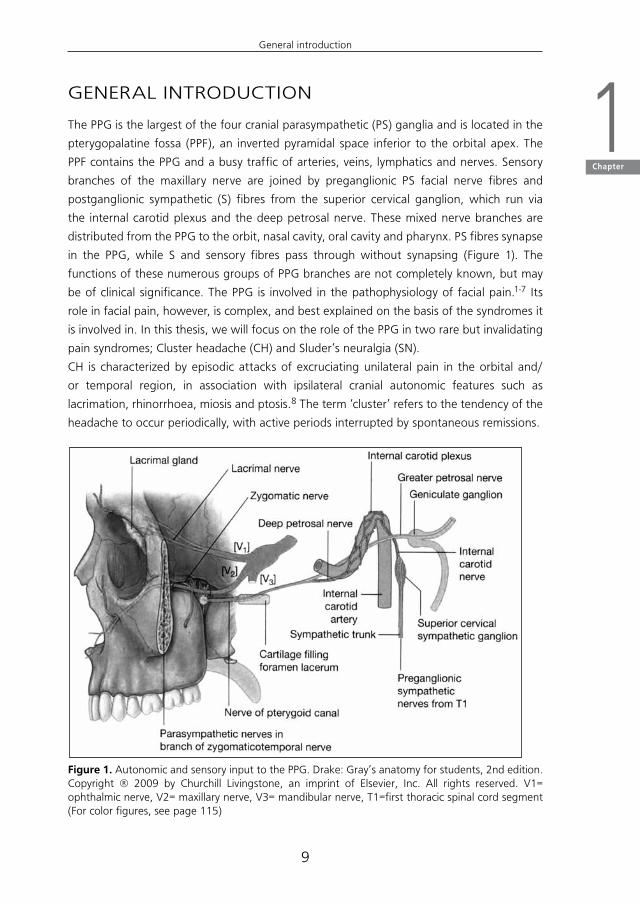
The PPG is the largest of the four cranial parasympathetic (PS) ganglia and is located in the
pterygopalatine fossa (PPF), an inverted pyramidal space inferior to the orbital apex. The
PPF contains the PPG and a busy traffic of arteries, veins, lymphatics and nerves. Sensory
branches of the maxillary nerve are joined by preganglionic PS facial nerve fibres and
postganglionic sympathetic (S) fibres from the superior cervical ganglion, which run via
the internal carotid plexus and the deep petrosal nerve. These mixed nerve branches are
distributed from the PPG to the orbit, nasal cavity, oral cavity and pharynx. PS fibres synapse
in the PPG, while S and sensory fibres pass through without synapsing (Figure 1). The
functions of these numerous groups of PPG branches are not completely known, but may
be of clinical significance. The PPG is involved in the pathophysiology of facial pain.1-7 Its
role in facial pain, however, is complex, and best explained on the basis of the syndromes it
is involved in. In this thesis, we will focus on the role of the PPG in two rare but invalidating
pain syndromes; Cluster headache (CH) and Sluder’s neuralgia (SN).
CH is characterized by episodic attacks of excruciating unilateral pain in the orbital and/
or temporal region, in association with ipsilateral cranial autonomic features such as
lacrimation, rhinorrhoea, miosis and ptosis.8 The term ‘cluster’ refers to the tendency of the
headache to occur periodically, with active periods interrupted by spontaneous remissions.
Figure 1. Autonomic and sensory input to the PPG. Drake: Gray’s anatomy for students, 2nd edition. Copyright ® 2009 by Churchill Livingstone, an imprint of Elsevier, Inc. All rights reserved. V1= ophthalmic nerve, V2= maxillary nerve, V3= mandibular nerve, T1=first thoracic spinal cord segment (For color figures, see page 115)
9
General introduction
1Chapter
The pathophysiology of CH has long
been unknown and is complex. The
primary defect in CH is thought to
be located in regulatory centres in
the hypothalamus. Alterations in
biological rhythms of secretions of
hypophysary hormones during active
CH periods and in remissions, and
positron emission tomography (PET)
studies showing an increased blood
flow and structural changes in the
hypothalamic grey area during CH
attacks, support this hypothesis.9
PET studies during CH attacks also
show activation in the region of the major basal cranial arteries and cavernous sinus,
specifically the intracranial segment of the internal carotid artery (ICA), which is likely to
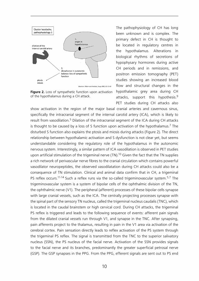
result from vasodilation.5 Dilation of the intracranial segment of the ICA during CH attacks
is thought to be caused by a loss of S function upon activation of the hypothalamus.7 The
disturbed S function also explains the ptosis and miosis during attacks (Figure 2). The direct
relationship between hypothalamic activation and S dysfunction is not clear yet, but seems
understandable considering the regulatory role of the hypothalamus in the autonomic
nervous system. Interestingly, a similar pattern of ICA vasodilation is observed in PET studies
upon artificial stimulation of the trigeminal nerve (TN).10 Given the fact that the TN supplies
a rich network of perivascular nerve fibres to the cranial circulation which contains powerful
vasodilator neuropeptides, the observed vasodilatation during CH attacks could also be a
consequence of TN stimulation. Clinical and animal data confirm that in CH, a trigeminal
PS reflex occurs.11-14 Such a reflex runs via the so-called trigeminovascular system.5,7 The
trigeminovascular system is a system of bipolar cells of the ophthalmic division of the TN,
the ophthalmic nerve (V1). The peripheral (afferent) processes of these bipolar cells synapse
with large cranial vessels, such as the ICA. The centrally projecting processes synapse with
the spinal part of the sensory TN nucleus, called the trigeminal nucleus caudalis (TNC), which
is located in the caudal brainstem or high cervical cord. During CH attacks, the trigeminal
PS reflex is triggered and leads to the following sequence of events: afferent pain signals
from the dilated cranial vessels run through V1, and synapse in the TNC. After synapsing,
pain afferents project to the thalamus, resulting in pain in the V1 area via activation of the
cerebral cortex. Pain sensation directly leads to reflex activation of the PS system through
the trigeminal PS reflex. The signal is transmitted from the TNC to the superior salivatory
nucleus (SSN), the PS nucleus of the facial nerve. Activation of the SSN provides signals
to the facial nerve and its branches, predominantly the greater superficial petrosal nerve
(GSP). The GSP synapses in the PPG. From the PPG, efferent signals are sent out to PS end
Figure 2. Loss of sympathetic function upon activation of the hypothalamus during a CH attack.
10
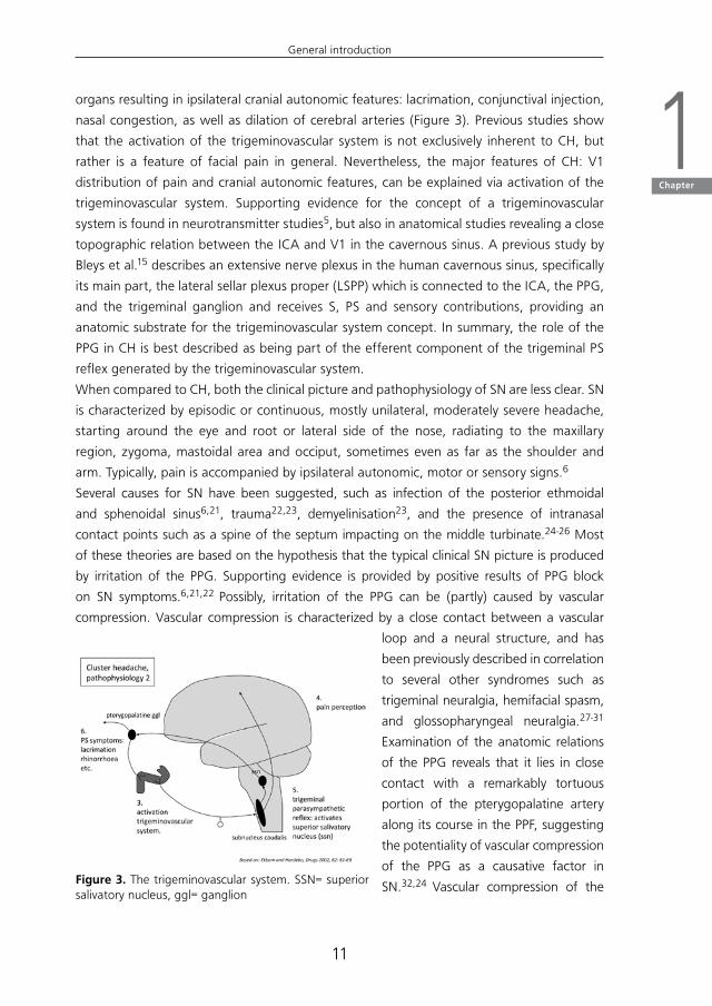
organs resulting in ipsilateral cranial autonomic features: lacrimation, conjunctival injection,
nasal congestion, as well as dilation of cerebral arteries (Figure 3). Previous studies show
that the activation of the trigeminovascular system is not exclusively inherent to CH, but
rather is a feature of facial pain in general. Nevertheless, the major features of CH: V1
distribution of pain and cranial autonomic features, can be explained via activation of the
trigeminovascular system. Supporting evidence for the concept of a trigeminovascular
system is found in neurotransmitter studies5, but also in anatomical studies revealing a close
topographic relation between the ICA and V1 in the cavernous sinus. A previous study by
Bleys et al.15 describes an extensive nerve plexus in the human cavernous sinus, specifically
its main part, the lateral sellar plexus proper (LSPP) which is connected to the ICA, the PPG,
and the trigeminal ganglion and receives S, PS and sensory contributions, providing an
anatomic substrate for the trigeminovascular system concept. In summary, the role of the
PPG in CH is best described as being part of the efferent component of the trigeminal PS
reflex generated by the trigeminovascular system.
When compared to CH, both the clinical picture and pathophysiology of SN are less clear. SN
is characterized by episodic or continuous, mostly unilateral, moderately severe headache,
starting around the eye and root or lateral side of the nose, radiating to the maxillary
region, zygoma, mastoidal area and occiput, sometimes even as far as the shoulder and
arm. Typically, pain is accompanied by ipsilateral autonomic, motor or sensory signs.6
Several causes for SN have been suggested, such as infection of the posterior ethmoidal
and sphenoidal sinus6,21, trauma22,23, demyelinisation23, and the presence of intranasal
contact points such as a spine of the septum impacting on the middle turbinate.24-26 Most
of these theories are based on the hypothesis that the typical clinical SN picture is produced
by irritation of the PPG. Supporting evidence is provided by positive results of PPG block
on SN symptoms.6,21,22 Possibly, irritation of the PPG can be (partly) caused by vascular
compression. Vascular compression is characterized by a close contact between a vascular
loop and a neural structure, and has
been previously described in correlation
to several other syndromes such as
trigeminal neuralgia, hemifacial spasm,
and glossopharyngeal neuralgia.27-31
Examination of the anatomic relations
of the PPG reveals that it lies in close
contact with a remarkably tortuous
portion of the pterygopalatine artery
along its course in the PPF, suggesting
the potentiality of vascular compression
of the PPG as a causative factor in
SN.32,24 Vascular compression of the Figure 3. The trigeminovascular system. SSN= superior salivatory nucleus, ggl= ganglion
11
General introduction
1Chapter
pterygopalatine artery on the PPG could possibly even be a contributing factor in the complex
pathophysiology of CH.
Because of the important pathophysiological role of the PPG and manifestation of cranial
pain combined with autonomic features in both, SN and CH are often considered as parts of
the same clinical entity.16 However, several differences exist between the two with respect
to age and sex of patients, periodicity, distribution and location of pain.16-20 The confusion
of SN and CH partly stems from the lack of specific criteria for SN. Criteria for CH are clear
and well-defined 8, but the description of SN remains rather vague. Correct classification
of both syndromes, with the aid of clear criteria for SN that are able to distinguish it from
CH and other forms of facial pain, could be helpful in making correct diagnosis and finding
suitable treatment.
Despite their differences, both CH and SN are rare but disabling, and treatment is absolutely
necessary. Most patients with CH can be managed pharmacologically with a variety of
medications such as prednisone, calcium-channel blockers, lithium, indomethacin, inhaled
oxygen and ergotamine, amongst others.7 Local pharmacological treatment such as
cocainization of the PPG provides temporary pain relief in most patients as well.18 However,
in an unfortunate 10%, local or systemic pharmacological treatment is of limited success.33
These patients require invasive treatment because of intractable symptoms or adverse effects
of medication. Unfortunately, surgical treatment for refractory CH has remained a frustrating
endeavour. Treatment has centered primarily upon interrupting the autonomic and sensory
pathways responsible for the pain and many of the PS symptoms in CH, by sectioning or
lesioning of the PPG, the intermediate nerve or the greater superficial petrosal nerve (GSP).
Invasive treatment directed to the PPG has included radiofrequency thermocoagulation
(RFT), phenolization, and direct ganglioneurectomy, all without providing long-lasting
pain relief.22,33-38 Section of the intermediate nerve39,40 and section of the GSP41,42 are
based on the hypothesis that the intermediate nerve mediates CH symptoms through
carriage of PS impulses to the PPG via the GSP.43,44 Unfortunately, both have previously
been reported without adequate long-term pain relief. Lovely et al.33 reported successful
surgical management of CH through microvascular decompression (MVD) of the trigeminal
nerve, alone or in combination with section and/or MVD of the intermediate nerve in CH
patients, but the success rate dropped from 77.3% in the immediate postoperative period
to 46.6% after the first postoperative year. Recently, the therapeutic options for refractory
CH patients have expanded with the emergence of both peripheral (occipital nerve) and
central (hypothalamic) neurostimulation. In occipital nerve stimulation (ONS), a suboccipital
neurostimulator is implanted on the side of the headache, which stimulates the greater
occipital nerve (GON). This treatment is based on the hypothesis that GON stimulation
affects the TNC and thus possibly interrupts the trigeminal PS reflex pathway.45 ONS has
been described in patients with refractory CH in a few case series46,47, and appears to
mainly decrease the attack frequency. Notably, there is a long (2 months or more) latency
period between electrode implantation and clinical effect. Neurostimulation with deep brain
12

electrodes or deep brain stimulation (DBS) was originally used in the subthalamic area in
patients with movement disorders such as Parkinson’s disease, but has lately been performed
in the hypothalamic area in patients with CH. DBS for CH has shown various success rates,
but seems effective in a subset of refractory patients.48,49 Unfortunately, DBS can give rise
to serious surgical complications. Previous studies on subthalamic DBS for Parkinson’s disease
report an intracerebral hemorrhage incidence varying between 1 and 5%.50,51 Whether
the incidence of intracerebral hemorrhage following hypothalamic DBS for CH might be
higher remains uncertain, but a previous pilot study in six refractory CH patients reported
postoperative cerebral hemorrhage with lethal outcome in one of their participants.52
Less is known about the management of patients with SN. General management of SN
has mostly been directed against the PPG. Local administration of anesthetic agents
such as lidocaine nose drops or cocaine provides instant adequate pain relief, but does
not serve as a permanent solution.25 Invasive therapies for SN have included correction of
nasal deformities26 drainage of infected sinus, and procedures directed at the PPG, such
as injection of alcohol or glycerol, phenolisation22, surgical resection37,38, stereotactic
radiosurgical treatment (gammaknife)23, and RFT.53 Unfortunately, none of these treatment
modalities offer permanent long-term pain relief. Intranasal phenolization of the PPG has
been described in a study of 8 patients and seems safe and temporarily effective, with a
mean duration of pain relief of 9.5 months, but a need for repetitive procedures.22 Studies
on surgical resection of the PPG show a high incidence of pain recurrence within one year
postoperatively, although the pain is usually less severe.37,38 Stereotactic radiosurgery of
the PPG has been reported to be successful in a case report of one single SN patient,
but only after repeated procedures.23 A previous study on RFT of the PPG in patients
with SN describes pain relief without significant side effects, but with the persistence of
a troublesome sensation in all treated patients.4 The collective data show that, despite
their clinical and pathophysiological differences, CH and SN can temporarily be managed
in a successful manner by blocking the PPG.53-55 This seems surprising, as their anatomic
pain distribution areas differ: pain in CH is located in the V1 area, whereas pain in SN is
predominantly located in the area of the maxillary nerve (V2). Even patients with trigeminal
neuralgia and temporomandibular joint pain (TMJ) seem to benefit from PPG blocks33-35,
which is all the more surprising as the anatomic distribution of trigeminal neuralgia covers all
three principal branches of the TN, and pain in TMJ is located in the area of the mandibular
nerve (V3). PPG block seems to provide pain relief in all three principal branches of the
TN, whereas theoretically, it could only result in relief of pain and autonomic symptoms in
its known anatomic distribution area, i.e. the maxillary nerve (V2) with its orbital, nasal,
palatine and pharyngeal rami. Thus, a PPG block produces pain relief in a broader area
than would be expected on anatomic grounds. The finding of such unexpected effects of
PPG blockage raises the question whether the neural connections of the PPG have been
described completely. A search for previously undescribed PPG branches and extensive
13
General introduction
1Chapter

functional characterization of new and known branches, could possibly offer an explanation
for both unexpected effects of PPG block and symptomatology of facial pain.
Unfortunately, no invasive intervention has yet been established as standard care for
refractory cases of both SN and CH. Clearly, there is a need for alternative surgical procedures
in management of CH and SN providing longer lasting pain relief, which is another reason
for careful anatomical examination of the PPG and its relations.
OBJECTIVE
The central goal in this thesis is to study the PPG in order to gain further insight in the
pathophysiology and treatment of PPG related syndromes CH and SN.
Both the macro- and microscopic anatomy and the neurochemical coding of the PPG
and its neural connections will be explored, and the PPF and its contents will be studied
radiologically. CH and SN will be compared through a systematic review of SN features, and
the effects of surgical therapy of the PPG in patients with facial pain will be evaluated.
THESIS OUTLINE
Chapter 2, 3 and 4 focus on the anatomy of the PPF.
In Chapter 2, we present a search for previously undescribed neural structures through
anatomical study of the neural content of the PPF. Macro- and microscopic dissection
of whole-mount specimens of the PPF are combined with enzyme histochemistry for
acetylcholinesterase (AChE), a general neural marker. The neurochemical characterization of the
neural PPF contents, specifically the orbital PPG branches, is presented in Chapter 3. Cryostat
sections of the PPF, specifically the PPG and its neural connections, are immunohistochemically
stained in order to localize nerves which contain the general neural marker protein gene
product (PGP) 9.5, the S nerve specific tyrosine hydroxylase (TH) and the PS nerve specific nitric
oxide synthase (NOS). In Chapter 4, the radiologic anatomy of the PPF is analyzed through MRI
study at 7 Tesla compared with cryomicrotome-derived sections of the PPF and its contents,
combined with a Mallory-Cason staining procedure.
Chapter 5, 6 and 7 focus on the pathophysiology and treatment of PPG related syndromes
CH and SN.
Based on a systematic literature review in which described SN symptoms are quantitatively
assessed, we present new criteria for SN and recognize it as an independent clinical entity,
different from CH, in Chapter 5. The effects of RFT of the PPG in facial pain and their
correlation with correct diagnosis are retrospectively studied in Chapter 6.
In Chapter 7 we present the effects of a new surgical therapy, microvascular decompression
of the PPG, in 3 patients with chronic refractory CH.
14
The results are discussed in Chapter 8, and recommendations are made for future research.
Summaries in English and Dutch complete this thesis.
15
General introduction
1Chapter

REFERENCES
1.EdvinssonL.Pathophysiologyofprimaryheadaches.CurrentPainandHeadacheReports2001;5:71-78.
2.Edvinsson L. Aspects on the pathophysiology of migraine and cluster headache.PharmacolToxicol2001;89:65-73.Review.
3. Goadsby PJ. Pathophysiology of cluster headache: a trigeminal autonomic cephalgia.LancetNeurol2002;1:251-257.
4. GoadsbyPJ.Newaspectsofthepathophysiologyofmigraineandclusterheadache.In:MaxM,ed.Pain1999-anupdatedreview.Seattle,WA:IASPPress;1999:181-191.
5. May A, Goadsby PJ. The trigeminovascular system in humans: pathofysiologicimplications forprimaryheadachesyndromesof theneural influenceson thecerebralcirculation.JCerebBloodFlowMetab1999;19:115-127.
6.SluderG.Roleofthesphenopalatine(Meckel’s)ganglioninnasalheadaches.NYStateJMed1908;87:989-990.
7.EkbomK,HardeboJE.Clusterheadache.Aetiology,diagnosisandmanagement.Drugs2002;62:61-69.
8. TheInternationalClassificationofHeadacheDisorders,2ndedition,Cephalalgia2004;24:1-160.
9.May A, Bahra A, Buechel C, Frackowiak RSJ, Goadsby PJ. Hypothalamic activation inclusterheadacheattacks.Lancet1998;325:275-278.
10.MayA,KaubeH,BuechelC,EichtenC,RijntjesM,JueptnerM,etal.Experimentalcranialpainelicitedbycapsaicin:aPET-study.Pain1998;74:61-66.
11. Goadsby PJ, LambertGA, Lance JW. Stimulation of the trigeminal ganglion increasesflowintheextracerebralbutnotthecerebralcirculationofthemonkey.BrainRes1986;381:63-67.
12. LambertGA,BogdukN,GoadsbyPJ,DuckworthJW,LanceJW.Decreasedcarotidarterialresistanceincatsinresponsetotrigeminalstimulation.JNeurosurg1984;61:307-315.
13. EdvinssonL,JansenI,KingmanTA,McCullochJ.Cerebrovascularresponsestocapsaicininvitroandinsitu.BritJPharmacol1990;100:312-318.
14. Edvinsson L, Jansen-Olesen I, Kingman TA,McCulloch J, Uddman R.Modification ofvasoconstrictorresponsesincerebralbloodvesselsbylesioningofthetrigeminalnerve:possibleinvolvementofCGRP.Cephalalgia1995;15:373-383.
15. BleysRLAW,JanssenLM,GroenGJ.Thelateralsellarnerveplexusanditsconnectionsinhumans.JNeurosurg2001;95:102-110.
16.AhamedSH,JonesNS.WhatisSluder’sneuralgia?JLaryngolOtol2003;117:437
17. HardeboJE,ElnerA.NervesandvesselsinthepterygopalatinefossaandsymptomsofClusterheadache.Headache1987;27:528-532.
18.BarreF.CocaineasanabortiveagentinClusterheadache.Headache1982;22:69-73.
19. KittrelleJP,GrouseDS,SeyboldME.Clusterheadache.Localanestheticabortiveagents.ArchNeurol1985;42:496-498.
20. Ryan RE Jr, Facer GW. Sphenopalatine ganglion neuralgia and cluster headache:comparisons,contrasts,andtreatment.Headache1977;17:7-8
16

21. Sluder G. Etiology, diagnosis prognosis and treatment of sphenopalatine ganglionneuralgia.JAmMedAssoc1913;16:1202-1206.
22. PuigCM,DriscollCLW,KernEB.Sluder’sSphenopalatineGanglionNeuralgiaTreatmentwith88%Phenol.Am.J.Rhinol1998;12:113-118.
23.Pollock BE, Kondziolka MD. Stereotactic radiosurgical treatment of sphenopalatineneuralgia.JNeurosurg1997;87:450-453.
24.Bruyn GW. Sphenopalatine Neuralgia (Sluder). Handbook of clinical neurology, vol.4.Headache. F. Clifford Rose, editor. Elsevier Science Publishers BV. Vol 4(48), 1986,475-482.
25. Akhtar Kamal S. Experiencewith the xylocaine test as a prognostic aid for surgery inSluder’sneuralgia.JLaryngolOtol1995;109:193-195.
26.Giacomini PG, Alessandrini M, DePadova A. Septoturbinal surgery in contact pointheadachesyndrome:long-termresults.Cranio2003;21:130-135.
27.McDermott AL, Dutt SN, Irving RM, Pahor AL, Chavda SV. Anterior inferior cerebellararterysyndrome:factorfiction.ClinOtolaryngol2003;28:75-80.
28. El-GaremHF,Badr-El-DineM,TalaatAM,Magnan J.Endoscopyas a tool inminimallyinvasivetrigeminalneuralgiasurgery.Otology&Neurology2002;23:132-135.
29.RyuH,YamamotoS,SugiyamaK,UemureK,NozueM.Neurovasculardecompressionoftheeighthcranialnerveinpatientswithhemifacialspasmandincidentaltinnitus:analternativewaytostudytinnitus.JNeurosurg1998:88:232-236.
30.Moller AR. Vascular compression cranial nerves. I. History of the microvasculardecompressionoperation.NeurologicalResearch1998;20:727-731.
31.Badr-El-DineM,El-GaremHF,TalaatAM,MagnanJ.Endoscopicallyassistedminimallyinvasive microvascular decompression of hemifacial spasm. Otology & Neurology2002;23:122-128.
32.AlfieriA,JhoH-D,SchettinoR,TschabitscherM.Endoscopicendonasalapproachtothepterygopalatinefossa:ananatomicstudy.Neurosurgery2003;52:374-380.
33. LovelyTJ,KotsiakisX,JanettaPJ.Thesurgicalmanagementofchronicclusterheadache.Headache1998;38:590-594.
34. StolkerRJ,KamphuisE,RohofO,etal.DiebehandlungvonClusterkopfschmerzenmitradiofrequentenLasionen.Schmerz1991;5:194.
35.Vervest ACM, Stolker RJ, Groen GJ, et al. Clusterkopfschmerz: thermolaesion desganglionsphenopalatinum.Schmerz1992;6:9.
36. van Kleef M, Lataster A, Narouze S, Mekhail N, Geurts JW, van Zundert J. Clusterheadache.PainPractice2009;9:435-442.
37. Cephero R, Miller RH, Bressler K. 1987. Long term results of SphenopalatineGanglioneurectomyforFacialPain.AmJOtolaryngol;3:171-174.
38. GersdorffM.1981.Lachirurgieduganglionspheno-palatindanslesalgiesfacials.ActaOtorhinolaryngolBelg;35:56-62.
39.SachsE.Theroleofthenervusintermediusinfacialneuralgia.Reportoffourcaseswithobservationson thepathways for taste, lacrimationandpain in the face.JNeurosurg1968;28:54-60.
40.SachsE. Further observations on surgery of the nervus intermedius.Headache 1969;9:159-161.
17
General introduction
1Chapter

41. GardnerWJ,StowellA,DutlingerR.Resectionofthegreatersuperficialpetrosalnerveinthetreatmentofunilateralheadache.JNeurosurg1947;4:105-114.
42.WatsonCP,MorleyTP,RichardsonJC,SchutzH,TaskerRR.Thesurgicalmanagementofclusterheadache.Headache1986;23:289-295.
43. SolomonS.Clusterheadacheandthenervusintermedius.Headache1986;26:3-8.
44. SolomonS,ApfelbaumRI.Surgicaldecompressionofthefacialnerveinthetreatmentofchronicclusterheadache.ArchNeurol1986;43:479-482.
45.GoadsbyPJ,BahraA,MayA.Mechanismsofclusterheadache.Cephalalgia1999;19:19-21.
46.MagisD,AllenaM,BollaM,etal.Occipitalnervestimulationfordrug-resistantchronicclusterheadache:aprospectivepilotstudy.LancetNeurol2007;6:314-321.
47. BurnsB,Watkins L,GoadsbyPJ. Treatment ofmedically intractable cluster headachebyoccipitalnervestimulation:long-termfollow-upofeightpatients.Lancet2007:369:1099-1106.
48.Leone M, Franzini A, Broggi G, Bussone G. Hypothalamic stimulation for intractableclusterheadache:longtermexperience.Neurology2006:67:150-152.
49. LeoneM,MayA,FranziniA,etal.Deepbrainstimulationforintractablechronicclusterheadache:proposalsforpatientselection.Cephalalgia2004;24:934-937.
50.Deep-brainstimulationforParkinson’sdiseasestudygroup.Deepbrain-stimulationofthesubthalamicnucleusortheparsinternaoftheglobuspallidusinParkinson’sdisease.NEngJMed2001;345:956-963.
51. BinderDK,RauG,StarrPA.Hemorrhagiccomplicationsofmicroelectrode-guideddeepbrainstimulation.StereotactFunctNeurosrug2003:80:28-31.
52.SchoenenJ,DiClementeL,VandenheedeM,etal.Hypothalamicstimulationinchronicclusterheadache:apilotstudyofefficacyandmodeofaction.Brain2005;1288:940-947.
53. SalarG,OriC,IobI,FioreD.Percutaneousthermocoagulationforsphenopalatineganglionneuralgia.ActaNeurochir1987;84:24-28.
54. Burchiel KJ, Steege TD, Howe JF, et al. Comparison of percutaneous radiofrequencygangliolysisandmicrovasculardecompressioninsurgicalmanagementofticdouloureux.Neurosurgery1981;9:111-119.
55.Sluyter ME, Vercruysse PJ, Sterk W. Radiofrequency lesions of the sphenopalatineganglioninthetreatmentofclusterheadache.Schmerz1988;9:56-59.
18

2C h a p t e r
A previously undescribed branch of the pterygopalatine ganglion
K. P. Q. Oomen, M. B. Ebbeling, J. A. de Ru, G. J. Hordijk, R. L. A. W. Bleys
Am J Rhinol Allergy 2011: 25: 1-4
ABSTRACT
Background: Endonasal and infrazygomatic pterygopalatine ganglion block for facial pain
provides pain relief in a broader area than expected on anatomic grounds. Aim of the
present study was to search for neural structures in the pterygopalatine fossa that could
explain unexpected pain relief after pterygopalatine ganglion blockage.
Methods: The neural pterygopalatine fossa content was explored through human cadaver
study and nerve specific staining. Five human pterygopalatine fossa specimens were
dissected as whole-mount preparations with the aid of an operation microscope and stained
for acetylcholinesterase. One of these specimens was partially sectioned and analyzed with
nitric oxide synthase immunohistochemistry.
Results: A hitherto unknown nerve was identified. The nerve runs between the pterygo-
palatine ganglion and the ophthalmic nerve, and was identified in all 5 specimens.
Nitric oxide synthase positivity was identified in several nerve fibers, suggesting the presence
of parasympathetic functional properties.
Conclusion: Because it is likely that the nerve branch contains parasympathetic sensory
fibers, our findings may provide an anatomic basis for unexplained pain relief in the
ophthalmic area after pterygopalatine ganglion blockage.
20

INTRODUCTION
Endonasal and infrazygomatic pterygopalatine ganglion (PPG) blocks have been successfully
used in several facial pain syndromes such as trigeminal neuralgia, Cluster headache (CH),
Sluder’s or pterygopalatine neuralgia, and even temporomandibular joint pain. 1-6 As facial
pain in these syndromes is conducted by all principal branches of the trigeminal nerve,
the PPG block seems to provide pain relief in a broader area than would be expected on
anatomic grounds. Theoretically, PPG block would result in relief of pain and autonomic
symptoms exclusively in its known anatomic distribution area, i.e. the maxillary nerve with
its orbital, nasal, palatine and pharyngeal rami. Some of the positive results of PPG block
in the ophthalmic and mandibular nerve area may be explained by inhibition of referred
pain, placebo effect, or by the use of cocaine as an anesthetic.7, 8 However, the actual
mechanism of pain relief resulting from PPG block in these unexpected areas is unknown.
The PPG is located in the pterygopalatine fossa (PPF), an inverted pyramidal space inferior
to the orbital apex. The PPF also contains several arteries, veins, lymphatics and nerves.
Branches of the maxillary nerve that are joined by postganglionic parasympathetic (PS) facial
nerve fibres and postganglionic sympathetic (S) fibres from the superior cervical ganglion
running via the internal carotid plexus and the deep petrosal nerve, are distributed from the
PPF to the orbit, nasal cavity, oral cavity and pharynx. In the PPG, PS fibres synapse while
S and sensory fibres pass through the ganglion without synapsing. The functions of these
numerous groups of nerves are not completely known, but may provide an explanation for
unexpected effects of PPG block.
Aim of the present study was to search for neural structures in the PPF that might be able
to account for current gaps in explanation of the unexpected broad area of pain relief upon
PPG block, specifically in the ophthalmic area.
The neural content of the PPF was studied by macro- and microscopic dissection of
whole-mount specimens combined with acetylcholinesterase (AChE) histochemistry, a
nerve specific staining procedure.
MATERIALS AND METHODS
Tissue preparation
Five human heads were obtained from post mortems (35 to 65 years of age) (Table 1). The
heads were perfused with 0.9% NaCl under physiologic pressure, followed by fixation with
1 litre 4% formaldehyde in phosphate-buffered saline (PBS). Finally, the specimens were
rinsed with 1 liter phosphate-buffered saline containing 15% sucrose and 0.1% Na-azide.
The heads were stored in 15% sucrose and 0.1% Na-azide.
21
A previously undescribed PPG branch
2Chapter

Whole-mount preparation
The heads were halved on the medial plane by using a band saw; four right halves and one
left half were trimmed to blocks containing the PPF, orbit, nasal and oral cavities.
Stepwise dissection took place with the aid of a dissecting microscope. The PPF was
explored using an anterolateral or medial approach.
For the anterolateral approach, the fossa was explored via the infratemporal fossa and via
the maxillary sinus through its posterior wall.
The fossa was explored medially by skeletonising the lateral nasal wall, removal of the
inferior and middle turbinates and removal of the lateral wall and floor of the sphenoidal
sinus. At several dissection stages, AChE histochemistry was performed via immersion (Table
1).9 The steps of this procedure included incubation in the medium primarily composed
of acetylthiocholine iodide, cupric sulphate and potassium ferrocyanide, followed by
intensification of stain accomplished using diaminobenzidine, nickel ammonium sulphate,
and hydrogen peroxide. Incubation time in medium containing acetylthiocholine iodide
was 120 minutes. Each AChE procedure resulted in additional staining of neural structures,
permitting safe continuation of dissection.
In one of the specimens, the PPF content was dissected out of its bony surroundings,
opened and microscopically dissected. Previously undescribed neural structures were
identified and dissected out of the tissue block. 16 µm sections were cut in a cryostat and
immunohistochemically stained for nitric oxide synthase (NOS), a marker for PS nerve fibers,
by the streptavidin biotin method. The primary antibody, anti-NOS antiserum (Biogenesis
Ltd, Poole, UK) was diluted to 1:1200. The secondary antibody, biotin conjugated goat
anti-rabbit antiserum (Dako, Glostrup, Denmark) was used in a 1:200 dilution. The third layer
consisted of fluorescein isothiocyanate(FITC)-conjugated streptavidine (ITK Diagnostics,
Uithoorn, The Netherlands), diluted 1: 500.
Table 1. Characteristics of post mortems from which specimens were obtained, all heads were halved on the median plane.
Number Side Age Gender PM delay (hours) Number of consecutive staining procedures
1 R 65 F 18.5 5
2 R 61 F 6 4
3 R 41 M 18 4
4 R 84 F 20 3
5 L 65 M 16 4
R= rightL=leftF=femaleM=malePM=post mortem
22

RESULTS
Macro- and microscopically, the whole-mount preparations initially demonstrated the known
contents of the PPF. The maxillary nerve and zygomatic nerve were demonstrated via a
lateral approach. Subsequently, on the lateral surface of the maxillary sinus, the posterior
superior alveolar nerve was identified. A group of previously undescribed nerves passing
laterally out of the PPF into the infratemporal fossa was also found, but these nerves could
not be followed back to their origin or target site, as they ended at the cut edge of the
specimen.
The anterior wall of the maxillary sinus was skeletonised and the infraorbital nerve was
found emerging at the infraorbital foramen. The anterior wall of the maxillary sinus was
removed and the anterior and middle superior alveolar nerves were identified. After
removal of the posterior wall of the maxillary sinus, the sphenopalatine artery and a large
neural bundle containing the greater and lesser palatine nerves were found. The known
arteries in the PPF were demonstrated as well, i.e. the greater palatine atery, the posterior
superior alveolar artery, the infraorbital artery with the anterior superior alveolar artery,
the pharyngeal branch of the maxillary artery and the sphenopalatine artery. The veins
coalescing in the PPF, before passing through the pterygomaxillary fissure and joining the
pterygoid plexus, were also found.
Via the medial approach, the sphenopalatine foramen and the greater and lesser palatine
nerve were identified after removal of the inferior and middle turbinates. The nerve of the
pterygoid canal or Vidian nerve was identified after removal of the floor of the spenoidal
sinus. After removal of the lateral wall of the sphenoidal sinus, the abducens nerve and the
horizontal part of the internal carotid artery with a perivascular nerve plexus were identified.
By following the Vidian nerve anteriorly, and following the greater and lesser palatine nerves
Figure 1 Medial view of right cavernous sinus and PPF, AChE stain. ICA=internal carotid artery, AN= abducens nerve, V1=ophthalmic nerve, V2=maxillary nerve, OR=orbital rami, PPG=pterygopalatine ganglion. Arrow points to the neural connection between the PPG and V1. Bar=2 mm.(For color figures, see page 115)
23
A previously undescribed PPG branch
2Chapter

in a cranial direction, the PPG was found. From the PPG, several orbital rami could be seen
running in a cranial direction through the inferior orbital fissure, into the orbit.
After removing the lateral wall of the sphenoidal sinus, the structures related to its lateral
wall could be identified, i.e. the internal carotid artery, the abducens nerve, the ophthalmic
nerve, the maxillary nerve and the Vidian nerve. Thus, the maxillary nerve could be followed
anteriorly to the PPG.
With the aid of a dissection microscope, in all specimens two hitherto unknown nerves
were found. A neural bundle consisting of two separate nerves originated from the PPG,
traversed the orbit, and traversed the cavernous sinus in the direction of the abducens
nerve (Figure 1, 2, 3). Of these two nerves, the posterior joined the ophthalmic nerve on
Figure 2 Detailed image of newly described neural structure, AChE stain. Arrow points toward the neural connection between the PPG and V1. The actual connection is not clearly visible in this photograph due to the lack of staining in this area, but was clearly visible with the aid of an operation microscope. ICA=internal carotid artery, AN= abducens nerve, V1=ophthalmic nerve, OR=orbital rami, SOF=superior orbital fissure. Bar=1mm.(For color figures, see page 116)
Figure 3. Schematic drawing of the hitherto unknown neural structures, medial view of right PPF and cavernous sinus.PPG= pterygopalatine ganglion, IOF= inferior orbital fissure, SOF= superior orbital fissure, FR= foramen rotundum,VI= abducens nerve, V1= ophthalmic nerve, V2= maxillary nerve, ICA= internal carotid artery. Arrow 1 points toward neural bundle consisting of two separate nerves originating in PPG, arrow 2 points toward posterior division of neural bundle, connected to V1, arrow 3 points toward anterior division of neural bundle.
24

its inferior aspect (Figure 4), the anterior ran in a cranial direction towards the lateral side
of the abducens nerve, but did not join the abducens nerve or the ophthalmic nerve. This
anterior nerve could not be followed to its target site without damaging the structures in
its vicinity.
Although the actual connection between the PPG and the ophthalmic nerve is less clearly
visible on photographic images, it was distinctly visible upon dissection in all specimens.
NOS immunohistochemistry demonstrated that both the posterior and anterior divisions of
the neural bundle were partially labelled. NOS staining did not cover the complete nerve
area.
Figure 4 Photograph of actual connection of neural structure with the ophthalmic nerve, medial view of contents of left human PPF in slightly different configuration than figure 2, 3 and 4. Arrow 1 points toward newly described neural bundle, arrow 2 points toward actual connection of posterior division of neural bundle with ophthalmic nerve. Arrow 3 points toward a network of fibrous tissue surrounding the posterior division of the new neural connection, which was left in place in order to preserve this structure. Bar=2mm. (For color figures, see page 116)
DISCUSSION
The AChE method enabled us to demonstrate several previously undescribed nerves in the
PPF. The main finding is a nerve which runs between the PPG and the ophthalmic nerve, and
can therefore be classified as an orbital branch. Other previously undescribed nerves leave
the PPF in a lateral direction through the pterygomaxillary fissure. Since our main aim was to
25
A previously undescribed PPG branch
2Chapter

investigate neural structures in relation to the distribution area of the ophthalmic nerve, our
discussion will be focused on the connection between the PPG and the ophthalmic nerve.
Preliminary immunohistochemical characterization demonstrates the presence of PS nerve
fibres in this connection. However, as the nerve area was labelled incomplete, it is likely that
the nerve contains other subpopulations of nerve fibres as well.
Because all of the known branches of the PPG contain S, PS and sensory fibers, we
hypothesize that the newly described nerve has similar characteristics. Further extensive
immunohistochemical characterization of the nerve is the appropriate next step in clarifying
its function.
The ophthalmic nerve is sensory to the eyeball, orbital adnexae and supra- and periorbital
structures, which could imply that the neural connection between the ophthalmic nerve
and the PPG is involved in the pain pathway and therefore explains relief of pain in the
orbital area when blocking the PPG.
Ruskell (1970) described the orbital branches of the PPG in primates.10 An orbitociliary nerve
was described, which runs between the maxillary nerve and the cavernous plexus, giving off
branches to the ciliary ganglion. Ruskell also presented a detailed description of orbital rami
that run between the PPG and the orbit and ranging from 5 to 16 in number. The largest
group of rami passed dorsally through the inferior orbital fissure and backwards through
the cavernous sinus towards the ophthalmic and abducens nerves. A smaller anterior group
entered the orbit at its apex. Upon emerging dorsally from the PPF through the inferior
orbital fissure, the orbital rami turned sharply, either toward orbit or cranium, to run parallel
with the oculomotor, ophthalmic and abducens nerves. The posterior group anastomosed
with the branches of the internal carotid nerve. An actual connection between one of these
orbital rami and the ophthalmic nerve, however, was not demonstrated.
Ruskell also described the orbital rami in humans. In four specimens, the number of rami
orbitales passing dorsally from the PPG were between 9 and 13. They passed through the
inferior orbital fissure in one or two groups, the majority passed towards the abducens and
ophthalmic nerves in a retro-orbital position. As in primates, a connection between these
rami and the ophthalmic nerve was not demonstrated.
Some of our findings correspond to those in previous anatomical studies. A recent
endoscopic study describes a.o. the contents of the PPF. 11 The anatomical findings could
be confirmed in our dissections. A whole-mount AchE enzymehistochemical analysis of the
human cavernous sinus was performed by Bleys et al.9 This study demonstrated an extensive
nerve plexus with small ganglia in the cavernous sinus. Its main part, the lateral sellar plexus
proper (LSPP), is located around the abducens nerve and medial to the ophthalmic nerve.
These findings could be confirmed in our study, as an extensive nerve plexus around the
abducens nerve was demonstrated, which continued medial to the ophthalmic nerve.
Bleys et al. also described a relatively large neural bundle that connects the LSPP with the
PPG.9
26
In one specimen the anterior nerve of the large neural bundle that originated from the PPG,
clearly ran in cranial direction to the lateral side of the abducens nerve. The anterior nerve
could not be followed to its target site, but may have been connected to the LSPP.
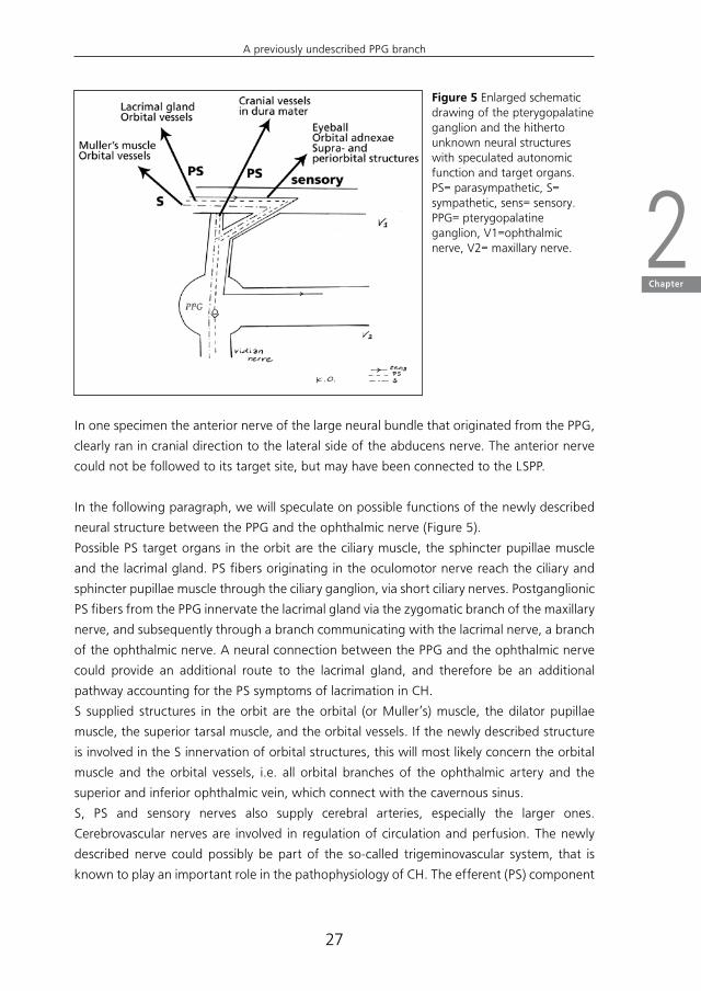
In the following paragraph, we will speculate on possible functions of the newly described
neural structure between the PPG and the ophthalmic nerve (Figure 5).
Possible PS target organs in the orbit are the ciliary muscle, the sphincter pupillae muscle
and the lacrimal gland. PS fibers originating in the oculomotor nerve reach the ciliary and
sphincter pupillae muscle through the ciliary ganglion, via short ciliary nerves. Postganglionic
PS fibers from the PPG innervate the lacrimal gland via the zygomatic branch of the maxillary
nerve, and subsequently through a branch communicating with the lacrimal nerve, a branch
of the ophthalmic nerve. A neural connection between the PPG and the ophthalmic nerve
could provide an additional route to the lacrimal gland, and therefore be an additional
pathway accounting for the PS symptoms of lacrimation in CH.
S supplied structures in the orbit are the orbital (or Muller’s) muscle, the dilator pupillae
muscle, the superior tarsal muscle, and the orbital vessels. If the newly described structure
is involved in the S innervation of orbital structures, this will most likely concern the orbital
muscle and the orbital vessels, i.e. all orbital branches of the ophthalmic artery and the
superior and inferior ophthalmic vein, which connect with the cavernous sinus.
S, PS and sensory nerves also supply cerebral arteries, especially the larger ones.
Cerebrovascular nerves are involved in regulation of circulation and perfusion. The newly
described nerve could possibly be part of the so-called trigeminovascular system, that is
known to play an important role in the pathophysiology of CH. The efferent (PS) component
Figure 5 Enlarged schematic drawing of the pterygopalatine ganglion and the hitherto unknown neural structures with speculated autonomic function and target organs. PS= parasympathetic, S= sympathetic, sens= sensory.PPG= pterygopalatine ganglion, V1=ophthalmic nerve, V2= maxillary nerve.
27
A previously undescribed PPG branch
2Chapter

of this trigeminal-autonomic reflex is thought to run through the PPG. 12 The ophthalmic
nerve, with cell bodies in the trigeminal ganglion, innervates structures in the head involved
in pain sensation, such as the dura mater. These pseudounipolar neurons project to second-
order neurons in the trigeminocervical complex, i.e. the trigeminal nucleus caudalis and
dorsal horns of C1 and C2, with a reflex connection to the superior salivatory nucleus (SSN).
From the SSN, preganglionic PS neurons project through the facial nerve, and synapse in the
PPG. These neurons supply, amongst others, cranial vessels in the dura mater. Although the
newly described nerve is not likely to play a role in this pathway, the anterior division of the
discovered neural bundle, which could be the same structure as described by Bleys et al.9,
could possibly contribute to this reflex by providing a direct connection between the PPG
and the LSPP.
In conclusion, the PPG is connected with the ophthalmic nerve by a hitherto undescribed
nerve. Although this neural connection is probably visceromotor and sensory in function,
further study involving immunohistochemical characterisation is necessary. The presence of
this neural connection, however, can add to explaining pain relief in the ophthalmic nerve
area after blocking the PPG.
28

REFERENCES
1. Burchiel KJ, Steege TD, Howe JF, et al. Comparison of percutaneous radiofrequencygangliolysisandmicrovasculardecompressioninsurgicalmanagementofticdouloureux.Neurosurgery1981;9:111-119.
2.SalarG,OriC,IobI,etal.Percutaneousthermocoagulationforsphenopalatineganglionneuralgia.ActaNeurochir1987;84:24-28.
3. SandersM,ZuurmondWWA.Efficacyofsphenopalatineganglionblockadein66patientssuffering fromclusterheadache:a12- to70month follow-upevaluation.JNeurosurg1997;87:876-880.
4.Sluyter ME, Vercruysse PJ, Sterk W. Radiofrequency lesions of the sphenopalatineganglioninthetreatmentofclusterheadache.Schmerz1988;9:56-59.
5. StolkerRJ,KamphuisE,RohofO,etal.DiebehandlungvonClusterkopfschmerzenmitradiofrequentenLasionen.Schmerz1991;5:194.
6.Vervest ACM, Stolker RJ, Groen GJ, et al. Clusterkopfschmerz: Thermolaesion desGanglionsphenopalatinum.Schmerz1992;6:9.
7.RuskinAP.Sphenopalatine (nasal)ganglion.: remoteeffects including“psychosomatic”symptoms,ragereaction,pain,andspasm.ArchPhysMedRehabil1979;63:127-133.
8. WaldmannSD.Sphenopalatineganglionblock-80yearslater.RegAnesth1993;18:274-276.
9. BleysRLAW,JanssenLM,GroenGJ.Thelateralsellarnerveplexusanditsconnectionsinhumans.JNeurosurg2001;95:102-110.
10.RuskellGL.Theorbitalbranchesof thepterygopalatineganglionand their relationshipwithinternalcarotidnervebranchesinprimates.JAnat1970;106:323-339.
11. ZimmerLA,HartC,TheodosopoulosPV.Endoscopicanatomyofthepetroussegmentoftheinternalcarotidartery.AmJRhinolAllergy.2009;23:192-196.
12.May A, Goadsby PJ. The trigeminovascular system in humans: pathofysiologicimplications forprimaryheadachesyndromesof theneural influenceson thecerebralcirculation.JCerebBloodFlowMetab1999;19:115-127.
29
A previously undescribed PPG branch
2Chapter

3C h a p t e r
Neurochemical characterization of pterygopalatine ganglion branches in humans
M. B. Ebbeling †, K. P. Q. Oomen †, J. A. de Ru, G. J. Hordijk, R. L. A. W. Bleys
† Equal contributors
Submitted to Am J Rhinol Allergy
ABSTRACT
Background: Pterygopalatine ganglion branches seem to be involved in the pathophysiology
of facial pain. The functions of these branches, including a recently discovered orbital
branch, are not completely known, but could be of clinical significance.
Objective: To characterize pterygopalatine ganglion branches by studying their
neurochemical coding, specifically the orbital branches.
Methods: In four specimens, the pterygopalatine fossa was dissected out of its bony
surroundings as a single intact tissue block and cryosectioned. In one specimen the
pterygopalatine fossa was dissected out, opened and microscopically dissected. Recently
discovered neural structures were identified, dissected out of the tissue block and
cryosectioned. All cryostat sectionings were immunohistochemically stained for protein
gene product 9.5, nitric oxide synthase, and tyrosine hydroxylase.
Results: A recently discovered neural connection between the pterygopalatine ganglion
and the ophthalmic nerve could be confirmed in our study, and could be classified as an
orbital pterygopalatine ganglion branch. The connection stained throughout for protein
gene product 9.5, and partially stained for nitric oxide synthase. In other orbital branches,
both nitric oxide synthase and tyrosine hydroxylase positive nerve fibres were found. The
pterygopalatine ganglion contained nitric oxide synthase positive cells. Tyrosine hydroxylase
labelling was also found in nerve fibers running through the pterygopalatine ganglion and
the Vidian nerve.
Conclusion: The recently discovered orbital pterygopalatine ganglion branch is of a
mixed parasympathetic and sensory nature. In the other orbital pterygopalatine branches,
sympathetic fibres were demonstrated as well. This knowledge may add to the understanding
of symptomatology and therapies of headache syndromes.
32
INTRODUCTION
The pterygopalatine fossa (PPF) is an inverted pyramidal space located inferior to the orbital
apex. It contains the pterygopalatine ganglion (PPG) and a busy traffic of arteries, veins,
lymphatics and nerves. Branches of the maxillary nerve that are joined by postganglionic
parasympathetic (PS) facial nerve fibres and postganglionic sympathetic (S) fibres from the
superior cervical ganglion running via the internal carotid plexus and the deep petrosal
nerve, are distributed to the PPF. In the PPG, PS fibres synapse while S and sensory fibres
pass through the ganglion without synapsing. Nerve fibres emerge form the PPG in
mixed branches to the orbit, nasal cavity, oral cavity and pharynx. The functions of these
numerous groups of PPG branches are not completely known. In a recent human cadaver
study by Oomen et al.,1 macro-and microdissection of whole-mount preparations of the
PPF combined with nerve specific staining demonstrated a previously undescribed, orbital
pterygopalatine ganglion branch, which runs between the pterygopalatine ganglion and
the ophthalmic nerve.
As the PPG is involved in the pathophysiology of headaches with an unknown specific cause
and largely unexplained features such as Cluster headache and Sluder’s neuralgia, 2-9 the
functional characterization of both new and previously described PPG branches may be of
clinical significance.
Therefore, the aim of the present study was to characterize PPG branches by studying their
neurochemical coding. Immunohistochemical techniques were used to localize the general
neural marker protein gene product (PGP) 9.5-, the sympathetic nerve specific enzyme
tyrosine hydroxylase (TH) and nitric oxide synthase (NOS) in order to identify PS nerves.
MATERIALS AND METHODS
The neural content of the PPF and adjacent regions was studied through cryostat sectioning
of tissueblocks combined with immunohistochemical staining.
Tissue preparation
Five left halves of human heads were obtained from post mortems (35 to 65 years of age)
(Table 1). The heads were perfused with 0.9% NaCl under physiologic pressure, followed by
fixation with 1 litre 4% formaldehyde in phosphate-buffered saline (PBS) at 4 °C. Finally, the
specimens were rinsed with 1 litre phosphate-buffered saline containing 15% sucrose and
0.1% Na-azide at 4 °C. The heads were cut in tissueblocks, each block containing a PPF with
surrounding tissue. The specimens were stored in 15% sucrose and 0.1% Na-azide at 4 °C.
33
Neurochemical characterization of PPG branches
3Chapter
Preparation of tissue blocks for cryosectioning and immunohistochemistry
In four specimens, the PPF contents with their periosteal lining were dissected out of their
bony surroundings as a single unit or tissue block without opening it. The contents of
the adjacent part of the orbit and cavernous sinus were included in the tissue block, as
was the maxillary nerve. In the fifth specimen the PPF was dissected out, opened and
microscopically dissected. A recently discovered orbital pterygopalatine ganglion branch,
which runs between the pterygopalatine ganglion and the ophthalmic nerve was identified.
The structure was dissected out of the tissue block and sectioned in the cryostat.
Cryostat sectioning
The tissue was frozen in dry ice and embedded by tissue-tek® (Sakura Finetek, Zoeterwoude,
the Netherlands). Sections of 16 µm were cut in a cryostat (HM 500 OM; MICROM
laborgerate, Walldorf, Germany). In the regions of interest, 1 in 5 sections was collected, in
the remaining regions 1 in 10. Various sectioning planes were used (Table 1).
Immunohistochemistry
Immunohistochemistry was used by the indirect method to demonstrate PGP 9.5 (Table 2).
The streptavidine-biotine method was used to demonstrate TH and NOS (Table 2). The
protocol for demonstration of PGP 9.5 was as follows: the sections were washed in Hepes
buffer (0.05 M, pH 7.4) containing 0.1% Triton X-100 for 30 minutes (3 x 10) followed by
pre-incubation in 5% normal swine serum (Dako, Denmark) and 5% Bovine Serum Albumin
(BSA; Sigma, Germany) in Hepes buffer containing 0.1% Triton X-100 for 60 minutes.
Subsequently, sections were incubated overnight at 4 °C in the first antibody in Hepes
buffer, containing 1% normal swine serum, 0.1% DL-lysine and 0.1% Triton X-100. After
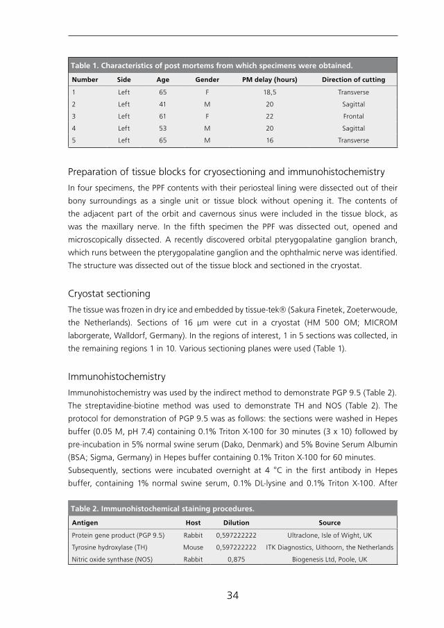
Table 1. Characteristics of post mortems from which specimens were obtained.
Number Side Age Gender PM delay (hours) Direction of cutting
1 Left 65 F 18,5 Transverse
2 Left 41 M 20 Sagittal
3 Left 61 F 22 Frontal
4 Left 53 M 20 Sagittal
5 Left 65 M 16 Transverse
Table 2. Immunohistochemical staining procedures.
Antigen Host Dilution Source
Protein gene product (PGP 9.5) Rabbit 0,597222222 Ultraclone, Isle of Wight, UK
Tyrosine hydroxylase (TH) Mouse 0,597222222 ITK Diagnostics, Uithoorn, the Netherlands
Nitric oxide synthase (NOS) Rabbit 0,875 Biogenesis Ltd, Poole, UK
34

washing in PBS for 30 minutes the sections were incubated in the second antibody at room
temperature. For PGP 9.5 this was fluorescein isothiocyanate (FITC)- conjugated swine anti
rabbit antiserum (Dako, Denmark) diluted 1:100, in PBS containing 1% normal swine serum,
1% DL-lysine and 0.1% Triton X-100 for 60 minutes. After washing in PBS the segments
were stained with 0,05% pontamine sky blue in PBS for 10 minutes to reduce background
autofluorescence and washed again in PBS.
The following protocol was used to demonstrate NOS and TH.
After washing in Hepes buffer (0.05 M, pH 7.4) for 30 minutes, the sections were
preincubated in 5% normal rabbit serum (for NOS normal goat serum) and 5% Bovine Serum
Albumin in Hepes buffer containing 0.1% Triton X-100 for 60 minutes. Subsequently, they
were incubated overnight at 4 º C in the first antibody in Hepes buffer containing 1%
normal rabbit serum (for NOS normal goat serum), 0.1% DL-lysine and 0.1% Triton X-100.
The primary antibody for NOS was anti-NOS antiserum (Biogenesis Ltd, Poole, UK, diluted
to 1:1200. After washing in PBS for 30 minutes the segments were pre-incubated in biotin
conjugated rabbit anti-mouse antiserum for TH (Dako, Glostrup, Denmark) and in biotin
conjugated goat anti-rabbit antiserum for NOS (Dako, Glostrup, Denmark) both diluted
1:200. After washing in PBS, they were incubated at room temperature in the third antibody.
For TH and NOS this was fluorescein isothiocyanate (FITC)- conjugated streptavidine (ITK
Diagnostics, Uithoorn,The Netherlands) diluted 1:500, in PBS containing 1% normal rabbit
serum (for NOS normal goat serum), 1% DL-lysine and 0.1% Triton X-100 for 60 minutes.
After washing in PBS the segments were stained for 10 minutes with 0,05% pontamine sky
blue in PBS to reduce background autofluorescence and washed again in PBS. The sections
were mounted in antifade mountant (Citifluor, London, UK) and stored at -20º C.
Acetylcholinesterase
A sensitive AChE technique 10 was used to obtain a clear view of the location of the PPG,
the distribution of the nerves in the PPF and especially of previously undescribed neural
structures. The tissue preparation and processing of the sections were similar as for the
immunohistochemistry. The steps of the procedure included incubation in medium primarily
composed of acetylthiocholine iodide, cupric sulfate, and potassium ferrocyanide, followed
by intensification of the stain accomplished using diaminobenzidine, nickel ammonium
sulfate, and hydrogen peroxide. As a result, all nerves present were stained black. The
sections were mounted in entellan and stored at room temperature.
35
Neurochemical characterization of PPG branches
3Chapter

RESULTS
Neural contents of the PPF
In the sections generated by cryosectioning of the intact, undissected PPF tissue blocks, the
general neural markers PGP 9.5 and AChE (Figs. 1 and 2) clearly demonstrated all the nerves
in the PPF, including the PPG with its cell bodies and nerve fibres. AChE stained sagittal
sections demonstrated all nerves, including the orbital branches, most clearly.
The shape of the PPG was irregular, due to the extensions from which the palatine, nasal,
pharyngeal and orbital nerves branched off.
The Vidian nerve (nerve of the pterygoid canal) connected with the PPG, and a large nerve
bundle ran through the PPG. Other nerves passing through the PPG were shown to branch
off the maxillary nerve, these were identified as ganglionic branches or pterygopalatine
nerves.
From the medial and cranial part of the PPG, nerves emerged that ran towards the nose and
pharynx, while other nerves connected the inferior part of the PPG to the palate. Orbital
branches emerged mainly from the medial side but sporadically from the lateral side of the
PPG, and ran in a cranial direction toward the inferior orbital fissure.
Figure 1. Sagittal section of the maxillary nerve, stained for PGP 9.5. Bar= 0.05 mm (For color figures, see page 117)
Figure 2. Sagittal section of an orbital branch (arrow) that runs cranially from the PPG, stained for AChE. PPG= pterygopalatine ganglion. Bar=0.05 mm.(For color figures, see page 117)
36

Other nerves that ran toward the inferior orbital fissure did not seem to originate from the
ganglionic area but from the area of the distal part of the maxillary or infraorbital nerve
(Fig.3), and thus formed an anterior group of rami orbitales. In this region a large branch of
the maxillary nerve, the zygomatic nerve, was visible.
After passing through the inferior orbital fissure, some of the orbital branches passed
forward in the orbit. Other rami orbitales left the orbit through the superior orbital
fissure and continued in direction of the cavernous sinus. Numerous nerves were found
in the cavernous sinus, particularly near the ophthalmic nerve. Some of these nerves were
continuations of the rami orbitales, others were probably emerging from the lateral sellar
plexus proper (LSPP). 10
No connections between the rami orbitales and the ophthalmic nerve were found.
Multiple cell bodies were found in the rami orbitales, also in the anterior group (Fig. 4). The
Vidian nerve contained cell bodies as well.
Figure 4. Sagittal section of a nerve cell body in an orbital branch, stained for AChE. Bar= 0.05 mm (For color figures, see page 117)
Figure 3. Schematic drawing of the distribution of the orbital branches. The branches pointed out with the blue frame did not seem to originate from the ganglionic area, but from the area of the distal part of the maxillary/infraorbital nerve; the anterior group of rami orbitales. PPG= pterygopalatine ganglion.
37
Neurochemical characterization of PPG branches
3Chapter
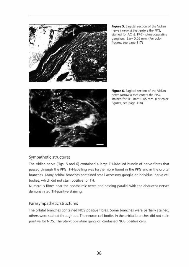
Sympathetic structures
The Vidian nerve (Figs. 5 and 6) contained a large TH-labelled bundle of nerve fibres that
passed through the PPG. TH-labelling was furthermore found in the PPG and in the orbital
branches. Many orbital branches contained small accessory ganglia or individual nerve cell
bodies, which did not stain positive for TH.
Numerous fibres near the ophthalmic nerve and passing parallel with the abducens nerves
demonstrated TH-positive staining.
Parasympathetic structures
The orbital branches contained NOS positive fibres. Some branches were partially stained,
others were stained throughout. The neuron cell bodies in the orbital branches did not stain
positive for NOS. The pterygopalatine ganglion contained NOS positive cells.
Figure 5. Sagittal section of the Vidian nerve (arrows) that enters the PPG, stained for AChE. PPG= pterygopalatine ganglion. Bar= 0,05 mm. (For color figures, see page 117)
Figure 6. Sagittal section of the Vidian nerve (arrows) that enters the PPG, stained for TH. Bar= 0.05 mm. (For color figures, see page 118)
38
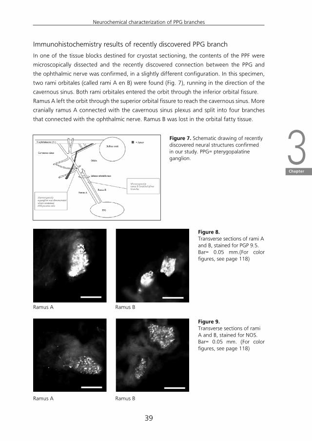
Immunohistochemistry results of recently discovered PPG branch
In one of the tissue blocks destined for cryostat sectioning, the contents of the PPF were
microscopically dissected and the recently discovered connection between the PPG and
the ophthalmic nerve was confirmed, in a slightly different configuration. In this specimen,
two rami orbitales (called rami A en B) were found (Fig. 7), running in the direction of the
cavernous sinus. Both rami orbitales entered the orbit through the inferior orbital fissure.
Ramus A left the orbit through the superior orbital fissure to reach the cavernous sinus. More
cranially ramus A connected with the cavernous sinus plexus and split into four branches
that connected with the ophthalmic nerve. Ramus B was lost in the orbital fatty tissue.
Figure 7. Schematic drawing of recently discovered neural structures confirmed in our study. PPG= pterygopalatine ganglion.
Figure 9.Transverse sections of rami A and B, stained for NOS.Bar= 0.05 mm. (For color figures, see page 118)
Figure 8.Transverse sections of rami A and B, stained for PGP 9.5.Bar= 0.05 mm.(For color figures, see page 118)
Ramus A
Ramus A
Ramus B
Ramus B
39
Neurochemical characterization of PPG branches
3Chapter
Studying the recently discovered structures after cryosectioning with the aid of
immunohistochemistry enabled us to identify the structures as nerves and characterize them.
Rami A and B both stained positive for PGP 9.5 (Figs. 8A-B). Ramus A was a long nerve,
while ramus B consisted of two branches that separated while running cranially (Fig. 7). Both
rami contained NOS positive fibres (Figs. 9A-B), and thus were partially stained positive for
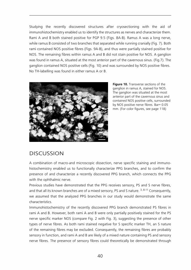
NOS. The remaining fibres within ramus A and B did not stain positive for NOS. A ganglion
was found in ramus A, situated at the most anterior part of the cavernous sinus. (Fig.7). The
ganglion contained NOS positive cells (Fig. 10) and was surrounded by NOS positive fibres.
No TH-labelling was found in either ramus A or B.
Figure 10. Transverse sections of the ganglion in ramus A, stained for NOS. The ganglion was situated at the most anterior part of the cavernous sinus and contained NOS positive cells, surrounded by NOS positive nerve fibres. Bar= 0.05 mm. (For color figures, see page 118)
DISCUSSION
A combination of macro-and microscopic dissection, nerve specific staining and immuno-
histochemistry enabled us to functionally characterize PPG branches, and to confirm the
presence of and characterize a recently discovered PPG branch, which connects the PPG
with the ophthalmic nerve.
Previous studies have demonstrated that the PPG receives sensory, PS and S nerve fibres,
and that all its known branches are of a mixed sensory, PS and S nature. 3, 8-11 Consequently,
we assumed that the analyzed PPG branches in our study would demonstrate the same
characteristics.
Immunohistochemistry of the recently discovered PPG branch demonstrated PS fibres in
rami A and B. However, both rami A and B were only partially positively stained for the PS
nerve specific marker NOS (compare Fig. 2 with Fig. 3), suggesting the presence of other
types of nerve fibres. As both rami stained negative for S specific marker TH, an S nature
of the remaining fibres may be excluded. Consequently, the remaining fibres are probably
sensory in function, and rami A and B are likely of a mixed nature containing PS and sensory
nerve fibres. The presence of sensory fibres could theoretically be demonstrated through
40
calcitonin gene-related peptide (CGRP) immunohistochemistry. However, previous studies
have shown that CGRP immunoreactivity seems to be present in part of the sensory neurons
only. 12, 13
Most other orbital PPG branches described in our study contained both NOS and TH-positive
fibres and therefore could be characterized as mixed PS/S and possibly sensory in function.
However, the visual impression was that PS fibres were more abundant than S fibres. Some
of the orbital branches contained PS and probably sensory fibres only, just like rami A and
B. Some of our findings correspond to those in previous studies.
Ruskell (1970) described the orbital PPG branches in primates and humans in detail. 14, 15
He described an orbitociliary nerve in primates that runs between the maxillary nerve and
the cavernous plexus, giving off branches to the ciliary ganglion, and presented a detailed
description of orbital branches that run between the PPG and the orbit. The largest group
of these orbital branches passes dorsally through the inferior orbital fissure and backwards
through the cavernous sinus, towards the ophthalmic and abducens nerves. A smaller
anterior group enters the orbit at its apex. After emerging through the inferior orbital
fissure, the orbital rami turn either toward the orbit or cranial cavity, and run parallel with
the oculomotor, ophthalmic and abducens nerves. A connection between orbital rami and
the ophthalmic nerve was not demonstrated.
Although the orbital PPG branches have been extensively described morphologically,
few data are available on their function. Ruskell did however previously demonstrate PS
nerve fibres in orbital PPG branches in rabbits. 14 Our impression that PS fibres were more
abundant than S fibres in the studied PPG branches, is in line with Ruskell’s description.
It is common knowledge that all orbital branches originate in the PPG and reach the orbit
by passing through the inferior orbital fissure. However, the present study demonstrates
a group of orbital branches that seem to stem from the distal part of the maxillary or
infraorbital nerve; the anterior group of orbital branches. Cell bodies could be demonstrated
in this anterior group, which could be PPG related ganglia or extensions of the PPG. This
finding is in line with the findings of previous anatomical studies, such as a whole mount
study of intracranial neural pathways by Bleys et al., 16 in which it was concluded that
neural cell bodies that innervate te cerebrovascular system are not confined to the classical
ganglia and more widespread than commonly thought. The NOS positive ganglion that was
demonstrated in ramus A in our study, located very close to the orbit, could functionally be
related to the PS ganglia of the cavernous sinus. Such cavernous sinus related ganglia have
previously been described, although located less close to the orbit. 10, 17-19
Most of the orbital branches in our study remained in the orbit. According to Ruskell, the
orbital branches penetrated the orbital smooth muscle (of Müller) and passed adjacent to
the periosteum of the orbit at the apex either medially or laterally. Ruskell also described a
junction between the rami orbitales and the lacrimal gland first passing through a plexus in
41
Neurochemical characterization of PPG branches
3Chapter

the orbital tissue between the fissures. No rami orbitales were identified passing from the
PPG to the gland without first passing through this plexus. 15
Several orbital braches in our study ran toward the plexus of the cavernous sinus. Bleys
et al described a relatively large bundle that connected the LSPP to the PPF and one or
more smaller nerves that connected the LSPP after traversing a plexus in the orbital tissue
between the fissures. 10 Ruskell described this connection as well. 14, 15 The present study
additionally demonstrates orbital braches that pass through to the cavernous sinus and
connect to the ophthalmic nerve, as recently discovered by Oomen et al.
One of the results of this study is that the orbital branches are generally of a mixed sensory,
S and PS nature. As the ophthalmic nerve is sensory to the eyeball, orbital adnexae and
supra- and periorbital structures, a sensory connection between this nerve and the PPG
could be involved in the pain pathway of CH and explain relief of pain in the orbital area
when blocking the PPG. This would imply a pain pathway that runs from the ophthalmic
nerve, through the rami orbitales and the PPG, to the trigeminal ganglion.
However, many questions remain unanswered, such as the functional direction of the rami
orbitales. It is unclear whether the discovered rami orbitales nerve fibres run forwards or
backwards in the ophthalmic nerve.
The PS target organs of the orbital branches are unknown. Possible PS target organs are the
cerebral arteries, the lacrimal gland, and least likely the pupillary sphincter muscle and the
ciliary muscle. Supporting evidence could be made by studying the nerves with the aid of
an electron microscope, but the best research would be tracer studies or conventional nerve
degeneration techniques, which for obvious reasons is not possible.
However, the presence of PS fibres in orbital branches may add to explaining reduction of
autonomic symptoms of Cluster headache such as lacrimation after lesioning the PPG.
In conclusion, the orbital PPG branches are of a mixed PS, S and sensory nature, the recently
discovered orbital branch which connects to the ophthalmic nerve is of a mixed PS and
sensory nature. The presence of these branches could add to explaining the relief of some
of the sensory and autonomic symptoms upon blocking the PPG.
42

REFERENCES
1. OomenKP,EbbelingM,deRuJA,HordijkGJ,BleysRL.Apreviouslyundescribedbranchofthepterygopalatineganglion.AmJRhinolAllergy2011;inpress.
2. AhamedSH,JonesNS.WhatisSluder’sneuralgia?JLaryngolOtol2003;117:437-443.
3. BruynGW.SphenopalatineNeuralgia (Sluder). InHandbookofclinicalneurology,vol.4.Headache.CliffordRoseF(Ed).ElsevierSciencePublisherBV,1986.
4. EdvinssonL.Pathophysiologyofprimaryheadaches.CurrPainHeadacheRep2001;5:71-78.
5. EkbomK,HardeboJE.Clusterheadache:aetiology,diagnosisandmanagement.Drugs2002;62:61-69.
6.May A, Goadsby PJ. The trigeminovascular system in humans: pathophysiologicimplications forprimaryheadachesyndromesof theneural influenceson thecerebralcirculation.JCerebBloodFlowMetab1999;19:115-127.
7.SandersM,ZuurmondWW.Efficacyofsphenopalatineganglionblockadein66patientssuffering fromclusterheadache:a12- to70month follow-upevaluation.JNeurosurg1997;87:876-880.
8. SluderG.Roleofthesphenopalatine(Meckel’s)ganglioninnasalheadaches.NewYorkMedJour1908;989-990.
9. Sluder G. Etiology, diagnosis prognosis and treatment of sphenopalatine ganglionneuralgia.JAmMedAssoc1913;16:1202-1206.
10.BleysRL,JanssenLM,GroenGJ.Thelateralsellarnerveplexusanditsconnectionsinhumans.JNeurosurg2001;95:102-110.
11. KleinRN,BurkDT,ChasePF.Anatomicallyandphysiologicallybasedguidelinesforuseofthesphenopalatineganglionblockversusthestellateganglionblocktoreduceatypicalfacialpain.Cranio2001;19:48-55.
12.QuartuM.,DiazG.,FlorisA.,etal.Calcitoningene-relatedpeptideinthehumantrigeminalsensorysystematdevelopmentalandadultlifestages:immunohistochemistry,neuronalmorphometryandcoexistencewithsubstanceP.JChemNeuroanat1992;5:143-157.
13. UusitaloH,KrootilaK,PalkamaA.Calcitoningene-relatedpeptide(CGRP)immunoreactivesensorynervesinthehumanandhuineapiguveaandcornea.ExpEyeRes1989;48:467-475.
14. RuskellGL.Theorbitalbranchesof thepterygopalatineganglionand their relationshipwithinternalcarotidnervebranchesinprimates.JAnat1970;106:323-339.
15.Ruskell GL. Distribution of pterygopalatine ganglion efferents to the lacrimal gland inman.ExpEyeRes2004;78:329-335.
16.Bleys,RL,GroenGJ,HommersomRF.Neuralconnectionsinandaroundthecavernoussinusinrat,withspecialreferencetocerebrovascularinnervation.JCompNeurol1996;369:2772-2791.
17. CarvalhoVC.Nervecellsinthehumancavernoussinus.AnatAnz1985;159:29-32.
18. GellertA.Gangliaoftheinternalcarotidplexus.JAnat1934;68:318-322.
19.SuzukiN,HardeboJE.Anatomicalbasisforaparasympatheticandsensoryinnervationoftheintracranialsegmentoftheinternalcarotidarteryinman.Possibleimplicationforvascularheadache.JNeurolSci1991;104:19-31.
43
Neurochemical characterization of PPG branches
3Chapter

4C h a p t e r
Improved depiction of pterygopalatine fossa anatomy using ultra high resolution magnetic resonance imaging at 7 Tesla
K. P. Q. Oomen, F. A. Pameijer, J. J. A. Zwanenburg, G. J. Hordijk, J. A. De Ru, R. L.
A. W. Bleys
Submitted to Eur J Radiol

ABSTRACT
Background: The pterygopalatine fossa is an important area to review in head and neck
imaging, both as a diagnostic and a preoperative measure. However, the complex anatomy
of the pterygopalatine fossa and its content, such as the pterygopalatine ganglion and its
branches, is difficult to image.
Study design: The pterygopalatine fossa of one cadaver specimen was studied through
magnetic resonance imaging at 7 Tesla and cryomicrotome sectioning.
Methods: The tissue block containing the pterygopalatine fossa was examined on a clinical
7 Tesla magnetic resonance imaging system. Subsequently, cryosections of the tissue block
were created in a coronal plane. The cryosections were photographed and collected on
adhesive tape. The on-tape sections were stained for Mallory-Cason, in order to detail the
anatomic structures within the fossa. Magnetic resonance images were compared with the
surface photos of the tissueblock and the on-tape sections.
Results: High resolution magnetic resonance images demonstrated the common
macroscopic anatomical structures in the PPF. Furthermore, smaller structures best viewed
at the level of the operation microscope, that have previously been obscured on magnetic
resonance imaging in this area, could be depicted. Some of the orbital ptergyopalatine
ganglion branches as well as the pharyngeal nerve were clearly viewed.
Conclusion: In our experience, magnetic resonance imaging at 7 Tesla provides excellent
depiction of the pterygopalatine fossa anatomy, and provides previously unseen detail
through its demonstration of the pharyngeal nerve and the orbital pterygopalatine ganglion
branches.
46

INTRODUCTION
The pterygopalatine fossa (PPF) is an inverted pyramidal space located inferior to the
orbital apex, which contains the pterygopalatine ganglion (PPG) and various arteries,
veins, lymphatics and nerves. Preganglionic parasympathetic facial nerve fibres synapse in
the PPG, while postganglionic sympathetic fibres from the superior cervical ganglion and
sensory fibres from the maxillary nerve pass through the ganglion without synapsing. The
PPF communicates with the orbit, nasal cavity and oral cavity, and through the orbit with
the maxillary sinus and upper teeth, which makes it an important cranial neurovascular
crossroad as well as a common site for invasion and perineural spread of malignant
disease.1,2 The neural content of the PPF plays an important role in the pathophysiology
of pain syndromes with cranial autonomic features such as Cluster headache and Sluder’s
neuralgia.3,4 These syndromes are invalidating and may require invasive treatment, such as
PPG blockage, for refractory cases.5,6 Thus, studying the PPF in head and neck imaging is of
importance, both for diagnostic as well as preoperative purposes.
Previous studies have shown that on magnetic resonance imaging (MRI) at 1.5 Tesla, small
PPF structures remain obscured, whereas the PPG and the sphenopalatine segment of the
maxillary artery and some of its branches can easily be identified.7 The recent development
of MRI at 7 Tesla (7T MRI) holds the promise of an increased signal-to-noise ratio (SNR). In
various human anatomical regions, the increased SNR of 7T MRI has been used to produce
high definition images with ultra high resolution and identification of previously unidentified
detail.8,9
The aim of the present study is to correlate MR findings to cryosections in order to determine
which part of the PPF and its contents can be identified on 7 T MRI.
MATERIALS AND METHODS
Tissue preparation
One undissected human head was obtained from a male post mortem, 73 years of age. The
head was perfused with 0.9% NaCl under physiologic pressure and frozen at -25 ºC.
Whole-mount preparation
The head was transected on the median plane using a band saw and trimmed to a block
containing the PPF and parts of the orbit, nasal and paranasal cavities, and oral cavity.
Imaging
The specimen was examined on a whole-body, clinical 7T MRI system (Philips Healthcare,
Cleveland, OH, USA), using a transmit/receive head coil with a 16 channel receive coil (Nova
47
Sluder’s neuralgia; a trigeminal autonomic cephalalgia
4Chapter

Medical, Wilmington, MA, USA). During the MR examination, the specimen was submersed
entirely in fomblin (Solvay Solexis, Bollate, Italy) to provide susceptibility matching, thereby
contributing to accurate B0 shimming.10 A 3D (volumetric) multi-echo gradient echo
sequence was applied with the following scan parameters: Field of view (FOV) 100 x 81 x
60 mm3, acquisition matrix 332 x 270, 199 slices, acquired resolution 0.3 x 0.3 x 0.3 mm3
(voxel volume 27 nL), TR 158 ms, TE 3.3 ms, fat suppression with SPAIR (inversion delay 50
ms), bandwidth 427 Hz/pixel. The acquisition duration was approximately 5 hours and 32
minutes. Parameters were consisted with a T1 weighted scan.
Cryomicrotome sectioning
After MR scanning, the whole-mount specimen was fixed in formaldehyde 4%, followed
by rinsing in running tap water for several days. After overnight impregnation in 1%
carboxymethylcellulose (CMC), the specimen was frozen and embedded in 1% CMC in the
cryomicrotome (PMV 450MP; Palmstiernas Instruments AB, Stockholm, Sweden) at -25 ºC.
The specimen was positioned in the cyromicrotome in an orientation matching its position
on the MR images. Cryosections were created in a coronal plane, with a section thickness
of 25 µm.
Pictures of the tissue block surface in the region of the PPF were taken every 75 µm of
sectioning. Sections were collected on wide adhesive tape11 every 375 µm of sectioning.
The on-tape sections were stained with a modified Mallory-Cason procedure12 and mounted
on cardboard. A corresponding photograph and section were assigned to each of the MR
images, taking into account the slice thickness, slice gap, and slice interval.
RESULTS
Correlations of anatomical findings and coronal MR images are shown in an anteroposterior
series of Figures 1 to 5. Figures 1 t/m 4 show several common anatomical structures in and
around the PPF. The sphenoidal sinus (SS), middle cranial fossa and nasal cavity were used
as orientation points. In the lateral nasal wall, the middle and inferior conchas were found.
Lateral to the PPF, several structures were found with intermediate signal intensity on MR
and an orange red Mallory-Cason stain, consistent with muscle tissue. From medial to
lateral, the medial pterygoid muscle (MPM), lateral pterygoid muscle (LPM) with its superior
and inferior heads, and temporal muscle were identified.
The optic nerve (ON) was identified from its origin in the optic chiasm to its position just
lateral to the SS. Caudal to the ON, the common anular tendon (of Zinn) was identified,
a ring of fibrous tissue surrounding the ON at its entrance at the apex of the orbit, which
48
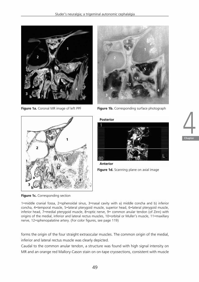
Figure 1a. Coronal MR image of left PPF Figure 1b. Corresponding surface photograph
Figure 1c. Corresponding section
Figure 1d. Scanning plane on axial image
1=middle cranial fossa, 2=sphenoidal sinus, 3=nasal cavity with a) middle concha and b) inferior concha, 4=temporal muscle, 5=lateral pterygoid muscle, superior head, 6=lateral pterygoid muscle, inferior head, 7=medial pterygoid muscle, 8=optic nerve, 9= common anular tendon (of Zinn) with origins of the medial, inferior and lateral rectus muscles, 10=orbital or Muller’s muscle, 11=maxillary nerve, 12=sphenopalatine artery. (For color figures, see page 119)
forms the origin of the four straight extraocular muscles. The common origin of the medial,
inferior and lateral rectus muscle was clearly depicted.
Caudal to the common anular tendon, a structure was found with high signal intensity on
MR and an orange red Mallory-Cason stain on on-tape cryosections, consistent with muscle
Posterior
Anterior
49
Sluder’s neuralgia; a trigeminal autonomic cephalalgia
4Chapter
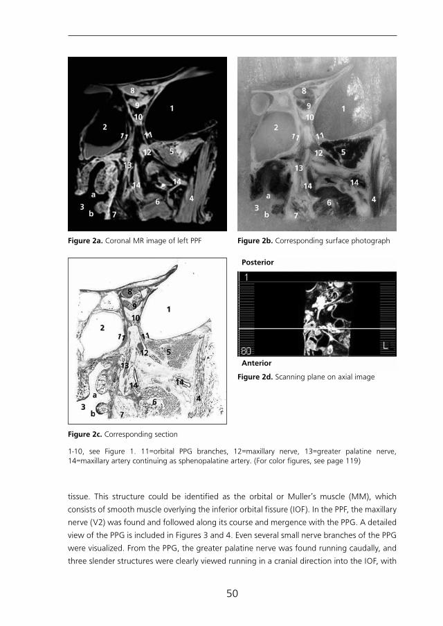
Figure 2a. Coronal MR image of left PPF Figure 2b. Corresponding surface photograph
Figure 2c. Corresponding section
Figure 2d. Scanning plane on axial image
1-10, see Figure 1. 11=orbital PPG branches, 12=maxillary nerve, 13=greater palatine nerve, 14=maxillary artery continuing as sphenopalatine artery. (For color figures, see page 119)
tissue. This structure could be identified as the orbital or Muller’s muscle (MM), which
consists of smooth muscle overlying the inferior orbital fissure (IOF). In the PPF, the maxillary
nerve (V2) was found and followed along its course and mergence with the PPG. A detailed
view of the PPG is included in Figures 3 and 4. Even several small nerve branches of the PPG
were visualized. From the PPG, the greater palatine nerve was found running caudally, and
three slender structures were clearly viewed running in a cranial direction into the IOF, with
Posterior
Anterior
50
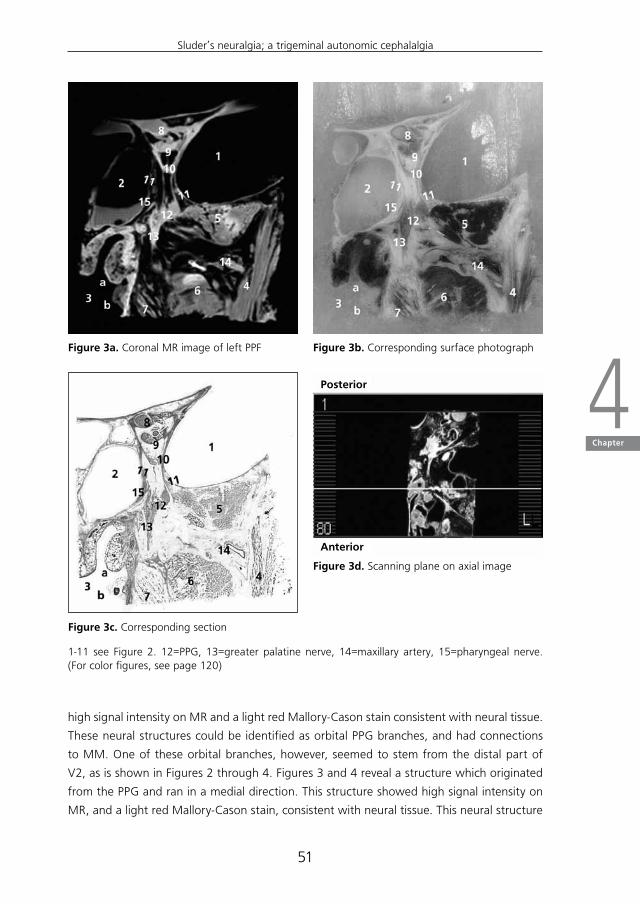
Figure 3a. Coronal MR image of left PPF Figure 3b. Corresponding surface photograph
Figure 3c. Corresponding section
Figure 3d. Scanning plane on axial image
1-11 see Figure 2. 12=PPG, 13=greater palatine nerve, 14=maxillary artery, 15=pharyngeal nerve. (For color figures, see page 120)
high signal intensity on MR and a light red Mallory-Cason stain consistent with neural tissue.
These neural structures could be identified as orbital PPG branches, and had connections
to MM. One of these orbital branches, however, seemed to stem from the distal part of
V2, as is shown in Figures 2 through 4. Figures 3 and 4 reveal a structure which originated
from the PPG and ran in a medial direction. This structure showed high signal intensity on
MR, and a light red Mallory-Cason stain, consistent with neural tissue. This neural structure
Posterior
Anterior
51
Sluder’s neuralgia; a trigeminal autonomic cephalalgia
4Chapter

was identified as the pharyngeal nerve, in its course towards the palatovaginal canal. From
a lateral direction, a remarkably tortuous structure, with a low intensity on MR and a dark
red Mallory-Cason stain with a distinct lumen, consistent with vascular tissue, was found
running between the superior and inferior head of the LPM, toward and into the PPF (Figure
1 and 2). This vascular structure was identified as the maxillary artery continuing as the
sphenopalatine artery (SPA).
Figure 4a. Coronal MR image of left PPF Figure 4b. Corresponding surface photograph
Figure 4c. Corresponding section
Figure 4d. Scanning plane on axial image
1-12 see Figure 3. 13=origin of greater palatine nerve, 14=maxillary artery, 15=pharyngeal nerve. (For color figures, see page 120)
Posterior
Anterior
52

Figure 5a. Coronal MR image of left cavernous sinus
Figure 5b. Corresponding surface photograph
Figure 5c. Corresponding section
Figure 5d. Scanning plane on axial image
1-3 see Figure 4. 4=lateral pterygoid muscle, 5=tensor veli palatini muscle, 6=internal carotid artery, 7=oculomotor nerve, 8=trochlear nerve, 9=ophthalmic nerve, 10=abducens nerve, 11= maxillary nerve, 12=nerve of the pterygoid canal or Vidian nerve. (For color figures, see page 121)
In the most posterior image, Figure 5, the cavernous sinus and the structures related to
its medial wall were demonstrated. The internal carotid artery, the oculomotor nerve, the
trochlear nerve, the abducens nerve, the ophthalmic nerve (V1) and V2 were all clearly
visible. The nerve of the pterygoid canal or Vidian nerve was found traversing the base of
the pterygoid process in the floor of the SS.
Posterior
Anterior
53
Sluder’s neuralgia; a trigeminal autonomic cephalalgia
4Chapter
DISCUSSION
Our study demonstrates that ex vivo MR imaging of the PPF at 7 T provides excellent
depiction of PPF content, specifically the PPG and some of its branches. Some of the orbital
branches and the pharyngeal nerve were clearly visible, and the orbital branches could be
followed toward one of their targets, MM.
Comparison of our findings with those in previous radiological studies is hampered
by the fact that few studies are available on MRI appearance of the PPF. Following the
introduction of high-resolution computed tomography (CT), several investigators have
compared CT findings of normal and pathological anatomy of the PPF with findings in
cadaver specimens.2,13-16 As expected, previous CT studies of the PPF have focused on its
boundaries and communications, rather than its content.17-19 The few MR studies that are
available, focus on perineural tumor spread in the PPF, which excludes a detailed search for
structures such as the PPG and its communications.7,20-23 Although the palatovaginal canals
are commonly depicted on MR16, a clear depiction of the pharyngeal nerve is hitherto
undescribed in radiological studies of the PPF. Rumboldt et al.16 have described structures
in the palatovaginal canal that presumably correspond to the pterygovaginal artery and
possibly, the pharyngeal nerve. However, their findings were only visible as flow voids or low
signal intensity structures on T1 weighted images, and the presumed structures were not
visible in great detail. The orbital PPG branches have never been described in radiological
PPF studies. Some of our findings correspond to those in previous anatomical studies. A
previous endoscopic study of the anatomical relations of the PPG revealed a remarkably
tortuous portion of the SPA along its course in the PPF, suggestive of a potentiality of
vascular compression of the PPG as a causative factor in headaches with ipsilateral cranial
autonomic features. 24 Our images of the SPA are in line with this description, although
direct compression of the SPA on the PPG was not present. Ruskell (1970) described the
orbital PPG branches in primates and humans in detail25,26, and demonstrated that orbital
branches originate in the PPG and reach the orbit by passing through the IOF. According to
Ruskell, the orbital branches penetrate the orbital smooth muscle (MM) and pass adjacent
to the periosteum of the orbit at the apex either medially or laterally. The orbital PPG
branches depicted on 7T MRI in our study demonstrated the same configuration. A recent
cadaver study on neurochemical characterization of PPG branches in humans demonstrated
a group of orbital branches that stem from the distal part of the maxillary or infraorbital
nerve; the anterior group of orbital PPG branches.27 These findings were confirmed in our
study. In a recent human cadaver study by Oomen et al.28, macro-and microdissection of
whole-mount preparations of the PPF combined with nerve specific staining demonstrated
a previously undescribed orbital PPG branch, which runs between the PPG and the V1. This
specific orbital PPG branch could not be demonstrated in our MR study.
The improved depiction of the neural PPG connections, such as the orbital branches and
the pharyngeal nerve, could become clinically important once the pathophysiology of facial
54

pain is completely understood, including the exact pain pathways. In treatment of facial
pain, these insights might hold the promise of development of selective nerve blocks in
this area, in which ultra high resolution imaging of the PPF at 7T could be of aid as a
preoperative measure.
Although our results seem very promising, the fact that this concerns a cadaver study and not
an in-vivo study has to be taken in to account. The current acquisition duration of the scan
was 5 hours and 32 minutes, which for obvious reasons is not applicable to living subjects.
Furthermore, cadaver images differ in signal intensities from in-vivo images, which could
account for subtle changes in the appearance of anatomical structures. The true viability of
depicting the PPF with ultra high resolution MR therefore depends on confirmation of our
positive results in larger studies with living human subjects.
In conclusion, MR of the PPF at 7 T provides excellent depiction of PPF content, and
demonstrates hitherto radiologically obscured anatomical details such as the orbital PPG
branches and the pharyngeal nerve. High resolution MR at 7 T could potentially contribute
to an improved diagnostic and preoperative evaluation of the PPF and its content.
55
Sluder’s neuralgia; a trigeminal autonomic cephalalgia
4Chapter
REFERENCES
1. Ginsberg LE. Imaging or perineural tumor spread in head and neck cancer. SeminUltrasoundCTMR1999;20:175-186.
2.CurtinHD,WilliamsR. Computed tomographic anatomy of the pterygopalatine fossa.Radiographics1985;5:429-440.
3.EkbomK,HardeboJE.Clusterheadache.Aetiology,diagnosisandmanagement.Drugs2002;62:61-69.
4. SluderG.Roleofthesphenopalatine(Meckel’s)ganglioninnasalheadaches.NYStateJMed1908;87:989-990.
5. LovelyTJ,KotsiakisX,JanettaPJ.Thesurgicalmanagementofchronicclusterheadache.Headache1998;38:590-594.
6.SalarG,OriC,IobI,FioreD.Percutaneousthermocoagulationforsphenopalatineganglionneuralgia.ActaNeurochir1987;84:24-28.
7.Chang PC, Fischbein NJ, McCalmont TH, Kashani-Sabet M, Zettersten EM, Liu AY,WeissmanJL.Perineuralspreadofmalignantmelanomaoftheheadandneck:clinicalandimagingfeatures.AmJNeuroradiol2004:25:5-11.
8. Chang G, Friedrich KM,Wang L, Vieira RLR, SchweitzerME, RechtMP,Wiggins GC,RegatteRR.MRIofthewristat7teslausinganeight-channelarraycoilcombinedwithparallelimaging:Preliminaryresults.JMagnResonImaging2010;31:740-746.
9.MetcalfM,XuD,OkudaDT,CarvajalL,SrinivasanR,KelleyDA,MukherjeeP,NelsonSJ,VigneronDB,PelletierD.High-resolutionphased-arrayMRIofthehumanbrainat7tesla:initialexperienceinmultiplesclerosispatients.JNeuroimaging2010;20:141-147.
10.Beneviste H, Blackband S. MR microscopy and high resolution small animal MRI:applicationsinneuroscienceresearch.ProgNeurobiol2002;67:393-420.
11. Zonneveld FW: Computed tomography of the temporal bone and orbit: technique ofdirect multiplanar, high resolution CT and correlative cryosectional anatomy. Munich:Urban&Schwarzenberg,1987,pp53-63.
12.VanLeeuwenMBM,DeddensAJH,GeritsPO,HillenB.AmodifiedMallory-Casonstainingprocedureforlargecryosections.StainTechnol1990;65:37-42.
13.DanielsDL,RauschningW,LovasJ,WilliamsAL,HaughtonVM.Pterygopalatinefossa:computedtomographicstudies.Radiology1983;149:511-516.
14. Osborn AG. Radiology of the pterygoid plates and pterygopalatine fossa. AJR1979:132:389-394.
15.LeckamR,TerbruggeKG,ChiuMC.Computedtomographyofthepterygopalatinefossa.JCanAssocRadiol1981;32:97-101.
16. RumboldtZ,CastilloM,SmithJK.Thepalatovaginalcanal:canitbeidentifiedonroutineCTandMRimaging?AJR2002;179:267-272.
17. Erdogan N, Unur E, Baykara M. CT anatomy of the pterygopalatine fossa and itscommunications: a pictorial review. Computerized Medical Imaging and Graphics2003;27:481-487.
18. PandolfoI,GaetaM,BlandinoA,LongoM.Theradiologyofthepterygoidcanal:normalandpathologicfindings.AmJNeuroradiol1987;8:479-483.
56

19. Kim HS, Kim DI, Chung IH. High-resolution CT of the pterygopalatine fossa and itscommunications.Neuroradiology1996;38:120-126
20. ChongVF,FanY-F.Pterygopalatinefossaandmaxillarynerveinfiltrationinnasopharyngealcarcinoma.HeadNeck1997;19:121-125.
21.BlandinoA,GaetaM,MinutoliF,PandolfoI.CTandMRfindingsinneoplasticperineuralspreadalongthevidiannerve.EurRadiol2000;10:521-526.
22.Mazziotti S,GaetaM,BlandionoA, Vinci S, Pandolfo I. Perineural spread in a caseofsinonasalsarcoidosis:casereport.AmJNeuroradiol2001;22:1207-1208.
23. GinsbergLE,DeMonteF.Imagingofperineuraltumorspreadfrompalatalcarcinoma.AmJNeuroradiol1998;19:1417-1422.
24.AlfieriA,JhoH-D,SchettinoR,TschabitscherM.Endoscopicendonasalapproachtothepterygopalatinefossa:ananatomicstudy.Neurosurgery2003;52:74-380.
25.RuskellGL.Theorbitalbranchesof thepterygopalatineganglionand their relationshipwithinternalcarotidnervebranchesinprimates.JAnat1970;106:323-339.
26. Ruskell GL. Distribution of pterygopalatine ganglion efferents to the lacrimal gland inman.ExpEyeRes2004;78:329-335.
27.Ebbeling MB, Oomen KP, de Ru JA, Hordijk GJ, Bleys RLAW. Neurochemicalcharacterizationofpterygopalatineganglionbranchesinhumans.Submitted.
28. OomenKP,EbbelingM,deRuJA,HordijkGJ,BleysRL.Apreviouslyundescribedbranchofthepterygopalatineganglion.AmJRhinolAllergy2011;25:50-53.
57
Sluder’s neuralgia; a trigeminal autonomic cephalalgia
4Chapter


5C h a p t e r
Sluder’s neuralgia; a Trigeminal Autonomic Cephalalgia?
K. P. Q. Oomen, A. J. M. van Wijck, G. J. Hordijk, J. A. de Ru
Cephalalgia 2010; 30: 360-364
ABSTRACT
Objective: To formulate distinctive criteria to substantiate our opinion that Sluder’s
neuralgia and cluster headache are two different clinical entities.
Study design: Systematic review of literature.
Methods: A systematic review was carried out of all available, original literature on Sluder’s
neuralgia. Pain characteristics, periodicity and associated signs and symptoms were studied
and listed according to frequency of appearance.
Results: Eleven articles on Sluder’s neuralgia were evaluated. Several differences between
Sluder’s neuralgia and cluster headache became evident. Based on described symptoms,
new criteria for Sluder’s neuralgia could be formulated
Conclusion: Sluder’s neuralgia and cluster headache could possibly be regarded as two
different headache syndromes.
60
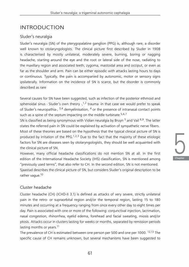
INTRODUCTION
Sluder’s neuralgia
Sluder’s neuralgia (SN) of the pterygopalatine ganglion (PPG) is, although rare, a disorder
well known to otolaryngologists. The clinical picture first described by Sluder in 1908
is characterised by mostly unilateral, moderately severe, burning, boring or nagging
headache, starting around the eye and the root or lateral side of the nose, radiating to
the maxillary region and associated teeth, zygoma, mastoidal area and occiput, or even as
far as the shoulder and arm. Pain can be either episodic with attacks lasting hours to days
or continuous. Typically, the pain is accompanied by autonomic, motor or sensory signs
ipsilaterally. Information on the incidence of SN is scarce, but the disorder is commonly
described as rare
Several causes for SN have been suggested, such as infection of the posterior ethmoid and
sphenoidal sinus - Sluder’s own theory -,1,2 trauma -in that case we would prefer to speak
of Sluder’s neuropathia-, 3,4 demyelinisation, 4 or the presence of intranasal contact points
such as a spine of the septum impacting on the middle turbinate.5,6,7
SN is classified as being synonymous with Vidian neuralgia by Bruyn 5 and Vail 8,9. The latter
states the referred pain in SN could be explained by activation of sympathetic nerve fibers.
Most of these theories are based on the hypothesis that the typical clinical picture of SN is
produced by irritation of the PPG.1,2,3 Due to the fact that the majority of these etiologic
factors for SN are diseases seen by otolaryngologists, they should be well acquainted with
the clinical picture of SN.
However, many official headache classifications do not mention SN at all. In the first
edition of the International Headache Society (IHS) classification, SN is mentioned among
“previously used terms”, that also refer to CH. In the second edition, SN is not mentioned.
Sjaastad describes the clinical picture of SN, but considers Sluder’s original description to be
rather vague.10
Cluster headache
Cluster headache (CH) (ICHD-II 3.1) is defined as attacks of very severe, strictly unilateral
pain in the retro- or supraorbital region and/or the temporal region, lasting 15 to 180
minutes and occurring at a frequency ranging from once every other day to eight times per
day. Pain is associated with one or more of the following: conjunctival injection, lacrimation,
nasal congestion, rhinorrhea, eyelid edema, forehead and facial sweating, miosis and/or
ptosis. Attacks occur in clusters lasting for weeks or months, separated by remission periods
lasting months or years.11
The prevalence of CH is estimated between one person per 500 and one per 1000. 12,13 The
specific cause of CH remains unknown, but several mechanisms have been suggested to
61
Sluder’sneuralgia;atrigeminalautonomiccephalagia
5Chapter

play a role in its etiology. A dysfunction of the central nervous system activates the so-called
trigeminovascular system (a system of neurons innervating the cerebral vessels whose cell
bodies are located in the trigeminal ganglion), and primary defects in the hypothalamic
grey matter, which are held responsible for the episodic pattern of clusters in which pain
attacks occur.12,14-17 The activation of a trigeminal parasympathetic reflex explains the
parasympathetic symptoms which are seen in CH. The efferent component of this reflex is
thought to run through the PPG.14-18
In contrast to patients with SN, who are diagnosed by otolaryngologists, the patients with
typical CH are mostly referred to neurologists.
Are SN and CH different clinical disorders?
It has been stated that the headache diagnosed by otolaryngologists as SN and the
headache diagnosed by neurologists as CH, are in fact the same clinical entity.19
Some of the symptoms and features of SN and CH show overlap. For instance, although one
of the characteristic features of SN is that pain can be blocked by cocainisation or infiltration
anaesthesia of the PPG, positive results of this method have also been reported in CH. 20-22
However, several crucial differences distinguish the two.19,23 One of the most striking
differences lies in the severity of pain. Pain in CH is often described as excruciating, is
in many cases accompanied by restlessness or agitation,11 and can even lead to suicidal
behaviour in some patients,24-26 whereas pain in SN is often described as moderately
severe.1,5,6,19,27 Pain in CH is localized orbital, supraorbital and/or temporal, whereas pain in
SN is localized at the root or lateral side of the nose and intra- or periorbitally, radiating to
maxilla, mastoidal or occipital area, and even to the neck, shoulder and arm.
Another difference lies in the periodicity of pain. Whereas the characteristic feature of CH
is the fact that attacks come in cluster periods, the pain in SN can be either continuous
with or without exacerbations or episodic with attacks. Furthermore, attacks in SN seem
to last much longer (hour(s) to days) than those in CH (15 to 180 minutes). The difference
between duration of pain in SN and CH is clearliest seen in the upper time limit of attacks,
although it is clear that the general lower time limit of hours in SN shows some overlap
with the upper time limit of 180 minutes in CH. The lower time limit of SN attacks varies
in literature between one hour 23 and a few minutes.4 Also, an attack duration of as short
as between 10 and 30 minutes, rarely exceeding two hours, has been described.28 It is
therefore difficult to present an exact duration range of SN attacks, as there seems to be no
consensus on duration of pain.
Also, the typical CH patient is different from the SN patient. The former is usually male, with
age of onset between 18 and 40, the latter is mostly female with age of onset between
30 and 50. These differences support our hypothesis that CH and SN are two independent
clinical disorders. Criteria for CH are clear and well-defined.
62

We would like to propose new, strict and clear criteria for SN that are able to distinguish it
from other forms of facial pain, and can thus be helpful in making correct diagnosis.
We performed a systematic review to provide a quantitative assessment of symptoms
described in literature on which these new SN criteria can be based.
In this article, the terms headache and facial pain are used synonymously.
METHODS
Study retrieval and selection
A computerised literature search was performed in the Pubmed database with search terms
“Facial neuralgia”, “Sluder’s neuralgia” and “Sphenopalatine ganglion neuralgia”. Reference
lists from identified publications were screened to retrieve additional articles. Medical
handbooks were also searched for reports.5 Only articles published in English and German
were retrieved.
Table I. Characteristics of SN according to newly defined criteria.
Characteristics Sluder’s neuralgia
Pain quality and intensity Moderately severe or severe, boring,
burning or nagging pain.
Site Unilateral but possibly bilateral, located peri- or intraorbitally, or at the root or lateral side of the nose, radiating to at least one of the following:
1. maxillary region or cheek and/or associated teeth
2. mastoidal and/or occipital area
3. neck, shoulder or arm
Frequency One of the following:
1. episodic with attacks lasting hour(s) to days
2. continuous for several weeks with or without exacerbations
Associated signs and symptoms At least one of the following:
1. ipsilateral lacrimation and/or conjunctival injection
2. ipsilateral nasal congestion and/or rhinorrhoea
3. hyp- or hyperesthesia in maxillary distribution of trigeminal nerve or
4. ipsilateral sore throat
5. ipsilateral delayed taste perception or parageusia
6. ipsilateral elevated palatine arch or contralaterally deflected uvula
Treatment Pain can be blocked by cocainisation or infiltration anaesthesia of the PPG
Characteristics Sluder’s Neuropathia (Secondary Sluder Syndrome)
As above, except headache develops in relation to trauma in the area innervated by the second division of the trigeminal nerve
63
Sluder’sneuralgia;atrigeminalautonomiccephalagia
5Chapter
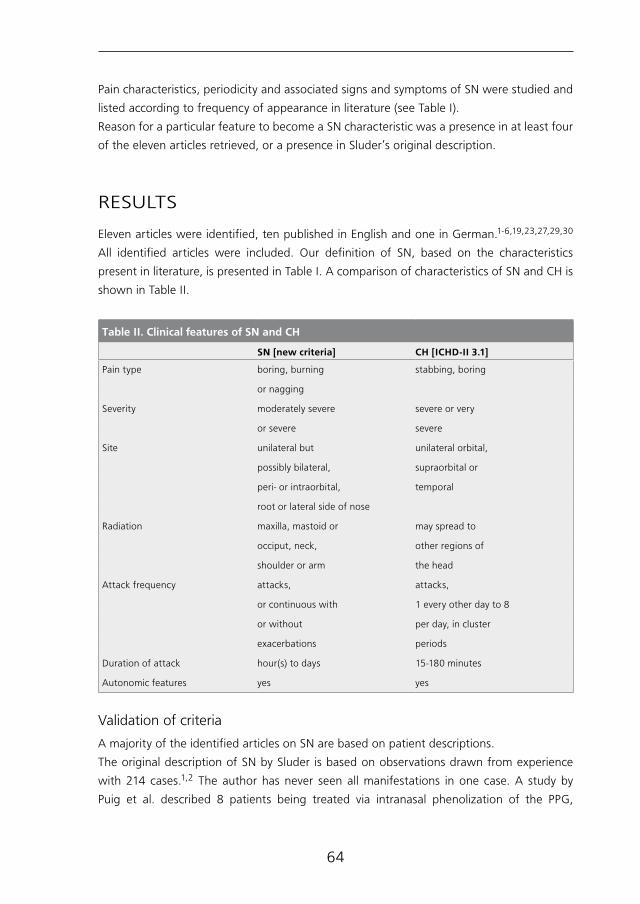
Pain characteristics, periodicity and associated signs and symptoms of SN were studied and
listed according to frequency of appearance in literature (see Table I).
Reason for a particular feature to become a SN characteristic was a presence in at least four
of the eleven articles retrieved, or a presence in Sluder’s original description.
RESULTS
Eleven articles were identified, ten published in English and one in German.1-6,19,23,27,29,30
All identified articles were included. Our definition of SN, based on the characteristics
present in literature, is presented in Table I. A comparison of characteristics of SN and CH is
shown in Table II.
Table II. Clinical features of SN and CH
SN [new criteria] CH [ICHD-II 3.1]
Pain type boring, burning stabbing, boring
or nagging
Severity moderately severe severe or very
or severe severe
Site unilateral but unilateral orbital,
possibly bilateral, supraorbital or
peri- or intraorbital, temporal
root or lateral side of nose
Radiation maxilla, mastoid or may spread to
occiput, neck, other regions of
shoulder or arm the head
Attack frequency attacks, attacks,
or continuous with 1 every other day to 8
or without per day, in cluster
exacerbations periods
Duration of attack hour(s) to days 15-180 minutes
Autonomic features yes yes
Validation of criteria
A majority of the identified articles on SN are based on patient descriptions.
The original description of SN by Sluder is based on observations drawn from experience
with 214 cases.1,2 The author has never seen all manifestations in one case. A study by
Puig et al. described 8 patients being treated via intranasal phenolization of the PPG,
64

and summarizes their presenting signs and symptoms.3 This summary is limited to pain
localization and associated signs and symptoms, but in these categories, 5 patients fit our
newly defined criteria for SN.
Pollock et al. described a case report of one patient who fits all of our criteria for SN.
Akhtar Kamal studied 56 patients diagnosed with SN, without presenting details on their
symptoms.6 However, 52 patients experienced pain relief upon administration of a local
anaethetic agent at the PPG, thereby meeting one of our criteria for SN.
Ryan and Facer described the clinical picture of SN on the basis of a series of 20 patients
from the Mayo Clinic, fitting all of our criteria for SN. 23
Although their patients experienced mostly unilateral pain, occasionally the pain was
bilateral.
Salar et al. studied 7 patients, all experienced pain relief upon cocainisation or infiltration
anaesthesia of the PPG, thereby meeting one of our criteria for SN. 29
Thus, in the eleven identified articles on SN, a total of 306 patients were described; 235
(77%) fit all current criteria for SN, 5 (2%) fit most criteria and 59 (19%) fit one of the
criteria for SN and of seven patients no details were provided. Of the 5 patients who fit
most criteria for SN, three patients fit one or more criteria for CH ( i.e. rhinorrhoea, epiphora
and/or retro-orbital pain).
DISCUSSION
Criteria for SN remain vague and uncertain and vary widely in literature, which makes
diagnosis difficult. There seems to be no such thing as a standard clinical picture for SN. A
study by Ahamed and Jones 19 focuses on analysing the features of SN, and compares them
to CH, classic migraine and Cluster-tic syndrome. Having done this, however, they conclude
that at present, the “best fit” for patients with SN symptoms is CH. They also conclude that
the term Sluder should be discarded as there are serious flaws in its original description.
Ryan and Facer 23 mention a number of differences between SN and CH, but also suggest
that possibly SN is merely a variant of CH.
Our systematic review shows that there does seem to be a common clinical picture for SN
according to the matching features in several of its descriptions.
In order to appreciate the results of this review, its limitations should be discussed. A
limitation of this study is the fact that original literature on SN is scarce. Criteria for SN thus
could only be based on characteristics noted in as few as eleven articles.
SN and CH are often regarded as being part of the same clinical entity. But although the
PPG seems to play a crucial role in the etiology of both these syndromes, literature shows
several crucial differences between the two. We suggest that SN and CH could be two
different headache syndromes, and thus, that SN could be an independent clinical entity.
However, just like CH, paroxysmal hemicrania, and short-lasting unilateral neuralgiform
65
Sluder’sneuralgia;atrigeminalautonomiccephalagia
5Chapter

headache attacks with conjunctival injection and tearing, SN could be a trigeminal
autonomic cephalalgia (TAC). The TAC’s are characterized by short-lasting headache with
autonomic features.30, 31 CH is currently known as the TAC with the longest duration, but
SN could possibly be a TAC with a longer attack duration than CH.
Since SN is a rare, but incapacitating pain syndrome, and no intervention has as yet proven
effective in terms of permanent pain relief, it is our opinion that correct classification is
highly important in order to study new treatment options.
66

REFERENCES
1.SluderG.Roleofthesphenopalatine(Meckel’s)ganglioninnasalheadaches.NYStateJMed1908;87:989-990.
2.Sluder G. Etiology, diagnosis prognosis and treatment of sphenopalatine ganglionneuralgia.JAmMedAssoc1913;16:1202-1206.
3. PuigCM,DriscollCLW,KernEB.Sluder’sSphenopalatineGanglionNeuralgia:Treatmentwith88%Phenol.Am.J.Rhinol1998;12:113-118.
4.Pollock BE, Kondziolka MD. Stereotactic radiosurgical treatment of sphenopalatineneuralgia.JNeurosurg1997;87:450-453.
5.Bruyn GW. Sphenopalatine Neuralgia (Sluder). Handbook of clinical neurology, vol.4.Headache. F. Clifford Rose, editor. Elsevier Science Publishers BV. Vol 4(48), 1986,475-482.
6.Akhtar Kamal S. Experiencewith the xylocaine test as a prognostic aid for surgery inSluder’sneuralgia.JLaryngolOtol1995;109:193-195.
7. Giacomini PG, Alessandrini M, DePadova A. Septoturbinal surgery in contact pointheadachesyndrome:long-termresults.Cranio2003;21:130-135.
8.VailHH.Vidianneuralgia.AnnOtolRhinLaryng1932;41:837.
9.VailHH.PathwaysofreflexpaininVidianneuralgia.ArchOtolaryngol1935;21:277-284.
10. SjaastadO.ClusterHeadacheSyndrome.London,England:WBSaundersCo;1992:100-102.
11. TheInternationalClassificationofHeadacheDisorders,2ndedition,Cephalalgia2004;24:1-160.
12. EkbomK,HardeboJE.ClusterHeadache:aetiology,diagnosisandmanagement.Drugs2002;62:61-69.
13.RussellMB.Epidemiologyandgeneticsofclusterheadache.LancetNeurol2004;3:279-283.Review.
14.EdvinssonL.Pathophysiologyofprimaryheadaches.CurrentPainandHeadacheReports2001;5:71-78.
15.Edvinsson L. Aspects on the pathophysiology of migraine and cluster headache.PharmacolToxicol2001;89:65-73.Review.
16. Goadsby PJ. Pathophysiology of cluster headache: a trigeminal autonomic cephalgia.LancetNeurol2002;1:251-257.
17. GoadsbyPJ.Newaspectsofthepathophysiologyofmigraineandclusterheadache.In:MaxM,ed.Pain1999-anupdatedreview.Seattle,WA:IASPPress;1999:181-191.
18. May A, Goadsby PJ. The Trigeminovascular System in Humans: PathophysiologicImplicationsforPrimaryHeadacheSyndromesoftheNeuralInfluencesontheCerebralCirculation.JournalofCerebralBloodFlowandMetabolism1999;19:115-127.
19. AhamedSH,JonesNS.WhatisSluder’sneuralgia?JLaryngolOtol2003;117:437
20. HardeboJE,ElnerA.NervesandvesselsinthepterygopalatinefossaandsymptomsofClusterheadache.Headache1987;27:528-532.
21.BarreF.CocaineasanabortiveagentinClusterheadache.Headache1982;22:69-73.
22.KittrelleJP,GrouseDS,SeyboldME.Clusterheadache.Localanestheticabortiveagents.ArchNeurol1985;42:496-498.
67
Sluder’sneuralgia;atrigeminalautonomiccephalagia
5Chapter

23. Ryan RE Jr, Facer GW. Sphenopalatine ganglion neuralgia and cluster headache:comparisons,contrasts,andtreatment.Headache1977;17:7-8
24. Dodick DW, Rozen TD, Goadsby PJ, Silberstein SD. Cluster headache. Cephalalgia2000;20:787.
25.GrahamJR.Clusterheadache.Headache1972;11:175.
26.LoderE.NaratriptanintheprophylaxisofClusterheadache.Headache2002;42:56.
27. Kaeser HE. Die sogenannt atypischen Gesichtsneuralgien. Schweiz. RundschauMed.(Praxis)1989;78:1018-1022.
28. AubryM,PialouxP.Sluder’ssyndrome.In:VinkenPJ,BruynGWeditors.Headachesandcranial neuralgias, Handbook of Clinical Neurology, Vol. 5. Amsterdam: North-HollandPubl.Company,1968;326-332.
29. SalarG,OriC,IobI,FioreD.Percutaneousthermocoagulationforsphenopalatineganglionneuralgia.ActaNeurochir1987;84:24-28.
30.CepheroR,MillerRH,BresslerK.LongtermresultsofSphenopalatineGanglioneurectomyforFacialPain.AmJOtolaryngol1987;3:171-174.
31.Matharu MS, Goadsby PJ. Trigeminal autonomic cephalalgias: diagnosis andManagement. In: Silberstein SD, Lipton RB, Dodick DW (eds).Wolff’s headache andOtherheadpain,8thedn.OxfordUniversityPress,Oxford,2008;379-430.
68

6C h a p t e r
Effects of radiofrequency thermocoagulation of the sphenopalatine ganglion on facial pain; correlation with diagnosis
K. P. Q.Oomen, A. J. M. van Wijck, G. J. Hordijk, J. A. de Ru
Accepted for publication J Orofac Pain, 2011
ABSTRACT
Aim: To study the effect of radiofrequency thermocoagulation of the sphenopalatine
ganglion on facial pain following critical evaluation of diagnosis.
Methods: Retrospective study of clinical records of all patients who underwent
radiofrequency thermocoagulation of the sphenopalatine ganglion at a tertiary pain
clinic for four consecutive years; diagnoses were re-evaluated after which the effect of
radiofrequent thermocoagulation on facial pain was assessed.
Results: After application of new criteria for Sluder’s neuralgia and strict criteria for Cluster
headache, seven patients out of fifteen turned out to have been diagnosed correctly. Nine out
of fifteen patients showed considerable pain relief after radiofrequency thermocoagulation
of the sphenopalatine ganglion. Positive results were most frequent among patients
with Sluder’s neuropathy, atypical facial pain and Cluster headache. However, repeated
procedures were needed in most patients.
Conclusion: Correct facial pain diagnosis is vital to assess the outcome of different treatment
strategies. Even in a tertiary centre, facial pain can be misdiagnosed. Radiofrequency
thermocoagulation of the sphenopalatine ganglion can be effective in patients with facial
pain, but repeated procedures are often needed.
70

INTRODUCTION
Headache and facial pain are an international public health problem with a worldwide lifetime
prevalence greater than 90 %.1 Facial pain is an incapacitating disorder for which surgical
treatment can be required. In a wide range of patients with facial pain, radiofrequency
thermocoagulation (RFT) of the sphenopalatine ganglion (SPG) is performed.
The SPG is involved in several facial pain syndromes such as Cluster headache (CH), Sluder’s
neuralgia (SN) and atypical facial pain. 2-10
Previous studies have shown that in management headache syndromes such as CH and SN,
RFT might be effective, with success rates ranging from 61 to 65%. 11-14 A study by Salar et
al. showed that percutaneous RFT is effective in relieving pain in patients with SN without
significant side-effects.15 However, a slight troublesome sensation persisted in all treated
patients. Pain relief through RFT is often only temporary, and repeated RFT procedures can
be needed to establish long lasting pain relief.
For assessment of treatment effect, correct classification of facial pain is important.
Criteria for CH are clear and well-defined (Table 1), but patients can be wrongly diagnosed
because of inappropriate handling of these criteria. 16
New criteria for SN were recently defined and might be helpful in discriminating between
SN and CH (Table 2). A new term was introduced for symptoms of SN developing in relation
to trauma in the area innervated by the secondary division of the trigeminal nerve: Sluder’s
neuropathy (SNPT). 17
Aim of the present study is twofold:
The first aim is to retrospectively assass the accuracy if facial pain diagnosis and to requalify
diagnosis through strict application of the existing and new facial pain criteria.
The second aim is to assess the effect of RFT of the SPG in facial pain patients.
Table 1. Characteristics of CH according to the IHS-classification of headache disorders. (ICHD-II 3.1)
Characteristics Cluster headache
Pain intensity Severe or very severe.
Site Unilateral orbital, supraorbital and/or temporal.
Frequency At least five attacks. Attacks last 15-180 minutes if untreated, and have a frequency of 1 every other day to 8 a day.
Associated signs and symptoms At least one of the following:
1. ipsilateral conjunctival injection and/or lacrimation
2. ipsilateral nasal congestion and/or rhinorrhoea
3. ipsilateral eyelid edema
4. ipsilateral forehead and facial sweating
5. ipsilateral miosis and/or ptosis
6. a sense of restlessness or agitation
71
Radiofrequency thermocoagulation of the PPG
6Chapter

METHODS
Study design
Clinical records of all patients (n=15) who underwent RFT of the SPG at the Pain Clinic of
the Universal Medical Center Utrecht, The Netherlands during four consecutive years, were
retrospectively studied. Previous diagnosis had been established either by a neurologist
prior to visiting the Pain Clinic, or by the anesthesiologist treating the patient.
Patient files were retrospectively studied for headache characteristics, which were
interpreted according to the International Classification of Headache Disorders (ICHD-II)
and new criteria for SN. 17 Two doctors, not involved in the treated patients, studied the
patient files simultaneously and agreed on all diagnoses.
Effectiveness of RFT was retrospectively assessed based on patient record information,
following re-evaluation of diagnoses. Pain scores were assessed on a Visual Analogue Scale
(VAS), and compared pre- and postoperatively.
Table 2. Characteristics of SN according to newly defined criteria.
Characteristics Sluder’s neuralgia
Pain quality and intensity Moderately severe or severe, boring, burning or nagging pain.
Site Unilateral but possibly bilateral, located peri- or intraorbitally, or at the root or lateral side of the nose, radiating to at least one of the following:
1. maxillary region or cheek and/or associated teeth
2. mastoidal and/or occipital area
3. neck, shoulder or arm
Frequency One of the following:
1. episodic with attacks lasting hour(s) to days
2. continuous for several weeks with or without exacerbations
Associated signs and symptoms At least one of the following:
1. ipsilateral lacrimation and/or conjunctival injection
2. ipsilateral nasal congestion and/or rhinorrhoea
3.hyp- or hyperesthesia in maxillary distribution of trigeminal nerve
or
4. ipsilateral sore throat
5. ipsilateral delayed taste perception or parageusia
6. ipsilateral elevated palatine arch or contralaterally deflected uvula
Treatment Pain can be blocked by cocainisation or infiltration anaesthesia of the SPG
Characteristics Sluder’s Neuropathy (Secondary Sluder Syndrome)
As above, except headache develops in relation to trauma in the area innervated by the second division of the trigeminal nerve
72
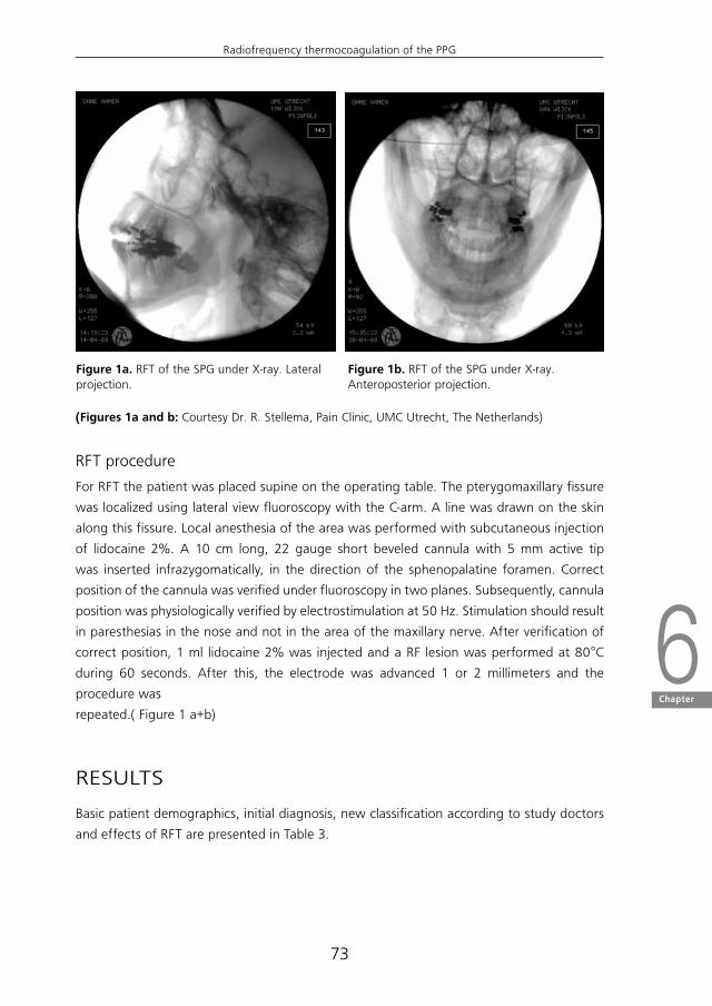
RFT procedure
For RFT the patient was placed supine on the operating table. The pterygomaxillary fissure
was localized using lateral view fluoroscopy with the C-arm. A line was drawn on the skin
along this fissure. Local anesthesia of the area was performed with subcutaneous injection
of lidocaine 2%. A 10 cm long, 22 gauge short beveled cannula with 5 mm active tip
was inserted infrazygomatically, in the direction of the sphenopalatine foramen. Correct
position of the cannula was verified under fluoroscopy in two planes. Subsequently, cannula
position was physiologically verified by electrostimulation at 50 Hz. Stimulation should result
in paresthesias in the nose and not in the area of the maxillary nerve. After verification of
correct position, 1 ml lidocaine 2% was injected and a RF lesion was performed at 80°C
during 60 seconds. After this, the electrode was advanced 1 or 2 millimeters and the
procedure was
repeated.( Figure 1 a+b)
RESULTS
Basic patient demographics, initial diagnosis, new classification according to study doctors
and effects of RFT are presented in Table 3.
Figure 1a. RFT of the SPG under X-ray. Lateral projection.
Figure 1b. RFT of the SPG under X-ray. Anteroposterior projection.
(Figures 1a and b: Courtesy Dr. R. Stellema, Pain Clinic, UMC Utrecht, The Netherlands)
73
Radiofrequency thermocoagulation of the PPG
6Chapter

Retrospective requalification of facial pain diagnosis
Of the fifteen patients, ten had previously been diagnosed with atypical facial pain, three
were diagnosed with CH, one had an unsure diagnosis of either trigeminal neuralgia or CH,
and one was diagnosed with postherpetic neuralgia.
These figures change after strict application of the IHS criteria for CH and new criteria for
SN.
After re-evaluation, two patients, both initially diagnosed with atypical facial pain, could
be diagnosed with SN according to the new SN criteria. Three patients, one initially
diagnosed with CH and two diagnosed with atypical facial pain, could be diagnosed with
SNPT. One patient, previously diagnosed with atypical facial pain, could be diagnosed with
CH according to the IHS criteria. Six patients, four initially diagnosed with atypical facial
pain and two with CH, kept their diagnosis after re-evaluation. The patient with an unsure
diagnosis of either trigeminal neuralgia or CH could be classified as having Short-lasting
Unilateral Neuralgiform headache attacks with Conjunctival injection and Tearing (SUNCT)
(ICHD-II). One patient, initially diagnosed with atypical facial pain, was diagnosed with
Table 3. Effect of RF on studied patients
Patient (Sex)
Age Previous diagnosis Diagnosis after re-evaluation
Number of procedures
Pain reduction
1. (F) 63 atypical facial pain atypical facial pain 1 almost complete
2. (F) 41 atypical facial pain posttraumatic neuropathy of infraorbital nerve
1 adequate
3. (M) 78 postherpetic neuralgia postherpetic neuralgia 1 none
4. (F) 67 CH SNPT 2 almost complete
5. (M) 61 CH CH 4R 1L complete
6. (F) 63 atypical facial pain CH complete 1 almost
7. (M) 59 atypical facial pain atypical facial pain oftraumatic(iatrogenic orpostinfectious) origin
1 none
8. (F) 67 atypical facial pain SNPT 4 complete
9. (M) 64 CH or trigeminalneuralgia
SUNCT 1 none
10. (M) 68 atypical facial pain atypical facial painsecondary to infection
2 adequate
11. (F) 33 atypical facial pain atypical facial pain 1 almost complete
12. (M) 62 atypical facial pain SN complete 1 almost ≤ 3 wks
13. (F) 39 atypical facial pain SN 1 none
14. (M) 40 CH CH 1 none
15. (F) 60 atypical facial pain SNPT 4 complete
R=right L=left
74
posttraumatic neuropathy of the infraorbital nerve. The patient with postherpetic neuralgia
kept this diagnosis.
After strict application of the IHS criteria for CH (ICHD-II) and new SN criteria, only seven out
of the total group of fifteen patients (47% (95%CI 22.3-72.6)) kept their initial diagnosis.
Out of the subgroup of ten patients initially diagnosed with atypical facial pain, four patients
(40% (95%CI 13.7-72.6)) had been diagnosed correctly. Of the three patients diagnosed
with CH, one patient (33% (95%CI 1.8-87.5)) had been diagnosed correctly. Thus, in our
study group, the two most frequent diagnoses, atypical facial pain and CH, were also most
frequently incorrect.
Outcome after RFT
Of the total group of fifteen patients, nine patients (60% (95%CI 32.9-82.5)) showed
considerable pain relief (≥90%) after single or repeated RFT procedures. Six patients (40%
(95%CI 17.5-67.1)) experienced no pain relief or temporary pain relief (≤ 3 weeks).
A positive effect of RFT was shown in all three patients (100% (95%CI 31-96.8%)) diagnosed
with SNPT according to the new criteria. However, all patients needed repeated (2 , 4
and 4, respectively) procedures for lasting effect. A positive effect of RFT was shown in
three out of four patients (75% (95%CI 21.9-98.7)) diagnosed with atypical facial pain; one
patient needed two procedures. A positive effect of RFT was shown in two out of three
(67% (95%CI 12.5-98.2)) patients with CH; one patient needed repeated (4 right-sided, 1
left-sided) procedures. A positive effect of RFT was shown in the one patient diagnosed
with posttraumatic neuropathy of the infraorbital nerve without a need for repeated
procedures. None of the patients with SN demonstrated a positive effect of RFT. One SN
patient experienced short-term pain relief after RFT (duration of pain relief ≤ 3 weeks), the
other patient experienced no pain relief at all. The patient diagnosed with SUNCT and the
patient with postherpetic neuralgia showed no pain relief after RFT.
No side effects of RFT were recorded.
Effects of RFT on cranial autonomic features were only noted in 5 patients. In CH patients
5 and 6, and SN patient 12, a complete recovery of all cranial autonomic symtoms
(lacrimtation and conjunctival injection) was reported at 1 year follow-up. In CH patient 14
and SN patient 13, lacrimation and nasal congestion were described, respectively. RFT did
not show any effect on these symptoms in both patients.
Part of our study aim was to assess for which subgroup of headache diagnoses RFT seemed
most effective. However, re-evaluation of diagnosis made these subgroups change in size
and composition. The subgroups were compared for their response to RFT before and after
reassessment.
Success rates of RFT before and after re-evaluation of diagnosis were compared for the two
most frequent diagnoses, atypical facial pain and CH ( Table 3).
75
Radiofrequency thermocoagulation of the PPG
6Chapter
At previous diagnosis, the subgroup of patients with atypical facial pain showed a success
rate of 70% (seven out of ten). After re-evaluation of diagnosis, this subgroup consisted of
4 patients, and showed a success rate of 75% (three out of four).
The subgroup of patients with CH at previous diagnosis showed a success rate of 50% (two
out of four, if patient 9 is considered as a CH patient). After re-evaluation of diagnosis, success
rate of RFT had gone up to 67% (two out of three). During the follow-up period, patients
visited the clinic approximately 3-monthly. The follow-up duration differed between patients;
most patients were followed- up during 1 year ( patients 1, 5-8, 11-15), some patients were
followed-up during a shorter period of 9 months (patient 2, 9) and 6 months (3, 10).
DISCUSSION
In this population of facial pain patients in a tertiary care centre, facial pain was frequently
misdiagnosed. In our study population, RFT seemed effective in patients with atypical facial
pain, CH and SNPT, but did not seem effective in patients with SN.
To appreciate the results of this study, some of its limitations should be discussed.
First of all, this study concerns a group of patients selected for RFT and treated in a tertiary
care centre. Therefore, our patient domain does not seem representative for headache
patients in the general population.
A second limitation of this study is the small number of patients, which leads to larger
confidence intervals. Furthermore, the small number of patients makes it difficult to
draw conclusions on RFT response. However, most of the results in this small study group
correspond to those in previous studies. 11-14
Third, in this study population, follow-up periods were relatively short, with a main follow-
up period of one year. Furthermore, effects on cranial autonomic symptoms were scarcely
noted.
Fourth, we have to take in to account that at initial diagnosis, the recently developed criteria
for SN did not yet exist, which explains why none of the patients were initially diagnosed
with SN. However, when applying the IHS criteria in a correct manner, 2 patients were still
diagnosed incorrectly.
Previous studies have shown a need for repeated RFT procedures in headache patients.11-15
In a study of 7 SN patients who underwent RFT of the SPG, all patients experienced
almost complete pain reduction after several days. 15 Patients were followed 6 to 34
months postoperatively. Three patients needed repeated (two or three) procedures. A
slight troublesome but not painful sensation persisted in all cases. Characteristics of these
patients correspond with those in our study group, i.e. mean age, duration of symptoms
and previous treatment. However, both SN patients in our study did not experience pain
relief after RFT.
76
Classification of headache influences success rate of treatment. Correlation of diagnosis
and treatment effect also applies to the results in this study and influences its outcome.
For example, diagnosis of patient 14 of our study group was difficult. There was discussion
on whether diagnosis SN or CH would be more appropriate. Finally the authors agreed on
diagnosis CH because of severity of pain and the particular sense of restlessness. Patient
14 was the only CH patient who did not demonstrate a positive effect of RFT, leading to a
success rate of 67% in the CH group. Had patient 14 been diagnosed with SN, success rate
in the CH group would have been 100%. This example illustrates the importance of correct
diagnosis in evaluating the effect of surgical therapies for headache patients.
Diagnosing headache patients such as in our group can be difficult for various reasons.
Different forms of facial pain can seem to represent ends of one spectrum. 18 For instance,
trigeminal autonomic cephalalgia’s (TAC’s) such as CH and SUNCT are clinically very similar,
which suggests a considerable shared pathophysiology. 19 Furthermore, CH and SN are
often regarded as parts of the same clinical entity, 20 and it has been suggested that SN
could even be a TAC with a longer attack duration than CH. 17 Other diagnostic challenges
stem from the fact that some facial pain patients do not meet all criteria for a specific
syndrome. However, a recent study showed that patients who fail one of the criteria, still
can be diagnosed with CH. 16 The ICHD-II has a separate classification for attacks fulfilling
all but one of the specific criteria for CH, called probable CH (ICHD- II 3.4.1).
As diagnosis is difficult, patients are often at risk being classified as having atypical facial
pain. However, this can only be done if they do not meet the criteria for CH, SN or other
facial pain syndromes. Facial pain syndromes can be incapacitating and correct classification
is important for choice of treatment strategies.
Conclusion
On the basis of the present study findings, we conclude that facial pain patients can
frequently be misdiagnosed, even in a tertiary care centre. In our study population, effects
of RFT of the SPG varied with pain diagnosis; RFT seemed effective in patients with atypical
facial pain, CH and SNPT, but did not seem effective in patients with SN.
77
Radiofrequency thermocoagulation of the PPG
6Chapter

REFERENCES
1.Rasmussen K, Jensen R, SchrodM, Olesen J. Epidemiology of headache in a generalpopulation:aprevalencestudy.JClinEpidemiol1991;44:1147-1157.
2. EkbomK,Hardebo JE. ClusterHeadache: aetiology, diagnosis andmanagement.Drugs2002;62:61-69.
3. Edvinsson L. Pathophysiology of primary headaches. Curr Pain Headache Rep 2001;5:71-78.
4. GoadsbyPJ.NewAspectsof thePathofysiologyofMigraineandClusterHeadache.Pain1999;181-191.
5.AridonP,D’AndreaG,RigamontiA, LeoneM,CasariG,BussoneG. Elusive amines andclusterheadache:mutationalanalysisoftraceaminereceptorclusterinchromosome6q23.NeurolSci2004;25:279-280.
6. RussellMB,Epidemiologyandgeneticsofclusterheadache.LancetNeurol2004;3:279-283.
7. PollockBE,KondziolkaMD.Stereotacticradiosurgicaltreatmentofsphenopalatineneuralgia.JNeurosurg1997;87:450-453.
8.DalessioDJ.Reliefofclusterheadacheandcranialneuralgias.Promisingprophylacticandsymptomatictreatments.PostgradMed2001;109:69-72.
9. Rozen TD. Anti-epileptic drugs in the management of cluster headache and trigeminalneuralgia.Headache2001;41:25-32.
10. PuigCM,DriscollCLW,KernEB.Sluder’sSphenopalatineGanglionNeuralgia: Treatmentwith88%Phenol.AmJRhinol1998;12:113-118.
11. SandersM,ZuurmondWW.Efficacyof sphenopalatineganglionblockage in66patientssuffering from cluster headache: a 1- to 70- months follow-up evaluation. J Neurosurg1997;87:876-880.
12. Stolker RJ, Kamphuis E, Rohof O, Faydherbe L, Vervest A. Die behandlung vonClusterkopfschmerzenmitradiofrequentenLaesionen.DerSchmerz1991;5:194.
13. Vervest ACM, Stolker RJ, Groen GJ, Kamphuis ET, Rohof OJJM. Clusterkopfschmerz:ThermolaesiondesGanglionsphenopalatinum.DerSchmerz1992;6:9
14. SluyterME,VercruyssePJ,SterkW.Radiofrequencylesionsofthesphenopalatineganglioninthetreatmentofclusterheadache.Schmerz1988;9:56-59.
15. SalarG,OriC,IobI,FioreD,Percutaneousthermocoagulationforsphenopalatineganglionneuralgia.ActaNeurochir1987;84:24-28.
16. vanVlietJA,EekersPJ,HaanJ,FerrariMD;DutchRUSSHStudyGroup.EvaluatingtheIHScriteriaforclusterheadache-acomparisonbetweenpatientsmeetingallcriteriaandpatientsfailingonecriteria.Cephalalgia2006;26:241-245.
17. Oomen KPQ, van Wijck AJM, Hordijk GJ, de Ru JA. Sluder’s neuralgia; a TrigeminalAutonomicCephalalgia?Cephalalgia2010;30:360-364.
18. Ruiz-LopezR,ErdineS.Treatmentofcranio-facialpainwithradiofrequencyprocedures.PainPractice2002;3:206-213.
19. GoadsbyPJ;LiptonRB.Areviewofparoxysmalhemicranias,SUNCTsyndromeandothershort-lasting headaches with autonomic feature, including new cases. Brain 1997;120:193-209.
20. AhamedSH,JonesNS.WhatisSluder’sneuralgia?JLaryngolOtol2003;117:437
78
7C h a p t e r
Microvascular decompression of the pterygopalatine ganglion in patients with refractory cluster headache
Oomen, KPQ (1), van Wijck, AJM (2), Hordijk GJ (1), de Ru, JA (3)
Submitted to Cephalalgia

ABSTRACT
Background: Cluster headache is an invalidating form of headache. Although Cluster
headache can be managed pharmacologically, a substantial number of patients require
surgical treatment with varying results. Microvascular decompression of the pterygopalatine
ganglion could be an alternative to traditional surgical management in patients with Cluster
headache.
Methods: Microvascular decompression of the pterygopalatine ganglion was performed in
three patients with refractory Cluster headache. The pterygopalatine artery was ligated and
a temporal muscle graft was placed between the artery and the ganglion.
Results: No differences were found between the presurgical period and 1 week, 1 month,
3 and 6 months postoperatively with respect to attack duration and frequency, visual
analogue scale score during attacks and in remission periods, duration of remissions, and
quality of life.
Conclusion: These preliminary data suggest that microvascular decompression of the
pterygopalatine ganglion does not provide pain reduction or improvement of quality of life
in patients with refractory Cluster headache.
80

INTRODUCTION
Cluster headache (CH) is a disabling form of headache, characterized by episodic attacks
of unilateral pain in association with ipsilateral cranial autonomic features. 1 The specific
cause of CH remains unknown. Besides a dysfunction in the central nervous system 2, the
pterygopalatine ganglion (PPG) plays an important role in its pathophysiology. 3-7 The PPG is
the largest of the four cranial parasympathetic (PS) ganglia, and lies in the pterygopalatine
fossa (PPF). From the PPG, PS, sympathetic (S) and sensory fibres are distributed to their
target organs in the orbit, nasal and oral cavity, providing an anatomic substrate for
symptoms in CH.
Although CH can be managed pharmacologically, some patients require surgical treatment
because of intractable symptoms or adverse effects of medication. CH can be treated
successfully with surgical interventions directed to the PPG. Previous studies have shown
a modest effect of radiofrequency thermocoagulation (RFT) of the PPG with success rates
ranging from 61 to 65%, but pain relief is often temporary and repeated procedures are
required. 8-11 Clearly, there is a need for alternative surgical procedures in management of
CH, providing longer lasting pain relief.
Examination of the anatomic relations of the PPG reveals that it lies in close contact with
a remarkably tortuous portion of the pterygopalatine artery along its course in the PPF,
suggesting vascular compression of the PPG as a causative factor in CH. 12-13 Vascular
compression is associated with other disorders such as trigeminal neuralgia, hemifacial
spasm, and glossopharyngeal neuralgia. 14-15 Such vascular compression syndromes can be
treated successfully with microvascular decompression (MVD), i.e. discontinuation of contact
between the vascular loop and neural structure of interest. 16-22 Possibly, discontinuation
of contact between the pterygopalatine artery and the PPG could provide pain relief in
patients with CH. Here, we report MVD of the PPG in three patients with refractory CH.
METHODS
Patients
We conducted a pilot study between October 2008 and May 2010. Patients from the
department of Otolaryngology of the University Medical Centre Utrecht, the Netherlands,
suffering from chronic refractory CH, were eligible. Initially, we planned to enrol a total of
six patients.
Patients with pain attributable to any other diagnosis were excluded. Other exclusion
criteria were major anatomical variations in the area of interest, disease of the maxillary
sinus, previous surgery in the area of interest, and contraindications for MRI such as allergy
to contrast, pregnancy, and renal disease.
81
Microvascular decompression of the PPG in CH patients
7Chapter
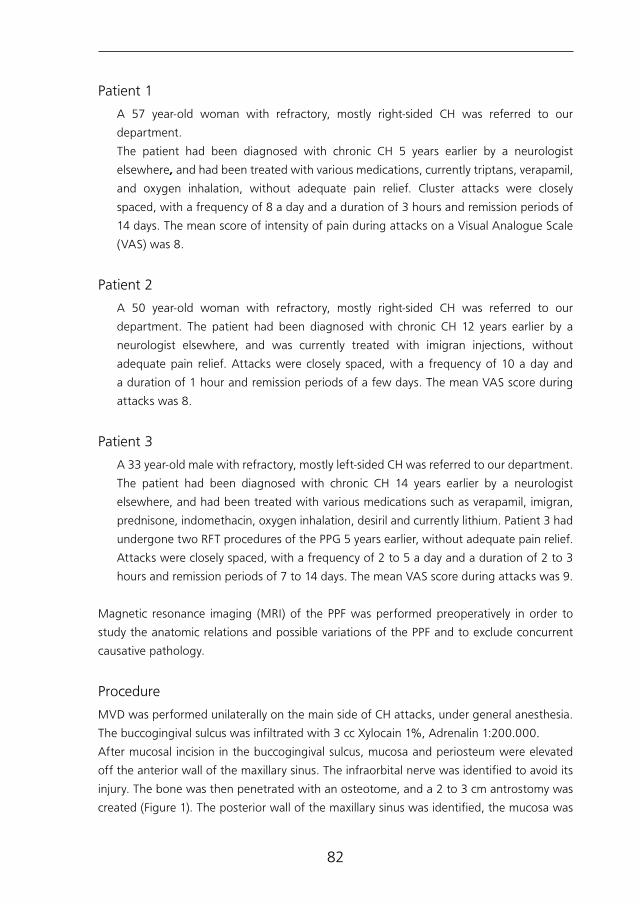
Patient 1
A 57 year-old woman with refractory, mostly right-sided CH was referred to our
department.
The patient had been diagnosed with chronic CH 5 years earlier by a neurologist
elsewhere, and had been treated with various medications, currently triptans, verapamil,
and oxygen inhalation, without adequate pain relief. Cluster attacks were closely
spaced, with a frequency of 8 a day and a duration of 3 hours and remission periods of
14 days. The mean score of intensity of pain during attacks on a Visual Analogue Scale
(VAS) was 8.
Patient 2
A 50 year-old woman with refractory, mostly right-sided CH was referred to our
department. The patient had been diagnosed with chronic CH 12 years earlier by a
neurologist elsewhere, and was currently treated with imigran injections, without
adequate pain relief. Attacks were closely spaced, with a frequency of 10 a day and
a duration of 1 hour and remission periods of a few days. The mean VAS score during
attacks was 8.
Patient 3
A 33 year-old male with refractory, mostly left-sided CH was referred to our department.
The patient had been diagnosed with chronic CH 14 years earlier by a neurologist
elsewhere, and had been treated with various medications such as verapamil, imigran,
prednisone, indomethacin, oxygen inhalation, desiril and currently lithium. Patient 3 had
undergone two RFT procedures of the PPG 5 years earlier, without adequate pain relief.
Attacks were closely spaced, with a frequency of 2 to 5 a day and a duration of 2 to 3
hours and remission periods of 7 to 14 days. The mean VAS score during attacks was 9.
Magnetic resonance imaging (MRI) of the PPF was performed preoperatively in order to
study the anatomic relations and possible variations of the PPF and to exclude concurrent
causative pathology.
Procedure
MVD was performed unilaterally on the main side of CH attacks, under general anesthesia.
The buccogingival sulcus was infiltrated with 3 cc Xylocain 1%, Adrenalin 1:200.000.
After mucosal incision in the buccogingival sulcus, mucosa and periosteum were elevated
off the anterior wall of the maxillary sinus. The infraorbital nerve was identified to avoid its
injury. The bone was then penetrated with an osteotome, and a 2 to 3 cm antrostomy was
created (Figure 1). The posterior wall of the maxillary sinus was identified, the mucosa was
82
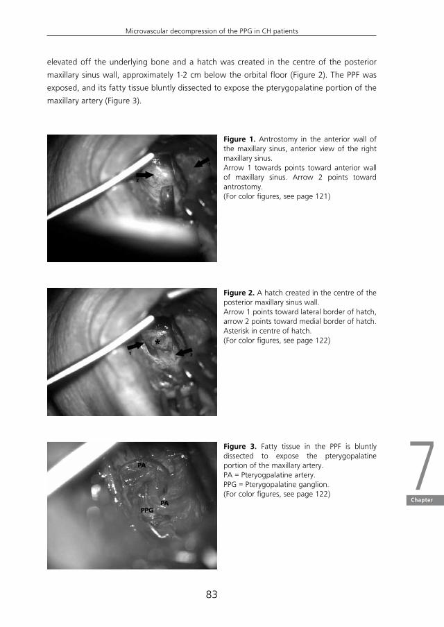
elevated off the underlying bone and a hatch was created in the centre of the posterior
maxillary sinus wall, approximately 1-2 cm below the orbital floor (Figure 2). The PPF was
exposed, and its fatty tissue bluntly dissected to expose the pterygopalatine portion of the
maxillary artery (Figure 3).
Figure 1. Antrostomy in the anterior wall of the maxillary sinus, anterior view of the right maxillary sinus.Arrow 1 towards points toward anterior wall of maxillary sinus. Arrow 2 points toward antrostomy.(For color figures, see page 121)
Figure 2. A hatch created in the centre of the posterior maxillary sinus wall.Arrow 1 points toward lateral border of hatch, arrow 2 points toward medial border of hatch.Asterisk in centre of hatch.(For color figures, see page 122)
Figure 3. Fatty tissue in the PPF is bluntly dissected to expose the pterygopalatine portion of the maxillary artery.PA = Pteryogpalatine artery.PPG = Pterygopalatine ganglion.(For color figures, see page 122)
83
Microvascular decompression of the PPG in CH patients
7Chapter

A small piece of temporal muscle was harvested through a retroauricular incision. The
pterygopalatine artery was ligated with hemoclips (Figure 4). Next, the temporal muscle
graft was placed between the artery and the PPG (Figure 5). The wound was closed with
absorbable sutures.
Figure 4. Ligation of the pterygopalatine artery with hemoclips.Arrows point toward hemoclips.(For color figures, see page 122)
Figure 5. Temporal muscle graft, placed between the ptergygopalatine artery and the PPG.PA = Pterygopalatine artery.TMG = Temporal muscle graft.(For color figures, see page 122)
Outcome
Outcome measures were pain and quality of life. Pain was assessed with respect to intensity,
frequency and duration. At inclusion, patients filled out a disease-specific questionnaire
including information on attack duration, attack frequency, mean VAS score during attacks
and in remission periods, and duration of remissions. From the first postoperative day,
patients kept a diary of pain intensity assessed on a VAS, during attacks and in remission
periods. The presence of attacks and their duration were also noted.
Quality of life was based on a generic health-related quality of life questionnaire: SF-36.
SF-36 was filled out at inclusion and at one week, 3 and 6 months postoperatively. Patients
visited the outpatients department for evaluation at one week, 6 weeks, 3 months and 6
months follow-up.
84

RESULTS
None of the patients showed abnormalities of the PPF on MRI. All patients underwent MVD
of the PPG, patient 1 and 2 underwent right-sided MVD, patient 3 underwent left-sided MVD.
Procedure 1 was complicated by postoperative haemorrhage of the wound bed, treated by
electrocoagulation under general anaesthesia. Procedure 2 and 3 were uncomplicated. All
patients could be discharged from our clinic on the third postoperative day.
Patient 1
No differences were found between the presurgical period and 1 week, 1 month, 3 and
6 months postoperatively with respect to attack duration, attack frequency, mean VAS
score during attacks and in remission periods, and duration of remissions. In the first
month, patient 1 even experienced an increase in attack frequency. Fortunately, the
attacks had decreased to their usual frequency by the third month. Patient 1 reported
a temporary hypesthesia of the infraorbital nerve area, which had resolved at 6 months
postoperatively. Quality of life based on SF-36 domain scores did not change between
the presurgical period and 1 week, 1 month, 3 and 6 months follow up (Figure 6).
Figure 6. SF-36 Domain Score Changes Patient 1. PF= Physical Functioning, RP= Role-Physical, RE= Role-Emotional, VIT= Vitality, MH= Mental Health, SF= Social Functioning, BP= Bodily Pain, GH= General Health (For color figures, see page 123)
85
Microvascular decompression of the PPG in CH patients
7Chapter
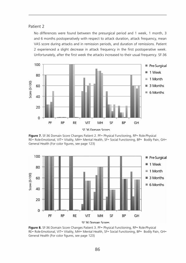
Patient 2
No differences were found between the presurgical period and 1 week, 1 month, 3
and 6 months postoperatively with respect to attack duration, attack frequency, mean
VAS score during attacks and in remission periods, and duration of remissions. Patient
2 experienced a slight decrease in attack frequency in the first postoperative week.
Unfortunately, after the first week the attacks increased to their usual frequency. SF-36
Figure 7. SF-36 Domain Score Changes Patient 2. PF= Physical Functioning, RP= Role-PhysicalRE= Role-Emotional, VIT= Vitality, MH= Mental Health, SF= Social Functioning, BP= Bodily Pain, GH= General Health (For color figures, see page 123)
Figure 8. SF-36 Domain Score Changes Patient 3. PF= Physical Functioning, RP= Role-PhysicalRE= Role-Emotional, VIT= Vitality, MH= Mental Health, SF= Social Functioning, BP= Bodily Pain, GH= General Health (For color figures, see page 123)
86
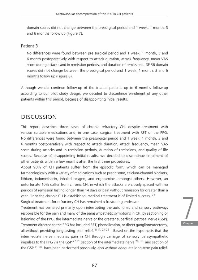
domain scores did not change between the presurgical period and 1 week, 1 month, 3
and 6 months follow up (Figure 7).
Patient 3
No differences were found between pre surgical period and 1 week, 1 month, 3 and
6 month postoperatively with respect to attack duration, attack frequency, mean VAS
score during attacks and in remission periods, and duration of remissions. SF-36 domain
scores did not change between the presurgical period and 1 week, 1 month, 3 and 6
months follow up (Figure 8).
Although we did continue follow-up of the treated patients up to 6 months follow-up
according to our pilot study design, we decided to discontinue enrolment of any other
patients within this period, because of disappointing initial results.
DISCUSSION
This report describes three cases of chronic refractory CH, despite treatment with
various suitable medications and, in one case, surgical treatment with RFT of the PPG.
No differences were found between the presurgical period and 1 week, 1 month, 3 and
6 months postoperatively with respect to attack duration, attack frequency, mean VAS
score during attacks and in remission periods, duration of remissions, and quality of life
scores. Because of disappointing initial results, we decided to discontinue enrolment of
other patients within a few months after the first three procedures.
About 90% of CH patients suffer from the episodic form, which can be managed
farmacologically with a variety of medications such as prednisone, calcium-channel blockers,
lithium, indomethacin, inhaled oxygen, and ergotamine, amongst others. However, an
unfortunate 10% suffer from chronic CH, in which the attacks are closely spaced with no
periods of remission lasting longer than 14 days or pain without remission for greater than a
year. Once the chronic CH is established, medical treatment is of limited success. 23
Surgical treatment for refractory CH has remained a frustrating endeavor.
Treatment has centered primarily upon interrupting the autonomic and sensory pathways
responsible for the pain and many of the parasympathetic symptoms in CH, by sectioning or
lesioning of the PPG, the intermediate nerve or the greater superficial petrosal nerve (GSP).
Treatment directed to the PPG has included RFT, phenolization, or direct ganglioneurectomy,
all without providing long-lasting pain relief. 8-11, 24-26 Based on the hypothesis that the
intermediate nerve mediates pain in CH through carriage of sensory parasympathetic
impulses to the PPG via the GSP 27, 28 section of the intermediate nerve 29, 30 and section of
the GSP 31, 32 have been performed previously, also without adequate long-term pain relief.
87
Microvascular decompression of the PPG in CH patients
7Chapter

As mentioned earlier, MVD has been described in the surgical management of
vascular compression syndromes such as trigeminal neuralgia, hemifacial spasm, and
glossopharyngeal neuralgia.
As MVD of the PPG has not been described in management of CH before, our findings
cannot correspond to those in previous studies. In trigeminal neuralgia, previous studies
have shown that MVD of the trigeminal nerve provides longer lasting pain relief when
compared to RFT of the PPG. 16, 17 Previously, Lovely et al 23 reported successful surgical
management of chronic CH through MVD of the trigeminal nerve, alone or in combination
with section and/or MVD of the intermediate nerve in 28 patients, including two with
bilateral disease. However, although 22 (77.3%) of 30 procedures resulted in an excellent or
good outcome in the immediate postoperative period, the success rate dropped to 46.6%
with long-term follow-up, i.e. after the first postoperative year.
The negative results of our study could be due to technical surgical aspects. Possibly, the
inserted piece of temporal muscle, intended for decompression, gave rise to irritation of the
PPG. More likely, however, our hypothesis of vascular compression of the PPG as a causative
factor in CH, is not as strong as assumed. Probably, other factors such as dysfunction in the
central nervous system are of greater importance in the pathogenesis of CH.
A limitation of our study is that the number of participants is extremely small. However,
due to disappointing initial results, enrolment of more patients did not seem acceptable.
Furthermore, the study was designed as a pilot with a maximum of 6 patients, to be
continued if initial results would be promising.
This report is the first of its kind describing MVD of the PPG in the surgical management
of refractory CH. Unfortunately, our preliminary data suggest that MVD of the PPG does
not provide pain reduction or improvement of quality of life in patients with refractory CH.
88

REFERENCES
1.TheInternationalClassificationofHeadachedisorders,2ndedition,Cephalalgia2004;24:1-160.
2.EkbomK,HardeboJE.ClusterHeadache.Aetiology,diagnosisandmanagement.Drugs2002;62:61-69.
3.EdvinssonL.Pathophysiologyofprimaryheadaches.CurrentPainandHeadacheReports2001;5:71-78.
4. Edvinsson L. Aspects on the pathophysiology of migraine and cluster headache.PharmacolToxicol2001;89:65-73.Review.
5. Goadsby PJ. Pathophysiology of cluster headache: a trigeminal autonomic cephalgia.LancetNeurol2002;1:251-257.
6.GoadsbyPJ.Newaspectsofthepathophysiologyofmigraineandclusterheadache.In:MaxM,ed.Pain1999-anupdatedreview.Seattle,WA:IASPPress;1999:181-191.
7.May A, Goadsby PJ. The Trigeminovascular System in Humans: PathophysiologicImplicationsforPrimaryHeadacheSyndromesoftheNeuralInfluencesontheCerebralCirculation.JournalofCerebralBloodFlowandMetabolism1999;19:115-127.
8. SandersM,ZuurmondWW.Efficacyofsphenopalatineganglionblockagein66patientssuffering fromcluster headache: a 1 to 70months follow-up evaluation. JNeurosurg1997;87:876-880.
9.Stolker RJ, Kamphuis E, Rohof O, Faydherbe L, Vervest A. Die behandlung vonclusterkopfschmerzenmitradiofrequentenlasionen.DerSchmerz1991;5:194.
10.Vervest ACM, Stolker RJ, Groen GJ, Kamphuis ET, Rohof OJM. Clusterkopfschmerz:thermolaesiondesganglionsphenopalatinum.DerSchmerz1992;6:9.
11. van Kleef M, Lataster A, Narouze S, Mekhail N, Geurts JW, van Zundert J. Clusterheadache.PainPractice2009;9:435-442.
12.AlfieriA,JhoH-D,SchettinoR,TschabitscherM.Endoscopicendonasalapproachtothepterygopalatinefossa:ananatomicstudy.Neurosurgery2003;52:374-380.
13. Bruyn GW. Sphenopalatine Neuralgia (Sluder). Handbook of clinical neurology, vol.4.Headache.F.CliffordRose,editor.ElsevierSciencePublishersBV.1986.
14.McDermott AL, Dutt SN, Irving RM, Pahor AL, Chavda SV. Anterior inferior cerebellararterysyndrome:factorfiction.ClinOtolaryngol2003;28:75-80.
15. El-GaremHF,Badr-El-DineM,TalaatAM,Magnan J.Endoscopyas a tool inminimallyinvasivetrigeminalneuralgiasurgery.Otology&Neurology2002;23:132-135.
16. BurchielKJ,SteegeTD,HowJF,LoeserJ.Comparisonofpercutaneousradiofrequencygangliolysisandmicrovasculardecompressioninsurgicalmanagementofticdouloureux.Neurosurgery1981;9:111-119.
17. TronnierVM,RascheD,HamerJ,KienleA-L,KunzeS.Treatmentofidiopathictrigeminalneuralgia: comparison of long-term outcome after radiofrequency rhizotomy andmicrovasculardecompression.Neurosurgery2001;48:1261-1268.
18. BarkerFG,JanettaPJ,BissonetteDJ,LarkinsMV,DongJhoH.Thelong-termoutcomeofmicrovasculardecompressionfortrigeminalneuralgia.NEngJMed1996;334:1077-1083.
89
Microvascular decompression of the PPG in CH patients
7Chapter
19.Moller AR. Cranial nerve dysfunction syndromes: pathofysiology of microvascularcompression.NeurosurgicalTopicsBook13.DLBarrow,ed.1993;2:105-129.
20.RyuH,YamamotoS,SugiyamaK,UemureK,NozueM.Neurovasculardecompressionoftheeighthcranialnerveinpatientswithhemifacialspasmandincidentaltinnitus:analternativewaytostudytinnitus.JNeurosurg1998:88:232-236.
21.Moller AR. Vascular compression cranial nerves. I. History of the microvasculardecompressionoperation.NeurologicalResearch1998;20:727-731.
22.Badr-El-DineM,El-GaremHF,TalaatAM,MagnanJ.Endoscopicallyassistedminimallyinvasivemicrovasculardecompressionofhemifacialspasm.Otology&Neurology2002;23:122-128.
23. LovelyTJ,KotsiakisX,JanettaPJ.Thesurgicalmanagementofchronicclusterheadache.Headache1998;38:590-594.
24. PuigCM,DriscollCLW,KernEB.1998.Sluder’sSphenopalatineGanglionNeuralgiaTreatmentwith88%Phenol.Am.J.Rhinol;12:113-118.
25. Cephero R, Miller RH, Bressler K. 1987. Long term results of SphenopalatineGanglioneurectomyforFacialPain.AmJOtolaryngol;3:171-174.
26.GersdorffM.1981.Lachirurgieduganglionspheno-palatindanslesalgiesfacials.ActaOtorhinolaryngolBelg;35:56-62.
27.SolomonS.Cluserheadacheandthenervusintermedius.Headache1986;26:3-8.
28.SolomonS,ApfelbaumRI.Surgicaldecompressionofthefacialnerveinthetreatmentofchronicclusterheadache.ArchNeurol1986;43:479-482.
29.SachsE.Theroleofthenervusintermediusinfacialneuralgia.Reportoffourcaseswithobservationson thepathways for taste, lacrimationandpain in the face.JNeurosurg1968;28:54-60.
30.SachsE. Further observations on surgery of the nervus intermedius.Headache 1969;9:159-161.
31. GardnerWJ,StowellA,DutlingerR.Resectionofthegreatersuperficialpetrosalnerveinthetreatmentofunilateralheadache.JNeurosurg1947;4:105-114.
32.WatsonCP,MorleyTP,RichardsonJC,SchutzH,TaskerRR.Thesurgicalmanagementofclusterheadache.Headache1986;23:289-295.
90
8C h a p t e r
General discussion


GENERAL DISCUSSION
Our anatomical and histochemical studies have provided a detailed picture of the
pterygopalatine ganglion (PPG) and its branches, and consequently improved our insight
in this region. In this thesis, we have aimed to correlate new anatomical findings to the
pathophysiology of PPG related syndromes Cluster headache (CH) and Sluder’s neuralgia
(SN). The discovery of an orbital PPG branch which connects to the ophthalmic nerve (V1)
could possibly provide an anatomical substrate for some of the unexplained effects of PPG
blockage. However, the finding of a previously undescribed neural structure raises questions
with respect to nerve topography and function. This also holds true for our clinical studies
on facial pain syndromes SN and CH. The formulation of clear and strict criteria for SN
and its recognition as an independent clinical entity, as well as the results of our retro-
and prospective studies on treatment of facial pain, raise new issues concerning facial pain
categories and their pathophysiological roles. Some of these anatomical and clinical issues
will be addressed in this chapter.
1. Which is the most likely course of the nerve fibres in the newly described orbital PPG branch?
The newly discovered orbital PPG branch connects the PPG to V1, but it remains unclear
whether its nerve fibres run forward or backward. It is clear that the branch contains
sensory (afferent) fibres, which carry impulses from the surface of the body toward the
central nervous system, and that there are visceromotor or parasympathetic (PS) (efferent)
fibres, which carry nerve impulses in the opposite direction, away from the central nervous
system (CNS) and toward the effector organ. The question is which route the impulses will
take, but in general, the most direct nerve route would seem the most probable. One route
for sensory signals to travel would be from the orbit, backward in the orbital PPG branch,
through the PPG and one of the pterygopalatine nerves, back in the maxillary nerve (V2)
toward the trigeminal ganglion. However, the signal could also travel from the nasal cavity
or palate, into the PPG, forward in the new orbital PPG branch, and through V1 toward the
trigeminal ganglion. The PS signals can only travel one way; from the superior salivatory
nucleus, in the vidian nerve, through the PPG, forward in the new orbital PPG branch and
toward the orbit. The peripheral nervous system is generally arranged in such a manner
that afferent and efferent impulses in the same nerve branch travel in opposite directions.
Although supporting evidence is lacking, we speculate that the new orbital PPG branch is
composed in a similar way, which means sensory impulses will probably run backward in the
branch. This would imply a pain pathway that runs from V1, through the rami orbitales and
the PPG, to the trigeminal ganglion.
93
General discussion
8Chapter

2. Could the sensory function of the newly discovered orbital PPG branch be demonstrated other than by means of exclusion?
The presence of sensory nerve fibres in the new orbital PPG branch could theoretically
be demonstrated through calcitonin gene-related peptide (CGRP) immunohistochemistry.
However, previous studies have shown that CGRP immunoreactivity seems to be present
in part of the sensory neurons only.1,2 As a consequence, CGRP immunoreactivity could
demonstrate a sensory character of the branch, but the absence of CGRP immunoreactivity
could not rule out the presence of sensory fibres. Supporting evidence could be made by
tracer studies or conventional nerve degeneration techniques, which for ethical reasons is
not possible. Short trajectory neural tract tracing in human post-mortem specimens could
be carried out with the aid of DiI, a highly fluorescent lipophilic and hydrophobic cyanine
dye, but previous attempts in the human cavernous sinus have not been successful (Bleys,
personal communication).
3. Can the absence of sympathetic fibres in the mixed PS and sensory orbital PPG branch be explained?
Previous studies have demonstrated that the PPG receives sensory, PS and sympathetic (S)
nerve fibres, and that all its previously known branches are of a mixed sensory, PS and S
nature. 3-7 Consequently, we assumed that the analyzed PPG branches in our study would
demonstrate the same characteristics. However, the newly described orbital PPG branch
did not stain positive for S marker tyrosine hydroxylase (TH), whereas all other orbital PPG
branches in our study demonstrated the expected mixed PS, S and sensory nature. The
functional character of a nerve structure will always be closely related to its target area. The
question is whether S fibres are needed in the target area of the new orbital branch; the
cavernous sinus, and if so, what their target organs could be. Possible S target organs in this
area could be the orbital or Muller’s muscle, which overlies the inferior orbital fissure, and
blood vessels. However, the amount the PS and sensory target organs equals the amount
of possible S target organs, which renders it very difficult to explain the absence of S fibres
in this specific orbital branch.
4. What is the meaning of the other anatomical findings?
The acteylcholinesterase (AChE) method enabled us to demonstrate several previously
undescribed nerves in the PPF besides the new orbital PPG branch. A small population
of these nerves originated from the PPG, left the pterygopalatine fossa (PPF) in a lateral
direction through the pterygomaxillary fissure, and passed into the infratemporal fossa.
These nerves could not be followed to their origin or target site, as they ended at the cut
edge of the specimen. A target organ reached through the infratemporal fossa could be the
94
temporomandibular joint (TMJ), which might be reached either directly or via mandibular
nerve (V3) branches, specifically the auriculotemporal nerve.
Most of the orbital branches in our study originated in the PPG and reached the orbit by
passing through the inferior orbital fissure (IOF). However, a group of orbital branches was
demonstrated that seemed to stem from the distal part of V2 or the infraorbital nerve; the
anterior group of orbital branches. Cell bodies were demonstrated in this group, which
might be PPG related ganglia or PPG extensions. The finding of PPG related ganglia or
extensions of the PPG in orbital branches is in line with previous anatomical studies.8 The
finding of orbital branches originating from V2 or the infraorbital nerve has not been
demonstrated previously. However, our 7 Tesla magnetic resonance imaging (7T MRI) study
on the PPF and its contents confirms the presence of this anterior group of orbital branches.
The newly described orbital PPG branch was found as a bundle consisting of two separate
nerves that originated from the PPG, traversed the orbit and the cavernous sinus, in the
direction of the abducens nerve. The posterior nerve division joined V1 on its inferior aspect,
the anterior division ran in a cranial direction towards the lateral side of the abducens nerve,
but did not join the abducens nerve or V1. The anterior nerve could not be explored beyond
its position near the abducens nerve without damaging the structures in its vicinity, but
its targets are probably found in this area. Like the posterior division, the anterior division
was previously undescribed. However, one previous study describes a relatively large neural
bundle that connects the PPG with the lateral sellar plexus proper (LSPP), the main part of
an extensive cavernous sinus nerve plexus, located around the abducens nerve and medial
to V1.3 Possibly, the anterior division of the new orbital PPG branch in our study connects to
the LSPP and thus may be in line with an earlier description.
5. Have pain pathways in CH been completely elucidated with the recent discovery of a sensory connection between the PPG and V1?
Pain during CH attacks is classically localized in the orbital, supraorbital and/ or temporal
region.9 However, the pain distributions during CH attacks are generally not described in
detail, which renders it difficult to explain them anatomically. A sensory pathway between
the PPG and V1 may possibly play a role in the pain distribution in CH. The principal
branches of V1, i.e. the nasociliary, frontal and lacrimal nerves, supply different regions of
the orbita and its surroundings. The orbital region is a sensory target area for the lacrimal
nerve as well as the nasociliary nerve and its infratrochlear branch. The supraorbital region
is a region specifically supplied by the frontal nerve and its supraorbital and supratrochlear
branches. The temporal region, however, is innervated by the zygomatic nerve, which is a
branch of V2, and the auriculotemporal nerve (a V3 branch). Pain distribution in this region
cannot be explained on the basis of V1 involvement in general.
95
General discussion
8Chapter

6. Can all positive effects of PPG blockage be explained?
Based on the known anatomy of the PPG, its blockage should result in relief of pain and
autonomic symptoms exclusively in its distribution area, i.e. V2 and the orbital, nasal,
palatine and pharyngeal rami. This is the pain pathway in SN, and fully explains the positive
effect of PPG blockage in this syndrome. The positive effects of PPG blockage in the V1
region, as occur in CH, were previously unaccounted for, but can now possibly be explained
through the finding of a sensory connection between the PPG and V1. The positive effects
in TMJ pain remain a subject of controversy. Some of the positive results of PPG block
in the V3 area may be explained by inhibition of referred pain, placebo effect, or by the
use of cocaine as an anesthetic.10,11 Given the communications of the PPF with the orbit,
nasal cavity and oral cavity, it seems particularly understandable that infiltration of the PPG
with any anesthetic not only leads to an effect in that specific structure, but also produces
anesthesia in a larger surrounding area, due to simple diffusion of the injected agent. It
is tempting to suggest that the unexplained positive effects of PPG blockage in TMJ pain
may have an anatomical substrate, such as the nerves passing laterally out of the PPF via
the pterygomaxillary fissure demonstrated in our study, but this remains speculation. Only
further research in that specific area through whole-mount preparation combined with
AChE histochemistry and immunohistochemical staining could clarify this matter.
7. Is SN a trigeminal autonomic cephalalgia?
Trigeminal autonomic cephalalgias (TAC’s) are characterized by short-lasting headache with
prominent cranial autonomic features.12,13 The term TAC constitutes an array of headache
syndromes such as CH, paroxysmal hemicrania, and short-lasting unilateral neuralgiform
headache attacks with conjunctival injection and tearing (SUNCT). CH is currently known
as the TAC with the longest duration, but SN might be a TAC with a longer attack duration
than CH.
The classification of SN as a TAC, however, raises a few issues. One of the characteristics of
TAC’s is that headache is strictly unilateral, whereas SN can occur uni- and bilaterally.4,6,14
Furthermore, regarding SN as a TAC would dramatically alter its thus far supposed
pathophysiology. TAC’s are believed to be caused by the occurrence of a trigeminal PS
reflex12,13,15, a mechanism best described in CH. This pathophysiological mechanism
has never been proposed for patients with SN, which is believed to be caused by direct
mechanical irritation of the PPG.5,6 Only neurotransmitter and functional imaging studies in
larger groups of SN patients could elucidate this matter.
8. What is the significance of microvascular decompression of the PPG in
96

management of CH and SN?
Microvascular decompression (MVD) of the PPG has not been described in management of
CH or SN before. MVD of the trigeminal nerve has been previously reported in patients with
CH, resulting in an excellent or good outcome in the immediate postoperative period, but a
rapid drop in success rate at one year follow-up.16 In our study, no differences were found
between the presurgical period and 1 week, 1 month, 3 and 6 months postoperatively
with respect to attack duration, attack frequency, mean pain score on a visual analogue
scale during attacks and in remission periods, duration of remissions, and quality of life
scores. The negative results of our study, even in the immediate postoperative period,
could be due to several technical surgical aspects. More likely, however, our hypothesis of
vascular compression of the PPG as a causative factor in CH, is not as strong as assumed.
We considered the pathophysiology of CH to be a multifactorial complex, with on one
hand a CNS dysfunction and on the other a reflex activation of PS outflow through the
PPG. Vascular compression of the PPG could have been a contributing factor in this
complex, analogue to several other pain syndromes in which vascular compression seems
to play a role.17-19 MVD of the PPG was meant to influence the autonomic pathway of the
pathophysiological complex in a non-desctructive manner (when compared to lesioning
the ganglion). However, the other factors such as dysfunction in the CNS are of greater
importance in the pathogenesis of CH. MVD might prove to be more successful in pain
syndromes with a unifactorial pathophysiology such as SN, simply through relief of direct
mechanical irritation of the PPG. The role of MVD in SN management would have to be
explored in a similar pilot study, and if beneficial, confirmed in larger studies.
97
General discussion
8Chapter

FUTURE PERSPECTIVES
Our work presents several findings related to the PPG which are either anatomically or
clinically relevant. The discovery of a new orbital PPG branch that possibly provides a mixed
sensory and PS connection between the PPG and V1, may provide an explanation for relief
of pain in the orbital area upon PPG blockage. A 7T MRI study of the PPF and its contents
not only demonstrated a clear visibility of previously obscured small structures, but also
confirmed the presence of an anterior group of orbital PPG branches originating from
V2, which were described in our earlier immunohistochemical study. The formulation of
distinct criteria for SN to enable its classification as a separate clinical entity, will hopefully
contribute to improvement of management through a better recognition of the syndrome.
The results of our retrospective study on RFT of the PPG show us that effects of therapies
are dependent on correct diagnosis, which is another reason for strictly applying existing
and new pain criteria. Unfortunately, we found that in management of refractory CH, no
benefit of MVD of the PPG is to be expected.
Our study has identified several anatomical and clinical aspects on the PPG that deserve
further attention. The issue of complete description of the neural connections of the
PPG has been partly disclosed with the finding of a new orbital PPG branch, an anterior
group of orbital branches, and neural connections of the PPG passing laterally through
the pterygomaxillary fissure. Considering the fact that our findings are correlated to a
specific aim, there is a possibility that further anatomical research with a different focus
could demonstrate other previously undescribed neural structures. The finding of a new
orbital PPG branch may be partly correlated to the pain pathway of CH and SN. As far as
the pathophysology of CH concerns, this new sensory connection can only be considered
a contributing factor in its clarification. As mentioned earlier, supporting evidence for
activation of a trigeminovascular system in CH is found in neurotransmitter and anatomical
studies3,20 , and this system principally explains the major features of CH. However, the
exact trigger for trigeminal nerve activation in this system remains unexplained and calls for
further investigation in order to fully apprehend CH as a syndrome. A better understanding of
the pathogenesis of CH and SN will ultimately lead to improvement of treatment strategies,
including those for refractory cases. Unfortunately, the conduction of larger, prospective
multicentered studies in these patients may be hampered by their small incidence rates.21,22
This may specifically count for SN, as a syndrome that often goes unrecognized.14,23,24
Further study regarding pathophysiology and management of SN patients will depend on
its future recognition, as only the confirmation of preliminary findings in larger studies can
establish the true viability of management strategies.
98
REFERENCES
1. QuartuM.,DiazG.,FlorisA.,etal.Calcitoningene-relatedpeptideinthehumantrigeminalsensorysystematdevelopmentalandadultlifestages:immunohistochemistry,neuronalmorphometryandcoexistencewithsubstanceP.JChemNeuroanat1992;5:143-157.
2. UusitaloH,KrootilaK,PalkamaA.Calcitoningene-relatedpeptide(CGRP)immunoreactivesensorynervesinthehumanandhuineapiguveaandcornea.ExpEyeRes1989;48:467-475.
3. leysRLAW,JanssenLM,GroenGJ.Thelateralsellarnerveplexusanditsconnectionsinhumans.JNeurosurg2001;95:102-110.
4. BruynGW.SphenopalatineNeuralgia(Sluder).In:Handbookofclinicalneurology,vol.4.Headache.CliffordRoseF(Ed).ElsevierSciencePublisherBV,1986.
5.SluderG.Roleofthesphenopalatine(Meckel’s)ganglioninnasalheadaches.NYStateJMed1908;87:989-990.
6. Sluder G. Etiology, diagnosis prognosis and treatment of sphenopalatine ganglionneuralgia.JAmMedAssoc1913;16:1202-1206.
7. KleinRN,BurkDT,ChasePF.Anatomicallyandphysiologicallybasedguidelinesforuseofthesphenopalatineganglionblockversusthestellateganglionblocktoreduceatypicalfacialpain.Cranio2001;19:48-55.
8. Bleys,RL,GroenGJ,HommersomRF.Neuralconnectionsinandaroundthecavernousinusinrat,withspecialreferencetocerebrovascularinnervation.JCompNeurol1996;369:2772-2791.
9.TheInternationalClassificationofHeadacheDisorders,2ndedition,Cephalalgia2004;24:1-160.
10. RuskinAP.Sphenopalatine (nasal)ganglion.: remoteeffects including“psychosomatic”symptoms,ragereaction,pain,andspasm.ArchPhysMedRehabil1979;63:127-133.
11.WaldmannSD.Sphenopalatineganglionblock-80yearslater.RegAnesth1993;18:274-276.
12.Matharu MS, Goadsby PJ. Trigeminal autonomic cephalalgias: diagnosis andManagement. In: Silberstein SD, Lipton RB, Dodick DW (eds).Wolff’s headache andOtherheadpain,8thedn.OxfordUniversityPress,Oxford,2008;379-430.
13.BussoneG,UsaiS.Trigeminalautonomiccephalalgias:frompathophysiologytoclinicalaspects.NeurolSci2004:24;74-76.
14.Ryan RE Jr, Facer GW. Sphenopalatine ganglion neuralgia and cluster headache:comparisons,contrasts,andtreatment.Headache1977;17:7-8.
15. MayA.Headacheswith (ipsilateral) autonomic symptoms. JNeurol 2003; 250: 1273-1278.
16. LovelyTJ,KotsiakisX,JanettaPJ.Thesurgicalmanagementofchronicclusterheadache.Headache1998;38:590-594.
17.Moller AR. Cranial nerve dysfunction syndromes: pathofysiology of microvascularcompression.NeurosurgicalTopicsBook13.DLBarrow,ed.1993;2:105-129.
18.McDermott AL, Dutt SN, Irving RM, Pahor AL, Chavda SV. Anterior inferior cerebellararterysyndrome:factorfiction.ClinOtolaryngol2003;28:75-80.
99
General discussion
8Chapter

19. El-GaremHF,Badr-El-DineM,TalaatAM,Magnan J.Endoscopyas a tool inminimallyinvasivetrigeminalneuralgiasurgery.Otology&Neurology2002;23:132-135.
20. May A, Goadsby PJ. The trigeminovascular system in humans: pathofysiologicimplications forprimaryheadachesyndromesof theneural influenceson thecerebralcirculation.JCerebBloodFlowMetab1999;19:115-127.
21.EkbomK,HardeboJE.ClusterHeadache:aetiology,diagnosisandmanagement.Drugs2002;62:61-69.
22.RussellMB.Epidemiologyandgeneticsofclusterheadache.LancetNeurol2004;3:279-283.
23. SjaastadO.ClusterHeadacheSyndrome.London,England:WBSaundersCo;1992:100-102.
24.AhamedSH,JonesNS.WhatisSluder’sneuralgia?JLaryngolOtol2003;117:437
100

Summary


SUMMARY
The complex anatomy of the pterygopalatine ganglion (PPG) and its branches is clinically
related to pain syndromes Cluster headache (CH) and Sluder’s neuralgia (SN). Several aspects
of the pathophysiology and treatment of these PPG related syndromes, however, remain
unexplained. In order to gain more insight in these matters, we conducted an anatomical
and clinical study of the PPG and its neural connections. The anatomy and neurochemical
coding of the PPG and its connections have been explored, and the pterygopalatine fossa
(PPF) contents have been studied radiologically. CH and SN have been compared through a
systematic review of SN features, and the effects of surgical therapy of the PPG in patients
with facial pain have been evaluated.
In Chapter 1, a general introduction and the aim and outline of this thesis are given. This
chapter provides an overview of the rationale for our anatomical and clinical studies of the
PPG and its related clinical syndromes.
In Chapter 2, a search for new neural structures in the PPF is described in correlation
to anatomically unexplained pain relief upon PPG blockage. The neural PPF content was
explored through a human cadaver study combined with nerve specific staining. Five human
PPF specimens were dissected as whole-mount preparations with the aid of an operation
microscope and stained for acetylcholinesterase. One of these specimens was partially
sectioned and analyzed with the parasympathetic (PS) neural marker nitric oxide synthase
(NOS). A hitherto unknown nerve, which runs between the PPG and the ophthalmic nerve
(V1), was identified in all specimens. The nerve was classified as an orbital PPG branch. NOS
positive labeling was identified in several of its nerve fibres, suggesting the presence of PS
functional properties. As it is likely that the nerve branch contains a mix of PS and sensory
fibres, our findings may provide an anatomic basis for unexplained pain relief in the V1 area
after PPG blockage.
Chapter 3 deals with the functional characterization of PPG branches, which seem to
be involved in the pathophysiology of facial pain. Immunohistochemical techniques were
used to characterize PPG branches, specifically the orbital branches, by studying their
neurochemical coding. In four specimens, the PPF was dissected out of its bony surroundings
as a single tissue block and sectioned in a cryostat. In one specimen the PPF was dissected
out, opened and dissected microscopically. The previously unknown neural structures
demonstrated in Chapter 2 were confirmed. These specific structures were dissected out
of the tissue block and cryosectioned. All cryostat sectionings were immunohistochemically
stained for the general neural marker protein gene product (PGP) 9.5, NOS, and the
sympathetic (S) neural marker tyrosine hydroxylase (TH). The recently discovered orbital PPG
branch stained throughout for PGP 9.5, and partially for NOS. In the other orbital branches,
positive labeling for PGP 9,5, NOS and TH was found. Our immunohistochemical findings
show that the recently discovered orbital PPG branch is of a mixed PS and sensory nature.
103
Summary

In the other orbital PPG branches, S fibres were demonstrated as well. This knowledge may
add to understanding the symptomatology of facial pain and its therapy.
Chapter 4 describes the radiological anatomy of the PPF and its contents using ultra high
resolution magnetic resonance imaging at 7 Tesla (7T MRI). A human cadaveric tissue
block containing the PPF was examined on a clinical 7 T MRI system. Subsequently, coronal
cryosections of the tissue block were created which were photographed and collected on
wide adhesive tape. The on-tape sections were stained for Mallory-Cason, and coronal
MR images were compared with corresponding surface photos and on-tape sections. 7T
MRI provided excellent depiction of the PPF anatomy. Besides the common macroscopic
anatomical structures, smaller structures were visible, that had previously been obscured
on MRI. Some of the orbital PPG branches as well as the pharyngeal nerve were clearly
depicted. Thus, 7T MRI could potentially contribute to an improved diagnostic and
preoperative evaluation of the PPF and its content.
Chapter 5 deals with a systematic review of all available, original literature on SN, in which
pain characteristics, periodicity and associated signs and symptoms were studied and listed
according to frequency of appearance. Eleven articles were evaluated and several differences
between SN and CH became evident. Based on the described symptoms, distinct SN criteria
were formulated, which enable its recognition as a separate clinical entity. Considering its
symptomatology, SN could possibly be a trigeminal autonomic cephalalgia.
In Chapter 6, the effects of radiofrequency thermocoagulation (RFT) of the PPG on facial
pain are described, following critical evaluation of diagnosis. We conducted a retrospective
study of the clinical records of all patients who underwent RFT of the PPG at a tertiary pain
clinic for four consecutive years; diagnoses were re-evaluated after which the effect of RFT
on facial pain was assessed. After retrospective application of new criteria for SN and the
existing criteria for CH, seven patients out of fifteen turned out to have been diagnosed
correctly, which shows that even in a tertiary centre, facial pain can be misdiagnosed. Nine
out of fifteen patients showed considerable pain relief after RFT. Positive results were most
frequent among patients with Sluder’s neuropathy, atypical facial pain and CH. However,
repeated procedures were needed in most patients. Hence, correct facial pain diagnosis is
vital to assess the outcome of different treatment strategies.
Chapter 7 describes the effects of microvascular decompression (MVD) of the PPG as an
alternative to traditional surgical management in CH patients. MVD was performed in three
patients with refractory CH. The pterygopalatine artery was ligated and a temporal muscle
graft was placed between the artery and the ganglion. No differences were found between
the presurgical period and one week, one month, three and six months. postoperatively
with respect to attack duration and frequency, pain score on a visual analogue scale during
attacks and in remission periods, duration of remissions, and quality of life. These preliminary
data suggest that MVD of the PPG does not provide pain reduction or improvement of
quality of life in patients with refractory CH.
104

Chapter 8 encompasses a general discussion of the findings in this thesis. Our recent
anatomical findings are correlated to the pain pathways in CH, and the effects of PPG blockage
in various forms of facial pain. The significance of the neurochemical characterization of the
analyzed PPG branches is discussed and correlated to their functions. The classification of
SN as a TAC is critically evaluated and the effects of MVD in refractory CH are analyzed.
Suggestions for further research are proposed in future perspectives. Recommendations
for future research could include studies regarding pathophysiology and treatment on
larger cohorts of SN patients, as well as further study on currently unexplained aspects of
the complex pathophysiology of CH. A better understanding of the pathogenesis of these
syndromes will ultimately lead to improvement of treatment strategies, including those for
refractory cases.
105
Summary


Dutch summary

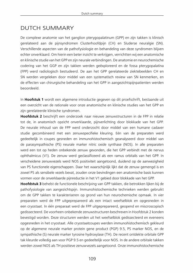
DUTCH SUMMARY
De complexe anatomie van het ganglion pterygopalatinum (GPP) en zijn takken is klinisch
gerelateerd aan de pijnsyndromen Clusterhoofdpijn (CH) en Sluderse neuralgie (SN).
Verschillende aspecten van de pathofysiologie en behandeling van deze syndromen blijven
echter onverklaard. Om hierin een beter inzicht te verkrijgen, verrichtten wij een anatomische
en klinische studie van het GPP en zijn neurale verbindingen. De anatomie en neurochemische
codering van het GGP en zijn takken werden geëxploreerd en de fossa pterygopalatina
(FPP) werd radiologisch bestudeerd. De aan het GPP gerelateerde ziektebeelden CH en
SN werden vergeleken door middel van een systematisch review van SN kenmerken, en
de effecten van chirurgische behandeling van het GPP in aangezichtspijnpatienten werden
beoordeeld.
In Hoofstuk 1 wordt een algemene introductie gegeven op dit proefschrift, bestaande uit
een overzicht van de rationale voor onze anatomische en klinische studies van het GPP en
zijn gerelateerde klinische syndromen.
Hoofdstuk 2 beschrijft een onderzoek naar nieuwe zenuwstructuren in de FPP in relatie
tot de, in anatomisch opzicht onverklaarde, pijnverlichting door blokkade van het GPP.
De neurale inhoud van de FPP werd onderzocht door middel van een humane cadaver
studie gecombineerd met een zenuwspecifieke kleuring. Eén van de preparaten werd
gedeeltelijk in coupes gesneden en immunohistochemisch geanalyseerd door middel van
de parasympathische (PS) neurale marker nitric oxide synthase (NOS). In alle preparaten
werd een tot op heden onbekende zenuw gevonden, die het GPP verbindt met de nervus
ophthalmicus (V1). De zenuw werd geclassificeerd als een ramus orbitalis van het GPP. In
verscheidene zenuwvezels werd NOS positiviteit aangetoond, duidend op de aanwezigheid
van PS functionele eigenschappen. Daar het waarschijnlijk lijkt dat de zenuw gemengd is en
zowel PS als sensibele vezels bevat, zouden onze bevindingen een anatomische basis kunnen
vormen voor de onverklaarde pijnreductie in het V1 gebied door blokkade van het GPP.
Hoofdstuk 3 behelst de functionele beschrijving van GPP takken, die betrokken lijken bij de
pathofysiologie van aangezichtspijn. Immunohistochemische technieken werden gebruikt
om de GPP takken te karakteriseren op grond van hun neurochemische opmaak. In vier
preparaten werd de FPP uitgeprepareerd als een intact weefselblok en opgesneden in
een cryostaat. In één preparaat werd de FPP uitgeprepareerd, geopend en miscroscopisch
gedissecteerd. De voorheen onbekende zenuwstructuren beschreven in Hoofdstuk 2 konden
bevestigd worden. Deze structuren werden uit het weefselblok gedissecteerd en eveneens
opgesneden in het cryostaat. Alle cryostaatcoupes werden immunohistochemisch gekleurd
op de algemene neurale marker protein gene product (PGP) 9.5, PS marker NOS, en de
sympathische (S) neurale marker tyrosine hydroxylase (TH). De recent ontdekte orbitale GPP
tak kleurde volledig aan voor PGP 9.5 en gedeeltelijk voor NOS. In de andere orbitale takken
werden zowel NOS als TH positieve zenuwvezels aangetoond. Onze immunohistochemische
109
Dutch summary

bevindingen laten zien dat de recent ontdekte orbitale GPP tak van een gemengde PS en
sensibele aard is. In de andere orbitale takken werden tevens S zenuwvezels aangetoond.
Deze kennis zou bij kunnen dragen aan het begrip van de symptomatologie en behandeling
van aangezichtspijn.
In Hoofdstuk 4 wordt de radiologische anatomie van de FPP en haar inhoud in beeld
gebracht met ultra hoge resolutie magnetic resonance imaging bij 7 Tesla (7T MRI). Een
humaan cadaverpreparaat van de FPP werd onderzocht op een klinisch 7T MRI systeem.
Vervolgens werden van dit weefselblok coronale coupes vervaardigd met behulp van een
cryomicrotoom. De coupes werden gefotografeerd en opgenomen op breed plakband. De
optape coupes werden gekleurd voor Mallory-Cason, en de coronale MR beelden werden
vergeleken met de overeenkomstige opzichtfoto’s en optape coupes. 7T MRI bleek een
excellente afbeelding van de FPP anatomie te geven. Behalve de bekende macroscopische
anatomische structuren, waren ook kleinere structuren zichtbaar, die voorheen op MRI
onzichtbaar waren. Een aantal van de orbitale ganglion pterygopalatinumtakken en de
nervus pharyngeus waren helder afgebeeld. 7T MRI zou dus mogelijk kunnen bijdragen aan
een betere diagnostische en preoperatieve evaluatie van de FPP en haar inhoud.
Chapter 5 beschrijft een systematisch review van alle voor handen zijnde en oorspronkelijke
literatuur over Sluderse neuralgie (SN). Pijnkenmerken, periodiciteit en geassocieerde
symptomen werden bestudeerd en geregistreerd in volgorde van voorkomen. Elf artikelen
werden bestudeerd en diverse verschillen tussen SN en Cluster hoofdpijn (CH) kwamen aan
het licht. Op grond van de in de literatuur beschreven symptomen konden duidelijke criteria
voor SN worden opgesteld, die de erkenning van SN als een apart klinisch ziektebeeld
zouden kunnen bevorderen. Gezien haar symptomatologie zou SN mogelijk een trigeminal
autonomic cephalalgia kunnen zijn.
In Hoofdstuk 6, worden de effecten of radiofrequente thermocoagulatie (RFT) van het
ganglion pterygopalatinum op aangezichtspijn beschreven, na kritische beoordeling
van de juistheid van diagnose. Wij voerden een retrospectieve studie uit van de klinische
dossiers van alle patienten die gedurende vier opeenvolgende jaren RFT van het
ganglion pterygopalatinum ondergingen in een tertiair pijncentrum. Diagnoses werden
opnieuw beoordeeld waarna het effect van RFT op aangezichtspijn werd bestudeerd. Na
retrospectieve toepassing van de nieuwe criteria voor SN en de bestaande criteria voor
CH, bleken zeven van de 15 patienten oorspronkelijk op juiste wijze gediagnosticeerd
te zijn. Hiermee wordt aangetoond dat aangezichtspijn, zelfs in een tertiair centrum, op
onjuiste wijze kan worden gediagnosticeerd. Negen van de vijftien patiënten ervoeren
een aanzienlijke pijnverlichting na RFT. De positieve resultaten waren het meest frequent
aanwezig onder de patiënten met Sluderse neuropathie, atypische aangezichtspijn en
CH. Echter, bij de meeste patiënten waren herhaalde procedures noodzakelijk om tot dit
resultaat te komen. Een juiste aangezichtspijndiagnose blijkt derhalve van groot belang voor
het voorspellen van het effect van de verschillende behandelstrategieën.
110

In Hoofdstuk 7 worden de effecten van microvasculaire decompressie (MVD) van het GPP
beschreven, als alternatief voor de traditionele chirurgische behandeling van CH patiënten.
MVD werd uitgevoerd in drie patiënten met refractaire CH. De arteria pterygopalatina
werd onderbonden en een musculus temporalis fascielapje werd tussen de arterie en het
ganglion geplaatst. Er werden geen verschillen gevonden tussen de preoperatieve periode
en een week, een maand, drie maanden en zes maanden follow-up, met betrekking tot
duur van de aanval, pijnscore op een visual analogue scale tijdens aanvallen en in remissies,
duur van de remissies en kwaliteit van leven. De voorlopige data wijzen erop dat MVD
van het ganglion pterygopalatinum geen pijnverlichting of verbetering van levenskwaliteit
oplevert bij patiënten met refractaire CH.
Chapter 8 bevat een discussie van de resultaten beschreven in dit proefschrift. Suggesties
voor verder onderzoek worden gedaan in de sectie toekomstige perspectieven.
111
Dutch summary


Color figures

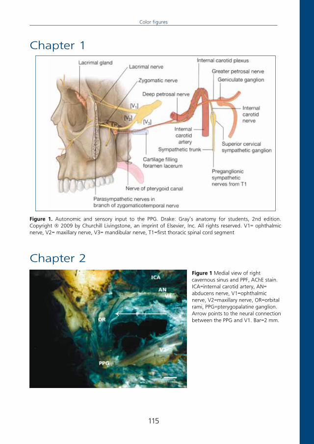
Figure 1 Medial view of right cavernous sinus and PPF, AChE stain. ICA=internal carotid artery, AN= abducens nerve, V1=ophthalmic nerve, V2=maxillary nerve, OR=orbital rami, PPG=pterygopalatine ganglion. Arrow points to the neural connection between the PPG and V1. Bar=2 mm.
Figure 1. Autonomic and sensory input to the PPG. Drake: Gray’s anatomy for students, 2nd edition. Copyright ® 2009 by Churchill Livingstone, an imprint of Elsevier, Inc. All rights reserved. V1= ophthalmic nerve, V2= maxillary nerve, V3= mandibular nerve, T1=first thoracic spinal cord segment
Chapter 1
Chapter 2
115
Color figures

Figure 2 Detailed image of newly described neural structure, AChE stain. Arrow points toward the neural connection between the PPG and V1. The actual connection is not clearly visible in this photograph due to the lack of staining in this area, but was clearly visible with the aid of an operation microscope. ICA=internal carotid artery, AN= abducens nerve, V1=ophthalmic nerve, OR=orbital rami, SOF=superior orbital fissure. Bar=1mm.
Figure 4 Photograph of actual connection of neural structure with the ophthalmic nerve, medial view of contents of left human PPF in slightly different configuration than figure 2, 3 and 4.Arrow 1 points toward newly described neural bundle, arrow 2 points toward actual connection of posterior division of neural bundle with ophthalmic nerve. Arrow 3 points toward a network of fibrous tissue surrounding the posterior division of the new neural connection, which was left in place in order to preserve this structure. Bar=2mm.
116

Figure 1. Sagittal section of the maxillary nerve, stained for PGP 9.5. Bar= 0.05 mm
Figure 2. Sagittal section of an orbital branch (arrow) that runs cranially from the PPG, stained for AChE. PPG= pterygopalatine ganglion. Bar=0.05 mm.
Chapter 3
Figure 4. Sagittal section of a nerve cell body in an orbital branch, stained for AChE. Bar= 0.05 mm
Figure 5. Sagittal section of the Vidian nerve (arrows) that enters the PPG, stained for AChE. PPG= pterygopalatine ganglion. Bar= 0,05 mm.
117
Color figures

Figure 9. Transverse sections of rami A and B, stained for NOS.Bar= 0.05 mm
Figure 8. Transverse sections of rami A and B, stained for PGP 9.5.Bar= 0.05 mm
Figure 10. Transverse sections of the ganglion in ramus A, stained for NOS. The ganglion was situated at the most anterior part of the cavernous sinus and contained NOS positive cells, surrounded by NOS positive nerve fibres. Bar= 0.05 mm.
Ramus A Ramus B
Ramus A Ramus B
Figure 6. Sagittal section of the Vidian nerve (arrows) that enters the PPG, stained for TH.Bar= 0.05 mm
118

Figure 1a. Coronal MR image of left PPF
Figure 1b. Corresponding surface photograph
Figure 1c. Corresponding section
Figure 1d. Scanning plane on axial image
Posterior
Anterior
Figure 2a. Coronal MR image of left PPF
Figure 2b. Corresponding surface photograph
Figure 2c. Corresponding section
Figure 2d. Scanning plane on axial image
Posterior
Anterior
Chapter 4
119
Color figures

Figure 3a. Coronal MR image of left PPF
Figure 4a. Coronal MR image of left PPF
Figure 3b. Corresponding surface photograph
Figure 4b. Corresponding surface photograph
Figure 3c. Corresponding section
Figure 4c. Corresponding section
Posterior
Posterior
Anterior
Anterior
Figure 3d. Scanning plane on axial image
Figure 4d. Scanning plane on axial image
120

Figure 5a. Coronal MR image of left cavernous sinus
Figure 5b. Corresponding surface photograph
Figure 5c. Corresponding section
Posterior
Anterior
Figure 5d. Scanning plane on axial image
Chapter 7Figure 1. Antrostomy in the anterior wall of the maxillary sinus, anterior view of the right maxillary sinus. Arrow 1 towards points toward anterior wall of maxillary sinus. Arrow 2 points toward antrostomy.
121
Color figures

Figure 2. A hatch created in the centre of the posterior maxillary sinus wall. Arrow 1 points toward lateral border of hatch, arrow 2 points toward medial border of hatch.Asterisk in centre of hatch.
Figure 3. Fatty tissue in the PPF is bluntly dissected to expose the pterygopalatine portion of the maxillary artery.PA = Pteryogpalatine artery.PPG = Pterygopalatine ganglion.
Figure 4. Ligation of the pterygopalatine artery with hemoclips. Arrows point toward hemoclips.
Figure 5. Temporal muscle graft, placed between the ptergygopalatine artery and the PPG.PA = Pterygopalatine artery.TMG = Temporal muscle graft.
122

Figure 6. SF-36 Domain Score Changes Patient 1. PF= Physical Functioning, RP= Role-Physical, RE= Role-Emotional, VIT= Vitality, MH= Mental Health, SF= Social Functioning, BP= Bodily Pain, GH= General Health
Figure 7. SF-36 Domain Score Changes Patient 2. PF= Physical Functioning, RP= Role-PhysicalRE= Role-Emotional, VIT= Vitality, MH= Mental Health, SF= Social Functioning, BP= Bodily Pain, GH= General Health
Figure 8. SF-36 Domain Score Changes Patient 3. PF= Physical Functioning, RP= Role-PhysicalRE= Role-Emotional, VIT= Vitality, MH= Mental Health, SF= Social Functioning, BP= Bodily Pain, GH= General Health
0
20
40
60
80
100
PF RP RE VIT MH SF BP GH
Pre-Surgical
1 Week
1 Month
3 Months
6 Months
SF-36 Domain Scores
Scor
e (0
-120
)
120
0
20
40
60
80
100
PF RP RE VIT MH SF BP GH
Pre-Surgical
1 Week
1 Month
3 Months
6 Months
SF-36 Domain Scores
Scor
e (0
-100
)
0
20
40
60
80
100
PF RP RE VIT MH SF BP GH
Pre-Surgical
1 Week
1 Month
3 Months
6 Months
SF-36 Domain Scores
Scor
e (0
-100
)
123
Color figures


Dankwoord


DANKWOORD
Iedereen die heeft bijgedragen aan de totstandkoming van dit proefschrift ben ik dank
verschuldigd, maar een aantal mensen in het bijzonder.
Aan de basis van dit project stond mijn promotor en oud-opleider professor G. J. Hordijk.
Zijn vriendelijke en geduldige ondersteuning maakten het werken aan dit proefschrift
tot een ontspannen bezigheid. De directe begeleiding van het onderzoek werd verzorgd
door mijn copromotoren, dr. Ronald L.A.W Bleys en dr. J.A. (Sander) de Ru; scherpzinnige
leermeesters met een groot gevoel voor humor. Onze bijeenkomsten, zowel binnen als
buiten het ziekenhuis, waren altijd inspirerend en vriendschappelijk, en droegen zeer bij
aan mijn enthousiasme voor ons onderwerp. Dr. A.J.M (Bart) van Wijck leverde als mede
auteur en anesthesioloog een zeer grote bijdrage aan de klinische studies. Michelle
Ebbeling voerde een groot deel van het anatomische veldwerk uit en was onmisbaar als
mede auteur van de anatomische artikelen, eerst als coassistent en later als oogarts in
opleiding. Jaco Zwanenburg en dr. Frank A. Pameijer leverden ieder een belangrijke bijdrage
aan dit proefschrift op radiologisch gebied. De leden van de afdeling Anatomie van het
UMC Utrecht, waaronder Jan-Willem de Groot, Simon Plomp, Willem van Wolferen en
oud-kamergenote Matty Spinder, dank ik voor hun gastvrijheid en hulp op uiteenlopende
terreinen. Geen van de figuren in dit proefschrift had tot stand kunnen komen zonder
Marloes Dunnewind, afdeling Multimedia.
Alle (oud) arts-assistenten, stafleden en medewerkers van de afdeling KNO-heelkunde
van het UMC Utrecht droegen bij aan mijn opleiding tot KNO-arts. In het bijzonder dank
ik dr. Herman Lubsen en dr. A.F. van Olphen voor de uitstekende begeleiding. Professor
Anne G.M. Schilder gaf mij als coassistent de kans onderzoek te doen naar de effecten
van adenotonsillectomie op de middenoorstatus bij kinderen en de resultaten hiervan te
publiceren, en stond daarmee aan de basis van mijn KNO-opleiding. Ik heb veel steun
gehad aan de hechte band binnen onze afdeling, die in mijn opleidingsjaren door vele
gebeurtenissen werd versterkt. Ik was vereerd om paranimf te zijn bij de promotie van mijn
oud-collega en vriendin Farzaneh van Voorst van Beest- Farshadpour, bij wie ik altijd terecht
kon voor gezelligheid en adviezen over mijn eigen promotie. Dr. S.J. (Hans) Rietema, dr.
Maarten H.J.M Majoor, dr. Wilbert M. Boek, dr. Remco M. Cardinaal, Jan Willem Sepmeijer
en Jan Burggraaf hebben zorg gedragen voor een onvergetelijke opleiding in Ede. De
maanden in Ede en het WKZ hebben daarbij geleid tot de vriendschap met mijn paranimf
en oud-collega Hendrik Bremer, die op zijn eigen humoristische en goedgehumeurde wijze
het werk een stuk lichter maakte.
Niet alles is in woorden te vatten. Natuurlijk is de laatste plaats in dit dankwoord voor hen
die de grootste plaats hebben in mijn hart: mijn zus en paranimf Maret, mijn ouders en
Volkert.
127
Dankwoord


CURRICULUM VITAE
Karin Oomen was born in De Bilt on August 13th, 1977. After graduation from high school
(VWO, Sint Bonifatiuscollege Utrecht) in 1995, she started her studies in Biology at Utrecht
University. In 1996, she started medical school. After receiving her medical license in 2003,
she worked as a junior teacher at the Anatomy Department of the University Medical
Center Utrecht. The research project collected in this thesis was started that year. In August
2005, she started her residency in Otolaryngology-Head and Neck Surgery at the University
Medical Center Utrecht (mentors: Prof. Dr. G.J. Hordijk, Prof. Dr. F.W.J. Albers, Dr. A.F. van
Olphen and Prof. Dr. W. Grolman) and Ziekenhuis Gelderse Vallei Ede (mentors: Dr. S.J.
Rietema and Dr. M.H.J.M. Majoor). She finished her residency on February 1st, 2011. In
August 2011, Karin will join the Department of Otolaryngology at Weill Cornell Medical
College, New York, for a two-year fellowship in Pediatric Otolaryngology.
129
Curriculum vitae
