péter windisch prof. dr. head of the department of
TRANSCRIPT

Péter Windisch Prof. Dr. Med. Dent.Head Of The Department Of PeriodontologySemmelweis University Budapest, Hungary
Biology of periodontal and peri-implant tissues

1) Biotype, thickness of facial bone2) Existence and shape of interdental papilla, level of proximal bone 3) Thickness and width of keratinized gingiva, maintained bone surrounding4) Depth of the vestibulum 5) Contour and proximal height of the periodontium of neighbouring teeth6) Shape and positioning of the teeth - „emergence profile”
There is an obvious need to achieve tooth-like harmonious pink and white esthetics via implant borne restorations. In order to achieve successful treatment:1. proper planning 2. 3D positioning 3. required amount of bone and non-mobile soft tissue are the key factors.
Efficacy of periodontal plastic surgery procedures in the treatment of localized facial gingival recessions. A systematic review. J Clin Periodontol. 2014
Apr;41 Suppl 15:S44-62.
Esthetic implant site development. Oral Maxillofac Surg Clin North Am. 2015 May; 27(2):283-311.
Factors effecting long term functional and esthetic stability around teeth and implants

Classification and treatment options of gingivalrecessions around teeth
• Lack of keratinized gingiva• Autogenous free gingival graft (FGG), xenograft
• Shallow vestibule• Autogenous free gingival graft (FGG), xenograft
• Gingival recession /apical displacement of marginalgingiva: distance from the CEJ/
• Autogenous subepithelial connective thisse graft (SCTG), xenograft, allograft
• Healthy conditions can be kept even with gingivalrecession and minimal amount keratinized tissue aroundteeth!
Prato GP. Advances in mucogingival surgery. J Int Acad Periodontol. 2000
Jan;2(1):24-7.

Classification and treatment options of gingivalrecessions around implants
• Non-sufficient amount of periimplant mucosa• Autogenous subepithelial connective tissue graft (SCTG), xenograft,
allograft
• Lack of keratinized periimplant mucosa• Autogenous free gingival graft (FGG), xenograft, allograft
• Periimplant recession• Autogenous free gingival graft (FGG)
• The role and importance of periimplant keratinized tissue –
Still under discussion
Wennström JL, Derks J. Is there a need for keratinized mucosa around implants to maintain health and tissue stability? Clin Oral Implants Res.
2012 Oct;23Chung DM, Oh TJ, Shotwell JL, Misch CE, Wang HL. Significance of keratinized mucosa in maintenance of dental implants withdifferent surfaces. J Periodontol. 2006 Aug;77(8):1410-20. P

Role of biologic width around teeth
Gargiulo, A.W.,Wentz,F.M.& Orban, B. (1961) Dimensions and relations of the dentogingival junction in humans, Journal of Periodontology 32,262-267
enamel
Alveolarboneepitheldenti
ncementum
Combined connective tissue- and epithelial attachment from the crest of the alveolar bone to the base of the gingival sulcus.
The biologic width is patient and sitespecific, may vary between 0,75 -4,3 mm including a required amountof soft tissue barrier to maintainunderlying tissue(s) healthy.

Bennani V, Schwass D, Chandler N.: Gingival retraction techniques for implants versus teeth: current status. J
Am Dent Assoc. 2008 Oct;139(10):1354-63.
The biologic width – supracrestal soft tissueattachment
Tooth
Sulcus 0.69 mm
JunctionalEpithelium0.97 mm
ConnectiveTissue
Attachment1.07 mm
Biologicwidth
2.04 mm
Bennani et al. 2008
Implant
Sulcus 0.5-1.0 mm
JunctionalEpithelium
1.5 ± 0.5 mm
ConnectiveTissue
Attachment1 mm
Biologicwidth
2.5 mm ±0.5 mm
Keratinised oralepithelium
The biologic width is patient and site specific but always higher aroundimplants than natural teeth

(Lindhe 1976)
Alterations of gingival biotype around different anatomic regions of both jaws

Characteristics of periimplant soft tissues
Berglundh T, Lindhe J, Ericsson I, Marinello CP, Lijenberg B, Thomsen P. The soft tissue barrier at implants and teeth. Clin Oral Implants Res 1991;2:81-90
Junctional
EpitheliumCollagen
WRONG!Periimplantmucositis
• Lack of cementum layer
• Hemidesmosomal attachment
• Parallely oriented collagen fibers

Nevins M, Nevins ML, Camelo M, Boyesen JL, Kim DM. Human histologic evidence of a connective tissue attachment to a dental implant. Int J Periodontics Restorative Dent. 2008 Apr;28(2):111-21.
Characteristics of periimplant soft tissues
Human proof-of-principle study: Achieving a physical connective tissue attachment to the Laser-Lokmicrochannel collar of a dental implant. Its 2-mm collar has been micromachined to encourage bone and
connective tissue attachment while preventing apical migration of the epithelium.

Biologic width development around implants
Berglundh T, Lindhe J. Dimension of the periimplant mucosa. Biological width revisited. J Clin Periodontol. 1996 Oct;23(10):971-3.
Basic researchThe reduced amount of soft tissues resulted in crestal bone loss at an external hex
abutment implant interfaceSaucerisation of crestal bone: typical phenomenon for two-stage implants after
abutment connection

Biologic width
test test controlcontrol
Biologic width around implants with different amount of soft tissue.
O: Oral epithelium; B: Bone; aJE: Junctional epithelium; PM: periimplantmucosa
Berglundh, T., Lindhe, J. (1996). Dimension of the peri-implant mucosa. Biological widthrevisited. J. Clin. Periodontol. 23: 971-973. (16)

Predictive factors determining functionalstability and esthetics
1. Relative position of
the tooth
2. Shape of the
periodontium
3. Type of the
periodontium4. Shape of the tooth
5. Positions of the alveolar bone
Kois (2004) :
Bone quality and
quantity
Soft tissue
quality and
quantity
Soft tissue
biotype
Emergence
profile
Lip line and
smile line
Treatment
options
Surgical
alternatives(
immediate,
delayed, late
implant placement)
Periodontal and
orthodontic
aspects
Orthodontic
options
Number of missing
teeth
Optimal implant
size
Temporary
solutions
Implant positioning
( apico-incisal,
mesio-distal, labio-
palatal )
Mucogingival
surgery options
Esthetic bone
augmentationProsthetic options
El Askary (2001)
Kois JC, Kan JY. (2001). Predictable peri-implant gingival aesthetics:
surgical and prosthodontic rationales. Pract Proced Aesthet Dent. Nov-
Dec (83)
El Askary, AS (2001). Multifaceted Aspects of Implant Ethetics: The
Anterior Maxilla. Implant Dentistry. 10(3): 182-190.
(Lindhe J.: Textbook of Clinical Periodontology and Implant Dentistry 2008)
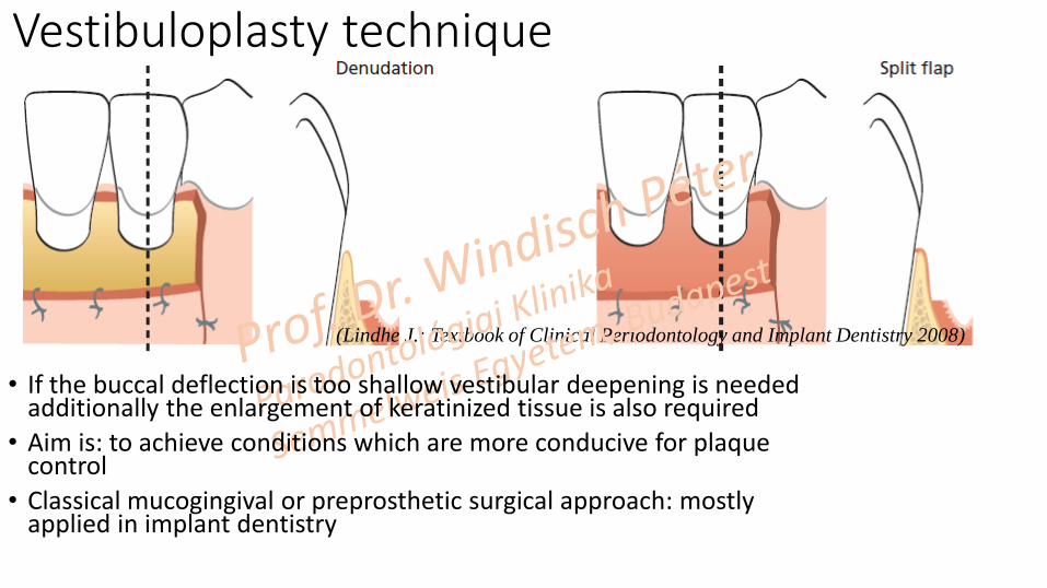
Vestibuloplasty technique
• If the buccal deflection is too shallow vestibular deepening is neededadditionally the enlargement of keratinized tissue is also required
• Aim is: to achieve conditions which are more conducive for plaquecontrol
• Classical mucogingival or preprosthetic surgical approach: mostlyapplied in implant dentistry

Apicocoronal or vertical positioningTarnow D, Elian N, Fletcher P, Froum S, Magner A, Cho SC, Salama M, Salama H, Garber DA. Vertical distance from the crest of bone to the height of the interproximal papilla betweenadjacent implants. J Periodontol 2003;74(12):1785-1788.)
„As shallow as possible, as
deep as necessary”
Aesthetically driven implant positioning
Mesio-distal positioningGastaldo JF, Cury PR, Sendyk WR. Effect of the vertical and horizontal distancesbetween adjacent implants and between a tooth and an implant on the incidenceof interproximal papilla. J Periodontol 2004;75:1242-1246)Orovestibular positioning
Spray JR, Black CG, Morris HF, Ochi S.Theinfluence of bone thickness on facial marginal bone response: stage-1 placement through stage-2 uncovering. Ann Periodontol 2000;5:119-128.

Evaluation of aestheticsImplant Crown Aesthetic index
1. Mesio-distal crown width2. Position of incisal edge3. Labial convexity of the crown4. Colour and translucency5. Structure of the crown6. Vestibular level of the periimplant mucosa7. Approximal level of the mucosa8. Vestibular contour of the mucosa9. Colour and surface of keratinised gingiva
(Meijer HJA, Stellingsma K, Meijndert L, Raghoebar GM. A new index for ratingaesthetics of implantsupported single crowns and adjacent soft tissues – The ImplantCrown Aesthetic Index. Clin Oral Implants Res 2005;16:645-649. )

Evaluation of esthetics
Pink Esthetic Score PES
1. mesial papilla2. distal papilla3. Height of gingival contour4. Shape of gingival contour5. Shape of a healthy jugum alveolare6. Texture of gingiva7. Colour of gingiva
(Fürhauser R, Florescu D, Benesch T, Haas R, Mailath G, Watzek G. Evaluation of soft tissue around single-tooth implant crowns: the pink esthetic score. Clin OralImplants Res. 2005 Dec;16(6):639-44.)

Single gingival recession coverage techniques -natural teeth
Coronally advanced flap -CAF
Double papilla technique
Laterally rotated flap technique
Raetzke PB. Covering localized
areas of root exposure employing
the "envelope" technique. J
Periodontol. 1985 Jul;56(7):397-402.
SCTG in the subepithelially prepared "envelope”→ No vertical incision, less
damage
→ Increased revascularization
→ Faster Healing
Envelope technique

Bernimoulin JP, Lüscher B, Mühlemann HR. Coronally repositioned periodontal flap. Clinical evaluation after one
year. J Clin Periodontol. 1975 Feb;2(1):1-13.
Frisch E, Ratka-Krüger P, Ziebolz D. A new technique for increasing keratinized tissue around dental implants: The partially
epithelialized free connective tissue graft (PECTG). Case Series. J Oral Implantol. 2013 Jul 8.
Single gingival recession coverage techniques - implants
Coronally advanced flap -CAF
Envelope technique and partially epithelialized connective tissue graft
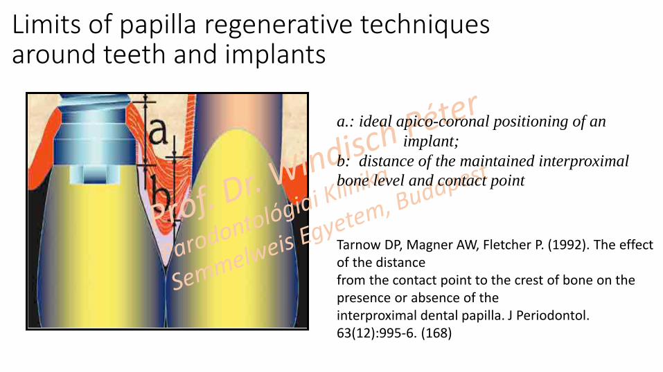
Tarnow DP, Magner AW, Fletcher P. (1992). The effectof the distancefrom the contact point to the crest of bone on thepresence or absence of theinterproximal dental papilla. J Periodontol. 63(12):995-6. (168)
Limits of papilla regenerative techniquesaround teeth and implants
a.: ideal apico-coronal positioning of an
implant;
b: distance of the maintained interproximal
bone level and contact point

Conclusions
• Ideal hard- and soft tissue surroundings are needed of an optimally positioned implant
• Soft tissue correction around a previously loaded implant has it’s limits: mucogingival surgicaltechnique can be only partially applied
• The treatment predictability is always more favorable for natural teeth then for implants – softtissue improvements prior to implant abutment connection are more preferable
• Anatomical restoration can help to achieve ideal emergence profile and thus esthetics ifproper width and thickness of keratinized periimplant tissue exists
