pten posttranslational inactivation and hyperactivation of...
TRANSCRIPT
Research article
3762 TheJournalofClinicalInvestigation http://www.jci.org Volume 118 Number 11 November 2008
PTEN posttranslational inactivation and hyperactivation of the PI3K/Akt pathway sustain primary T cell leukemia viabilityAna Silva,1 J. Andrés Yunes,2 Bruno A. Cardoso,1 Leila R. Martins,1 Patrícia Y. Jotta,2
Miguel Abecasis,3 Alexandre E. Nowill,4 Nick R. Leslie,5 Angelo A. Cardoso,6 and Joao T. Barata1
1Unidade de Biologia do Cancro, Instituto de Medicina Molecular, Faculdade de Medicina da Universidade de Lisboa, Lisbon, Portugal. 2Laboratório de Biologia Molecular, Centro Infantil Boldrini, Campinas, São Paulo, Brazil. 3Serviço de Cardiologia Pediátrica,
Hospital de Santa Cruz, Carnaxide, Portugal. 4Centro Integrado de Pesquisas Oncohematologicas da Infancia, Universidade Estadual de Campinas, Campinas, São Paulo, Brazil. 5Division of Molecular Physiology, College of Life Sciences, University of Dundee, Dundee, United Kingdom.
6Melvin and Bren Simon Cancer Center, Indiana University School of Medicine, Indianapolis, Indiana, USA.
Mutationsinthephosphataseandtensinhomolog(PTEN)geneleadingtoPTENproteindeletionandsubse-quentactivationofthePI3K/Aktsignalingpathwayarecommonincancer.HereweshowthatPTENinactiva-tioninhumanTcellacutelymphoblasticleukemia(T-ALL)cellsisnotalwayssynonymouswithPTENgenelesionsanddiminishedproteinexpression.SamplestakenfrompatientswithT-ALLatthetimeofdiagnosisveryfrequentlyshowedconstitutivehyperactivationofthePI3K/Aktpathway.Incontrasttoimmortalizedcelllines,mostprimaryT-ALLcellsdidnotharborPTENgenealterations,displayednormalPTENmRNAlevels,andexpressedhigherPTENproteinlevelsthannormalTcellprecursors.However,PTENoverexpressionwasassociatedwithdecreasedPTENlipidphosphataseactivity,resultingfromcaseinkinase2(CK2)overexpres-sionandhyperactivation.Inaddition,T-ALLcellshadconstitutivelyhighlevelsofROS,whichcanalsodown-modulatePTENactivity.Accordingly,bothCK2inhibitorsandROSscavengersrestoredPTENactivityandimpairedPI3K/AktsignalinginT-ALLcells.Strikingly,inhibitionofPI3Kand/orCK2promotedT-ALLcelldeathwithoutaffectingnormalTcellprecursors.Overall,ourdataindicatethatT-ALLcellsinactivatePTENmostlyinanondeletional,posttranslationalmanner.PharmacologicalmanipulationofthesemechanismsmayopennewavenuesforT-ALLtreatment.
IntroductionPI3K catalyzes the production of the second messenger phospha-tidylinositol 3,4,5-trisphosphate (PIP3), thereby recruiting and activating several downstream kinases. PI3K and its most promi-nent effector, Akt (also known as PKB), regulate cell viability, metabolism, motility, and proliferation and are extensively impli-cated in tumorigenesis (1–3). Constitutive activation of the PI3K/Akt signaling pathway in hematological malignancies, including myeloid leukemia, multiple myeloma, and T cell large granular lymphocytic leukemia, has been shown to support tumor cell pro-liferation and viability in vitro (4–6).
The main negative regulator of the PI3K/Akt pathway, the lipid phosphatase and tensin homolog (PTEN), is frequently inac-tivated in human cancer as result of various genetic lesions (7, 8), which ultimately result in decreased or absent PTEN protein expression and activity. PTEN deficiency in mice replicates the tumor spectrum observed in humans, including T cell malig-nancies (9, 10), and T cell–specific deletion of PTEN results in lymphoma-induced death (11). Importantly, PTEN is critically involved in maintaining hematopoietic stem cells and prevent-ing leukemogenesis (12, 13). Several human T cell acute lym-
phoblastic leukemia (T-ALL) cell lines lack PTEN as a result of deletions or mutations in the gene, which consequently effect constitutive hyperactivation of the PI3K/Akt pathway (14, 15). Enforced expression of PTEN in these cell lines induces apopto-sis by inhibiting PI3K/Akt (16), which suggests that this pathway may be important in T-ALL. However, most T-ALL cell lines were established from relapsed patients, have long been in culture, and likely accumulated genomic alterations not associated with the primary disease. Hence, it is unclear whether PTEN mutations and PI3K/Akt hyperactivation are common events in cells of T-ALL patients and whether these putative alterations originate relevant functional consequences.
PTEN inactivation and consequent PI3K/Akt pathway aberrant activation may arise from mechanisms other than those target-ing PTEN gene integrity (17). Although not directly implicated in cancer, downregulation of PTEN activity by mechanisms such as phosphorylation and oxidation has been recognized for sev-eral years (18–21). PTEN C-terminal phosphorylation appears to stabilize the protein by preventing its ubiquitination and pro-teasome degradation while decreasing PTEN phosphatase activ-ity (20–23). The serine/threonine protein kinase casein kinase 2 (CK2) has been linked to PTEN phosphorylation (21, 22). Inter-estingly, CK2 overexpression is observed in human solid tumors (24–26) and is essential for multiple myeloma cell survival (27). Moreover, transgenic mice with targeted expression of CK2 in T cells develop lymphomas (28). In addition, ROS, which are com-monly upregulated in cancer cells and proposed to contribute to transformation (29–31), were shown to oxidize PTEN Cys124 in
Nonstandardabbreviationsused: CK2, casein kinase 2; DRB, dichlororibofurano-sylbenzimidazole; DTT, dithiothreitol; β-ME, β-mercaptoethanol; NAC, N-acetyl-cys-teine; PIP3, phosphatidylinositol 3,4,5-trisphosphate; PTEN, phosphatase and tensin homolog; SP8, CD8 single positive; T-ALL, T cell acute lymphoblastic leukemia; TBB, tetrabromobenzotriazole; TP, triple positive.
Conflictofinterest: The authors have declared that no conflict of interest exists.
Citationforthisarticle: J. Clin. Invest. 118:3762–3774 (2008). doi:10.1172/JCI34616.

research article
TheJournalofClinicalInvestigation http://www.jci.org Volume 118 Number 11 November 2008 3763
the active site to form a disulfide bond with Cys71, thereby inac-tivating PTEN (18, 19, 32). However, there is no direct evidence linking CK2, ROS, PTEN phosphorylation, or PTEN oxidation to downregulation of PTEN function in patient tumor cells, and the actual implications of these mechanisms to cancer cell function remain to be determined.
Here we show that constitutive activation of the PI3K/Akt pathway is a common event in primary T-ALL and is critical for leukemia cell viability. PI3K/Akt pathway hyperactivation appeared to result not only from canonical mechanisms involv-ing PTEN gene alterations and consequent protein deletion, but also, in most cases, from PTEN protein stabilization and inacti-vation due to high CK2 activity and elevated intracellular ROS. Constitutive hyperactivation of the PI3K/Akt pathway occurred not only in PTEN-null, but also in most PTEN-expressing, T-ALL cells, and dependence on PI3K/Akt-mediated signaling was used to selectively target T-ALL cells. In addition, our data suggest that PI3K/Akt activation status, which integrates cues
arising from both genetic and posttranslational inactivation of PTEN, could serve as a biomarker for the identification of can-didate patients for treatment with inhibitors of PI3K and/or of its downstream targets.
ResultsThe PI3K/Akt pathway is constitutively hyperactivated in primary T-ALL cells. Based on evidence from T-ALL cell lines, we hypothesized that PI3K/Akt signaling is hyperactivated in primary disease. To establish how frequently PI3K/Akt constitutive activation actu-ally occurs, we evaluated the integrity of the PTEN/PI3K/Akt axis in T-ALL patient samples collected at diagnosis. Constitutive hyperactivation of the PI3K/Akt pathway was detected in most T-ALL specimens (87.5%; 21 of 24), as evaluated by increased phos-phorylation of Akt and/or at least one of its downstream targets GSK-3β and FOXO3a in comparison to normal T cell precursors (Figure 1, A and B, Table 1, and data not shown). Hyperactiva-tion of the PI3K/Akt pathway was not associated with overexpres-
Figure 1The PI3K/Akt pathway is constitutively hyperactivated in primary T-ALL cells. (A) Cell lysates of normal human thymocytes or primary T-ALL cells collected at diagnosis and immunoblotted with the indicated phosphospecific antibodies or actin as loading control. (B) Levels of phosphorylated Akt (S473) in thymocyte (n = 8) and T-ALL (n = 15) samples were quantified by densitometry analysis. Points represent individual samples, hori-zontal bars denote mean, and mean ± SEM is shown in parentheses. (C) Surface expression of PIP3 was determined by confocal microscopy after staining with anti-PIP3 antibody. Scale bar: 50 μm. Insets (20 μm square) of 1 representative cell of each sample are shown. (D) Same samples were analyzed by flow cytometry. Values in each histogram indicate PIP3 mean fluorescence intensity; gray histogram represents nega-tive isotypic control; vertical lines indicate peak value in T-ALL samples. (E) Mean fluorescence intensity (MFI) was quantified by flow cytometry and compared in thymocyte and T-ALL samples (n = 4 per group). Values are mean ± SEM.

research article
3764 TheJournalofClinicalInvestigation http://www.jci.org Volume 118 Number 11 November 2008
sion of total Akt (Supplemental Figure 1; supplemental material available online with this article; doi:10.1172/JCI34616DS1) and was consistently observed independently of the stage of develop-mental arrest of the T-ALL samples (Table 1). Moreover, primary T-ALL cells presented higher PIP3 levels in the plasma membrane, as determined by both confocal microscopy and flow cytometry analysis (Figure 1, C–E). Consistent with previous reports (15, 33) we also observed PI3K/Akt hyperactivation in leukemia T cell lines (Supplemental Figure 2).
Each T-ALL sample essentially consists of a clonal popula-tion of leukemia blasts arrested at a particular stage of thymic T cell development (34, 35), whereas normal thymocytes are immunophenotypically and functionally heterogeneous, cov-ering the spectrum of distinct developmental subpopulations. To ensure that our data were not biased by the prevalence of a particular subpopulation in the normal control samples, we
next analyzed patient specimens arrested at 2 particular stages of T cell development and compared them with their purified normal equivalents. We selected patients with CD3+CD4+CD8+ (triple positive; TP) and CD3+CD4–CD8+ (CD8 single positive; SP8) immunophenotypes because they were the most prevalent among primary T-ALL samples (Table 1 and Supplemental Fig-ure 3A). Additionally, normal TP and SP8 thymocytes represent 2 distinct subpopulations: TP cells are mostly quiescent, where-as SP8 cells are actively proliferating or have recently done so (36). Both TP and SP8 T-ALL cells revealed clearly higher levels of phosphorylated Akt than their normal immunophenotypic counterparts (Supplemental Figure 3B). Likewise, both CD3– and CD3+ T-ALLs displayed hyperphosphorylation of Akt compared with immunophenotype-matched normal thymocytes (data not shown). Also, PIP3 levels were similar among the different nor-mal thymic subsets and significantly and consistently lower than
Table 1Immunophenotype, differentiation stage, PTEN and PI3K/Akt status, and CK2 and ROS levels of T-ALL patient samples
Patient Surface markersA Stage PI3K/Akt PTEN status CK2I ROSJ
no. CD1 CD2 CD5 CD7 CD3 CD4 CD8 UckunB EGILC activationD ProteinE mRNAF MutationG PhosphoH
01 – + + + + + + III IV + – + ND NA + +02 + + + + + – – III III + + + ND + ND –03 + + + + – + + II III + + + ND + ND ND04 – + + + + – + III IV + + + ND + + +05 – + + + – + – II II + + + ND – – +06 + + + + + – + III III + – + ND NA ND ND07 + + + + + – – III III – + ND ND + ND +08 + – + + – + – II III – + ND ND – ND ND09 + + + + – – + II III ND + ND ND ND ND +10 + + + + – + + II III + + + WT + + +11 ND + + + + ND ND III III or IVK + + + WT ND ND ND12 + + + + – + – II IIIK + + + WT + + –13 ND + + + + ND ND III III or IVK + – + Mutated NA ND ND14 ND + + + + ND ND III III or IVK – + + WT – ND ND15 + + + + – + + II III + + + WT ND ND +16 ND – – + – ND ND I I or IIIK ND + + WT ND ND ND17 ND + + + + ND ND III III or IVK + + + WT + ND ND18 – + + + – + + II II + + + WT + ND +19 + + + + + + + III III + + + WT + + +20 ND + + + + ND ND III III or IVK ND – + Mutated NA + +21 – + + + + + + III IV ND + ND WT ND ND ND22 + + + + + + + III III + + + WT + ND –23 – + + + + – + III IV + + ND ND ND + +24 – + + + + + – III IV ND + + WT ND ND ND25 – + + + – + – II II + + + WT ND + +26 – + + + + – + III IV + + + WT + + ND27 – + + + + – + III IV + – ND Mutated NA + ND28 + + + + + + + III III ND + + WT ND ND ND29 + + + + – – + II III + – + ND NA ND ND30 + – + + + + + III III + + ND ND ND + +
ND, not determined. APatients were considered positive for a marker if greater than 30% of the blasts were positive compared with an irrelevant isotypic control. BT-ALL maturation stage as described by Uckun et al. (64): stage I (pro–T-ALL), CD7+CD2–CD3–; stage II (immature T-ALL), combination of CD7+ with CD2+ and/or CD5+; stage III (mature T-ALL), CD7+CD2+CD5+CD3+. CT-ALL maturation stage as determined by EGIL (65): stage I (pro–T-ALL), CD7+ only; stage II (pre–T-ALL), combination of CD7+ with CD2+, CD5+, and/or CD8+; stage III (cortical T-ALL), CD1a+; stage IV (mature T-ALL), CD3+CD1a–. DActivation status was considered positive when at least 1 pathway member (Akt, GSK-3β, and FOXO3a) showed increased levels of phosphorylation by immunoblot analysis compared with a panel of at least 2 normal thymocyte samples; 21 of 24 samples determined (87.5%) were positive. EAs determined by immunoblot; 24 of 30 (80%) were positive. FAs determined by semiquantitative RT-PCR and/or quantitative real-time PCR; 23 of 23 (100%) were posi-tive. GOf 18 samples determined, 15 (83.3%) were WT for PTEN mutation. HPhosphorylation, as determined by immunoblot; 11 of 14 (78.6%) presented levels above the panel of control thymocytes, considered positive. IAs determined by immunoblot; 11 of 12 (91.7%) presented levels above the panel of control thymocytes, considered positive. JAs determined by flow cytometry after staining with DCF-DA; 13 of 16 (81.3%) presented levels above the panel of control thymocytes, considered positive. KStage not determined.

research article
TheJournalofClinicalInvestigation http://www.jci.org Volume 118 Number 11 November 2008 3765
those of primary T-ALL cells (Supplemental Figure 4). Our data indicate that constitutive hyperactivation of the PI3K/Akt path-way is a common event in T-ALL, not only in immortalized cell lines, but more importantly in primary tumors.
PTEN gene alterations are infrequent, whereas protein expression is commonly deregulated, in primary T-ALL. PTEN dephosphorylates PIP3 and is the main negative regulator of the PI3K/Akt path-way. Thus, we next analyzed the expression of PTEN mRNA and protein in primary T-ALL samples. Whereas all 23 samples ana-lyzed expressed PTEN mRNA, a minority of the cases (20%; 6
of 30) lacked PTEN protein (Figure 2A and Table 1). Notably, not only the PTEN protein–negative samples (n = 5), but also most of the PTEN protein–positive samples (84.2%; 16 of 19), showed PI3K/Akt pathway hyperactivation (Table 1). Because point mutations may render PTEN protein inactive with unno-ticeable effects on protein expression, we performed mutation analysis of all PTEN exons and flanking intron sequences in 3 PTEN protein–negative and 15 PTEN protein–positive T-ALL cases. Interestingly, all 3 PTEN protein–null samples analyzed showed PTEN gene alterations involving exon 7 (Figure 2C and
Figure 2PTEN gene alterations and protein expression deregulation in primary T-ALL cells. (A) Expression of PTEN mRNA (top) and protein (bottom) in normal thymocytes, T-ALL primary samples, and cell lines was assessed by RT-PCR and immunoblotting, respectively. (B) PTEN mRNA levels in thymocyte (n = 12) and T-ALL (n = 18) samples were evaluated by quantitative RT-PCR. (C) None of the 15 PTEN-expressing T-ALL patients presented PTEN gene alterations, whereas all 3 PTEN-negative leukemia samples analyzed harbored mutations in exons 1 and/or 7. (D) PTEN protein levels in thymocyte (n = 6) and T-ALL (n = 8) samples were evaluated by densitometry analysis after immunoblotting. Data are representative of 4 independent analyses involving a total of 9 thymocyte samples and 15 PTEN-expressing T-ALL specimens. In B and D, points represent individual samples, horizontal bars denote mean, and mean ± SEM is shown in parentheses.
Table 2Alterations in PTEN gene coding sequence of PTEN protein–negative T-ALL patients
Patient PTEN Exon Nucleotide change Predicted protein change
no. Protein mRNA13 - + 7 755–787 hom_del (ATATCAAAGTAGAGTTCTTCCACAAACAGAACA) 252–263 del (DIKVEFFHKQNK) 755–760 hom_ins (CGAAAG) 252–254 ins (AKE)20 - + 1 2–13 het_del (TGACAGCCATCA) 1–34 del (MTAIIKEIVSRNKRRYQEDGFDLDLTYIYPNIIA) 2–15 het_ins (GGTTCTTTGGGCTG) 7 697 het_ins (A) Frameshift, stop at 242 233–241 ins (TTGRQVHVL)27 - ND 7 700–710 hom_ins (ACCTTACCTTC) Frameshift, stop at 260 234–259 ins (SPLPSGKTSSCTLSSLSRYLCVVISK)
ND, not determined; hom_, homozygous; het_, heterozygous; ins, insertion; del, deletion.

research article
3766 TheJournalofClinicalInvestigation http://www.jci.org Volume 118 Number 11 November 2008
Table 2), which is mutated in Jurkat cells and is essential for PTEN protein stability (15, 37). One of the samples was also affected in exon 1, which appears to be involved in PTEN mem-brane binding and activation (38). None of the 15 PTEN pro-tein–positive T-ALLs analyzed presented PTEN mutations in
exons 1–9 or in the flanking intronic splice site sequences. Our data indicate that relatively few primary T-ALL samples harbor PTEN gene alterations, in accordance with recent reports that PTEN mutations are rare in primary T cell leukemia specimens (33, 39). In addition, despite some heterogeneity, average PTEN
Figure 3PTEN expression and activity are regulated by CK2 in PTEN-positive T-ALL. (A) PTEN in vitro lipid phosphatase activity was determined after immunoprecipitation of endogenous PTEN from normal thymocytes (n = 5) and PTEN-positive primary leukemia cells (n = 4). PTEN activity was normalized to the levels of immunoprecipitated PTEN in each sample. (B) PTEN phosphorylation at S380 was determined by immunoblotting. Results shown are from 2 independent analyses representative of a total of 8 thymocyte samples and 14 PTEN-positive T-ALL specimens. T-ALL patient 14 showed no PI3K/Akt basal hyperactivation (see Table 1). (C) Immunoblot analysis of CK2α and CK2β expression. (D and E) CK2 levels in normal thymocytes (n = 3) and T-ALL primary cells (n = 4) were quantified by densitometry analysis regarding CK2α (D) and CK2β (E). See Table 1 for expression of CK2 in 12 total cases analyzed. (F) CK2 kinase activity in thymocyte (n = 6) and T-ALL (n = 5) sample lysates was measured in vitro. (G) TAIL7, HPB-ALL, or primary T-ALL cells were treated for 2.5 h with DMSO vehicle control or 25 μM TBB, and levels of expression and phosphorylation of indicated proteins were analyzed by immunoblotting. p-PTEN (sev) indicates PTEN phosphorylation at the cluster S380/T382/T383/S385. (H) TAIL7 cells treated for 2.5 h with DMSO vehicle control (untreated) or 25 μM TBB were lysed, and in vitro lipid phosphatase activity of immunoprecipitated PTEN was assessed in triplicate. Values in A, D–F, and H are mean ± SEM.

research article
TheJournalofClinicalInvestigation http://www.jci.org Volume 118 Number 11 November 2008 3767
mRNA expression in primary T-ALL cells was comparable to that in normal T cell precursors, as assessed by quantitative RT-PCR (Figure 2B). This observation suggests that PTEN tran-scription is largely normal in T-ALL, arguing against the pos-sibility that PTEN promoter hypermethylation (40) or Notch-dependent PTEN transcriptional repression (39) plays a broad role in primary disease. Finally, we assessed whether hyperacti-vation of the PI3K/Akt pathway in PTEN-positive T-ALL cells is caused by decreased PTEN protein abundance. Unexpectedly, we found that PTEN-positive T-ALL samples expressed even higher levels of PTEN protein than did normal controls (Figure 2D), despite displaying constitutive activation of the PI3K/Akt pathway. Similar results were found for PTEN-positive T-ALL cell lines (Supplemental Figure 2). Analysis of PTEN protein expression by flow cytometry revealed no significant differences among the 4 major normal thymic subpopulations (Supplemen-tal Figure 5, A–C). Furthermore, each subpopulation had lower PTEN levels than those of the T-ALL cell line TAIL7 (41), a good representative of PTEN-positive primary T-ALL T cells (Supple-mental Figure 5, D–F, and Figure 2A). In addition, immunoblot analysis of TP and SP8 T-ALL cells and normal thymocytes iso-lated by fluorescence-activated cell sorting (FACS) showed once again that the T-ALL cells expressed higher PTEN protein levels than did their normal counterparts (Supplemental Figure 5G).
CK2 regulates PTEN expression and activity in PTEN protein–positive T-ALL cells. The apparent contradiction between PTEN protein abundance and PI3K/Akt pathway activation in T-ALL samples prompted us to analyze PTEN phosphatase activity. We found that high PTEN expression was not associated with increased activity, but rather with PTEN inactivation, since T-ALL samples showed diminished PTEN in vitro lipid phosphatase activity compared with normal thymocytes (Figure 3A). Furthermore, most malig-nant specimens showed higher PTEN phosphorylation at different residues in the C-terminal tail (S380, S370, and the cluster includ-ing T382/T383/S385) than did normal controls (Table 1, Figure 3B, and data not shown). Thus, most T-ALL cells expressed higher levels of both PTEN (Figure 2A) and phosphorylated PTEN (Fig-ure 3B). Importantly, the ratio of phosphorylated PTEN to total PTEN was significantly higher in phosphorylated PTEN–high T-ALL cases than in normal thymocytes (Supplemental Figure 6). This finding indicates that the higher amounts of phosphorylated PTEN in T-ALL cells are not simply the result of increased PTEN protein availability. Because CK2-mediated phosphorylation of PTEN at the C-terminal tail was previously proposed to down-modulate PTEN activity while increasing its stability (20, 21), we next examined expression and activation of CK2. T-ALL cells expressed higher levels of both the catalytic CK2α and regula-tory CK2β subunits compared with normal thymocytes (Table 1, Figure 3, C–E, and Supplemental Figure 7) and showed clearly increased constitutive CK2 kinase activity (Figure 3F). Treatment of TAIL7, HPB-ALL, or primary T-ALL cells with the CK2-specific inhibitor tetrabromobenzotriazole (TBB) resulted in abrogation of C-terminal phosphorylation of PTEN, downregulation of PTEN expression, and simultaneous inhibition of Akt and GSK-3β phos-phorylation (Figure 3G). These results suggest that PTEN phos-phorylation — leading to PTEN protein stabilization and inactiva-tion — and consequent PI3K/Akt pathway hyperactivation might be secondary to constitutive increase of CK2 activity in T-ALL cells. Because it was previously shown that CK2 may stimulate Akt activity directly (42), we next analyzed the effect of TBB on phos-
phorylation of Akt in PTEN-null Jurkat T-ALL cells stably express-ing PTEN in a Tet-inducible manner (16). CK2 inhibition clearly diminished the levels of Akt S473 phosphorylation in Jurkat cells that expressed PTEN upon doxycycline treatment, whereas it induced minor downregulation of Akt phosphorylation in Jurkat cells that did not express PTEN (Supplemental Figure 8). These data suggest that the effect of CK2 on PI3K/Akt pathway activity in T-ALL cells is largely dependent on the ability of CK2 to regu-late PTEN activity. To confirm this, we analyzed the effect of TBB directly on PTEN phosphatase activity. CK2 inhibition dramatical-ly upregulated PTEN activity in TAIL7 (Figure 3H) and HPB-ALL cells (data not shown), suggesting that constitutive aberrant CK2 activity contributed to PI3K/Akt pathway activation, at least in part, by nondeletional posttranslational inactivation of PTEN. Consistent with this hypothesis, patient samples without PI3K/Akt pathway hyperactivation displayed levels of PTEN phosphory-lation similar to those of normal thymocytes (e.g., T-ALL patient 14, Figure 3B). Moreover, high CK2 expression in PTEN-positive T-ALL samples was associated with increased phosphorylation of PTEN (Supplemental Figure 9 and Table 1).
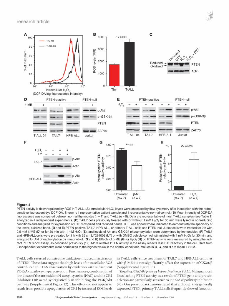
High ROS levels downregulate PTEN activity in PTEN protein–positive T-ALL cells. Cancer cells frequently express high levels of ROS, including superoxide anion (30) and H2O2 (29). By using the redox-sensitive dye DCF-DA, we found that the majority of pri-mary T-ALL cells had higher intracellular ROS levels than did normal thymocytes (Table 1, Figure 4, A and B, and Supplemen-tal Figure 10). Because H2O2 was previously shown to oxidize and thereby inactivate PTEN (18, 19, 32), we sought to determine whether diminished PTEN activity in T-ALL cells is also associ-ated with increased ROS. Although most PTEN present in TAIL7 cells was in the reduced form, there were constitutively detect-able levels of oxidized PTEN, which were further upregulated by addition of exogenous H2O2 and abrogated by in vitro treatment with the reducing agent dithiothreitol (DTT; Figure 4C). Treat-ment of PTEN-positive T-ALL cells with another antioxidant, β-mercaptoethanol (β-ME), downregulated constitutive phos-phorylation of Akt and GSK-3β (Figure 4D and Supplemental Figure 11A), whereas H2O2 induced the opposite effect (Figure 4E and Supplemental Figure 11B). In contrast, Akt and GSK-3β phosphorylation levels were not significantly affected by modu-lation of ROS in PTEN-null T-ALL Jurkat cells (Figure 4, D and E), which suggests that oxidation-dependent activation of the PI3K/Akt pathway largely relied on regulation of PTEN. In accor-dance, the PI3K-specific chemical inhibitor LY294002 abrogated not only constitutive, but also H2O2-promoted, Akt phosphoryla-tion, which indicates that ROS upregulated phosphorylated Akt in a PTEN/PI3K/PIP3-dependent manner and not via alternative mechanisms acting directly on Akt (Figure 4F). To further sup-port the evidence that PTEN activity contributes to ROS-mediat-ed activation of the PI3K/Akt pathway, we performed an indirect PTEN redox assay (19), in which lysis buffers alkylate reduced cys-teine residues, thereby irreversibly inactivating PTEN. Oxidized cysteine residues are protected from alkylation, and, because oxidation is reversible, they can be recovered afterward by treat-ment with a reducing agent for assessment of PTEN activity. The resultant in vitro phosphatase activity reflects the proportion of PTEN that was oxidized and inactivated at the time of lysis. Using this approach, we determined that β-ME (Figure 4G) and H2O2 (Figure 4H) modulated endogenous PTEN redox state and activity in opposite directions. Importantly, addition of β-ME to
research article
3768 TheJournalofClinicalInvestigation http://www.jci.org Volume 118 Number 11 November 2008
T-ALL cells reverted constitutive oxidation–induced inactivation of PTEN. These data suggest that high levels of intracellular ROS contributed to PTEN inactivation by oxidation with subsequent PI3K/Akt pathway hyperactivation. Furthermore, combination of low doses of the antioxidant N-acetyl-cysteine (NAC) and the CK2 inhibitor TBB acted synergistically in inhibiting the PI3K/Akt pathway (Supplemental Figure 12). This effect did not appear to result from possible upregulation of CK2 by increased ROS levels
in T-ALL cells, since treatment of TAIL7 and HPB-ALL cell lines with β-ME did not significantly affect the expression of CK2α/β (Supplemental Figure 13).
Targeting PI3K/Akt pathway hyperactivation in T-ALL. Malignant cell lines lacking PTEN activity as a result of PTEN gene and protein deletion are particularly sensitive to PI3K/Akt pathway inhibition (43). Our present data demonstrated that although they generally expressed PTEN, primary T-ALL cells frequently showed function-
Figure 4PTEN activity is downregulated by ROS in T-ALL. (A) Intracellular H2O2 levels were assessed by flow cytometry after incubation with the redox-sensitive fluorescent dye DCF-DA. Shown is 1 representative patient sample and 1 representative normal control. (B) Mean intensity of DCF-DA fluorescence was compared between normal thymocytes (n = 7) and T-ALL (n = 5). Data are representative of most T-ALL samples (see Table 1) analyzed in 4 independent experiments. (C) TAIL7 cells previously treated with or without 1 mM H2O2 for 30 min were lysed in nonreducing conditions and analyzed for expression of PTEN oxidized and reduced bands. DTT was added where indicated to demonstrate the specificity of the lower, oxidized band. (D and E) PTEN-positive TAIL7, HPB-ALL, or primary T-ALL cells and PTEN-null Jurkat cells were treated for 2 h with 0.5 mM β-ME (D) or for 30 min with 1 mM H2O2 (E), and levels of Akt and GSK-3β phosphorylation were determined by immunoblot. (F) TAIL7 and HPB-ALL cells were pretreated for 1 h with 25 μm LY294002 (LY) or with DMSO vehicle control, stimulated with 1 mM H2O2 for 30 min, and analyzed for Akt phosphorylation by immunoblot. (G and H) Effects of β-ME (G) or H2O2 (H) on PTEN activity were measured by using the indi-rect PTEN redox assay, as described previously (19). More relative PTEN activity in the assay reflects less PTEN activity in the cell. Data from 2 independent experiments were normalized to the highest value in the control conditions. Values in B, G, and H are mean ± SEM.

research article
TheJournalofClinicalInvestigation http://www.jci.org Volume 118 Number 11 November 2008 3769
Figure 5Inhibition of PI3K induces selective cell death of T-ALL cells displaying hyperactivation of the PI3K/Akt pathway and does not affect normal T cell precursors. (A) Normal thymocytes (n = 10) and primary leukemia cells (n = 15) were treated with 10 or 25 μM LY294002 for 48 h. Viability index to control untreated samples is shown. (B) Annexin V–FITC versus propidium iodide (PI) dot plots of representative cases. Percent viable cells are shown. (C) PTEN-negative (T-ALL patient 01) and PTEN-positive (T-ALL patient 05) patient samples were analyzed for viability by forward scatter–side scatter distribution. Numbers within plots indicate percent viable cells. FSC, forward scatter; SSC, side scatter. (D and E) T-ALL patients were reevaluated for PI3K/Akt pathway activation by Western blot analysis of phosphorylated Akt, GSK-3β, and PTEN. (F) T-ALL patient 19, with basal PI3K/Akt hyperactivation, and T-ALL patient 08, without such activation, were cultured with 2 different doses of LY294002 and ana-lyzed for viability after 24 h. Also shown is 1 thymocyte sample as a control for unresponsiveness to LY294002 treatment. Numbers within plots indicate percent viable cells for each condition. (G) Responsiveness to LY294002 treatment at 24 h of culture of T-ALL samples without PI3K/Akt constitutive activation (negative) and with hyperactivation of the pathway (positive). Results are representative of 2 independent experiments. (H) Primary leukemia cells were cultured for 48 h in the presence of TBB (n = 12) or DRB (n = 9). TBB and DRB showed no significant effect on viability of normal thymocytes (n = 7). Data in A, G, and H are mean ± SEM. “Medium” denotes culture medium with DMSO vehicle control.

research article
3770 TheJournalofClinicalInvestigation http://www.jci.org Volume 118 Number 11 November 2008
al inactivation of PTEN phosphatase activity and constitutive acti-vation of the PI3K/Akt pathway. Hence, we next analyzed the effect of the PI3K inhibitor LY294002 on the viability of T-ALL cells. In contrast to normal thymocytes, primary T-ALL cells underwent significant death upon LY294002 treatment in vitro (Figure 5, A and B, and Supplemental Figure 14). The effect of LY294002 on T-ALL cells did not appear to be solely dependent on the expres-sion of PTEN, because it affected not only PTEN-negative but also PTEN-positive patient samples (Figure 5C) and cell lines (Supple-mental Figure 15). Moreover, PTEN-positive patient samples with hyperactivation of the PI3K/Akt pathway (e.g., T-ALL patient 19; Figure 5D) were clearly sensitive to PI3K inhibition (Figure 5, F and G). In contrast, PTEN-positive samples without constitutive activation of the PI3K/Akt pathway (e.g., T-ALL patient 08; Figure 5E) were unresponsive to LY294002 treatment (Figure 5, F and G). As expected, these samples showed low levels of PTEN phosphory-lation (e.g., T-ALL patient 08, Figure 5E; and T-ALL patient 14, Figure 3B), indicative of normal PTEN activity, in contrast to the samples with hyperactivation of the PI3K/Akt pathway (e.g., T-ALL patients 4, 19, and 22, Figure 5, D and E) that displayed hyperphos-phorylation of PTEN. These results suggest that the sensitivity of T-ALL cells to PI3K inhibition is determined by PTEN functional integrity and consequent activation status of the PI3K/Akt path-way, rather than the level of PTEN expression per se.
Because abrogation of CK2 activity leads to PTEN activation and PI3K/Akt pathway inhibition, we next treated T-ALL cells with 2 distinct CK2 inhibitors to assess their impact on cell via-bility. Both TBB and dichlororibofuranosylbenzimidazole (DRB) induced significant cell death in primary T-ALL cells but not in normal thymocytes (Figure 5H). Because we showed that CK2-mediated activation of the PI3K/Akt pathway was largely depen-dent on regulation of PTEN activity, we next compared the effect
of CK2 inhibition on the viability of PTEN-positive versus PTEN-negative Jurkat T-ALL cells. Although TBB and DRB promoted death in PTEN-null T-ALL cells, the effect of both CK2 inhibitors was significantly higher in the cells that expressed PTEN (Supple-mental Figure 16). This suggests that PTEN-positive T-ALL cells with hyperactivation of the PI3K/Akt pathway may especially benefit from therapies that include pharmacological inhibitors of CK2. Similarly, the viability of PTEN-expressing Jurkat cells was strikingly diminished upon treatment with β-ME, whereas PTEN-null Jurkat cells were insensitive to the reducing agent (Supplemental Figure 16).
Finally, to evaluate potential cooperative antileukemic effects, we performed experiments combining CK2 inhibition with ROS scavenging. CK2 blockade by TBB cooperated with the antioxi-dants β-ME and NAC to markedly decrease T-ALL T cell viability, with more than 95% cell death (Figure 6, A and B). We then used each of these agents together with the PI3K antagonist LY294002. Addition of LY294002 markedly increased the antileukemic activ-ity of CK2 inhibitors TBB and DRB (Figure 6, C and D). Finally, PI3K blockade also potentiated the inhibitory effects of ROS scav-enging by either β-ME or NAC (Figure 6, E and F). Similar coopera-tive effects were observed using HPB-ALL and primary T-ALL cells (Supplemental Figures 17 and 18). Together, these data suggest that combinatorial strategies targeting multiple elements related to PTEN regulation and PI3K/Akt activation are promising thera-peutic approaches in T-ALL.
DiscussionPTEN gene alterations occur frequently in human cancers and mainly associate with PTEN protein deletion. However, many so-called “PTEN-null” malignant cells actually express low levels of PTEN protein (44). One possible explanation for this observation
Figure 6Cooperative effects of combinatorial treatment of T-ALL cells with pharmacological antagonists of PI3K, CK2, and ROS. (A and B) Inhibition of CK2 and ROS scavenging cooperate in induc-ing T-ALL cell death. TAIL7 cells were cultured with the indicated concentrations of TBB and β-ME (A), TBB and NAC (B), or their combina-tion, and viability was assessed at 72 h. (C–F) Inhibition of PI3K signaling cooperates with inhi-bition of CK2 and ROS scavenging in inducing T-ALL cell death. TAIL7 cells were cultured with the indicated concentrations of LY294002 and TBB (C), LY294002 and DRB (D), LY294002 and β-ME (E), LY294002 and NAC (F), or their combination, and viability was assessed at 72 h. Percent viability relative to untreated control (vehicle alone) is indicated for each condition. Similar results were obtained using HPB-ALL and primary T-ALL cells (Supplemental Figures 17 and 18). Data are mean ± SEM.

research article
TheJournalofClinicalInvestigation http://www.jci.org Volume 118 Number 11 November 2008 3771
is that complete PTEN depletion may be detrimental for tumor function. Indeed, complete loss of PTEN can lead to p53-depen-dent cellular senescence that antagonizes tumorigenesis (45). Moreover, PTEN was shown to be haploinsufficient for tumor sup-pression (46), and many human cancers retain a WT copy of PTEN (8). Hence, PTEN does not appear to conform necessarily to the canonical definition of a tumor suppressor that requires inactiva-tion of both alleles. Given this context, it is tempting to speculate that posttranslational inactivation of PTEN may result in levels of activity similar to those seen in cells carrying PTEN heterozygous mutations. Accordingly, aberrant PTEN polyubiquitination and proteasome-dependent degradation were recently shown to poten-tiate transformation (47). Hence, diminished but detectable PTEN activity likely provides an optimum intracellular context for can-cer development. Our data further indicate that decreased PTEN activity, and subsequent activation of the PI3K/Akt pathway, can be uncoupled from PTEN gene lesions and from PTEN protein downregulation and/or deletion in primary T-ALL cells.
Previous studies conducted in cell lines or in a very small num-ber of patient samples suggested that the PI3K/Akt pathway is constitutively activated in T-ALL (33, 48), but did not assess how frequently primary leukemia specimens display PI3K/Akt pathway hyperactivation. Here, we demonstrated that 87.5% of T-ALL primary samples showed hyperactivation of the PI3K/Akt pathway, which suggests that PI3K-mediated signaling is critical for the biology of this malignancy in patients. PTEN expression inversely correlates with phosphorylation of Akt in some T-ALL cells (14, 33) and in many leukemia/lymphoma cell lines (49). Similarly, we found that some T-ALL patient samples present alterations in PTEN gene coding sequence, lack PTEN protein expression, and show constitutive activation of the PI3K/Akt pathway. PTEN inactivating mutations predicted to cause pro-tein truncation were recently reported in 5.2% (33) and 8% (39) of primary T-ALL samples, a low frequency event confirmed in our present study. However, the vast majority of primary T-ALL cells
show simultaneously high expression of unmutated PTEN pro-tein and hyperactivation of the PI3K/Akt pathway. We propose 2 posttranslational mechanisms to solve this apparent paradox: CK2-mediated phosphorylation and ROS-dependent oxidation of PTEN in T-ALL cells downregulate PTEN activity and conse-quently promote PI3K/Akt constitutive hyperactivation without decreasing PTEN expression (Figure 7).
It was previously shown that CK2 and PTEN can physically interact (22), and CK2-mediated phosphorylation of PTEN affects its activity and stabilizes the protein by preventing pro-teasome-mediated degradation (20, 21). We demonstrate here — for the first time to our knowledge — that CK2 is overexpressed and hyperactivated in primary T-ALL cells and suggest that CK2-mediated phosphorylation of PTEN stabilizes PTEN expression and inhibits its activity, ultimately leading to PI3K/Akt pathway constitutive activation. The biological impact of these observa-tions was addressed by using 2 distinct pharmacological inhibi-tors of CK2, both of which promoted T-ALL cell death without affecting the viability of normal T cell precursors, similar to the PI3K-specific inhibitor LY294002. Our present work indicates that increased CK2 expression in T-ALL cells is likely not a con-sequence of PI3K/Akt hyperactivation itself. Future studies to identify why CK2 is overexpressed and activated in T-ALL are warranted. Interestingly, PTEN phosphorylation on the CK2 target residues S380/T382/T383 was observed in some AML patients and shown to correlate with increased phosphorylation of Akt and decreased overall patient survival (50). These observa-tions suggest that CK2-mediated posttranslational inactivation of PTEN could have important implications in hematological malignancies other than T-ALL. In addition, PTEN-expressing breast cancer cell lines with hyperactivation of PI3K/Akt have concomitant phosphorylation of PTEN, and both can be inhib-ited by the antitumor agent tetracarcin A (51). Finally, a recent report demonstrated the coexistence of high PTEN protein levels and increased Akt activation in some cases of renal cell carcinoma (52). These studies raise the question of whether the mechanism described herein may extend to solid tumors (17).
We further reported that the intracellular levels of H2O2 are significantly upregulated in primary T-ALL cells and presented evidence indicating that high ROS levels can lead to constitu-tive oxidation and inactivation of a PTEN pool in T-ALL cells. Although PTEN oxidation is known to be reversible, it appears to significantly contribute to T-ALL cell viability, since the use of β-ME that reverted endogenous oxidation/inactivation of PTEN also promoted T-ALL cell death. This is in accord with previ-ous observations that minor alterations in the pool of oxidized PTEN can significantly impact cellular function (53). Future studies should identify the origin of high ROS in T-ALL. Genes involved in redox regulation, including thioredoxin, peroredox-in, and manganese superoxide dismutase, affect ROS levels and may thereby regulate PTEN (18, 32), but their status in T-ALL is unknown. The Akt substrates FOXO transcription factors were shown to downregulate ROS levels (54, 55), and FOXO1/3/4 full knockout mice develop thymic lymphomas (56). These findings may suggest the existence of a positive feedback loop involving ROS and PI3K/Akt activation in T-ALL. However, we did not find evidence that inhibition of PI3K-mediated signaling affected ROS levels in T-ALL cells (Supplemental Figure 19). Hence, the genetic source of high ROS affecting PTEN function in T-ALL remains to be identified. Our data suggest that ROS-dependent
Figure 7Model for CK2- and ROS-mediated activation of the PI3K/Akt pathway in primary T-ALL. By as-yet unknown mechanisms, CK2 is overexpressed and hyperactivated in T-ALL cells. Likewise, ROS intracellular levels are clearly elevated. These phenomena lead to posttranslational, nondeletional inactivation of PTEN, thereby contrib-uting to constitutive hyperactivation of the PI3K/Akt pathway in leuke-mia cells. It is possible that CK2 and ROS may also mediate PTEN-independent activation of the PI3K/Akt pathway in some T-ALL cells.

research article
3772 TheJournalofClinicalInvestigation http://www.jci.org Volume 118 Number 11 November 2008
oxidation and CK2-mediated phosphorylation of PTEN contrib-ute to PI3K/Akt pathway hyperactivation and T-ALL cell viabil-ity. Although we provided clear evidence that high ROS levels and CK2 expression and activity act upon the PI3K/Akt path-way largely by regulating PTEN, we cannot exclude that in some T-ALL cases they may also act via PTEN-independent mecha-nisms (Figure 7). Moreover, although PI3K/Akt pathway hyperac-tivation in the majority of the PTEN-positive specimens analyzed can be explained by at least one of the mechanisms we reported (Supplemental Figure 20), it is conceivable that other posttrans-lational PTEN modifications, orchestrated by different regula-tors, may contribute to inactivation of PTEN in T-ALL.
Genetic lesions could be implicated in hyperactivation of PI3K/Akt without PTEN deletion in T-ALL. For instance, PTEN gene alterations could occur, leading to functional inactivation with no effect on protein stability. This possibility is highly improb-able, since no PTEN mutations were found in any of the PTEN-expressing samples analyzed. PI3K amplifications and mutations in hematopoietic/lymphoid cancers are very rare (33, 57), and thus unlikely to justify the very high frequency of PI3K/Akt pathway activation that we found in T-ALL. In contrast, Notch1-activat-ing mutations are common, occurring in roughly half of T-ALL cases (58). Notch1 ectopic expression in Jurkat cells upregulates PI3K and Akt activity (59), suggesting a possible link between the pathways in T-ALL. In agreement with this notion, a recent study using T-ALL cell lines showed that mutational deletion of PTEN induces resistance to Notch1 inhibition and that Notch signaling via Hes1 mediates repression of PTEN transcription (39). However, most primary T-ALL samples express abundant, unmutated PTEN. One possible explanation for this discrepancy is that the Notch target gene MYC, which was previously shown to transcriptionally upregulate PTEN (39), may counterbalance the effect of Hes1 in primary leukemia cells but lack this ability in cell lines. Alterna-tively, Notch-mediated transcriptional downregulation of PTEN may be complemented at the protein level by CK2-mediated PTEN stabilization. In any case, a higher level of complexity appears to exist in the integration of signals regulating PTEN in primary leu-kemia cells compared with T-ALL cell lines.
Regardless of the underlying mechanism, we showed that dependence on PI3K/Akt pathway hyperactivation could be used to selectively target the malignant cells without affecting their normal counterparts (Supplemental Figure 21). Strikingly, T-ALL samples with normal levels of PTEN expression, PTEN phosphor-ylation, and PI3K/Akt activation were insensitive to LY294002 treatment, whereas all the remaining PTEN-expressing samples tested that showed PI3K/Akt pathway hyperactivation were clear-ly affected. We propose that evaluation of the activation status of the PI3K/Akt pathway at diagnosis could be used to determine the endpoint of different mechanisms that affect PTEN function, which are not restricted to gene mutations and deletions affecting PTEN expression. This could be easily performed by flow cytome-try analysis of Akt and GSK-3 phosphorylation in patient samples against a panel of known PI3K/Akt activation–negative cell lines. Such simple evaluation should allow for a more accurate assess-ment of the clinical impact of constitutive PI3K/Akt activation on disease prognosis, treatment response, and patient survival.
In conclusion, we showed that PTEN inactivation was not neces-sarily synonymous with PTEN deletion in tumor cells. Primary T cell leukemias frequently presented high levels of PI3K/Akt acti-vation as a result of posttranslational, nondeletional inactivation
of PTEN by CK2-mediated phosphorylation and ROS-dependent oxidation. Our results indicate that combinations of PI3K/Akt pathway inhibitors with CK2 antagonists or ROS scavengers may be therapeutically beneficial in T-ALL.
MethodsFACS of thymic subpopulations, PTEN intracellular staining, and levels of PIP3 and intracellular ROS in surface-stained cells. See Supplemental Methods.
Primary samples and T-ALL cell lines. Institutional review board approval was obtained for all primary leukemia and thymic sample collections from Comitê de Ética em Pesquisa, Centro Infantil Boldrini (Campinas, São Paulo, Brazil), Gabinete de Investigação Clínica, Instituto Português de Oncologia (Lisbon, Portugal), Comissão de Ética, Centro Hospitalar de Lisboa Occidental (Carnaxide, Portugal), and Comissão de Ética, Fac-uldade de Medicina da Universidade de Lisboa (Lisbon, Portugal). All subjects provided informed consent. T-ALL cells were obtained from the peripheral blood and/or the bone marrow of patients with high leukemia involvement of 85%–100%. Samples were enriched by density centrifuga-tion over Ficoll-Paque (GE Healthcare), washed twice in culture medium (RPMI-1640 supplemented with 10% FBS), subjected to immunopheno-typic analysis by flow cytometry, and classified according to their matura-tion stage (Table 1). Normal thymocytes were isolated from thymic tissue obtained from children undergoing cardiac surgery. The tissue was gently minced in medium, the resulting cell suspension was filtered through a cell strainer, and thymocytes were enriched by density centrifugation to greater than 95% purity. The PTEN-positive T-ALL cell line TAIL7 shares significant similarities with primary leukemia samples (41). The cell lines Jurkat, Molt4, CEM (PTEN-negative), and HPB-ALL (PTEN-positive) have been extensively described(60–63). Jurkat PTEN Tet-inducible clones were provided by A. Weiss (UCSF, San Francisco, California, USA).
Immunoblotting. Cell lysates were resolved by 10% SDS-PAGE, transferred onto nitrocellulose membranes, and immunoblotted with the following antibodies: ZAP70, p-PTEN (S370), p-PTEN (S380/T382/T383/S385) (Upstate), actin, CK2α, CKβ (Santa Cruz Biotechnology Inc.), Akt, p-Akt (S473), p–GSK-3β (S9), p-FOXO3a (T32), PTEN, and p-PTEN (S380) (Cell Signaling Technology). Where indicated, densitometry analysis was per-formed using Image Quant software (version 5.2). Each band was analyzed with a constant frame and normalized to the respective loading control.
Detection of PIP3 levels. Cells were washed in PBS and fixed in 2% paraformaldehyde, blocked with 5% normal goat serum in PBS, and incubat-ed overnight at 4°C with mouse anti-PIP3 monoclonal antibody (Echelon) at 1:10 dilution. Primary antibody was detected with Alexa Fluor 488–con-jugated goat anti-mouse antibody (Invitrogen). Samples were split and (a) imaged by confocal microscopy using a Zeiss LSM 510 Meta microscope, using identical acquisition parameters in 1 imaging session, or (b) analyzed by flow cytometry to quantify mean fluorescence intensity using FACSCanto flow cytometer (BD Biosciences) and FlowJo software (Tree Star).
RT-PCR and quantitative RT-PCR. Total RNA (1 μg) was reverse transcribed using the ImProm II Reverse Transcriptase enzyme (Promega) and random hexamers. PTEN expression was evaluated by RT-PCR using the following primers: forward, CCAAGTCCAGAGCCATTTC; reverse, GTGGGTCCT-GAATTGGAGG. The quantitative assessment of PTEN transcripts was made by quantitative RT-PCR on an ABI PRISM 7500 (Applied Biosystems) with the following primers: ABL, TGGAGATAACACTCTAAGCATAACTAAAGGT and GATGTAGTTGCTTGGGACCCA; PTEN, GCTACCTGTTAAAGAATCA-TCTGG and CATGAACTTGTCTTCCCGT. PTEN primers were designed to provide the most possible difference to the PTEN pseudogene transcript. PCR products were cloned into the pGEM-T Easy vector (Promega), and standard curves were obtained by serial dilutions of uncut plasmid. PTEN transcript values were normalized with respect to the number of ABL transcripts.

research article
TheJournalofClinicalInvestigation http://www.jci.org Volume 118 Number 11 November 2008 3773
PTEN sequencing and mutational analysis. The entire PTEN coding and flanking intronic sequences were amplified by PCR. Primers (provided by A. Vettore, Ludwig Institute for Cancer Research, São Paulo, Brazil) are listed in Supplemental Table 1. PCR reactions were done using 100 ng DNA and 0.5 U TripleMaster Polymerase (Eppendorf). The PCR products were sequenced directly, on both strands, using the BigDye kit (Applied Biosystems). Sequences were evaluated using Chromas Lite (version 2.0; Technelysium) and Mutation Explorer (version 2.6.1; SoftGenetics LLC) software. All mutations were confirmed by sequencing a newly amplified product. When necessary, PCR amplicons were cloned into pGEM-T vetor (Promega), and several individual clones were sequenced.
Endogenous PTEN in vitro lipid phosphatase assay. Immunoprecipitations were carried out with an anti-PTEN antibody (Santa Cruz Biotechnology Inc.) overnight and a secondary agarose-conjugate antibody for 3 hours at 4°C. Immunoprecipitated protein was washed, resuspended in enzyme reaction buffer (50 mM Tris, pH 8; 50 mM NaCl; 10 mM DTT; and 10 mM MgCl2), and incubated with 10 μM PIP3 (Echelon) for 30 minutes at 37°C, after which phosphatase reaction was stopped with 100 μl malachite green reagent (Echelon). Free phosphate levels were measured in an ELISA plate reader at 620 nm. Absorbance was converted into pmol phosphate using a phosphate standard curve.
Endogenous CK2 kinase activity assay. CK2 activity in cell lysates was mea-sured using an appropriate kit from Upstate Biotechnology following the manufacturer’s instructions.
Intracellular ROS levels. Cell samples were washed in PBS, incubated with 10 μM DCF-DA (Sigma-Aldrich) for 30 minutes in PBS at 37°C, washed, acquired on a FACSCalibur flow cytometer (BD Biosciences), and analyzed with FlowJo software.
Detection of PTEN oxidized form. Cells were treated with or without 1mM H2O2 in PBS for 30 minutes at 37°C with 5% CO2. Whole cell lysates were prepared in nonreducing lysis buffer (100 mM Tris-Hcl, pH 6.8; 2% SDS; and 50 mM iodoacetamide) and ultracentrifuged at 125,000 g for 10 min-utes at 4°C. Lysates were split and incubated with or without 100 mM DTT (Sigma-Aldrich) for 5 minutes at room temperature. Protein was analyzed in denaturing, nonreducing conditions by SDS-PAGE, transferred onto nitrocellulose membranes, and immunoblotted.
Assay of PTEN redox state and activity. To measure PTEN activity modula-tion by oxidation the indirect method relying upon the oxidative protec-tion from alkylation was performed as described previously (19).
Assessment of cell viability. Cells were cultured in 24-well plates as 2 × 10⁶ cells/ml at 37°C with 5% CO2 in control medium or with the indicated concentrations of LY294002 (Calbiochem), TBB, DRB, β-ME, NAC (Sigma-Aldrich), or combinations thereof. At 24, 48, and/or 72 hours, cells were harvested and quantitative determination of viability was performed using an Annexin V–based apoptosis detection kit (R&D Systems), as previously described (3). Alternatively, viability was assessed by forward scatter–side scatter flow cytometry analysis. Viability index was calculated as the ratio of experimental to control conditions.
Statistics. Differences between populations were calculated using unpaired 2-tailed Student’s t test or Mann-Whitney test, as appropriate. A P value less than 0.05 was considered significant.
AcknowledgmentsThis work was supported by grants from Fundação para a Ciência e a Tecnologia (FCT; POCI/SAU-OBS/58913), Fundação Calouste Gulbenkian, Fundação de Amparo à Pesquisa do Estado de São Paulo (FAPESP; 05/02390-4), and Conselho Nacional de Desen-volvimento Científico e Tecnológico (CNPq; 401122/2005-0). A. Silva and B.A. Cardoso have FCT SFRH PhD fellowships. L.R. Martins and P.Y. Jotta have FCT BI and FAPESP scholarships, respectively. We thank Vassiliki Boussiotis for valuable sugges-tions and critical reading of the manuscript; Ana Luisa Caetano for FACS isolation of thymic subpopulations; Cristina Santos and Inês Cebola for technical support; Francisco Enguita for assistance with CK2 activity assays; André Vettore for providing PTEN prim-ers; and José Rino for help with confocal microscopy and prepara-tion of figures. We also thank Arthur Weiss for kindly providing the Jurkat PTEN Tet-inducible clones. We especially acknowledge the contribution of patients and their families, clinical directors, physicians, and nurses in providing primary samples.
Received for publication November 29, 2007, and accepted in revised form August 20, 2008.
Address correspondence to: Joao T. Barata, Cancer Biology Unit, Instituto de Medicina Molecular, Lisbon University Medical School, Av. Prof. Egas Moniz, 1649-028 Lisbon, Portugal. Phone: 351-21-799-95-24; Fax: 351-21-799-95-24; E-mail: [email protected].
1. Vivanco, I., and Sawyers, C.L. 2002. The phosphati-dylinositol 3-Kinase AKT pathway in human cancer. Nat. Rev. Cancer. 2:489–501.
2. Luo, J., Manning, B.D., and Cantley, L.C. 2003. Targeting the PI3K-Akt pathway in human cancer: rationale and promise. Cancer Cell. 4:257–262.
3. Barata, J.T., et al. 2004. Activation of PI3K Is Indis-pensable for Interleukin 7-mediated viability, prolif-eration, glucose use, and growth of T cell acute lym-phoblastic leukemia cells. J. Exp. Med. 200:659–669.
4. Xu, Q., Simpson, S.E., Scialla, T.J., Bagg, A., and Carroll, M. 2003. Survival of acute myeloid leuke-mia cells requires PI3 kinase activation. Blood. 102:972–980.
5. Hsu, J., et al. 2001. The AKT kinase is activated in multiple myeloma tumor cells. Blood. 98:2853–2855.
6. Schade, A.E., Powers, J.J., Wlodarski, M.W., and Maciejewski, J.P. 2006. Phosphatidylinositol-3-phosphate kinase pathway activation protects leukemic large granular lymphocytes from undergo-ing homeostatic apoptosis. Blood. 107:4834–4840.
7. Ali, I.U., Schriml, L.M., and Dean, M. 1999. Muta-tional spectra of PTEN/MMAC1 gene: a tumor suppressor with lipid phosphatase activity. J. Natl. Cancer Inst. 91:1922–1932.
8. Sansal, I., and Sellers, W.R. 2004. The biology and
clinical relevance of the PTEN tumor suppressor pathway. J. Clin. Oncol. 22:2954–2963.
9. Di Cristofano, A., Pesce, B., Cordon-Cardo, C., and Pandolfi, P.P. 1998. Pten is essential for embryonic development and tumour suppression. Nat. Genet. 19:348–355.
10. Podsypanina, K., et al. 1999. Mutation of Pten/Mmac1 in mice causes neoplasia in multiple organ systems. Proc. Natl. Acad. Sci. U. S. A. 96:1563–1568.
11. Suzuki, A., et al. 2001. T cell-specific loss of Pten leads to defects in central and peripheral tolerance. Immunity. 14:523–534.
12. Yilmaz, O.H., et al. 2006. Pten dependence distin-guishes haematopoietic stem cells from leukaemia-initiating cells. Nature. 441:475–482.
13. Zhang, J., et al. 2006. PTEN maintains haemat-opoietic stem cells and acts in lineage choice and leukaemia prevention. Nature. 441:518–522.
14. Sakai, A., Thieblemont, C., Wellmann, A., Jaffe, E.S., and Raffeld, M. 1998. PTEN gene alterations in lymphoid neoplasms. Blood. 92:3410–3415.
15. Shan, X., et al. 2000. Deficiency of PTEN in Jurkat T cells causes constitutive localization of Itk to the plasma membrane and hyperresponsiveness to CD3 stimulation. Mol. Cell. Biol. 20:6945–6957.
16. Xu, Z., Stokoe, D., Kane, L.P., and Weiss, A. 2002.
The inducible expression of the tumor suppressor gene PTEN promotes apoptosis and decreases cell size by inhibiting the PI3K/Akt pathway in Jurkat T cells. Cell Growth Differ. 13:285–296.
17. Cully, M., You, H., Levine, A.J., and Mak, T.W. 2006. Beyond PTEN mutations: the PI3K pathway as an integrator of multiple inputs during tumorigenesis. Nat. Rev. Cancer. 6:184–192.
18. Lee, S.R., et al. 2002. Reversible inactivation of the tumor suppressor PTEN by H2O2. J. Biol. Chem. 277:20336–20342.
19. Leslie, N.R., et al. 2003. Redox regulation of PI 3-kinase signalling via inactivation of PTEN. EMBO J. 22:5501–5510.
20. Vazquez, F., Ramaswamy, S., Nakamura, N., and Sellers, W.R. 2000. Phosphorylation of the PTEN tail regulates protein stability and function. Mol. Cell. Biol. 20:5010–5018.
21. Torres, J., and Pulido, R. 2001. The tumor suppres-sor PTEN is phosphorylated by the protein kinase CK2 at its C terminus. Implications for PTEN sta-bility to proteasome-mediated degradation. J. Biol. Chem. 276:993–998.
22. Miller, S.J., Lou, D.Y., Seldin, D.C., Lane, W.S., and Neel, B.G. 2002. Direct identification of PTEN phosphorylation sites. FEBS Lett. 528:145–153.
research article
3774 TheJournalofClinicalInvestigation http://www.jci.org Volume 118 Number 11 November 2008
23. Tolkacheva, T., et al. 2001. Regulation of PTEN binding to MAGI-2 by two putative phosphoryla-tion sites at threonine 382 and 383. Cancer Res. 61:4985–4989.
24. Daya-Makin, M., et al. 1994. Activation of a tumor-associated protein kinase (p40TAK) and casein kinase 2 in human squamous cell carcino-mas and adenocarcinomas of the lung. Cancer Res. 54:2262–2268.
25. Landesman-Bollag, E., et al. 2001. Protein kinase CK2 in mammary gland tumorigenesis. Oncogene. 20:3247–3257.
26. Scaglioni, P.P., et al. 2006. A CK2-dependent mecha-nism for degradation of the PML tumor suppressor. Cell. 126:269–283.
27. Piazza, F.A., et al. 2006. Multiple myeloma cell sur-vival relies on high activity of protein kinase CK2. Blood. 108:1698–1707.
28. Seldin, D.C., and Leder, P. 1995. Casein kinase II alpha transgene-induced murine lymphoma: rela-tion to theileriosis in cattle. Science. 267:894–897.
29. Szatrowski, T.P., and Nathan, C.F. 1991. Produc-tion of large amounts of hydrogen peroxide by human tumor cells. Cancer Res. 51:794–798.
30. Irani, K., et al. 1997. Mitogenic signaling mediated by oxidants in Ras-transformed fibroblasts. Science. 275:1649–1652.
31. Suh, Y.A., et al. 1999. Cell transformation by the superoxide-generating oxidase Mox1. Nature. 401:79–82.
32. Connor, K.M., et al. 2005. Mitochondrial H2O2 regu-lates the angiogenic phenotype via PTEN oxidation. J. Biol. Chem. 280:16916–16924.
33. Maser, R.S., et al. 2007. Chromosomally unstable mouse tumours have genomic alterations similar to diverse human cancers. Nature. 447:966–971.
34. Ferrando, A.A., et al. 2002. Gene expression sig-natures define novel oncogenic pathways in T cell acute lymphoblastic leukemia. Cancer Cell. 1:75–87.
35. Graux, C., Cools, J., Michaux, L., Vandenberghe, P., and Hagemeijer, A. 2006. Cytogenetics and molec-ular genetics of T-cell acute lymphoblastic leuke-mia: from thymocyte to lymphoblast. Leukemia. 20:1496–1510.
36. Hare, K.J., Jenkinson, E.J., and Anderson, G. 2000. An essential role for the IL-7 receptor during intrathym-ic expansion of the positively selected neonatal T cell repertoire. J. Immunol. 165:2410–2414.
37. Georgescu, M.M., Kirsch, K.H., Akagi, T., Shishido, T., and Hanafusa, H. 1999. The tumor-suppres-sor activity of PTEN is regulated by its carboxyl-
terminal region. Proc. Natl. Acad. Sci. U. S. A. 96:10182–10187.
38. Vazquez, F., et al. 2006. Tumor suppressor PTEN acts through dynamic interaction with the plasma membrane. Proc. Natl. Acad. Sci. U. S. A. 103:3633–3638.
39. Palomero, T., et al. 2007. Mutational loss of PTEN induces resistance to NOTCH1 inhibition in T-cell leukemia. Nat. Med. 13:1203–1210.
40. Roman-Gomez, J., et al. 2005. Lack of CpG island methylator phenotype defines a clinical subtype of T-cell acute lymphoblastic leukemia associated with good prognosis. J. Clin. Oncol. 23:7043–7049.
41. Barata, J.T., et al. 2004. IL-7-dependent human leukemia T-cell line as a valuable tool for drug dis-covery in T-ALL. Blood. 103:1891–1900.
42. Di Maira, G., et al. 2005. Protein kinase CK2 phos-phorylates and upregulates Akt/PKB. Cell Death Differ. 12:668–677.
43. Uddin, S., et al. 2004. Inhibition of phosphatidyli-nositol 3ʹ-kinase induces preferentially killing of PTEN-null T leukemias through AKT pathway. Biochem. Biophys. Res. Commun. 320:932–938.
44. Abbott, R.T., et al. 2003. Analysis of the PI-3-Kinase-PTEN-AKT pathway in human lymphoma and leukemia using a cell line microarray. Mod. Pathol. 16:607–612.
45. Chen, Z., et al. 2005. Crucial role of p53-dependent cellular senescence in suppression of Pten-deficient tumorigenesis. Nature. 436:725–730.
46. Di Cristofano, A., et al. 2001. Pten and p27KIP1 cooperate in prostate cancer tumor suppression in the mouse. Nat. Genet. 27:222–224.
47. Wang, X., et al. 2007. NEDD4-1 is a proto-oncogen-ic ubiquitin ligase for PTEN. Cell. 128:129–139.
48. Avellino, R., et al. 2005. Rapamycin stimulates apoptosis of childhood acute lymphoblastic leuke-mia cells. Blood. 106:1400–1406.
49. Dahia, P.L., et al. 1999. PTEN is inversely correlated with the cell survival factor Akt/PKB and is inacti-vated via multiple mechanisms in haematological malignancies. Hum. Mol. Genet. 8:185–193.
50. Cheong, J.W., et al. 2003. Phosphatase and tensin homologue phosphorylation in the C-terminal regulatory domain is frequently observed in acute myeloid leukaemia and associated with poor clini-cal outcome. Br. J. Haematol. 122:454–456.
51. Nakajima, H., et al. 2007. Apoptosis and inactiva-tion of the PI3-kinase pathway by tetrocarcin A in breast cancers. Biochem. Biophys. Res. Commun. 356:260–265.
52. He, L., et al. 2007. Co-existence of high levels of
the PTEN protein with enhanced Akt activation in renal cell carcinoma. Biochim. Biophys. Acta. 1772:1134–1142.
53. Kwon, J., et al. 2004. Reversible oxidation and inac-tivation of the tumor suppressor PTEN in cells stimulated with peptide growth factors. Proc. Natl. Acad. Sci. U. S. A. 101:16419–16424.
54. Kops, G.J., et al. 2002. Forkhead transcription fac-tor FOXO3a protects quiescent cells from oxidative stress. Nature. 419:316–321.
55. Tothova, Z., et al. 2007. FoxOs are critical media-tors of hematopoietic stem cell resistance to physi-ologic oxidative stress. Cell. 128:325–339.
56. Paik, J.H., et al. 2007. FoxOs are lineage-restrict-ed redundant tumor suppressors and regulate endothelial cell homeostasis. Cell. 128:309–323.
57. Vogt, P.K., Kang, S., Elsliger, M.A., and Gymnopou-los, M. 2007. Cancer-specific mutations in phosphati-dylinositol 3-kinase. Trends Biochem. Sci. 32:342–349.
58. Weng, A.P., et al. 2004. Activating mutations of NOTCH1 in human T cell acute lymphoblastic leukemia. Science. 306:269–271.
59. Sade, H., Krishna, S., and Sarin, A. 2004. The anti-apoptotic effect of Notch-1 requires p56lck-depen-dent, Akt/PKB-mediated signaling in T cells. J. Biol. Chem. 279:2937–2944.
60. Schneider, U., Schwenk, H.U., and Bornkamm, G. 1977. Characterization of EBV-genome negative “null” and “T” cell lines derived from children with acute lymphoblastic leukemia and leukemic trans-formed non-Hodgkin lymphoma. Int. J. Cancer. 19:621–626.
61. Minowada, J., Onuma, T., and Moore, G.E. 1972. Rosette-forming human lymphoid cell lines. I. Estab-lishment and evidence for origin of thymus-derived lymphocytes. J. Natl. Cancer Inst. 49:891–895.
62. Foley, G.E., et al. 1965. Continuous culture of human lymphoblasts from peripheral blood of a child with acute leukemia. Cancer. 18:522–529.
63. Morikawa, S., Tatsumi, E., Baba, M., Harada, T., and Yasuhira, K. 1978. Two E-rosette-forming lym-phoid cell lines. Int. J. Cancer. 21:166–170.
64. Uckun, F.M., et al. 1997. Clinical features and treatment outcome of childhood T-lineage acute lymphoblastic leukemia according to the apparent maturational stage of T-lineage leukemic blasts: a Children’s Cancer Group study. J. Clin. Oncol. 15:2214–2221.
65. Bene, M.C., et al. 1995. Proposals for the immuno-logical classification of acute leukemias. European Group for the Immunological Characterization of Leukemias (EGIL). Leukemia. 9:1783–1786.