protectiveeffectsofthetraditionalherbalformula...
TRANSCRIPT

Hindawi Publishing CorporationEvidence-Based Complementary and Alternative MedicineVolume 2012, Article ID 438191, 9 pagesdoi:10.1155/2012/438191
Research Article
Protective Effects of the Traditional Herbal FormulaOryeongsan Water Extract on Ethanol-Induced Acute GastricMucosal Injury in Rats
Woo-Young Jeon, Mee-Young Lee, In-Sik Shin, Hye-Sun Lim, and Hyeun-Kyoo Shin
Basic Herbal Medicine Research Group, Korea Institute of Oriental Medicine, 483 Expo-ro, Yusung-gu,Daejeon 305-811, Republic of Korea
Correspondence should be addressed to Hyeun-Kyoo Shin, [email protected]
Received 13 April 2012; Accepted 19 September 2012
Academic Editor: Carlo Ventura
Copyright © 2012 Woo-Young Jeon et al. This is an open access article distributed under the Creative Commons AttributionLicense, which permits unrestricted use, distribution, and reproduction in any medium, provided the original work is properlycited.
This study was performed to evaluate the protective effect and safety of Oryeongsan water extract (OSWE) on ethanol-induced acute gastric mucosal injury and an acute toxicity study in rats. Acute gastric lesions were induced via intragastric oraladministration of absolute ethanol at a dose of 5 mL/kg. OSWE (100 and 200 mg/kg) was administered to rats 2 h prior to theoral administration of absolute ethanol. The stomach of animal models was opened and gastric mucosal lesions were examined.Gastric mucosal injuries were evaluated by measuring the levels of malondialdehyde (MDA), glutathione (GSH), and the activityof antioxidant enzymes. In the acute toxicity study, no adverse effects of OSWE were observed at doses up to 2000 mg/kg/day.Administration of OSWE reduced the damage by conditioning the gastric mucosa against ethanol-induced acute gastric injury,which included hemorrhage, hyperemia, and loss of epithelial cells. The level of MDA was reduced in OSWE-treated groupscompared with the ethanol-induced group. Moreover, the level of GSH and the activity of antioxidant enzymes were significantlyincreased in the OSWE-treated groups. Our findings suggest that OSWE has a protective effect on the gastric mucosa againstethanol-induced acute gastric injury via the upregulation of antioxidant enzymes.
1. Introduction
It is well known that ethanol is metabolized mainly by alco-hol dehydrogenases to form acetaldehyde, is then furthermetabolized to form acetate, and has toxic effects on thegastrointestinal tract [1]. Intake of ethanol induces theoverproduction of reactive oxygen species (ROS) and thedecrease in the activity of antioxidant enzymes, such ascatalase (CAT), glutathione S-transferase (GST), glutathioneperoxidase (GPx), superoxide dismutase (SOD), and gluta-thione reductase (GR), leading to gastric mucosal injuries,including ulceration, erosion, hemorrhage, congestion, andedema [2, 3].
As mentioned earlier, gastric damage caused by ethanolincreases oxidative stress, leading to the excessive productionof ROS, which is the main cause of oxidative stress. Over-production of ROS plays a key role in the pathophysiological
changes that occur in unsaturated fatty acids at the cellmembrane, resulting in the increase of lipid peroxidation [4].Hence, the measurement of lipid peroxidation via the deter-mination of the concentration of MDA, the most widely usedindex of lipid peroxidation, possibly relates to the ability toscavenge oxygen free radicals [5]. To date, numerous antiox-idants have been introduced to minimize the actions of ROS.For example, phenolic compounds can trap the free radicalsdirectly or scavenge them through a series of coupled reac-tions with antioxidant enzymes. Previous studies reportedthat antioxidant enzymes reduce elevated levels of ROS viathese enhancements [2, 5]. In addition, many studies havedemonstrated that antioxidant enzymes exhibit a protectiveeffect on ethanol-induced gastric mucosal injury usingvarious experimental animals [2, 6]. In particular, Sprague-Dawley rats have been used in virtually all disciplines ofbiomedical research including toxicology and pharmacology.
2 Evidence-Based Complementary and Alternative Medicine
Ethanol-induced gastric lesions in rats are considered to bea reliable tool for studying the pathogenesis of acute gastricinjury [7]. Ethanol-induced acute gastric lesions are charac-terized by pathological changes such as hemorrhage, edema,inflammatory infiltration, and loss of epithelial cells [8, 9].Many researchers used SD rats as experimental animalsto evaluate effect of herbal materials against acute gastricmucosal injury [5, 10]. Therefore, the present study focusedon whether Oryeongsan has an antioxidative effect in anethanol-induced gastric injury model.
Oryeongsan is a well-known mixed traditional herbalmedicine used specifically for the treatment of renal diseasesmanifesting edema, dysuria, and oliguria [11]. It is composedof five herbs: Alismatis Rhizoma, Poria Sclerotium, Atracty-lodis Rhizoma Alba, Polyporus, and Cinnamomi Cortex(Table 1). According to some reports, Oryeongsan exhibitsantihypertensive [12], antidiabetic [13], and antioxidative[14] effects and confers hepatic protection. However, despitethese beneficial effects, research on Oryeongsan has not beenactively pursued. Considering the properties of these herbs,we predicted that Oryeongsan water extract (OSWE) woulddecrease ethanol-induced acute gastric injury, possibly viaantioxidative effects.
Acute toxicity is produced by the adverse effects of oneor more doses of a substance, usually in less than 24 h. Inaddition, analysis of acute toxicity is often the basic stepin the study of the safety of a substance [15]. Data fromthese tests can be used to screen for toxicity to determine ifthe OSWE is toxic. Therefore, we conducted an experimentto evaluate the protective effects and safety of OSWE onthe ethanol-induced acute gastric mucosal injury and acutetoxicity study in rats. The present study evaluates thescientific basis for the traditional use of OSWE.
2. Materials and Methods
2.1. Preparation of Oryeongsan. A voucher specimen ofOryeongsan (2008-KE17-1–KE17-5) is available at the BasicHerbal Medicine Research Group, Korea Institute of OrientalMedicine. Oryeongsan was prepared in our laboratory froma mixture of chopped crude herbs purchased from Omniherb(Korea) and HMAX (China, Vietnam). Professor Je-HyunLee of Dongguk University, Gyeongju, Republic of Korea,confirmed the identity of each crude herb. Oryeongsanwas prepared as described in Table 1 and its extract wasobtained by boiling the herbs in distilled water at 100◦Cfor 2 h. The solution was evaporated and freeze-dried (yield,22.7%). In HPLC analysis of Oryeongsan in a previous study,cinnamaldehyde and coumarin were determined as standardcompounds [16]. As results of HPLC analysis, contents ofstandard compound in Oryeongsan were 3.683 mg/g and1.103 mg/g, respectively.
2.2. Ethanol-Induced Gastric Injury. Specific-pathogen-free(SPF) male Sprague-Dawley (SD) rats weighing 200–250 g(aged 6 weeks) were purchased from Orient Co. (Seoul,Korea) and used after 1 week of quarantine and acclimati-zation. The animals were kept in a room at 23 ± 3◦C witha relative humidity of 50% under a controlled 12 h/12 h
Table 1: Crude components of Oryeong-san.
Scientific name Amount (g) Company of purchase Source
AlismatisRhizoma
9.375 Omniherb Korea
PoriaSclerotium
5.625 Omniherb Korea
AtractylodisRhizoma Alba
5.625 Omniherb China
Polyporus 5.625 HMAX China
CinnamomiCortex
1.875 HMAX Vietnam
Total amount 28.125
light/dark cycle. The rats were given a standard rodentchow and sterilized tap water ad libitum. All experimentalprocedures were carried out in accordance with the NIHGuidelines for the Care and Use of Laboratory Animalsand were approved by Korea Institute of Oriental MedicineInstitutional Animal Care and Use Committee. The animalswere cared for in accordance with the dictates of the NationalAnimal Welfare Law of Korea.
Acute gastric lesions were induced via intragastricadministration of absolute ethanol according to a methoddescribed previously [7]. The animals were divided into fivegroups (seven animals in each group): normal control (NC),ethanol (EtOH), omeprazole (Ome), and OSWE (OSWE-100, OSWE at 100 mg/kg of body weight; OSWE-200, OSWEat 200 mg/kg of body weight) groups, and fasted for 18 hbefore the experiment. Rats in the NC group were given PBSorally (5 mL/kg of body weight) as the vehicle, and the EtOHgroup received absolute ethanol (5 mL/kg of body weight)via oral gavage. Rats in the positive-control group were givenoral omeprazole (50 mg/kg of body weight) 2 h prior to theadministration of absolute ethanol. Omeprazole has beenused widely for the treatment of gastritis because of its anti-inflammatory and antioxidant activities [17, 18]. Therefore,it was used as the positive-control drug in this study. TheOSWE groups received OSWE (100 and 200 mg/kg of bodyweight, resp.) 2 h prior to absolute ethanol intake.
Animals in a group were sacrificed via cervical dislo-cation 1 h after receiving the absolute ethanol treatment.The stomach was removed from each animal and openedalong its greater curvature. The tissue was gently rinsed inPBS. The stomach was stretched on a piece of cork with themucosal surface facing upward and was then examined usinga standard position for gross examination of gastric mucosallesions. Photographs of hemorrhagic erosions in the stomachwere acquired with a Photometrics Quantix digital camera.After the gastric lesions were photographed, the stomachtissue was cut in half and stored at −70◦C for biochemicalanalysis.
2.3. Stomach Tissue Histopathology. The extent of mucosalinjury was evaluated using light microscopy by an experi-enced histologist blinded to the treatment regimen. Quan-titative analysis of gastric mucosal injury index was deter-mined by the representative photographs using an image
Evidence-Based Complementary and Alternative Medicine 3
analyzer (Molecular Devices, Inc., CA, USA). Tissues wereembedded in paraffin, sectioned at a thickness of 4µm,stained with H&E solution (hematoxylin, Sigma, MHS-16;and eosin, Sigma, HT1100-1-32), to measure the loss ofepithelial cells and hemorrhage findings. Tissues were sub-sequently mounted and coverslipped using Dako mountingmedium (Invitrogen Cooperation, CA, USA). The histopath-ological findings were assessed according to the criteria, asdescribed previously [19, 20].
2.4. Biochemical Analysis. Stomach tissues were cut intosmall pieces and homogenized (1/10 w/v) with tissue lysis/extraction reagent containing a protease inhibitor (Sigma,MI, USA). The homogenates were centrifuged at 12,000 rpmfor 10 min at 4◦C, to discard any cell debris, and thesupernatant was used to measure MDA, reduced GSH, CAT,GST, GPx, SOD, and GR. The concentration of total proteinswas determined was using a protein assay reagent (Bio-RadLaboratories, Inc.).
Lipid peroxidation was estimated via the determinationof the level of MDA using a thiobarbituric acid reactivesubstance (TBARS) assay kit (BioAssay Systems, CA, USA).In brief, 100 µL of homogenate was mixed with 100 µL of10% trichloroacetic acid and incubated for 15 min on ice.The mixture was centrifuged at 12,000 rpm for 5 min at 4◦C.Subsequently, 200 µL of supernatant was mixed with 200 µLof thiobarbituric acid and incubated at 100◦C for 60 min.The absorbance at 535 nm was measured after the mixturewas cooled. The results are expressed as nmol of MDA/mgprotein.
The contents of GSH were measured using a GSHassay kit (Cayman, MI, USA), and the results are expressedas µmol/mg protein. The activity of antioxidant enzymes,including CAT, GST, GPx, SOD, and GR, was quantifiedusing a commercial kit (Cayman, MI, USA) according to themanufacturer’s protocols. The results are expressed as U/mgprotein.
2.5. Acute Toxicity Study. Male and female 5-week-old SDrats were purchased from an SPF facility at the Orient BioCo. (Seoul, Republic of Korea) and used after 1 week ofquarantine and acclimatization. All animals were housed ina room maintained at 23 ± 3◦C with a relative humidityof 50%, artificial lighting from 08:00 to 20:00, and 10–20 air changes/h. The animals were fed a commercialpellet diet (PMI Nutrition International, Richmond, USA)and sterilized tap water ad libitum (after UV irradiationand filtration). The acute toxicity study was performed incompliance with the test guidelines of the Korea Food andDrug Administration (KFDA) under the Good LaboratoryPractice Regulations for Nonclinical Laboratory Studies [21]and the study protocol was approved by the InstitutionalAnimal Care and Use Committee of the Korea Institute ofToxicology (earned by AALAC International, 1998).
In the preliminary study, a single oral administrationof OSWE did not induce any toxicity at a dose up to2,000 mg/kg. Based on these results, the dose of 2,000 mg/kgwas selected as the limit dose, as recommended by Organisa-tion for Economic Co-operation and Development (OECD)
test guidelines [22]. Ten rats of each sex were randomlyassigned to two groups, with five rats in each group, andthe animals received a single dose of 2,000 mg/kg via gavage.The vehicle-administered control rats received an equivalentvolume of distilled water. After oral administration, allabnormal clinical signs were recorded before and after dosingat least twice a day, and body weight was measured on the dayof dosing (day 1), immediately before treatment, as well ason days 2, 5, 8, and 15. At scheduled termination (day 15), allsurviving animals were anesthetized by carbon dioxide andsacrificed by exsanguination from the aorta. Complete grosspostmortem examinations were performed on all animals.
2.6. Statistical Analyses. Data are expressed as the mean ±standard deviation (SD). Significance was determined usinganalysis of variance (ANOVA). If the tests revealed the pres-ence of a significant difference among the groups, the datawere analyzed via a multiple comparison procedure usingDunnett’s test [23]. Statistical analyses were performed usingPath/Tox System (Ver. 4.2.2). The level of significance was setat P < 0.05 or 0.01.
3. Results
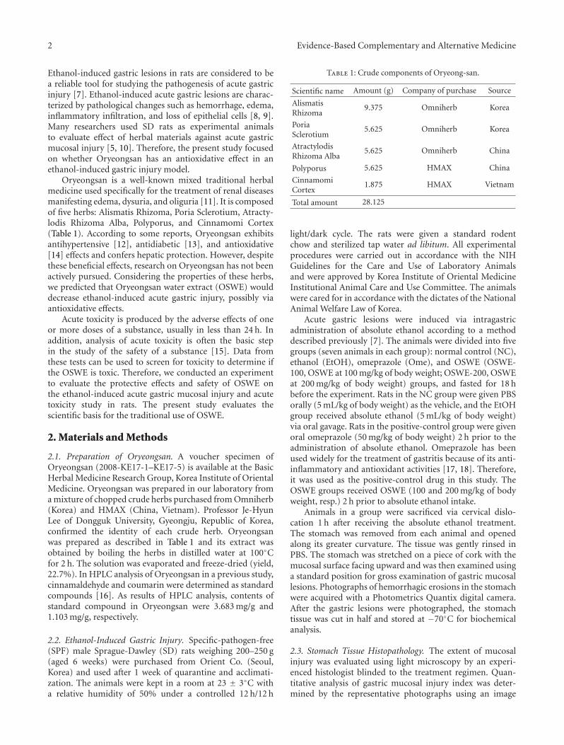
3.1. Acute Toxicity of OSWE. We evaluated the acute toxicityof OSWE, to investigate the safety of its oral administration.As shown in Figure 1, there were no significant differencesin body weight changes between the OSWE-treated and NCgroups for male and female rats. In addition, there wereno observed clinical signs and gross findings in the OSWE-treated groups and NC groups, with the exception of loss offur (n = 1) in the NC group (data not shown).
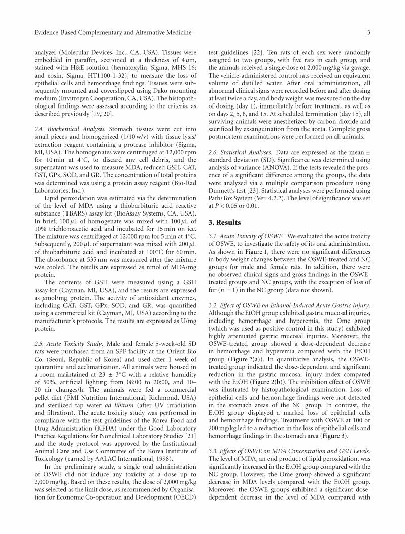
3.2. Effect of OSWE on Ethanol-Induced Acute Gastric Injury.Although the EtOH group exhibited gastric mucosal injuries,including hemorrhage and hyperemia, the Ome group(which was used as positive control in this study) exhibitedhighly attenuated gastric mucosal injuries. Moreover, theOSWE-treated group showed a dose-dependent decreasein hemorrhage and hyperemia compared with the EtOHgroup (Figure 2(a)). In quantitative analysis, the OSWE-treated group indicated the dose-dependent and significantreduction in the gastric mucosal injury index comparedwith the EtOH (Figure 2(b)). The inhibition effect of OSWEwas illustrated by histopathological examination. Loss ofepithelial cells and hemorrhage findings were not detectedin the stomach areas of the NC group. In contrast, theEtOH group displayed a marked loss of epithelial cellsand hemorrhage findings. Treatment with OSWE at 100 or200 mg/kg led to a reduction in the loss of epithelial cells andhemorrhage findings in the stomach area (Figure 3).
3.3. Effects of OSWE on MDA Concentration and GSH Levels.The level of MDA, an end product of lipid peroxidation, wassignificantly increased in the EtOH group compared with theNC group. However, the Ome group showed a significantdecrease in MDA levels compared with the EtOH group.Moreover, the OSWE groups exhibited a significant dose-dependent decrease in the level of MDA compared with
4 Evidence-Based Complementary and Alternative Medicine
0
100
200
300
400
500
Day 1 Day 2 Day 5 Day 8 Day 15
Bod
y w
eigh
t ch
ange
(g)
2000 mg/kg group 2000 mg/kg groupNC group NC group
Figure 1: Body weight changes in animals treated with OSWEat dose levels of 0 mg/kg (©) and 2,000 mg/kg (•) in males and0 mg/kg (
�) and 2,000 mg/kg (�) in females. There were no
significant differences in body weight between the OSWE-treatedand control groups.
the EtOH group (Figure 4(a)). Conversely, the levels ofGSH, a strong antioxidant, were significantly increased inthe OSWE-200 group compared with the EtOH group(Figure 4(b)).
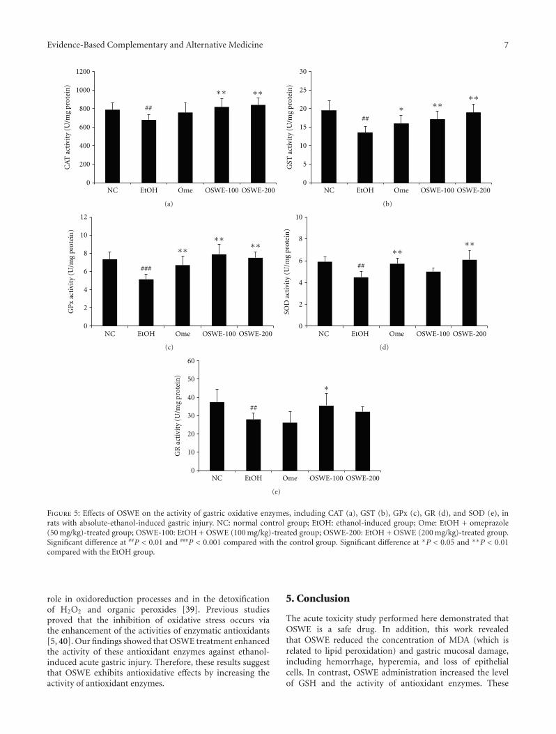
3.4. Effects of OSWE on the Activity of Antioxidant Enzymes.The activity of antioxidant enzymes, including CAT, GST,GPx, SOD, and GR, in the EtOH group was signifi-cantly decreased compared with the NC group. However,administration of OSWE significantly increased the ethanol-mediated decrease in the activity of these enzymes; theseeffects were similar to those observed for the NC groupand were significantly different (CAT; P < 0.01 in 100 and200 mg/kg, GST; P < 0.01 in 100 and 200 mg/kg, GPx; P <0.01 in 100 and 200 mg/kg, SOD; P < 0.01 in 200 mg/kg, GR;P < 0.01 in 100 mg/kg) from those obtained for the grouptreated only with ethanol (Figures 5(a) and 5(e)).
4. Discussion
The main purpose of the present study was to investigatewhether OSWE has a protective effect on ethanol-inducedacute gastric mucosal injury in the rat and whether it canbe used safely (as assessed using an acute toxicity study).OSWE administration led to recovery from acute gastricinjury (which included hemorrhage, hyperemia, and lossof epithelial cells) and to reduction of MDA concentration,increase of GSH levels, and enhancement of the activity ofantioxidant enzymes.
The acute toxicity study is a basic step in the determi-nation of the safety of materials. Our acute toxicity study,which was performed according to OECD guidelines, showedthat OSWE is a safe material when administered to rats in asingle dose via oral gavage, at a dose level of 2,000 mg/kg.
In addition, clinical signs and gross findings of treatment-related adverse effects were not observed in any of the OSWE-treated Crl:CD (SD) rats (data not shown).
Ethanol is an ulcerogenic agent that is used often andproduces severe gastric hemorrhagic lesions when given torats via gavage [10]. Ethanol-induced acute gastric lesions arecharacterized by pathological changes such as hemorrhagiclesions, mucosal edema, inflammatory cell infiltration, andloss of epithelial cells [8, 24]. A previous study reportedthat the mechanism of ethanol-induced gastric lesionsvaries, including damaged mucosal blood flow and mucosalcell injury. In the present study, ethanol-induced gastriclesions were caused by severe gastric damage, includinghemorrhage, hyperemia, and loss of epithelial cells. Severalstudies and our findings yielded similar results regardingethanol-induced damage [2, 25]. However, administrationof OSWE significantly attenuated the acute gastric injuryinduced by absolute ethanol. These results demonstrated thatOSWE has a protective effect that occurs via the reductionof hemorrhage, hyperemia, and loss of epithelial cells thatare associated with gastric mucosal injury. Similar to ourresults, earlier studies reported that the administration of anartichoke leaf extract confers gastroprotection by attenuatinggastric lesions (including hemorrhage and hyperemia) [26].
In addition, ethanol-induced gastric mucosal injury isassociated with overproduction of free radicals, which leadsto increased lipid peroxidation [27, 28]. Lipid peroxidationis one of the major outcomes of free-radical-mediated injury,which immediately causes damage to the cell membrane andis related to DNA damage [29]. MDA, an end product oflipid peroxidation, is used as a marker of tissue damage[30]. Moreover, accumulation of MDA is considered areliable biomarker of the degree of oxidative stress [31].As mentioned above, in many studies, ethanol led to anincrease in lipid peroxidation and a decrease in the activityof antioxidant enzymes, which are two of the factors thatare important in the pathogenesis of ethanol-induced gastricinjury. Our results showed that OSWE attenuated theincrease in MDA concentration in the gastric mucosa injuredby ethanol, thus indicating that OSWE can attenuate theprocess of lipid peroxidation implicated in the pathogenesisof ethanol-induced gastric damage.
Among the components of the OSWE herbal mix, wewere able to estimate, based on previous studies, that thetherapeutic effect of OSWE is exerted via the antioxidativeeffect of Alismatis Rhizoma [32], Poria Sclerotium [33],and Polyporus [34], the antiulcer effect of AtractylodisRhizoma Alba [35], the anti-inflammatory effect of Cin-namomi Cortex [36]. In oriental herbal medicine, a mixtureof several herbs is considered to enhance or prolong thepharmacological activities of any component herb anddecrease its toxic effects [37]. Considering this theory, OSWEwas considered to exhibit the synergic effects when eachherb was formulated. Therefore, the present study focusedon determining whether Oryeongsan has an antioxidativeeffect that is associated with a reduction of oxidative stress.However, we did not investigate the effects of each herbin OSWE against ethanol-induced gastric mucosal injury.Because of this reason, we did not evaluate the differences of
Evidence-Based Complementary and Alternative Medicine 5
OSWE-100 OSWE-200
NC EtOH Ome
(a)
0
10
20
30
40
50
60
NC EtOH Ome OSWE-100 OSWE-200
Gas
tric
mu
cosa
l in
jury
inde
x
∗∗∗
∗∗
###
(b)
Figure 2: Representative photographs (a) and gastric mucosal injury index (b) of gastric mucosa with absolute-ethanol-inducedgastric injuries. Gastric mucosal injury index (b) determined the findings of representative photographs using an image analyzer. NC:normal control group; EtOH: ethanol-induced group; Ome: EtOH + omeprazole (50 mg/kg)-treated group; OSWE-100: EtOH + OSWE(100 mg/kg)-treated group; OSWE-200: EtOH + OSWE (200 mg/kg)-treated group. Each bar represents the mean ± SD of seven rats.Significant differences at ###P < 0.001 compared with the control group. Significant differences at ∗P < 0.05 and ∗∗P < 0.01 comparedwith the EtOH group.
beneficial effects between each herb and OSWE. In furtherstudy, we will conduct the studies on effects of activecomponent herbs against ethanol-induced gastric mucosalinjury.
Oxidative stress has been reported to be involved inmany diseases. In addition, it is considered to cause gastricmucosal injuries. ROS production by oxidative stress iswell controlled by antioxidant defenses under physiologicalconditions [38]. The antioxidant defense system includesGSH, CAT, GST, GPx, SOD, and GR, which are known as the
main constituents of the intracellular protective mechanismthat acts against oxidative injury [18].
GSH is a well-known antioxidant that is commonlypresent as the most abundant low-molecular-mass thiol inmost organisms. It has multiple functions in the defenseagainst oxidative stress [10]. Previous studies were per-formed to evaluate the gastroprotective effects of test drugsfocusing on the alteration of GSH levels in gastric tissue [10,25]. The present study showed that OSWE administrationsignificantly increased the levels of GSH in gastric tissue

6 Evidence-Based Complementary and Alternative Medicine
OSWE-100 OSWE-200
NC EtOH Ome
Figure 3: Histopathological examination of gastric mucosa with absolute-ethanol-induced gastric injury. NC: normal control group; EtOH:ethanol-induced group; Ome: EtOH + omeprazole (50 mg/kg)-treated group; OSWE-100: EtOH + OSWE (100 mg/kg)-treated group;OSWE-200: EtOH + OSWE (200 mg/kg)-treated group.
0
100
200
300
400
500
600
700
NC EtOH Ome OSWE-100 OSWE-200
MD
A c
once
ntr
atio
n
(nm
ol/m
g pr
otei
n) ∗
∗∗∗∗
##
(a)
0
2
4
6
8
10
12
14
NC EtOH Ome OSWE-100 OSWE-200
∗∗
∗∗###
GSH
con
ten
ts (
U/m
g pr
otei
n)
(b)
Figure 4: Effects of OSWE on gastric MDA concentration (a) and GSH levels (b) in rats with absolute-ethanol-induced gastric injury. NC:normal control group; EtOH: ethanol-induced group; Ome: EtOH + omeprazole (50 mg/kg)-treated group; OSWE-100: EtOH + OSWE(100 mg/kg)-treated group; OSWE-200: EtOH + OSWE (200 mg/kg)-treated group. Each bar represents the mean ± SD of seven rats.Significant differences at ###P < 0.001 and ##P < 0.01 compared with the control group. Significant differences at ∗P < 0.05 and ∗∗P <0.01 compared with the EtOH group.
damaged by ethanol. Thus, these results indicate that OSWEexhibits gastroprotective effects against ethanol-induced gas-tric damage via enhancement of GSH levels.
The antioxidant enzymes CAT and GPx play an impor-tant role in the detoxification of hydrogen peroxide (H2O2).GST consists of a group of isoenzymes that are capable ofdetoxifying various exogenous and endogenous substances
by conjugation with glutathione. SOD, a family of enzymes,defends organisms against oxygen free radicals by catalyzingthe elimination of the superoxide radical. This radicaldamages the cell membrane and other biological structures.GR is a glutathione regenerating enzyme that permits theconversion of oxidized glutathione (GSSG) to the reducedform (GSH) [5]. Reduced glutathione plays an important
Evidence-Based Complementary and Alternative Medicine 7
0
200
400
600
800
1000
1200
NC EtOH Ome OSWE-100 OSWE-200
CA
T a
ctiv
ity
(U/m
g pr
otei
n)
∗∗∗∗
##
(a)
0
5
10
15
20
25
30
NC EtOH Ome OSWE-100 OSWE-200
GST
act
ivit
y (U
/mg
prot
ein
)
∗∗∗
∗∗##
(b)
0
2
4
6
8
10
12
NC EtOH Ome OSWE-100 OSWE-200
GP
x ac
tivi
ty (
U/m
g pr
otei
n)
∗∗∗∗ ∗∗
###
(c)
0
2
4
6
8
10
NC EtOH Ome OSWE-100 OSWE-200
SOD
act
ivit
y (U
/mg
prot
ein
)
∗∗∗∗
##
(d)
0
10
20
30
40
50
60
NC EtOH Ome OSWE-100 OSWE-200
GR
act
ivit
y (U
/mg
prot
ein
)
##
∗
(e)
Figure 5: Effects of OSWE on the activity of gastric oxidative enzymes, including CAT (a), GST (b), GPx (c), GR (d), and SOD (e), inrats with absolute-ethanol-induced gastric injury. NC: normal control group; EtOH: ethanol-induced group; Ome: EtOH + omeprazole(50 mg/kg)-treated group; OSWE-100: EtOH + OSWE (100 mg/kg)-treated group; OSWE-200: EtOH + OSWE (200 mg/kg)-treated group.Significant difference at ##P < 0.01 and ###P < 0.001 compared with the control group. Significant difference at ∗P < 0.05 and ∗∗P < 0.01compared with the EtOH group.
role in oxidoreduction processes and in the detoxificationof H2O2 and organic peroxides [39]. Previous studiesproved that the inhibition of oxidative stress occurs viathe enhancement of the activities of enzymatic antioxidants[5, 40]. Our findings showed that OSWE treatment enhancedthe activity of these antioxidant enzymes against ethanol-induced acute gastric injury. Therefore, these results suggestthat OSWE exhibits antioxidative effects by increasing theactivity of antioxidant enzymes.
5. Conclusion
The acute toxicity study performed here demonstrated thatOSWE is a safe drug. In addition, this work revealedthat OSWE reduced the concentration of MDA (which isrelated to lipid peroxidation) and gastric mucosal damage,including hemorrhage, hyperemia, and loss of epithelialcells. In contrast, OSWE administration increased the levelof GSH and the activity of antioxidant enzymes. These
8 Evidence-Based Complementary and Alternative Medicine
results indicate that administration of OSWE has a protectiveeffect against ethanol-induced gastric mucosal injury in rats.Therefore, OSWE may be considered a gastroprotective agentagainst oxidative injury caused by ethanol.
Authors’ Contribution
W.-Y. Jeon and M.-Y. Lee contributed equally to this paper.
Acknowledgment
This study was supported by a Grant from the Korea Instituteof Oriental Medicine (K12030).
References
[1] S. Liu, W. Hou, P. Yao et al., “Heme oxygenase-1 mediates theprotective role of quercetin against ethanol-induced rat hepat-ocytes oxidative damage,” Toxicology in Vitro, vol. 26, no. 1, pp.74–80, 2012.
[2] J. M. Alvarez-Suarez, D. Dekanski, S. Ristic et al., “Strawberrypolyphenols attenuate ethanol-induced gastric lesions in ratsby activation of antioxidant enzymes and attenuation of MDAincrease,” PLoS One, vol. 6, no. 10, Article ID e25878, 2011.
[3] A. I. Cederbaum, “Introduction-serial review: alcohol, oxida-tive stress and cell injury,” Free Radical Biology & Medicine, vol.31, no. 12, pp. 1524–1526, 2001.
[4] H. Tang, B. M. Sebastian, A. Axhemi et al., “Ethanol-inducedoxidative stress via the CYP2E1 pathway disrupts adiponectinsecretion from adipocytes,” Alcoholism, vol. 36, no. 2, pp. 214–222, 2012.
[5] T. Ramesh, S. W. Kim, J. H. Sung et al., “Effect of fermentedPanax ginseng extract (GINST) on oxidative stress and antiox-idant activities in major organs of aged rats,” ExperimentalGerontology, vol. 47, no. 1, pp. 77–84, 2012.
[6] A. Dongan and I. Celik, “Hepatoprotective and antioxidant ac-tivities of grapeseeds against ethanol-induced oxidative stressin rats,” British Journal of Nutrition, vol. 107, no. 1, pp. 45–51,2012.
[7] A. Robert, J. E. Nezamis, C. Lancaster, and A. J. Hanchar, “Cy-toprotectio by prostaglandins in rats. Prevention of gastric ne-crosis produced by alcohol, HCl, NaOH, hypertonic NaCl, andthermal injury,” Gastroenterology, vol. 77, no. 3, pp. 433–443,1979.
[8] J. V. R. Medeiros, G. G. Gadelha, S. J. Lima et al., “Role of theNO/cGMP/KATP pathway in the protective effects of sildenafilagainst ethanol-induced gastric damage in rats,” British Jour-nal of Pharmacology, vol. 153, no. 4, pp. 721–727, 2008.
[9] M. I. G. Silva, B. A. Moura, M. R. De Aquino Neto et al., “Gas-troprotective activity of isopulegol on experimentally inducedgastric lesions in mice: investigation of possible mechanismsof action,” Naunyn-Schmiedeberg’s Archives of Pharmacology,vol. 380, no. 3, pp. 233–245, 2009.
[10] F. M. Birdane, M. Cemek, Y. O. Birdane, I. Gulcin, and M. E.Buyukokuroglu, “Beneficial effects of Foeniculum vulgare onethanol-induced acute gastric mucosal injury in rats,” WorldJournal of Gastroenterology, vol. 13, no. 4, pp. 607–611, 2007.
[11] L. He, X. Rong, J. M. Jiang, P. Q. Liu, and Y. Li, “Ameliorationof anti-cancer agent adriamycin-induced nephrotic syndromein rats by Wulingsan (Gorei-San), a blended traditional Chi-nese herbal medicine,” Food and Chemical Toxicology, vol. 46,no. 5, pp. 1452–1460, 2008.
[12] C. Kiga, H. Goto, H. Sakurai et al., “Effects of traditional Jap-anese (Kampo) medicines (orengedokuto, goreisan and shi-chimotsukokato) on the onset of stroke and expression pat-terns of plasma proteins in spontaneously hypertensive stroke-prone rats,” Journal of Traditional Medicines, vol. 25, no. 5-6,pp. 125–132, 2008.
[13] I. M. Liu, T. F. Tzeng, S. S. Liou, and C. J. Chang, “The amel-ioration of streptozotocin diabetes-induced renal damage byWu-Ling-San (Hoelen Five Herb Formula), a traditional Chi-nese prescription,” Journal of Ethnopharmacology, vol. 124, no.2, pp. 211–218, 2009.
[14] H. M. Shin, G. W. Kim, H. Kim, and U. S. Shin, “Effect ofpretreatment with the Oryungsan on the hepatic superoxidedismutase activity and lipid peroxidation in CCl4-treatedrats,” Journal of Oriental Physiology, vol. 11, no. 1, pp. 171–180, 1996.
[15] Z. Yang, B. Han, D. Fu, and W. Liu, “Acute toxicity of highdosage carboxymethyl chitosan and its effect on the blood pa-rameters in rats,” Journal of Materials Science, vol. 23, no. 2,pp. 457–462, 2012.
[16] C. S. Seo and H. K. Shin, “Simultaneous determination ofCinnamaldehyde and Coumarin in Oryeong-san using HPLCwith Photodiode array detector,” The Korean Journal of Orien-tal Medical Prescription, vol. 18, no. 2, pp. 251–257, 2010.
[17] D. Lapenna, S. de Gioia, G. Ciofani, D. Festi, and F. Cuccurullo,“Antioxidant properties of omeprazole,” FEBS Letters, vol. 382,no. 1-2, pp. 189–192, 1996.
[18] G. Sener-Muratoglu, K. Paskaloglu, S. Arbak, C. Hurdag, andG. Ayanoglu-Dulger, “Protective effect of famotidine, omepra-zole, and melatonin against acetylsalicylic acid-induced gastricdamage in rats,” Digestive Diseases and Sciences, vol. 46, no. 2,pp. 318–330, 2001.
[19] L. Laine and W. M. Weinstein, “Histology of alcoholic hemor-rhagic “gastritis” a prospective evaluation,” Gastroenterology,vol. 94, no. 6, pp. 1254–1262, 1988.
[20] M. Y. Lee, I. S. Shin, W. Y. Jeon et al., “Protective effect of Bo-jungikki-tang, a traditional herbal formula, against alcohol-induced gastric injury in rats,” Journal of Ethnopharmacology,vol. 142, no. 2, pp. 346–353, 2012.
[21] Organization for Economic Cooperation and Development(OECD), “OECD Principles of Good Laboratory Practice,”1997, http://search.oecd.org/officialdocuments/displaydocu-mentpdf/?doclanguage=en&cote=env/mc/chem(98)17.
[22] Organization for Economic Cooperation and Development(OECD), “OECD Guidelines for Testing of Chemical No. 423,Acute Oral Toxicity-Acute Class Method,” 2001, http://www.oecd.org/chemicalsafety/assessmentofchemicals/1948370.pdf
[23] C. W. Dunnett, “New tables for multiple comparisons withcontrol,” Biometrics, vol. 20, pp. 482–491, 1964.
[24] M. Guslandi, “Effects of ethanol on the gastric mucosa,” Diges-tive Diseases, vol. 5, no. 1, pp. 21–32, 1987.
[25] Y. G. Li, D. F. Ji, T. B. Lin, S. Zhong, G. Y. Hu, and S. Chen,“Protective effect of sericin peptide against alcohol-inducedgastric injury in mice,” Chinese Medical Journal, vol. 121, no.20, pp. 2083–2087, 2008.
[26] K. Ishida, R. Kojima, M. Tsuboi, Y. Tsuda, and M. Ito, “Effectsof artichoke leaf extract on acute gastric mucosal injury inrats,” Biological & Pharmaceutical Bulletin, vol. 33, no. 2, pp.223–229, 2010.
[27] N. Osakabe, C. Sanbongi, M. Yamagishi, T. Takizawa, and T.Osawa, “Effects of polyphenol substances derived from Theo-broma cacao on gastric mucosal lesion induced by ethanol,”Bioscience, Biotechnology and Biochemistry, vol. 62, no. 8, pp.1535–1538, 1998.
Evidence-Based Complementary and Alternative Medicine 9
[28] A. Kahraman, N. Erkasap, T. Koken, M. Serteser, F. Aktepe,and S. Erkasap, “The antioxidative and antihistaminic prop-erties of quercetin in ethanol-induced gastric lesions,” Toxicol-ogy, vol. 183, no. 1–3, pp. 133–142, 2003.
[29] J. J. Fortunato, F. R. Agostinho, G. Z. Reus, F. C. Petronilho,F. Dal-Pizzol, and J. Quevedo, “Lipid peroxidative damage onmalathion exposure in rats,” Neurotoxicity Research, vol. 9, no.1, pp. 23–28, 2006.
[30] H. Ohkawa, N. Ohishi, and K. Yagi, “Assay for lipid peroxidesin animal tissues by thiobarbituric acid reaction,” AnalyticalBiochemistry, vol. 95, no. 2, pp. 351–358, 1979.
[31] N. Traverso, S. Menini, E. P. Maineri et al., “Malondialdehyde,a lipoperoxidation-derived aldehyde, can bring about second-ary oxidative damage to proteins,” Journals of Gerontology A,vol. 59, no. 9, pp. 890–895, 2004.
[32] S. E. Kim, D. Y. Rhyu, T. Yokozawa, and J. C. Park, “Antioxi-dant effect of Alisma plantago-aquatica var. orientale and itsmain component,” Korean Journal of Pharmacognosy, vol. 38,no. 4, pp. 372–375, 2007.
[33] Y. H. Park, I. H. Son, B. Kim, Y. S. Lyu, H. I. Moon, and H. W.Kang, “Poria cocos water extract (PCW) protects PC12 neu-ronal cells from beta-amyloid-induced cell death through an-tioxidant and antiapoptotic functions,” Die Pharmazie, vol. 64,no. 11, pp. 760–764, 2009.
[34] Y. D. Ha, “Antitumoral, antioxidant and antimicrobial activi-ties of solvent fractions from Grifola umbellatus,” Korean Jour-nal of Postharvest Science and Technology, vol. 8, no. 4, pp. 481–487, 2001.
[35] H. Matsuda, Y. H. Li, K. Taniguchi, J. Yamahara, and Y. Tamai,“Imaging analysis of antiulcer action and the active constitu-ent of atractylodis rhizoma,” Yakugaku Zasshi, vol. 111, no. 1,pp. 36–39, 1991.
[36] M. Kubo, S. Ma, J. Wu, and H. Matsuda, “Anti-inflammatoryactivities of 70% methanolic extract from cinnamomi cortex,”Biological & Pharmaceutical Bulletin, vol. 19, no. 8, pp. 1041–1045, 1996.
[37] E. Hosoya, “Scientific reevaluation of Kampo prescriptionusing modern technology,” in Recent Advances in the Pharma-cology of Kampo (Japanese Herbal) Medicine, E. Hosoya, Ed.,pp. 17–29, 1988.
[38] V. P. Skulachev, “Cytochrome c in the apoptotic and antioxi-dant cascades,” FEBS Letters, vol. 423, no. 3, pp. 275–280, 1998.
[39] D. Armstrong, Free Radical and Antioxidant Protocols, Human-a Press, 1998, Edited by D. Armstrong.
[40] J. R. Noh, Y. H. Kim, G. T. Gang et al., “Hepatoprotectiveeffects of chestnut (Castanea crenata) inner shell extractagainst chronic ethanol-induced oxidative stress in C57BL/6mice,” Food and Chemical Toxicology, vol. 49, no. 7, pp. 1537–1543, 2011.

Submit your manuscripts athttp://www.hindawi.com
Stem CellsInternational
Hindawi Publishing Corporationhttp://www.hindawi.com Volume 2014
Hindawi Publishing Corporationhttp://www.hindawi.com Volume 2014
MEDIATORSINFLAMMATION
of
Hindawi Publishing Corporationhttp://www.hindawi.com Volume 2014
Behavioural Neurology
EndocrinologyInternational Journal of
Hindawi Publishing Corporationhttp://www.hindawi.com Volume 2014
Hindawi Publishing Corporationhttp://www.hindawi.com Volume 2014
Disease Markers
Hindawi Publishing Corporationhttp://www.hindawi.com Volume 2014
BioMed Research International
OncologyJournal of
Hindawi Publishing Corporationhttp://www.hindawi.com Volume 2014
Hindawi Publishing Corporationhttp://www.hindawi.com Volume 2014
Oxidative Medicine and Cellular Longevity
Hindawi Publishing Corporationhttp://www.hindawi.com Volume 2014
PPAR Research
The Scientific World JournalHindawi Publishing Corporation http://www.hindawi.com Volume 2014
Immunology ResearchHindawi Publishing Corporationhttp://www.hindawi.com Volume 2014
Journal of
ObesityJournal of
Hindawi Publishing Corporationhttp://www.hindawi.com Volume 2014
Hindawi Publishing Corporationhttp://www.hindawi.com Volume 2014
Computational and Mathematical Methods in Medicine
OphthalmologyJournal of
Hindawi Publishing Corporationhttp://www.hindawi.com Volume 2014
Diabetes ResearchJournal of
Hindawi Publishing Corporationhttp://www.hindawi.com Volume 2014
Hindawi Publishing Corporationhttp://www.hindawi.com Volume 2014
Research and TreatmentAIDS
Hindawi Publishing Corporationhttp://www.hindawi.com Volume 2014
Gastroenterology Research and Practice
Hindawi Publishing Corporationhttp://www.hindawi.com Volume 2014
Parkinson’s Disease
Evidence-Based Complementary and Alternative Medicine
Volume 2014Hindawi Publishing Corporationhttp://www.hindawi.com