propagated signaling: the action potential chapter 7 we saw how an action p0-tential arises from...
TRANSCRIPT

9
Propagated Signaling: The Action Potential
~
The Action Potential Is Generated by the Flow of IonsThrough Voltage-Gated Channels
Sodium and Potassium Currents Through Voltage-GatedChannels Are Recorded With the Voltage Clamp
Voltage-Gated Sodium and Potassium Conductances AreCalculated From Their Currents
~
The Action Potential Can Be Reconstructed From theProperties of Sodium and Potassium Channels
Variations in the Properties of Voltage-Gated Ion ChannelsIncrease the Signaling Capabilities of Neurons
The Nervous System Expresses a Rich Variety of Voltage-Gated Ion Channels
Gating of Voltage-Sensitive Ion Channels Can BeInfluenced by Various Cytoplasmic Factors
Excitability Properties Vary Between Regions of theNeuron
Excitability Properties Vary Among Neurons
The Signaling Functions of Voltage-Gated Channels Can BeRelated to Their Molecular Structures
Opening of Voltage-Gated Channels Is All-or-None
Redistribution of Charges Within Voltage-Gated SodiumChannels Controls Channel Gating
The Voltage-Gated Sodium Channel Selects for Sodiumon the Basis of Size, Charge, and Energy of Hydration ofthe Ion
Genes Encoding the Potassium, Sodium, and CalciumChannels Stem From a Common Ancestor
Various Smaller Subunits Contribute to the FunctionalProperties of Voltage-Gated Sodium, Calcium, andPotassium ChannelsThe Diversity of Voltage-Gated Channel Types Is Due toSeveral Genetic Mechanisms
Mutations in Voltage-Gated Channels Cause SpecificNeurological Diseases
An Overall View
N ERVE CELLS ARE ABLE TO carry signals over longdistances because of their ability to generate anaction potential-a regenerative electrical sig-
nal whose amplitude does not attenuate as it movesdown the axon. In Chapter 7 we saw how an action p0-tential arises from sequential changes in the mem-brane's selective penneability to Na + and K+ ions. In
Chapter 8 we considered how the membrane's passiveproperties influence the speed at which action poten-tials are conducted. In this chapter we focus on thevoltage-gated ion channels that are critical for generat-ing and propagating action potentials and consider howthese channels are responsible for many important fea-tures of a neuron's electrical excitability.
The Action Potential Is Generated by the Flowof Ions Through Voltage-Gated Channels
An important early clue about how action potentials aregenerated came from an experiment performed by Ken-neth Cole and Howard Curtis. While recording from thegiant axon of the squid they found that the ion conduc-tance across the membrane increases dramatically dur-ing the action potential (Figure 9-1). This discovery pro-vided the first evidence that the action potential resultsfrom changes in the flux of ions through the channels of

the membrane. It also raised the question: Which ionsare responsible for the action potential?
A key to this problem was provided by AlanHodgkin and Bernard Katz, who found that the ampli-tude of the action potential is reduced when the externalNa + concentration is lowered, indicating that Na + in-
flux is responsible for the rising phase of the action po-tential. Their data also suggested that the falling phaseof the action potential was caused by a later increase inK+ permeability. Hodgkin and Katz proposed that de-polarization of the cell above threshold causes a brief in-crease in the cell membrane's permeability to Na +, dur-ing which Na + permeability overwhelms the dominant
permeability of the resting cell membrane to K+ ions.
Sodium and Potassium Currents ThroughVoltage-Gated Channels Are Recorded Withthe Voltage Clamp
To test this hypothesis, Hodgkin and Andrew Hux-ley conducted a second series of experiments. They sys-tematically varied the membrane potential in the squidgiant axon and measured the resulting changes in themembrane conductance to Na + and K+ through volt-age-gated Na + and K+ channels. To do this they made
use of a new apparatus, the voltage clamp. Prior to theavailability of the voltage-damp technique, attempts tomeasure Na + and K+ conductance as a function ofmembrane potential had been limited by the stronginterdependence of the membrane potential and thegating of Na + and K+ channels. For example, if themembrane is depolarized sufficiently to open some ofthe voltage-gated Na + channels, inward Na + current
flows through these channels and causes further depo-larization. The additional depolarization causes stillmore Na + channels to open and consequently inducesmore inward Na + current:
~ Depolarization ~Inward ~. Open Na+
~nelS
This positive feedback cycle, which eventually drivesthe membrane potential to the peak of the action poten-tial, makes it impossible to achieve a stable membranepotential. A similar coupling between current and mem-brane potential complicates the study of the voltage-gated K+ channels.
The basic function of the voltage clamp is to inter-rupt the interaction between the membrane potentialand the opening and closing of voltage-gated ion chan-
Chapter 9 / Propagated Signaling: The Action Potential 151
Ionic conductance./
Figure 9.1 A net increase in ionic conductance in the mem-brane of the axon accompanies the action potential. Thishistoric recording from an experiment conducted in 1938 byKenneth Cole and Howard Curtis shows the oscilloscoperecord of an action potential superimposed on a simultaneousrecord of the ionic conductance.
nels. The voltage clamp does so by injecting a currentinto the axon that is equal and opposite to the currentflowing through the voltage-gated membrane channels.In this way the voltage clamp prevents the charge sepa-ration across the membrane from changing. The amountof current that must be generated by the voltage clamp tokeep the membrane potential constant provides a directmeasure of the current flowing across the membrane(Box 9-1). Using the voltage-clamp technique, Hodgkinand Huxley provided the first complete description ofthe ionic mechanisms underlying the action potential.
One advantage of the voltage clamp is that it readilyallows the total membrane current to be separated intoits ionic and capacitive components. As described inChapter 8, the membrane potential V m, is proportionalto the charge Qm on the membrane capacitance (Cm).When V m is not changing, Qm is constant and no capac-itive current (~Qm/ ~t) flows. Capacitive current flowsonly when V m is changing. Therefore, when the mem-brane potential changes in response to a very rapid stepof command potential, capacitive current flows only atthe beginning and end of the step. Since the capacitivecurrent is essentially instantaneous, the ionic currentsthat flow through the gated membrane channels can be
analyzed separately.Measurements of these ionic membrane currents
can be used to calculate the voltage and time depen-dence of changes in membrane conductances caused bythe opening and closing of Na+ and K+ channels. Thisinformation provides insights into the properties ofthese two types of channels.A typical voltage-clamp experiment starts with themembrane potential clamped at its resting value. H a10 mV depolarizing potential step is commanded, we


observe that an initial, very brief outward current in-stantaneously discharges the membrane capacitance bythe amount required for a 10 mV depolarization. Thiscapacitive current (Ie) is followed by a smaller outwardionic current that persists for the duration of this pulse.At the end of the pulse there is a brief inward capacitivecurrent, and the total membrane current returns to zero(Figure 9-3A). The steady ionic current that persiststhroughout the depolarization is the current that flowsthrough the resting ion channels of the membrane (seeChapter 6) and is called the leakage current, II' The totalconductance of this population of channels is called theleakage conductance (gl)' These resting channels, whichare always open, are responsible for generating the rest-ing membrane potential (see Chapter 7). In a typicalneuron most of the resting channels are permeable toK+ ions; the remaining channels are permeable to 0- orNa + ions.
If a larger depolarizing step is commanded, the cur-rent record becomes more complicated. The capacitiveand leakage currents both increase in amplitude. In ad-dition, shortly after the end of the capacitive current andthe start of the leakage current, an inward current devel-ops; it reaches a peak within a few milliseconds, de-clines, and gives way to an outward current. This out-ward current reaches a plateau that is maintained forthe duration of the pulse (Figure 9-3B).
A simple interpretation of these results is that thedepolarizing voltage step sequentially turns on activeconductance channels for two separate ions: one type ofchannel for inward current and another for outwardcurrent. Because these two oppositely directed currentspartially overlap in time, the most difficult task in ana-lyzing voltage-clamp experiments is to determine theirseparate time courses.
Hodgkin and Huxley achieved this separation bychanging ions in the bathing solution. By substituting alarger, impermeant cation (choline.H+) for Na +, theyeliminated the inward Na + current. Subsequently, the
task of separating inward and outward currents wasmade easier by selectively blocking the voltage-sensitive conductance channels with drugs or toxins.Tetrodotoxin, a poison from certain Pacific puffer fish,blocks the voltage-gated Na + channel with a very high
potency in the nanomolar range of concentration.(Ingestion of only a few milligrams of tetrodotoxin fromimproperly prepared puffer fish, consumed as theJapanese sushi delicacy fugu, can be fatal.) The cationtetraethylammonium specifically blocks the voltage-gated K+ channel (Figure 9-4).
When tetraethylammonium is applied to the axonto block the K+ channels, the total membrane current(Im) consists of Ie- II, and INa. The leakage conductance,
153Chapter 9 / Propagated Signaling: The Action Potential
l~~ ~~ t-I 1/
Outward
1m 0
Inward
B
C
0
Vm
-60
1m
Vm
01m
1 ms
Time --
Figure 9-3 A voltage-clamp experiment demonstrates thesequential activation of two types of voltage-gated chan-nels.A. A small depolarization is accompanied by capacitive andleakage currents (Ie and 'I, respectively).B. A larger depolarization results in larger capacitive and leak-age currents, plus an inward current followed by an outwardcurrent.C. Depolarizing the cell in the presence of tetrodotoxin(which blocks the Na + current) and again in the presence of
tetraethylammonium (which blocks the K+ current!. revealsthe pure K+ and Na+ currents UK and 'N.. respectively) aftersubtracting 'e and II-

154 Part IT / Cell and Molecular Biology of the Neuron
Figure 9-4 Drugs that block voltage-gated Na +and K+ channels. Tetrodotoxin and saxitoxin bothbind to Na+ channels with a very high affinity.Tetrodotoxin is produced by certain puffer fish,newts, and frogs. Saxitoxin is synthesized by the di-noflagellates Gonyaulax that are responsible for redtides. Consumption of clams or other shellfish thathave fed on the dinoflagellates during a red tidecauses paralytic shellfish poisoning. Cocaine, the ac-tive substance isolated from coca leaves, was thefirst substance to be used as a local anesthetic. Italso blocks Na+ channels but with a lower affinityand specificity than tetrodotoxin. Tetraethylammo-nium is a cation that blocks certain voltage-gated K+channels with a relatively low affinity. The red plussigns represent positive charge.
gl1 is constant; it does not vary with V m or with time.Therefore, 111 the leakage current, can be readily calcu-lated and subtracted from 1m/leaving INa and Ic. BecauseIc occurs only briefly at the beginning and end of thepulse, it can be easily isolated by visual inspection, leav-ing the pure INa. The full range of current flow throughthe voltage-gated Na + channels (INa) is measured by re-
peating this analysis after stepping V m to many differ-ent levels. With a similar process, IK can be measuredwhen the Na + channels are blocked by tetrodotoxin
(Figure 9-3C).
Voltage-Gated Sodium and Potassium ConductancesAre Calculated From Their CurrentsThe Na + and K+ currents depend on two factors: the
conductance for each ion and the electrochemicaldriving force acting on the ion. Since the Na + and K+
membrane conductance is directly proportional to thenumber of open Na + and K+ channels, we can gain in-
sight into how membrane voltage controls channelopening by calculating the amplitudes and time coursesof the Na + and K+ conductance changes in response to
voltage-clamp depolarizations (Box 9-2).Measurements of Na + and K+ conductances at var-
ious levels of membrane potential reveal two functionalsimilarities and two differences between the Na + and
K+ channels. Both types of channels open in response todepolarizing steps of membrane potential. Moreover, asthe size of the depolarization increases, the probabilityand rate of opening increase for both types of channels.The Na + and K+ channels differ, however, in their rates
of opening and in their responses to prolonged depolar-
Saxitoxin
~
Tetraethylammonium
ization. At all levels of depolarization the Na + channe]
open more rapidly than do the K+ channels (Figure 9-6When the depolarization is maintained for some timlthe Na + channels begin to close, leading to a decrease <inward current. The process by which Na + channe]
close during a maintained depolarization is tenneinactivation. In contrast, the K+ channels in the squiaxon do not inactivate; they remain open as long as thmembrane is depolarized (Figure 9-7).
Thus, depolarization causes Na + channels t
undergo transitions among three different states, whicrepresent three different conformations of the Nachannel protein: resting, activated, or inactivated. Upodepolarization the channel goes from the restin(closed) state to the activated (open) state (see Figw6-6C). If the depolarization is brief, the channels go drectly back to the resting state upon repolariz~tion. :the depolarization is maintained, the channels go frorthe open to the inactivated (closed) state. Once the charnel is inactivated it cannot be opened by further dep<larization. The inactivation can be reversed only by npolarizing the membrane to its negative restinpotential, which allows the channel to switch from thinactivated to the resting state. This switch takes somtime because channels leave the inactivated state reI.tively slowly (Figure 9-8).
These variable, time-dependent effects of depolaJization on gNa are determined by the kinetics of the galing reactions that control the Na + channels. Each Na
channel has two kinds of gates that must be openesimultaneously for the channel to conduct Na + ions. A
activation gate is closed when the membrane is at itnegative resting potential and is rapidly opened by dE

polarization; an inactivation gate is open at the resting po-tential and closes slowly in response to depolarization.The channel conducts only for the brief period during adepolarization when both gates are open. Repolarizationreverses the two processes, closing the activation gaterapidly and opening the inactivation gate more slowly.After the channel has returned to the resting state, it canagain be activated by depolarization (Figure 9-9).
Chapter 9 / Propagated Signaling: The Action Potential 155
The Action Potential Can Be Reconstructed Fromthe Properties of Sodium and Potassium Channels
Hodgkin and Huxley were able to fit their measure-ments of membrane conductance changes to a set of em-pirical equations that completely describe variations inmembrane Na + and K+ conductances as functions of
membrane potential and time. Using these equations

156 Part n / Cell and Molecular Biology of the Neuron
(mY)
.w~ V. ~ I
-60 I II II I: +20 mV I
I II OmV,I
~: -20 mV I: -40 mV I
I IIIIIIIIIII
~ I,II
Time -Figure 9-6 Voltage-clamp experiments show that Na+ chan-nels turn on and off more rapidly than K+ channels over awide range of membrane potentials. The increases and de-creases in the Na+ and K+ conductances (9Na and 9\<) shownhere reflect the shifting of thousands of voltage-gated channelsbetween the open and closed states.
Vm
Time-
Figure 9.7 Sodium and potassium channels respond differ-ently to long-term depolarization. If the membrane is repolar-ized after a brief depolarization (line a), both 9/118 and 9K returnto their initial values. If depolarization is maintained (line b), theNa + channels close (or inactivate) before the depolarization is
terminated. whereas the K+ channels remain open and gK in-creases throughout the depolarization.
and measured values for the passive properties of theaxon, they computed the expected shape and the con-duction velocity of the propagated action potential. Thecalculated waveform of the action potential matched thewaveform recorded in the undamped axon almost per-fectly! This dose agreement indicates that the voltageand time dependence of the active Na + and K+ chan-
nels, calculated from the voltage-clamp data, accuratelydescribe the properties of the channels that are essentialfor generating and propagating the action potential. Ahalf century later, the Hodgkin-Huxley model stands asthe most successful quantitative computational modelin neural science if not in all of biology.
According to the Hodgkin-Huxley model, an actionpotential involves the following sequence of events. Adepolarization of the membrane causes Na + channels to
open rapidly (an increase in gNa), resulting in an inwardNa + current. This current, by discharging the mem-
brane capacitance, causes further depolarization, there-
I i 1 ms
~
VmP, P2
Figure 9-8 Sodium channels remain inactivated for a fewmilliseconds after the end of a depolarization. Therefore ifthe interval between two depolarizing pulses (P, and P2) isbrief, the second pulse produces a smaller increase in 9'N. be-cause many of the Na + channels are inactivated. The longer
the interval between pulses, the greater the increase in 9'N.,because a greater fraction of channels will have recovered frominactivation and returned to the resting state when the secondpulse begins. The time course of recovery from inactivationcontributes to the time course of the refractory period.
O.5ms

Figure 9-9 Voltage-gated Na + channels havetwo gates, which respond in opposite waysto depolarization. In the resting (closed) statethe activation gate is closed and the inactiva-tion gate is open (1). Upon depolarization arapid opening of the activation gate allows Na+to flow through the channel (2). As the inactiva-tion gates close, the Na + channels enter the in-activated (closed) state (3). Upon repolarization,first the activation gate closes, then the inacti-vation gate opens as the channel returns to theresting state (1).
by opening more Na + channels, resulting in a further
increase in inward current. This regenerative processdrives the membrane potential toward ENa, causing therising phase of the action potentiaL} The depolarizingstate of the action potential then limits the duration ofthe action potential in two ways: (1) It gradually inacti-vates the Na + channels, thus reducing gNa, and (2) it
opens, with some delay, the voltage-gated K+ channels,thereby increasing gK' Consequently, the inward Na +
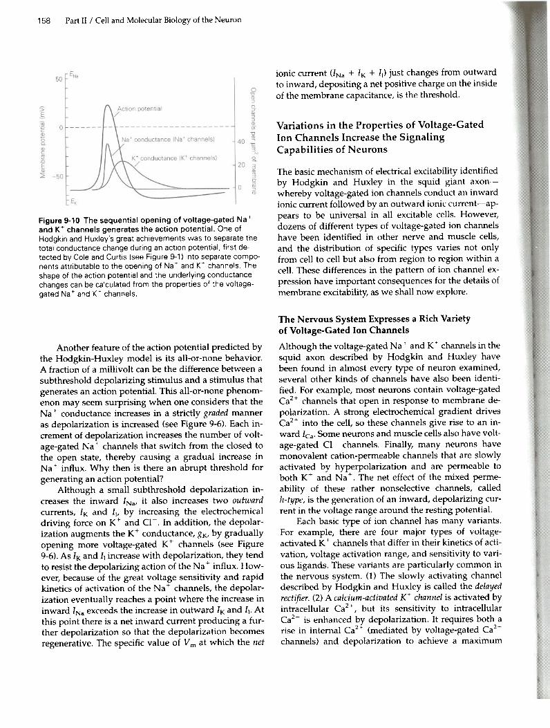
current is followed by an outward K+ current that tendsto repolarize the membrane (Figure 9-10).
1. It may at first seem paradoxical that to depolarize the cell exper-imentally one passes outward current across the membrane (see Fig-ure 7-2C), while at the same time attributing the depolarizationduring the upstroke of the action potential to an inward Na+ cur-rent. However, in both cases the current flow across the passivecomponents, the nongated leakage channels (gt) and the capaci-tance of the membrane (Cm), is outward because positive charge isinjected into the cell in one case through an intracellular electrode(see Figure 7-2) and in the other case by the opening of voltage-gated Na + channels. It is a matter of convention that when we refer
to current injected through a microelectrode we refer to the direc-tion in which the current crosses the membrane capacitance andleakage channels, whereas when we refer to current that flowsthrough channels we refer to the direction of movement of chargethrough the channels.
Chapter 9 / Propagated Signaling: The Action Potential 157
1 Resting (closed) 2 Activated (open)
Na.
Fastchannelopening .
+ + + + + +
'-.JSIow
3 Inactivated (closed)
In most nerve cells the action potential is followedby a transient hyperpolarization, the after-potential. Thisbrief increase in membrane potential occurs because theK+ channels that open during the later phase of the ac-tion potential close some time after V m has returned toits resting value. It takes a few milliseconds for all of thevoltage-gated K+ channels to return to the closed state.During this time, when the permeability of the mem-brane to K+ is greater than during the resting state, V mis hyperpolarized slightly with respect to its normalresting value, resulting in a V m closer to EK (Figure9-10).
The action potential is also followed by a brief pe-riod of diminished excitability, or refractoriness, whichcan be divided into two phases. The absolute refractoryperiod comes immediately after the action potential; dur-ing this period it is impossible to excite the cell no mat-ter how great a stimulating current is applied. Thisphase is followed directly by the relative refractory period,during which it is possible to trigger an action potentialbut only by applying stimuli that are stronger thanthose normally required to reach threshold. These peri-ods of refractoriness, which together last just a fewmilliseconds, are caused by the residual inactivation ofNa + channels and increased opening of K+ channels.
Part n / Cell and Molecular Biology of the Neuron158
Figure 9.10 The sequential opening of voltage-gated Na+and K+ channels generates the action potential. One ofHodgkin and Huxley's great achievements was to separate thetotal conductance change during an action potential. first de-tected by Cole and Curtis (see Figure 9-1) into separate compo-nents attributable to the opening of Na+ and K+ channels. Theshape of the action potential and the underlying conductancechanges can be calculated from the properties of the voltage-gated Na + and K+ channels.
Another feature of the action potential predicted bythe Hodgkin-Huxley model is its all-or-none behavior.A fraction of a millivolt can be the difference between asubthreshold depolarizing stimulus and a stimulus thatgenerates an action potential. This all-or-none phenom-enon may seem surprising when one considers that theNa + conductance increases in a strictly graded manneras depolarization is increased (see Figure 9-6). Each in-crement of depolarization increases the number of volt-age-gated Na + channels that switch from the closed tothe open state, thereby causing a gradual increase inNa + influx. Why then is there an abrupt threshold for
generating an action potential?Although a small subthreshold depolarization in-
creases the inward INa, it also increases two outwardcurrents, IK and Iv by increasing the electrochemicaldriving force on K+ and Cl-. In addition, the depolar-ization augments the K+ conductance, gKt by graduallyopening more voltage-gated K+ channels (see Figure9-6). As IK and II increase with depolarization, they tendto resist the depolarizing action of the Na + influx. How-
ever, because of the great voltage sensitivity and rapidkinetics of activation of the Na + channels, the depolar-
ization eventually reaches a point where the increase ininward INa exceeds the increase in outward IK and II' Atthis point there is a net inward current producing a fur-ther depolarization so that the depolarization becomesregenerative. The specific value of V m at which the net
ionic current (lNa + IK + II) just changes from outwardto inward, depositing a net positive charge on the insideof the membrane capacitance, is the threshold.
Variations in the Properties of Voltage-GatedIon Channels Increase the SignalingCapabilities of Neurons
The basic mechanism of electrical excitability identifiedby Hodgkin and Huxley in the squid giant axon-whereby voltage-gated ion channels conduct an inwardionic current followed by an outward ionic current-ap-pears to be universal in all excitable cells. However,dozens of different types of voltage-gated ion channelshave been identified in other nerve and muscle cells,and the distribution of specific types varies not onlyfrom cell to cell but also from region to region within acell. These differences in the pattern of ion channel ex-pression have important consequences for the details ofmembrane excitability, as we shall now explore.
The Nervous System Expresses a Rich Varietyof Voltage-Gated Ion ChannelsAlthough the voltage-gated Na + and K+ channels in the
squid axon described by Hodgkin and Huxley havebeen found in almost every type of neuron examined,several other kinds of channels have also been identi-fied. For example, most neurons contain voltage-gatedCa2+ channels that open in response to membrane de-polarization. A strong electrochemical gradient drivesCa2+ into the cell, so these channels give rise to an in-ward lea. Some neurons and muscle cells also have volt-age-gated CI- channels. Finally, many neurons havemonovalent cation-permeable channels that are slowlyactivated by hyperpolarization and are permeable toboth K+ and Na+. The net effect of the mixed perme-ability of these rather nonselective channels, calledh-type, is the generation of an inward, depolarizing cur-rent in the voltage range around the resting potential.
Each basic type of ion channel has many variants.For example, there are four major types of voltage-activated K+ channels that differ in their kinetics of acti-vation, voltage activation range, and sensitivity to vari-ous ligands. These variants are particularly common inthe nervous system. (1) The slowly activating channeldescribed by Hodgkin and Huxley is called the delayedrectifier. (2) A calcium-activated K+ channel is activated byintracellular Ca2+, but its sensitivity to intracellularCa2+ is enhanced by depolarization. It requires both arise in internal Ca2+ (mediated by voltage-gated Ca2+channels) and depolarization to achieve a maximum

probability of opening. (3) The A-type K+ channel is acti-vated rapidly by depolarization, almost as rapidly asthe Na + channel; like the Na + channel, it also inacti-
vates rapidly if the depolarization is maintained. (4) TheM-type K+ channel is very slowly activated by small de-polarizations from the resting potential. One distinctivefeature of the M-type channels is that they can be closedby a neurotransmitter, acetylcholine (ACh).
Similarly, there are at least five subtypes of voltage-gated Ca2+ channels and two or more types of voltage-gated Na + channels. Moreover, each of these subtypes
has several structurally and functionally different iso-forms.
The squid axon can generate an action potentialwith just two types of voltage-gated channels. Why thenare so many different types of voltage-gated ion chan-nels found in the nervous system? The answer is thatneurons with an expanded set of voltage-gated channelshave much more complex information-processing abili-ties than those with only two types of channels. Someways in which this large variety of voltage-gated chan-nels influences neuronal function are described below.
Gating of Voltage-Sensitive Ion Channels CanBe Influenced by Various Cytoplasmic Factors
In a typical neuron the opening and closing of certainvoltage-gated ion channels can be modulated by vari-ous cytoplasmic factors, resulting in increased flexibilityof the neuron's excitability properties. Changes in suchcytoplasmic modulator substances may result from thenormal intrinsic activity of the neuron itself or from theinfluences of other neurons.
The flow of ionic current through membrane chan-nels during an action potential generally does not resultin appreciable changes in the intracellular concentra-tions of most ion species. Calcium is a notable exceptionto this rule. Changes in the intracellular concentration ofCa2+ can have important modulatory influences on thegating of various channels. The concentration of freeea2+ in the cytoplasm of a resting cell is extremely low,about 10-7 M, several orders of magnitude below theexternal Ca2+ concentration. For this reason the intracel-lular Ca2+ concentration may increase significantly as aresult of inward current flow through voltage-gatedCa2+ channels.
The transient increase in Ca2+ concentration nearthe inside of the membrane has several effects. It en-hances the probability that Ca2+ -activated K+ channelswill open. Some Ca2+ channels are themselves sensitiveto levels of intracellular Ca2+ and are inactivated whenincoming Ca2+ binds to their intracellular surface. Inother channels the influx of Ca2+ activates a Ca2+ -sensi-
Chapter 9 / Propagated Signaling: The Action Potential 159
tive protein phosphatase, calcineurin, which dephos-phorylates the channel, thereby inactivating it (see Fig-ure 6-7C).
Thus, in some cells the Ca2+ influx during an actionpotential can have two opposing effects: (1) The positivecharge that it carries into the cell contributes to the re-generative depolarization, while (2) the increase in cyto-plasmic Ca2+ concentration results in the opening ofmore K+ channels and the closing of Ca2+ channels. Be-cause of the opening of K+ channels and the closing ofCa2+ channels, outward ionic current increases whileinward ionic current decreases; the resulting net effluxof positive charge causes the cell to repolarize. In thisway the depolarizing influx of Ca2+ through voltage-gated Ca2+ channels is self-limited by two processesthat aid repolarization: an increase in K+ efflux and adecrease in Ca2+ influx.
Calcium's role in modulating the gating of ionchannels is the simplest example of a variety of second-messenger systems that control channel activity. Gatingof ion channels can also be modulated by changes in thecytoplasmic level of small organic second-messengercompounds as a result of synaptic input from other neu-rons. The gating properties of several voltage-gatedchannels that are directly involved in generating actionpotentials are modified when their phosphorylationstate is changed by a protein kinase (eg, the cAMP-dependent protein kinase) whose activity is controlledby changes in the concentration of synaptically acti-vated second messengers (eg, cAMP). The importanceof Ca2+ and other second messengers in the control ofneuronal activity will become evident in many contextsthroughout this book.
Excitability Properties Vary Between Regionsof the Neuron
Different regions of the cell perform specific signalingtasks. The axon, for example, usually specializes in car-rying signals faithfully over long distances. As such, itfunctions as a relatively simple relay line. In contrast,the input, integrative, and output regions of a neuron(see Figure 2-8) typically perform more complex pro-cessing of the information they receive before passing italong. The signaling function of a discrete region of theneuron depends on the particular set of ion channelsthat it expresses.
In many types of neurons the dendrites havevoltage-gated ion channels, including ea2+, K+, and insome cases Na + channels. When activated, these chan-
nels modify the passive, electrotonic conduction ofsynaptic potentials. In some neurons action potentialsmay be propagated from their site of initiation at the

160 Part II / Cell and Molecular Biology of the Neuron
trigger zone back into the dendrites, thereby influencingsynaptic integration in the dendrites. In other neuronsthe density of dendritic voltage-gated channels mayeven support the orthograde propagation of a dendriticimpulse to the cell soma and axon hillock.
The trigger zone of the neuron has the lowestthreshold for action potential generation, in part be-cause it has an exceptionally high density of voltage-gated Na + channels. In addition, it typically has
voltage-gated ion channels that are sensitive to rela-tively small deviations from resting potential. Thesechannels are important in determining whether synap-tic input will drive the membrane potential to spikethreshold. They thus playa critical role in the transfor-mation of graded, analog changes in synaptic or recep-tor potentials into a temporally patterned, digital trainof all-or-none action potentials. Examples include theM-type and certain A-type K+ channels, the hyperpolar-ization-activated h-~ channels, and a class of lowvoltage-activated Ca + channels (see below).
As the action potential is carried down the axon it ismediated primarily by voltage-gated Na + and K+ chan-
nels that function much like those in the squid axon. Atthe nodes of Ranvier of myelinated axons the mecha-nism of action potential repolarization is particularlysimple-the spike is terminated by fast inactivation ofNa + channels combined with a large outward leakage
current. Voltage-gated K+ channels do not playa signif-icant role in action potential repolarization at the nodalmembrane.
Presynaptic nerve terminals at chemical synapsescommonly have a high density of voltage-gated Ca2+channels. Arrival of an action potential in the terminalopens these channels, causing ea2+ influx, which inturn triggers transmitter release.
Excitability Properties Vary Among Neurons
The computing power of an entire neural circuit is en-hanced when cells in the circuit represent a wide rangeof functional properties, because specific functionswithin the circuit can be assigned to cells with the mostappropriate dynamic properties. Thus, while the func-tion of a neuron is determined to a great extent by itsanatomical relationships to other neurons (its inputsand its outputs), the biophysical properties of the cellalso playa critical role.
How a neuron responds to synaptic input is deter-mined by the proportions of different types of voltage-gated channels in the cell's integrative and triggerzones. Cells with different combinations of channels re-spond to a constant excitatory input differently. Somecells respond with only a single action potential, others
with a constant-frequency train of action potentials, andstill others with either an accelerating or deceleratingtrain of action potentials. Some neurons even fire spon-taneously in the absence of any external input becauseof the presence of h-type channels that generate endoge-nous pacemaker currents (Figure 9-11).
In certain neurons small changes in the strength ofsynaptic inputs produce a large increase in firing rate,whereas in other cells large changes in synaptic inputare required to modulate the firing rate. In many neu-rons a steady hyperpolarizing input makes the cell lessresponsive to excitatory input by reducing the restinginactivation of the A-type K+ channels. In other neuronssuch a steady hyperpolarization makes the cell more ex-citable, because it removes the inactivation of a particu-lar class of voltage-gated Ca2+ channels. In many casesthe firing properties of a neuron can be modulated bysecond messenger-mediated changes in the function ofvoltage-gated ion channels (Figure 9-11).
The Signaling Functions of Voltage-GatedChannels Can Be Related to TheirMolecular Structures
The empirical equations derived by Hodgkin andHuxley are quite successful in describing how the flowof ions through the Na + and K+ channels generates the
action potential. However, these equations describe theprocess of excitation primarily in terms of changes inmembrane conductance and current flow. They tell lit-tie about the molecular structure of the voltage-gatedchannels and the molecular mechanisms by which theyare activated. Fortunately, technical advances such asthose described in Chapter 6 have made it possible toexamine the structure and function of the voltage-gated Na+, K+, and Ca2+ channels in detail at the mol-ecular level.
One of the first clues that Na + channels are distinct
physical entities came from studies that measured thebinding of radiolabeled tetrodotoxin to nerve mem-branes. The density of voltage-gated Na + channels in
different nerves was estimated by measuring the totalamount of tritium-labeled tetrodotoxin bound whenspecific axonal binding sites are saturated. In nonmyeli-nated axons the density of channels is quite low, rang-ing from 35 to 500 Na + channels per square micrometer
of axon membrane in different cell types. In myelinatedaxons, where the Na + channels are concentrated at the
nodes of Ranvier, the density is much higher-between1000 and 2000 channels per square micrometer of nodalmembrane. The greater the density of Na + channels in
the membrane of an axon, the greater the velocity at
A Delayed firing
vm~-55 mV
2
Delay"
Vm-75 mV
I
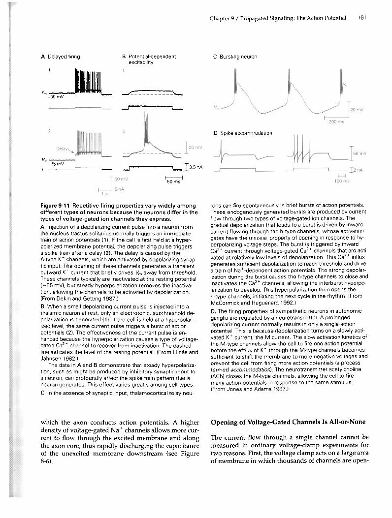
Figure 9.11 Repetitive firing properties vary widely amongdifferent types of neurons because the neurons differ in thetypes of voltage-gated ion channels they express.A. Injection of a depolarizing current pulse into a neuron fromthe nucleus tractus solitarius normally triggers an immediatetrain of action potentials (1). If the cell is first held at a hyper-polarized membrane potential, the depolarizing pulse triggersa spike train after a delay (2). The delay is caused by theA-type K+ channels, which are activated by depolarizing synap-tic input. The opening of these channels generates a transientoutward K+ current that briefly drives Vm away from threshold.These channels typically are inactivated at the resting potential(-55 mY), but steady hyperpolarization removes the inactiva-tion, allowing the channels to be activated by depolarization.(From Dekin and Getting 1987.)
B. When a small depolarizing current pulse is injected into athalamic neuron at rest. only an electrotonic, subthreshold de-polarization is generated (1). If the cell is held at a hyperpolar-ized level, the same current pulse triggers a burst of actionpotentials (2). The effectiveness of the current pulse is en-hanced because the hyperpolarization causes a type of voltage-gated Ca2+ channel to recover from inactivation. The dashedline indicates the level of the resting potential. (From Llinas andJahnsen 1982.)
The data in A and B demonstrate that steady hyperpolariza-tion, such as might be produced by inhibitory synaptic input toa neuron, can profoundly affect the spike train pattern that aneuron generates. This effect varies greatly among cell types.C. In the absence of synaptic input, thalamocortical relay neu-
which the axon conducts action potentials. A higherdensity of voltage-gated Na + channels allows more cur-
rent to flow through the excited membrane and alongthe axon core, thus rapidly discharging the capacitanceof the unexcited membrane downstream (see Figure8-6).
Chapter 9 / Propagated Signaling: The Action Potential 161
C Bursting neuronB Potential-dependentexcitability
1
--r ,--
\--.
2 D Spike accommodation
-oJ I.-. 10.6 nA
I-:-:---i5Oms
rons can fire spontaneously in brief bursts of action potentials.These endogenously generated bursts are produced by currentflow through two types of voltage-gated ion channels. Thegradual depolarization that leads to a burst is driven by inwardcurrent flowing through the h-type channels, whose activationgates have the unusual property of opening in response to hy-perpolarizing voltage steps. The burst is triggered by inwardCa2+ current through voltage-gated Ca2+ channels that are acti-vated at relatively low levels of depolarization. This ea2+ influxgenerates sufficient depolarization to reach threshold and drivea train of Na + -dependent action potentials. The strong depolar-
ization during the burst causes the h-type channels to close andinactivates the Ca2+ channels, allowing the interburst hyperpo-larization to develop. This hyperpolarization then opens theh-type channels, initiating the next cycle in the rhythm. (FromMcCormick and Huguenard 1992.)
D. The firing properties of sympathetic neurons in autonomicganglia are regulated by a neurotransmitter. A prolongeddepolarizing current normally results in only a single actionpotential. This is because depolarization turns on a slowly acti-vated K+ current, the M current. The slow activation kinetics ofthe M-type channels allow the cell to fire one action potentialbefore the efflux of K+ through the M-type channels becomessufficient to shift the membrane to more negative voltages andprevent the cell from firing more action potentials (a processtermed accommodation). The neurotransmitter acetylcholine(ACh) closes the M-type channels, allowing the cell to firemany action potentials in response to the same stimulus.(From Jones and Adams 1987.)
Opening of Voltage-Gated Is Is All-or-NoneChanne
The current flow through a single channel cannot bemeasured in ordinary voltage-damp experiments fortwo reasons. First, the voltage clamp acts on a large areaof membrane in which thousands of channels are open-

162 Part II / Cell and Molecular Biology of the Neuron
ing and closing randomly. Second, the backgroundnoise caused by the flow of current through passivemembrane channels is much larger than the flow of cur-rent through anyone channel. Both these problems canbe circumvented by electrically isolating a tiny piece ofmembrane in a patch-clamp electrode (see Box 6-1).
Patch-clamp experiments demonstrate that voltage-gated channels generally have only two conductancestates, open and closed. Each channel opens in an all-or-none fashion and, when open, permits a pulse of currentto flow with a variable duration but constant amplitude(Figure 9-12). The conductances of single voltage-gatedNa +, K+, and Ca2+ channels in the open state typicallyrange from 1 to 20 pS, depending on channel type. Oneclass of ea2+ -activated K+ channels has an unusuallylarge conductance of about 200 pS.
Redistribution of Charges Within Voltage-GatedSodium Channels Controls Channel Gating
In their original study of the squid axon, Hodgkin andHuxley suggested that a voltage-gated channel has a netcharge, the gating charge, somewhere within its wall.They postulated that a change in membrane potentialcauses this charged structure to move within the planeof the membrane, resulting in a conformational changethat causes the channel to open or close. They furtherpredicted that such a charge movement would be mea-surable. For example, when the membrane is depolar-ized a positive gating charge would move from near theinner surface toward the outer surface of the membrane,owing to its interaction with the membrane electricfield. Such a displacement of positive charge would re-duce the net separation of charge across the membraneand hence tend to hyperpolarize the membrane. To keepthe membrane potential constant in a voltage-clamp ex-periment, a small extra component of outward capaci-tive current, called gating current, would have to be gen-erated by the voltage clamp. When the membranecurrent was examined by means of very sensitive tech-niques, the predicted gating current was found to flowat the beginning and end of a depolarizing voltage-clamp step prior to the opening or closing of the Na +
channels (Figure 9-13).Analysis of the gating current reveals that activation
and inactivation of Na + channels are coupled processes.During a short depolarizing pulse net outward move-ment of gating charge within the membrane at the begin-ning of the pulse is balanced by an opposite inwardmovement of gating charge at the end of the pulse. How-ever, if the pulse lasts long enough for Na + inactivation
to take place, the movement of gating charge back acrossthe membrane at the end of the pulse is delayed. The gat-
A
Muscle cell
B
Ip
Figure 9-12 Individual voltage-gated channels open in anall-or-none fashion.
A. A small patch of membrane containing only a singlevoltage-gated Na + channel is electrically isolated from the restof the cell by the patch electrode. The Na + current that enters
the cell through these channels is recorded by a current moni-tor connected to the patch electrode.B. Recordings of single Na+ channels in cultured muscle cellsof rats. 1. lime course of a 10 mV depolarizing voltage step ap-plied across the patch of membrane (Vp = potential differenceacross the patch). 2. The sum of the inward current through theNa + channels in the patch during 300 trials Up = current
through the patch of membrane). The trace was obtained byblocking the K+ channels with tetraethylammonium and sub-tracting the capacitive current electronically. 3. Nine individualtrials from the set of 300. showing six individual Na+ channelopenings (circles). These data demonstrate that the total Na+current recorded in a conventional voltage-clamp record (seeFigure 9-3C) can be accounted for by the all-or-none openingand closing of individual Na + channels. (From Sigworth and
Neher 1980.)

Figure 9.13 Gating currents directlymeasure the changes in charge distrib-ution associated with Na + channel acti-
vation.A. When the membrane is depolarizedthe Na+ current (lNa) first activates andthen inactivates. The activation of theNa+ current is preceded by a brief out-ward gating current (/g), reflecting the out-ward movement of positive charge withinthe Na+ channel protein associated withthe opening of the activation gate. To de-tect the small gating current it is neces-sary to block the flow of ionic currentthrough the Na+ and K+ channels andmathematically subtract the capacitivecurrent due to charging of the lipid bi-
layer.B. Illustration of the position of the activa-tion and inactivation gates when thechannel is at rest (1), when the Na+ chan-nels have been opened (2), and when thechannels have been inactivated (3). It isthe movement of the positive charge onthe activation gate through the mem-brane electric field that generates the gat-ing current.
ing charge is thus temporarily immobilized; only as theNa + channels recover from inactivation is the charge free
to move back across the membrane. This charge immobi-lization indicates that the gating charge cannot movewhile the channel is in the inactivated state, ie, while theinactivation gate is closed (see Figure 9-9).
To explain this phenomenon, Clay Armstrong andFrancisco Bezanilla proposed that Na + channel inacti-
vation occurs when the open (activated) channel isblocked by a tethered plug (the ball and chain mecha-nism), thereby preventing the closure of the activationgate. In support of this idea, exposing the inside of theaxon to proteolytic enzymes selectively removes inacti-vation, causing the Na + channels to remain open during
a depolarization, presumably because the enzymes clipoff the inactivation "ball."
The Voltage-Gated Sodium Channel Selects forSodium on the Basis of Size, Charge, and Energyof Hydration of the IonAfter the gates of the Na + channel have opened, howdoes the channel discriminate between Na + and other
ions? The channel's selectivity mechanism can be probedby measuring the channel's relative permeability to sev-eral types of organic and inorganic cations that differ insize and hydrogen-bonding characteristics. As we
Chapter 9 / Propagated Signaling: The Action Potential 163
A
'8
'No
B 2 Open1 Resting (closed) 3 Inactivated (closed)
+++ ------
+++ +++ +++ +++- --Cytoplasmicside /
Activationgate
Inactivationgate
learned in Chapter 6, the channel behaves as if it containsa filter or recognition site that selects partly on the basisof size, thus acting as a molecular sieve (see Figure 6-3).The ease with which ions with good hydrogen-bondingcharacteristics pass through the channel suggests thatpart of the inner wall of the channel is made up of nega-tively polarized or charged amino acid residues that cansubstitute for water. When the pH of the fluid surround-ing the cell is lowered, the conductance of the open chan-nel is gradually reduced, consistent with the titration ofimportant negatively charged carboxylic acid residues.
The selectivity filter of the Na + channel is made up
of four loops within the molecule (the P region) that aresimilar in structure (see below). A glutamic acid residueis situated at equivalent points in two of these loops. Alysine and an alanine residue are situated at the equiva-lent site in the other two loops. The channel is thought toselect for Na + ions by the following mechanism. The
negatively charged carboxylic acid groups of the glu-tamic acid residues, which are located at the outer mouthof the pore, perform the first step in the selection processby attracting cations and repelling anions. The cationsthen encounter a constricted portion of the pore, the se-lectivity filter, with rectangular dimensions of0.3 x 0.5 nm. This cross section is just large enough to ac-commodate one Na + ion contacting one water molecule.
Cations that are larger in diameter cannot pass through

164 Part n / Cell and Molecular Biology of the Neuron
the pore. Cations smaller than this critical size passthrough the pore, but only after losing most of the watersof hydration they normally carry in free solution.
The negative carboxylic acid group, as well as otheroxygen atoms that line the pore, can substitute for thesewaters of hydration, but the degree of effectiveness ofthis substitution varies among ion species. The more ef-fective the substitution, the more readily ions can tra-verse the Na + channel. The Na + channel excludes K+ions, in part because the larger-diameter K+ ion cannotinteract as effectively with the negative carboxylicgroup. The lysine and alanine residues also contributeto the selectivity of the channel. When these residues arechanged to glutamic acid residues by site-directed mu-tagenesis, the Na + channels can act as Ca2+ -selectivechannels! (The mechanism whereby K+ selectivity isachieved was discussed in Chapter 6.)
Genes Encoding the Potassium, Sodium,and Calcium Channels Stem Froma Common Ancestor
Since a change in two amino acid residues can cause aNa + channel to behave as a Ca2+ channel, it is reason-able to believe that the Na + and CaB channels may beclosely related. Detailed molecular studies have re-vealed that all voltage-gated ion channels-those forK+, Na +, and ea2+ -share several functionally impor-tant domains and are indeed quite similar. In fact, thereis now strong evidence from studies of bacteria, plants,invertebrates, and vertebrates that most voltage-sensi-tive cation channels stem from a common ancestralchannel-perhaps a K+ channel-that can be traced to asingle-cell organism living over 1.4 billion years ago, be-fore the evolution of separate plant and animal king-doms. The amino acid sequences conserved throughevolution help identify the domains within contempo-rary cation channels that are critical for function.
Molecular studies of the voltage-sensitive cationchannels began with the identification of Na + channel
molecules. Three subunits have been isolated: one largeglycoprotein (a) and two smaller polypeptides (~1 and~2). The a-subunit is ubiquitous, and insertion of thissubunit into an artificial lipid bilayer reconstitutes thebasic features of Na + channel function. Therefore the a-subunit is presumed to form. the aqueous pore of thechannel. The smaller subunits, whose presence varies indifferent regions of the nervous system, regulate vari-ous aspects of a-subunit function.
Examination of the amino acid sequence encodedby the cloned gene for the a-subunit of the Na + channel
reveals two fundamental features of the structure of the
Na + channel. First, the a-subunit is composed of fourinternal repeats (domains I-M, with only slight varia-tions, of a sequence that is approximately 150 aminoadds in length. Each of the four repeats of this sequencedomain is believed to have six membrane-spanning hy-drophobic regions (51-56) that are primarily a-helical inform. A seventh hydrophobic region the P region thatconnects the S5 and 56 segments, appears to form a lOOpthat dips into and out of the membrane (Figure 9-14).The four repeated domains are thought to be arrangedroughly symmetrically, with the P region and some ofthe membrane-spanning regions forming the walls ofthe water-filled pore (Figure 9-15).The second structural feature of the Na + channel re-
vealed by amino acid sequence analysis is that one ofthe six putative membrane-spanning regions, the 54 re-gion, is structurally quite similar in the Na + channels of
many different species. This strict conservation suggeststhat the 54 region is critical to Na + channel function.Moreover, the 54 region of the Na + channel is similar to
corresponding regions of the voltage-gated Ca2+ andK+ channels (Figure 9-14) but is lacking in K+ chapnelsthat are not activated by voltage (see below). For thisreason the 54 region may be the voltage sensor-thatpart of the protein that transduces depolarization of thecell membrane into a gating transition within the chan-nel, thereby opening the channel. This idea is supportedby the observation that the 54 region contains a distinc-tive pattern of amino acids. Every third amino acidalong the 54 helix is positively charged (lysine or argi-nine) while the intervening two amino acids are hydro-phobic. This highly charged structure is therefore likelyto be quite sensitive to changes in the electric fieldacross the membrane. Experiments using site-directedmutagenesis show that reducing the net positive chargein one of the 54 regions of the channel lowers the volt-age sensitivity of Na + channel activation.
Structure-function studies based on genetic engi-neering of the a-subunit have led to a hypothesis abouthow the charges in the 54 region move across the mem-brane during channel gating. According to the scheme,at rest one of the charged residues on the 54 a-helix iscompletely buried in the wall of the channel, where itspositive charge is stabilized by interaction with a nega-tively charged amino acid residue in one of the othermembrane-spanning segments of the channel (Figure9-16). The other positive charges are located on parts ofthe 54 helix that are within a water-filled lacuna in thewall of the channel that is continuous with the cyto-plasm. When the membrane is depolarized the changein electrostatic force causes movement of the 54 helixrelative to the surrounding channel wall, translocatingsome of the positively charged residues to the outside of

Figure 9-14 The pore-formingsubunits of the voltage-gatedNa+, Ca2+, and K+ channelsare composed of a common re-peated domain. The a-subunitof the Na + and Ca2+ channelsconsists of a single polypeptidechain with four repeats (I-IV) of adomain that contains six mem-brane-spanning a-helical regions(S1-S6). A stretch of amino acids,the P region between a-helices 5and 6, forms a loop that dips intoand out of the membrane. TheS4 segment is shown in red, rep-resenting its net positive charge.The fourfold repetition of the Pregion is believed to form a ma-jor part of the pore lining (seeFigure 9-15). The K+ channel, incontrast. has only a single repeatof the six a-helices and the P re-gion. Four K+ channel subunitsare assembled to form a com-plete channel (see Figure 6-12).(Adapted from Catterall 1988,Stevens 1991.)
the membrane. This movement is somehow transducedinto opening of the activation gate.
The genes encoding the major a-subunits of severalvoltage-gated Ca2+ channels have also been cloned.Their sequences reveal that the Ca2+ channels are alsocomposed of four repeating domains, each with sixhydrophobic transmembrane regions and one P loop,which have amino acid sequences homologous to thoseof the voltage-gated Na + channel (see Figure 9-14).
The K+ channel genes contain only one copy of the
Chapter 9 / Propagated Signaling: The Action Potential 165
IVNa+ channel II IIIExtracellularside
Ca2+ channel
K+ channel
domain that is repeated four times in the genes for Na +
and Ca2+ channels. Nevertheless, the basic channelstructure is similar for the three channel types, as foura-subunits must aggregate symmetrically around a cen-tral pore to form a K+ channel. It is this striking homol-ogy among the voltage-gated Na +, Ca2+, and K+channels that suggests that all three channels belong tothe same gene family and have evolved by gene dupli-cation and modification from a common ancestral struc-ture, presumably a K+ channel.

166 Part n / Cell and Molecular Biology of the Neuron
Figure 9-15 The four membrane-spanning domains of thea-subunit in voltage-gated Na+ and Ca2+ channels form thechannel pore. The tertiary structure of the channels proposedhere is based on the secondary structures shown in Figure9-14. The central pore is surrounded by the four internally re-peated domains (M-I to M-IV). (Only three of the domains areshown here for clarity.) Each quadrant of the channel includessix cylinders, which represent six putative membrane-spanninga-helices. The 54 segment (in red) is thought to be involved ingating because it contains a significant net charge. The protrud-ing loop in each quadrant represents the P region segment thatdips into the membrane to form the most narrow region of thewall of the pore.
The conservative mechanism by which evolutionproceeds-creating new structural or functional entitiesby modifying, shuffling, and recombining existing genesequences-is illustrated by the modular design of vari-ous members of the extended gene family that includesthe voltage-gated Na +, K+ I and ea2+ channels. For ex-ample, the basic structures of both a Ca2+ -activated K+channel, an h-type cation channel activated by hyperpo-larization and intracellular cycle neucleotides, and avoltage-independent cation channel activated by intra-cellular cyclic nucleotides are the same as the structuresof other members of the gene family (six membrane-spanning a-helices and a P region), with some modifica-tions. The functional differences between these twochannels are due primarily to the addition of regulatorydomains that bind Ca2+ or cyclic nucleotides, respec-tively, to the C-terminal ends of the proteins. As we sawin Chapter 6, the subunits that comprise the inward-rectifying K+ channels are truncated versions of the fun-damental domain, consisting of the P region and its twoflanking membrane-spanning regions. Four such sub-units combine to form a functional channel (Figure 9-17).
The modular design of this extended gene family isalso illustrated by a comparison of activation and inacti-vation mechanisms of various channels within thefamily. The 54 membrane-spanning region, which isthought to be the voltage sensor in channels of this fam-ily, has a relatively large net charge in the Na +, K+, andCa2+ channels that open in response to depolarization.In contrast, the 54 regions in cyclic nucleotide-gatedchannels, which are only weakly sensitive to voltage,have significantly less net charge, and h-type channelslack certain conserved 54 residues. Moreover, inward-rectifying K+ channels, which have essentially no intrin-sic voltage sensitivity, completely lack the 54 region.These inward-rectifying channels are activated by theeffect of hyperpolarization on freely diffusible, posi-tively charged blocking particles in the cytoplasm. De-pending on the subspecies of channel, this blocker maybe either Mi+ or various organic polyamines. Thesechannels open when the cation-blocking particle is elec-trostatically drawn out of the channel at negative poten-tials around the resting potential.
Inactivation of voltage-gated ion channels is alsomediated by different molecular modules. For eXample,the rapid inactivation of both the A-type K+ channeland the voltage-gated Na + channel can be attributed to
a tethered plug that binds to the inner mouth of thechannel when the activation gate opens. In the A-typeK+ channel the plug is formed by the cytoplasmic N ter-minus of the channel, whereas in voltage-gated Na +
channels the cytoplasmic loop connecting domains illand IV of the a-subunit forms the plug.
Various Smaller Subunits Contribute to theFunctional Properties of Voltage-Gated Sodium,Calcium, and Potassium Channels
Most, perhaps all, voltage-gated Na +, K+, and Ca2+channels have ~- and, in some cases, -y- and ~bunitsthat modulate the functional properties of the channel-forming a-subunits. The modulatory function of thesesubunits, which may be either cytoplasmic or mem-brane-spanning, depends on the type of channel. Forexample, the subunits may enhance the efficiency ofcoupling of depolarization to activation or inactivationgating. They may also shift the gating functions to dif-ferent voltage ranges. In some K+ channels in whichthe a-subunit lacks a tethered inactivation plug, addi-tion of a set of ~-subunits with their own N-terminaltethered plugs can endow the channel with the abilityto rapidly inactivate. In contrast to the a-subunits,there is no known homology among the ~-, -y-, and8-subunits from the three-major subfamilies of voltage-gated channels.

A
Figure 9.16 Gating of the Na+ channel is thought to rely onredistribution of net charge in the 54 region.A. At rest, the inside-negative electric field across the mem-brane biases the positively charged 54 helix toward the insideof the membrane. One of the positive charges is stabilized byinteraction with a negative charge in another part of the chan-nel. The remainder of the charged region lies in a water-filled
The Diversity of Voltage-Gated Channel Types IsDue to Several Genetic Mechanisms
A single ion species can cross the membrane throughseveral distinct types of ion channels, each with its owncharacteristic kinetics, voltage sensitivity, and sensitiv-ity to different modulators. In voltage-gated channelsthis diversity may be due to any of five genetic mecha-nisms: (1) More than one gene may encode related a-subunits within each class of channel. (2) A single geneproduct may be alternatively spliced in different classesof neurons, resulting in different variants of the mRNAthat encodes the a-subunit. (3) The four a-subunits thatcoalesce to form a K+ channel may be encoded by dif-ferent genes. After translation the gene products aremixed and matched in various combinations, thus form-ing different subclasses of heteromultimeric channels.(4) A given a-subunit may be combined with differentIh -y- or 8-subunits to form functionally different chan-nel types. (5) The diversity of some j3-subunits is in-
Chapter 9 / Propagated Signaling: The Action Potential 167
B
+ ++ +
cavity in the channel wall that is continuous with the cyto-
plasm.B. When the cell is depolarized the change in electrical fieldacross the membrane drives the S4 region toward the extra-cellular face of the membrane. This change in configurationopens the activation gate by a mechanism that is not wellunderstood. (Adapted from Yang et al. 1996.)
creased either by alternative splicing of the pre-mRNAmolecule or by the encoding of different variants of abasic ~-subunit type on different genes. These varioussources of diversity endow the nervous system withtremendous opportunities for regional diversity of func-tional properties.
Mutations in Voltage-Gated Channels CauseSpecific Neurological Diseases
Several inherited neurological disorders are now knownto be caused by mutations in voltage-gated ion chan-nels. Patients with hyperkalemic periodic paralysishave episodes of muscle stiffness (myotonia) and mus-cle weakness (paralysis) in response to the elevation ofK+ levels in serum after vigorous exercise. Genetic stud-ies have shown that the disease is caused by a point mu-tation in the a-subunit of the gene for the voltage-gatedNa+ channel found in skeletal muscle. Voltage-damp

168 Part II / Cell and Molecular Biology of the Neuron
A Depolarization-activated,noninactivatingK+ channel
B Depolarization-activated.inactivating K+ channel
C Der.°1arization- andCa +-activatedK+ channel
D Cyclic nucleotide-activatedcation channel
E Inward-rectifierK+ channel
Figure 9-17 Ion channels belonging to the extended genefamily of voltage-gated channels are variants of a commonmolecular design.A. Depolarization-activated. noninactivating K+ channels areformed from four copies of an a-subunit, the basic buildingblock of voltage-gated channels. The a-subunit is believed tohave six membrane-spanning regions and one membrane-embedded region (the P region). The P region contains a K+-selective sequence (denoted by the rectangle).B. Many K+ channels that are first activated and then inacti-vated by depolarization have a ball-and-chain segment on theirN-terminal ends that inactivates the channel by plugging itsinner mouth.
C. Potassium channels that are activated by both depolarization
Extracellular side
Cytoplasmic side
Selectivity determinant
COOHNH2
NH2
Cyclic nucleotide-binding site
NH2
and intracellular Ca2+ have a Ca2+ -binding sequence attachedto the C-terminal end of the channel.D. Cation channels gated by cyclic nucleotides have a cyclicnucleotide-binding domain attached to the C-terminal end. Oneclass of such channels is the voltage-independent, cyclicnucleotide-gated channels important in the transduction ofolfactory and visual sensory signals. Another class of channelsis the hyperpolarization-activated h-type channels important forpacemaker activity (see Figure 9-11 C).E. Inward-rectifying K+ channels, which are gated by blockingparticles available in the cytoplasm, are formed from truncatedversions of the basic building block, with only twomembrane-spanning regions and the P region.

studies of cultured skeletal muscle cells obtained frombiopsies of patients with this disorder demonstrate thatthe voltage-gated Na + channels fail to completely inac-tivate. This defect is exacerbated by elevation of externalK+. The prolonged opening of the Na + channels is
thought to cause muscles to fire repetitive trains of ac-tion potentials, thus producing the muscle stiffness. Asthe fraction of channels with altered inactivation in-creases (as a result of continued K+ elevation), the mus-cle resting potential eventually reaches a new stable de-polarized level (around -40 mY), at which point mostNa + channels become inactivated so that the membranefails to generate further action potentials (paralysis).
Patients with episodic ataxia exhibit normal neuro-logical function except during periods of emotional orphysical stress, which can trigger a generalized ataxiadue to involuntary muscle movements. The disease hasbeen shown to result from one of several point muta-tions in a delayed-rectifier, voltage-gated K+ channel.These mutations decrease current through the channel,in part by enhancing the rate of inactivation. As a result,because less outward K+ current is available for repolar-ization, the tendency of nerves and muscle cells to firerepetitively is enhanced. (Remarkably, the first K+ chan-nel gene to be cloned was identified based on a geneticstrategy involving a similar mutation in a Drosophila K+channel gene, which gives rise to the so-called Shakerphenotype.) Muscle diseases involving mutations inCl- channels (myotonia congenita) and Ca2+ channels(hypokalemic periodic paralysis) have also been identi-fied.
An Overall View
An action potential is produced by the movement ofions across the membrane through voltage-gated chan-nels. This ion movement, which occurs only when thechannels are open, changes the distribution of chargeson either side of the membrane. An influx of Na +, andin some cases Ca2+, depolarizes the membrane, initiat-ing an action potential. An outflow of K+ then repolar-izes the membrane by restoring the initial charge distri-bution. A particularly important subset of voltage-gatedion channels opens primarily when the membrane p0-tential nears the threshold for an action potential; thesechannels have a profound effect on the firing patternsgenerated by a neuron.
We know something about how channels functionfrom studies using variations on the voltage-clamptechnique-these studies let us eavesdrop on a channelat work. And we know something from biochemicaland molecular biology studies about the channel's
Chapter 9 / Propagated Signaling: The Action Potential 169
structure-about the primary amino acid sequence ofthe proteins that form them. Now these two approachesare being combined in a concerted effort to understandthe relationship between structure and function in thesechannels: how they are put together, what their con-tours and surface map look like, how they interact withother molecules, what the structure of the channel poreis, and how its gate is opened.
Thus, we may soon be able to understand the mo-lecular mechanism for the remarkable ability of voltage-gated channels to generate the action potential. Theseinsights have two important implications: they willallow us to understand better the molecular bases ofcertain genetic diseases that involve mutations in ionchannel genes, and they will enable us to design saferand more effective drugs to treat a variety of diseasesthat involve disturbances in electrical signaling (such asepilepsy, multiple sclerosis, myotonia, and ataxia).
Selected ReadingsArmstrong CM. 1992. Voltage-dependent ion channels and
their gating. Physiol Rev 72:55-13.Armstrong CM, Hille B. 1998. Voltage-gated ion channels
and electrical excitability. Neuron 20:371-380.Cannon SC. 19%. Ion-channel defects and aberrant excitabil-
ity in myotonia and periodic paralysis. Trends Neurosci19:~1O.
Catterall WA. 1994. Molecular properties of a superfamily ofplasma-membrane cation channels. CUff Opin Cell BioI6:607-615.
Hille B. 1991. Ionic Channels of Excitable Membranes, 2nd ed.Sunderland, MA: Sinauer.
Hodgkin AL. 1992. Chance & Design: Reminiscences of Sciencein Peace and War. Cambridge: Cambridge Univ. Press.
150m LL, De Jongh KS, Catterall WA. 1994. Auxiliary sub-units of voltage-gated ion channels. Neuron 12:1l~1194.
Jan LY, Jan YN. 1997. Cloned potassium channels fromeukaryotes and prokaryotes. Annu Rev Neurosci 20:91-123.
Kukuljan M, Labarca P, Latorre R. 1995. Molecular determi-nants of ion conduction and inactivation in K+ channels.Am J Physiol 268:C535-C556.
Llinas RR. 1988. The intrinsic electrophysiological propertiesof mammalian neurons: insights into central nervous sys-tem function. Science 242:1654-1664.
JOM KoesterSteven A. Siegelbaum

170 Part n / Cell and Molecular Biology of the Neuron
ReferencesArmstrong CM, Bezanilla F. 1977. Inactivation of the sodium
channel. D. Gating current experiments. J Gen Physiol
70:567-590.Catterall WA. 1988. Structure and function of voltage-
sensitive ion channels. Science 242:50-61.Cole KS, Curtis HJ. 1939. Electric impedance of the squid
giant axon during activity. J Gen Physiol27:649-670.Dekin MS, Getting fA. 1987. In vitro characterization of neu-
rons in the vertical part of the nucleus tractus solitarius.II. Ionic basis for repetitive firing patterns. J Neurophysiol58:215-229.
Hartmann HA, Kirsch GE, Drewe JA, Taglialatela M, JohoRH, Brown AM. 1991. Exchange of conduction pathwaysbetween two related K+ channels. Science 251:942-944.
Heinemann SH, Terlau H, Stiihmer W, Imoto K, Numa S.1992. Calcium channel characteristics conferred on thesodium channel by single mutations. Nature 356:441-443.
Hodgkin AL, Huxley AF. 1952. A quantitative description ofmembrane current and its application to conduction andexcitation in nerve. J Physiol (Lond) 117:500-544.
Hodgkin AL, Katz B. 1949. The effect of sodium ions on theelectrical activity of the giant axon of the squid. J Physiol(Lond) 108:37-77.
Jones SW. 1985. Muscarinic and peptidergic excitation ofbull-frog sympathetic neurones. J Physiol366:63-87.
Llinas R, Jahnsen H. 1982. Electrophysiology of mammalianthalamic neurones in vitro. Nature 297:406-408.
MacKinnon R. 1991. Determination of the subunit stoichio-metry of a voltage-activated potassium channel. Nature350:232-235.
McCormick DA, Huguenard JR. 1992. A model of electro-physiological properties of thalamocortical relay neurons.
J Neurophysiol68:1384-1400.Noda M, Shimizu S, Tanabe T, Takai T, Kayano T, Ikeda T,
Takahashi H, Nakayama H, Kanaoka Y, Minamino N,Kangawa K, Matsuo H, Raferty MA, Hirose T, Inayama S,Hayashida H, Miyata T, Numa S. 1984. Primary structureof Electrophorus electricus sodium channel deduced fromcDNAsequence. Nature 312:121-127.
Papazian DM, Schwarz TL, Tempel BL, Ian YN, Ian LY. 1987.Cloning of genomic and complementary DNA from
Shaker, a putative potassium channel gene fromDrosophila. Science 237:749-753.
Pongs 0, Kecskemethy N, Miiller R, Krah-Jentgens I,Baumann A, Kiltz HH, Canal I, Llamazares S, Ferrus A.1988. Shaker encodes a family of putative potassium chan-nel proteins in the nervous system of Drosophila. EMBO J7:1087-1096.
Rosenberg RL, Tomiko SA, Agnew WS. 1984. Single-channelproperties of the reconstituted voltage-regulated Nachannel isolated from the electroplax of Electrophorus elec-tricus. Proc Natl Acad Sci V SA 81:5594-5598.
Santoro B, Liu DT, Yao H, Bartsch D, Kandel ER, SiegelbaumSA, Tibbs GR. 1998. Identification of a gene encoding ahyperpolarization-activated pacemaker of brain. Cell93:717-729.Sigworth FJ, Neher E. 1980. Single Na + channel currents ob-
served in cultured rat muscle cells. Nature 287:447-449.Stiihmer W, Conti F, Suzuki H, Wang X, Noda M, Yahagi N,
Kubo H, Numa S. 1989. Structural parts involved in acti-vation and inactivation of the sodium channel. Nature339:597-603.
Takeshima H, Nishimura S, Matsumoto T, Ishida H,Kangawa K, Minamino N, Matsuo H, Veda M, HanaokaM, Hirose T, Numa S. 1989. Primary structure and ~xpres-sion from complementary DNA of skeletal muscle ryan-odine receptor. Nature 339:439-445.
Vassilev PM, Scheuer T, Catterall WA. 1988. Identification ofan intracellular peptide segment involved in sodiumchannel inactivation. Science 241:1658-1661.
Woodhull AM. 1973. Ionic blockage of sodium channels innerve. J Gen PhysioI61:687-708.
Yang N, George AL Jr, Horn R. 1996. Molecular basis ofcharge movement in voltage-gated sodium channels.Neuron 16:113-122.
Yellen G, Jurman ME, Abramson T, MacKinnon R. 1991.Mutations affecting internal TEA blockade identify theprobable pore-forming region of a K+ channel. Science251:939-942.
Yool AJ, Schwarz TL. 1991. Alteration of ionic selectivity of aK+ channel by mutation of the H5 region. Nature349:7~704.