insertion mutant of bacteriophage sensitive ecori m13andfd) haveseveral ... sential, because the...
TRANSCRIPT
Proc. Natl. Acad. Sci. USAVol. 76, No. 6, pp. 2699--2702, June 1979Biocthemistry
Insertion mutant of bacteriophage fI sensitive to EcoRI(biological linker/directed mutagenesis/f1 cloning vector/fl /pBR322 chimera)
JEF D. BOEKE, GERALD F. VOVIS, AND NORTON D. ZINDEIIThe Rockefeller University, New York, New York 10021
Contributed by Norton D. Zinder, March 26, 1979
ABSTRACT The nucleotide sequence A-A-T-T was insertedinto the intergenic region of the ft genome at a site cleaved byHae III (cleavage sequence (G-G-LC-C). The resultant viablephage mutant (R199) contains a single site sensitive to the re-
striction endonuclease EcoRI (cleavage sequence GLA-A-T-TC).This phage is sensitive to EcoRI restriction and modificationin vivo and in vitro. Its potential for use as a cloning vector hasbeen tested by construction in vitro of an fl/pBR322 chimericphage. The four bases inserted into wild-type fI to generate theR199 mutant came from a small restriction fragment obtainedby digesting plasmid pBR322 with EcoRI and HindIII. The useof this linker prepared from a biological substrate is an exampleof a technique for constructing restriction enzyme sites in vitro.It is presented as an alternative to the use of synthetic linkersand should be generally applicable.
The filamentous bacteriophage fi (and the closely relatedpllages M13 and fd) have several properties that make themparticularly desirable as vectors for the cloning of foreign DNA.These include the presence of an intergenic region (1, 2), certainsections of which are not required for normal growth of thephage and can hence be used as sites for inserting foreign DNA(3-5). Furthermore, the apparent lack of packaging constraintsallows for the cloning of long stretches of foreign DNA (6).
In addition, the single strandedness of the genomic DNAallows for ready purification of large amounts of single-strandedDNA. Single-stranded DNA is required in such procedures as:
rapid sequence determination by the dideoxy or plus-minusmethods (7, 8); pulse chase restriction mapping (9); directedmutagenesis techniques (10, 11); heteroduplex formation (12);and potentially for determining the transcriptional polarity ofcloned genetic material. Finally, the limited host range of thenmale-specific filamentous phages (13) makes them desirablefrom a containment point of view, because most potential hostsin nature are female (14).A desirable feature of any vector for use in recombinant DNA
research is the presence of a single site sensitive to a restrictionenzyme that generates cohesive termini, such as EcoRI (15).For a phage vector it is especially useful if this site is in a non-
essential part of the genome. Wild-type fi possesses no re-
striction site that fulfills these criteria. We report here theconstruction in vitro of an fi mutant that contains a singleEcoRI site within a nonessential portion of the intergenic region.We chose to introduce an EcoRI site into ft because a mutantcontaining such a site is potentially susceptible to EcoRI re-
striction and modification in vivo and hence distinguishablefrom wild type, which is not restricted in vivo or in vitro byEcoRl. To insert an EcoRI site into the fi genome, we devel-ope(l a technique in which the linker wvas isolated from a bio-logical substrate (Fig. 1) and which we present as an alternativeto the use of synthetic linkers (16, 17).
The publication costs of this article were defrayed in part by page
charge payment. This article must therefore he hereby marked "ad-verti.sernent in accordance with 18 U. S. C. §17:34 soiely to indicatethis fact.
2699
MATERIALS AND METHODSThe Escherichia coli strains have been described (18-21). K507is K38 transformed by the CaC12 technique with the plasmidpMB 4 (22), is ampicillin resistant, and carries the RI restrictionand modification systems. H560 (23) was obtained from K.Marians. [a-32PJTTP was purchased from Amersham Radio-chemicals. Unlabeled triphosphates were purchased from P-LBiochemicals. T4 ligase and restriction endonucleases Hae II,HincII, and Hae III were obtained from Bethesda ResearchLaboratories (Rockville, MD). EcoRI and HindIII were pur-chased from New England BioLabs. DNA polymerase I largefragment (24) was purchased from Boehringer Mannheim. T4ligase unable to ligate blunt-ended molecules was the generousgift of C. Yehle. Polyacrylamide and agarose gels (25), prepa-ration of fi replicative form (RF) and single-stranded DNA(26), and isolation of pBR322 RF were as described (27). Iso-pycnic centrifugation to remove RFI from other species wasas described (26). The cohesive termini of pBR322 generatedby cleavage with EcoRI were filled in by incubation with thelarge fragment of DNA polymerase I (24) in 0.2 ml of buffer(6.6 mM Tris-HCI, pH 7.6/6.6 mM NaCI/6.6 mM MgCl2/6.6mM dithiothreitol) containing 33 MM each of dATP, dGTP, anddCTP. a--32PJTTP (60 MCi of 350 Ci/mmol; 1 Ci = 3.7 X 10¹⁰becquerels) was added carrier free to the reaction to monitorincorporation and serve as a marker for the detection of thelinker on gels. Enzyme (3 pl) (supplied at a nominal concen-tration of 892 units/ml) was diluted with 15 Al of 0.1 M phos-phate buffer in 50% glycerol. Incubation was at room tem-perature for 10 min, followed by the addition of 10 Al of 1 mMTTP and 10 min of additional incubation. The biological linkerwas recovered by cutting the appropriate portion of the gel into1-mm squares and soaking the slices in 1.5 ml of buffer (0.6 Msodium acetate/0.1 M Tris-HCl, pH 8.0/2.5 mM EDTA)overnight at 370C, followed by filtration and ethanol precipi-tation. The resultant pellet was dissolved in a small volume,reprecipitated, and washed with 70% ethanol before resus-pension. Ligation of blunt-ended DNA was performed in avolume of 20 Ml in a buffer containing 20 mM Tris-HCl (pH7.4), 7.5 mM MgCI2, 1 mM EDTA, and 50 Mg of bovine serumalbumin per ml at 40C fo. 16 hr. Ligation of cohesive terminiwas carried out in the same buffer but at 190C,. Transfectionwas by the CaCl2 technique (28). DNA sequence analysis wasby the didleoxy method of Sanger et al. (8).
RESULTSBiological Linker Technique. The principle of this method
is that by ligation of two blunt-ended pieces of DNA, eachcontaining a portion of the desired restriction endonucleaserecognition sequence, it is possible to generate the desired se-
Abbreviation: [IF, double-stranded, replicative form DNA-e.g.,RF1, suiperhelical circular form; RFI1, circular form with one or morenicks; RBF'11, linear, unit-length form (generated bv a restriction en-zvmne in vitro); and RFIV. circular form lacking single-strand nicksand superhelicity.

Proc. Natl. Acad. Sci. USA 76 (1979)
pBR322,
IEco RI
pAATTC --------------------GG-------------------- CTTAAp
RFMtPol I
pAATTC-------------------GAATTTTAAG--------------------CTTAAp
{Hind II
pAATTC-----ATTAAG-----TTCGAp** -27b.p.
T4{Ligase
pAGCTT----- GAATTCC GGAATTC-----AA-----CTTAAGG CCTTAAG-----TTCGAp
Eco RI
N ,pAATTCC GG
GG CCTTAAp
T4 Ligase
* *
R199
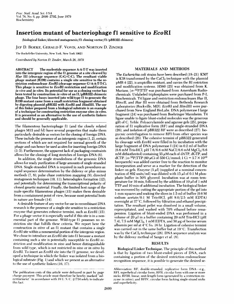
FIG. 1. Outline of the biological linker technique and its use togenerate insertion mnutants of bacteriophage fl sensitive to EcoRI.fl RF was partially digested with Hae III to give 40% RFIII, a moleculehaving blunt ends with 5'-pC residues. This was ligated to a biologicallinker isolated from pBR322 RF. The linker was prepared by incu-bating pBR322 RF with EcoRI, then with the Klenow fragment ofDNA polymerase I and dATP and [a_-32P]TTP, and finally withHindIll. The 31-base-pair linker fragment was gel-purified and li-gated to the fl RFIII by using a T4 ligase preparation containing"blunt end" activity. The DNA in the ligation mixture was cleavedwith EcoRI. The 1FIII-sized material was purified, diluted (to pro-mote intramolecular ligation), and religated to circularize the prod-uicts. Asterisks denote residues labeled with 32p.
quence. This approach was used in the cloning of the cI geneof bacteriop~hage X'(29). Any DNA fragment having blunt (basepaired) ends with 5'-terminal pC residues and 3'-terminal Gresidues can be used with a biological linker of the type de-scribed belqw to generate terminal EcoRI sites on the fragment.The plassmid pBRO22 contains a single EcoRI site 31 nucleotidesfrom its unique HindIII site. Thus, it is possible to cut pBR322with EcoRI and then fill in the cohesive termini by incubationwith DNA polymerase, dATP, and TTP. After heat inactivationof the polymerase, the RFIII is digested with HindIII. Thisdigestion generjstes two fragments, the approximately 4000-nucleotide fragment comprising most of the pBR322 genomeand a 31-nucleotide fragment that we refer to as the "biologi-cal" linker.One end of the biological linker consists of the cohesive ter-
minus produced by HindIII digestion, whereas the other con-sists of a filled-in EcoRI terminus (blunt end). Only the latterend is reactive in blunt-end ligation. When excess biologicallinker is incubated with an appropriate DNA fragment in thepresence qf T4 ligase having blunt-end activity, any fragment
reacting with a molecule of linker on each end will be boundedby EcoRI sites. Removal of unreacted linker (30) is unnecessarybecause neither the linker itself nor its multimers contain anycomplete EcoRI sites to compete for the EcoRI enzyme in thesubsequent cleavage step necessary to generate cohesive EcoRItermini.
Introduction of EcoRI Site into fI Genome. To put anEcoRI site into the intergenic region of fI by the above tech-nique, we used as our substrate RFIII molecules generated bypartial digestion of fi RFI with the enzyme Hae III, whichgenerates blunt ends with 5'-terminal pC residues and 3'-ter-minal G residues. The partial digest contained approximately30% RFI, 30% RFII, and 40% RFIII. Because the fI genomecontains nine Hae III sites, two of which are in the intergenicregion, and the other seven in coding regions, it was clear thatthe insertion of four nucleotides into seven of the nine siteswould probably result in lethal frameshift mutations. Of thetwo sites in the intergenic region, one is known to be nones-sential, because the M13 strain mp 1 (3) has an insertion ofapproximately 800 bases of the lac operon inserted in whatcorresponds to the fl Hae III G/D border. The other fI site [theF/G border (see below)] is near the origin of minus strandreplication, and the effects of insertion there are not predictable.After removal of RFI by isopycnic centrifugation, the RFIIIwas incubated with a ten-fold excess of biological linker (basedon the molarity of ends) and T4 "blunt end" ligase (Fig. 2, lanea). The resultant mixture was cleaved with EcoRI and loadedon a 0.8% agarose gel to isolate RFIII from RFII and othercontaminants. The RFIII was ligated under dilute conditionsby using a preparation of T4 ligase containing no detectableblunt-end activity. The absence of blunt-end activity prevents
a
AM
b c
_0
t
E- I
FIG. 2. Blunt-end ligation of biological linker to linearized fIgenome and recircularization by intramolecular ligation of genomeafter EcoRI cleavage. fi RFIII produced by partial digestion of fl RFIwith Hae III was incubated with a 10-fold excess of biological linker.For lane a, 1% of the reaction volume was diluted and loaded on a2.4-10% polyacrylainide gradient gel, which was then autoradio-graphed overnight. The lowermost band seen is unreacted linker. Thesecond band from the bottom is linker dimer, which can be formedeither by cohesive ligation (of the HindIll ends) or blunt-end ligation.The products in the higher bands are linker trimers, tetramers, etc.In order to create these multimers, one or more blunt-end ligationevents is retuired; hence the extent of multimer formation in a ligationreaction of this type reflects the extent of blunt-end ligation. Theuppermost band is RFIII ligated to linker molecules. Ligation prod-ucts were digested with EcoRI and loaded on a vertical 0.8% agaroseslab gel. The band comigrating with marker RFIII was cut out andthe RFIII was recovered by electroelution. This DNA was used forintramolecular ligation of the EcoRI cohesive termini. Small aliquotsof this reaction were run on a 0.8% agarose gel before (lane b) and after(lane c) this ligation step. Arrows indicate the position of (top tobottom) fi RFII, RFIII, and RFI OD markers. RFI and RFIV comi-grate in this gel system.
IHoeM
pCC GGGG CCp
RFm
2700 Biochemistry: Boeke et al.
H
<--.- 'JU
Proc. Natl. Acad. Sci. USA 76 (1979) 2701
the circularization of those RFIII molecules to which no bio-logical linker was added. Circularization of suQh rrterialw tAyield infectious molecules that lack an EcoRlik. AsWquot was run on a 0.8% agarose gel to monitor the extent ofcircularization, which was estimated at approximately 20%(Fig. 2, lanes b and c). After transfection of CaCl2-treated K414cells, plaques were picked and tested for their relative efficiencyof plating on K38 (rRl- mRI,) and K507 (rRI+ mRI+). Severalisolates showed classical restriction modification behavior. Onesuch isolate (R199) had a restriction coefficient of 0.2 on K507compared to K38 and produced typical fI plaques.
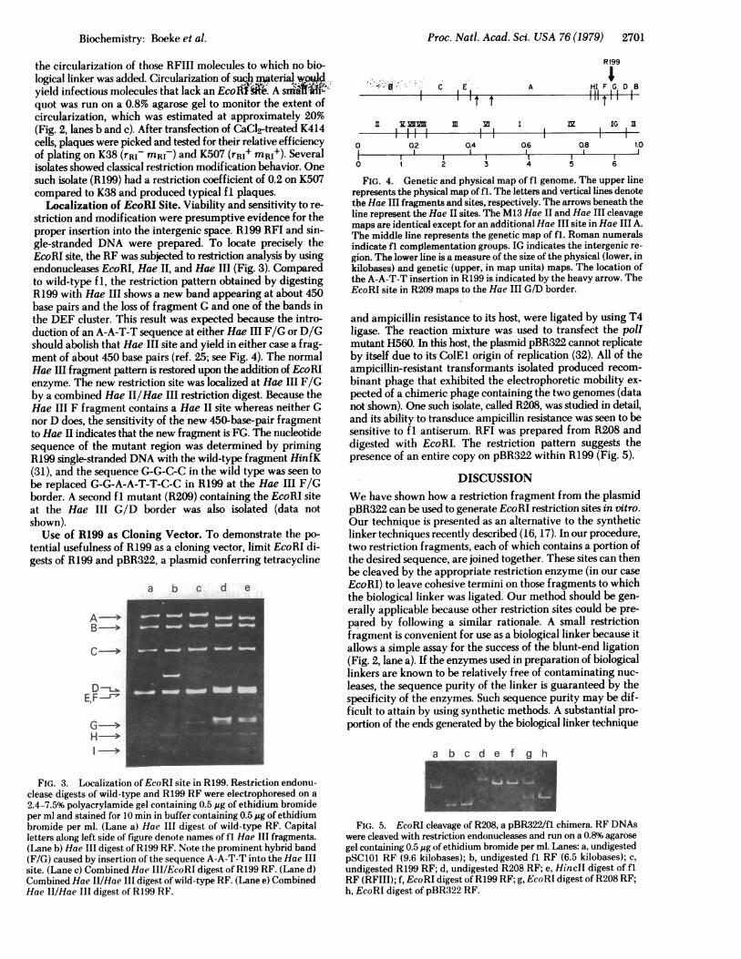
Localization of EcoRI Site. Viability and sensitivity to re-striction and modification were presumptive evidence for theproper insertion into the intergenic space. R199 RFI and sin-gle-stranded DNA were prepared. To locate precisely theEcoRI site, the RF was subjected to restriction analysis by usingendonucleases EcoRI, Hae II, and Hae III (Fig. 3). Comparedto wild-type f 1, the restriction pattern obtained by digestingR199 with Hae III shows a new band appearing at about 450base pairs and the loss of fragment G and one of the bands inthe DEF cluster. This result was expected because the intro-duction of an A-A-T-T sequence at either Hae III F/G or D/Gshould abolish that Hae III site and yield in either case a frag-ment of about 450 base pairs (ref. 25; see Fig. 4). The normalHae III fragment pattern is restored upon the addition of EcoRIenzyme. The new restriction site was localized at Hae III F/Gby a combined Hae II/Hae III restriction digest. Because theHae III F fragment contains a Hae II site whereas neither Gnor D does, the sensitivity of the new 450-base-pair fragmentto Hae II indicates that the new fragment is FG. The nucleotidesequence of the mutant region was determined by primingR199 single-stranded DNA with the wild-type fragment HinfK(31), and the sequence G-G-C-C in the wild type was seen tobe replaced G-G-A-A-T-T-C-C in R199 at the Hae III F/Gborder. A second fi mutant (R209) containing the EcoRI siteat the Hae III G/D border was also isolated (data notshown).Use of R199 as Cloning Vector. To demonstrate the po-
tential usefulness of R199 as a cloning vector, limit EcoRI di-gests of R199 and pBR322, a plasmid conferring tetracycline
a b c d e
-A--->
D--E,F -r
FIG. 3. Localization of EcoRI site in R199. Restriction endonu-clease digests of wild-type and R199 RF were electrophoresed on a
2.4-7.5% polyacrylamide gel containing 0.5 Mg of ethidium bromideper ml and stained for 10 min in buffer containing 0.5 ug of ethidiumbromide per ml. (Lane a) Hae III digest of wild-type RF. Capitalletters along left side of figure denote names of fl Hae III fragments.(Lane b) Hae III digest of R199 RF. Note the prominent hybrid band(F/G) caused by insertion of the sequence A-A-T-T into the Hae IIIsite. (Lane c) Combined Hae III/EcoRI digest of R199 RF. (Lane d)Combined Hae II/Hae III digest of wild-type RF. (Lane e) CombinedHae II/Hae III digest of R199 RF.
R 199
IC E A HI F G D B
IT T lilt''V. MI2l IIm i I M IG I[
0 Q2 0.4 Q6 0.8 1.0
0 1 2 3 4 5 6
FIG. 4. Genetic and physical map of fl genome. The upper linerepresents the physical map of fl. The letters and vertical lines denotethe Hae III fragments and sites, respectively. The arrows beneath theline represent the Hae II sites. The M13 Hae II and Hae III cleavagemaps are identical except for an additional Hae III site in Hae III A.The middle line represents the genetic map of fl. Roman numeralsindicate fl complementation groups. IG indicates the intergenic re-gion. The lower line is a measure of the size of the physical (lower, inkilobases) and genetic (upper, in map units) maps. The location ofthe A-A-T-T insertion in R199 is indicated by the heavy arrow. TheEcoRI site in R209 maps to the Hae III G/D border.
and ampicillin resistance to its host, were ligated by using T4ligase. The reaction mixture was used to transfect the polImutant H560. In this host, the plasmid pBR322 cannot replicateby itself due to its ColEl origin of replication (32). All of theampicillin-resistant transformants isolated produced recom-binant phage that exhibited the electrophoretic mobility ex-pected of a chimeric phage containing the two genomes (datanot shown). One such isolate, called R208, was studied in detail,and its ability to transduce ampicillin resistance was seen to besensitive to fi antiserum. RFI was prepared from R208 anddigested with EcoRI. The restriction pattern suggests thepresence of an entire copy on pBR322 within R199 (Fig. 5).
DISCUSSIONWe have shown how a restriction fragment from the plasmidpBR322 can be used to generate EcoRI restriction sites in vitro.Our technique is presented as an alternative to the syntheticlinker techniques recently described (16, 17). In our procedure,two restriction fragments, each of which contains a portion ofthe desired sequence, are joined together. These sites can thenbe cleaved by the appropriate restriction enzyme (in our caseEcoRI) to leave cohesive termini on those fragments to whichthe biological linker was ligated. Our method should be gen-erally applicable because other restriction sites could be pre-pared by following a similar rationale. A small restrictionfragment is convenient for use as a biological linker because itallows a simple assay for the success of the blunt-end ligation(Fig. 2, lane a). If the enzymes used in preparation of biologicallinkers are known to be relatively free of contaminating nuc-leases, the sequence purity of the linker is guaranteed by thespecificity of the enzymes. Such sequence purity may be dif-ficult to attain by using synthetic methods. A substantial pro-portion of the ends generated by the biological linker technique
a b cdefgh
FIG. 5. EcoRI cleavage of R208, a pBR322/fl chimera. RF DNAswere cleaved with restriction endonucleases and run on a 0.8% agarosegel containing 0.5 Atg of ethidium bromide per ml. Lanes: a, undigestedpSC101 RF (9.6 kilobases); b, undigested fl RF (6.5 kilobases); c,undigested R199 RF; d, undigested R208 RF; e, HincIl digest of flRF (RFIII); f, EcoRI digest of R199 RF; g, EcoRI digest of R208 RF;h, EcoRI digest of pBR322 RF.
Biochemistry: Boeke et al.
-r ;- _I- -;

Proc. Natl. Acad. Sci. USA 76 (1979)
are chemically and biologically intact, as shown by the fact thatmost of the cohesive EcoRI termini generated could be ligatedto circularize the fragment (Fig. 2, lanes b and c). Currently,the main drawback of the technique is that rather large amountsof pBR322 RF are required for each experiment, because eachmolecule of pBR322 produces only one molecule of biologicallinker. We are currently attempting to construct plasmid var-iants that will carry multiple copies of the biological linker,thereby alleviating this problem.By use of this technique, the sequence A-A-T-T was inserted
at two different sites within the intergenic region of f1. One ofthese sites, the Hae III F/G border region, has been implicatedin the replication of the filamentous phage (33, 34). Based onsequence data, this region has potential secondary structure (35).In fact, the hairpin-like structure containing the Hae III F/Gborder has been implicated in the initiation of DNA replication.Therefore, the insertion of foreign DNA at this site might beexpected to prevent DNA replication. The isolation of thefl/pBR322 recombinant phage (R208) indicates that replica-tion of such molecules is possible and appears to reduce thelikelihood of an absolute requirement for secondary structurein this region for the initiation of DNA replication. Moreover,the use of the polI mutant H560 [in which pBR322 cannotreplicate independently (32)] ensures that the bacteriophageorigin of replication is used exclusively in chimeric phageproduction.The filamentous phage, because their genome consists of a
single-stranded DNA molecule, offer a set of unique advantagesas cloning vectors. Milligram quantities of both single-strandedand double-stranded DNA can be easily prepared, even whenthe genome size is increased substantially. In terms of efficientproduction of cloned material, filamentous phage potentiallyoffer great advantages, because a single wild-type plaqueroutinely yields greater than 109 plaque forming units, andphage production is on the order of 1012 per ml. Also, the vectorgenome size is very small, so that the cloned DNA constitutesa large fraction of the recombinant DNA. The availability ofcloned DNA in both single-stranded and double-stranded formsfacilitates its rapid structural analysis. By using single-strandedDNA from chimeric phage as template and wild-type orchimeric restriction fragments as primers, pulse-chase re-striction mapping and DNA sequence determination by usingdideoxy terminators are possible. Also, one should be able todetermine the transcriptional polarity of the cloned DNA byhybridization of the RNA in question to genomic DNA fromchimeric phages carrying the insert in opposite orientations.Finally, single-stranded DNA is one of the substrates requiredfor directed mutagenesis techniques which employ a mis-matched primer (10, 11). Thus, point mutations might be in-troduced in a specific manner into the foreign genetic materialinserted in the phage genome. Limits on the size of the DNAto be cloned have yet to be determined.
We thank Dr. P. Modrich for supplying us with the plasmid pMB4, P. McGraw for her excellent technical assistance, K. Horiuchi andP. Model for critically reading the manuscript, and C. Yehle for gen-erous gifts of enzyme. This work was supported in part by funds fromthe National Science Foundation and the National Cancer Institute.
1. Vovis, G. F., Horiuchi, K. & Zinder, N. D. (1975) J. Virol. 16,674-684.
2. Enea, V., Horiuchi, K., Turgeon, B. G. & Zinder, N. D. (1977)J. Mol. Biol. 111, 395-414.
3. Messing, J., Gronenborn, B., Muller-Hill, B. & Hofschneider, P.H. (1977) Proc. NatI. Acad. Sci. USA 74,3642-3646.
4. Herrmann, R., Neugebauer, K., Schaller, H. & Zentgraf, H. (1978)in The Single-Stranded DNA Phages, eds. Denhardt, D. T.,Dressler, D. & Ray, D. S. (Cold Spring Harbor Laboratory, ColdSpring Harbor, NY), pp. 473-476.
5. Ray, D. S. & Kook, K. (1978) Gene 4, 109-119.6. Vovis, G. F., Ohsumi, M. & Zinder, N. D. (1977) in Molecular
Approaches to Eukaryotic Gene Systems, eds. Wilcox, G.,Abelson, J. & Fox, C. F. (Academic, New York), pp. 55-61.
7. Sanger, F. & Coulson, A. R. (1975) J. Mol. Biol. 94, 441-448.8. Sanger, F., Nicklen, S. & Coulson, A. R. (1977) Proc. Natl. Acad.
Sci. USA 74,5463-5467.9. Seeburg, P. H. & Schaller, H. (1975) J. Mol. Biol. 92,261-277.
10. Hutchison, C. A., III, Phillips, S., Edgell, M. H., Gillam, S., Jahnke,P. & Smith, M. (1978) J. Biol. Chem. 253,6551-6560.
11. Razin, A., Hirose, T., Itakura, K. & Riggs, A. D. (1978) Proc. Natl.Acad. Sci. USA 75,4268-4270.
12. Vovis, G. F., Horiuchi, K., Hartman, N. & Zinder, N. D. (1973)Nature (London) New Biol. 246,13-16.
13. Zinder, N. D., Valentine, R. C., Roger, M. & Stoeckenius, W.(1963) Virology 20,638-640.
14. Lederberg, J. (1951) Science 114, 68-69.15. Mertz, J. E. & Davis, R. W. (1972) Proc. Natl. Acad. Sci. USA 69,
3370-3374.16. Bahl, C. P., Marians, K. J., Wu, R., Stawinsky, J. & Narang, S. A.
(1977) Gene 1, 81-92.17. Scheller, R. H., Dickerson, R. E., Boyer, H. W., Riggs, A. D. &
Itakura, K. (1977) Science 196, 177-180.18. Garen, A. & Siddiqi, 0. (1962) Proc. Natl. Acad. Sci. USA 48,
1121-1127.19. Lyons, L. B. & Zinder, N. D. (1972) Virology 49,45-60.20. Cole, R. S. (1971) J. Bacteriol. 106, 143-149.21. Ohsumi, M., Vovis, G. F. & Zinder, N. D. (1978) Virology 89,
438-449.22. Betlach, M., Hershfield, V., Chow, L., Brown, W., Goodman, H.
M. & Boyer, H. W. (1976) Fed. Proc. Fed. Am. Soc. Exp. Biol.35,2037-2043.
23. Vosberg, H. P. & Hoffmann-Berling, H. (1971) J. Mol. Biol. 58,739-753.
24. Klenow, H. & Henningsen, I. (1970) Proc. Natl. Acad. Sci. USA65, 168-175.
25. Horiuchi, K., Vovis, G. F., Enea, V. & Zinder, N. D. (1975) J. Mol.Biol. 95, 147-165.
26. Model, P. & Zinder, N. D. (1974) J. Mol. Biol. 83,231-251.27. Clewell, D. B. (1972) J. Bacteriol. 110, 667-676.28. Mandel, M. & Higa, A. (1970) J. Mol. Biol. 53, 159-162.29. Backman, K., Ptashne, M. & Gilbert, W. (1976) Proc. Natl. Acad.
Sci. USA 73,4174-4178.30. Maniatis, T., Hardison, R. C., Lacy, E., Lauer, J., O'Connell, C.,
Quon, D., Sim, G. K. & Efstratiadis, A. (1978) Cell 15, 687-701.
31. Horiuchi, K., Vovis, G. F. & Model, P. (1978) in The Single-Stranded DNA Phages, eds. Denhardt, D. T., Dressler, D. & Ray,D. S. (Cold Spring Harbor Laboratory, Cold Spring Harbor, NY),pp. 113-137.
32. Kingsbury, D. T. & Helinski, D. R. (1970) Biochem. Biophys. Res.Commun. 41, 1538-1544.
33. Horiuchi, K. & Zinder, N. D. (1976) Proc. Natl. Acad. Sci. USA73, 2341-2345.
34. Geider, K., Beck, E. & Schaller, H. (1978) Proc. Nati. Acad. Sci.USA 75, 645-649.
35. Schaller, H., Beck, E. & Takanami, M. (1978) in The Single-Stranded DNA Phages, eds. Denhardt, D. T., Dressler, D. & Ray,D. S. (Cold Spring Harbor Laboratory, Cold Spring Harbor, NY),pp. 139-163.
2702 Biochemistry: Boeke et al.