progress report membrane digestion · oligomers, ie, the final stages of digestion, is being...
TRANSCRIPT
Gut, 1972, 13, 735-747
Progress report
Membrane digestionBy the end of the 1950s the extensive investigations which laid the foundationsfor certain basic current concepts were complete, namely, the digestivefunction of the intestinal cells; the integration of the processes of digestionproper and of transport during digestion; and the pathology of enterocytesand other cells. And they may be summarized as follows: (1) the free surfaceof the enterocyte represents an aggregate of microvillil-14; (2) hydrolysis ofoligomers, ie, the final stages of digestion, is being accomplished duringabsorption by enzymes bound to the enterocyte structures15-20; (3) numerousenzymes, in particular alkaline phosphatase and the aminopeptidases, areconcentrated within the brush border zone21-27. The final link in the under-standing of these processes is that oligomers as such penetrate inside theintestinal cells where the final stages of hydrolysis and further transport ofmonomers take place.
The Digestive Functions of Enterocytes
Simultaneously, at the end of the 1950s and the beginning of the 1960s, twodifferent hypotheses to explain these facts were presented, namely, (1) theconcept of apical intracellular digestion28-32 and (2) that of membrane diges-tion33-. Evidence is increasing to suggest that the first hypothesis is untenable.Membrane digestion, in addition to the two early known types-distant
extracellular and intracellular-is considered to be a system of the digestiveprocesses occurring under the influence of the enzymes situated on theexternal surface of the membrane. We had the good fortune to be able todemonstrate that the external surface of the brush border is a morphologicalsubstrate of a new, previously unknown type of digestion (membrane)which appeared to be associated with the adsorption of pancreatic enzymesto the surface of the intestinal mucosa3336,i40,47. (Due to the large size ofamylase molecules (molecular weight is about 45 000)48,49 their penetrationinto the cells was excluded.) Evidence of the rapid and complete loss ofenzyme from fluid as it adsorbs onto membrane (desorption) supported ourarguments even more forcibly3336'40,47. Adsorbed amylase was shown tohydrolyze intensively the soluble (not colloidal) starch40. In practice, it wasfound at the same time that enteric enzymes, in particular disaccharidasesand dipeptidases, produce their effect not intracellularly but at the externalsurface of the cell membranes37,40,41,46. In man and mammals, membranedigestion is accomplished at the surface of the microvilli of the enterocyte.This idea was first reported in 1959 and discussed in a number of reports36,40,50,51-53
Those enzymes which ensure membrane digestion (Fig. I)* may have two
*The Editor and publishers of Gut are grateful to the Ciba Foundation and Associated Scientific Publishers,Amsterdam, for permission to reproduce the illustrations which originally appeared in the proceedings ofa Cibasymposium on 'Peptide Transport in Bacteria and Mammalian Gut'.
735
on March 24, 2020 by guest. P
rotected by copyright.http://gut.bm
j.com/
Gut: first published as 10.1136/gut.13.9.735 on 1 S
eptember 1972. D
ownloaded from
A. M. Ugolev
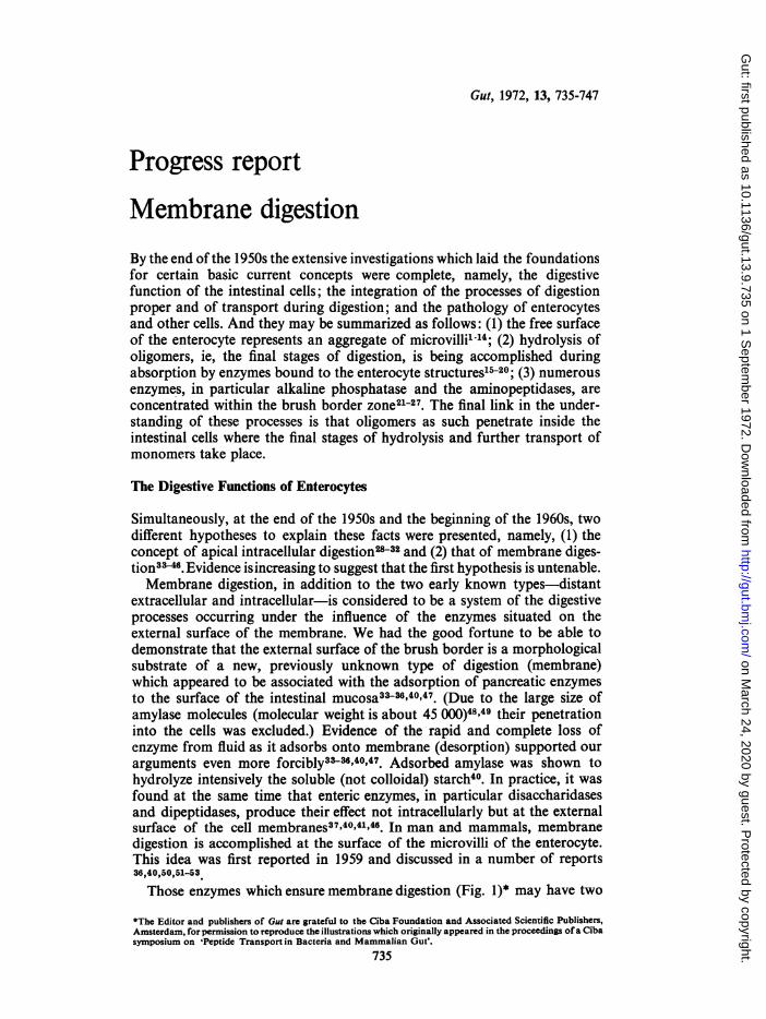
Fig. 1 The enzymaticapparatus for membrane
_v tm 6 * \ digestion. Adsorption ofpancreatic enzymes fromchyme (1), synthesis (3), andmigration (2) of entericenzymes.
origins: (1) enteric enzymes synthesized by enterocytes and structurallyassociated with the external surface of their membrane, and (2) enzymesadsorbed from the intestinal cavity.
Indeed, I was able to write in the Physiological Review50: 'Cavital digestionis produced by enzymes secreted into the cavity of the gastrointestinal tract.Membrane digestion is due to enzymes adsorbed from chyme and enzymesstructurally associated with the membrane of the intestinal cells. Con-sequently, study of enzymatic functions of digestive glands and properties ofdigestive juices cannot answer all questions of the physiology of digestion'.
It should be stressed that the nature of the bonds of the enteric enzymeswith the membrane structures of the microvilli remains unknown. The termsemployed in the literature 'structurally associated enzymes' or 'enzymes,which are the integral part of the membrane structure,' lack physical orchemical sense as they do not reflect the forces ensuring preservation of theenzyme-membrane complexes. It is known only that the majority of in-testinal peptidases are easily solubilized in vitro while disaccharidases,alkaline phosphatase, and aminopeptidase can be solubilized by treating theenzyme-membrane complexes with proteases (papain, trypsin) and otherdetergents (Triton X-100)18,5442.
736
on March 24, 2020 by guest. P
rotected by copyright.http://gut.bm
j.com/
Gut: first published as 10.1136/gut.13.9.735 on 1 S
eptember 1972. D
ownloaded from
Membrane digestion
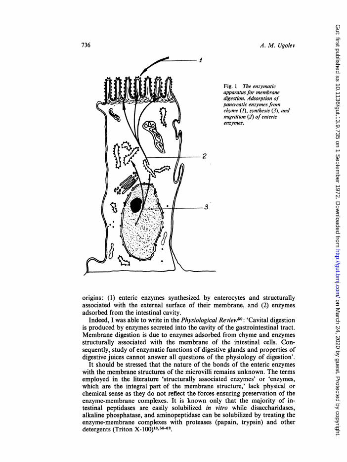
Fig. 2 The detailed schemeof the interrelationship betweencavital andmembrane digestion
9 96D d with nofood substances present¢ 'o ta 6 d° 0 (I) and in their presence (I1).°aQ*b6d° 1 Enzymes in the intestinal
0- 4,>10.0 o ca, cavity; 2 microvilli; 3 enzymes0 0
-'0C" ~ ~ (adsorbed and enteric) on thed or 0° %o 6 microvillous surface; 4 thegQd 0 .09g 0. brushborderpores; 5
v-0 OL ba0.oe6 bacteria; 6,7 food substances'0 a '06 -O 0
a babC'°&O b ag4 5 at the different stages of
01= .0-
a bjqb6 o 5 hydrolysis.b
7
0
9
2 ZE
The general scheme of membrane digestion as a process completing thehydrolysis and associated with absorption was proposed in 1959-60 and isillustrated in Figure 2. Later it was developed and defined concretely in linewith the success achieved in the study of the role of membranes in the bodyand with the progress in our knowledge of their digestive and transportfunctions152.
During the 1960s about 20 independent experimental criteria were used toprove the fact that oligosaccharide and peptide hydrolysis occurs exclusivelyor mainly at the external membrane surface50652'63'64. Ofparticular significanceare the electron-microscope studies by Oda and Seki, Johnston, andothers6549, who have demonstrated the presence of globules of 50 to 60 A indiameter at the external membrane surface using the negative contrast of thebrush border. These globules were solubilized and turned out to be at leastthe bearers of disaccharidase activity.Four different viewpoints on the mechanism of the final stages of hydrol-
ysis existed before 196670. Since that time investigators have accepted themembrane hypothesis. Judging from the later publications of Crane and hisgroup, they themselves have joined this majority63,7'76'76.
Following the first research refuting the significance of enzyme adsorptionfor membrane digestion came the first support for this mechanism77'81. Atpresent, adsorption of such pancreatic enzymes as amylase38'40,43,52,77-81,lipase8.5, and protease37,47 is shown. It is very likely that other enzymes,such as pancreatic DNA-ase and RNA-ase, may be adsorbed and participatein membrane digestion.A detailed analysis of adsorption mechanisms revealed, first, that both
the physical and chemical aspects of adsorption are of importance; secondly,adsorption is possible on both lipoprotein and mucopolysaccharidestructures40'43'51'52' 70,86-93
737
on March 24, 2020 by guest. P
rotected by copyright.http://gut.bm
j.com/
Gut: first published as 10.1136/gut.13.9.735 on 1 S
eptember 1972. D
ownloaded from
The analysis of the structure and functions of the brush border glycocalixmade first by Ito94 and later by other authors95-101 appeared of particularsignificance for the development of the membrane digestion concept. IndeedIto was the first to pay attention to the importance of this structure in theprocesses of membrane digestion.
The Characteristics of Membrane Digestion
The basic functional and structural characteristics of membrane digestion areillustrated in Fig. 2 and are set out as follows:Membrane digestion is carried out at the external surface of the membrane
of microvilli by the enteric and adsorbed enzymes. Therefore, the final stagesof digestion are integrated and the initial stages of transport are begun. Itoccurs in the zone inaccessible to bacteria, thus ensuring sterility of thefinal stages of hydrolysis. Membrane digestion, as any other catalysis on thesurface, depends on the rate at which substances pass from liquid mediumonto the surface and under normal conditions that depends upon intestinalmotility.The size of the pores of the brush border and of the glycocalix allows the
segregation of larger structures from smaller ones thus providing for theconsequent treatment of food, first in the cavity then at the surface. Thanks tothe continuous elimination of the intermediate products of hydrolysis intothe membrane digestion zone, digestion in the cavity is intensified.Many characteristics of the solubilized enzymes and those bound to
membrane differ from each other, in particular Ki, V.ax, and thermo-stability.
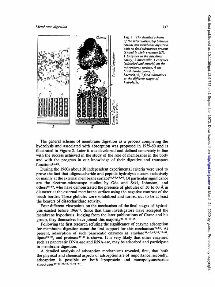
4% ° Fig. 3 Enteric and adsorbed-00 00 Q
enzymes during membraneC) digestion (the scheme of thefragment of the microvillus
4 luminal surface). ADistribution of enzymes; Binterrelationship of enzymes,carriers and substrates. Icavity; II glycocalix; III three-layered membrane; IV
\\} ( t | 4 \ t N A X o B t-94 glycocalix luminal surface; Vthree-layered membraneluminal surface. 1 Intestinalenzymes proper; 2 adsorbedenzymes; 3 carriers; 4substrates.
itl
738 A. M. Ugolev
on March 24, 2020 by guest. P
rotected by copyright.http://gut.bm
j.com/
Gut: first published as 10.1136/gut.13.9.735 on 1 S
eptember 1972. D
ownloaded from

Owing to fixation of enzymes at the external surface of the membrane, thestructural organization of membrane hydrolysis and transport processes ispossible. Physicochemical conditions in the membrane digestion zone varyfrom those of the digestive cavities.
Figure 3 shows a fragment of membrane with a glycocalix, the approxi-mate correlation of enzymes and structures being observed. As can be seen,the normal course ofmembrane digestion requires the presence of both entericenzymes proper and adsorbed enzymes. In fact, the enteric and adsorbedenzymes functionally supplement each other and simultaneously seem toenhance the reliability of the system as a whole. It is assumed that entericenzymes are structurally connected with the external surface of a three-layered membrane which ensures the effective conveyance of the finalproducts (glucose, amino acids) from enzyme onto the carrier. Adsorbedenzymes can be situated on all the structures of the external surface of themicrovillus.
It is obvious that substrates included in chyme do not directly contactthe three-layered membrane; they must pass through the glycocalix space.Their penetration into the glycocalix is facilitated by enzymes, adsorbed tothe free surface of the latter, and further on passing enzymes inside theglycocalix space. Thus, the movement across the glycocalix space is accom-panied by an intense depolimerization of food substances. Some supple-mentary hydrolysis by means of adsorbed enzymes is possible at the externalsurface of the lipoprotein 'membrane, but the chief enzymes are enteric en-zymes represented mainly by dimer and oligomerhydrolases.
Such localization of disaccharidases, di-amino-, and oligopeptidases islikely to ensure the highly effective conveyance from the final enzyme onto theentrance into the transport system, in particular, onto the labile carrier(Fig. 4).Within the limits of the main arguments concerning membrane digestion
(hydrolysis is completed at the external surface of the membrane and its
Fig. 4 Hypotheticaldistribution of enzymes andcarriers on the externalmembrane surface. E Enzyme;
P *| ) I C carrier; M membrane.
t\\ ~~~~Fig. 4
Fig. 5 The enzyme-carrierpool. S Substrate; P hydrolysisproducts; E enzyme; C1, C2,C3 carriersforming a transportconveyor.
Fig. 5
Membrane digestion 739
S,'-
on March 24, 2020 by guest. P
rotected by copyright.http://gut.bm
j.com/
Gut: first published as 10.1136/gut.13.9.735 on 1 S
eptember 1972. D
ownloaded from

products are transported across the membrane) a number of hypotheses havebeen suggested to explain the extreme efficiency of the digestive-transportconveyer40.44,52,53P76,58 0,64,70,103-112 They all, finally, agree that enzyme andcarrier form the spatial and functional integration; one such scheme isrepresented in Figure 5. Thus the membrane digestion apparatus represents ahighly complex structure with elements of a different nature and origin, whichis in agreement with the current idea of 'thick' membranes113'114.The membranous surface of the microvillus seems to exercise not only
digestive-transport functions but those of a protective barrier as well. In fact,the glycocalix, together with its enzymes, forms a specialized filter whichallows adequate amounts of substrates to pass but traps the other molecules.The glycocalix is also an ion exchanger98 and a molecular sievel10.
Thus, membrane digestion is one of the three basic stages in the activity ofthe alimentary tract: it accepts the products of intermediate hydrolysis fromthe cavity (by which all the cavital processes are intensified); it accomplishesthe hydrolysis of 70 to 80% of the primary molecular bindings in food bio-polymers; and it couples the digestive processes proper and those of trans-port40'50,52,53.
Membrane Digestion as a Clinical Problem
Membrane digestion should be taken into account for the better under-
d B
3 I3
C ,A2 43 r C 41 42 3 r
C
Fig. 6 Amylase activity in the differentfractions obtained during successive desorptionand reflecting cavital and membrane digestion.A Normal; B in patients with defectivepancreatic function; C in patients with chronicenteritis. Abscissa: separatefractions andsubfractions; ordinate: amylolytic activity in
2 | \ tg/mg ofhydrolyzed substrate. C Activity of\ pancreatic cx-amylase not adsorbed to the surface
/ \ ofsmall intestine. D1 Easily desorbed amylase;D, more slowly desorbed amylase; D., slowlydesorbed amylase; H homogenate amylolytic
c A 2~ 4, r activity.
740 A. M. Ugolev
on March 24, 2020 by guest. P
rotected by copyright.http://gut.bm
j.com/
Gut: first published as 10.1136/gut.13.9.735 on 1 S
eptember 1972. D
ownloaded from
standing of the aetiology of a number of diseases and for promoting progressin diagnostic and therapeutic research. We have considered this problem indetail earlier and it will be the subject of a special review. Here we shalldwell only on certain consequences following upon the idea of membranedigestion.
Enzyme Adsorption Disorders
Hooft and others115 were the first to show that in certain forms of starchmalabsorption cx-amylase activity in the cavity remains normal but thecapacity of the mucosa, obtained from biopsy in such patients, to adsorbamylase is greatly decreased.
Masevich, Zabelinskii, and Ugolev"16 have developed the fraction techniqueof investigating enzyme adsorption. Figure 6 shows typical examples of themucosal amylolytic spectral disorders. As can be seen, such a techniqueallows the differentiation of disorders in the amylolytic activity in the in-testinal content, the adsorption properties of the surface, and o-amylaseactivity dependent upon proteolytic processes in the enterocyte itself.
Recently, changes in lipase adsorption have been seen in patients sufferingfrom atherosclerosis when compared with healthy controls117.
Different enzyme maladsorptions were revealed also in such diseases asenteritis, enterocolitis, and diseases of the biliary ducts8l. Adsorption defectscould be both isolated and combined with damage to the villous structuresand disturbances in the epithelial element in proteosynthesis.
It is of interest that disorders of membrane digestion are likely to appearearlier than can be seen by histological and histochemical changes.
Synthesis and Migration of Enteric Enzymes in Disease
Studies of genetic and acquired defects of the enzymatic properties ofenterocytes shed light upon a great number of diseases of the digestivesystem. Information was obtained by means of loading studies and byinvestigation of the enzymatic properties of mucosa obtained by biopsy. Inboth cases homogenates of a specimen were mainly used.
In terms of membrane digestion, functioning of the enteric enzymes(disaccharidases, peptidases, etc) is determined by the two successive pro-cesses of intracellular synthesis and further progress to the external mem-brane surface.There are two possibilities: enzymes are being included either in the com-
position of a waiting microvillous membrane structure or built into thesubunits of the membrane intracellularly so that the enzyme-membraneblocks are included in the composition of the microvillus52.
It is obvious that investigation of homogenates provides only a notion ofthe state ofenzyme synthesis but not of the transfer of enzymes and thereforenot of their actual functioning. For this reason our laboratory has devel-oped a technique based on the comparison of intact and homogenizedmucosa52,118-120 as the investigation of homogenates led to a considerable lossof information. Many enzymatic defects remain beyond the understandingboth of the laboratory investigator and the clinician.
It is important to remember that in the same disease certain enzymaticdefects can depend on disorders of synthesis and others on disturbances of
Membrane digestion 741
on March 24, 2020 by guest. P
rotected by copyright.http://gut.bm
j.com/
Gut: first published as 10.1136/gut.13.9.735 on 1 S
eptember 1972. D
ownloaded from

A. M. Ugolev
4
so5 K Fig. 7 Effict ofx-raytreatment on the hydrolysis of
40 a--_ sucrose (A) and glycyl-l-leucine(B). Abscissa: time after x-ray
so \ ~ ~--_ , treatment (in hours); ordinate\0 enzyme activity (in
conventional units). Dotted20 line: enzymatic activity of
homogenates; continuous line:10 enzymatic activity of the
everted pieces of smallintestine.
4 24 48 72
Btao~~~~~~~~~~~~~~~~~~~~~~~~~.
149
424 43 ~~~~~~~~~~72
transfer52. For example, in severe x-ray injuries hydrolysis of both disac-charides and peptides is completely stopped. But the absence of invertaseactivity depends on the suppression of synthesis whereas peptidase is syn-thesized in amounts close to normal but it is not included in the membraneof the microvillus (Fig. 7)52,119. Restoration of normal interactions betweenthe synthesis and transfer of the enteric enzymes after irradiation injuries isessentially important for a better insight into the restoration processes'21'4
It is interesting that under the influence of different stresses disorders of theenterocyte digestive function depend rather on disturbances of transfer thanon enteric enzyme synthesis.
Other Defects of Membrane Digestion
It is clear that disorders of membrane digestion can take place not only as aresult of disturbances in the enteric cell functions but in all those situations inwhich hydrolysis is decreased at the intestinal surface as a whole. Whenhydrolysis in the digestive cavity is slowed as a result of deficiency of thebasic digestive glands membrane digestion is impaired due to an insufficientsupply of substrates into the brush border zoe53,125-127*. This is accom-panied by the enzymatic and transport functions of distal portions of
742
on March 24, 2020 by guest. P
rotected by copyright.http://gut.bm
j.com/
Gut: first published as 10.1136/gut.13.9.735 on 1 S
eptember 1972. D
ownloaded from
Membrane digestion 743
small intestine being greatly enhanced, a situation which is defined by Boothas a 'reserved zone'28. In severe cases food which has not been utilized canenter the large intestine unchanged.
In disturbances of motility of the small intestine the increased passageshortens the time of contact between food and digestive surface and, therefore,decreases the volume of membrane digestion. As soon as the entry of sub-stances into the brush border is determined by convection and not bydiffusion, the effectiveness of membrane hydrolysis is reduced so as toimpair t-he action of chyme stirring the contents of the small intestine43I46.The total volume of membrane hydrolysis can diminish as a result of
a reduction in the epithelial population in the small intestine, first, due toits shortening, as in surgical operations, and secondly to smaller numbers ofenterocytes in the villi ('atrophy of villi').
Thus, disorders of the enterocyte digestive functions are all disorders ofmembrane digestion, but the pathology of membrane digestion is wider sinceit can be brought about by factors other than those of the enterocytes40 50'52.
Until now we have been considering membrane digestion as a processwhich when disordered can be the cause of various diseases. It should benoted, however, that in many cases membrane digestion is a process com-pensating for the defects of cavital digestion. This was demonstrated, inparticular, in connexion with the assimilation of polysaccharides when theexternal secretory function of the pancreas was excluded53"125"127.
Conclusion
Membrane digestion is the third fundamental type of digestive processes,whose functional regulation in a number of aspects is highly specific. Thismechanism is widespread and probably universal.The actual functioning of the digestive apparatus in higher animals and
man comprises three successive and interconnected stages: cavital digestion,membrane digestion, and absorption.The study of membrane digestion is not only of theoretical interest but of
practical importance as well.
A. M. UGOLEVLaboratory of the Physiology of Nutrition
I. P. Pavlov Institute ofPhysiologyAcademy of Sciences of the USSR
Leningrad, USSR
References
'Granger, B., and Baker, R. F. (1949). Electron microscope investigation of the striated border of intestinalepithelium. (Abstr.) Anat. Rec., 103,459.
2Granger, B., and Baker, R. F. (1950). Electron microscope investigation of the striated border of intestinalepithelium. Anat. Rec., 107, 423-441.
"Dalton, A. J., Kahler, H., Striebich, M. J., and Lloyd, B. (1950). Finer structure of hepatic, intestinal and renalcells of the mouse as revealed by the electron microscope. J. nat. Cancer Inst., 11, 439-461.
"Dalton, A. J., Kahler, H., and Lloyd, B. J. (1951). The structure of the free surface of a series of epithelial celltypes in the mouse as revealed by the electron microscope. Anat. Rec., 111, 67-77.
'Schiebler, T. (1955). Neuere morphologische Anschauungen uber die Darmresorption. Dtsch. med. Wschr.,80, 1885-1887.
'Sjostrand, F. S. (1956). The ultrastructure of cells as revealed by the electron microscope. Int. Rev. Cytol., 5,455-533.
7Goldin, L. S. (1956). Electron microscopy of the mucosa of the small intestine. (Russian). Dokl. Akad. NaukSSSR, 106,721-724.
on March 24, 2020 by guest. P
rotected by copyright.http://gut.bm
j.com/
Gut: first published as 10.1136/gut.13.9.735 on 1 S
eptember 1972. D
ownloaded from
744 A. M. Ugolev
'Zetterqvist, H. (1956). The Ultrastructural Organization of the Columnar A bsorbing Cells of the Mouse Jejunum'Aktiebolaget Godvil, Stockholm.
'Johnston, J. M., and Wiggans, D. S. (1958). The absorption in vitro of alanyl-phenylalanine. Biochim. biophys.Acta (Amst.), 27, 224.
1°Palay, S. L., and Karlin, L. J. (1959). An electron microscopic study of the intestinal villus. I. The fastinganimal. J. biophys. biochem. Cytol., 5, 363-372.
'Palay, S. L., and Karlin, L. J. (1959). An electron microscopic study of the intestinal villus. 11. The pathway offat absorption. J. biophys. biochem. Cytol., 5, 373-384.
12Shestopalova, N. M. (1959). The regeneration of mucosa of the small intestine of mammals and birds afterexperimental injury. (Russian). In Problems of Regeneration and Cell Division: Proceedings of a Con-ference on Problems of Regeneration and Cell Multiplication. pp. 154-158. Moscow.
3Shestopalova, N. M., Avakyan, A. A., Reingold, V. N., and Tkal, V. V. (1960). Electron microscopic study ofplastic processes of intestinal epithelium of certain mammals. (Russian). Arkh. Anat. Gistol., Embriol.,38, (3), 34-44.
14Shestopalova, N. M., Avakyan, A. A., and Reingold, V. N. (1961). Electron microscopic comparative study ofthe structure of the epithelium of duodenum and of mammals and amphibious animals. (Russian).Tsitologiya, 3, 125-136.
"Cajori, F. A. (1933). The enzyme activity of dog's intestinal juice and its relation to intestinal digestion. Amer.J. Physiol., 104, 659-668.
'Borgstrdm, B., Dahlqvist, A., Lundh, G., and Sjovall, J. (1957). Studies of intestinal digestion and absorptionin the human. J. clin. Invest., 36, 1521-1536.
'Borgstrom, B., and Dahlqvist, A. (1958). Cellular localisation, solubilization and separation of intestinalglycosidases. Acta chem. scand., 12, 1997-2006.
"Dahlqvist, A. (1960). Hog intestinal a-glucosidases. Solubilization, separation and characterization. Thesis,Lund.
9Smyth, D. H. (1961). Intestinal absorption. Proc. roy. Soc. Med., 54, 769-773.20Borgstr6m, B., Dahlqvist, A., and Lundh, G. (1962). On the site of absorption of fat from the human small
intestine. Gut, 3, 315-317.21Bourne, G. (1944). Distribution of alkaline phosphatase in various tissues. Quart. J. exp. Physiol., 32, 1-19."Deane, H. W., and Dempsey, E. W. (1945). The localization of phosphatases in the Golgi region of intestinal
and other epithelial cells. Anat. Rec., 93, 401-417.23Dempsey, E. W., and Deane, H. W. (1946). The cytological localization, substrate specificity, and pH optima
of phosphatases in the duodenum of the mouse. J. cell. comp. Physiol., 27, 159-179.24Emmel, V. M. (1946). The intracellular distribution ofalkaline phosphatase activity following various methods
of histologic fixation. Anat. Rec., 95, 159-175.25Martin, B. F., and Jacoby, F. (1949). Diffusion phenomenon complicating the histochemical reaction for
alkaline phosphatase. J. Anat. (Lond.), 83, 351-363.2"Barrnett, R. J. (1959). The demonstration with the electron microscope of the end-products of histochemical
reactions in relation to the fine structure of cells. Exp. Cell Res., Suppl. 7, 65-89.27Burstone, M. S., and Weisburger, E. K. (1961). Histochemical demonstration of aminopeptidase by a new
oxidation technique. (Letter). J. Histochem. Cytochem., 9, 712-713.2"Holt, J. H., and Miller, D. (1961). The intestinal brush border as a digestive surface. Localization of amino-
peptidase and phosphatase in the mucosal epithelial cell. (Abstr.) J. Lab. clin. Med., 58, 827."Miller, D., and Crane, R. K. (1961). The digestive function of the epithelium of the small intestine. I. An
intracellular locus of disaccharide and sugar phosphate ester hydrolysis. Biochim. biophys. Acta (Amst.),52, 281-293.
"Miller, D., and Crane, R. K. (1961). The digestive function of the epithelium ofthe small intestine. II. Localiza-tion of disaccharide hydrolysis in the isolated brush border portion of the intestinal epithelial cells.Biochim. biophys. Acta (Amst.), 52, 293-298.
3'Holt, J. H., and Miller, D. (1962). The localization of phosphomonoesterase and aminopeptidase in brushborders isolated from intestinal epithelial cells. Biochim. biophys. Acta (Amst.), 58, 239-243.
"Miller, D., and Crar.e, R. K. (1963). The digestion of carbohydrates in the small intestine. Amer. J. clin.Nutr., 12, 220-227.
33Ugolev, A. M. (1960). On the existence of membrane (contact) digestion. (Russian). Byull. eksp. Biol. Med.,49,(l), 12-17.
3"Ugolev, A. M. (1960). Certain general patterns in cavital and membrane digestion. (Russian). In Proceea-ings ofScientific Conference on the Problems ofPhysiology and Pathology of Digestion, Dedicated to theMemory of Academician K. M. Bykov, pp. 829-834. Ivanovo.
35Ugolev, A. M. (1961). Digestion and its Adaptation Evolution. (Russian). 'Vysshaya Shkola', Moscow.3"Ugolev, A. M. (1961). Data on membrane digestion. III. Comparison of enzymatic hydrolysis ofstarch in the
intestine and in vitro. (Russian). Byull. eksp. Biol. Med., 52, (8), 8-12.37Ugolev, A. M., Iezuitova, N. N., Kashina, N. A., Sabsai, B. I., Spiridenkova, T. I., Timofeeva, N. M.,
Shchelovanova, I. N., Shalygina, T. V., and Sherstobitov, 0. E. (1961). Newer data on the physiology ofmembrane digestion. In Scientific Conference on the Problems ofPhysiology and Pathology of Digestionand Absorption, Abstracts of Reports, pp. 240-243. Odessa.
3"lezuitova, N. N. (1963). On hydrolysis ofcertain poly- and oligosaccharides in the mammalian small intestine.(Russian). In First All-Union Biochemical Conference. Abstracts, vol. 2, Supplementary (Sectional) Con-ference 1-9, p. 43. Akad. Nauk SSSR, Moscow-Leningrad.
3"Timofeeva, N. M. (1963). Localization of the hydrolysis of certain dipeptides in the small intestine of whiterats. (Russian). In First All-Union Biochemical Conference. Abstracts, vol. 2, Supplementary (Sectional)Conference 1-9, p. 50. Akad. Nauk SSSR, Moscow-Leningrad.
4"Ugolev, A. M. (1963). Membrane (Contact) Digestion. (Russian). Izd. AN SSSR, Moscow.4"lezuitova, N. N., Ugolev, A. M., and Fediushina, I. N. (1963). The effect ofthe rate of perfusion on cavital and
membranal hydrolysis of starch and sucrose in the intestine. (Russian). Dokl. Akad. Nauk SSSR, 149,746-749.
42lezuitova, N. N., De Laey, P., and Ugolev, A. M. (1964). An analysis of invertase localization in small intestinecells by a comparison of the concentration of hydrolysis products in intra- and extracellular fluids.(Russian). Dokl. Akad. Nauk SSSR, 159, 1191-1193.
"'3lezuitova, N. N., De Laey, P., and Ugolev, A. M. (1964). Digestion of starch in vivo and in vitro in a ratintestine. Biochim. biophys. Acta (Amst.), 86, 205-210.
on March 24, 2020 by guest. P
rotected by copyright.http://gut.bm
j.com/
Gut: first published as 10.1136/gut.13.9.735 on 1 S
eptember 1972. D
ownloaded from
Membrane digestion 745
4"Ugolev, A. M. (1964). Interrelationship of digestive and absorption processes in the intestinal cell.(Russian). In Symposium on Physiology and Pathology of Absorption in the Gastrointestinal Tract, pp. 7-20. Odessa.
"'Ugolev, A. M., lesuitova, N. N., and De Laey, P. (1964). Localization of invertase activity in small intestinalcells. Nature (Lond.), 203, 879-880.
46Ugolev, A. M., Iezuitova, N. N., Timofeeva, N. M., and Fediushina, I. N. (1964). Location of hydrolysis ofcertain disaccharides and peptides in the small intestine. Nature (Lond.), 202, 807-809.
'7Ugolev, A. M. (1960). Influence of the surface of the small intestine on enzymatic hydrolysis of starch byenzymes. Nature (Lond.), 188, 588-589.
4"Dixon, M., and Webb, E. C. (1964). Enzymes, 2nd. ed., Longmans, Green, London."Barman, T. E. (1969). Enzyme Handbook, vol. 1. Springer, Berlin, Heidelberg, New Yolk."OUgolev, A. M. (1965). Membrane (contact) digestion. Physiol. Rev., 45, 555-595."Ugolev, A. M. (1966). Die Membran-(Kontakt)-Verdauung in der Physiologie und Pathologie der Magen-
Darm-Traktes. Die Nahrung, 10, 483-498."'Ugolev, A. M. (1968). Physiology and Pathology ofMembrane Digestion, translated by J. A. Stekol. Plenum
Press, New York.'3Ugolev, A. M. (1970). Organization and regulation of membrane digestion and transport processes.
(Russian). Fiziol. Zh. (Mosk.), 56, 651-662."4Semenza, G., and Auricchio, S. (1962). Chromatographic separation of human intestinal disaccharidases.
Biochim. biophys. Acta (Amst.), 65, 172-175.6"Auricchio, S., Dahlqvist, A., and Semenza, G. (1963). Solubilization of the human intestinal disaccharidases.
Biochim. biophys. Acta (Amst.), 73, 582-587.5"Semenza, G., Auricchio, S., and Rubino, A. (1965). Multiplicity of human intestinal disaccharidaves. I.
Chromatographic separation of maltases and of two lactases. Biochim. biophys. Acta (Amst.), 96, 487-497.
57Eggermont, E. (1968). The biochemical defects of sucrose intolerance and in glucose-galactose malabsorption.Thesis, Universit6 Catholique de Louvain, Belgium.
"ISemenza, G. (1968). Intestinal oligosaccharidases and disaccharidases. In Handbook of Physiology, Sect. 6,Alimentary Canal, edited by C. F. Code, vol. 5, pp. 2543-2566. American Physiological Society, Washing-ton, D.C.
59Semenza, G. (1968). Pancreatic amylase. In Handbook of Physiology, Sect. 6, Alimentary Canal, edited byC. F. Code, vol. 5, pp. 2637-2645. American Physiological Society, Washington, D.C.
"OSemenza, G., and Kolinskai, J. (1968). An 'enzyme-substrate chromatography': intestinal sucrase-isomaltaseon sephadex G-200. Prot. biol. Fluids, 15, 581-583.
"'Dahlqvist, A., and Telenius, U. (1969). Column chromatography of human small-intestinal maltase, iso-maltase and invertase activities. Biochem. J., 111, 139-146.
"2Grusdkov, A. A., Egorova, V. V., lezuitova, N. N., Timofeeva, N. M., Tulyaganova, E. H., Chernyakhovs-kaya, M. Y., and Ugolev, A. M. (1970). Distribution of some enzymatic activities along the smallintestine in white rats in fraction ultracentrifugating. In Physiology and Pathology of the Small Intestine,pp. 53-56. (Proceedings of the All-Union Conference of Gastroenterologists, Riga).
"3Greenberger, N. J. (1969). The intestinal brush border as a digestive and absorptive surface. Amer. J. med.Sci., 258, 144-149.
"4Newey, H., and Smyth, D. H. (1970). Basic concepts in intestinal absorption. In The Alimentary Tract, part 3,p. 347.
'"Oda, T., Seki, S., Takesue, J., and Sato, R. (1964). KosoKagaku Shinposjiumu 17, 389."Oda, T., and Seki, S. (1965). Molecular structure and biochemical function of the microvilli membrane of
intestinal epithelial cells with special emphasis on the elementary particles. J. electron. Micr., 14, 210-217."7Oda, T., and Seki, S. (1966). Molecular basis ofstructure and function of the plasma membrane of the micro-
villi of intestinal epithelial cells. In Electron Microscopy, 6th International Congress for Electron Micro-scopy, vol. II. Biology, pp. 387-388. Maruzen, Tokyo.
""Johnson, C. F. (1967). Disaccharidase: localization in hamster intestine brush borders. Science, 155, 1670-1672.
'@Nishi, Y., Yoshida, T. O., and Takesue, Y. (1968). Electron microscope studies on the structure of rabbitintestinal sucrase. J. molec. Biol., 37, 441-444.
7"Crane, R. K. (1966). Structural and functional organization of an epithelial cell brush border. In IntracellularTransport. (Symposia of the International Society for Cell Biology, vol. V), pp. 71-103. Academic Press,New York and London.
"Eichholz, A., and Crane, R. K. (1965). Studies on the organization of the brush border on intestinal epithelialcells. I. Tris disruption of isolated hamster brush borders and density gradient separation of fractions.J. Cell Biol., 26, 687-691.
72Overton, J., Eichholz, A., and Crane, R. K. (1965). Studies on the organization of the brush border in in-testinal epithelial cells. II. Fine structure of fractions of Tris-disrupted hamster brush borders. J. Cell.Biol., 26, 693-706.
"3Eichholz, A. (1967). Structural and functional organization of the brush border of intestinal epithelial cells.III. Enzymic activities and chemical composition of various fractions of tris-disrupted brush borders.Biochim. biophys. Acta (Amst.), 135, 475-482.
7"Eichholz, A. (1968). Studies on the organization of the brush border in intestinal epithelial cells. V. Sub-fractionation of enzymatic activities of microvillous membrane. Biochim. biophys. Acta (Amst.), 163,101-107.
75Crane, R. K. (1969). Introductory remarks, to Intersociety Symposium of Gastroenterology: structure andfunction of a digestive-absorptive surface. Fed. Proc., 28, 5.
7"Crane, R. K. (1968). Absorption of sugars. In Handbook ofPhysiology, Sect. 6, Alimentary Canal, edited byC. F. Code, vol. 3, pp. 1323-1351. American Physiological Society, Washington, D.C.
7'De Laey, P., and lesuitova, N. N. (1962). On the mechanism of enzymatic cleavage of starch in the intestine.(Russian). Dokl. Akad. Nauk SSSR, 146, 731-733.
7"lezuitova, N. N. (1965). On the role ofmembrane digestion in the hydrolysis of poly- and oligosaccharides.(Russian). In Data of Scientific Conference on the Probkms of 'Physiology and Pathology of Cortico-Visceral Interrelationships and Functional Systems of the Organism', pp. 414-417. Ivanovo.
79lezuitova, N. N., Timofeeva, N. M., Chernyakhovskaya, M. Yu., Zabelinskii, E. K., and Ugolev, A. M.(1967). Distribution of some enzymes (amylase, invertase, dipeptidase, lipase and monoglyceride lipase)
on March 24, 2020 by guest. P
rotected by copyright.http://gut.bm
j.com/
Gut: first published as 10.1136/gut.13.9.735 on 1 S
eptember 1972. D
ownloaded from

746 A. M. Ugolev
between the small intestinal content and mucose during digestion in dog and rat. (Russian). Dokl. Akad.Nauk SSSR, 173,475-478.
"'Ugolev, A. M., and Ustinkova, E. M. (1967). On the problem ofthe chemosorption role in fixation ofamylaseat the surface ofsmall intestine. (Russian). In Physiology of Digestion (IX Conference, Dedicated to the50th Aniversary of the Great October Socialist Revolution, Abstracts of Reports, part II), pp. 122-123.Odessa.
"'Physiology and Pathology of the Small Intestine (Proceedings of the All-Union Conference of Gastro-enterologists, Riga, 1970).
"'Ugolev, A. M., and Chernyakhovskaya, M. Y. (1966). Analysis of enzymes role in the hydrolysis of someglycerides in the small intestine. (Russian). In Data of the XVI Scientific Session of the Institute of Nu-trition of the Academy of Medical Sciences of the USSR, pp. 146-147. Moscow.
'3Chernyakhovskaya, M. Y. (1967). On the mechanism of the initial and final stages of the hydrolysis oftriglycerides with short chains. (Comparative-physiological date.) (Russian). In Biochemical, Pharmaco-logical and Toxicological Aspects of Adaptation Investigations, pp. 112-115. (Proceedings of 3rd All-Union Conference on Ecological Physiology, Biochemistry and Morphology, Novosibirsk).
8'Chernyakhovskaya, M. Y. (1968). Characterization of the initial and final stages of triglycerides hydrolysis. InPhysiology and Biochemistry of the Organism Functional Systems (Data of the Conference, part II), pp.180-182. Kiev.
85Chernyakhovskaya, M. Y., and Ugolev, A. M. (1969). Localization of final stages of hydrolysis of tributyrinein epithelial cells of the small intestine. (Russian). Dokl. Akad. Nauk SSSR, 187, 701-704.
8"De Laey, P. (16S). Development of the intestinal digestion mechanism of starch as a function of age in rats.Nature (Lond.), 212, 78-79.
"'De Laey, P. (1966). Die Membranverdauung der Starke. 1. Mitt. Der Einfluss von Seiten der Perfusiongesch-windigkeit und der amylolytischen Aktivitat des Pankreassaftes auf die 'in vivo'-Verdauung derStarke. Nahrung, 10, 641-648.
""De Laey, P. (1966). Die Membranverdauung der Starke. II. Der Einfluss von Mucinen auf die Membranver-dauung der Starke. Nahrung, 10, 649-653.
""De Laey, P. (1966). Die Membranverdauung der Starke. III. Der Einfluss von alimentaren Komponenten desChymus auf die Membranverdauung der Starke. Nahrung, 10, 655-663.
"ODe Laey, P. (1967). Die Membranverdauung der Starke. IV Der Einfluss der Grosse der intestinalenSchleimhaut auf die Membranferdauung. Nahrung, 11, 1-7.
"'DeLaey, P. (1967). Die Membranverdauung der Starke. V. Zur Zweigestaltigkeit der Membranverdauung derStirke. Nahrung, 11, 9-15.
"De Laey, P. (1967). Die Membranverdauung der Starke. VI. Die Bindung der Amylase auf der Intestinal-Mucosa. Nahrung, 11, 17-30.
"3De Laey, P. (1968). Adsorption of pancreatic enzymes on the intestinal membrane. In Prot. biol. Fluids, 15,159-165.
"'Ito, S. (1964). The surface coating of enteric microvilli. (Abstr.) Anat. Rec., 148, 294."Ito, S., and Revel, J. P. (1964). Incorporation of radioactive sulfate and glucose on the surface coat of enteric
microvilli. (Abstr.) J. Cell Biol., 23, 44A-45A."Ito, S. (1965). The enteric surface coat on cat intestinal microvilli. J. Cell Biol., 27, 475-491.'7Ito, S. (1967). The surface components of cells. In The Specificity of Cell Surfaces, edited by B. D. Davis and
L. Warren, p. 211. Prentice Hall, Englewood Cliffs, N.J.9"Deren, J. J. (1968). Development of intestinal structure and function. In Handbook of Physiology, Sect. 6,
Alimentary Canal, edited by C. F. Code, vol. 3, pp. 1099-1123. American Physiological Society, Washing-ton, D.C.
"Ito, S. (1969). Structure and function of the glycocalyx. Fed. Proc., 28, 12-25.'°°Komissarchik, Y. Y., and Ugolev, A. M. (1970). The ultrastructure and probable functional role of the
glycocalyx of microfibrillae intestinal cells. (Russian). Dokl. Akad. Nauk SSSR, 194, 731-733.°'Sjostrand, F. S. (1968). Molecular structure and function of cellular membranes. Prot. biol. Fluids, 15, 15-36.'02Crane, R. K. (1960). Intestinal absorption of sugars. Physiol. Rev., 40, 789-825.'°'Crane, R. K. (1966). Enzymes and malabsorption: a concept of brush border membrane disease. Gastro-
enterology, 50, 254-262.°'Semenza, G. (1967). Selected topics in carbohydrate biochemistry: digestion and absorption of sugars in the
intestinal tract. Caries Res., 1, 187-207.06Kolinska, J., and Semenza, G. (1967). Studies on intestinal sucrase and on intestinal sugar transport. V.
Isolation and properties of sucrase-isomaltase from rabbit small intestine. Biochim. biophys. Acta(Amst.), 146, 181-195.
"'Crane, R. K. (1968). Digestive-absorptive surface of the small bowel mucosa. Ann. Rev. Med., 19, 57-68.'7Semenza, G. (1968). Digestion and absorption of sugars. Mod. Probl. Pediat., 11, 32.°'Semenza, G. (1968). Sucrase and sugar transport in the small intestine. Prot. biol. Fluids, 15, 210-208.^'Ugolev, A. M. (1968). Some properties and functions of hydrolytic enzymes of the surface of intestinal cells.
Prot. biol. Fluids, 15, 149-158."'Crane, R. K. (1969). A perspective of digestive-absorptive function. Amer. J. clin. Nutr., 22, 242-249.""Parsons, D. S. (1969). Black box models of intestinal mucosal function. In International Symposium on
Intestinal Transfer ofElectrolytes, Amino Acids and Sugars, edited by W. Armstrong. Thomas, Springfield,Illinois.
"'Semenza, G. (1969). In search of molecular mechanisms in intestinal sugar transport. Enzym. biol. clin., 10,323-324.
"'Sjostrand, F. S. (1967). The structure of cellular membranes. Protoplasma (wien), 63, 248-261."'Sjostrand, F. S. (1968). Molecular structure and function of cellular membranes. Prot. biol. Fluids, 15, 15-36.5"Hooft, C., van Hauwaert, J., De Laey, P., and Adriaenssens, K. (1963). Malabsorption after total gastro-
ectomy in childhood. Helv. pediat. Acta, 18, 502-517."'Masevich, T. G., Ugolev, A. M., and Zabelinskii, E. K. (1967). Methods of membrane digestion investigation
in clinical conditions. (Russian). Ter. Arkh., 39, 53-57.17Churina, S. K., and Chernyakhovskaya, M. Y. (1971). The activity of small intestine lipolytic enzymes
(membrane digestion of lipids, in patients with ischemic heart disease. (Russian). Kardiologiya, 11, (7),38-41.
"'Timofeeva, N. M., lezuitova, N. N., Nadirova, T. Y., and Ugolev, A. M. (1965). Characterization of diges-
on March 24, 2020 by guest. P
rotected by copyright.http://gut.bm
j.com/
Gut: first published as 10.1136/gut.13.9.735 on 1 S
eptember 1972. D
ownloaded from
Membrane digestion 747
tive functions of intestinal epithelium in severe irradiation. In Phvsiology and Pathology of Digestion,(Brief Account in Reports of the Scientific Conference, 24-28 September), pp. 272-274. Lvov.
1'Ugolev A. M. lezuitova N. N., Nadirova, T. Y., and Timofeeva, N. M. (1966). Digestive functions ofintestinal epithelium in severe radiation injuries. (Russian). Dokl. Akad. Nauk SSSR, 166, 472-475.1
"2Ugolev, A. M., Timofeeva, N. M., Iezuitova, N. N., Dombrovskaya, M. P., and Rakhimov, K. R. (1969).The effect of certain stress agents on the digestive functions of intestinal epithelium. (Russian). Dokl.Akad. Nauk SSSR, 188, 489-492.
1'5Gavrilov, R. I., and Domanskii, V. Y. (1966). Alterations in membrane digestion caused by X-ray action.(Russian). Radiobiologiya, 6, 394-398.
'22Gavrilov, R. I. (1968). Membrane digestion in cases with the injurious effects upon intestine. (Russian). InMembrane Processes in the Organs of the Digestive System, pp. 12-18. (Proceedings of a Symposium).Lvov.
"Gavrilov, R.I.,and Ovdeichuk, R. A. (1970).The pecularities ofalterations in the enzymatic processes ofsmallintestine in case of damaging effects in the early period of ontogenesis. (Russian). In Physiology and Path-ology ofthe Small Intestine (Proceedings of the All-Union Conference of Gastroenterologists), pp. 338-341. Riga.
'24Gavrilov, R. I., and Remizova, I. V. (1970). Alkaline phosphatase activity in small intestine under X-rayeffect upon organism. (Russian). In Physiology and Pathology of the Small Intestine (Proceedings of theAll-Union Conference of Gastroenterologists), pp. 43-45. Riga.
"'2Loginov, G. I. (1968). Relationships between the membrane and cavital carbohydrate hydrolysis in normalstate and after exclusion of external secretion of pancreas. In Proceedings af 3rd Scientific Conferenceof Young Scientists from Tashkent Medical College, pp. 200-202. Tashkent.
5'Loginov, G. I. (1969). Physiological mechanisms of pancreatic deficiency compensation at the cost of contact(membrane) digestion. (Russian). In Problems ofProphylaxis, Diagnostics and Treatment of the DigestiveDiseases, (Proceedings of the Republic Conference), pp. 124-125. Kiev.
' 7Loginov, G. I., and Ugolev, A. M. (1970). Digestive-resorptive functions of the intestinal epithelium organi-zation, regulation and compensation. (Russian). Proceedings of the 4th Inter-college Scientific Conferenceof Physiologists and Morphologists of the Educational Colleges in Honour of I. P. Pavlov's 120th birthday,pp. 231-233. Yaroslav.
t2tBooth, C. C. (1968). Effect of location along the small intestine of absorption of nutrients. In Handbook ofPhysiology, Sect. 6, Alimentary Canal, edited by C. F. Code, vol. 3. pp. 1513-1527. American Physio-logical Society, Washington, D.C.
on March 24, 2020 by guest. P
rotected by copyright.http://gut.bm
j.com/
Gut: first published as 10.1136/gut.13.9.735 on 1 S
eptember 1972. D
ownloaded from