prezentace aplikace powerpointbiochemie.lf2.cuni.cz/anglicky/biox2zimni/prednasky/aminoacids....
TRANSCRIPT
Amino acids
Jana Novotná, Bruno SopkoDepartment of Medical Chemistry and Clinical
Biochemistry
General characteristics of amino acids(AA)
• Building blocks of proteins.
• 20 common AA - encoded by standard genetic code, construct proteins in all species.
• Primary structure of AA determinates unique three-dimensional structure, function, binding sites of proteins for different interactions.
• Important intermediates in metabolism (porphyrins, purines, pyrimidines, creatin, urea etc).
• Some of AA have hormonal or catalytic function.
• Several genetic disorders are cause in amino acid metabolism errors (aminoaciduria - presence of amino acids in urine)
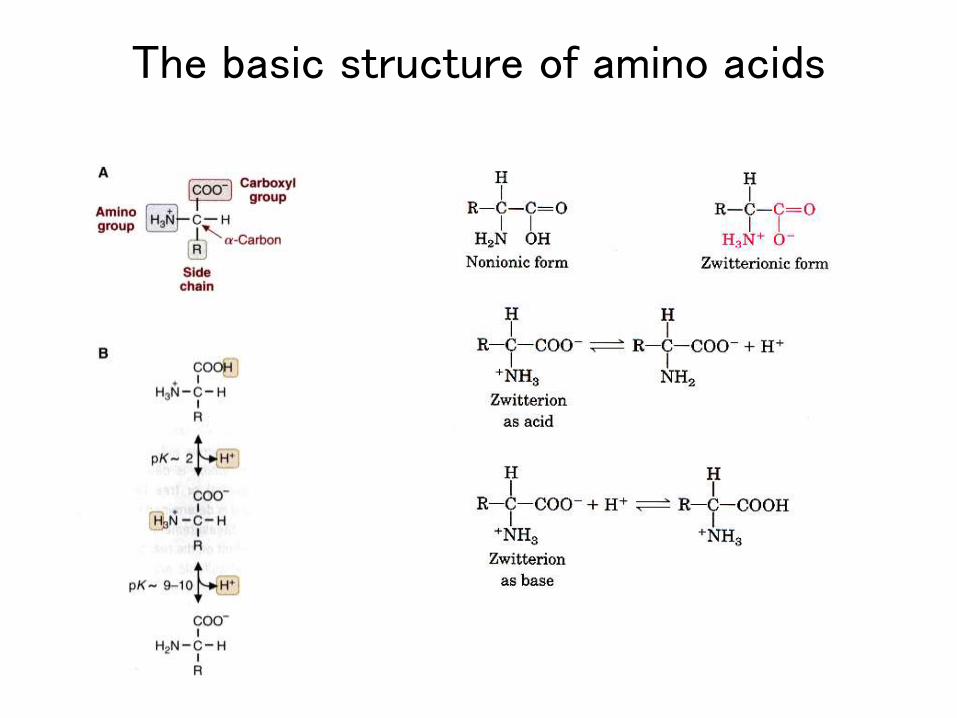
The basic structure of amino acids
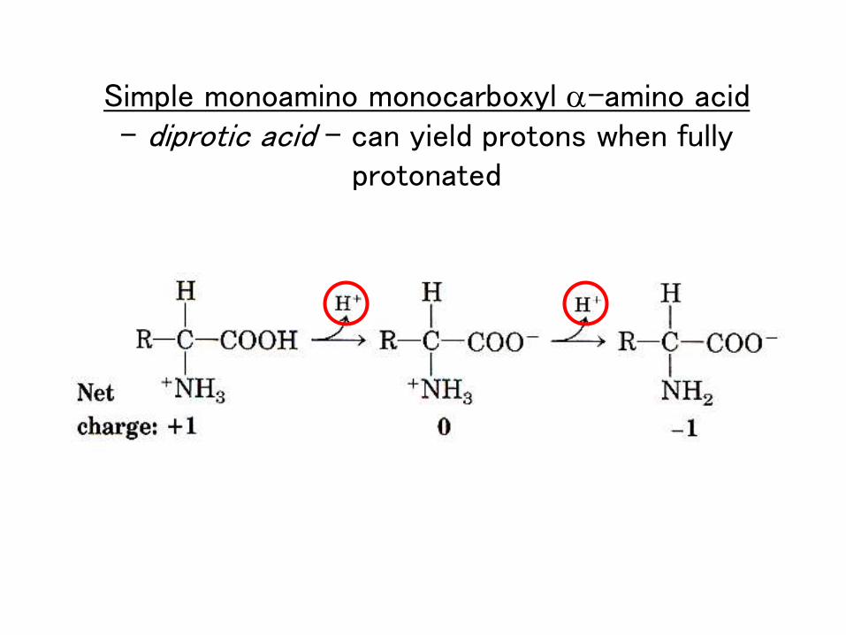
Simple monoamino monocarboxyl a-amino acid- diprotic acid - can yield protons when fully
protonated
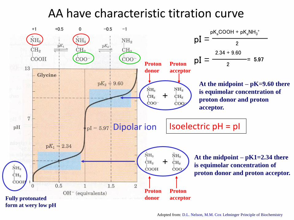
AA have characteristic titration curve
Fully protonated
form at wery low pH
At the midpoint – pK1=2.34 there
is equimolar concentration of
proton donor and proton acceptor.
+
Dipolar ion
+
At the midpoint – pK=9.60 there
is equimolar concentration of
proton donor and proton
acceptor.
Proton
donor
Proton
acceptor
Proton
donor
Proton
acceptor
Isoelectric pH = pI
Adopted from: D.L. Nelson, M.M. Cox Lehninger Principle of Biochemistry
+1 +0.5 0 -0.5 -1pKaCOOH + pKaNH3
+
pI =2
2.34 + 9.60
pI =2
= 5.97
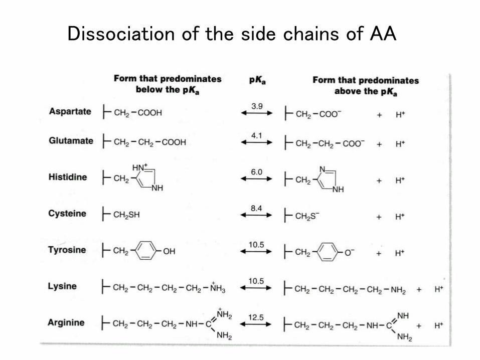
Dissociation of the side chains of AA
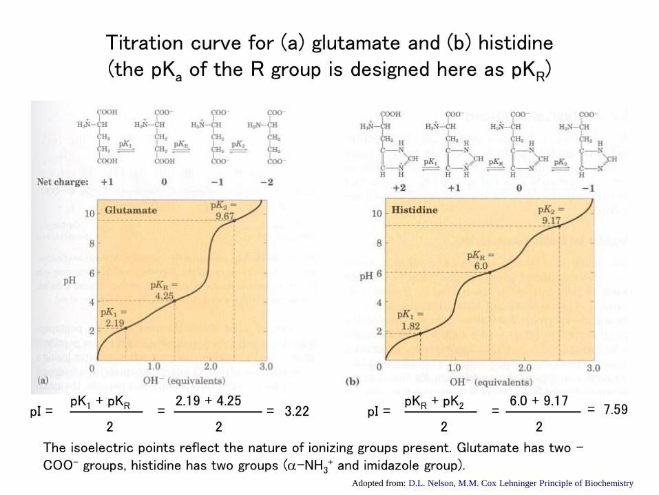
Titration curve for (a) glutamate and (b) histidine(the pKa of the R group is designed here as pKR)
The isoelectric points reflect the nature of ionizing groups present. Glutamate has two –COO- groups, histidine has two groups (a-NH3
+ and imidazole group).Adopted from: D.L. Nelson, M.M. Cox Lehninger Principle of Biochemistry
pI =pK1 + pKR
2=
2.19 + 4.25
2= 3.22 pI =
pKR + pK2
2=
6.0 + 9.17
2= 7.59
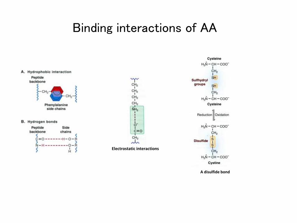
Binding interactions of AA
Electrostatic interactions
A disulfide bond
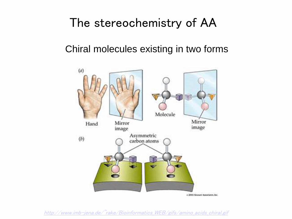
The stereochemistry of AA
Chiral molecules existing in two forms
http://www.imb-jena.de/~rake/Bioinformatics_WEB/gifs/amino_acids_chiral.gif
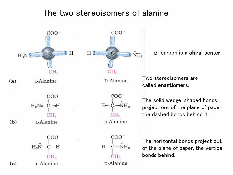
The two stereoisomers of alanine
a-carbon is a chiral center
Two stereoisomers are called enantiomers.
The solid wedge-shaped bondsproject out of the plane of paper, the dashed bonds behind it.
The horizontal bonds project out of the plane of paper, the vertical bonds behind.
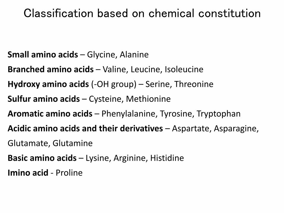
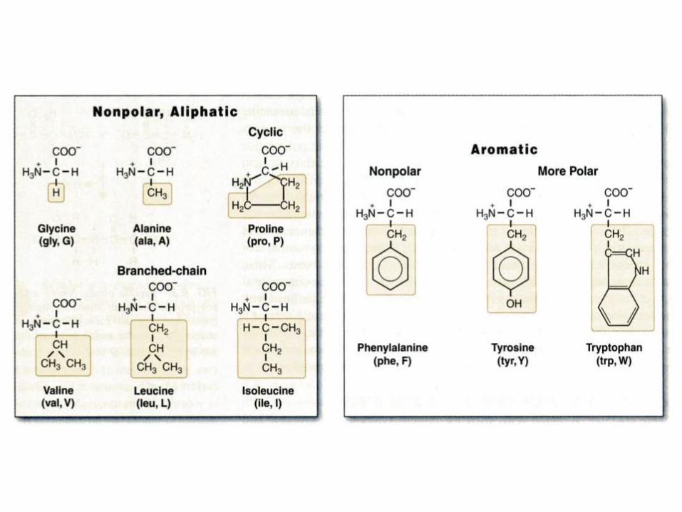
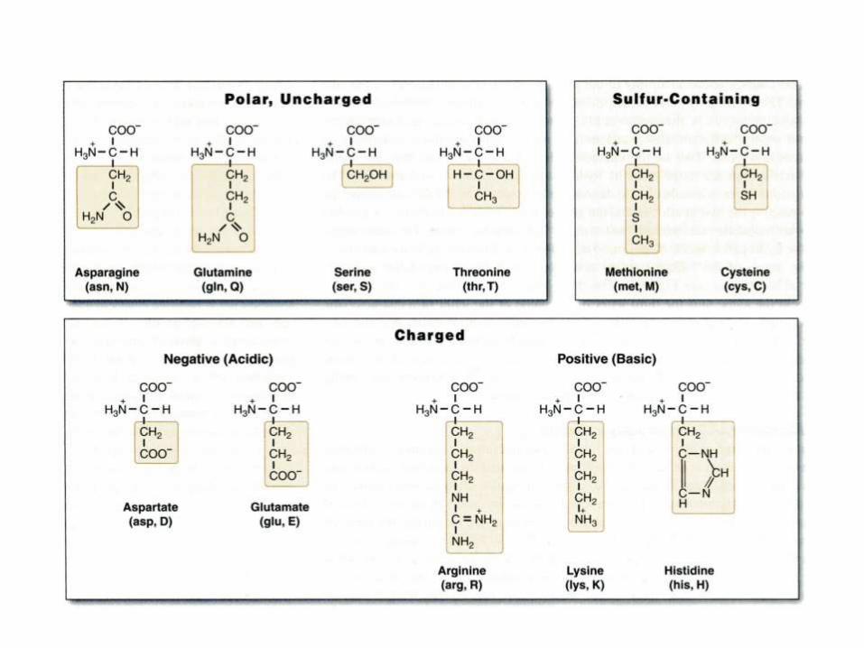
Classification based on chemical constitution
Small amino acids – Glycine, Alanine
Branched amino acids – Valine, Leucine, Isoleucine
Hydroxy amino acids (-OH group) – Serine, Threonine
Sulfur amino acids – Cysteine, Methionine
Aromatic amino acids – Phenylalanine, Tyrosine, Tryptophan
Acidic amino acids and their derivatives – Aspartate, Asparagine,
Glutamate, Glutamine
Basic amino acids – Lysine, Arginine, Histidine
Imino acid - Proline
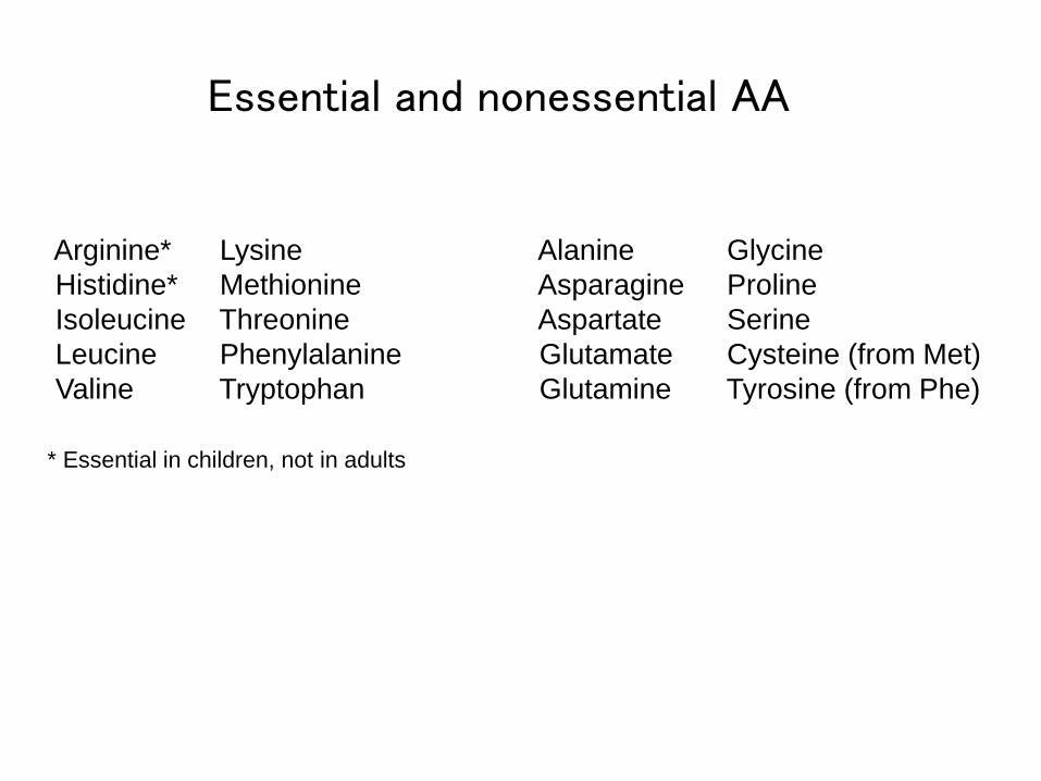
Essential and nonessential AA
Arginine*
Histidine*
Isoleucine
Leucine
Valine
Lysine
Methionine
Threonine
Phenylalanine
Tryptophan
Alanine
Asparagine
Aspartate
Glutamate
Glutamine
Glycine
Proline
Serine
Cysteine (from Met)
Tyrosine (from Phe)
* Essential in children, not in adults
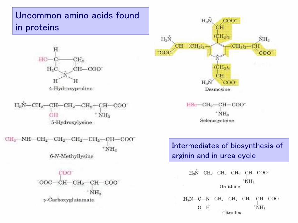
Uncommon amino acids found in proteins
Intermediates of biosynthesis of arginin and in urea cycle
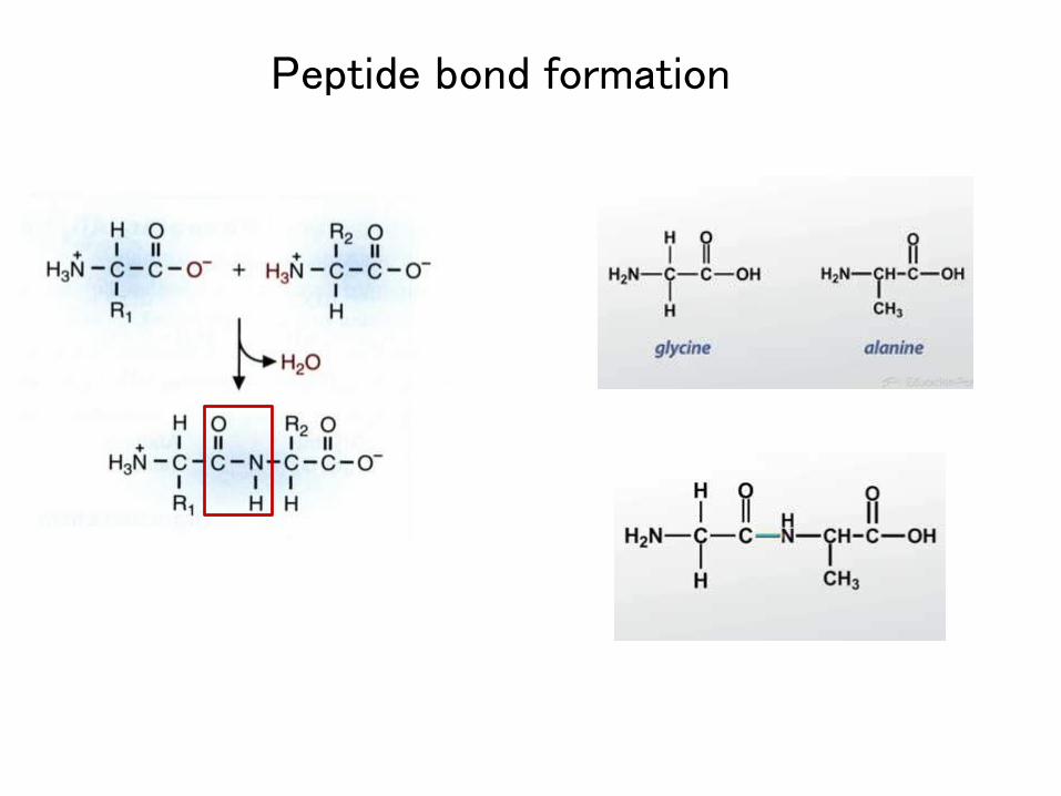
Peptide bond formation

Proteins
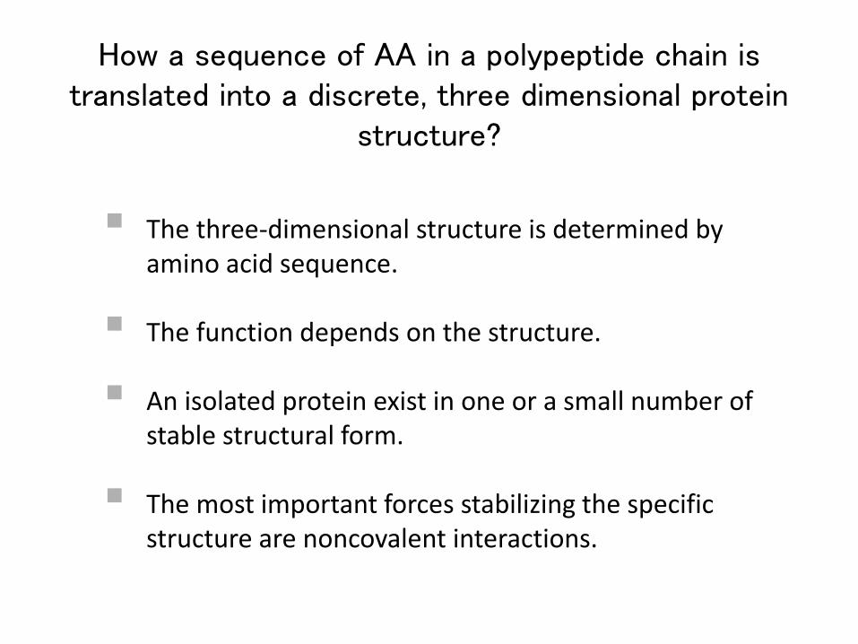
The three-dimensional structure is determined by amino acid sequence.
The function depends on the structure.
An isolated protein exist in one or a small number of stable structural form.
The most important forces stabilizing the specific structure are noncovalent interactions.
How a sequence of AA in a polypeptide chain is translated into a discrete, three dimensional protein
structure?
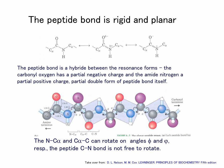
The peptide bond is rigid and planar
The peptide bond is a hybride between the resonance forms – the carbonyl oxygen has a partial negative charge and the amide nitrogen a partial positive charge, partial double form of peptide bond itself.
The N-Ca and Ca-C can rotate on angles f and j, resp., the peptide C-N bond is not free to rotate.
Take over from: D. L. Nelson, M. M. Cox :LEHNINGER. PRINCIPLES OF BIOCHEMISTRY Fifth edition
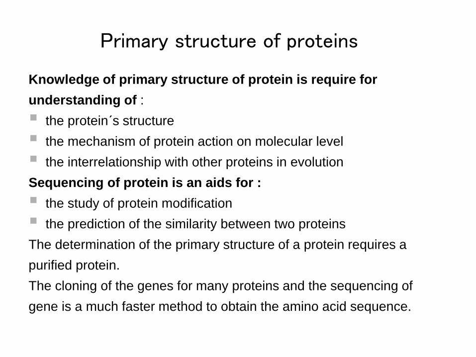
Knowledge of primary structure of protein is require for
understanding of :
the protein´s structure
the mechanism of protein action on molecular level
the interrelationship with other proteins in evolution
Sequencing of protein is an aids for :
the study of protein modification
the prediction of the similarity between two proteins
The determination of the primary structure of a protein requires a
purified protein.
The cloning of the genes for many proteins and the sequencing of
gene is a much faster method to obtain the amino acid sequence.
Primary structure of proteins
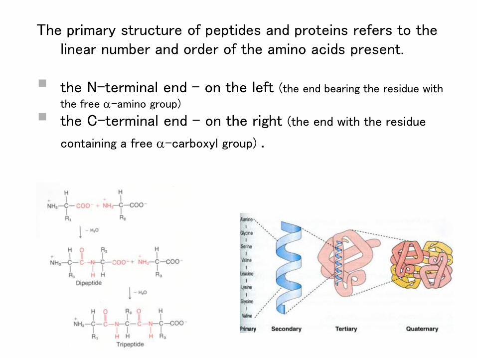
The primary structure of peptides and proteins refers to the linear number and order of the amino acids present.
the N-terminal end - on the left (the end bearing the residue with
the free a-amino group)
the C-terminal end - on the right (the end with the residue
containing a free a-carboxyl group) .
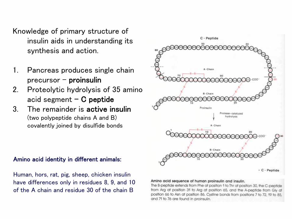
Knowledge of primary structure of insulin aids in understanding its synthesis and action.
1. Pancreas produces single chain precursor – proinsulin
2. Proteolytic hydrolysis of 35 amino acid segment – C peptide
3. The remainder is active insulin(two polypeptide chains A and B) covalently joined by disulfide bonds
Amino acid identity in different animals:
Human, hors, rat, pig, sheep, chicken insulin have differences only in residues 8, 9, and 10 of the A chain and residue 30 of the chain B
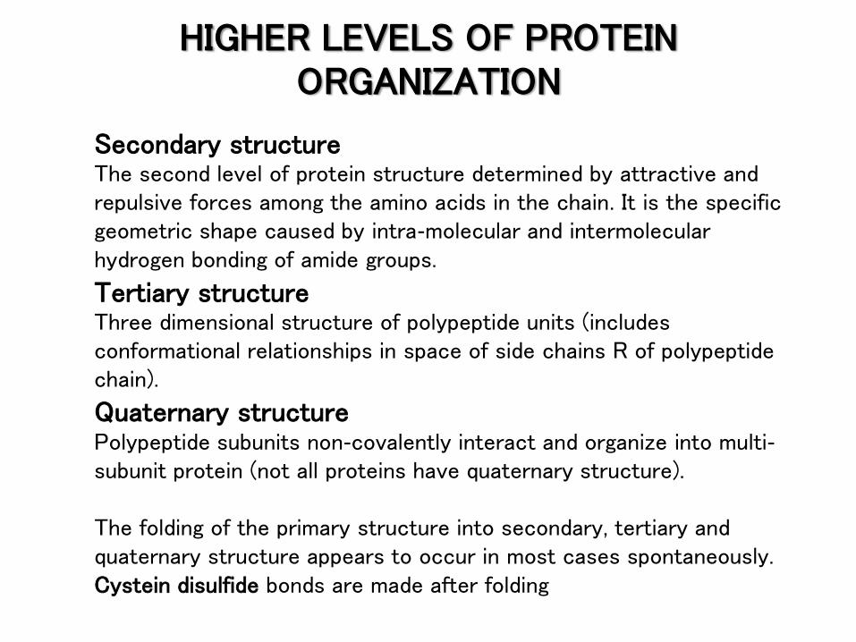
Secondary structureThe second level of protein structure determined by attractive and repulsive forces among the amino acids in the chain. It is the specific geometric shape caused by intra-molecular and intermolecular hydrogen bonding of amide groups.
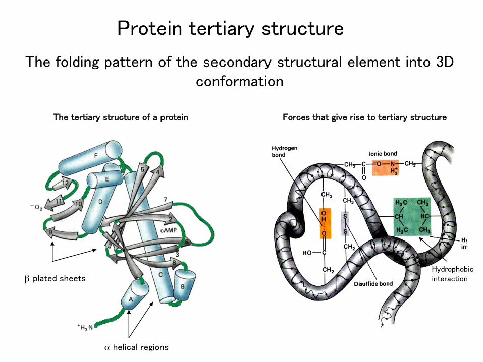
Tertiary structureThree dimensional structure of polypeptide units (includes conformational relationships in space of side chains R of polypeptide chain).
Quaternary structurePolypeptide subunits non-covalently interact and organize into multi-subunit protein (not all proteins have quaternary structure).
The folding of the primary structure into secondary, tertiary and quaternary structure appears to occur in most cases spontaneously.Cystein disulfide bonds are made after folding
HIGHER LEVELS OF PROTEIN ORGANIZATION
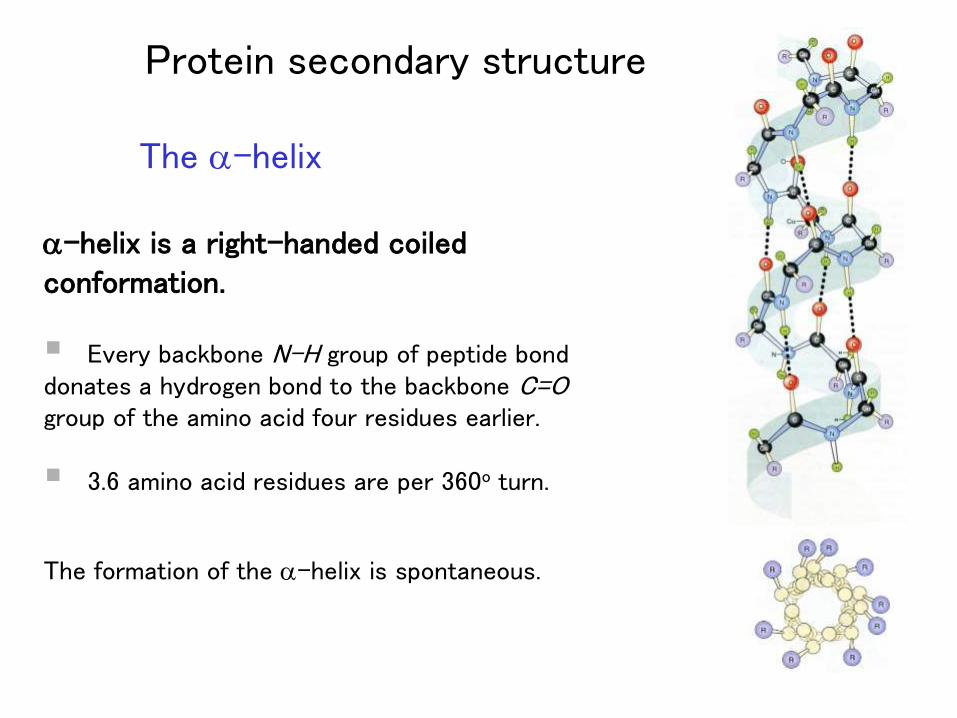
The a-helix
a-helix is a right-handed coiled conformation.
Every backbone N-H group of peptide bond donates a hydrogen bond to the backbone C=Ogroup of the amino acid four residues earlier.
3.6 amino acid residues are per 360o turn.
The formation of the a-helix is spontaneous.
Protein secondary structure
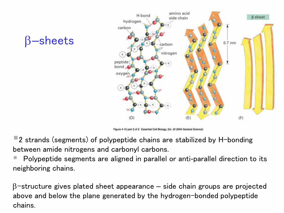
b–sheets
2 strands (segments) of polypeptide chains are stabilized by H-bonding between amide nitrogens and carbonyl carbons. Polypeptide segments are aligned in parallel or anti-parallel direction to its neighboring chains.
b-structure gives plated sheet appearance – side chain groups are projected above and below the plane generated by the hydrogen-bonded polypeptide chains.
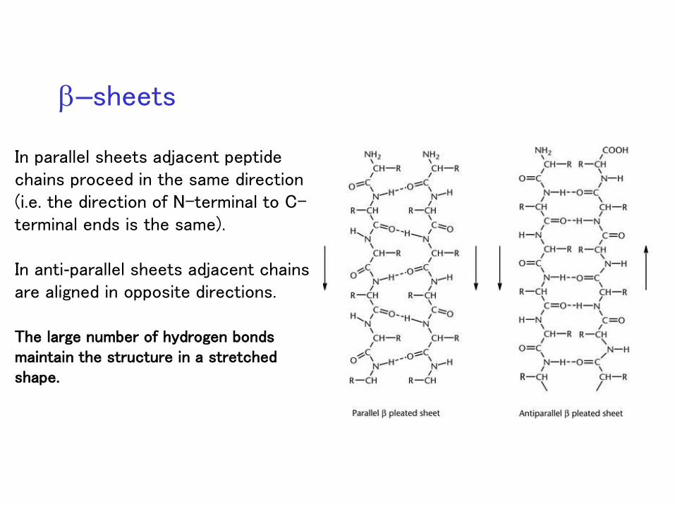
In parallel sheets adjacent peptide chains proceed in the same direction (i.e. the direction of N-terminal to C-terminal ends is the same).
In anti-parallel sheets adjacent chains are aligned in opposite directions.
The large number of hydrogen bonds maintain the structure in a stretched shape.
b–sheets
The folding pattern of the secondary structural element into 3D conformation
a helical regions
b plated sheets
Forces that give rise to tertiary structureThe tertiary structure of a protein
Hydrophobic interaction
Protein tertiary structure
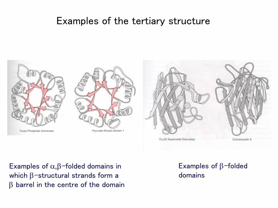
Examples of a,b-folded domains in which b-structural strands form a b barrel in the centre of the domain
Examples of b-folded domains
Examples of the tertiary structure
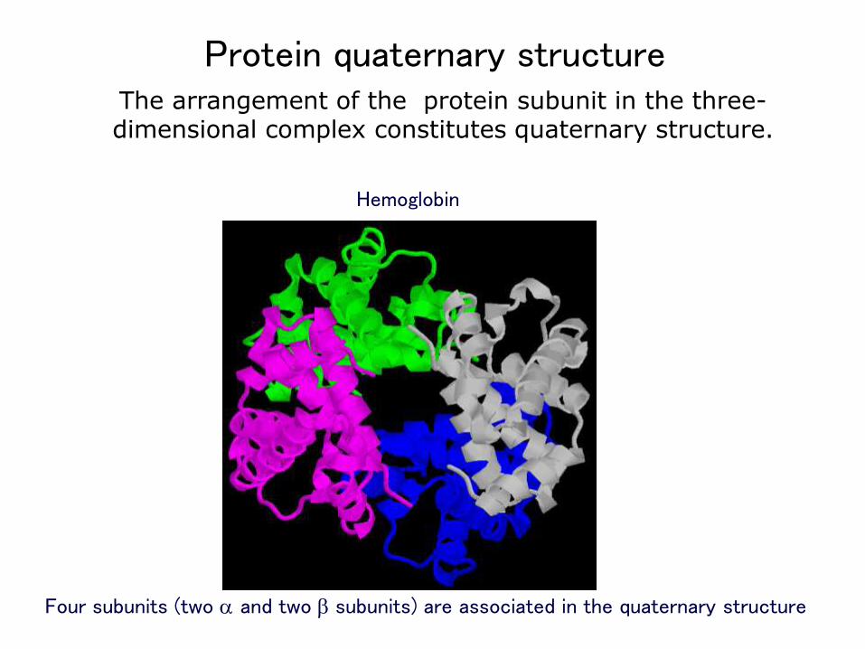
Protein quaternary structure
Four subunits (two a and two b subunits) are associated in the quaternary structure
Hemoglobin
The arrangement of the protein subunit in the three-dimensional complex constitutes quaternary structure.
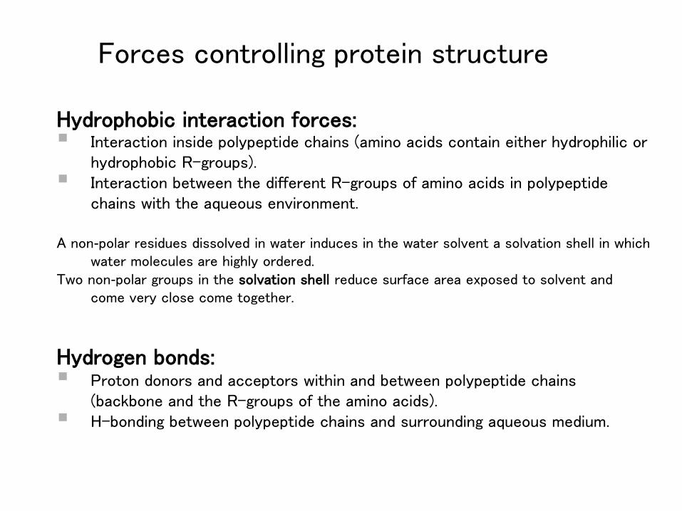
Forces controlling protein structure
Hydrophobic interaction forces: Interaction inside polypeptide chains (amino acids contain either hydrophilic or
hydrophobic R-groups). Interaction between the different R-groups of amino acids in polypeptide
chains with the aqueous environment.
A non-polar residues dissolved in water induces in the water solvent a solvation shell in which water molecules are highly ordered.
Two non-polar groups in the solvation shell reduce surface area exposed to solvent and come very close come together.
Hydrogen bonds: Proton donors and acceptors within and between polypeptide chains
(backbone and the R-groups of the amino acids). H-bonding between polypeptide chains and surrounding aqueous medium.
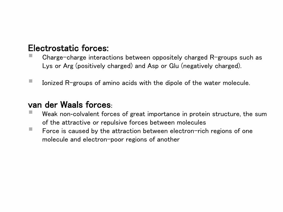
Electrostatic forces: Charge-charge interactions between oppositely charged R-groups such as
Lys or Arg (positively charged) and Asp or Glu (negatively charged).
Ionized R-groups of amino acids with the dipole of the water molecule.
van der Waals forces:
Weak non-colvalent forces of great importance in protein structure, the sum of the attractive or repulsive forces between molecules
Force is caused by the attraction between electron-rich regions of one molecule and electron-poor regions of another
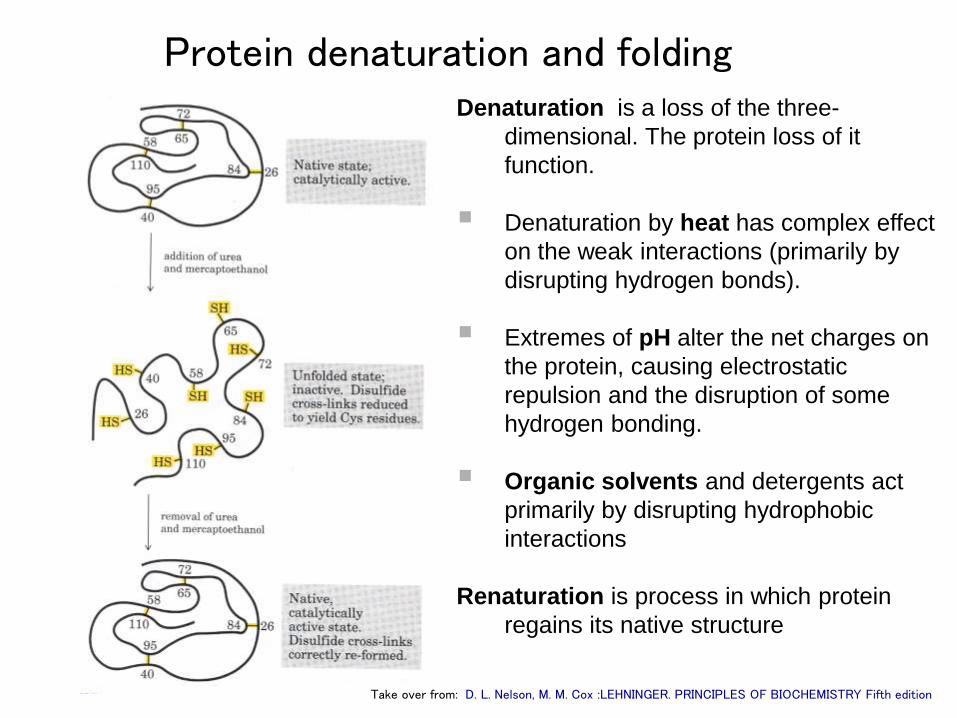
Protein denaturation and foldingDenaturation is a loss of the three-
dimensional. The protein loss of it
function.
Denaturation by heat has complex effect
on the weak interactions (primarily by
disrupting hydrogen bonds).
Extremes of pH alter the net charges on
the protein, causing electrostatic
repulsion and the disruption of some
hydrogen bonding.
Organic solvents and detergents act
primarily by disrupting hydrophobic
interactions
Renaturation is process in which protein
regains its native structure
Take over from: D. L. Nelson, M. M. Cox :LEHNINGER. PRINCIPLES OF BIOCHEMISTRY Fifth edition
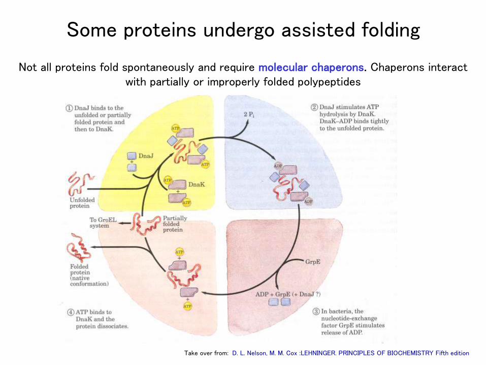
Some proteins undergo assisted folding
Not all proteins fold spontaneously and require molecular chaperons. Chaperons interact with partially or improperly folded polypeptides
Take over from: D. L. Nelson, M. M. Cox :LEHNINGER. PRINCIPLES OF BIOCHEMISTRY Fifth edition
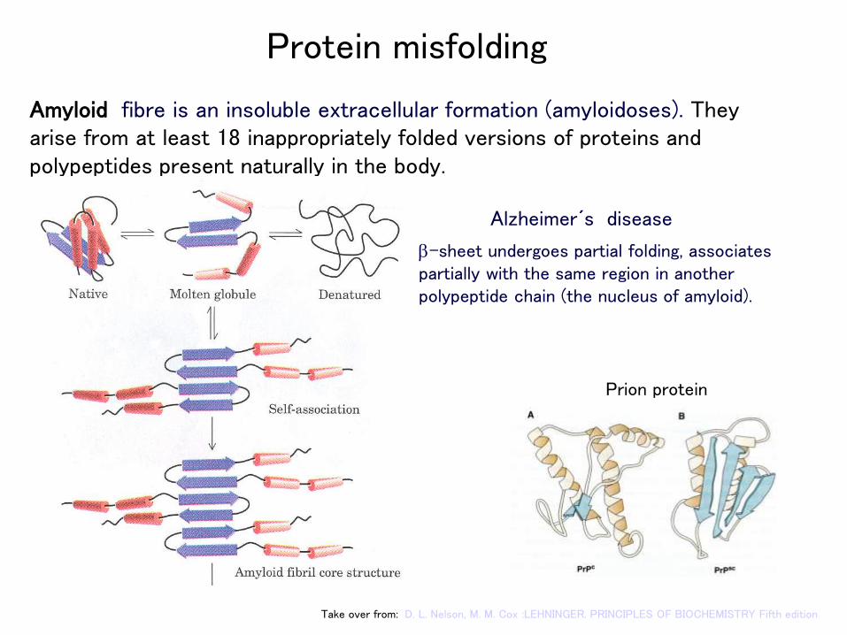
Protein misfolding
Amyloid fibre is an insoluble extracellular formation (amyloidoses). They arise from at least 18 inappropriately folded versions of proteins and polypeptides present naturally in the body.
Take over from: D. L. Nelson, M. M. Cox :LEHNINGER. PRINCIPLES OF BIOCHEMISTRY Fifth edition
b-sheet undergoes partial folding, associates partially with the same region in another polypeptide chain (the nucleus of amyloid).
Alzheimer´s disease
Prion protein
1. Globular proteins are compactly folded and coiled.
2. Fibrous proteins are more filamentous or elongated.
3. Peptides• Small peptides (containing less than a couple of dozen
amino acids) are called oligopeptides. • Long peptides are called polypeptides.• Peptides have a "polarity"; each peptide has only one
free a-amino group (on the amino-terminal residue) and one free (non-side chain) carboxyl group (on the carboxy-terminal residue)
Protein structure
Functional roles of proteins
1. Dynamic function
transport
metabolic control
contraction
catalysis of chemical transformation
2. Structural function
bone, connective tissue
1. Enzymes (lactate dehydrogenase, DNA polymerase)
2. Storage proteins (ferritin, cassein, ovalbumin)
3. Transport proteins (hemoglobin, myoglobin, serum albumin)
4. Contractile proteins (myosin, actin)
5. Hormones (insulin, growth hormone)
6. Protective proteins in blood (antibodies, complement,
fibrinogen)
7. Structural proteins (collagen, elastin, glycoproteins)
Classification of proteins by bioloical function

Globular proteins Spheroid shapeVariable molecular weightRelatively high water solubilityVariety function roles – catalysts, transporters, control proteins (for the
regulation of metabolic pathways and gene expression)
Fibrous proteins Rodlike shapeLow solubility in the waterStructural role in the organism
LipoproteinsComplex of lipids with protein
GlycoproteinsContain covalently bound carbohydrate
Types of proteins
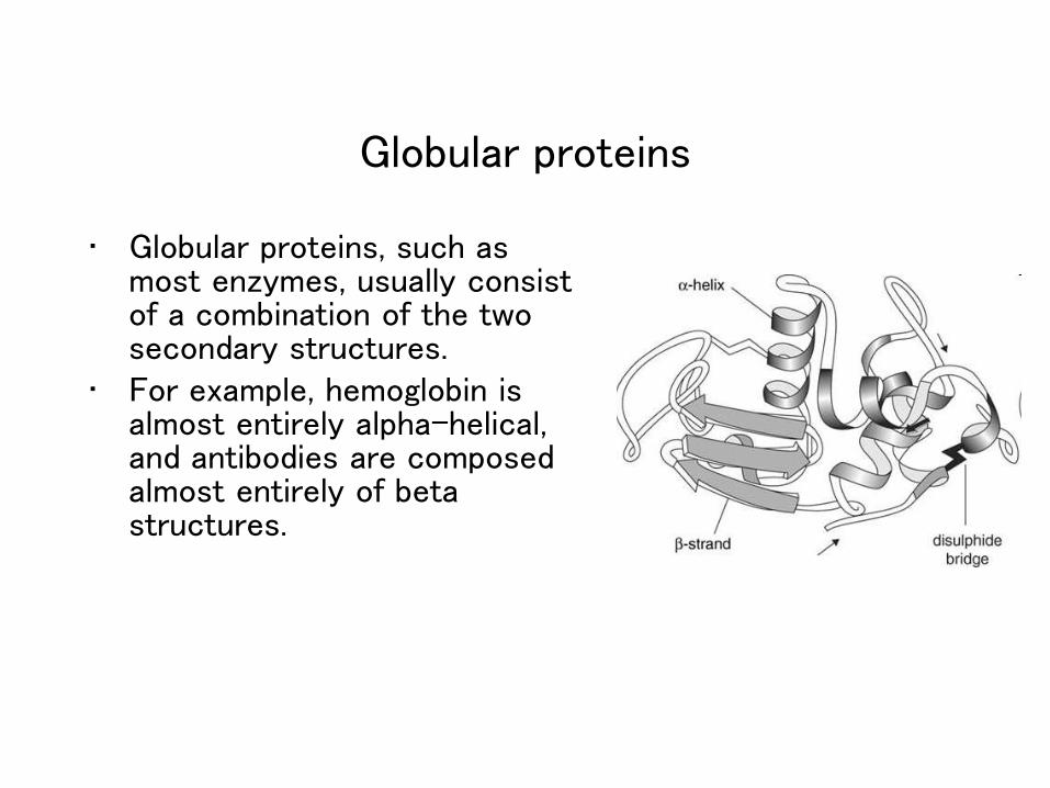
Globular proteins
• Globular proteins, such as most enzymes, usually consist of a combination of the two secondary structures.
• For example, hemoglobin is almost entirely alpha-helical, and antibodies are composed almost entirely of beta structures.
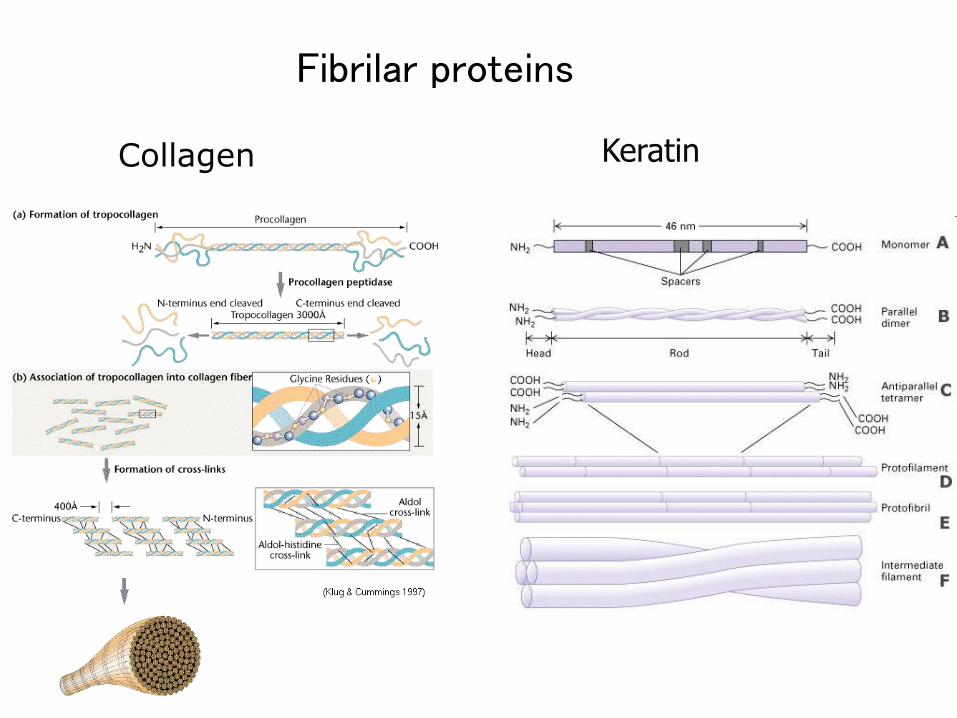
Keratin
Fibrilar proteins
Collagen
Multicomponent complexes of protein and lipids.
The lipids or their derivatives may be covalently or non-covalently bound
to the proteins.
Examples of lipoproteins: many enzymes, transporters, structural
proteins, antigens, adhesins and toxins.
The function of lipoprotein particles - transport of lipids (fats) and cholesterol around the body in the aqueous blood, in which they would normally dissolve
Lipoproteins
Glycoproteins
Glycoproteins have covalently attached sugar molecules at one or multiple points along the polypeptide chain
Glycoproteins are:• hormones• extracellular matrix proteins• proteins involved in blood coagulation• antibodies• mucus secretion from epithelial cells• protein localized on surface of cells• receptors (transmit signals of hormones or growth factors from
outside environment into the cell)Sugar molecules are:glucose, galactose, mannose, fucose, xylose, N-acetylglucos-amine, N-
acetylgalactosamine
Structure-Function Relationship of Protein Families
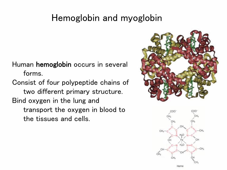
Human hemoglobin occurs in several forms.
Consist of four polypeptide chains of two different primary structure.
Bind oxygen in the lung and transport the oxygen in blood to the tissues and cells.
Hemoglobin and myoglobin
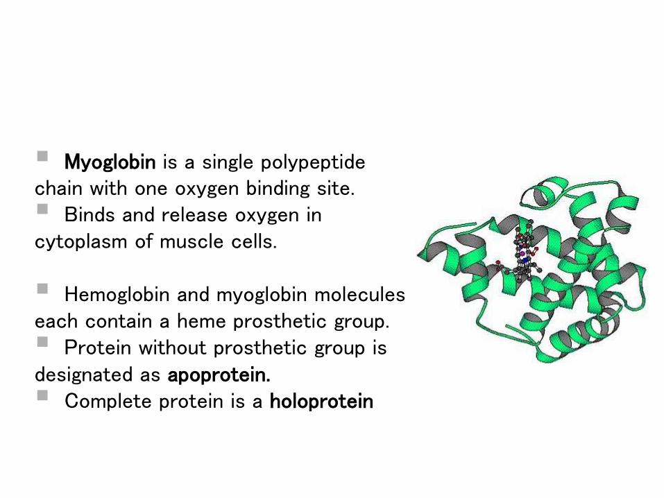
Myoglobin is a single polypeptide chain with one oxygen binding site. Binds and release oxygen in cytoplasm of muscle cells.
Hemoglobin and myoglobin molecules each contain a heme prosthetic group. Protein without prosthetic group is designated as apoprotein. Complete protein is a holoprotein
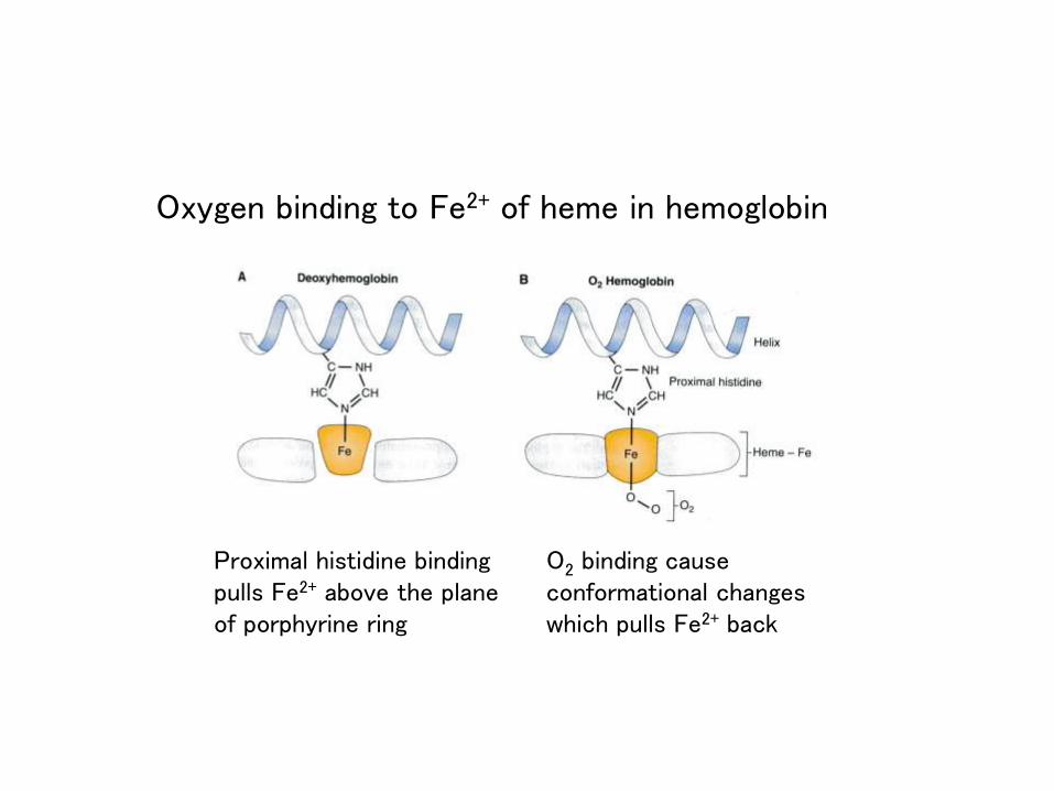
Oxygen binding to Fe2+ of heme in hemoglobin
O2 binding cause conformational changes which pulls Fe2+ back
Proximal histidine binding pulls Fe2+ above the plane of porphyrine ring
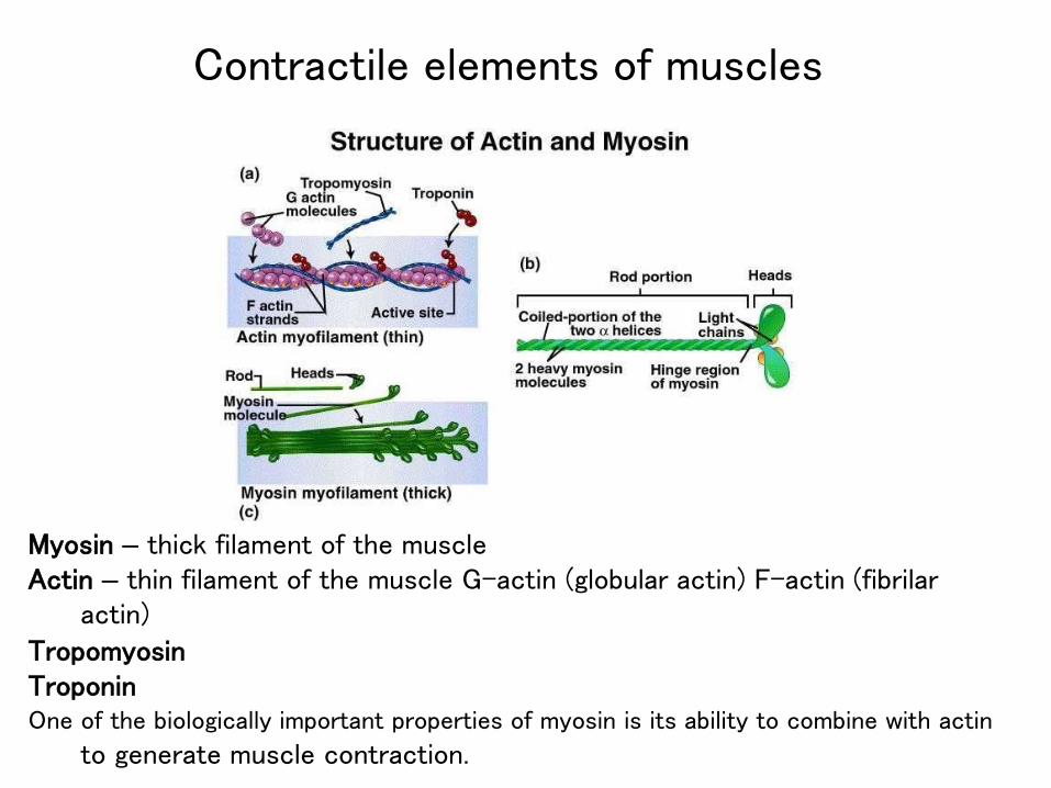
Myosin – thick filament of the muscleActin – thin filament of the muscle G-actin (globular actin) F-actin (fibrilar
actin)
TropomyosinTroponinOne of the biologically important properties of myosin is its ability to combine with actin
to generate muscle contraction.
Contractile elements of muscles
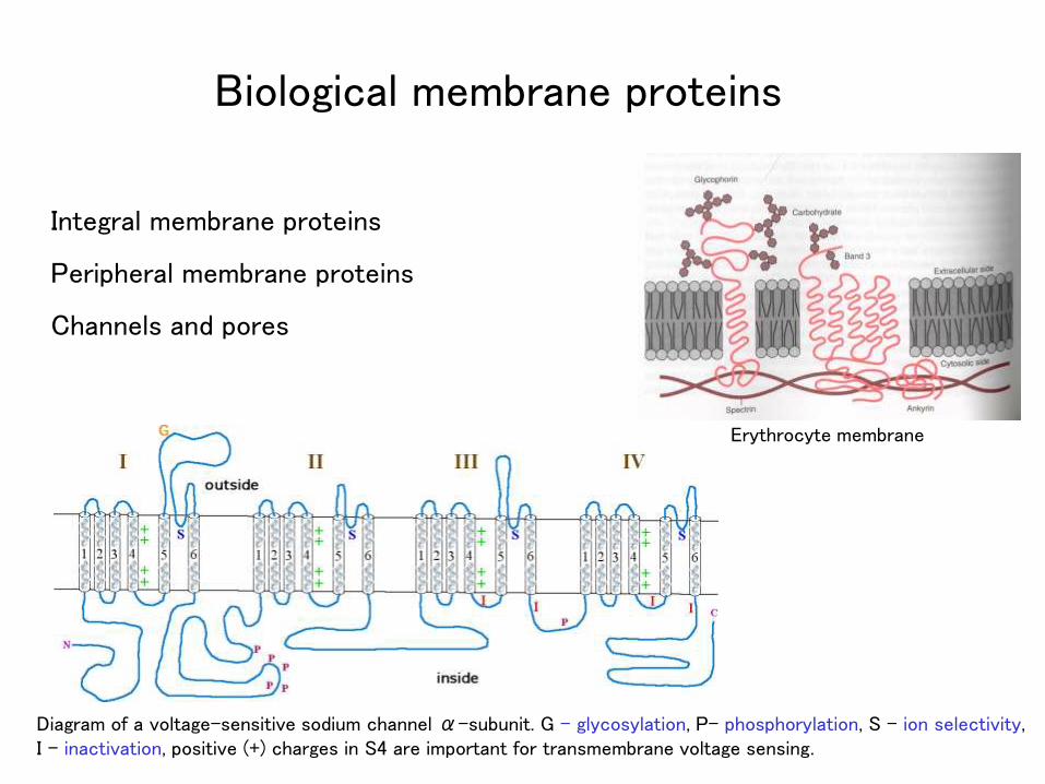
Integral membrane proteins
Peripheral membrane proteins
Channels and pores
Erythrocyte membrane
Diagram of a voltage-sensitive sodium channel α-subunit. G - glycosylation, P- phosphorylation, S - ion selectivity, I - inactivation, positive (+) charges in S4 are important for transmembrane voltage sensing.
Biological membrane proteins
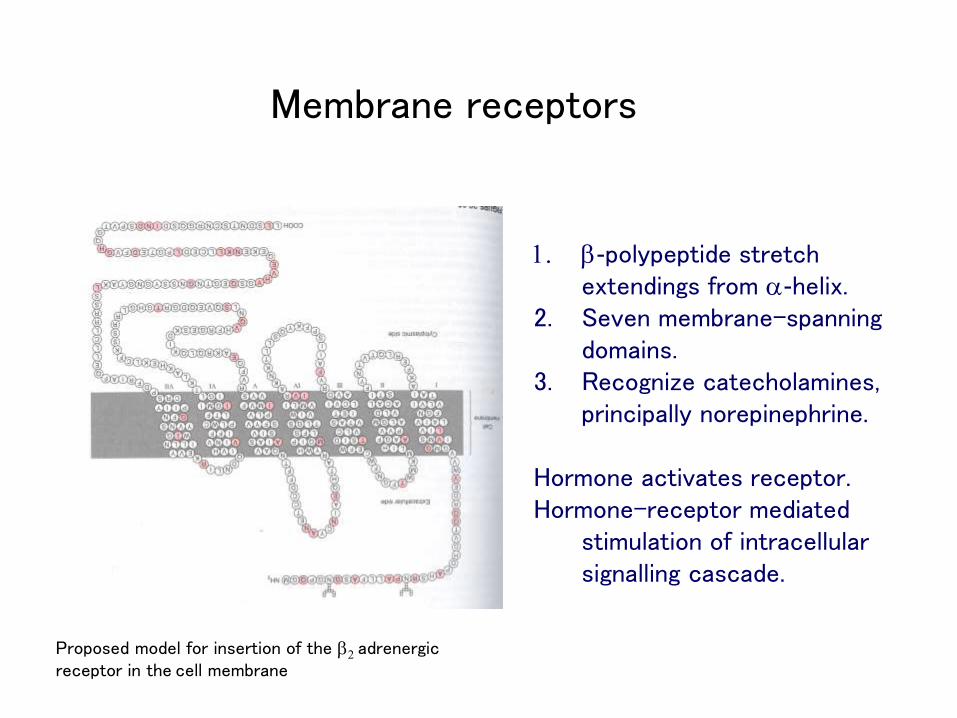
Proposed model for insertion of the b2 adrenergic receptor in the cell membrane
1. b-polypeptide stretch extendings from a-helix.
2. Seven membrane-spanning domains.
3. Recognize catecholamines, principally norepinephrine.
Hormone activates receptor.Hormone-receptor mediated
stimulation of intracellular signalling cascade.
Membrane receptors
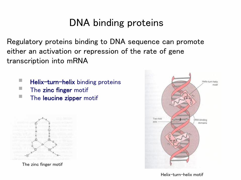
Regulatory proteins binding to DNA sequence can promote either an activation or repression of the rate of gene transcription into mRNA
Helix-turn-helix binding proteins The zinc finger motif The leucine zipper motif
The zinc finger motif
Helix-turn-helix motif
DNA binding proteins