preface - modern biology inc. creative experiments...
TRANSCRIPT
1 PREFACE EXPERIMENTS 401-403MODERNBIO.COM
Preface Individual Experiments 401-403
The material in this folder is divided into three sections. The first section provides basic information on the biochemistry of nucleic acids, and is also intended to acquaint the students with the principles and techniques of electrophoresis and DNA hybridization. The second part shows students how to apply what they have learned to perform one or more exercises in modern biology. Each exercise consists of a background information section, an experimental procedure and study questions. Before the laboratory, the student should read the background information section, study the directions for doing the experiment and understand the reason for each step in the procedure. The instructor manual for experiments 401-403 is the third item in the folder. This manual contains a listing of the chemicals provided with each experiment in this series, procedures for preparing solutions, typical experimental results and answers to study questions.
John N. Anderson, Ph.D. Professor of BiologyDepartment of Biological SciencesPurdue UniversityWest Lafayette, Indiana 47907
© 2013 by John N. Anderson, All rights reserved.
NOTES:1. Permission is granted to reproduce the written material in this folder one time for educational purposes only.2. Electrophoresis Package 3/4 is required to perform the exercises described in this folder.

2 PREFACE EXPERIMENTS 401-403MODERNBIO.COM
Table of conTenTsPart A. Background Information
I. Nucleic Acid Structure and Function: A Review of the Basics Nucleotides- ..Building Blocks of Nucleic Acids ................................................................. 1
The Polynucleotide Chain .............................................................................. 3
DNA Structure .............................................................................................. .4
Biological Role of DNA and RNA ................................................................ 4
Analyzing Specific Genes and Recombinant DNA Technology ................... 7
II. General Description of Agarose Gel Electrophoresis
III. Practical Aspects of Electrophoresis
Electrophoresis Equipment ........................................................................... l2
Electrophoresis Chemicals ............................................................................ l4
IV. Electrophoresis Procedures
Pouring the Agarose Gels ............................................................................ 15
Sample Application ...................................................................................... 16
Electrophoresis ............................................................................................ 17
Staining ........................................................................................................ 18
Determining the Size of DNA by Electrophoresis ....................................... 19
V. Hybridization Analysis
DNA Denaturation and Renaturation ........................................................... 21
Hybridization Procedures ............................................................................ 24
Application of Southern Blotting in Medicine ............................................ 30
VI. Suggested Reading and References for Part A ............................................ 33
Part B. Laboratory ExercisesExperiment 1 (401) Evolution of the Vertebrate Genome .................................. 34
Experiment 2 (402) Application of the Southern Blot Procedure ....................... 44
Experiment 3 (403) Detecting a Specific Sequence in the Mammalian Genome ................................................................................... 63

3 PREFACE EXPERIMENTS 401-403MODERNBIO.COM
ParT a. background InformaTIonI. Nucleic Acids: A Review of the Basics
The concept that chromosomal units known as genes transmit heritable information from parent to offspring was founded in the late 19th century. However, a description of genes in terms of their unique structural and functional properties is relatively new. We now know that genes are composed of a type of nucleic acid called deoxyribonucleic acid (DNA). The DNA molecule not only directs its own reproduction but also stores all the information that determines the types of proteins produced during the lifetime of an organism. In so doing, DNA orchestrates the complex reactions and structures characteristic of an organism and its offspring. Ribonucleic acid (RNA), the second major category of nucleic acids, is involved principally in the transmission of genetic information and in protein production. The structure and function of DNA and RNA can most easily be understood by examining the chemical composition of the nucleic acids.
Nucleotides - Building Blocks of Nucleic AcidsUnder the proper conditions, nucleic acids can be broken down to low- molecular weight products of three types: a pentose (or 5 carbon) sugar; purines and pyrimidines; and phosphoric acid (Figure 1). The phosphate group is responsible for the strong negative charge of nucleic acids. The pentose sugar from RNA is always ribose and that from DNA is 2-deoxyribose. These sugars differ only by the presence or absence of a hydroxyl group on carbon 2 (so-called 2’). The numbers assigned to the five carbon atoms are shown in Figure 1. The purines and pyrimidines are often called nitrogenous bases (or, simply, bases). The major purine bases in DNA and RNA are adenine (A) and guanine (G), and the major pyrimidines in DNA are cytosine (C) and thymine (T). RNA contains the base uracil (U) in place of thymine. The sugars and phosphates are readily soluble in water. That is, they are hydrophilic. In contrast, the bases are hydrophobic in that they display limited solubility in water. As will be discussed below, these differences in water solubility are extremely important for the structure of the DNA molecule.
A nucleotide consists of a pentose sugar, a nitrogenous base and a phosphate group structured as shown below. The high-energy storage compound, adenosine triphosphate (ATP), is a well known nucleotide found in biological systems.

4 PREFACE EXPERIMENTS 401-403MODERNBIO.COM
Figure 1. The Nucleotide Components

5 PREFACE EXPERIMENTS 401-403MODERNBIO.COM
The Polynucleotide ChainNucleic acids are polynucleotides and have the general structure shown below:
A polynucleotide is composed of repeating nucleotide units linked into chains by phosphodiester bonds that join the 5’ carbon of one ribose or deoxyribose group to the 3’ carbon of the next sugar (Figure 2). The sequence or order of nucleotides in a polynucleotide chain is often abbreviated by a 1-letter code (e.g., G-C-A-T-A) with the 5’ end of the chain written at the left. A typical RNA molecule is a single-stranded polynucleotide chain. As will be described below, DNA usually contains two polynucleotide strands coiled around one another to form a double-stranded helix. The number of nucleotide units in a nucleic acid chain varies tremendously depending on the nucleic acid type. For example, each chromosome from a higher organism is thought to contain a single, very long DNA molecule. A DNA molecule from the largest human chromosome is composed of approximately 5.4 x 10⁸ nucleotides, which corresponds to a molecular weight of the order of 10 11 and a length of about 4cm. On the other hand, transfer RNA molecules generally contain only 70-80 nucleotides.
Figure 2. Structure of the Polynucleotide Chain

6 PREFACE EXPERIMENTS 401-403MODERNBIO.COM
DNA StructureIn early physical studies of DNA, a variety of experiments indicated that DNA molecules occur in long helixes with each helix being formed from two or more polynucleotide chains bound side by side. Chemical analyses also demonstrated that the phosphate groups were on the outside of the helix and that the number of A and T residues in DNA were always equal, as were those of G and C. With these facts in mind, Watson and Crick in 1953 proposed that the DNA molecule actually consists of two polynucleotide chains coiled around the same axis to form a double helix (Figure 3). In this model, the hydrophilic sugar-phosphate groups follow the outer edges of the molecule where they can interact with water. The hydrophobic bases face inward toward each other in the molecule’s center and thus avoid contact with water. The two polynucleotide strands run in opposite directions (they are anti- parallel) and are held together primarily by hydrogen and hydrophobic bonding between the bases, where A is always paired with T, and G with C. These complementary bases have an affinity for each other such that, when they are paired, they contribute to the overall stability of the DNA helix. Because of this complementary base-pairing, the sequence of bases in one polynucleotide chain determines the sequence in the other. For example, if the bases along one strand are arranged in the order T-G-C-T-A-G, the opposite bases on the complementary strand will be A C-G-A-T-C. This fact is of extreme biological significance because it explains how a DNA helix in the chromosome directs the formation of copies of itself and directs the formation of RNA molecules with its specific informational content. The B-form DNA shown in Figure 3 is the most common of the DNA types. It is a right-handed double helix and contains about 10.5 nucleotide pairs per helical turn.
Figure 3. Double-Helical Structure of Common B-DNA

7 PREFACE EXPERIMENTS 401-403MODERNBIO.COM
Biological Role of DNA and RNADNA is an information molecule with two general functions (Figure 4). First, DNA plays a central role in the propagation of the species and the determination of the heritable characteristics of the cell and its descendants. Prior to the time of each cell division, the two strands of the DNA helix separate from one another and each serves as a pattern or template for the synthesis of a new, complementary chain. This process of DNA biosynthesis is known as replication. One of the double helices formed is then transmitted to one daughter cell, and one to the other. Although the principle underlying DNA replication is straightforward, the actual mechanism responsible for the replication process in the cell involves an array of enzymes and regulatory proteins.
The informational content of DNA also determines the types of proteins that are produced by a cell. In this manner, the DNA molecule functions as a blueprint for all cellular processes that go on during the lifetime of an organism. In the first step along the information pathway from DNA to protein, a segment of DNA is copied into a complementary strand of messenger RNA (mRNA) by a process known as transcription. Transcription begins when an enzyme called RNA polymerase binds to a specific sequence on the DNA known as the promoter. At this site, the enzyme unwinds a small segment of double helix, exposing the bases of the two single strands of the DNA molecule. One of these strands is then transcribed. As the polymerase travels along the DNA, ribonucleotides with bases complementary to the DNA are added to the growing chain. For example, the DNA segment C-G-T-A-T G is transcribed into G-C-A-U-A-C in the mRNA. Each sequence of three nucleotides in the mRNA is called a “codon,” and codes for one amino acid. Since most polypeptide chains contain between 100 to 1,000 amino acids, an mRNA must be at least 300 to 3000 nucleotides long. Therefore, a gene that codes for a polypeptide chain must contain at least 300 to 3,000 base pairs.
The translation of mRNA into protein is a complex process that occurs on particles called ribosomes. This process requires ribosomal RNAs (rRNAs) and transfer RNAs (tRNAs). These RNA species do not specify proteins themselves but rather take part in decoding the information carried by the mRNAs. At one end of each tRNA molecule is a nucleotide triplet called the “anticodon,” which is complementary to an mRNA codon. A specific amino acid is bound to the opposite end of each tRNA molecule. On the ribosome, tRNAs carrying amino acids associate with the mRNA by way of complementary base-pairing at the anticodon-codon sequences. As the ribosome moves along the mRNA, the amino acids carried by the tRNAs are linked to the growing polypeptide chain. In this manner, the order of codons along the mRNA directs the amino acid sequence of a polypeptide chain. The translation process occurs on the surface of the ribosomes. These particles, which are composed of rRNA and ribosomal proteins, serve to bring together the mRNAs, the tRNA and other factors that are required for protein production.

8 PREFACE EXPERIMENTS 401-403MODERNBIO.COM
ReplicationThe two DNA strands separate and each serves as a template for the synthesis of a new, complementary polynucleotide.
TranscriptionOne strand of the DNA serves as a template for the synthesis of complementary RNA.
TranslationThe mRNA associates with ribosomes and its nucleotides are matched three at a time to a complementary set of three nucleotides in a specific tRNA molecule. The tRNA molecule carries an amino acid and when it associates with the mRNA, the amino acid is added to the growing polypeptide chain.
Figure 4. Molecular Information Transfer: Complimentary Base Pairing

9 PREFACE EXPERIMENTS 401-403MODERNBIO.COM
Analysis of Specific Genes and Recombinant DNA TechnologyA key to one of life’s great mysteries was discovered in 1953 when the double helical structure of DNA was perceived by Watson and Crick. Elucidation of the basic mechanisms of replication, transcription and translation quickly followed, and by the early 1960’s, the model shown in Figure 4 was generally accepted by most biologists. However, genes from higher organisms resisted detailed analysis until the mid
1970’s because of the complexity of the DNA in eukaryotic organisms; a vertebrate cell contains enough DNA to code for more than 100,000 proteins. In order to study the structure and function of a single protein coding gene, the gene must be prepared in a purified form. The isolation of a specific gene from cellular DNA by conventional biochemical procedures is not practical because of the magnitude of the purification required (usually 100,000-fold) and because the procedures would necessitate the use of a large quantity of starting cellular DNA. Herein lies the major use of recombinant DNA technology, for it permits the amplification and isolation of specific genes by relatively simple procedures. A basic understanding of these procedures requires a description of an interesting feature of bacterial physiology.
Plasmids are small circular DNA molecules that exist apart from the chromosomes in most bacterial species. Under normal circumstances, plasmids are not essential for survival of the host bacteria. However, many plasmids contain genes that enable the bacteria to survive and prosper in certain environments. For example, some plasmids carry one or more genes that confer resistance to antibiotics. A bacterial cell containing such a plasmid can live and multiply in the presence of the drug. Indeed, antibiotic-resistant E.coli isolated in many parts of the world contain plasmids that carry the genetic information for protein products that interfere with the action of many different antibiotics.
In the laboratory, plasmids can be introduced into living bacterial cells by a process known as transformation. When bacteria are placed in a solution of calcium chloride, they acquire the ability to take in plasmid DNA molecules. As illustrated in Figure 5, this procedure provides a means for preparing large amounts of specific plasmid DNA since one transformed cell gives rise to a clone of cells that contains exact replicas of the parent plasmid DNA molecule. Following growth of the bacteria in the presence of the antibiotic, the plasmid DNA can readily be isolated from the bacterial culture.
Plasmids, as well as certain viruses, are extraordinarily useful tools for the molecular biologist, because they serve as gene-carrier molecules called cloning vectors. A basic procedure of recombinant DNA technology consists of joining a gene of interest to vector DNA to form a hybrid or recombinant molecule that is able to replicate in bacteria. Thus, cloning vectors contain genes for replication in bacteria. In addition, vectors generally carry antibiotic-resistance genes so that uninfected bacteria can be eliminated from the culture. In order to prepare a recombinant DNA molecule, a procedure is required for cutting cloning vectors and cellular DNA molecules at precise positions.

10 PREFACE EXPERIMENTS 401-403MODERNBIO.COM
Figure 5. Introduction of a Plasmid Into Bacteria by Transformation
11 PREFACE EXPERIMENTS 401-403MODERNBIO.COM
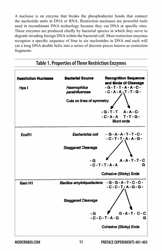
A nuclease is an enzyme that breaks the phosphodiester bonds that connect the nucleotide units in DNA or RNA. Restriction nucleases are powerful tools used in recombinant DNA technology because they cut DNA at specific sites. These enzymes are produced chiefly by bacterial species in which they serve to degrade invading foreign DNA within the bacterial cell. Most restriction enzymes recognize a specific sequence of four to six nucleotides in DNA and each will cut a long DNA double helix into a series of discrete pieces known as restriction fragments.
Table 1. Properties of Three Restriction Enzymes

12 PREFACE EXPERIMENTS 401-403MODERNBIO.COM
Typically, the restriction sites for a given enzyme are hundreds to thousands of base-pairs apart so that the fragments generated are hundreds to thousands of base-pairs long. More than 300 different restriction nucleases are now commercially available. General properties of three of these enzymes are given in Table 1. It should be noted that some restriction nucleases (e.g., EcoR1 and Bam H1) produce a staggered cleavage that creates sticky, or cohesive, single-stranded ends on the cut molecules. These cohesive ends are very important in recombinant DNA procedures because they enable any two DNA fragments to be linked together by complementary base pairing at their ends, provided that they were generated with the same restriction enzyme.
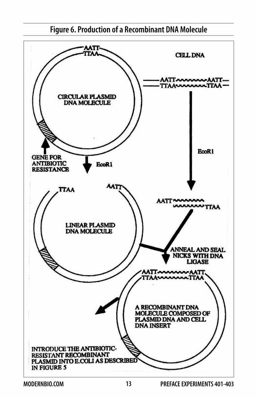
Figure 6 illustrates one basic procedure by which cellular DNA from essentially any source can be amplified by recombinant DNA techniques. First, a plasmid is cleaved at a single site by a restriction nuclease, such as EcoR1, that produces cohesive ends on the plasmid DNA. The cellular DNA to be cloned is cleaved with the same enzyme, and fragments of the cellular DNA are annealed to the plasmid DNA by complementary base-pairing at the cohesive ends of the DNA molecules. The newly formed joints are sealed with an enzyme called DNA ligase, which forms covalent bonds between the ends of each DNA molecule. The recombinant DNA molecules are introduced into E.coli by transformation, and the bacteria are grown in the presence of an antibiotic. The hybrid plasmid can replicate in the dividing bacterial cells to produce an enormous number of copies of the original DNA fragment. At the end of the proliferation period, the hybrid plasmid molecules are purified from the bacteria. Copies of the original DNA fragments can then be recovered by cleavage of the recombinant plasmid with EcoR1.
Digestion of DNA from a vertebrate cell with EcoR1 generates about 106 different DNA fragments. Thus, the DNA cloning procedure described above and outlined in Figure 6 gives rise to a large number of plasmids, each descended from a single hybrid DNA molecule. The most difficult step in the procedure is to identify the hybrid plasmid in this “library” that contains the inserted cellular DNA of interest. When a particular mRNA can be purified from a tissue, such as the mRNAs for the polypeptide chains of hemoglobin, the mRNA or a DNA copy of it can be used to identify its corresponding gene sequence in a recombinant library. In this procedure, the mRNA, or its DNA copy, is first labeled with a radioactive isotope. Under the appropriate conditions, the radioactive probe will preferentially stick or hybridize to the DNA clone of interest because of complementary base-pairing. By this procedure, the genes for many different proteins in DNA libraries have been identified.
13 PREFACE EXPERIMENTS 401-403MODERNBIO.COM
Figure 6. Production of a Recombinant DNA Molecule

14 PREFACE EXPERIMENTS 401-403MODERNBIO.COM
II. General Description of Agarose Gel Electrophoresis
Electrophoresis is the movement of charged particles in solution under the influence of an electric field. In the most common form of electrophoresis, the sample is applied to a stabilizing medium which serves as a matrix for the buffer in which the sample molecules travel. The agarose gel is a common type of stabilizing medium used for the electrophoretic separation of nucleic acids. A diagram of the essential components of an agarose electrophoretic system is shown in Figure 7. The agarose gel, containing preformed sample wells, is submerged in buffer within the electro phoretic gel cell. Samples to be separated are then loaded into the sample wells. Current from the power supply travels to the negative electrode (cathode),supplying electrons to the conductive buffer solution, gel and positive electrode (anode), thus completing the circuit.
At neutral pH, a molecule of DNA or RNA is negatively charged because of the negative charges on the phosphate backbone. Under these conditions, nucleic acids applied to sample wells at the negative electrode end of the gel migrate within pores of the gel matrix towards the positive electrode. The agarose gel serves as a molecular sieve in that its structure is similar to that of a sponge. The size of the pores in the gel are generally on the same order as the size of the DNA molecules that are being separated. As a result, large molecules move more slowly through the gel than smaller molecules. Thus, the method sorts the molecules according to size, since it relies on the ability of uniformly charged nucleic acids to fit through the pores of the agarose gel matrix.
Ill. Practical Aspects of ElectrophoresisElectrophoresis EquipmentThe PROCELL Horizontal Electrophoresis unit is composed of an acrylic cell with central platform, platinum electrodes, four removable gel-casting trays, four sample well-forming combs and a safety lid with power cords. The four gels are made in the casting trays and then placed on the central platform of the electrophoresis cell. Each gel contains 8 separate sample wells. The experiments described below were designed such that each student uses four sample wells per experiment. Therefore, the experiments of 8 students can be analyzed in one electrophoretic run. If the students work in pairs, the system can be used by 16 students.
The Model MB-170 power supply is a general purpose electrophoresis power source. The unit produces a constant voltage output of 85 or 170 volts. Voltage selection is controlled by the switch located in the center of the front panel. The ammeter, also located on the front panel, permits the current to be monitored during an electrophoretic run. The unit can reach a maximum of 500 mAmp.

15 PREFACE EXPERIMENTS 401-403MODERNBIO.COM
Figure 7. Components of a Horizontal Electrophoresis System
Electrophoresis Chemicals
The Agarose GelBecause the agarose gel is an ideal solid support for the separation of nucleic acids on the basis of size, it is used extensively for this purpose in the molecular biology laboratory. Agarose is a natural polysaccharide of galactose and 3,6-

16 PREFACE EXPERIMENTS 401-403MODERNBIO.COM
anhydrogalactose derived from agar, which, in turn, is obtained from certain marine red algae. Agarose gels are made by dissolving the dry polymer in boiling buffer, pouring the gels into casting trays and allowing them to set by cooling at room temperature. The resolving power of an agarose gel depends on the pore size, which is dictated by the concentration of dissolved agarose. High percentage agarose gels (e.g., 3%) are used for the separation of small DNA molecules (102-1()3 base-pairs in length), while low percentage gels (e.g., 0.6%) are used for large molecules (104 -105 base-pairs). The 1.2% and 1.5% agarose gels used in the experiments described in this manual are suitable for separating DNA molecules that range in length from about 200 to 10,000 base-pairs.
Electrophoresis BufferThe buffer (0.04M Tris-Acetate-EDTA, pH 8.0) used in the experiments described below is a common buffer employed in the research laboratory for separating double stranded DNA molecules by agarose gel electrophoresis. The buffer is provided as a 100-fold stock solution and should be diluted with distilled or deionized water prior to use.
StainingNucleic acids are not colored, and therefore it is necessary to make them visible in some way in order to determine their position in the agarose gel after electrophoresis. The most common method involves staining the nucleic acids with ethidium bromide, and then detecting the DNA or RNA-bound dye with ultraviolet lamps. However, ethidium bromide is a powerful mutagen (and probably a carcinogen) and UV light can cause serious eye and skin burns. Therefore, in the studies described below, the agarose gels are stained with the harmless dye, methylene blue.
The Sample BufferThe DNA samples in the exercises described below are loaded into the wells of the agarose gel as 10-20% glycerol solutions. The viscous glycerol ensures that the samples will layer smoothly at the bottom of the sample wells. The sample buffer also contains the tracking dye bromophenol blue. As will be described below, this dye enables the investigator to follow the progress of an electrophoretic run.
AccessoriesThe accessories listed below are required to perform the experiments described in this manual. They are used for sample handling, and for the preparation and analysis of the agarose gels.
*Glass test tubes (25ml) *Gloves*Micro tubes (0.5ml) *Gel staining trays with lids*Tube holders for the 0.5ml tubes *Macropipets (pipet-syringe)*Tape *Micropipetors and micropipets
17 PREFACE EXPERIMENTS 401-403MODERNBIO.COM
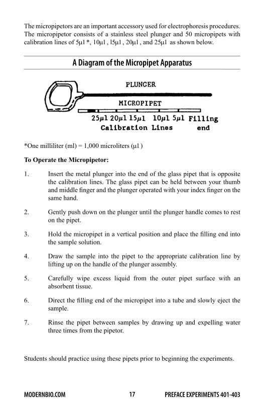
The micropipetors are an important accessory used for electrophoresis procedures. The micropipetor consists of a stainless steel plunger and 50 micropipets with calibration lines of 5µ1*, 10µ1, l5µ1, 20µ1, and 25µ1 as shown below.
A Diagram of the Micropipet Apparatus
*One milliliter (ml) = 1,000 microliters (µ1)
To Operate the Micropipetor:
1. Insert the metal plunger into the end of the glass pipet that is opposite the calibration lines. The glass pipet can be held between your thumb and middle finger and the plunger operated with your index finger on the same hand.
2. Gently push down on the plunger until the plunger handle comes to rest on the pipet.
3. Hold the micropipet in a vertical position and place the filling end into the sample solution.
4. Draw the sample into the pipet to the appropriate calibration line by lifting up on the handle of the plunger assembly.
5. Carefully wipe excess liquid from the outer pipet surface with an absorbent tissue.
6. Direct the filling end of the micropipet into a tube and slowly eject the sample.
7. Rinse the pipet between samples by drawing up and expelling water three times from the pipetor.
Students should practice using these pipets prior to beginning the experiments.

18 PREFACE EXPERIMENTS 401-403MODERNBIO.COM
IV. Procedures for the Preparation, Electrophoresis and Staining of Agarose Gels
The exercises described in this manual were designed such that he samples of two students are analyzed on one agarose gel. If students work in pairs, four students will share one gel. Four agarose gels are electrophoresed simultaneously using the Procell and MB-170 power supply.
Pouring the Agarose Gels1. Place the casting tray on a level work surface and place a precleaned
glass slide into the gel support deck.
2. Seal both ends of the gel support deck with tape. The tape must be firmly pressed against the edges of the deck to ensure a tight seal.
Casting Tray Assembly
3. * With the macropipetor (pipet-syringe), dispense 15ml of electrophoresis buffer into a 25ml glass test tube and add 0.18 grams of agarose. The agarose can be weighed out directly on an appropriate balance. If a balance is not available, 0.18 grams of agarose can be estimated by filling a 0.5ml micro tube with agarose until two-thirds full. Gently swirl the glass tube until the agarose forms a suspension.
4. Place the test tube into a boiling water bath and allow the agarose suspension to come to a vigorous boil. After boiling for about two minutes, remove the test tube from the bath, stir gently with a glass rod, and cool at room temperature for about 2-3 minutes. At this time, the agarose solution should be absolutely clear.
*The melted agarose for the four gels (15ml per gel) can also be prepared in one operation by boiling 0.9g of agarose in 75ml of buffer in a 250-500ml flask over a Bunsen burner or in a microwave oven. The flask should be rotated periodically during the heating process in order to prevent damage to the agarose.

19 PREFACE EXPERIMENTS 401-403MODERNBIO.COM
5. Pour the melted agarose directly from the test tube onto the casting deck and return the test tube to the hot (but not boiling) water bath. The small amount of melted agarose left in the test tube will be used for sample application (see below). Insert the comb into the casting tray slots and push down gently on the top of the comb until resistance is encountered. The teeth of the comb will come to rest in the melted agarose about 0.2mm above the surface of the glass plate.
6. After the gel has cooled for at least 15 minutes, remove the tape strips and carefully lift the comb straight up and away from the casting tray. The gel is now ready for sample application. Gels can also be stored for up to one week before use. For gel storage, the comb is left in place and the tray containing the gel and comb is wrapped in plastic wrap and placed in the refrigerator.
Sample ApplicationPrior to sample application, place the following items on the laboratory bench in front of you.
*Agarose gel*Sample for electrophoresis*Micropipetor and micropipets*Absorbent tissue (e.g. Kleenex or Kim wipes)*Small beaker of distilled or deionized water*Melted agarose - Transfer the melted agarose from the large test tube in the
hot water bath to a small tube. The small tube should then be placed in a beaker of hot water to ensure that the agarose remains in a liquid state.
1. Hold the micropipetor in a vertical position and place the filling end of the micropipet into the sample solution.
2. Draw the sample into the pipet to the l5µ1 calibration line by lifting up on the handle of the plunger assembly.
3. Wipe excess liquid from the outer pipet surface with an absorbent tissue.
4. Carefully direct the filling end of the micropipet into the top of the sample well and slowly eject the 15µ1 of the sample well.
5. Draw melted agarose into the micropipet to the 20µ1 calibration line, direct the filling end into the sample well, and slowly eject the agarose onto the sample until the well is full. Between 10-20µ1 of agarose are required to fill the well. The agarose will seal the sample in the sample well.
6. Rinse the pipet by drawing up and expelling water three times from the pipetor.
7. Wipe excess liquid from the outer pipet surface with an absorbent tissue.
8. Repeat steps 1-7 to load each additional sample.

20 PREFACE EXPERIMENTS 401-403MODERNBIO.COM
Electrophoresis1. Transfer the four casting trays with gels to the central platform of the
electrophoresis cell and position them such that the sample wells are closest to the black (negative) electrode. Upon electrophoresis, DNA will then migrate from the negative (black) towards the positive (red) electrode.
2. Place the gel tray stabilizing bar parallel to the long axis of the electrophoresis cell between the gel trays.
3. Slowly fill the electrophoresis chamber with electrophoresis buffer until the four gels are covered with a 1/4cm layer of buffer. Approximately 2.5 liters of buffer are required.
4. Place the electrophoresis cell lid in position.
5. With the power supply off, connect the cables from the cell to the power supply, red to red (positive) and black to black (negative).
6. Push the rocker switches on the power supply to “on” and”170V”. The voltage will now remain constant at 170 volts during the run.
7. Unless otherwise indicated, electrophorese until the bromophenol blue in the sample solution has migrated to within 1/4cm of the positive electrode end of the gel. At 170V, this takes approximately 50 minutes.
8. At the termination of the electrophoretic separation, shut off the power supply, disconnect the cables and remove the gel casting trays containing the gels.
9. The buffer should be emptied from the electrophoresis cell and stored under refrigeration in a separate container until the next electrophoretic run. The same buffer should be used for at least 3 electrophoretic separations. However, fresh buffer should be employed for the preparation of all agarose gels. The electrophoresis cell should be rinsed with deionized or distilled water and stored in an upright position.
Staining
Normal Procedure (Overnight Staining)1 Carefully slide the agarose gel out of the casting tray and off of the slide
and place the gel in a staining dish. (Note: The glass slide should not be placed in the staining dish.)
2. Dilute the gel stain concentrate 1000-fold with distilled water.

21 PREFACE EXPERIMENTS 401-403MODERNBIO.COM
3. Cover the gel with about 100ml of staining solution, making certain that the agarose does not stick to the dish and all gel surfaces are exposed to the stain.
4. Place the staining tray in the refrigerator and allow 3-18 hours for staining.
5. Decant and discard the stain, rinse the gel and dish with distilled water and add about 100ml of distilled water.
6. Change the water after about 10-20 minutes and hold the staining dish over a light source such as a desk lamp or light box. Note the position of the dark blue DNA bands against the light blue gel background. The gel can be stored in water in the refrigerator for a few days. After about a week or two in the refrigerator, the dye will diffuse out of the gel and the DNA bands will no longer be visible.
Rapid Procedure1. Dilute the gel stain concentrate 1000-fold and stain the gels for 30-60
minutes in 100mls at 37°C.
2. Decant and discard the stain, rinse the gel and dish with water and add about 100ml of distilled water.
3. Change the water after about 20 minutes and again after an additional 20 minutes. DNA bands can been seen during these destaining steps.
4. When the background stain has been reduced sufficiently, hold the staining dish over a light source and carefully observe the stained DNA bands in the gel.
Gel Storage1. The gel can be stored in a sealed plastic bag (3x3”) with a few mls of
distilled water for up to one month in the refrigerator.
2. For long term storage, place the gel on a glass slide and smooth with a gloved index finger to eliminate air bubbles between the gel and the slide. Allow the gel to dry onto the slide at room temperature for 3-4 days. Cover the dry gel film and glass slide with saran wrap.
V. Suggested Reading and Reference for Part ALewin, B. Genes V. Oxford University Press, Oxford NewYork Tokyo, 1994.
Jones, P. Gel Electrophoresis: Nucleic Acids. Chichester, West Sussex, UK, New York: Wiley 1995.

22 PREFACE EXPERIMENTS 401-403MODERNBIO.COM
Determining the Size of DNA by ElectrophoresisA first step in the analysis of a DNA molecule in the molecular biology laboratory frequently involves determining its length in nucleotide pairs. Electro phoresis in agarose gels has proven to be an extremely useful tool for this purpose, as well as for separating DNA fragments of different sizes. The smaller a DNA fragment, the more rapidly it moves during electrophoresis. The length of a given DNA fragment can be determined by comparing its electrophoretic mobility on agarose gels with DNA markers of known lengths. As illustrated in Figure 8, DNA fragments of known lengths (DNA standards or markers) and a DNA fragment of unknown length, are electrophoresed on adjacent lanes of the same gel. After electrophoresis, the DNA in the gel is stained and the positions of the standard and unknown DNA bands are determined. A linear relationship is obtained if the logarithms of the sizes of the standard DNA fragments are plotted against their respective electrophoretic mobilities. The length of the unknown DNA fragment is then estimated from this calibration curve. The length of DNA is frequently given in base-pairs (bp) for small fragments and kilobase pairs (KB) for large ones. One kilobase-pair equals 1000 base-pairs.
Figure 8. Determining the Length of a DNA Molecule.
Left Six DNA standards and a DNA fragment of unknown length are electrophoresed on adjacent lanes of a gel and the distances (in em) that each has migrated during the run are determined.
Right: The distances migrated by the standards are plotted against the logarithms of their lengths. The length of the unknown is determined by extrapolation from the standard graph. In the example, the unknown is about 1.5 KB.

23 PREFACE EXPERIMENTS 401-403MODERNBIO.COM
V. Hybridization AnalysisDNA Denaturation and RenaturationA DNA molecule is composed of two polynucleotide chains that are coiled around each other to form a rigid double-helix (see page 4 ). The double-helical structure of DNA is very stable at room temperature because the hydrogen and hydrophobic bonds between the stacked basses hold the two polynucleotide chains together. However, if a solution of DNA is heated to a critical temperature, or if the DNA is exposed to alkaline pH (pH > 11.5), these bonds are broken and the two polynucleotide strands separate by a process called denaturation or melting (Figure 9). If the DNA is cooled rapidly, the molecules will remain denatured as single stranded polynucleotides. However, if the solution is cooled very slowly, restoration of the DNA helix will occur. The reassembly of the two separated polynucleotide strands is called renaturation and the reformed DNA is called renatured DNA.
Figure 9. Denaturation of DNA

24 PREFACE EXPERIMENTS 401-403MODERNBIO.COM
A number of physical changes occur in DNA as denaturation proceeds. For example, DNA denaturation is accompanied by a decrease in the viscosity of the solution because single-stranded DNA molecules form flexible coiled structures that no longer retain the rigid native structure of the DNA double-helix (Figure 9). There is also an increase in the absorption of ultraviolet (UV) light upon DNA denaturation since the bases in single-stranded DNA absorb more UV light than the bases in native duplex DNA. Thus, changes in viscosity or in the absorption of UV light can be used to follow the course of DNA denaturation. If a DNA solution is slowly heated, and the absorption of UV light is measured at various temperatures, a DNA melting or denaturation curve such as the one shown in Figure 10 is obtained. As shown in the figure, the absorption of UV light remains constant up to temperatures above those found in cells. As the temperature is increased, there is a sharp increase in absorption because the strands of the double helix are separating. In the final stage of denaturation, the melting process is complete and only single stranded molecules are found in the reaction mixture. The temperature at which the rise in absorption is half complete is called the melting temperature and is designated Tm. When DNA is in a solution of physiological ionic strength and pH (0.14 m NaCl, pH 7.4), the Tm is usually between 85-95° C.
Figure 10. DNA Melting Curve
A number of factors can influence the stability of DNA and thus the Tm value. The base composition of DNA itself is one such factor since a higher temperature is required to disrupt the three hydrogen bonds connecting a G- C pair than the two hydrogen bonds connecting an A -T pair. Thus, DNA with a high A+ T content melts at a lower temperature than DNA with a high G + C content The composition

25 PREFACE EXPERIMENTS 401-403MODERNBIO.COM
of the solution in which the DNA is dissolved also affects its stability and Tm value. For example, most common salts (e.g. NaCl) stabilize the DNA duplex by reducing electrostatic repulsion due to unneutralized phosphate groups of the DNA back bone. Thus, the Tm of a DNA preparation is higher in high salt buffers as compared the low salt buffers. In fact, DNA can denature spontaneously in distilled water at room temperature.
Under appropriate conditions, DNA denaturation can be reversed, so that the two complementary strands can reform a double helix. During this renaturation process, there is a restoration of the original properties of the double helix that were lost when the DNA was denatured. As shown in Figure 11, DNA renaturation is a two-step process. First, two small segments of complementary sequences collide as a result of random motion and base-pairs are formed. This step is rate limiting for the renaturation, and thus the rate of renaturation increases with DNA concentration. In the laboratory, this step can take several hours. Second, base-pairing continues along the length of each molecule by a zipper-like action to form complete DNA duplexes. This step generally occurs within a few seconds.
Figure 11. Two Steps In DNA Renaturation
The factors that influence DNA renaturation can be divided into two groups:
1. Temperature and Salt ConcentrationThe temperature of the renaturation mixture must be high enough to disrupt non-base pairing interactions that can occur between strands at lower temperatures. However, the temperature cannot be too high or the ionic strength too low, because base-pairing will not occur. Renaturation reactions are usually performed in buffers containing 0.15 to 0.5 m NaCl at a temperature which is 15-25° C below the value of the Tm of the duplex. The hybridization buffer that you will use in the exercises described below contains 0.5 M NaCl and the reactions will be carried out at 60 - 65° C.
2. Time and ConcentrationDNA renaturation is dependent on the concentration of reacting complementary strands. The higher the concentration, the faster the reaction will proceed. In fact, the parameter controlling the renaturation reaction is the product of the concentration of the DNA (Co) and the time of incubation (t). This parameter

26 PREFACE EXPERIMENTS 401-403MODERNBIO.COM
is called the Cot and the value for half-maximal reaction is the Cot 1/2. DNA renaturation reactions are frequently described by their Cot 1/2 values.
Hybridization ProceduresRenaturation reactions involve two complementary DNA strands that were separated by denaturation. The term hybridization is often used when two complementary polynucleotides from different sources come together to form hybrid molecules. The factors that govern hybridization reactions (temperature, salt, concentration of nucleic acid and time) are the same as those that control DNA renaturation reactions and, in fact, some investigators use the terms hybridization and renaturation synonymously. In hybridization reactions, the polynucleotides can both be DNA (DNA hybridization), one can consist of DNA and the other RNA (DNA-RNA hybridization) or both can even be RNA (RNA hybridization). DNA hybridizations will be used in the experiments presented in the second part of this manual.
There are many different hybridization procedures for detecting and characterizing specific nucleotide sequences in DNA and RNA. However, each procedure consists of three basic steps:
1. Single strands of DNA or RNA are mixed together.2. The mixture is incubated under conditions that favor hybrid formation.3. The amount of specific hybrids that form are measured.
The principal method for performing hybridization reactions prior to the mid-1970s was solution hybridization where the nucleic acids are mixed together and incubated in solution. When large amounts of nucleic acids are available, solution hybridization reactions can be followed by a change in the absorption of UV light (see Figure 10). More commonly, the reaction is monitored using radioactive RNA or DNA as a hybridization probe. In this application, the quantity of hybridization probe incorporated into the duplex during the reaction is determined by radioactive counting.
During the past 10 years, filter hybridization techniques have, for the most part, replaced solution hybridization procedures for detecting specific nucleotide sequences. A basic procedure for filter hybridization is outlined in Figure 12. In the example given in the figure, the procedure is used to show that the genes coding for the a -polypeptides in hemoglobin (α-globin) are found in human but not in yeast DNA. First, denatured DNA from human and yeast is immobilized on a thin filter (membrane filter) made of nitrocellulose, nylon, or another substance that binds nucleic acids. The immobilization is accomplished simply be pipeting the DNA onto the filter. This procedure, which you will perform in Exercise 1, has been called Dot hybridization because the DNA occupies small circles (or

27 PREFACE EXPERIMENTS 401-403MODERNBIO.COM
“Dots”) on the filter. The filter is then incubated with a radioactive single-stranded DNA fragment containing the α-globin gene which is used as a hybridization probe. During the incubation, the probe will hybridize to the α-globin genes in the immobilized human DNA. The filter is then washed and placed on x-ray film. As can be seen from the diagram of the resulting developed film (autoradiogram), a radioactive signal is observed with human, but not with yeast DNA. In addition, from the intensity of the signal, one can estimate the number of copies of the α-globin gene in the human genome.
Figure 12. Detection of the α-Globin Genes in Human DNA Using Filter Hybridization and a Radioactive Probe
A key feature to performing hybridization reactions is the preparation and detection of the hybridization probe. In the example described above, the α-globin probe was prepared by recombinant DNA procedures and then labeled with 32 P -labeled dATP in an enzymatic reaction. After hybridization, the radioactive probe was detected by autoradiography. It is also possible to prepare a nonradioactive probe by introducing a chemical label into the DNA that can be detected visually after the hybridization reaction. A common nonradioactive label used for this purpose is biotin (Vitamin H) and this label has been incorporated into the DNA hybridization probes that you will use. The detection of biotinylated DNA probes after hybridization is based on the ability of the protein avid in to bind very tightly to biotin and biotinylated DNA. Avid in is not colored so a method is needed to detect this protein. For this reason, avidin is frequently coupled to an enzyme

28 PREFACE EXPERIMENTS 401-403MODERNBIO.COM
Figure 13. The Reaction Catalized by Peroxidase
Figure 14. Detection of the α-Globin Genes in Human DNA Using Filter Hybridization and a Biotinylated Probe
which catalyzes a color-producing reaction. In the experiments described in this manual, avidin has been linked to peroxidase which converts 4-chloronapthol to an insoluble purple product Thus, the avidin-peroxidase complex binds to the biotinylated probe and deposits purple product at the site which allows visualization of the hybridized probe. The color generating reaction catalyzed by peroxidase is shown in Figure 13 and a method for detecting α-globin genes in human DNA using the filter hybridization technique and a biotinylated probe is outlined in Figure 14.

29 PREFACE EXPERIMENTS 401-403MODERNBIO.COM
In 1975, Edward M. Southern at the University of Edinburgh, developed a powerful technique for DNA analysis which has become known as Southern blotting. More recently, the general technique has been applied to RNA (Northern blotting) and proteins (Western blotting). In blotting procedures, a replica of DNA, RNA, or proteins in an electrophoretic gel is created on a membrane filter.
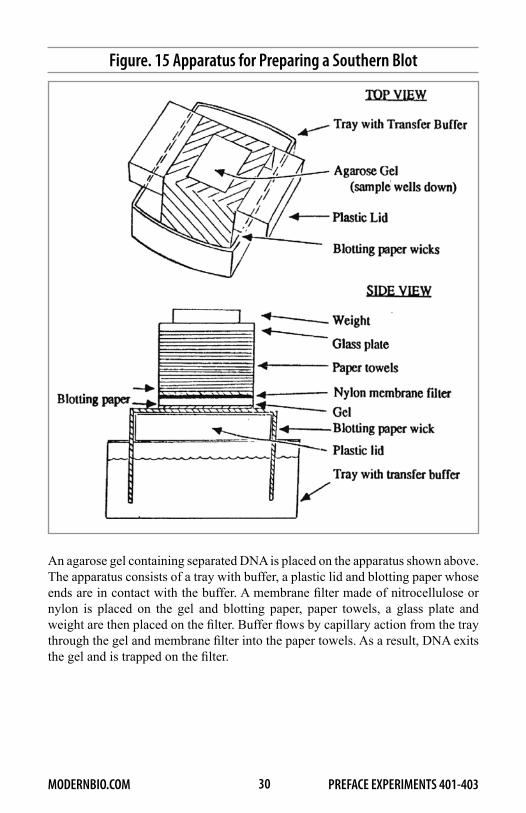
As we have seen, filter hybridization permits the detection and quantification of specific genes. Southern blotting, in conjunction with filter hybridization, enables the investigator to further characterize genomic elements for it permits identification of restriction fragments containing specific DNA sequences. In this elegant procedure, cellular or cloned DNA is most often digested with a restriction endonuclease and the different size fragments are separated on an agarose gel. Next, the DNA in the gel is denatured by alkaline pH and the gel is placed directly against a membrane filter made of nitrocellulose or nylon. DNA fragments are then transferred from the gel to the filter by capillary action. As a result, a replica (blot) of the electrophoretically separated DNA is produced on the membrane filter. The apparatus that you will use to prepare Southern blots in Exercises 2 and 3 is similar to one that is used in many research laboratories and is shown in Figure 15.
\After a Southern blot has been prepared, it is incubated with a single-stranded hybridization probe under conditions which favor hybrid formation. The gel position (and hence size) of a restriction fragment containing sequences complementary to the probe is then determined by autoradiography (for a labeled probe) or by a color-producing enzyme reaction (for a biotinylated probe). Figure 16 illustrates how Southern hybridization analysis would be used to detect a restriction fragment containing the α-globin gene in human DNA.
30 PREFACE EXPERIMENTS 401-403MODERNBIO.COM
Figure. 15 Apparatus for Preparing a Southern Blot
An agarose gel containing separated DNA is placed on the apparatus shown above. The apparatus consists of a tray with buffer, a plastic lid and blotting paper whose ends are in contact with the buffer. A membrane filter made of nitrocellulose or nylon is placed on the gel and blotting paper, paper towels, a glass plate and weight are then placed on the filter. Buffer flows by capillary action from the tray through the gel and membrane filter into the paper towels. As a result, DNA exits the gel and is trapped on the filter.

31 PREFACE EXPERIMENTS 401-403MODERNBIO.COM
Figure 16. Detection of a Restriction Fragment Containing the α-Globin Genes by Southern Blotting and Filter Hybridization.
The Southern hybridization procedure has been used extensively to determine the general nucleotide sequence arrangement found within and around specific genes in DNA. In this application, cellular DNA is cleaved with a battery of restriction nucleases, each of which cuts the DNA at a specific sequence. The restriction fragments are separated electrophoretically, transferred to a filter and the frag-ments containing the gene sequence under investigation are identified by hybrid-ization to a complementary probe. This analysis permits construction of restric-tion maps of specific genetic loci which show the locations of each restriction site in relation to its neighbor. Since restriction nucleases cut at specific nucleotide recognition sites, such maps are a reflection of selected nucleotide sequences in the region. A restriction map of the cluster of α-globin genes in human DNA is shown in Figure 17.
Application of Southern Blotting:

32 PREFACE EXPERIMENTS 401-403MODERNBIO.COM
The revolution brought about by molecular biology depended heavily on the Southern hybridization technique. The technique is used extensively in the research laboratory for detecting and characterizing specific genes and is increasingly being applied in medicine. A major medical application is in the diagnosis of genetic diseases in the fetus, so that abortion can be considered to prevent birth of children with incurable diseases. To illustrate the use of Southern blotting in disease diagnosis, we will consider the genes for hemoglobin. Hemoglobin is a globular protein made up of four sub-units. Each sub-unit contains a polypeptide chain attached to an iron-containing component called heme. The polypeptide chains of hemoglobin are referred to as the globin portion of the molecule. Normal adult hemoglobin (hemoglobin A) has four sub-units made up of two a- and two a polypeptide chains. The human globin polypeptides are encoded by two gene clusters: the a-like genes on chromosome 10 and 6-like genes on chromosome 11.
Many changes in the structure of hemoglobin have arisen by mutations in the human population. About one person in 100 contains a mutant hemoglobin gene and these individuals have an abnormal hemoglobin molecule in their blood. The mutations often involve substitution of one amino acid for another and are usually harmless. However, in a few cases, mutations in hemoglobin can cause serious diseases. One of the most common and serious abnormal hemoglobins is hemoglobin S, which is present in individuals suffering from sickle cell anemia. In hemoglobin S, a single glutamic acid residue on the beta chains is replaced by valine and this change is due to a point mutation in the b globin genes. This single change in the primary sequence of hemoglobin causes a marked change in the net charge and conformation of the protein. When hemoglobin S is deoxygenated, it crystallizes in the red blood cells, leading to a distortion of the red cells into sickle shape. These abnormal cells are destroyed rapidly in the body, reducing the numbers of erythrocytes; hence the term, “sickle cell anemia”. The thalassemias are another inherited disorder of hemoglobin which are common among some Mediterranean populations. Thalassemias are a group of diseases of hemoglobin synthesis resulting in low levels or an absence of synthesis of one of the globin polypeptides. There are a number of different causes of the thalassemias including point mutations or deletion of all or part of the globin gene itself. As shown in Figure 18, Southern hybridization analysis can be used for the direct detection of the genetic aberration responsible for these diseases. The restriction enzyme Mst ll cleaves at the sequence CCTNAGG and this sequence is present in the normal f3 globin gene. However, the point mutation in patients with sickle cell anemia converts this sequence to CCTNTGG and eliminates the restriction site for this enzyme. Thus, the Mst II restriction fragments containing b globin gene sequences are different sizes in normal individuals as compared to those with sickle cell anemia. Deletion of B globin genes, which causes one form of B thalassemia, would also be readily detected by the procedure because the Southern blot prepared from the DNA of these patients would lack a signal when probed with a b-globin gene sequence.

33 PREFACE EXPERIMENTS 401-403MODERNBIO.COM
Figure 17. Restriction Map of α-Globin Genes
Figure 18. Detection of Mutant β Globin Genes by Southern Hybridization

34 PREFACE EXPERIMENTS 401-403MODERNBIO.COM
A major goal of human genetics is to develop a complete genetic and restriction map of each human chromosome and ultimately, to determine the entire DNA sequence of the human genome. This goal has frequently been compared, in effort and in required funding level, to that which was needed to put a man on the moon. A high-resolution map of the human genome is considered to be essential for diagnosis of genetic diseases and development of potential cures of these diseases by gene therapy. The past few years have witnessed a tremendous explosion in the number of DNA markers isolated from the human genome that can be used as hybridization probes for detecting inherited diseases and even normal traits at the level of DNA sequence. Thus far, there are on the order of 50 genetic diseases that can be detected by hybridization procedures and the names of a few of these diseases are given in Table 2. This list is expanding daily and the potential for further developments in human genetics are enormous.
Table 2. Genetic Diseases Detected by Southern Hybridization Analysis
Disease ChromosomeHuntington’s disease 4
Sickle cell anemia 11
Thalassemia 11
Cystic Fibrosis 7
Familial Alzheimer’s disease 21
Retinoblastoma 13
Manic-depressive illness 11
Duchenne muscular dystrophy X
Hemophilia A and B X
X-linked spinal muscular atrophy X
X-linked retinitis pigmentosa X
X-linked cleft palate X

35 PREFACE EXPERIMENTS 401-403MODERNBIO.COM
VI. Suggested Reading and References for Part AGeneral References:Alberts, B., Bray, L., Lewis, J., Raff, M., Roberts, K. and Watson, J.D. Molecular Biology of the Cell. New York and London: Garland, 1983.
Watson, J.D.,Tooze, J.and Kurtz, D.T. Recombinant DNA: A Short Course. New York: Freeman, 1983.
Weinberg, R.A. “The Molecules of Life”, Scientific American 253:48, 1985. Felsenfeld, G. “DNA”, Scientific American 253:58, 1985.
Darnell, J.G. “RNA”, Scientific American 253:58, 1985.
Watkins, P.C. “Restriction Fragment Length Polymorphism: Applications in
Human Chromosome Mapping and Genetic Disease”, Bio Techniques 6:310, 1988.
White, R. and Lalovel, J.M. “Chromosome Mapping with DNA Markers”, Scientific American 258:40, 1988.
Electrophoretic and Hybridization Techniques
Southern, E. “Detection of Specific Sequences among DNA Fragments Separated by Electrophoresis”, J. Mol. Bioi. 98:503, 1975.
Hames, B.D. and Richwood, D. Gel Electrophoresis of Nucleic Acids. Oxford: IRL Press, 1982.
Maniatis, T., Fritsch, E.F. and Sambrook, J. Molecular Cloning: A Laboratory Manual. Cold Springs Harbor Labs, New York, 1982.
Perbal, B. Molecular Cloning. New York, John Wiley and Sons, 1984.
Davis, L.G., Dibner, M.D. and Batter, J.F. Basic Methods in Molecular Biology. New York, Elsevier, 1986.